Article

Anti-Epileptic Effects of FABP3 Ligand MF1 through the Benzodiazepine Recognition Site of the GABAA Receptor

1
Department of Pharmacology, Graduate School of Pharmaceutical Sciences, Tohoku University, Sendai 980-8578, Japan
2
Department of Genomic Neurology, Institute of Molecular Embryology and Genetics, Kumamoto University, Kumamoto 860-0811, Japan
3
Department of Neurophysiology, Hyogo College of Medicine, Nishinomiya 663-8501, Japan
4
Department of Neurophysiology, Graduate School of Medicine, Hirosaki University, Hirosaki 036-8216, Japan
*
Author to whom correspondence should be addressed.
These authors contributed equally to this work.
Int. J. Mol. Sci. 2020, 21(15), 5525; https://0-doi-org.brum.beds.ac.uk/10.3390/ijms21155525
Submission received: 14 July 2020 / Revised: 29 July 2020 / Accepted: 29 July 2020 / Published: 1 August 2020
(This article belongs to the Special Issue Precision Medicine and Therapy for Neurodegenerative Disorders)

Abstract

:
Recently, we developed the fatty acid-binding protein 3 (FABP3) ligand MF1 (4-(2-(1-(2-chlorophenyl)-5-phenyl-1H-pyrazol-3-yl)phenoxy) butanoic acid) as a therapeutic candidate for α-synucleinopathies. MF1 shows affinity towards γ-aminobutyric acid type-A (GABAA) receptor, but its effect on the receptor remains unclear. Here, we investigate the pharmacological properties of MF1 on the GABAA receptor overexpressed in Neuro2A cells. While MF1 (1–100 μm) alone failed to evoke GABA currents, MF1 (1 μm) promoted GABA currents during GABA exposure (1 and 10 μm). MF1-promoted GABA currents were blocked by flumazenil (10 μm) treatment, suggesting that MF1 enhances receptor function via the benzodiazepine recognition site. Acute and chronic administration of MF1 (0.1, 0.3 and 1.0 mg/kg, p.o.) significantly attenuated status epilepticus (SE) and the mortality rate in pilocarpine (PILO: 300 mg/kg, i.p.)-treated mice, similar to diazepam (DZP: 5.0 mg/kg, i.p.). The anti-epileptic effects of DZP (5.0 mg/kg, i.p.) and MF1 (0.3 mg/kg, p.o.) were completely abolished by flumazenil (25 mg/kg, i.p.) treatment. Pentylenetetrazol (PTZ: 90 mg/kg, i.p.)-induced seizures in mice were suppressed by DZP (5.0 mg/kg, i.p.), but not MF1. Collectively, this suggests that MF1 is a mild enhancer of the GABAA receptor and exercises anti-epileptic effects through the receptor’s benzodiazepine recognition site in PILO-induced SE models.

1. Introduction

γ-aminobutyrate (GABA) is a critical inhibitory neurotransmitter in both the mammalian peripheral and central nervous systems and maintains brain homeostasis by mediating neuronal excitability [1,2]. A collapse of GABAergic transmission leads to neuronal hyperexcitability and the subsequent development of epileptic seizures [3,4]. An aberrant GABAergic system is also associated with neurological disorders such as Alzheimer’s disease (AD), Parkinson’s disease (PD) and autism spectrum disorders (ASD) [5,6,7], often co-occurring with epileptic seizures [8,9]. Many anticonvulsants promote GABAergic neuronal transmission by enhancing the activity of the GABA type-A (GABAA) receptor and increasing GABA content in the synaptic cleft [4,10]. However, 20–30% of patients with epilepsy showed resistance to existing anticonvulsants [11]. Therefore, the development of powerful novel and safe anticonvulsants are necessary to improve the quality of life for patients with epilepsy.
The GABAA receptor complex consists of five subunits of different families (α1–6, β1–4, γ1–4, δ, ϵ and π) [12]. In the mammalian brain, the αβγ-subunit-containing complex is the major component of the GABAA receptor; a minor complex with αβδ subunits is also observed in the hippocampal dentate gyrus, thalamus and cerebellum [13]. The GABAA receptor function is dependent on the type of subunit complex. For instance, the αβγ complex is sensitive to benzodiazepine, but this is not so for the αβδ complex [12,13]. The α1βγ-type GABAA receptors are localized in the post synaptic region, while α4βδ-type GABAA receptors are observed in extra-synaptic region [14,15].
Fatty acid-binding proteins (FABPs) are essential for the intracellular trafficking of long-chain polyunsaturated fatty acids and mediating their transport to the plasma membrane, mitochondria and nucleus [16]. Of the twelve types of FABP, FABP3, FABP5 and FABP7 are predominantly expressed in human and rodent brains [17,18]. FABP3 has been proposed as a potential biomarker of neurodegeneration, as increased levels of FABP3 have been observed in the cerebrospinal fluid (CSF) of patients with AD, PD, and vascular dementia [19,20,21]. We previously demonstrated that FAPB3 has a critical role in the development and progression of α-synucleinopathies such as PD and dementia with Lewy bodies (DLB). FABP3 null mice show resistance to 1-methyl-4-phenyl-1,2,3,6-tetrahydropyridine (MPTP)-induced dopaminergic neuronal toxicity and PD-like motor deficits [22]. FABP3 deficiency also prevents the spreading of α-synucleinopathy-like pathologies following the injection of α-synuclein preformed fibril (PFF) in the mouse brain [23] and uptake into dopaminergic neurons [24]. Moreover, we have succeeded in producing a novel FABP3 ligand, MF1 (4-(2-(1-(2-chlorophenyl)-5-phenyl-1H-pyrazol-3-yl)phenoxy) butanoic acid), which has a high affinity for FABP3 [25,26]. MF1 significantly attenuates loss of dopaminergic neurons, behavioral impairments and α-synucleinopathy-like pathologies observed in MPTP-treated and/or α-synuclein PFF injected mice [23,26]. These results suggest that FABP3 is a potential molecular target producing novel therapeutics for α-synucleinopathies. Our off-target analysis showed that MF1 has an affinity for the GABAA receptor, but its effect on the GABAergic system remains unclear.
Here, we test whether MF1 affects GABAA receptor function using the whole cell patch-clamp technique on Neuro2A cells that overexpress the GABAA receptor. We also validate the anti-epileptic effect of MF1 in pilocarpine (PILO)-induced status epilepticus (SE) and pentylenetetrazol (PTZ)-induced epilepsy in mice. Our results suggest that MF1 is able to enhance the GABAA receptor activity through the benzodiazepine recognition site, thereby demonstrating anticonvulsant-like effects.

2. Results

2.1. MF1 Promotes GABAA Receptor Currents through the Benzodiazepine Recognition Site

First, we evaluated the effect of MF1 on the GABAA receptor. We confirmed that the application of GABA (1–100 μm) induces inward currents, indicating that the overexpressed GABAA receptors function normally in Neuro2A cells (Figure 1A,C). However, application of MF1 (1–100 μm) alone did not evoke GABA currents (Figure 1B,C). Next, we tested whether MF1 affects GABA-induced currents. Significant group effects were observed upon application of 1 μm GABA [F(2, 18) = 28.12, p < 0.0001] and 10 μm GABA [F(2, 18) = 14.43, p = 0.0002], with or without treatment with MF1 (1 μm) and flumazenil (10 μm) (Figure 1D,E). Co-application of MF1 (1 μm) markedly increased GABA (1 - 10 μm) currents (1 μm: 2.2 ± 0.07, p < 0.01 vs. application of GABA alone; 10 μm: 1.4 ± 0.07, p < 0.01 vs. application of GABA alone; Figure 1D,E). Since benzodiazepines facilitate GABAA currents without evoking them [27], we speculated that MF1 promotes GABAA currents in a manner similar to benzodiazepines. As expected, flumazenil (10 μm), which inhibits the GABAA receptor by binding the benzodiazepine recognition site, significantly blocking the MF1-promoted GABA current (1 μm: 1.2 ± 0.1, p > 0.05 vs. application of GABA alone, p < 0.01 vs. application of GABA and MF1; 10 μm: 0.82 ± 0.08, p > 0.05 vs. application of GABA alone, p < 0.01 vs. application of GABA and MF1; Figure 1D,E).

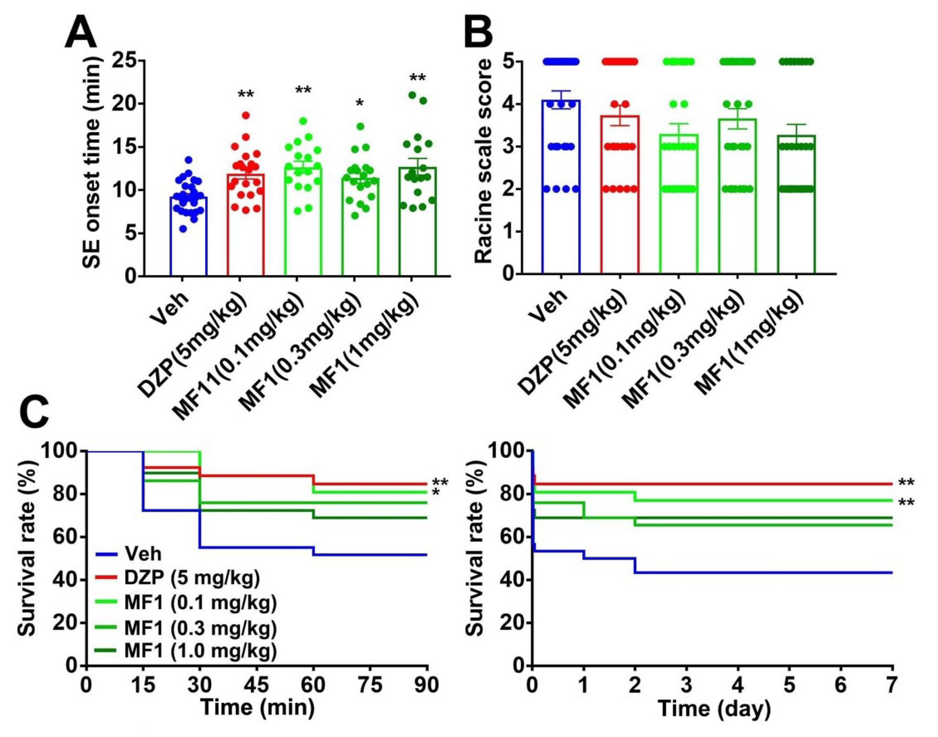
2.2. Acute MF1 Administration Attenuates SE and Mortality in PILO-Treated Mice

Next, we assessed the anti-epileptic effect of MF1 using animal models of epilepsy. Animal experimental schedules are shown in Figure 2A. We utilized diazepam (DZP: 5.0 mg/kg, i.p.) as a positive control [28,29]. We observed significant group effects on SE onset time [F(4, 66) = 24.87, p < 0.0001] and Racine scale score [F(4, 98) = 7.425, p < 0.0001]. Likewise, DZP (5.0 mg/kg, i.p.) treatment and acute administration of MF1 (0.3 and 1.0 mg/kg, p.o.) significantly prolonged SE onset time (0.3 mg/kg: 14.6 ± 1.2 min, p < 0.01 vs. PILO-treated mice; 1.0 mg/kg: 13.9 ± 0.8 min, p < 0.01 vs. PILO-treated mice; Figure 2B) and reduced the Racine scale score (0.3 mg/kg: 3.3 ± 0.3, p < 0.05 vs. PILO-treated mice; 1.0 mg/kg: 3.3 ± 0.2, p < 0.01 vs. PILO-treated mice; Figure 2C) in PILO-treated mice.
Administration of MF1 (0.3 and 1.0 mg/kg, p.o.) also extended the survival rate over 90 min (0.3 mg/kg: 89%, p < 0.01 vs. PILO-treated mice; 1.0 mg/kg: 89%, p < 0.01 vs. PILO-treated mice; Figure 2D) and 7 days (0.3 mg/kg: 72%, p < 0.05 vs. PILO-treated mice; 1.0 mg/kg: 89%, p < 0.01 vs. PILO-treated mice; Figure 2D) after PILO (300 mg/kg, i.p.) injection and treatment with DZP (5.0 mg/kg, i.p.) (Figure 2D).

2.3. Acute MF1 Administration Does Not Improve Epileptic Seizures in PTZ-Treated Mice

The anti-epileptic effect of MF1 was also investigated using PTZ-treated mice. DZP (5.0 mg/kg, i.p.) significantly suppressed the onset time of generalized tonic-clonic seizures (GTCs) and mortality, as compared to vehicle-administered PTZ-treated mice. However, MF1 had no effect in PTZ-treated mice (Table 1), suggesting that the anti-epileptic effect of MF1 (0.1–1.0 mg/kg, p.o.) may be milder than that of DZP (5.0 mg/kg, i.p.).

2.4. Chronic MF1 Administration Suppresses SE and Mortality in PILO-Treated Mice

We also investigated whether the chronic administration of MF1 rescues epileptic seizures in PILO-treated mice. Animals were treated with PILO (300 mg/kg, i.p.) 24 h after final MF1 and DZP administration (Figure 2A). A significant group effect was observed on SE onset time [F(4, 95) = 5.922, p = 0.0003], but not on the Racine scale score [F(4, 136) = 2.152, p = 0.0778]. Whereas chronic DZP (5.0 mg/kg, i.p.) or MF1 administration significantly prolonged SE onset time (0.1 mg/kg: 12.7 ± 0.68 min, p < 0.01 vs. PILO-treated mice; 0.3 mg/kg: 12.0 ± 0.79 min, p < 0.05 vs. PILO-treated mice; 1.0 mg/kg: 12.7 ± 0.91 min, p < 0.01 vs. PILO-treated mice; Figure 3A), both drugs failed to lower the Racine scale score in PILO-treated mice (Figure 3B). Chronic administration of DZP (5.0 mg/kg, i.p.) or MF1 (0.1 mg/kg, p.o.) significantly improved the survival rate over 90 min (0.1 mg/kg: 19%, p < 0.05 vs. PILO-treated mice; Figure 3C) and 7 days (0.1 mg/kg: 22%, p < 0.01 vs. PILO-treated mice; Figure 3C) after PILO (300 mg/kg, i.p.) injection. MF1 (0.3 and 1.0 mg/kg) slightly improved the survival rate, but the effect was not significant (Figure 3C).

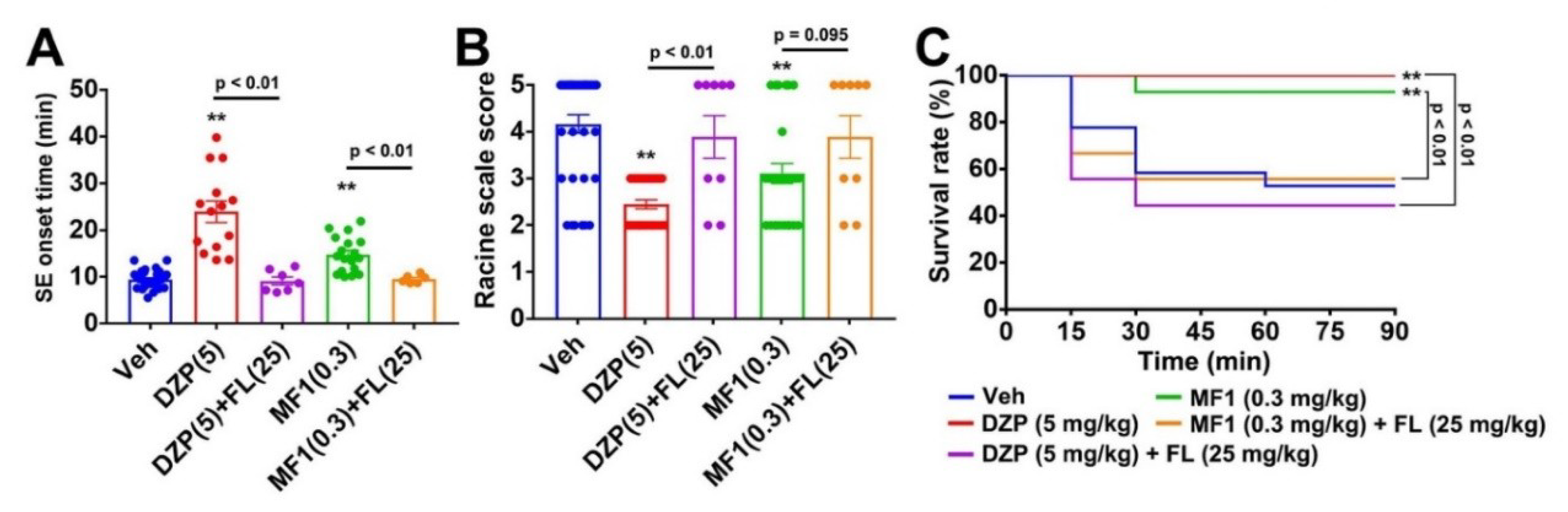
2.5. Flumazenil Blocks Anti-Epileptic Effect of MF1 in PILO-Treated Mice

Finally, we confirmed whether the acute administration of MF1 (0.3 mg/kg, p.o.) shows an anti-epileptic effect by enhancing the GABAA receptor through the benzodiazepine recognition site. We observed significant group effects on the SE onset time [F(4, 70) = 29.54, p < 0.0001] and Racine scale score [F(4, 106) = 11.89, p < 0.0001]. Consistent with results of the whole cell patch-clamp recording, flumazenil (25 mg/kg, i.p.) significantly prevented the acute effect of MF1 (0.3 mg/kg, p.o.) on the SE onset time (9.5 ± 2.0 min, p > 0.05 vs. PILO-treated mice, p < 0.01 vs. MF1-administered PILO-treated mice; Figure 4A) and Racine scale score (3.9 ± 0.40, p > 0.05 vs. PILO-treated mice, p = 0.095 vs. MF1-administered PILO-treated mice; Figure 4B) in PILO-treated mice. The improvement of the survival rate by MF1 (0.3 mg/kg, p.o.) was also inhibited by flumazenil (25 mg/kg, i.p.) treatment (55.6%, p > 0.05 vs. PILO-treated mice, p < 0.01 vs. MF1-administered PILO-treated mice; Figure 4C). Flumazenil (25 mg/kg, i.p.) also inhibited the effect of DZP (5.0 mg/kg, i.p.) in PILO-treated mice (Figure 4A–C). Therefore, MF1 may exercise its anti-epileptic effects by promoting GABAA receptor function in a manner similar to DZP.

3. Discussion

In the present study, we demonstrated that the FABP3 ligand MF1 promotes GABA currents in GABAA receptor-expressing cells. MF1 also exhibits anti-epileptic effects on PILO-induced SE in mice. These effects of MF1 were blocked by treatment with flumazenil, suggesting that MF1 enhances GABAA receptor function directly via the benzodiazepine recognition site.
Here, we show that MF1 enhances GABA currents arising from the GABAA receptor complex with α1β2γ2 subunits, which is the major complex found in the brain (approximately 60% of all GABAA receptors) [13]. However, the effects of MF1 on other types of GABAA receptors is unclear. Since MF1-promoted GABA currents were completely blocked by flumazenil, we propose that MF1 enhances the function of at least the αβγ-type benzodiazepine sensitive GABAA receptor.
Benzodiazepine receptor agonists act on not only the GABAA receptor, but also delayed rectifier K+ channels. Midazolam behaves like an open-channel inhibitor with a rapid onset of blocking and without frequency dependence on the block for the delayed rectifier K+ channels, thereby inhibiting the amplitude of action potentials in NSC-34 neuronal cells [30,31]. This effect is not blocked by flumazenil [31], suggesting that the benzodiazepine receptor agonist has the possibility of suppressing neuronal excitabilities, in vivo and independently of GABAergic action. As the MF1 here shows benzodiazepine receptor agonist-like effects, it may also affect the electrophysiological properties of delayed rectifier K+ channels. We will try to investigate the effect of MF1 on the delayed rectifier K+ currents in a future study.
Acute DZP (5.0 mg/kg, i.p.) treatment significantly attenuated the epileptic seizures observed in both PILO- and PTZ-treated mice. However, the acute administration of MF1 (0.3 and 1.0 mg/kg, p.o.) failed to inhibit PTZ-induced GTCs and mortality. Assuming that the anti-epileptic effects of DZP and MF1 arise from similar mechanisms of action, given that they both bind to the benzodiazepine recognition site, this result suggests that DZP may be a more powerful enhancer of the GABAA receptor function than MF1 in mouse brain. PILO leads to aberrant excitability, followed by the generation of seizures by stimulating the muscarinic acetylcholine receptor [32,33,34], while PTZ induces epileptic seizures by antagonizing GABAA receptor via the picrotoxinin-sensitive site [35,36]. Thus, acute MF1 (1.0 mg/kg, p.o.) administration appears to be insufficient to enhance the activity of the GABAergic system in the background of the impaired GABAA receptor function in PTZ-treated mice. However, a recent report indicates that fatty acid amides, with an affinity for FABPs, show anti-epileptic action in PTZ-treated mice, an effect blocked by flumazenil [37]. Further studies are necessary to investigate the effect of MF1 on other types of GABAA receptors.
The chronic administration of MF1 also attenuated PILO-induced SE and mortality. Since MF1 has a long half-life of not less than 20 h, the concentration of MF1 in the brain by chronic administration is sufficient to enhance GABAA receptor activity in the background of PILO treatment. We have previously reported that deletion of FABP3 upregulates GABAergic transmission by increasing the expression levels of the GABA, synthesizing enzyme glutamic acid decarboxylase 67 (GAD67), thereby suppressing excitability in the mouse anterior cingulate cortex [38,39]. Hence, chronic administration of MF1 may also promote the GABAergic system activity in the cortex by increasing GAD67 expression. We plan to assess the chronic effects of MF1 on GAD67 levels in the future.
While DZP is the drug of choice for the treatment of early SE, for acute repetitive seizures and febrile seizure prophylaxis [40,41], it has many side effects such as somnolence, depression, nausea, motor coordination disorder, and dizziness [41]. Moreover, repeated DZP treatment leads to the development of benzodiazepine tolerance and withdrawal syndrome, making it unsuitable for long-term epilepsy therapy [40,41]. Chronic administration (at least once a day for seven consecutive weeks) of MF1 (1.0 mg/kg, p.o.) did not affect motor and cognitive function in mice [23,26]. While the tolerance and withdrawal of MF1 should be analyzed, we hope that MF1 may be a safer alternative for long-term therapy.
We originally developed MF1 as therapeutic candidate for α-synucleinopathies [23,26]. MF1 attenuates aggregation and spreading of α-synuclein by preventing interactions between FABP3 and α-synuclein [23,26], suggesting that MF1 has the potential for early treatment of α-synucleinopathies. Patients with DLB cause seizures (14.7%) and myoclonus (58.1%) after disease onset, indicating that the seizure incidence rates are approximately 10-fold, relative to healthy controls [42,43]. Therefore, MF1 may be effective in alleviating not only core symptoms, but peripheral symptoms as well.
In conclusion, we have highlighted the pharmacological properties of MF1 on the GABAergic inhibitory system. Like the benzodiazepine agent DZP, MF1 may enhance function of the GABAA receptor, and in turn, suppress epileptic seizures in PILO-treated mice. Therefore, we suggest MF1 as an attractive therapeutic candidate for neurodegenerative disorders with epilepsy, including α-synucleinopathies.

4. Materials and Methods

4.1. Animals

Male ICR mice were purchased from Clea Japan, Inc. (Tokyo, Japan). Adult male mice (8–10 weeks old, weight 30–45 g) were used for all the experiments. Animals were housed under conditions of constant temperature 23 ± 2 °C and humidity 55 ± 5%, in a 12 h light–dark cycle (light: 9 am–9 pm). The mice had free access to food and water. All experimental procedures using animals were approved (2019 PhA-024, 1st April 2019) by the Committee on Animal Experiments at Tohoku University. We made an effort to reduce animal suffering and to use the minimum number of mice.

4.2. Chemicals

For administration to the animals, scopolamine hydrobromide (cholinergic inhibitor), DZP, PILO (muscarinic cholinergic agonist), and PTZ (GABAA receptor antagonist) (Sigma-Aldrich, St Louis, MO, USA) were dissolved in saline (0.9% NaCl). Flumazenil (Wako Pure Chemicals, Osaka, Japan) was prepared with 0.05% Tween-80 in saline. MF1 was synthesized as described previously [44] and suspended in 0.5% carboxymethylcellulose sodium salt (CMC). Doses of DZP (5.0 mg/kg, i.p.) and flumazenil (25 mg/kg, i.p.) were chosen, since these doses are enough to work in vivo without the influence of normal animal conditions [28].
For patch-clamp recording, GABA (Wako Pure Chemicals) was dissolved in distilled water. MF1 and flumazenil were suspended in dimethyl sulfoxide (DMSO) to achieve final concentrations of 0.1–0.01% for the assay.

4.3. Cell Transfection

Neuro2A mouse neuroblastoma cells were incubated in Dulbecco’s minimal essential medium (DMEM) with 10% heat-inactivated fetal bovine serum (FBS) and penicillin/streptomycin (100 units/100 μg/mL) in a 5% CO2 incubator at 37 °C. Cells were transfected with pcDNA plasmids (1.0 μg/mL), encoding the rat GABAA receptor subunits α1, β2 and γ2, with 0.1% green fluorescent protein (GFP) plasmid, by lipofection, as previously described [45]. GABAA receptor-expressing cells were used for electrophysiological recording 24–48 h after transfection.

4.4. Whole Cell Patch-Clamp Recording

GABAA receptor currents were recorded as previously described [45,46]. The external solution contained 143 mM NaCl, 5 mM KCl, 2 mM CaCl2, 1 mM MgCl2, 10 mM glucose and 10 mM HEPES (Tyrode’s solution; pH adjusted to 7.4 with NaOH). Glass pipettes were filled with internal solution containing 140 mM CsCl, 2 mM Mg-ATP, 10 mM EGTA, 10 mM HEPES (pH adjusted to 7.4 with CsOH). The resistance of electrodes filled with internal solution was 3.0–4.5 MΩ. Rapid drug application was achieved using the ALA-VM4 system (Sutter Instrument Company, Novato, CA, USA). GABAA receptor currents were recorded at room temperature using an EPC10 single patch clamp amplifier and acquisition system (HEKA, Lambrecht, Germany), filtered at 3 kHz, and sampled at 10 kHz. The membrane potential was clamped at −60 mV. Measured GABA currents were normalized to membrane capacitance in each cells.

4.5. Evaluation of Epileptic Behaviors

To assay the anti-epileptic effect of MF1, we utilized PILO- and PTZ-induced epileptic mice. To assess the acute drug effects, some animals were treated with MF1 (0.1, 0.3 and 1.0 mg/kg, p.o.) and DZP (5.0 mg/kg, i.p.) 30 min before injection with PILO (300 mg/kg, i.p.) or PTZ (90 mg/kg, i.p.). The competitive inhibitor of GABAA receptor, flumazenil (25 mg/kg, i.p.) was administered 5 min prior to treatment with MF1 (0.3 mg/kg, p.o.) and DZP (5.0 mg/kg, i.p.). To assess the chronic drug effects, the same doses of MF1 and DZP were administered once a day for seven consecutive days before PILO injection. Animal experimental schedules are shown in Figure 2A.

4.5.1. PILO-Induced SE Model

To suppress the peripheral cholinergic side effects, mice were pre-treated with scopolamine (1.0 mg/kg, i.p.) 30 min prior to PILO (300 mg/kg, i.p.) injection. Each mouse was placed in a plastic cage and its behavior was recorded for 90 min after PILO injection. The seizure stages of the mice were evaluated, based on the following Racine scale score (1972) [47]: 0- No behavioral seizures; 1-Mouth and facial movements; 2-Head myoclonus; 3-Forelimb myoclonus; 4-Forelimb myoclonus followed by rearing; 5-Falling or generalized tonic–clonic convulsions. If mice died during these 90 min, the Racine scale score was assigned as stage 5. Stages 3–5 are defined as convulsive seizures [48,49]. Mice that exhibited intermittently persistent stage 3–5 seizures at least three times within 90 min of PILO injection were considered to undergo SE. SE onset time was measured as the time of the first observation of convulsive seizures. To terminate SE, all the animals were treated with DZP (5 mg/kg, i. p.), which was repeated, if needed, to suppress convulsions. To minimize suffering, moistened rodent chow and an injection of 2 mL of 5% glucose were given to all the mice for 5 days after SE.

4.5.2. PTZ-Induced Seizure

Mouse behavior was monitored and recorded in individual cages for 30 min after injection of PTZ (90 mg/kg, i.p.). We scaled seizure behavior as follows (modified from Racine, 1972) [47]: 1-Hypoactivity; 2-Tail extension or limb jerk; 3-Whole-body clonus; 4-Rolling, running, jumping, and tonic-clonic. GTCs were assigned as stage 4.

4.6. Statistical Analysis

Data are shown as mean ± standard error of the mean (SEM). Significant differences were determined using Student’s t-test for two-group comparisons or a one-way analysis of variance (ANOVA) for multi-group comparisons, followed by Bonferroni’s multiple comparison test. Statistically significant differences of Kaplan Meier survival curves were tested by log-rank test using GraphPad Prism 7.04 (GraphPad Software, Inc., La Jolla, CA, USA). p < 0.05 represented a statistically significant difference.

Author Contributions

Investigation and data analysis, Y.Y., J.L., I.K., H.I., Y.S. and N.S.; Technical support and supply of pcDNA plasmids encoding rat GABAA receptor subunits, K.K. and S.U.; Writing the manuscript, Y.Y., N.S. and K.F.; Designing the study, K.F. All authors have read and agreed to the published version of the manuscript.

Funding

This work was supported in part by grants-in-aid for Scientific Research from the Ministry of Education, Science, Sports and Culture of Japan (Kakenhi 16H05219 to K.F. and 15H06036 to Y.Y.), Takeda Science Foundation (to Y.Y.), ONO Medical Research Foundation (to Y.Y.), Project of Translational and Clinical Research Core Centers from Japan Agency for Medical Research and Development, AMED (19dm0107071 and 20dm0107071 to K.F.).

Conflicts of Interest

The authors declare no conflict of interest.

Abbreviations

ADAlzheimer’s disease
ANOVAanalysis of variance
CMCcarboxymethylcellulose
DMSOdimethyl sulfoxide
DZPdiazepam
FABPfatty acid binding protein
GABAγ-aminobutyrate
GABAAGABA type-A
GADglutamic acid decarboxylase
GFPgreen fluorescent protein
MF14-(2-(1-(2-chlorophenyl)-5-phenyl-1H-pyrazol-3-yl)phenoxy) butanoic acid
MPTP1-methyl-4-phenyl-1,2,3,6-tetrahydropyridine
PDParkinson’s disease
PFFpreformed fibril
PILOpilocarpine
PTZpentylenetetrazol
SEstatus epilepticus
SEMstandard error of the mean

References

  1. Whiting, P.J.; Bonnert, T.P.; McKernan, R.M.; Farrar, S.; Le Bourdellès, B.; Heavens, R.P.; Smith, D.W.; Hewson, L.; Rigby, M.R.; Sirinathsinghji, D.J.; et al. Molecular and functional diversity of the expanding GABA-A receptor gene family. Ann. N. Y. Acad. Sci. 1999, 868, 645–653. [Google Scholar] [CrossRef] [PubMed]
  2. Mitchell, S.J.; Silver, R.A. Shunting inhibition modulates neuronal gain during synaptic excitation. Neuron 2003, 38, 433–445. [Google Scholar] [CrossRef] [Green Version]
  3. Crunelli, V.; Lőrincz, M.L.; McCafferty, C.; Lambert, R.C.; Leresche, N.; Di Giovanni, G.; David, F. Clinical and experimental insight into pathophysiology, comorbidity and therapy of absence seizures. Brain 2020, 21, awaa072. [Google Scholar] [CrossRef] [PubMed]
  4. Rogawski, M.A.; Löscher, W.; Rho, J.M. Mechanisms of Action of Antiseizure Drugs and the Ketogenic Diet. Cold Spring Harb. Perspect. Med. 2016, 6, a022780. [Google Scholar] [CrossRef] [PubMed]
  5. Błaszczyk, J.W. Parkinson’s Disease and Neurodegeneration: GABA-Collapse Hypothesis. Front. Neurosci. 2016, 10, 269. [Google Scholar] [CrossRef] [PubMed] [Green Version]
  6. Bi, D.; Wen, L.; Wu, Z.; Shen, Y. GABAergic dysfunction in excitatory and inhibitory (E/I) imbalance drives the pathogenesis of Alzheimer’s disease. Alzheimers Dement. 2020, 1–18. [Google Scholar] [CrossRef]
  7. Fung, L.K.; Flores, R.E.; Gu, M.; Sun, K.L.; James, D.; Schuck, R.K.; Jo, B.; Park, J.H.; Lee, B.C.; Jung, J.H.; et al. Thalamic and prefrontal GABA concentrations but not GABA(A) receptor densities are altered in high-functioning adults with autism spectrum disorder. Mol. Psychiatry 2020. [Google Scholar] [CrossRef]
  8. Faulkner, M.A.; Singh, S.P. Neurogenetic disorders and treatment of associated seizures. Pharmacotherapy 2013, 33, 330–343. [Google Scholar] [CrossRef]
  9. Giorgi, F.S.; Saccaro, L.F.; Busceti, C.L.; Biagioni, F.; Fornai, F. Epilepsy and Alzheimer’s Disease: Potential mechanisms for an association. Brain Res. Bull. 2020, 160, 107–120. [Google Scholar] [CrossRef]
  10. Löscher, W. Animal Models of Seizures and Epilepsy: Past, Present, and Future Role for the Discovery of Antiseizure Drugs. Neurochem. Res. 2017, 42, 1873–1888. [Google Scholar] [CrossRef]
  11. Czapiński, P.; Blaszczyk, B.; Czuczwar, S.J. Mechanisms of action of antiepileptic drugs. Curr. Top. Med. Chem. 2005, 5, 3–14. [Google Scholar] [CrossRef] [PubMed]
  12. Bormann, J. The ‘ABC’ of GABA Receptors. Trends Pharmacol. Sci. 2000, 21, 16–19. [Google Scholar] [CrossRef]
  13. Rudolph, U.; Möhler, H. GABAA receptor subtypes: Therapeutic potential in Down syndrome, affective disorders, schizophrenia, and autism. Annu. Rev. Pharmacol. Toxicol. 2014, 54, 483–507. [Google Scholar] [CrossRef] [PubMed] [Green Version]
  14. Sassoè-Pognetto, M.; Fritschy, J.M. Mini-review: Gephyrin, a Major Postsynaptic Protein of GABAergic Synapses. Eur. J. Neurosci. 2000, 12, 2205–2210. [Google Scholar] [CrossRef] [PubMed]
  15. Glykys, J.; Mody, I. Activation of GABAA Receptors: Views from Outside the Synaptic Cleft. Neuron 2007, 56, 763–770. [Google Scholar] [CrossRef] [Green Version]
  16. Coe, N.R.; Bernlohr, D.A. Physiological properties and functions of intracellular fatty acid-binding proteins. Biochim. Biophys. Acta 1998, 1391, 287–306. [Google Scholar] [CrossRef]
  17. Owada, Y.; Yoshimoto, T.; Kondo, H. Spatio-temporally differential expression of genes for three members of fatty acid binding proteins in developing and mature rat brains. J. Chem. Neuroanat. 1996, 12, 113–122. [Google Scholar] [CrossRef]
  18. Owada, Y. Fatty acid binding protein: Localization and functional significance in the brain. Tohoku J. Exp. Med. 2008, 214, 213–220. [Google Scholar] [CrossRef] [Green Version]
  19. Sepe, F.N.; Chiasserini, D.; Parnetti, L. Role of FABP3 as biomarker in Alzheimer’s disease and synucleinopathies. Future Neurol. 2018, 13, 199–207. [Google Scholar] [CrossRef] [Green Version]
  20. Chiasserini, D.; Biscetti, L.; Eusebi, P.; Salvadori, N.; Frattini, G.; Simoni, S.; De Roeck, N.; Tambasco, N.; Stoops, E.; Vanderstichele, H.; et al. Differential role of CSF fatty acid binding protein 3, α-synuclein, and Alzheimer’s disease core biomarkers in Lewy body disorders and Alzheimer’s dementia. Alzheimers Res. Ther. 2017, 9, 52. [Google Scholar] [CrossRef]
  21. Bjerke, M.; Kern, S.; Blennow, K.; Zetterberg, H.; Waern, M.; Börjesson-Hanson, A.; Östling, S.; Kern, J.; Skoog, I. Cerebrospinal Fluid Fatty Acid-Binding Protein 3 is Related to Dementia Development in a Population-Based Sample of Older Adult Women Followed for 8 Years. J. Alzheimers Dis. 2016, 49, 733–741. [Google Scholar] [CrossRef] [PubMed]
  22. Shioda, N.; Yabuki, Y.; Kobayashi, Y.; Onozato, M.; Owada, Y.; Fukunaga, K. FABP3 protein promotes α-synuclein oligomerization associated with 1-methyl-1,2,3,6-tetrahydropiridine-induced neurotoxicity. J. Biol. Chem. 2014, 289, 18957–18965. [Google Scholar] [CrossRef] [PubMed] [Green Version]
  23. Yabuki, Y.; Matsuo, K.; Kawahata, I.; Fukui, N.; Mizobata, T.; Kawata, Y.; Owada, Y.; Shioda, N.; Fukunaga, K. Fatty Acid Binding Protein 3 Enhances the Spreading and Toxicity of α-Synuclein in Mouse Brain. Int. J. Mol. Sci. 2020, 21, 2230. [Google Scholar] [CrossRef] [PubMed] [Green Version]
  24. Kawahata, I.; Bousset, L.; Melki, R.; Fukunaga, K. Fatty Acid-Binding Protein 3 is Critical for α-Synuclein Uptake and MPP+-Induced Mitochondrial Dysfunction in Cultured Dopaminergic Neurons. Int. J. Mol. Sci. 2019, 20, 5358. [Google Scholar] [CrossRef] [Green Version]
  25. Cheng, A.; Shinoda, Y.; Yamamoto, T.; Miyachi, H.; Fukunaga, K. Development of FABP3 ligands that inhibit arachidonic acid-induced α-synuclein oligomerization. Brain Res. 2019, 1707, 190–197. [Google Scholar] [CrossRef]
  26. Matsuo, K.; Cheng, A.; Yabuki, Y.; Takahata, I.; Miyachi, H.; Fukunaga, K. Inhibition of MPTP-induced α-synuclein oligomerization by fatty acid-binding protein 3 ligand in MPTP-treated mice. Neuropharmacology 2019, 150, 164–174. [Google Scholar] [CrossRef]
  27. Liu, Y.M.; Fan, H.R.; Ding, J.; Huang, C.; Deng, S.; Zhu, T.; Xu, T.L.; Ge, W.H.; Li, W.G.; Li, F. Curcumol allosterically modulates GABA(A) receptors in a manner distinct from benzodiazepines. Sci. Rep. 2017, 7, 46654. [Google Scholar] [CrossRef] [Green Version]
  28. Costa, J.P.; Ferreira, P.B.; De Sousa, D.P.; Jordan, J.; Freitas, R.M. Anticonvulsant effect of phytol in a pilocarpine model in mice. Neurosci. Lett. 2012, 523, 115–118. [Google Scholar] [CrossRef] [Green Version]
  29. Khoshnoud, M.J.; Tanideh, N.; Namdarian, S. Anticonvulsant activity of atorvastatin against seizure induced by pentylenetetrazole and maximal electroshock in mice. Trends Pharm. Sci. 2015, 1, 44–47. [Google Scholar]
  30. Vonderlin, N.; Fischer, F.; Zitron, E.; Seyler, C.; Scherer, D.; Thomas, D.; Katus, H.A.; Scholz, E.P. Inhibition of cardiac Kv1.5 potassium current by the anesthetic midazolam: Mode of action. Drug Des. Dev. Ther. 2014, 8, 2263–2271. [Google Scholar] [CrossRef] [Green Version]
  31. So, E.C.; Wu, K.C.; Kao, F.C.; Wu, S.N. Effects of midazolam on ion currents and membrane potential in differentiated motor neuron-like NSC-34 and NG108-15 cells. Eur. J. Pharmacol. 2014, 724, 152–160. [Google Scholar] [CrossRef] [PubMed]
  32. Smolders, I.; Khan, G.M.; Manil, J.; Ebinger, G.; Michotte, Y. NMDA receptor-mediated pilocarpine-induced seizures: Characterization in freely moving rats by microdialysis. Br. J. Pharmacol. 1997, 121, 1171–1179. [Google Scholar] [CrossRef] [PubMed] [Green Version]
  33. Millan, M.H.; Chapman, A.G.; Meldrum, B.S. Extracellular amino acid levels in hippocampus during pilocarpine-induced seizures. Epilepsy Res. 1993, 14, 139–148. [Google Scholar] [CrossRef]
  34. Macdonald, R.L.; Kapur, J. Acute cellular alterations in the hippocampus after status epilepticus. Epilepsia 1999, 40, S9–S20. [Google Scholar] [CrossRef] [PubMed] [Green Version]
  35. Ramanjaneyulu, R.; Ticku, M.K. Interactions of pentamethylenetetrazole and tetrazole analogues with the picrotoxinin site of the benzodiazepine-GABA receptor-ionophore complex. Eur. J. Pharmacol. 1984, 98, 337–345. [Google Scholar] [CrossRef]
  36. Hansen, S.L.; Sperling, B.B.; Sánchez, C. Anticonvulsant and antiepileptogenic effects of GABAA receptor ligands in pentylenetetrazole-kindled mice. Pharmacology 1984, 98, 337–345. [Google Scholar] [CrossRef] [PubMed]
  37. Rodrigues de Oliveira, F.; Eleuterio Rodrigues, K.; Hamoy, M.; Sarquis, Í.R.; Otake Hamoy, A.; Crespo Lopez, M.E.; Maciel Ferreira, I.; Macchi, B.M.; Luiz Martins do Nascimento, J. Fatty Acid Amides Synthesized from Andiroba Oil (Carapa guianensis Aublet.) Exhibit Anticonvulsant Action with Modulation on GABA-A Receptor in Mice: A Putative Therapeutic Option. Pharmaceuticals 2020, 13, 43. [Google Scholar] [CrossRef] [Green Version]
  38. Yamamoto, Y.; Kida, H.; Kagawa, Y.; Yasumoto, Y.; Miyazaki, H.; Islam, A.; Ogata, M.; Yanagawa, Y.; Mitsushima, D.; Fukunaga, K.; et al. FABP3 in the Anterior Cingulate Cortex Modulates the Methylation Status of the Glutamic Acid Decarboxylase67 Promoter Region. J. Neurosci. 2018, 38, 10411–10423. [Google Scholar] [CrossRef] [Green Version]
  39. Yabuki, Y.; Takahata, I.; Matsuo, K.; Owada, Y.; Fukunaga, K. Ramelteon Improves Post-traumatic Stress Disorder-Like Behaviors Exhibited by Fatty Acid-Binding Protein 3 Null Mice. Mol. Neurobiol. 2018, 55, 3577–3591. [Google Scholar] [CrossRef]
  40. Riss, J.; Cloyd, J.; Gates, J.; Collins, S. Benzodiazepines in epilepsy: Pharmacology and pharmacokinetics. Acta Neurol. Scand. 2008, 118, 69–86. [Google Scholar] [CrossRef]
  41. Parsonage, M.J.; Norris, J.W. Use of diazepam in the treatment of severe convulsive status epilepticus. Br. Med. J. 1967, 3, 85–88. [Google Scholar] [CrossRef] [PubMed] [Green Version]
  42. Beagle, A.J.; Darwish, S.M.; Ranasinghe, K.G.; La, A.L.; Karageorgiou, E.; Vossel, K.A. Relative Incidence of Seizures and Myoclonus in Alzheimer’s Disease, Dementia with Lewy Bodies, and Frontotemporal Dementia. J. Alzheimers Dis. 2017, 60, 211–223. [Google Scholar] [CrossRef] [PubMed] [Green Version]
  43. Murata, M.; Horiuchi, E.; Kanazawa, I. Zonisamide has beneficial effects on Parkinson’s disease patients. Neurosci. Res. 2001, 41, 397–399. [Google Scholar] [CrossRef]
  44. Beniyama, Y.; Matsuno, K.; Miyachi, H. Structure-guided design, synthesis and in vitro evaluation of a series of pyrazole-based fatty acid binding protein (FABP) 3 ligands. Bioorg. Med. Chem. Lett. 2013, 23, 1662–1666. [Google Scholar] [CrossRef] [PubMed]
  45. Migita, K.; Yamada, J.; Nikaido, Y.; Shi, X.; Kaneko, S.; Hirose, S.; Ueno, S. Properties of a novel GABAA receptor γ2 subunit mutation associated with seizures. J. Pharmacol. Sci. 2013, 121, 84–87. [Google Scholar] [CrossRef] [Green Version]
  46. Yabuki, Y.; Matsuo, K.; Izumi, H.; Haga, H.; Yoshida, T.; Wakamori, M.; Kakei, A.; Sakimura, K.; Fukuda, T.; Fukunaga, K. Pharmacological properties of SAK3, a novel T-type voltage-gated Ca2+ channel enhancer. Neuropharmacology 2017, 117, 1–13. [Google Scholar] [CrossRef]
  47. Racine, R.J. Modification of Seizure Activity by Electrical Stimulation. II. Motor Seizure. Electroencephalogr. Clin. Neurophysiol. 1972, 32, 281–294. [Google Scholar] [CrossRef]
  48. Lowenstein, D.H.; Alldredge, B.K. Status Epilepticus. N. Engl. J. Med. 1998, 338, 970–976. [Google Scholar] [CrossRef]
  49. Shibley, H.; Smith, B.N. Pilocarpine-induced status epilepticus results in mossy fiber sprouting and spontaneous seizures in C57BL/6 and CD-1 mice. Epilepsy Res. 2002, 49, 109–120. [Google Scholar] [CrossRef]
Figure 1. Effects of MF1 on GABA currents in GABAA receptor-expressing cell. (A,B) Representative traces treated with (A) GABA (1–100 μm) and (B) MF1 (1–100 μm). (C) Peak current observed by treatment with GABA (1–100 μm) and MF1 (1–100 μm) was normalized to membrane capacitance (GABA: n = 10 per group; MF1: n = 8 per group). Error bars represent SEM. (D) Representative traces from cells treated with GABA (1 and 10 μm) in combination with MF1 (1 μm) and/or flumazenil (10 μm). (E) MF1 (1 μm) enhanced GABA (1 and 10 μm) currents, which were blocked by flumazenil (10 μm) (n = 7 per group). Error bars represent SEM. ** p < 0.01 vs. each GABA alone treatment. ## p < 0.01 vs. each combination treatment of GABA and MF1. FL: flumazenil.
Ijms 21 05525 g001
Figure 2. Acute effect of MF1 and diazepam (DZP) on epileptic seizure in PILO-treated mice. (A) Animal experimental schedules of the present study. (B,C) Effects of MF1 (0.1, 0.3 and 1.0 mg/kg, p.o.) and DZP (5.0 mg/kg, i.p.) on (B) SE onset time and (C) Racine scale score in PILO-treated mice. Error bars represent SEM. (D) Survival rates over 90 min (left) and 7 days (right) after PILO treatment are shown. (Vehicle: n = 29; DZP: n = 18; MF1 [0.1 mg/kg]: n = 10; MF1 [0.3 mg/kg]: n = 18; MF1 [1 mg/kg]: n = 27). * p < 0.05, ** p < 0.01 vs. vehicle-administered PILO-treated mice.
Ijms 21 05525 g002
Figure 3. Effects of chronic MF1 and DZP administration on epileptic seizure in PILO-treated mice. (A) Chronic administration of MF1 (0.1, 0.3 and 1.0 mg/kg, p.o.) and DZP (5.0 mg/kg, i.p.) attenuated SE onset time in PILO-treated mice. Error bars represent SEM. (B) Chronic administration of MF1 (0.1, 0.3 and 1.0 mg/kg, p.o.) and DZP (5.0 mg/kg, i.p.) did not affect Racine scale score in PILO-treated mice. Error bars represent SEM. (C) Chronic administration of MF1 (0.1 mg/kg, p.o.) and DZP (5.0 mg/kg, i.p.) improved the reduced survival rate over 90 min (left) and 7 days (right) after PILO treatment. (Vehicle: n = 30; DZP: n = 26; MF1 [0.1 mg/kg]: n = 27; MF1 [0.3 mg/kg]: n = 29; MF1 [1 mg/kg]: n = 29). * p < 0.05, ** p < 0.01 vs. vehicle-administered PILO-treated mice.
Ijms 21 05525 g003
Figure 4. Flumazenil antagonizes the acute effects of MF1 on epilepsy in PILO-treated mice. (AC) Treatment with flumazenil (25 mg/kg, i.p.) inhibited the effects of MF1 (0.3 mg/kg, p.o.) and DZP (5.0 mg/kg, i.p.) on (A) SE onset time, (B) Racine scale score and (C) survival rate in PILO-treated mice. Error bars represent SEM. (Vehicle: n = 36; DZP: n = 29; DZP + flumazenil: n = 9; MF1: n = 28; MF1 + flumazenil: n = 9). ** p < 0.01 vs. vehicle-administered PILO-treated mice. DZP (5): DZP DZP (5.0 mg/kg, i.p.) treatment; FL (25): flumazenil (25 mg/kg, i.p.) treatment; MF1 (0.3): MF1 (0.3 mg/kg, p.o.) administration.
Ijms 21 05525 g004
Table 1. Acute effects of MF1 and DZP on epileptic seizure in PTZ-treated mice. ** p < 0.01 vs. vehicle-administered PTZ-treated mice. (n = 10 per group). GTCs: generalized tonic-clonic seizures, i.p.: intraperitoneal, p.o.: per os.
GroupGTCs Onset Time (Min)Mortality (%)Seizure Scale
Vehicle1.7 ± 0.5350 (5/10)4 ± 0
DZP (5 mg/kg, i.p.)3.3 ± 0.70 **0 (0/10)3.7 ± 0.15
MF1 (0.1 mg/kg, p.o.)0.92 ± 0.1540 (4/10)3.8 ± 0.20
MF1 (0.3 mg/kg, p.o.)1.2 ± 0.2250 (5/10)3.8 ± 0.20
MF1 (1.0 mg/kg, p.o.)1.2 ± 0.2870 (7/10)3.8 ± 0.20

