Review

Extracellular ATP as an Inter-Kingdom Signaling Molecule: Release Mechanisms by Bacteria and Its Implication on the Host

1
Department for Visceral Surgery and Medicine, Bern University Hospital, University of Bern, CH-3010 Bern, Switzerland
2
Department for BioMedical Research (DBMR), Visceral and Transplantation Surgery, Bern University Hospital, University of Bern, CH-3008 Bern, Switzerland
*
Author to whom correspondence should be addressed.
Int. J. Mol. Sci. 2020, 21(15), 5590; https://0-doi-org.brum.beds.ac.uk/10.3390/ijms21155590
Submission received: 4 July 2020 / Revised: 29 July 2020 / Accepted: 1 August 2020 / Published: 4 August 2020
(This article belongs to the Section Molecular Immunology)

Abstract

:
The purine adenosine 5′-triphosphate (ATP) is not only a universal intracellular energy carrier but plays also an important role as extracellular signaling molecule. Purinergic signaling is involved in many physiological and pathological processes like coagulation, inflammation, or sepsis in mammals. ATP is well-known as a messenger for intercellular communications in multicellular organisms, but phylogenetically much older unicellular organisms like yeast or bacteria use ATP as an extracellular signaling molecule as well. However, the mechanisms of ATP secretion by bacteria and its extracellular implications still have to be elucidated. This review will provide an overview of the current knowledge about bacterial extracellular ATP (eATP) under homeostatic conditions and during growth. Possible secretion mechanisms of ATP by bacteria will be discussed and implications of bacterial ATP are shown, with a focus on bacteria–host interactions.

1. Principles of Purinergic Signaling

While the importance of the purine adenosine 5′-triphosphate (ATP) as a universal intracellular energy carrier was discovered in the middle of the 20th century, the landmark discoveries about the importance of ATP as an extracellular signaling molecule took another 20 years [1]. Intracellularly, ATP enables energy-dependent processes releasing high-energy electrons by the hydrolyzation of its three phosphoanhydride bonds. This intracellular ATP can be released into the extracellular space by specific or unspecific mechanisms and acts as a danger associated molecular pattern (DAMP) [2], affecting platelet activation and coagulation [3,4], inflammation [5,6,7], and a wide range of neural processes [8].
Extracellular ATP (eATP) binds to specific P2 receptors, which are grouped into ionotropic P2X (P2X1, P2X2, P2X3, P2X4, P2X5, P2X6, P2X7) and metabotropic (G-protein-coupled) P2Y (P2Y1, P2Y2, P2Y4, P2Y6, P2Y11, P2Y12, P2Y13, P2Y14) receptors [9,10]. After having bound eATP, P2X receptors undergo conformational change and facilitate the influx as well as efflux of mono- and divalent cations along their concentration gradient (mainly Na+, Ca2+ in and K+ out) [11]. P2Y receptors activate either stimulatory or inhibitory G proteins, which mediate downstream signaling regulating 3′,5′-cyclic adenosine monophosphate (cAMP) and phospholipase C β (PLCβ) levels [12]. While P2X receptors are exclusively modulated by ATP, ligands for P2Y receptors also include adenosine 5′-diphosphate (ADP), uridine 5′-triphosphate (UTP), uridine 5′-biphosphate (UDP), and UDP-glucose [10,12]. Extracellular ATP is rapidly degraded by ectonucleotidases that convert ATP to ADP and adenosine 5′-monophosphate (AMP) as well as CD73/ecto-5′-nucleotidase that converts AMP to adenosine [13]. Adenosine is able to bind four distinct receptors: A1, A2A, A2B, or A3 [14]. All four are metabotropic (G-protein-coupled) receptors transducing extracellular signals mainly via regulation of cAMP [15]. In contrast to ATP receptors, adenosine receptors play a pivotal role in attenuating inflammation [16,17,18]. This dynamic regulation allows purinergic signaling to be both pro-inflammatory (mainly but not exclusively via P2 receptors) and anti-inflammatory (mainly but not exclusively via P1 receptors) [19,20,21]. Furthermore, the ligand binding affinities for different P2X receptors are within the nanomolar to the high micromolar range, making purinergic signaling a highly versatile communication system [22].

2. Secretion and Uptake of ATP by Microorganisms

The importance of the purinergic communication system in multicellular organisms like animals or plants has been established over the last decades. However, not only multicellular but also phylogenetically much older unicellular organisms make use of purinergic signaling or are at least able to secrete ATP [23].
These includes fungi such as Candida albicans (C. albicans) that secretes ATP putatively via conductive channels in a histatin 5-dependent manner [24]. Such eATP subsequently leads to cell death in neighboring C. albicans cells by a hitherto unknown mechanism [24]. The unicellular yeast Saccharomyces cerevisiae (S. cerevisiae) secretes ATP in the nanomolar range in a MCD4-dependent manner [25]. Incubation of S. cerevisiae with 2% glucose solution dramatically increased eATP concentration in a cAMP-dependent and regulated manner [26]. Furthermore, eATP was elevated during growth of S. cerevisiae [26]. The conclusion of these observations was that ATP, its metabolites ADP, AMP, and adenosine may act as intercellular signaling molecules for S. cerevisiae physiology [26]. Extracellular ATP as an intercellular messenger in S. cerevisiae has also been shown in synchronizing sporulation among cells, yet the mechanism has still to be elucidated [27].
Not only such unicellular eukaryotes though, but also prokaryotes are able to secrete ATP and there is now an increasing number of reports focusing on bacterial ATP, the invading bacteria as relevant source of eATP in the host and its importance in bacteria–host interaction [28].

2.1. Bacterial eATP under Homeostatic Conditions

In bacteria, first results were obtained in 2005 by Ivanova et al. [29]. A screen of the supernatant of 86 heterotrophic environmental (marine) bacteria cultured in Marine Broth 2216 for eATP showed that Gram-negative α-proteobacteria and γ-proteobacteria secreted ATP in the micro- to millimomolar range in vitro. The measured concentrations were between 0.2 and 7 mM [29,30,31]. Overall, marine Gram-positive bacteria (Planococcus spp., Kocuria spp., Brevibacterium spp., Bacillus sp., Microbacterium sp.) secreted less ATP than marine Gram-negative α-proteobacteria (Ruegeria spp., Erythrobacter sp., Sulfitobacter spp., Staley sp.) or γ-proteobacteria (Marinobacter spp., Marinobacterium sp., Marinobulbifer sp., Cobetia spp., Alteromonas spp., Pseudoalteromonas spp., Shewanella spp.) [29]. The concentrations in the supernatant even exceeded values that were measured inside cells [32,33,34]. In vivo, the ability of commensal bacteria to secret ATP was assessed from isolates from mouse feces and from human feces, urine, and skin. In mouse feces, Enterococcus gallinarum (E. gallinarum), Enterococcus faecalis (E. faecalis) and Escherichia coli (E. coli) and in human specimens, E. gallinarum, E. faecalis, E. coli, and in addition Enterococcus faecium (E. faecium) as well as Staphylococcus aureus (S. aureus) were cultured in RPMI 1640 medium and eATP levels in the supernatant were assessed after 16 h [35]. Thereby, eATP at levels between 1 and 3 µM were detected only in the supernatant of Gram-positive E. gallinarum culture, whereas in the supernatant of the other bacterial cultures, eATP was absent [35].

2.2. Bacterial eATP During Growth

In the follow-up study by Hironaka et al., seven more ATP secreting Enterococcus species were identified (E. cecorum, E. faecium, E. gilvus, E. mundtii, E. saccharolyticus, E. sulfureus, and E. thailandicus) under the same culture conditions as described above [36]. As in the previous study, eATP concentration was in the low µmolar range (around 0.1–2.2 µM/108 cells), and highest in E. mundtii. Importantly, these studies revealed that ATP secretion is growth phase-dependent, peaking during exponential phase. These observations allowed to detect eATP also in the supernatant of E. faecalis, E. faecium, E. coli, and S. aureus during exponential growth phase [36]. According to the authors, checking membrane integrity using a Sytox9/propidium iodide based staining excluded bacteriolysis as a source of ATP secretion [36].
Growth-dependent ATP secretion during exponential phase in the supernatant of a variety of different Gram-positive and Gram-negative bacteria was also observed by Mempin et al., however, in the low nanomolar range [37]. A possible explanation for the discrepancy in the amount of eATP between these two groups is that Hironaka et al. used RPMI 1640 medium for culturing bacteria, which contains a different amount of ATP secretion triggering glucose than standard Luria–Bertani broth (LB), which was used by Mempin et al. [36]. Another possibility is that the used RPMI 1640 medium was enriched with serum, which accounted for the higher amount of ATP. However, this is rather unlikely since the authors did not mention any supplementation with serum in their paper. The question remains, why Hironaka et al. did not observe ATP secretion with the tested media other than RPMI 1640 medium (e.g., brain heart infusion (BHI), LB, tryptic soy agar (TSA)), since the group used the same reagents to measure eATP like Mempin et al. This could be explained by different methods they used for the generation of their respective standard curves allowing Mempin et al. more sensitive measurements and therefore to detect lower eATP concentrations than Hironaka et al.
In addition to glucose, cytochrome bo oxidase plays an important role for eATP concentration in E. coli and Salmonella enterica (S. enterica) [37]. Cytochrome bo oxidase subunits mutated E. coli (ΔcyoA, ΔcyoC, ΔcyoD) and S. enterica (ΔcyoA, ΔcyoB, ΔcyoCD) showed significantly less eATP compared to the wild type when cultured aerobically [37]. Cytochome bo oxidase is one of three different ubiquinol oxidases, enzymes that pump protons across the inner membrane of the bacteria generating a proton motif force and that catalyzes the last step of the respiratory chain, which is the reduction of the electron acceptor. Furthermore, cytochrome bo oxidase predominates at high oxygen concentrations [38,39]. Together with the results from Hironaka et al. this could indicate that in E. coli and S. enterica, glycolysis and subsequent anaerobic or aerobic respiration are more important than fermentation for ATP secretion, which makes sense, since it is much more effective for generating ATP and bacteria can use a variety of redox pairs for respiration [36,37,40].

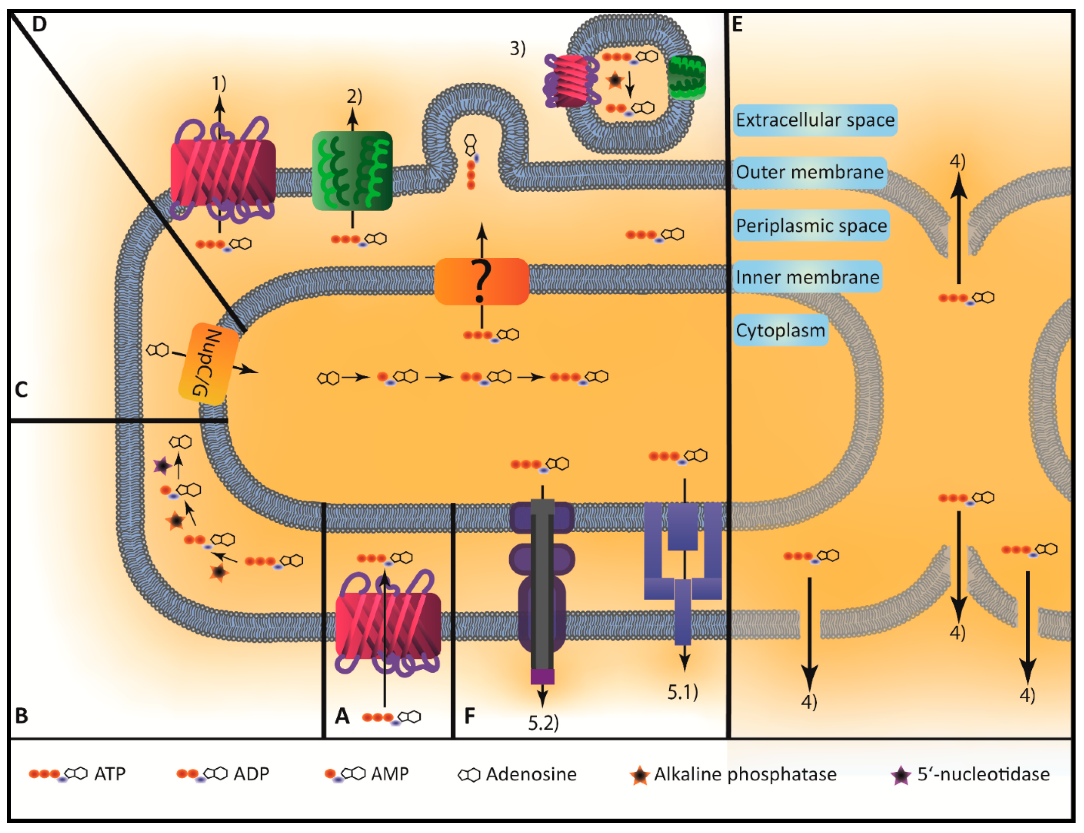
2.3. Uptake of eATP by Bacteria and Intracellular Fate

Several reports show that bacteria are also able to take up ATP according to their needs. In an experiment using E. coli W derived strains that have mutations in purine metabolism, it was shown that these bacteria were able to take up a variety of different purine nucleobases, nucleosides, and nucleotides (adenine, guanine, adenosine, guanosine, AMP, ADP, and ATP) possibly via general porins, which restored growth deficiency [41]. Bacteria mediated depletion of ATP was also shown in an experiment, in which 10 µM ATP was added to the bacterial culture and the remaining concentration was measured after several time intervals [37]. An almost complete depletion of the 10 µM was achieved by E. coli K12 as well as S. enterica (SE2472) within 120 min [37]. Interestingly, they concluded that ATP was mainly hydrolyzed on the bacterial surface, given that the remaining levels of radioactivity were detected in the supernatant and not in the bacteria. Conversely, using radioactively labelled [γ-32P]ATP, Alvarez et al. showed that in E. coli DH5α ATP is rapidly uptaken in the periplasmic space and hydrolyzed in a linear [eATP]-dependent manner and that eATP concentration in steady state in the periplasmic space is 24 ± 3 µM/1010 bacteria [42]. The periplasmic space as an important compartment involved in purine hydrolyzation is supported by a variety of scientific reports [41,42,43] and an array of acid and alkaline phosphatases (aphA, appA, phoA) as well as 5′-nucleotidases (e.g., napD, ushA, surE, yfbR, yjjG) are present in E. coli. Under physiological conditions especially phoA encoded alkaline phosphatase and ushA encoded 5′-nucleotidase are responsible for ATP hydrolyzation to adenosine [42,43,44,45,46]. Adenosine can then be further metabolized to adenine or hypoxanthine or can be directly transported back to the cytoplasm via the specific nucleoside transport systems nupC and nupG and recycled to ATP [41,47].
Interestingly, it is still unclear how ATP is transported from the cytosol to the periplasmic space since E. coli do not have ATP/ADP translocases in the inner membrane and this membrane is impermeable for the highly (four-fold) negatively charged ATP [43,48]. Furthermore, it still has to be determined how bacteria secrete ATP and possible mechanisms will be discussed in the next section.

3. Comparison of ATP Secretion Mechanisms between Prokaryotic and Eukaryotic Cells

3.1. Release Mechanisms of Eukaryotic Cells

In eukaryotic cells, there are mainly three distinct mechanisms of ATP release: (1) channel-mediated release, (2) vesicle-mediated release, and (3) non-specific release [49].
(1) Channel-mediated ATP release occurs mainly via pannexin-1 channels and connexin-43 hemichannels [12,49]. Pannexin-1-dependent ATP release occurs in apoptotic cells, which thereby establish a pro-inflammatory environment and attract leucocytes by releasing ATP in a controlled manner [50]. Pannexin-1 is also involved in ATP release by dendritic cells and subsequent autocrine signaling via P2X7 that creates a positive feedback loop, promoting fast migration at site of inflammation [51]. Additionally, T-cells are activated via pannexin-1-mediated ATP release and autocrine P2X1 and P2X4 signaling at the immune synapse, which represents a general T-cell activation mechanism [52]. Furthermore, macrophages can be activated by pathogen associated molecular patterns (PAMPs) to release ATP via pannexin-1, autocrine signaling via P2X7, and subsequent inflammasome activation [53]. Connexin-dependent ATP release occurs in neutrophils and macrophages upon stimulation with PAMPs [30,31,49]. This ATP release has two different effects: (1) it supports the inflammatory milieu [6] and (2) it stops neutrophil chemotaxis at site of inflammation in an autocrine manner [54]. Macrophages also release ATP via Connexin-43 in an autocrine manner, resulting in self-activation and improved survival during sepsis in mice [30].
(2) Vesicle-dependent release requires storage of ATP in vesicles, which is mediated by a V-ATPase dependent proton gradient [49]. V-ATPase is localized in the vesicle membrane and actively pumps H+ into the vesicle lumen. Negatively charged ATP is subsequently transported into the lumen via vesicular nucleotide transporter (VNUT) along the electrochemical gradient [55,56]. Release of vesicular ATP is then mediated by Ca2+-dependent zipper-like fusion of v- and t-SNARE complexes localized in the vesicle and the plasma membrane, respectively [57]. Vesicular ATP release has been shown to play an important role in mediating chronic inflammatory pain [58]. Different cells (e.g., microglia or peritoneal macrophages) use this release mechanism upon stimulation with PAMPs [58,59,60].
(3) Non-specific release of ATP typically depends on necrosis of cells. The intracellular ATP concentration is within the millimolar range, which is more than 105-fold higher than extracellular ATP concentration [6,61]; therefore, disruption of membrane integrity leads to an instant release of high amounts of ATP. This establishes a strong pro-inflammatory environment and a strong chemotactic gradient [5,12].

3.2. Release Mechanisms of Prokaryotic Cells

In bacteria, the detailed mechanisms of ATP secretion are less known as in eukaryotic cells. Bacteria do not possess pannexin or connexin hemichannels nor are any homologues of VNUT known. However, some authors have described and shown different mechanisms how ATP can be secreted: (1) by porins, (2) by mechanosensitive (MS) channels, or (3) by outer membrane vesicles (OMVs). Another possibility is that ATP is secreted by (4) lytic mechanisms especially during growth. Furthermore, it is conceivable that bacterial efflux pumps or secretion systems (5) are responsible for ATP secretion although such specific pumps for ATP have not been shown so far [62,63,64,65,66,67] (Figure 1).
(1) Porins are water-filled β-barrel pores in the membrane of Gram-positive or the outer membrane of Gram-negative bacteria that are permeable to water-soluble molecules. In E. coli K12, there are specific porins, e.g., tsx, which transports nucleosides and deoxynuclosides [68,69] or lamB, which transports maltose and maltodextrins [70]. However, there are also three unspecific porins called phoE, ompC, and ompF that are permeable for solutes smaller than 600–700 Dalton. PhoE is especially induced by phosphate starvation and has an anion selectivity, whereas ompC and ompF are rather cation-selective [71]. So far, no specific porin for nucleotides has been characterized and it is assumed that ATP is unspecifically secreted by these porins [72,73]. It is currently not known if there is a mechanism that allows E. coli to control the transport of ATP through the pore or if it is just passively governed by tonicity.
(2) It has been shown that if bacteria are exposed to hypotonic osmotic shock, they secrete ATP and other metabolites and ions via MS channels to prevent bursting [74,75]. However, if this mechanism is involved or responsible for the measured eATP concentrations during growth remains to be elucidated.
(3) OMVs are produced and released especially by Gram-negative bacteria and play a role in promoting pathogenesis, bacterial survival, and interaction [76]. They pinch off the outer membrane of Gram-negative bacteria and therefore contain periplasmic solutes. It was shown that OMVs contain also ATP, which they secret in a concentration of 13 ± 5 nM/µg protein [42]. Furthermore, OMVs possess intrinsic linear [eATP]-dependent ATPase activity [42]. That OMVs contain ATP and periplasmic phosphatases was also shown by Proietti et al., however to what extent OMVs contribute to eATP concentration remains unclear [77].
(4) Bacterial cell division is a highly complex mechanism that requires cell division proteins like Fts- or Min.-proteins, murein hydrolases, and other proteolytic enzymes working together for structured local breakdown of bacterial cell wall [78,79]. Furthermore, cellular integrity is highly dependent on environmental factors like temperature, pH, osmolarity, and concentration of divalent cations [80,81]. It is therefore conceivable that some degree of ATP secretion could happen within the division process because of disturbances in any of these factors. Another reason for eATP could be full lysis of bacteria during growth. It was shown that lysis in growing cultures is a rare phenomenon but since the intracellular ATP concentration is up to 105-fold higher than in the extracellular space, this may be at least partially responsible for eATP concentration [82].

4. Extracellular ATP as an Inter-Kingdom Signaling Molecule

Bacterial ATP has been shown to act as an inter-kingdom signaling molecule in different organ systems. Follicular T helper cells (Tfh) in the Peyer’s patches of gut-associated lymphoid tissue are modulated by bacterial ATP [83]. Tfh are key cells in B cell development and subsequent IgA production and need therefore to be tightly regulated, which occurs, at least in part, via P2X7 mediated apoptosis [83]. By gavaging E. coli or S. enterica either containing an empty plasmid or a plasmid carrying an apyrase that hydrolyzes the secreted ATP and is polarly localized in the periplasmatic space, the role of specifically bacterial ATP could be assessed [84]. Using this approach, Proietti et al. have shown that bacterial ATP leads to P2X7 mediated apoptosis of Tfh, subsequent limitation of IgA secretion from B cells, and therefore a decreased efficiency of oral vaccination [77,83]. Conversely, such limited IgA secretion leads to a diverse microbiota and a favorable metabolic profile (blood sugar levels, body weight) [85].
Atarashi et al. showed that after intraperitoneal and rectal application of 1.25 mg (2.3 µM) ATPγS, the CD70highCD11clow intestinal lamina propria cells increase production of IL-6 and IL-23 as well as TGF-β, leading to increased differentiation of naïve helper T cells to IL-17 producing helper T cells (TH17) in germ-free ILC mice [86]. The authors stated that this mechanism could be important for physiologic TH17 but also for pathologic TH17 differentiation, since severe combined immunodeficient mice (SCID-mice) suffered from exacerbated colitis after intraperitoneal application of 1.5 mg (2.9 µM) αβ-ATP [86]. Since germ-free mice had markedly reduced TH17 levels and antibiotic treated (vancomycin and metronidazole) specific pathogen free mice showed a significant reduction in TH17 and fecal ATP concentration, the group concluded that eATP is derived by commensal bacteria [86].
Abbasian et al. have suggested that ATP secreted by uropathogenic bacteria (e.g., E. coli) acts as a virulence factor in urgency urinary incontinence and overactive bladder disease by promoting Ca2+ influx into uroepithelial cells affecting subsequent bladder contractility [87].
Since eukaryotic cells are ubiquitously equipped with ecto-nucleoside triphosphate diphosphohydrolases (e.g., CD39), ecto-5′-nucleotidases (e.g., CD73), ectonucleotide pyrophosphatases, as well as alkaline phosphatases and are able to hydrolyze micromolar concentrations of ATP within 20–40 min, ATP signaling is generally considered as fast and short-termed [13,88]. However, the specific kinetics of bacterial ATP under the above-mentioned physiologic and inflammatory conditions still have to be elucidated.
Bacteria are not only able to secrete ATP itself but also to stimulate host cells to release ATP. Alvarez et al. have shown that concentration of apical ATP on a polarized monolayer of Caco-2 cells that was exposed to E. coli suspension (2 × 10⁷ bacteria) increased six-fold from basal concentration of 123 ± 5 nM. Furthermore, rat jejunum segments that were flushed with E. coli suspensions (5 × 10⁸ bacteria/mL) increased luminal ATP secretion two-fold from basal concentration of 13 ± 2 nM [42]. It has also been shown that the pore-forming toxins α-hemolysin from E. coli as well as leukotoxin A from Aggregatibacter actinomycetemcomitans lead to ATP secretion of erythrocytes [89]. ATP is most likely released through the toxin pore itself or by the damaged membrane around the toxin and subsequently activates P2X1 and P2X7 receptors, leading to an amplification of the hemolytic process [90].
Furthermore, bacteria seem also to use host-derived eATP for its own purposes. Mempin et al. has shown that addition of 10 or 100 µM ATP to stationary phase E. coli K12 and S. enterica cultures increased survival over 7 days of incubation [37]. The group concluded that this could be of advantage since these bacteria are able to invade host cells, where they could use intracellular ATP as a nutrient or signaling molecule. It has also been shown that eATP promotes biofilm formation in periodontal disease as well as detachment of bacteria from Fusobacterium nucleatum biofilm into its planktonic form to build new colonies [91,92]. Extracellular ATP is also involved in maintaining inflammation and bone resorption in periodontal disease via: (1) P2X7 mediated cytokine release by immune cells and subsequent osteoclast activation [93], (2) direct P2X7 mediated fibroblast activation and chemokine production (IL-8, CXCL-1, CXCL-2, and MCP-1) on the mRNA as well as the protein level, and (3) its chemotactic properties as an attractant for leukocytes [94].
Other reports show that bacteria are able to modulate ATP. Porphyromonas gingivalis, an intracellular bacterium, expresses a nucleoside-diphosphate kinase to hydrolyze ATP. This bacterium attenuated ATP-P2X7 mediated inflammasome activation and subsequent release of Il-1β as well as release of the DAMP high-mobility group box 1 [95]. Legionella pneumophila encodes an ecto-triphosphate diphosphohydrolase, which is similar to human CD39. It has been shown that this enzyme is essential for intracellular multiplication of the bacterium probably also by attenuating ATP-P2X7 receptor mediated inflammasome activation or by attenuating the chemotactic ATP gradient [96]. S. aureus and Bacillus anthracis use a cell wall anchored 5′-ectonuleotidase to escape phagocytotic clearance during inflammation [97] and Streptococcus sanguinis exploits the immunosuppressive ATP to adenosine hydrolyzation of a surface ecto-5′-nucleotidase [98].

5. Bacterial ATP as Potential Virulence Factor in Inflammation and Sepsis

ATP has various functions in inflammation and sepsis [5,6,99] and it is therefore conceivable that bacteria exploit the pro-inflammatory properties of ATP for its own purposes or that bacterial ATP interferes with host-derived ATP.
It has been shown that endothelial P2Y1 receptor signaling leads to an increased expression of vascular cell adhesion molecule 1 (VCAM-1), intercellular adhesion molecule 1 (ICAM-1), as well as P-selectin, favoring leucocyte rolling and adhesion to endothelial cells [100]. Endothelial cells also actively release ATP after stimulation with bacterial membrane components in a Toll-like receptor 2 dependent way, promoting early immune response [101]. Furthermore, Maître et al. showed that diapedesis of neutrophils is at least partly dependent on ATP–P2X1 interaction [102]. It is therefore conceivable that ATP secreted by bacteria in the blood stream or by bacteria bound to platelets or erythrocytes [103] favors extravasation of leukocytes. Furthermore, it is well known, that some bacteria are able to survive within phagocytes and, therefore, these mechanisms could be exploited by some bacteria for its own dispersal throughout the body [104].
Sepsis is a life-threatening organ dysfunction caused by a dysregulated immune response to invading bacteria, which leads to extensive damage locally but also remote from the primary site of infection [105,106]. Purinergic signaling has been shown to influence septic outcome in various studies [19,21,107,108,109,110,111] and it is therefore conceivable that bacterial ATP is able to activate purinergic receptors and to influence septic outcome as well. It is highly likely that eATP concentration in the surrounding of a bacterium or a bacterial colony is much higher in a halo-like manner than the diluted eATP concentration measured in the supernatant mentioned above. It may therefore be possible that bacterial ATP is able to activate all P2X receptors (even P2X7, which is activated at µmolar concentrations) contributing to the complex system of purinergic signaling.

6. Conclusions

ATP is well-known as a major player of the intercellular purinergic signaling system. Purinergic signaling has been extensively investigated especially in multicellular organisms, however there is growing evidence that also phylogenetically much older organisms such as eukaryotic and prokaryotic unicellular organisms use this versatile communication system. Bacteria secrete ATP in a glucose triggered, respiratory chain- and growth-dependent manner. Furthermore, it has been shown that bacteria actively take up and metabolize ATP in the periplasmic space and the cytoplasm. Still a matter of discussion is though, how ATP is transported across the inner membrane from the cytoplasm to the periplasmic space, how ATP is secreted, and if this is an active/specific process. Although eATP has been shown to be an inter-kingdom signaling molecule especially at mucosal sites in the gut or the mouth, the role of bacterial ATP in systemic inflammation and sepsis still has to be resolved and this could lead towards new therapeutic avenues as outlined in the last section of this review.

Author Contributions

Conceptualization, D.S. and G.B.; methodology, D.S. and G.B.; investigation, D.S.; resources, G.B.; writing—original draft preparation, D.S. and G.B.; writing—review and editing, G.B.; visualization, D.S.; supervision, G.B.; project administration, G.B. All authors have read and agreed to the published version of the manuscript.

Funding

G.B receives support by the Swiss National Science Foundation (No: 166594).

Conflicts of Interest

The authors declare no conflict of interest.

Abbreviations

ADPAdenosine 5′-diphosphate
AMPAdenosine 5′-monophosphate
ATPAdenosine 5′-triphosphate
BHIBrain heart infusion
cAMP3′:5′-cyclic adenosine monophosphate
DAMPDanger associated molecular pattern
eATPExtracellular adenosine 5′-triphosphate
ICAM-1Intercellular adhesion molecule 1
LBLuria–Bertani broth
MSMechanosensitive
OMVOuter membrane vesicles
PAMPPathogen associated molecular pattern
PLCβPhospholipase C β
TfhFollicular T helper cells
TH17IL-17 producing helper T cells
TSATryptic soy agar
UDPUridine 5′-biphosphate
UTPUridine 5′-triphosphate
VCAM-1Vascular cell adhesion molecule 1
VNUTVesicular nucleotide transporter

References

  1. Burnstock, G. Purinergic signalling. Br. J. Pharmacol. 2006, 147, 172–181. [Google Scholar] [CrossRef] [PubMed]
  2. Venereau, E.; Ceriotti, C.; Bianchi, M.E. DAMPs from cell death to new life. Front. Immunol. 2015, 6, 422. [Google Scholar] [CrossRef] [PubMed] [Green Version]
  3. Fagerberg, S.K.; Patel, P.; Andersen, L.W.; Lui, X.; Donnino, M.W.; Praetorius, H.A. Erythrocyte P2X1 receptor expression is correlated with change in haematocrit in patients admitted to the ICU with blood pathogen-positive sepsis. Crit. Care 2018, 22, 181. [Google Scholar] [CrossRef] [PubMed] [Green Version]
  4. Wang, Y.; Ouyang, Y.; Liu, B.; Ma, X.; Ding, R. Platelet activation and antiplatelet therapy in sepsis: A narrative review. Thromb. Res. 2018, 166, 28–36. [Google Scholar] [CrossRef]
  5. Idzko, M.; Ferrari, D.; Eltzschig, H.K. Nucleotide signalling during inflammation. Nature 2014, 509, 310–317. [Google Scholar] [CrossRef] [Green Version]
  6. Eltzschig, H.K.; Sitkovsky, M.V.; Robson, S.C. Purinergic signaling during inflammation. New Engl. J. Med. 2012, 367, 2322–2333. [Google Scholar] [CrossRef] [Green Version]
  7. Ledderose, C.; Bao, Y.; Kondo, Y.; Fakhari, M.; Slubowski, C.; Zhang, J.; Junger, W.G. Purinergic signaling and the immune response in sepsis: A review. Clin. Ther. 2016, 38, 1054–1065. [Google Scholar] [CrossRef] [Green Version]
  8. Burnstock, G. Historical review: ATP as a neurotransmitter. Trends Pharmacol. Sci. 2006, 27, 166–176. [Google Scholar] [CrossRef]
  9. Antonio, L.S.; Stewart, A.P.; Xu, X.J.; Varanda, W.A.; Lagnado, R.D.M.; Edwardson, J.M. P2X4 receptors interact with both P2X2 and P2X7 receptors in the form of homotrimers. Br. J. Pharmacol. 2011, 163, 1069–1077. [Google Scholar] [CrossRef] [Green Version]
  10. Burnstock, G. Introduction to the special issue on purinergic receptors. Adv. Exp. Med. Biol. 2017, 1051, 1–6. [Google Scholar] [CrossRef]
  11. Burnstock, G. P2X ion channel receptors and inflammation. Purinergic Signal. 2016, 12, 59–67. [Google Scholar] [CrossRef] [PubMed] [Green Version]
  12. Junger, W.G. Immune cell regulation by autocrine purinergic signalling. Nat. Rev. Immunol. 2011, 11, 201–212. [Google Scholar] [CrossRef] [PubMed] [Green Version]
  13. Antonioli, L.; Pacher, P.; Vizi, E.S.; Hasko, G. CD39 and CD73 in immunity and inflammation. Trends Mol. Med. 2013, 19, 355–367. [Google Scholar] [CrossRef] [PubMed] [Green Version]
  14. Hasko, G.; Linden, J.; Cronstein, B.; Pacher, P. Adenosine receptors: Therapeutic aspects for inflammatory and immune diseases. Nat. Rev. Drug Discov. 2008, 7, 759–770. [Google Scholar] [CrossRef] [PubMed]
  15. Fredholm, B.B.; Adriaan, I.J.; Jacobson, K.A.; Linden, J.; Muller, C.E. International union of basic and clinical pharmacology. LXXXI. Nomenclature and classification of adenosine receptors--an update. Pharmacol. Rev. 2011, 63, 1–34. [Google Scholar] [CrossRef] [PubMed]
  16. Cronstein, B.N.; Daguma, L.; Nichols, D.; Hutchison, A.J.; Williams, M. The adenosine/neutrophil paradox resolved: Human neutrophils possess both A1 and A2 receptors that promote chemotaxis and inhibit O2 generation, respectively. J. Clin. Investig. 1990, 85, 1150–1157. [Google Scholar] [CrossRef]
  17. Ohta, A.; Sitkovsky, M. Role of G-protein-coupled adenosine receptors in downregulation of inflammation and protection from tissue damage. Nature 2001, 414, 916–920. [Google Scholar] [CrossRef]
  18. Wallace, K.L.; Linden, J. Adenosine A2A receptors induced on iNKT and NK cells reduce pulmonary inflammation and injury in mice with sickle cell disease. Blood 2010, 116, 5010–5020. [Google Scholar] [CrossRef] [Green Version]
  19. Cauwels, A.; Rogge, E.; Vandendriessche, B.; Shiva, S.; Brouckaert, P. Extracellular ATP drives systemic inflammation, tissue damage and mortality. Cell Death Dis. 2014, 5, e1102. [Google Scholar] [CrossRef] [Green Version]
  20. Cekic, C.; Linden, J. Purinergic regulation of the immune system. Nat. Rev. Immunol. 2016, 16, 177–192. [Google Scholar] [CrossRef]
  21. Csoka, B.; Nemeth, Z.H.; Toro, G.; Koscso, B.; Kokai, E.; Robson, S.C.; Enjyoji, K.; Rolandelli, R.H.; Erdelyi, K.; Pacher, P.; et al. CD39 improves survival in microbial sepsis by attenuating systemic inflammation. FASEB J. Publ. Fed. Am. Soc. Exp. Biol. 2015, 29, 25–36. [Google Scholar] [CrossRef] [Green Version]
  22. Fredholm, B.B.; Abbracchio, M.P.; Burnstock, G.; Daly, J.W.; Harden, T.K.; Jacobson, K.A.; Leff, P.; Williams, M. Nomenclature and classification of purinoceptors. Pharmacol. Rev. 1994, 46, 143–156. [Google Scholar]
  23. Fountain, S.J. Primitive ATP-activated P2X receptors: Discovery, function and pharmacology. Front. Cell. Neurosci. 2013, 7, 247. [Google Scholar] [CrossRef] [PubMed] [Green Version]
  24. Koshlukova, S.E.; Lloyd, T.L.; Araujo, M.W.; Edgerton, M. Salivary histatin 5 induces non-lytic release of ATP from candida albicans leading to cell death. J. Biol. Chem. 1999, 274, 18872–18879. [Google Scholar] [CrossRef] [PubMed] [Green Version]
  25. Zhong, X.; Malhotra, R.; Guidotti, G. ATP uptake in the Golgi and extracellular release require Mcd4 protein and the vacuolar H+-ATPase. J. Biol. Chem. 2003, 278, 33436–33444. [Google Scholar] [CrossRef] [PubMed] [Green Version]
  26. Boyum, R.; Guidotti, G. Glucose-dependent, cAMP-mediated ATP efflux from saccharomyces cerevisiae. microbiology 1997, 143, 1901–1908. [Google Scholar] [CrossRef] [PubMed] [Green Version]
  27. Jakubowski, H.; Goldman, E. Evidence for cooperation between cells during sporulation of the yeast saccharomyces cerevisiae. Mol. Cell. Biol. 1988, 8, 5166–5178. [Google Scholar] [CrossRef] [Green Version]
  28. Inami, A.; Kiyono, H.; Kurashima, Y. ATP as a Pathophysiologic mediator of bacteria-host crosstalk in the gastrointestinal tract. Int. J. Mol. Sci. 2018, 19, 2371. [Google Scholar] [CrossRef] [Green Version]
  29. Ivanova, E.P.; Alexeeva, Y.V.; Pham, D.K.; Wright, J.P.; Nicolau, D.V. ATP level variations in heterotrophic bacteria during attachment on hydrophilic and hydrophobic surfaces. Int. Microbiol. J. Span. Soc. Microbiol. 2006, 9, 37–46. [Google Scholar]
  30. Dosch, M.; Zindel, J.; Jebbawi, F.; Melin, N.; Taltavull, D.S.; Stroka, D.; Candinas, D.; Beldi, G. Connexin-43-dependent ATP release mediates macrophage activation during sepsis. eLife 2019, 8. [Google Scholar] [CrossRef]
  31. Eltzschig, H.K.; Eckle, T.; Mager, A.; Kuper, N.; Karcher, C.; Weissmuller, T.; Boengler, K.; Schulz, R.; Robson, S.C.; Colgan, S.P. ATP release from activated neutrophils occurs via connexin 43 and modulates adenosine-dependent endothelial cell function. Circ. Res. 2006, 99, 1100–1108. [Google Scholar] [CrossRef] [PubMed] [Green Version]
  32. Lasko, D.R.; Wang, D.I. On-line monitoring of intracellular ATP concentration in escherichia coli fermentations. Biotechnol. Bioeng. 1996, 52, 364–372. [Google Scholar] [CrossRef]
  33. Takaine, M.; Ueno, M.; Kitamura, K.; Imamura, H.; Yoshida, S. Reliable imaging of ATP in living budding and fission yeast. J. Cell Sci. 2019, 132. [Google Scholar] [CrossRef] [PubMed] [Green Version]
  34. Yaginuma, H.; Kawai, S.; Tabata, K.V.; Tomiyama, K.; Kakizuka, A.; Komatsuzaki, T.; Noji, H.; Imamura, H. Diversity in ATP concentrations in a single bacterial cell population revealed by quantitative single-cell imaging. Sci. Rep. 2014, 4, 6522. [Google Scholar] [CrossRef] [PubMed] [Green Version]
  35. Iwase, T.; Shinji, H.; Tajima, A.; Sato, F.; Tamura, T.; Iwamoto, T.; Yoneda, M.; Mizunoe, Y. Isolation and identification of ATP-secreting bacteria from mice and humans. J. Clin. Microbiol. 2010, 48, 1949–1951. [Google Scholar] [CrossRef] [Green Version]
  36. Hironaka, I.; Iwase, T.; Sugimoto, S.; Okuda, K.; Tajima, A.; Yanaga, K.; Mizunoe, Y. Glucose triggers ATP secretion from bacteria in a growth-phase-dependent manner. Appl. Environ. Microbiol. 2013, 79, 2328–2335. [Google Scholar] [CrossRef] [Green Version]
  37. Mempin, R.; Tran, H.; Chen, C.; Gong, H.; Kim Ho, K.; Lu, S. Release of extracellular ATP by bacteria during growth. BMC Microbiol. 2013, 13, 301. [Google Scholar] [CrossRef] [Green Version]
  38. Bekker, M.; Vries, S.D.; Beek, A.T.; Hellingwerf, K.J.; Mattos, M.J.D. Respiration of escherichia coli can be fully uncoupled via the nonelectrogenic terminal cytochrome bd-II oxidase. J. Bacteriol. 2009, 191, 5510–5517. [Google Scholar] [CrossRef] [Green Version]
  39. Sharma, P.; Hellingwerf, K.J.; Mattos, M.J.D.; Bekker, M. Uncoupling of substrate-level phosphorylation in escherichia coli during glucose-limited growth. Appl. Environ. Microbiol. 2012, 78, 6908–6913. [Google Scholar] [CrossRef] [Green Version]
  40. Unden, G.; Steinmetz, P.A.; Dunnwald, P.D. The aerobic and anaerobic respiratory chain of escherichia coli and salmonella enterica: Enzymes and energetics. EcoSal Plus 2014, 6. [Google Scholar] [CrossRef]
  41. Watanabe, K.; Tomioka, S.; Tanimura, K.; Oku, H.; Isoi, K. Uptake of AMP, ADP, and ATP in escherichia coli W. Biosci. Biotechnol. Biochem. 2011, 75, 7–12. [Google Scholar] [CrossRef] [PubMed] [Green Version]
  42. Alvarez, C.L.; Corradi, G.; Lauri, N.; Freixa, I.M.; Denis, M.F.L.; Enrique, N.; Mate, S.M.; Milesi, V.; Ostuni, M.A.; Herlax, V.; et al. Dynamic regulation of extracellular ATP in escherichia coli. Biochem. J. 2017, 474, 1395–1416. [Google Scholar] [CrossRef] [PubMed] [Green Version]
  43. Kakehi, M.; Usuda, Y.; Tabira, Y.; Sugimoto, S. Complete deficiency of 5′-nucleotidase activity in escherichia coli leads to loss of growth on purine nucleotides but not of their excretion. J. Mol. Microbiol. Biotechnol. 2007, 13, 96–104. [Google Scholar] [CrossRef] [PubMed]
  44. Passariello, C.; Forleo, C.; Micheli, V.; Schippa, S.; Leone, R.; Mangani, S.; Thaller, M.C.; Rossolini, G.M. Biochemical characterization of the class B acid phosphatase (AphA) of escherichia coli MG1655. Biochim. Biophys. Acta 2006, 1764, 13–19. [Google Scholar] [CrossRef] [PubMed] [Green Version]
  45. Thaller, M.C.; Schippa, S.; Bonci, A.; Cresti, S.; Rossolini, G.M. Identification of the gene (aphA) encoding the class B acid phosphatase/phosphotransferase of escherichia coli MG1655 and characterization of its product. FEMS Microbiol. Lett. 1997, 146, 191–198. [Google Scholar] [CrossRef]
  46. Proudfoot, M.; Kuznetsova, E.; Brown, G.; Rao, N.N.; Kitagawa, M.; Mori, H.; Savchenko, A.; Yakunin, A.F. General enzymatic screens identify three new nucleotidases in escherichia coli. biochemical characterization of SurE, YfbR, and YjjG. J. Biol. Chem. 2004, 279, 54687–54694. [Google Scholar] [CrossRef] [Green Version]
  47. Patching, S.G.; Baldwin, S.A.; Baldwin, A.D.; Young, J.D.; Gallagher, M.P.; Henderson, P.J.; Herbert, R.B. The nucleoside transport proteins, NupC and NupG, from escherichia coli: Specific structural motifs necessary for the binding of ligands. Org. Biomol. Chem. 2005, 3, 462–470. [Google Scholar] [CrossRef]
  48. Meyrat, A.; Ballmoos, C.V. ATP synthesis at physiological nucleotide concentrations. Sci. Rep. 2019, 9, 3070. [Google Scholar] [CrossRef] [PubMed]
  49. Dosch, M.; Gerber, J.; Jebbawi, F.; Beldi, G. Mechanisms of ATP release by inflammatory cells. Int. J. Mol. Sci. 2018, 19, 1222. [Google Scholar] [CrossRef] [Green Version]
  50. Chekeni, F.B.; Elliott, M.R.; Sandilos, J.K.; Walk, S.F.; Kinchen, J.M.; Lazarowski, E.R.; Armstrong, A.J.; Penuela, S.; Laird, D.W.; Salvesen, G.S.; et al. Pannexin 1 channels mediate ‘find-me’ signal release and membrane permeability during apoptosis. Nature 2010, 467, 863–867. [Google Scholar] [CrossRef] [Green Version]
  51. Saez, P.J.; Vargas, P.; Shoji, K.F.; Harcha, P.A.; Dumenil, A.M.L.; Saez, J.C. ATP promotes the fast migration of dendritic cells through the activity of pannexin 1 channels and P2X7 receptors. Sci. Signal. 2017, 10. [Google Scholar] [CrossRef] [Green Version]
  52. Woehrle, T.; Yip, L.; Elkhal, A.; Sumi, Y.; Chen, Y.; Yao, Y.; Insel, P.A.; Junger, W.G. Pannexin-1 hemichannel-mediated ATP release together with P2X1 and P2X4 receptors regulate T-cell activation at the immune synapse. Blood 2010, 116, 3475–3484. [Google Scholar] [CrossRef] [Green Version]
  53. Riteau, N.; Baron, L.; Villeret, B.; Guillou, N.; Savigny, F.; Ryffel, B.; Rassendren, F.; Bert, M.L.; Gombault, A.; Couillin, I. ATP release and purinergic signaling: A common pathway for particle-mediated inflammasome activation. Cell Death Dis. 2012, 3, e403. [Google Scholar] [CrossRef] [PubMed] [Green Version]
  54. Wang, X.; Qin, W.; Xu, X.; Xiong, Y.; Zhang, Y.; Zhang, H.; Sun, B. Endotoxin-induced autocrine ATP signaling inhibits neutrophil chemotaxis through enhancing myosin light chain phosphorylation. Proc. Natl. Acad. Sci. USA 2017, 114, 4483–4488. [Google Scholar] [CrossRef] [PubMed] [Green Version]
  55. Imura, Y.; Morizawa, Y.; Komatsu, R.; Shibata, K.; Shinozaki, Y.; Kasai, H.; Moriishi, K.; Moriyama, Y.; Koizumi, S. Microglia release ATP by exocytosis. Glia 2013, 61, 1320–1330. [Google Scholar] [CrossRef] [PubMed]
  56. Lazarowski, E.R. Vesicular and conductive mechanisms of nucleotide release. Purinergic Signal. 2012, 8, 359–373. [Google Scholar] [CrossRef] [PubMed] [Green Version]
  57. Sudhof, T.C.; Rothman, J.E. Membrane fusion: Grappling with SNARE and SM proteins. Science 2009, 323, 474–477. [Google Scholar] [CrossRef] [Green Version]
  58. Kato, Y.; Hiasa, M.; Ichikawa, R.; Hasuzawa, N.; Kadowaki, A.; Iwatsuki, K.; Shima, K.; Endo, Y.; Kitahara, Y.; Inoue, T.; et al. Identification of a vesicular ATP release inhibitor for the treatment of neuropathic and inflammatory pain. Proc. Natl. Acad. Sci. USA 2017. [Google Scholar] [CrossRef] [Green Version]
  59. Ren, H.; Teng, Y.; Tan, B.; Zhang, X.; Jiang, W.; Liu, M.; Jiang, W.; Du, B.; Qian, M. Toll-like receptor-triggered calcium mobilization protects mice against bacterial infection through extracellular ATP release. Infect. Immun. 2014, 82, 5076–5085. [Google Scholar] [CrossRef] [Green Version]
  60. Sakaki, H.; Tsukimoto, M.; Harada, H.; Moriyama, Y.; Kojima, S. Autocrine regulation of macrophage activation via exocytosis of ATP and activation of P2Y11 receptor. PLoS ONE 2013, 8, e59778. [Google Scholar] [CrossRef] [Green Version]
  61. Virgilio, F.D.; Vuerich, M. Purinergic signaling in the immune system. Auton. Neurosci. 2015, 191, 117–123. [Google Scholar] [CrossRef] [PubMed] [Green Version]
  62. Li, X.Z.; Nikaido, H. Efflux-mediated drug resistance in bacteria: An update. Drugs 2009, 69, 1555–1623. [Google Scholar] [CrossRef] [PubMed]
  63. Poole, K. Efflux-mediated antimicrobial resistance. J. Antimicrob. Chemother. 2005, 56, 20–51. [Google Scholar] [CrossRef] [PubMed] [Green Version]
  64. Masi, M.; Refregiers, M.; Pos, K.M.; Pages, J.M. Mechanisms of envelope permeability and antibiotic influx and efflux in gram-negative bacteria. Nat. Microbiol. 2017, 2, 17001. [Google Scholar] [CrossRef]
  65. Piddock, L.J. Multidrug-resistance efflux pumps—Not just for resistance. Nat. Rev. Microbiol. 2006, 4, 629–636. [Google Scholar] [CrossRef]
  66. Costa, T.R.; Rodrigues, C.F.; Meir, A.; Prevost, M.S.; Redzej, A.; Trokter, M.; Waksman, G. Secretion systems in Gram-negative bacteria: Structural and mechanistic insights. Nat. Rev. Microbiol. 2015, 13, 343–359. [Google Scholar] [CrossRef]
  67. Green, E.R.; Mecsas, J. Bacterial secretion systems: An overview. Microbiol. Spectr. 2016, 4. [Google Scholar] [CrossRef] [Green Version]
  68. Benz, R.; Schmid, A.; Maier, C.; Bremer, E. Characterization of the nucleoside-binding site inside the Tsx channel of escherichia coli outer membrane. Reconstitution experiments with lipid bilayer membranes. Eur. J. Biochem. 1988, 176, 699–705. [Google Scholar] [CrossRef]
  69. Maier, C.; Bremer, E.; Schmid, A.; Benz, R. Pore-forming activity of the Tsx protein from the outer membrane of escherichia coli. demonstration of a nucleoside-specific binding site. J. Biol. Chem. 1988, 263, 2493–2499. [Google Scholar]
  70. Benz, R.; Schmid, A.; Nakae, T.; Scheperkeuter, G.H.V. Pore formation by LamB of escherichia coli in lipid bilayer membranes. J. Bacteriol. 1986, 165, 978–986. [Google Scholar] [CrossRef] [Green Version]
  71. Samartzidou, H.; Delcour, A.H. Distinct sensitivities of OmpF and PhoE porins to charged modulators. FEBS Lett. 1999, 444, 65–70. [Google Scholar] [CrossRef] [Green Version]
  72. Heller, K.B.; Wilson, T.H. Selectivity of the escherichia coli outer membrane porins ompC and ompF. FEBS Lett. 1981, 129, 253–255. [Google Scholar] [CrossRef] [Green Version]
  73. Gehring, K.B.; Nikaido, H. Existence and purification of porin heterotrimers of escherichia coli K12 OmpC, OmpF, and PhoE proteins. J. Biol. Chem. 1989, 264, 2810–2815. [Google Scholar] [PubMed]
  74. Booth, I.R.; Edwards, M.D.; Black, S.; Schumann, U.; Miller, S. Mechanosensitive channels in bacteria: Signs of closure? Nat. Rev. Microbiol. 2007, 5, 431–440. [Google Scholar] [CrossRef]
  75. Berrier, C.; Coulombe, A.; Szabo, I.; Zoratti, M.; Ghazi, A. Gadolinium ion inhibits loss of metabolites induced by osmotic shock and large stretch-activated channels in bacteria. Eur. J. Biochem. 1992, 206, 559–565. [Google Scholar] [CrossRef]
  76. Schwechheimer, C.; Kuehn, M.J. Outer-membrane vesicles from gram-negative bacteria: Biogenesis and functions. Nat. Rev. Microbiol. 2015, 13, 605–619. [Google Scholar] [CrossRef] [Green Version]
  77. Proietti, M.; Perruzza, L.; Scribano, D.; Pellegrini, G.; D’Antuono, R.; Strati, F.; Raffaelli, M.; Gonzalez, S.F.; Thelen, M.; Hardt, W.D.; et al. ATP released by intestinal bacteria limits the generation of protective IgA against enteropathogens. Nat. Commun. 2019, 10, 250. [Google Scholar] [CrossRef]
  78. Hartmann, R.; Hennig, S.B.B.; Schwarz, U. Murein hydrolases in the envelope of escherichia coli. properties in situ and solubilization from the envelope. Eur. J. Biochem. 1974, 41, 203–208. [Google Scholar] [CrossRef]
  79. Schwarz, U.; Asmus, A.; Frank, H. Autolytic enzymes and cell division of escherichia coli. J. Mol. Biol. 1969, 41, 419–429. [Google Scholar] [CrossRef]
  80. Leive, L. Studies on the permeability change produced in coliform bacteria by ethylenediaminetetraacetate. J. Biol. Chem. 1968, 243, 2373–2380. [Google Scholar]
  81. Leduc, M.; Heijenoort, J.V. Autolysis of escherichia coli. J. Bacteriol. 1980, 142, 52–59. [Google Scholar] [CrossRef] [PubMed] [Green Version]
  82. Koch, A.L. Death of bacteria in growing culture. J. Bacteriol. 1959, 77, 623–629. [Google Scholar] [CrossRef] [PubMed] [Green Version]
  83. Proietti, M.; Cornacchione, V.; Jost, T.R.; Romagnani, A.; Faliti, C.E.; Perruzza, L.; Rigoni, R.; Radaelli, E.; Caprioli, F.; Preziuso, S.; et al. ATP-gated ionotropic P2X7 receptor controls follicular T helper cell numbers in Peyer’s patches to promote host-microbiota mutualism. Immunity 2014, 41, 789–801. [Google Scholar] [CrossRef] [Green Version]
  84. Scribano, D.; Petrucca, A.; Pompili, M.; Ambrosi, C.; Bruni, E.; Zagaglia, C.; Prosseda, G.; Nencioni, L.; Casalino, M.; Polticelli, F.; et al. Polar localization of PhoN2, a periplasmic virulence-associated factor of Shigella flexneri, is required for proper IcsA exposition at the old bacterial pole. PLoS ONE 2014, 9, e90230. [Google Scholar] [CrossRef] [PubMed]
  85. Perruzza, L.; Gargari, G.; Proietti, M.; Fosso, B.; D’Erchia, A.M.; Faliti, C.E.; Jost, T.R.; Scribano, D.; Mauri, L.; Colombo, D.; et al. T follicular helper cells promote a beneficial gut ecosystem for host metabolic homeostasis by sensing microbiota-derived extracellular ATP. Cell Rep. 2017, 18, 2566–2575. [Google Scholar] [CrossRef] [Green Version]
  86. Atarashi, K.; Nishimura, J.; Shima, T.; Umesaki, Y.; Yamamoto, M.; Onoue, M.; Yagita, H.; Ishii, N.; Evans, R.; Honda, K.; et al. ATP drives lamina propria T(H)17 cell differentiation. Nature 2008, 455, 808–812. [Google Scholar] [CrossRef]
  87. Abbasian, B.; Shair, A.; O’Gorman, D.B.; Diaz, A.M.P.; Brennan, L.; Engelbrecht, K.; Koenig, D.W.; Reid, G.; Burton, J.P. Potential role of extracellular ATP released by bacteria in bladder infection and contractility. mSphere 2019, 4. [Google Scholar] [CrossRef] [PubMed] [Green Version]
  88. Zimmermann, H.; Zebisch, M.; Strater, N. Cellular function and molecular structure of ecto-nucleotidases. Purinergic Signal. 2012, 8, 437–502. [Google Scholar] [CrossRef] [PubMed] [Green Version]
  89. Skals, M.; Bjaelde, R.G.; Reinholdt, J.; Poulsen, K.; Vad, B.S.; Otzen, D.E.; Leipziger, J.; Praetorius, H.A. Bacterial RTX toxins allow acute ATP release from human erythrocytes directly through the toxin pore. J. Biol. Chem. 2014, 289, 19098–19109. [Google Scholar] [CrossRef] [PubMed] [Green Version]
  90. Skals, M.; Jorgensen, N.R.; Leipziger, J.; Praetorius, H.A. Alpha-hemolysin from escherichia coli uses endogenous amplification through P2X receptor activation to induce hemolysis. Proc. Natl. Acad. Sci. USA 2009, 106, 4030–4035. [Google Scholar] [CrossRef] [PubMed] [Green Version]
  91. Xi, C.; Wu, J. dATP/ATP, a multifunctional nucleotide, stimulates bacterial cell lysis, extracellular DNA release and biofilm development. PLoS ONE 2010, 5, e13355. [Google Scholar] [CrossRef] [PubMed] [Green Version]
  92. Ding, Q.; Tan, K.S. The danger signal extracellular ATP is an inducer of fusobacterium nucleatum biofilm dispersal. Front. Cell. Infect. Microbiol. 2016, 6, 155. [Google Scholar] [CrossRef] [Green Version]
  93. Binderman, I.; Gadban, N.; Yaffe, A. Extracellular ATP is a key modulator of alveolar bone loss in periodontitis. Arch. Oral Biol. 2017, 81, 131–135. [Google Scholar] [CrossRef] [PubMed]
  94. Ding, Q.; Quah, S.Y.; Tan, K.S. Secreted adenosine triphosphate from aggregatibacter actinomycetemcomitans triggers chemokine response. Mol. Oral Microbiol. 2016, 31, 423–434. [Google Scholar] [CrossRef] [PubMed]
  95. Johnson, L.; Atanasova, K.R.; Bui, P.Q.; Lee, J.; Hung, S.C.; Yilmaz, O.; Ojcius, D.M. Porphyromonas gingivalis attenuates ATP-mediated inflammasome activation and HMGB1 release through expression of a nucleoside-diphosphate kinase. Microbes Infect. 2015, 17, 369–377. [Google Scholar] [CrossRef] [PubMed] [Green Version]
  96. Sansom, F.M.; Newton, H.J.; Crikis, S.; Cianciotto, N.P.; Cowan, P.J.; d’Apice, A.J.; Hartland, E.L. A bacterial ecto-triphosphate diphosphohydrolase similar to human CD39 is essential for intracellular multiplication of legionella pneumophila. Cell. Microbiol. 2007, 9, 1922–1935. [Google Scholar] [CrossRef]
  97. Thammavongsa, V.; Kern, J.W.; Missiakas, D.M.; Schneewind, O. Staphylococcus aureus synthesizes adenosine to escape host immune responses. J. Exp. Med. 2009, 206, 2417–2427. [Google Scholar] [CrossRef] [Green Version]
  98. Fan, J.; Zhang, Y.; Smith, O.N.C.; Frank, K.L.; Guenther, B.D.; Kern, M.; Schlievert, P.M.; Herzberg, M.C. Ecto-5′-nucleotidase: A candidate virulence factor in streptococcus sanguinis experimental endocarditis. PLoS ONE 2012, 7, e38059. [Google Scholar] [CrossRef] [Green Version]
  99. Chen, Y.; Yao, Y.; Sumi, Y.; Li, A.; To, U.K.; Elkhal, A.; Inoue, Y.; Woehrle, T.; Zhang, Q.; Hauser, C.; et al. Purinergic signaling: A fundamental mechanism in neutrophil activation. Sci. Signal. 2010, 3. [Google Scholar] [CrossRef] [Green Version]
  100. Hechler, B.; Gachet, C. Purinergic receptors in thrombosis and inflammation. Arterioscler. Thromb. Vasc. Biol. 2015, 35, 2307–2315. [Google Scholar] [CrossRef] [Green Version]
  101. Robertson, J.; Lang, S.; Lambert, P.A.; Martin, P.E. Peptidoglycan derived from staphylococcus epidermidis induces Connexin43 hemichannel activity with consequences on the innate immune response in endothelial cells. Biochem. J. 2010, 432, 133–143. [Google Scholar] [CrossRef] [PubMed] [Green Version]
  102. Maitre, B.; Magnenat, S.; Heim, V.; Ravanat, C.; Evans, R.J.; Salle, H.D.L.; Gachet, C.; Hechler, B. The P2X1 receptor is required for neutrophil extravasation during lipopolysaccharide-induced lethal endotoxemia in mice. J. Immunol. 2015, 194, 739–749. [Google Scholar] [CrossRef] [PubMed] [Green Version]
  103. Minasyan, H. Sepsis: Mechanisms of bacterial injury to the patient. Scand. J. Trauma Resusc. Emerg. Med. 2019, 27, 19. [Google Scholar] [CrossRef] [Green Version]
  104. Krezalek, M.A.; Hyoju, S.; Zaborin, A.; Okafor, E.; Chandrasekar, L.; Bindokas, V.; Guyton, K.; Montgomery, C.P.; Daum, R.S.; Zaborina, O.; et al. Can methicillin-resistant staphylococcus aureus silently travel from the gut to the wound and cause postoperative infection? Modeling the “trojan horse hypothesis”. Ann. Surg. 2018, 267, 749–758. [Google Scholar] [CrossRef] [PubMed]
  105. Singer, M.; Deutschman, C.S.; Seymour, C.W.; Hari, M.S.; Annane, D.; Bauer, M.; Bellomo, R.; Bernard, G.R.; Chiche, J.D.; Coopersmith, C.M.; et al. The third international consensus definitions for sepsis and septic shock (sepsis-3). JAMA 2016, 315, 801–810. [Google Scholar] [CrossRef] [PubMed]
  106. Vincent, J.L.; Marshall, J.C.; Silva, S.A.N.; Francois, B.; Loeches, I.M.; Lipman, J.; Reinhart, K.; Antonelli, M.; Pickkers, P.; Njimi, H.; et al. Assessment of the worldwide burden of critical illness: The intensive care over nations (ICON) audit. Lancet. Respir. Med. 2014, 2, 380–386. [Google Scholar] [CrossRef]
  107. Lecut, C.; Faccinetto, C.; Delierneux, C.; Oerle, R.V.; Spronk, H.M.; Evans, R.J.; Benna, J.E.; Bours, V.; Oury, C. ATP-gated P2X1 ion channels protect against endotoxemia by dampening neutrophil activation. J. Thromb. Haemost. 2012, 10, 453–465. [Google Scholar] [CrossRef]
  108. Santana, P.T.; Benjamim, C.F.; Martinez, C.G.; Kurtenbach, E.; Takiya, C.M.; Silva, R.C. The P2X7 receptor contributes to the development of the exacerbated inflammatory response associated with sepsis. J. Innate Immun. 2015, 7, 417–427. [Google Scholar] [CrossRef]
  109. Csoka, B.; Nemeth, Z.H.; Szabo, I.; Davies, D.L.; Varga, Z.V.; Paloczi, J.; Falzoni, S.; Virgilio, F.D.; Muramatsu, R.; Yamashita, T.; et al. Macrophage P2X4 receptors augment bacterial killing and protect against sepsis. JCI Insight 2018, 3. [Google Scholar] [CrossRef] [Green Version]
  110. Csoka, B.; Nemeth, Z.H.; Toro, G.; Idzko, M.; Zech, A.; Koscso, B.; Spolarics, Z.; Antonioli, L.; Cseri, K.; Erdelyi, K.; et al. Extracellular ATP protects against sepsis through macrophage P2X7 purinergic receptors by enhancing intracellular bacterial killing. Faseb J. Publ. Fed. Am. Soc. Exp. Biol. 2015, 29, 3626–3637. [Google Scholar] [CrossRef] [Green Version]
  111. Greve, A.S.; Skals, M.; Fagerberg, S.K.; Tonnus, W.; Eriksen, S.E.; Evans, R.J.; Linkermann, A.; Praetorius, H.A. P2X1, P2X4, and P2X7 receptor knock out mice expose differential outcome of sepsis induced by alpha-haemolysin producing escherichia coli. Front. Cell. Infect. Microbiol. 2017, 7, 113. [Google Scholar] [CrossRef] [PubMed] [Green Version]
Figure 1. Model of ATP turnover in gram-negative bacteria. (A) ATP is taken up by the bacterium, possibly via general porins [41,42]. (B) Periplasmic ATP is subsequently hydrolyzed via alkaline phosphatases and 5‘-nucleotidases to ADP, AMP and adenosine [42,43,44,45,46]. (C) Adenosine can then be further metabolized or taken up into the cytoplasm via nucleoside permeases like NupC or NupG, where it is recycled to ATP [41,47]. (D) ATP is mainly produced in the cytoplasm by the respiratory chain. ATP is then transported into the periplasmic space by a hitherto unknown mechanism (?), since as known so far, gram-negative bacteria do not code for an ATP-ADP translocase homologue. Periplasmic ATP can subsequently be secreted in a growth-dependent manner by non-lytic mechanisms such as 1) general porins [42,72,73], 2) mechanosensitive channels [74,75] or 3) outer membrane vesicles [42,76,77]. Most likely ATP surrounds the bacterial cell in a halo-like manner. It has been shown that outer membrane vesicles contain ATP and have intrinsic ATPase activity. (E) It is also conceivable that lysis contributes to measured eATP concentrations during growth 4) [78,79,80,81,82]. (F) Possibly, active systems such as specific 5.1) efflux pumps or 5.2) secretion systems for ATP exist, but have not been identified so far [62,63,64,65,66,67]. If a specific mechanism is predominating in ATP secretion or if a combination of these mechanims are responsible for bacterial eATP remains to be elucidated.
Ijms 21 05590 g001

