Ursolic Acid-Based Derivatives as Potential Anti-Cancer Agents: An Update


Department of Chemistry, University of Fort Hare, Alice Campus, Alice 5700, Eastern Cape, South Africa
*
Author to whom correspondence should be addressed.
Int. J. Mol. Sci. 2020, 21(16), 5920; https://doi.org/10.3390/ijms21165920
Submission received: 25 April 2020
/
Revised: 12 May 2020
/
Accepted: 21 May 2020
/
Published: 18 August 2020
(This article belongs to the Collection Bioactive Natural Compounds for Therapeutics and Nutraceutical Applications)
Abstract
:Ursolic acid is a pharmacologically active pentacyclic triterpenoid derived from medicinal plants, fruit, and vegetables. The pharmacological activities of ursolic acid have been extensively studied over the past few years and various reports have revealed that ursolic acid has multiple biological activities, which include anti-inflammatory, antioxidant, anti-cancer, etc. In terms of cancer treatment, ursolic acid interacts with a number of molecular targets that play an essential role in many cell signaling pathways. It suppresses transformation, inhibits proliferation, and induces apoptosis of tumor cells. Although ursolic acid has many benefits, its therapeutic applications in clinical medicine are limited by its poor bioavailability and absorption. To overcome such disadvantages, researchers around the globe have designed and developed synthetic ursolic acid derivatives with enhanced therapeutic effects by structurally modifying the parent skeleton of ursolic acid. These structurally modified compounds display enhanced therapeutic effects when compared to ursolic acid. This present review summarizes various synthesized derivatives of ursolic acid with anti-cancer activity which were reported from 2015 to date.
1. Introduction
At present, cancer remains a significant health problem and is the second leading cause of death worldwide. The Global Cancer Incidence, Mortality and Prevalence (GLOBOCAN) recently reported that in 2018 about 18.1 million people were diagnosed with cancer and more than 9.6 million deaths were reported due to cancer [1]. Made et al. indicated that by 2025, the number of people living with cancer will increase [2]. In general terms, cancer is defined as a tumor resulting from the abnormal proliferation of cells that can spread to various organs of the body. Most of the currently used treatment includes the combination of chemotherapeutic agents, surgery, radiotherapy, or hormone therapy. Despite the above treatment strategies, there are still many limitations associated with cancer treatment such as multi-drug resistance, unselective targeting of the cancer cells, drug toxicity, etc. [3].
The shortage of suitable cancer chemopreventive procedures suitable for improving the therapeutic outcomes during an anticancer treatment has motivated researchers around the world to test the anticancer effect of biomolecules obtained from natural sources. Natural products are one of the main sources of pharmacologically active compounds, and they are potentially useful for the development of drugs. Generally, natural products are more environmentally friendly for frequent use when compared to synthesized drugs [4]. Extensive research has been performed to study the therapeutic effect of drugs derived from plants against a number of cancers, and the outstanding results indicate that most natural active compounds have potent anticancer effects [5]. Natural compounds can inhibit the formation and development of cancer by specifically interacting with multiple cell-signaling pathways [6]. These properties make it possible to affect multiple cancer hallmarks [7]. As a result, about 74% of FDA-approved drugs are either natural products or natural product-derived [8]. Active chemical compounds originally isolated from natural products contain some dominant structures which can be modified to form new effective drugs [9].
A class of natural products called triterpenoids is among the most important group of phytochemicals originally derived from plants, with approximately 20,000 chemical structures confirmed so far [10]. Triterpenoids comprise six isoprene units with a basic molecular formula (C30H48). In terms of biological perspectives, the large group of pentacyclic triterpenoids with basic molecular structures with five membered rings have attracted the attention of many researchers due to their notable broad spectrum of pharmacological activities including anticancer, anti-inflammatory, antioxidant, antiviral, antimicrobial, etc. [11,12].
Ursolic acid (UA) 3-(β-hydroxy-urs-12-en-28-oic acid) is a ubiquitous pentacyclic compound that possesses functional groups such as a carboxylic moiety at C28, β-hydroxy function at C3, and an alkene at C12-C13. UA was first identified in the 1920s from the epicuticular waxes of apples [13,14] and it has been isolated in recent years from many other plant organs. Some plant species containing UA as an active constituent are listed in Table 1.
UA possesses various interesting biological activities including anticancer, anti-inflammatory, antimicrobial, antidiabetic properties etc. [65,66,67,68]. Some reports have extensively explored the pharmacological properties of UA, as shown in Table 2. In terms of the anticancer properties, studies have demonstrated that UA can modulate the cellular transcription factor; growth factor receptors; cytokines inflammatory; and many other molecular targets which regulate the cell proliferation, metastasis, apoptosis, angiogenesis, and autophagy of cancer cell lines through different mechanisms and signaling pathways [69,70] The anticancer effects of UA have been reported for various types of cancers such as endometrial [71,72], pancreatic [73,74], lung [75,76,77], prostate [78,79,80], ovarian [81,82], bladder [83], gastric [84,85], and liver cancers [86]. Other UA molecular targets reported for the treatment of cancer include its effect on p53 pathways [87,88,89,90]; the canonical pathway (Wnt/β-catenin) [91,92]; Ras signaling [93]; and transcription pathways like nuclear factor kappa light chain enhancer of activated B cells (NF-Kb) [94], Tumor Necrosis Factor-Related Apoptosis-Inducing Ligand (TRAIL) [95], and the Signal transducer and activator of transcription 3 (STAT3) family of transcription factors [96,97,98]. Figure 1 below depicts the various molecular targets regulated by UA.
The biopharmaceutical classification system (BCS) classified UA as a class IV drug with limited pharmacological effect due to its low water solubility and difficulty in permeating some biological membranes. Drugs in this class not only have slow dissolution but also have limited gastrointestinal mucosa penetration, which results in their low oral bioavailability [99,100]. Due to the aforementioned reasons, most researchers have developed some new strategies to enhance the biopharmaceutical effect of UA via loading in nanoformulations or the structural modification of its structure. Chen et al. reported various derivatives of UA with potential anticancer activities until 2015 [101]. The main contents of the present review provide an update on reported UA derivatives with potential anticancer activities reported over the last five years (2015–2020).
2. An overview of Pharmacokinetics of UA and Its Derivatives
As mentioned earlier, UA is among the most studied triterpenoids and possesses a wide range of pharmacological activities, but its poor water solubility hinders its efficacy and potential as an appropriate therapeutic drug [137,138]. To overcome its poor water solubility, a number of structure–activity relationships (SARs) of the synthesized derivatives have been studied. The structural modification of UA is a suitable approach to enhance its pharmacokinetic profiles. UA and its derivatives have numerous pharmacological activities and most of them have been extensively screened and reviewed in recent published articles and reviews [101,138]. In terms of toxicity, UA and its derivatives are safe phytochemical compounds with low toxicity. In vitro studies have revealed the cytotoxic activity of UA and its derivatives on tumor cell lines and low cytotoxic effects against normal cell lines [139,140]. Additionally, the in vivo toxicity analysis of UA in animals showed no sign of toxicity in Kunming mice (0.2 mL/10 g) [141]. UA displayed a significant anticancer activity in animal models in vivo [142,143]. Several studies have reported the pharmacokinetics of UA and its derivatives in different animal models. Alzate and colleagues used Phoenix WinNonlin software to determine the pharmacokinetic profile of UA administered to Wistar rats at different doses and routes (i.e., 1 mg/kg intravenously and 20 and 50 mg/kg orally). The results indicated that the oral bioavailability of UA was significantly different between the two groups that were administered UA orally at doses of 20 mg/kg (2.8%) and 50 mg/kg (1.55%) [144].
Liao et al. developed and validated a rapid, sensitive, and accurate liquid chromatography–mass spectrometry (LC–MS) method for the determination of ursolic acid in rat plasma. In this procedure, rat plasma was acidified with acetic acid and then extracted with a mixture of hexane-dichloromethane-2-propanol (20:10:1, v/v/v). This LC-MS method was successfully used for pharmacokinetic studies after the oral administration of a Lu-Ying extract containing 80.32 mg/kg UA to the rats [145]. Furthermore, UA was established as an internal standard to determine the glycyrrhetic acid and gambogic acid in human plasma to determine their pharmacokinetics using sensitive liquid chromatography-electrospray ionization-mass spectrometry (LC–ESI-MS) [146,147]. However, a study showed that, after the administration of a UA solution, liposome-loaded UA, or chitosan-coated UA liposome (CS-UA-L) to mice models via the intra-gastric route, the content of UA was high in the liver, spleen, and kidney for the group administered the UA solution when compared to those administered with the UA liposome and CS-UA-L groups. However, a significantly greater amount of UA was accumulated at the tumors for the mice treated with CS-UA-L, which was 4.2- and 1.7-fold higher than those administered the UA solution and UA liposome groups, respectively. CS-UA-L significantly and selectively accumulated in the tumor tissues [148].
Interestingly, UA-medoxomil (NX-201), a UA prodrug (a derivative of UA with a structural modification of C-28 position) showed an improved bioavailability about 200 times better than UA in a rodent model. According to an in vivo test performed with a human pancreatic cancer (PANC-1) xenograft Severe Combined Immunodeficient (SCID) mouse model, the tumor growth rate decreased in a dose-dependent approach and a 100 mg/kg dose of NX-201 had an anticancer effect comparable to gemcitabine [149]. Despite the various reports on the pharmacokinetics of UA and its derivatives, there are currently few clinical trials. UA has been loaded into liposomes and there are few reports [150,151,152,153].
Clinical Trials of UA
A few clinical trials have been conducted to assess the pharmacokinetics of various UA formulations using both healthy volunteers and patients with different types of cancers. UA is currently undergoing phase I trials to investigate its safety and adverse effects in patients.
Wang et al. investigated liposomal ursolic acid (LUA) pharmacokinetics, maximum tolerated dose (MTD), and dose-limiting toxicity (DLT) in healthy adult volunteers and patients with advanced solid tumors. A total of 63 subjects (i.e., 35 healthy volunteers, 24 adults, and 4 patients) received a single dose of LUA (11, 22, 37, 56, 74, 98, and 130 mg/m2) administered as a 4 h intravenous infusion. The clinical data indicated that LUA had low toxicity with a MTD of 98 mg/m2 [152]. Other studies demonstrated that incorporating UA into liposomes can increase the ceramide content of the human subject’s skin, with an increase in the hydroxyl-ceramides appearing after three treatments [153]. Zhu et al. also investigated the single and multiple-dose pharmacokinetics and the safety of UA nanoliposomes (UANL) in 24 healthy volunteers and 8 patients with advanced solid tumors. The 24 healthy volunteers were divided into three groups which received a single dose of 37, 74, and 98 mg/m2 of UANL. Eight patients received multiple doses of 74 mg/m2 of UANL daily for 14 days. Ultra-performance liquid chromatograph-tandem mass spectrometry was used to determine the UA plasma concentrations [150]. The UANL was found to be safe and showed apparent linear pharmacokinetics (PK) behavior for a dose level ranging from 37 mg/m2 to 98 mg/m2. The repeated UANL administration indicated no drug accumulation even with 14 days of continuous IV infusion. Patients with solid tumors and healthy volunteers tolerated the IV infusion of UANL very well [150]. These studies clearly suggest that UA has tremendous potential to be developed into a potent anticancer drug.
3. Chemistry of UA
The structure of a pentacyclic triterpenoid UA comprises C-30 isoprenoid; it has a low water solubility but is highly soluble in alcoholic NaOH and glacial acetic acid. It is a white crystalline solid with a melting point and molecular weight of 284 °C and 456.70,032 g/mol, respectively. Its maximum UV absorption wavelength is ~450 nm [154].
3.1. UA Derivatives as an Anticancer Agent
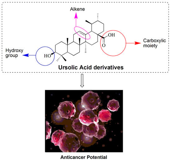
Many researchers have studied various strategies to modify the molecular structure of UA and simultaneously enhance its therapeutic effect. The structure of UA has been broken down into functional groups or pharmacophores to classify the major active sites for structural modification (Compound 1, Figure 2). These sites are broadly categorized as the carboxylic moiety (C-28), β-hydroxy function (C-3), and an alkene at C12–C13. The sites have been extensively studied for their potential anti-cancer activity. Some derivatives that are not identified in any of the aforementioned three pharmacophores are separately categorized into miscellaneous groups. The following reports further detailed the literature previously reported under these active sites and their successfully enhanced anticancer activity.
3.1.1. Modification of the Carboxylic Moiety (C-28)
Tian et al. synthesized a series of UA derivatives bearing diamine moieties at C-28 and investigated their antiproliferative potential against three human cancer cell lines (MCF-7, HeLa, and A549). This study was performed as a comparative analysis against the conventional antitumor drug, gefitinib. The derivatives were prepared by first protecting the C3-OH via acetylation, followed by esterification with 2-hydroxyacetic acid at the C-28 position and amidation reaction with amines including piperazine, N-methylpiperazine and alkane-1, 6-diamines and alkane-1, 4-diamines and alkane-1, and 2-diamines, as shown in Scheme 1 and Scheme 2. The half maximal inhibitory concentration (IC50) values for most of these derivatives were significantly higher when compared to gefitinib. The study revealed that derivatives containing primary amine moieties were more effective when compared to those with secondary or tertiary amine moieties. The antiproliferative activities all of the secondary amines were more active than those of the tertiary amine compounds. Compound 9a (Scheme 2, Table 3) showed a more potent antiproliferative activity than other derivatives [155].
A series of new derivatives of UA bearing oxadiazole, piperazine, and triazolone moieties was designed by Chi et al. and they were evaluated for their anticancer activity as Hypoxia-inducible factor-1α (HIF-1α) inhibitors. The in vitro results revealed that these derivatives had a significantly enhanced antitumor activity by inhibiting the expression of HIF-1α. Compound 13b (Table 3) demonstrated the best potent inhibitory effect against HIF-1α activity but was not cytotoxic to cancer cells. These results indicated that the simple esterification of the UA carboxyl moiety can result in a significantly enhanced inhibitory effect on HIF-1α activity and decreased toxicity. The mechanism of action suggested that 13b can also suppress cell proliferation and block cell cycle progression in the G1 phase (Scheme 3) [156].
Liu et al. synthesized a number of novel UA derivatives and evaluated their antitumor activities against two human cancer cell lines—gastric cancer cells (MGC-803) and breast cancer cells (Bcap-37) using an MTT assay in vitro [157]. These derivatives were obtained by reacting UA at C-28 with 1,2-dibromoethane, 1,3-dibromoethane, 1,4-dibromoethane, and butyl bromide in a solution of DMF and K2CO3, followed by reaction with corresponding amines. Most of the derivatives exhibited moderate to high inhibitory activities when compared to UA. The results illustrated that compound 14 (Table 3) induced cell apoptosis in MGC-803 cells.
3.1.2. Modification of both β-hydroxy (C-3) and Carboxylic Moiety (C-28)
Hua et al. synthesized a series of new UA derivatives by incorporating piperazine and thiourea at the C28 position and evaluated their in vitro cytotoxicity against selected cancer cell lines—namely HCT-116, T24, MGC-803, HepG2, and A549 cells and a normal cell line, HL-7702, using a 3-(4,5-dimethylthiazol-2-yl)-2,5-diphenyltetrazolium bromidefor (MTT) assay. The in vitro antiproliferative results suggested that most of the derivatives exhibited a potent inhibitory effect. However, one compound displayed an excellent in vitro cytotoxicity on HepG2 cells with an IC50 value of 5.40 µM and significantly induced apoptosis in the HeG2 cell line (compound 15, Table 4) [139]. Wiemann et al. designed a class of ursolic and oleanolic acid-derived hydroxamates in an attempt to investigate their cytotoxicity, using sulforhodamine B (SRB) assays against several human cancer cell lines (518A2, A2780, A549, FaDu, HT29, MCF-7, and NIH 3T3). The ursolic acid-based derivatives containing at least an OH and NH moiety in the hydroxamate part displayed good cytotoxicity and were significantly less selective to the cancer cells when compared to the oleanolic acid-based compounds. The results showed that compound 16 (Table 4) was the most potent compound, with IC50 values ranging from 2.5 to 6.4 µM [158].
Nedopekina et al. synthesized conjugates of triterpenoids UA and betulinic acid with the triphenylphosphonium (TPP+) group; evaluated their cytotoxic activity against two human cancer cell lines; and also studied their ability to induce programmed cancer cell death by employing markers of apoptosis, including the activation of caspase-3, permeabilization of the outer mitochondrial membrane, PARP-1 cleavage, the release of cytochrome c, and the inhibition of the mitochondrial respiratory chain. Two of the conjugates derived from ursolic acid and betulinic acid indicated a good range of IC50 values. Compound 17 (Table 4) derived from UA was obtained by the alkylation of the carboxyl group, C-28 of UA, with triethylene glycol dibromide in dimethylformamide (DMF) using K2CO3 at 50 °C for 3 h [159]. Jiang et al. synthesized a series of UA derivatives as inhibitors of Nuclear factor kappa B (NF-κB) by introducing long-chain amide moieties at the C-28 position, and the β-hydroxy group C-3 was protected by an acetyl group. Their in vitro anticancer properties and Tumor necrosis factor alpha (TNF-α-induced) NF-κB activation were evaluated against four human cancer cell lines, such as human ovarian cancer cells (SKOV3), lung cancer cells (A549), liver cancer cells (HepG2), and bladder cancer cells (T24). Several compounds exhibited considerable anticancer effects against different cancer cell lines. Compound 18 (Table 4) demonstrated the highest potency by inhibiting the growth of SKOV3, A549, HepG2, and T24 cells with IC50 values of 8.95, 5.22, 6.82, and 6.01 μM, respectively. The IC50 values were five-fold to eight-fold lower than the parent UA, and the results showed that compounds with longer diamide side chains showed relatively enhanced activity compared to compounds with shorter diamide side chains. A related mechanism study indicated that compound 18 caused cell cycle arrest at the G1 phase and triggered apoptosis in A549 cells by blocking the NF-κB signaling pathway [94].
Zhang et al. designed and synthesized UA-based tetrazole derivatives and studied their potential antitumor effects as HIF-1α transcriptional inhibitors. Compound 19 was the most potent with (IC50 = 0.8 µM) (Table 4). This compound was prepared by mixing the corresponding anhydrides with UA in a solution of trimethylamine and 4-dimethylaminopyridine (DMAP) in ice and stirred overnight at room temperature; the intermediate compound was reacted with SOCl2 in refluxing dichloromethane (DCM). The results suggest that introducing tetrazole at C-28 of UA increased the HIF-1α inhibitory effect; additionally, the introduction of large groups at C-3 is disadvantageous for the synthesis of effective HIF-1α inhibitors [160]. Kahnt et al. studied the ethylenediamine-spaced carboxamides of UA and betulinic acid and further analyzed their cytotoxicity effects against human tumor cells (8505C, A2780, MCF-7, HT29, NIH 3T3, and 518A2). The UA derivatives that demonstrated good in vitro cytotoxicity effects were compounds 20, 21, and 22) [161].
Wang et al. evaluated the antiproliferative activity of UA derivatives containing thiazole, triazole, tetrazolepiperazine, or homopiperazine moiety against two cancer cells, Hela and MKN45. Compound 23 (Table 4) displayed a superior antiproliferative activity on both cell lines and induced apoptosis via the intrinsic mitochondrial pathway [162]. Meng et al. designed UA derivatives through multiple synthetic steps and evaluated the synthesized compounds in vitro using an MTT assay on two cancer cell lines (BEL7402 and SGC7901). The results showed a higher inhibitory rate of the synthesized compounds on both cells when compared to the parent compound, UA. The results of molecular docking studies indicated the role of the structural design and optimization of UA. The most promising derivative was compound 24, as shown in Table 4 [163].
Wolfram et al. converted pentacyclic triterpenoic acids—namely oleanolic, betulinic glycyrrhetinic, boswellic, and ursolic acids—into their acetylated piperazinyl amides by coupling them with rhodamine B. To evaluate their in vitro cytotoxicity, all the triterpene-homopiperazinyl-rhodamine derivatives were subjected to RB assays and most of them were highly toxic against numerous human cancer cell lines (A2780, A375, HT29, NiH3T3, MCF7, and SW1736). The ursolic acid-homopiperazinyl-rhodamine derivative (Compound 25, Table 4) was one of the compounds that showed a strong cytotoxicity against the tumor cell lines, while it was less cytotoxic on SW1736 cells [164]. Kahnt et al. synthesized amine-spaced conjugates of UA and 1,4,7,10-tetraazacyclododecane-1,4,7,10-tetraacetic acid (DOTA). Two conjugates (Compound 26, 27, Table 4) displayed the highest cytotoxicity on A375 melanoma and A2780 ovarian carcinoma, with IC50 values of 1.5 µM and 1.7 µM, respectively. Compound 26 induced the death of A375 cells by apoptosis [165]. Mang et al. synthesized UA derivatives with potential anticancer properties by modifying C-2, C-3, and C-28 and evaluated their in vitro cytotoxicity against HepG2, BGC-823, and HeLa cell lines using an MTT assay These derivatives were synthesized by reacting UA with Jones reagents in acetone and then with NH2-OH·HCl; the obtained compounds were then reacted with Ac2O in the presence of DMAP in THF. The intermediate compound was condensed with the relevant amino or phenolic moieties in Et3N to afford the targeted compounds. Among these derivatives, Compounds 28 and 29 were demonstrated to be more effective on the cell lines when compared to the reference drug, gefitinib [166].
3.1.3. Modification of β-Hydroxy (C-3 Position)
Fontana et al. synthesized derivatives of UA and oleanolic acid with a modified oxidation state and lipophilicity at C-3 and C-28 positions which were screened in vitro on hepatocarcinoma cell lines—namely HepG2, HA22T/VGH, and Hep3B. UA derivatives containing three carbons as the side chain at the C-3 position of UA were synthesized in stereoisomeric forms using the Barbier–Grignard method and (Compounds 30 and 31, Table 5) were found to be effective against all three cancer cell lines; these compounds inhibited cell growth and induced the inhibition of NF-κB activation in the cell lines [167].
Xu et al. isolated triterpenoids from the acorn-starch/licorice and reacted them with 3,4,5-methoxybenzoic acid under dicyclohexyl carbodiimide (DCC)/DMAP conditions and evaluated their cytotoxicity in four cells, including A-549, MCF-7, H1975, and BGC-823. Most of the synthesized compounds indicated a significant cytotoxic activity in all the four cell lines and a lower toxicity in the normal cells, Human Hair Dermal Papilla Cells (HHDPC) when compared to the positive control, mitomycin C. The UA derivative containing 3,4,5-methoxy-phenacyl at the C-3 position (compound 32, Figure 3, Table 5) was the derivative with a significant antiproliferative effect, with IC50 values in the range of 6.07–22.27 µM [140].
3.1.4. Modification of Miscellaneous Groups
Wu et al. synthesized UA derivatives bearing an aminoguanidine moiety and investigated them as HIF-1α inhibitors and anticancer agents in human cancer cell lines. The majority of these compounds showed a potent inhibition of the HIF-1α transcriptional effect; among these derivatives, compound 35b (Scheme 4) was found to be the most potent inhibitor of HIF-1α expression in hypoxic conditions (IC50 4.0 µM), with no significant cytotoxicity noted against any cell lines tested [168].
Gu et al. designed and synthesized a series of new ursolic acid-based derivatives through the conjugation of UA with quinolone and oxadiazole motifs and investigated their anticancer activity. These compounds were investigated in vitro on human cancer cell lines-namely MDA-MB-231, Hela, and SMMC-7721, and the results indicated that compounds 36–38 exhibited a strong growth inhibitory effect against the three cancer cell lines. Among these derivatives, compound 38b exhibited the most potent antitumor activity, with IC50 values of 0.61 ± 0.07 (MDA-MB-231), 0.36 ± 0.05 (HeLa), and 12.49 ± 0.08 µM (SMMC-7721) when compared to the positive control, etoposide [169] (Scheme 5).
Mendes et al. synthesized ring-A-cleaved UA derivatives and evaluated their antiproliferative activity against three lung cancer cell lines—namely H460, H322, and H460+/+—using 2D or 3D culture models. Three of these newly designed UA derivatives bearing a cleaved ring-A with a secondary amide at C-3 (compounds 43a–c, Scheme 6) demonstrated significantly enhanced antiproliferative effects in the 2D systems. These compounds possessed a potent anticancer activity and the preliminary mechanism of action showed that compound 43c induced apoptosis through the activation of caspase-7 and caspase-8 and the decrease in Bcl-2 [170].
Borkoa et al. synthesized the novel triterpenic derivatives of dihydrobetulonic, betulonic, and ursonic acid. All of these compounds were tested for their in vitro cytotoxic activity against human cancer cell lines—namely CCRF-CEM, CEM-DNR, HCT116, HCT116 p53−/−, K562, K562-TAX, A549, U2OS, and two noncancerous fibroblasts such as BJ and MRC-5. Compounds 45 and 46 were the most effective on the CCRF-CEM cell lines and less toxic in non-cancerous fibroblasts. These derivatives triggered apoptosis via the intrinsic pathways. The ursonic acid derivative 45 was synthesized by the bromination of UA using copper (II) bromide in a solution of ethyl acetate (EtOAc) and methanol (MeOH) at room temperature. Compound 46 was obtained by the nucleophilic substitution of bromoketone 43 by potassium thiocyanate (KSCN) in DMSO at 90 °C, as shown in Scheme 7 [171].
Fan and colleagues synthesized UA hybrid compound 49 (Scheme 8) by reacting UA with Jones reagent at 0 °C in dimethyl ketone to obtain a C-3 oxidized derivative. The oxidized compound was then reacted with benzaldehyde using ethanolic potassium hydroxide (KOH) via the Claisen Schmidt condensation reaction at room temperature to achieve a benzylidine hybrid compound. The benzylidine compound was then reacted with an indole and substituted with a benzaldehyde in EtOAc at room temperature for 2 h to achieve compound 49. The anticancer potential of the compound against glioma cells was studied. The compound demonstrated a good inhibition of cell proliferation and induced apoptosis when compared to the parent compound, UA [172].
An aromatic heterocyclic compound containing carbazole has attracted attention due its potential anticancer activity [173]. Gu et al. synthesized a series of carbazole derivatives of UA. Among these derivatives, compound 50 (Figure 4) showed a significant cytotoxic activity against the hepatocarcinoma cell line, HepG2, with an IC50 value of 1.26 ± 0.17 µM [174]. Zhang et al. employed the biotransformation of UA by Mucor spinosus AS 3.3450, and three novel compounds were isolated. Compound 51 (Figure 4), identified as 3β, 7β-dihydroxy-ursolic acid-28-etha-none indicated a stronger cytotoxic activity against the tumor cell lines (Hela, K562, and KB) when compared to the parent compound UA [175]. A number of NO-donating ursolic acid-based benzylidene derivatives with various substitutions were synthesized by Zhang et al. They were further analyzed for their in vitro cytotoxicity against the HepG-2, MCF-7, HT-29, and A549 cancer cell lines. Most of these derivatives revealed a weaker inhibitory effect when compared to UA; compound 52 (Figure 4) showed the most potent activity against HT-29 (IC50 = 4.28 μM). The further investigation of its anticancer mode of action revealed that it induced the apoptosis of HT-29 cell lines in a dose-dependent manner and indicated cell cycle arrest at the G1 phase, which led to cell apoptosis and also induced apoptosis via the mitochondria-mediated pathways [176].
Jin et al. synthesized UA-based quinoline derivatives bearing thiadiazole, hydrazide, or oxadiazole moieties. The in vitro antiproliferative activity on tumor cells such as SMMC-7721, MDA-MB-231, and HeLa revealed that quinoline-based derivatives bearing carboxyl moieties or hydrazide moieties exhibited a significant anticancer activity against all the cancer cell lines when compared to the positive control, etoposide. Furthermore, a pharmacological in vitro analysis revealed that compound 53 (Figure 4) exhibited an antiproliferative activity against HeLa cell lines by cell cycle arrest at the G0/G1 phase, decreasing the mitochondrial membrane potential, inducing intracellular ROS generation, intervening with the Ras/Raf/mitogen-activated protein kinase kinase (MEK)/extracellular signal-regulated kinase (ERK) signaling pathways as MEK kinase inhibitors, and finally inducing the apoptosis of HeLa cell lines. The molecular docking analysis also revealed compound 53 capability to effectively bind with the active site of MEK [177].
The 1C50 values of compound 35b, 38b, 43a–b, 45, 46 and 50–53 on different cancer cell lines are shown in Table 6.
4. Insights and Future Directions
This review reports various strategies that have been used to design UA-based derivatives with enhanced anticancer activity when compared to UA or conventional drugs used as controls in most of the studies reported over the past five years. Many studies on pentacyclic triterpenoids have shown that the C-2 position, β-hydroxyl (C-3), and carboxylic moieties (C-28) of UA were the major sites for the modification of its structure. Most researchers modified the molecular structure of UA around the three sites, resulting in the improvement in the chemical or physical activities of the UA molecule.
The derivatives were grouped according to their active sites of modifications. Their corresponding IC50 values are listed in the tables against reference compounds, usually UA or a conventional drug. The derivatives were tested against various cancer cell lines in vitro using different cell lines of colon, prostate, gastric, leukaemia, lung, breast, pancreas, skin, glioblastoma, and renal cells. Most of the derivatives demonstrated an improved antiproliferative activity when compared to their respective reference molecules, revealing that UA structural modification significantly enhanced their antiproliferative activity.
Despite the great progress which has been made, there are still a lot of research gaps, such as the synthesis of ursolic acid-based hybrid compounds via hybridization with known chemotherapeutic scaffolds, bioavailability studies, and toxicological studies. Furthermore, more evaluation in vivo is lacking and there is a pressing need to evaluate the synthesized compounds in vivo.
Funding
This research was funded by the South African Medical Research Council (Self-Initiated Research), National Research Foundation South Africa, Sasol Inzalo Foundation, and Govan Mbeki Research and Development Centre (GMRDC), University of Fort Hare.
Conflicts of Interest
The authors declare no conflict of interest.
Abbreviations
| FDA | Food and Drug Administration |
| UA | Ursolic acid |
| P53 | Tumor protein p53 |
| Wnt | Wnt/β-catenin pathways |
| Ras | Retrovirus-associated DNA sequences |
| TRAIL | TNF-related apoptosis-inducing ligand |
| STAT3 | Signal transducers and activators of transcription |
| PK | Pharmacokinetic |
| UV | Ultraviolet |
| HIF-1α | Hypoxia-inducible factor 1-alpha |
| DMF | Dimethylformamide |
| MTT | Dye compound 3-(4,5-Dimethylthiazol-2-yl)-2,5-diphenyltetrazolium bromide assay |
| SRB | sulforhodamine B assay |
| PARP-1 | Poly [ADP-ribose] polymerase 1 |
| NF-kB | Nuclear factor kappa-B |
| DMAP | 4-Dimethylaminopyridine |
| DCM | Dichloromethane |
| THF | Tetrahydrofuran |
| DCC | N,N′-Dicyclohexylcarbodiimide |
| DMSO | Dimethyl sulfoxide |
| FROS | Reactive oxygen species |
| MEK | Mitogen-activated extracellular signal-regulated kinase |
References
- Dewangan, J.; Srivastava, S.; Mishra, S.; Divakar, A.; Kumar, S.; Rath, S.K. Salinomycin inhibits breast cancer progression via targeting HIF-1α/VEGF mediated tumor angiogenesis in vitro and in vivo. Biochem. Pharmacol. 2019, 164, 326–335. [Google Scholar] [CrossRef] [PubMed]
- Made, F.; Wilson, K.; Jina, R.; Tlotleng, N.; Jack, S.; Ntlebi, V.; Kootbodien, T. Distribution of cancer mortality rates by province in South Africa. Cancer Epidemiol. 2017, 51, 56–61. [Google Scholar] [CrossRef] [PubMed]
- Rodrigues, F.C.; Anil Kumar, N.V.; Thakur, G. Developments in the anticancer activity of structurally modified curcumin: An up-to-date review. Eur. J. Med. Chem. 2019, 177, 76–104. [Google Scholar] [CrossRef] [PubMed]
- Pejin, B.; Jovanovi, K.K.; Mojovi, M.; Savi, A.G. New and Highly Potent Antitumor Natural Products from Marine-Derived Fungi: Covering the Period from 2003 to 2012. Curr. Top. Med. Chem. 2013, 13, 2745–2766. [Google Scholar] [CrossRef] [PubMed]
- Sisodiya, P.S. Plant Derived Anticancer Agents: A Review. Int. J. Res. Dev. Pharm. Life Sci. 2013, 2, 293–308. [Google Scholar]
- Pejin, B.; Glumac, M.A. brief review of potent anti-CNS tumourics from marine sponges: Covering the period from 1994 to 2014. Nat. Prod. Res. 2018, 32, 375–384. [Google Scholar] [CrossRef]
- Calcabrini, C.; Catanzaro, E.; Bishayee, A.; Turrini, E.; Fimognari, C. Marine Sponge Natural Products with Anticancer Potential: An Updated Review. Mar. Drugs. 2017, 15, 310. [Google Scholar] [CrossRef] [Green Version]
- Katz, L.; Baltz, R.H. Natural product discovery: Past, present, and future. J. Ind. Microbiol. Biotechnol. 2016, 43, 155–176. [Google Scholar] [CrossRef]
- Liu, W.; Li, Q.; Hu, J.; Wang, H.; Xu, F.; Bian, Q. Application of natural products derivatization method in the design of targeted anticancer agents from 2000 to 2018. Bioorganic Med. Chem. 2019, 27, 115150. [Google Scholar] [CrossRef]
- Khwaza, V.; Oyedeji, O.O.; Aderibigbe, B.A. Antiviral Activities of Oleanolic Acid and Its Analogues. Molecules 2018, 23, 2300. [Google Scholar] [CrossRef] [Green Version]
- Salvador, J.A.R.; Leal, A.S.; Valdeira, A.S.; Gonçalves, B.M.F.; Alho, D.P.S.; Figueiredo, S.A.C.; Silvestre, S.M.; Mendes, V.I.S. Oleanane-, ursane-, and quinone methide friedelane-type triterpenoid derivatives: Recent advances in cancer treatment. Eur. J. Med. Chem. 2017, 142, 95–130. [Google Scholar] [CrossRef] [PubMed]
- Sathya, S.; Sudhagar, S.; Sarathkumar, B.; Lakshmi, B.S. EGFR inhibition by pentacyclic triterpenes exhibit cell cycle and growth arrest in breast cancer cells. Life Sci. 2014, 95, 53–62. [Google Scholar] [CrossRef] [PubMed]
- Luo, J.; Hu, Y.L.; Wang, H. Ursolic acid inhibits breast cancer growth by inhibiting proliferation, inducing autophagy and apoptosis, and suppressing inflammatory responses via the PI3K/AKT and NF-κB signaling pathways in vitro. Exp. Ther. Med. 2017, 14, 3623–3631. [Google Scholar] [CrossRef] [PubMed]
- Woźniak, Ł.; Skąpska, S.; Marszałek, K. Ursolic acid—A pentacyclic triterpenoid with a wide spectrum of pharmacological activities. Molecules 2015, 20, 20614–20641. [Google Scholar] [CrossRef] [PubMed] [Green Version]
- Amarowicz, R.; Pegg, R.B. Inhibition of proliferation of human carcinoma cell lines by phenolic compounds from a bearberry-leaf crude extract and its fractions. J. Funct. Foods. 2013, 5, 660–667. [Google Scholar] [CrossRef]
- Hassan, H.M.; Jiang, Z.H.; Asmussen, C.; McDonald, E.; Qin, W. Antibacterial activity of northern Ontario medicinal plant extracts. Can. J. Plant Sci. 2014, 94, 417–424. [Google Scholar] [CrossRef]
- Guinda, A.; Rada, M.; Delgado, T.; Castellano, J.M. Pentacyclic triterpenic acids from Argania spinosa. Eur. J. Lipid Sci. Technol. 2011, 113, 231–237. [Google Scholar] [CrossRef]
- Guinda, Á.; Rada, M.; Delgado, T.; Gutiérrez-Adánez, P.; Castellano, J.M. Pentacyclic triterpenoids from olive fruit and leaf. J. Agric. Food Chem. 2010, 58, 9685–9691. [Google Scholar] [CrossRef]
- García-Morales, G.; Huerta-Reyes, M.; González-Cortazar, M.; Zamilpa, A.; Jiménez-Ferrer, E.; Silva-García, R.; Román-Ramos, R.; Aguilar-Rojas, A. Anti-inflammatory, antioxidant and anti-acetylcholinesterase activities of Bouvardia ternifolia: Potential implications in Alzheimer’s disease. Arch. Pharm. Res. 2015, 38, 1369–1379. [Google Scholar] [CrossRef]
- Figueroa-Suárez, M.Z.; González, C.J.; Cardoso-Taketa, A.T.; Del Carmen Gutiérrez Villafuerte, M.; Rodríguez-López, V. Anti-inflammatory and antihistaminic activity of triterpenoids isolated from Bursera cuneata (Schldl.) Engl. J. Ethnopharmacol. 2019, 238, 111786. [Google Scholar] [CrossRef]
- Yu, F.; Thamm, A.M.K.; Reed, D.; Villa-Ruano, N.; Quesada, A.L.; Gloria, E.L.; Covello, P.; De, L.V. Functional characterization of amyrin synthase involved in ursolic acid biosynthesis in Catharanthus roseus leaf epidermis. Phytochemistry 2013, 91, 122–127. [Google Scholar] [CrossRef] [PubMed] [Green Version]
- Kwon, S.H.; Park, H.Y.; Kim, J.Y.; Jeong, I.Y.; Lee, M.K.; Seo, K.-I. Apoptotic action of ursolic acid isolated from Corni fructus in RC-58T/h/SA#4 primary human prostate cancer cells. Bioorganic Med. Chem. Lett. 2010, 20, 6435–6438. [Google Scholar]
- Zar, P.P.K.; Yano, S.; Sakao, K.; Hashimoto, F.; Nakano, T.; Fujii, M.; Hou, D.X. In vitro anticancer activity of loquat tea by inducing apoptosis in human leukemia cells. Biosci. Biotechnol. Biochem. 2014, 78, 1731–1737. [Google Scholar] [CrossRef] [PubMed]
- Tan, H.; Zhao, C.; Zhu, Q.; Katakura, Y.; Tanaka, H.; Ohnuki, K.; Shimizu, K. Ursolic Acid Isolated from the Leaves of Loquat (Eriobotrya japonica) Inhibited Osteoclast Differentiation through Targeting Exportin 5. J. Agric. Food Chem. 2019, 67, 3333–3340. [Google Scholar] [CrossRef]
- Tan, H.; Sonam, T.; Shimizu, K. The potential of triterpenoids from loquat leaves (Eriobotrya japonica) for prevention and treatment of skin disorder. Int. J. Mol. Sci. 2017, 18, 1030. [Google Scholar] [CrossRef] [PubMed] [Green Version]
- Kuraoka-Oliveira, Â.M.; Radai, J.A.S.; Leitão, M.M.; Lima Cardoso, C.A.; Silva-Filho, S.E.; Leite, K.C.A. Anti-inflammatory and anti-arthritic activity in extract from the leaves of Eriobotrya japonica. J. Ethnopharmacol. 2020, 249, 112418. [Google Scholar] [CrossRef] [PubMed]
- Gao, Y.S.; Yuan, Y.; Song, G.; Lin, S.Q. Inhibitory effect of ursolic acid and oleanolic acid from Eriobotrya fragrans on A549 cell viability in vivo. Genet. Mol. Res. 2016, 15, 1–8. [Google Scholar] [CrossRef] [PubMed]
- González-Burgos, E.; Liaudanskas, M.; Viškelis, J.; Žvikas, V.; Janulis, V.; Gómez-Serranillos, M.P. Antioxidant activity, neuroprotective properties and bioactive constituents analysis of varying polarity extracts from Eucalyptus globulus leaves. J. Food Drug Anal. 2018, 26, 1293–1302. [Google Scholar] [CrossRef] [PubMed]
- Domingues, R.M.A.; Patinha, D.J.S.; Sousa, G.D.A.; Villaverde, J.J.; Silva, C.M.; Freire, C.S.R. Eucalyptus biomass residues from agro-forest and pulping industries as sources of high-value triterpenic compounds. Cellul. Chem. Technol. 2011, 45, 475–481. [Google Scholar]
- Madmanang, S.; Cheyeng, N.; Heembenmad, S.; Mahabusarakam, W.; Saising, J.; Seeger, M.; Chusri, S.; Chakthong, S. Constituents of Fagraea fragrans with Antimycobacterial Activity in Combination with Erythromycin. J. Nat. Prod. 2016, 79, 767–774. [Google Scholar] [CrossRef]
- Palu, D.; Bighelli, A.; Casanova, J.; Paoli, M. Identification and quantitation of ursolic and oleanolic acids in ilex aquifolium l. Leaf extracts using 13C and 1H-NMR spectroscopy. Molecules 2019, 24, 4413. [Google Scholar] [CrossRef] [PubMed] [Green Version]
- Wójciak-Kosior, M.; Sowa, I.; Kocjan, R.; Nowak, R. Effect of different extraction techniques on quantification of oleanolic and ursolic acid in Lamii albi flos. Ind. Crops Prod. 2013, 44, 373–377. [Google Scholar] [CrossRef]
- Jamal, M.; Amir, M.; Ali, Z.; Mujeeb, M. A comparative study for the extraction methods and solvent selection for isolation, quantitative estimation and validation of ursolic acid in the leaves of Lantana camara by HPTLC method. Futur. J. Pharm. Sci. 2018, 4, 229–233. [Google Scholar] [CrossRef]
- Kazmi, I.; Rahman, M.; Afzal, M.; Gupta, G.; Saleem, S.; Afzal, O.; Shaharyar, M.A.; Nautiyal, U.; Ahmed, S.; Anwar, F. Anti-diabetic potential of ursolic acid stearoyl glucoside: A new triterpenic gycosidic ester from Lantana camara. Fitoterapia 2012, 83, 142–146. [Google Scholar] [CrossRef] [PubMed]
- Kazmi, I.; Afzal, M.; Ali, B.; Damanhouri, Z.A.; Ahmaol, A.; Anwar, F. Anxiolytic potential of ursolic acid derivative-a stearoyl glucoside isolated from Lantana camara L. (verbanaceae). Asian Pac. J. Trop. Med. 2013, 6, 433–437. [Google Scholar] [CrossRef]
- Gilabert, M.; Marcinkevicius, K.; Andujar, S.; Schiavone, M.; Arena, M.E.; Bardón, A. Sesqui- and triterpenoids from the liverwort Lepidozia chordulifera inhibitors of bacterial biofilm and elastase activity of human pathogenic bacteria. Phytomedicine 2015, 22, 77–85. [Google Scholar] [CrossRef]
- Xia, E.Q.; Yu, Y.Y.; Xu, X.R.; Deng, G.F.; Guo, Y.J.; Bin Li, H. Ultrasound-assisted extraction of oleanolic acid and ursolic acid from Ligustrum lucidum Ait. Ultrason. Sonochem. 2012, 19, 772–776. [Google Scholar] [CrossRef]
- Xia, E.Q.; Wang, B.W.; Xu, X.R.; Zhu, L.; Song, Y.; Li, H.B. Microwave-assisted extraction of oleanolic acid and ursolic acid from Ligustrum lucidum ait. Int. J. Mol. Sci. 2011, 12, 5319–5329. [Google Scholar] [CrossRef]
- Frighetto, R.T.S.; Welendorf, R.M.; Nigro, E.N.; Frighetto, N.; Siani, A.C. Isolation of ursolic acid from apple peels by high speed counter-current chromatography. Food Chem. 2008, 106, 767–771. [Google Scholar] [CrossRef]
- Yamaguchi, H.; Noshita, T.; Kidachi, Y.; Umetsu, H.; Hayashi, M.; Komiyama, K.; Funayama, S.; Ryoyama, K. Isolation of ursolic acid from apple peels and its specific efficacy as a potent antitumor agent. J. Heal. Sci. 2008, 54, 654–660. [Google Scholar] [CrossRef] [Green Version]
- Zahran, E.M.; Abdelmohsen, U.R.; Ayoub, A.T.; Salem, M.A.; Khalil, H.E.; Desoukey, S.Y.; Fouad, M.A.; Kamel, M.S. Metabolic profiling, histopathological anti-ulcer study, molecular docking and molecular dynamics of ursolic acid isolated from Ocimum forskolei Benth (family Lamiaceae). South Afr. J. Bot. 2020, 131, 311–319. [Google Scholar] [CrossRef]
- Ahmad, A.; Abuzinadah, M.F.; Alkreathy, H.M.; Banaganapalli, B.; Mujeeb, M. Ursolic acid rich ocimum sanctum L leaf extract loaded nanostructured lipid carriers ameliorate adjuvant induced arthritis in rats by inhibition of COX-1, COX-2, TNF-α and IL-1: Pharmacological and docking studies. PLoS ONE 2018, 13, e0193451. [Google Scholar] [CrossRef] [PubMed] [Green Version]
- Flegkas, A.; Milosević Ifantis, T.; Barda, C.; Samara, P.; Tsitsilonis, O.; Skaltsa, H. Antiproliferative Activity of (-)-Rabdosiin Isolated from Ocimum sanctum L. Medicines 2019, 6, 37. [Google Scholar] [CrossRef] [PubMed] [Green Version]
- Jothie, R.E.; Illuri, R.; Bethapudi, B.; Anandhakumar, S.; Bhaskar, A.; Chinampudur, V.C.; Mundkinajeddu, D.; Agarwal, A. Activity of Ocimum sanctum: Possible Effects on Hypothalamic-Pituitary-Adrenal Axis. Phyther. Res. 2016, 30, 805–814. [Google Scholar] [CrossRef] [PubMed]
- Shibata, S. Chemistry and cancer preventing activities of ginseng saponins and some related triterpenoid compounds. J. Korean Med. Sci. 2001, 16, S28–S37. [Google Scholar] [CrossRef] [Green Version]
- Ali, S.A.; Ibrahim, N.A.; Mohammed, M.M.D.; El-Hawary, S.; Refaat, E.A. The potential chemo preventive effect of ursolic acid isolated from Paulownia tomentosa, against N-diethylnitrosamine: Initiated and promoted hepatocarcinogenesis. Heliyon 2019, 5, e01769. [Google Scholar] [CrossRef] [Green Version]
- Li, B.; Hu, Y.; Li, J.; Shi, K.; Shen, Y.; Zhu, B.; Wang, G.X. Ursolic acid from Prunella vulgaris L. efficiently inhibits IHNV infection in vitro and in vivo. Virus Res. 2019, 273, 197741. [Google Scholar] [CrossRef]
- Kim, H.I.; Quan, F.; Kim, J.; Lee, N.; Kim, H.; Jo, S.; Lee, C.M.; Jang, D.; Inn, K.S. Inhibition of estrogen signaling through depletion of estrogen receptor alpha by ursolic acid and betulinic acid from Prunella vulgaris var. lilacina. Biochem. Biophys. Res. Commun. 2014, 451, 282–287. [Google Scholar] [CrossRef]
- Xu, C.; Liao, Y.; Fang, C.; Tsunoda, M.; Zhang, Y.; Song, Y.; Deng, S. Simultaneous Analysis of Ursolic Acid and Oleanolic Acid in Guava Leaves Using QuEChERS-Based Extraction Followed by High-Performance Liquid Chromatography. J. Anal. Methods Chem. 2017, 2017, 2984562. [Google Scholar] [CrossRef]
- Yang, Y.C.; Wei, M.C.; Huang, T.C. Optimisation of an ultrasound-assisted extraction followed by RP-HPLC separation for the simultaneous determination of oleanolic acid, ursolic acid and oridonin content in Rabdosia rubescens. Phytochem. Anal. 2012, 23, 627–636. [Google Scholar] [CrossRef]
- MacHado, D.G.; MacHado, D.G.; Neis, V.B.; Balen, G.O.; Colla, A.; Cunha, M.P.; Dalmarco, J.B.; Pizzolatti, M.G.; Prediger, R.D.; Rodrigues, A.L.S. Antidepressant-like effect of ursolic acid isolated from Rosmarinus officinalis L. in mice: Evidence for the involvement of the dopaminergic system. Pharmacol. Biochem. Behav. 2012, 103, 204–211. [Google Scholar] [CrossRef] [PubMed] [Green Version]
- Do Nascimento, P.G.G.; Lemos, T.L.G.; Bizerra, A.M.C.; Arriaga, Â.M.C.; Ferreira, D.A. Antibacterial and Antioxidant Activities of Ursolic Acid. Molecules 2014, 19, 1317–1327. [Google Scholar] [CrossRef] [PubMed]
- Mazumder, K.; Siwu, E.R.O.; Nozaki, S.; Watanabe, Y.; Tanaka, K.; Fukase, K. Ursolic acid derivatives from Bangladeshi medicinal plant, Saurauja roxburghii: Isolation and cytotoxic activity against A431 and C6 glioma cell lines. Phytochem. Lett. 2011, 4, 287–291. [Google Scholar] [CrossRef]
- Kubatka, P.; Uramova, S.; Kello, M.; Kajo, K.; Samec, M.; Jasek, K.; Vybohova, D.; Liskova, A.; Mojzis, J.; Adamkov, M.; et al. Anticancer activities of thymus vulgaris L. In experimental breast carcinoma in vivo and in vitro. Int. J. Mol. Sci. 2019, 20, 1749. [Google Scholar] [CrossRef] [PubMed] [Green Version]
- Somova, L.O.; Nadar, A.; Rammanan, P.; Shode, F.O. Cardiovascular, antihyperlipidemic and antioxidant effects of oleanolic and ursolic acids in experimental. Phytomedicine 2003, 10, 115–121. [Google Scholar] [CrossRef] [PubMed]
- Vinciguerra, V.; Rojas, F.; Tedesco, V.; Giusiano, G.; Angiolella, L. Chemical characterization and antifungal activity of Origanum vulgare, Thymus vulgaris essential oils and carvacrol against Malassezia furfur. Nat. Prod. Res. 2019, 33, 3273–3277. [Google Scholar] [CrossRef]
- Abu-Gharbieh, E.; Shehab, N.G.; Almasri, I.M.; Bustanji, Y. Antihyperuricemic and xanthine oxidase inhibitory activities of Tribulus arabicus and its isolated compound, ursolic acid: In vitro and in vivo investigation and docking simulations. PLoS ONE 2018, 13, e0202572. [Google Scholar] [CrossRef]
- Ksiksi, T.; Palakkott, A.R.; Ppoyil, S.B.T. Tribulus arabicus and Tribulus macropterus are Comparable to Tribulus terrestris: An Antioxidant Assessment. Curr. Bioact. Compd. 2017, 13, 82–87. [Google Scholar] [CrossRef]
- Sharifiyan, F.; Mirjalili, S.A.; Fazilati, M.; Poorazizi, E.; Habibollahi, S. Variation of ursolic acid content in flowers of ten Iranian pomegranate (Punica granatum L.) cultivars. BMC Chem. 2019, 13, 80. [Google Scholar] [CrossRef] [Green Version]
- Fu, Q.; Zhang, L.; Cheng, N.; Jia, M.; Zhang, Y. Extraction optimization of oleanolic and ursolic acids from pomegranate (Punica granatum L.) flowers. Food Bioprod. Process. 2014, 92, 321–327. [Google Scholar] [CrossRef]
- Jung, T.Y.; Pham, T.N.N.; Umeyama, A.; Shoji, N.; Hashimoto, T.; Lee, J.; Takei, M. Ursolic acid isolated from Uncaria rhynchophylla activates human dendritic cells via TLR2 and/or TLR4 and induces the production of IFN-γ by CD4+ naïve T cells. Eur. J. Pharmacol. 2010, 643, 297–303. [Google Scholar] [CrossRef] [PubMed]
- Zhang, Y.; Yang, W.Z.; Yao, C.L.; Feng, R.H.; Yang, M.; Guo, D.; Wu, W.Y. New triterpenic acids from Uncaria rhynchophylla: Chemistry, NO-inhibitory activity, and tandem mass spectrometric analysis. Fitoterapia 2014, 96, 39–47. [Google Scholar] [CrossRef] [PubMed]
- Chandramu, D.; Rao, D.M.; Krupadanam, G.I.D.; Reddy, V.R. Isolation, Characterization and Biological Activity of Betulinic Acid and Ursolic Acid from Vitex negundo L. Phyther. Res. 2003, 134, 129–134. [Google Scholar] [CrossRef] [PubMed]
- Kawabata, K.; Kitamura, K.; Irie, K.; Naruse, S.; Matsuura, T.; Uemae, T.; Taira, S.; Ohigashi, H.; Murakami, S.; Takahashi, M.; et al. Triterpenoids isolated from Ziziphus jujuba enhance glucose uptake activity in skeletal muscle cells. J. Nutr. Sci. Vitaminol. 2017, 63, 193–199. [Google Scholar] [CrossRef] [Green Version]
- Yin, R.; Li, T.; Tian, J.X.; Xi, P.; Liu, R.H. Ursolic acid, a potential anticancer compound for breast cancer therapy. Crit. Rev. Food Sci. Nutr. 2018, 58, 568–574. [Google Scholar] [CrossRef] [PubMed]
- Checker, R.; Sandur, S.K.; Sharma, D.; Patwardhan, R.S.; Jayakumar, S.; Kohli, V.; Sethi, G.; Aggarwal, B.B.; Sainis, K.B. Potent anti-inflammatory activity of ursolic acid, a triterpenoid antioxidant, is mediated through suppression of NF-κB, AP-1 and NF-AT. PloS ONE. 2012, 7, e31318. [Google Scholar] [CrossRef] [Green Version]
- Wolska, K.I.; Grudniak, A.M.; Fiecek, B.; Kraczkiewicz-dowjat, A.; Kurek, A. Antibacterial activity of oleanolic and ursolic acids and their derivatives. Cent. Eur. J. Biol. 2010, 5, 543–553. [Google Scholar] [CrossRef]
- Castro, A.J.; Frederico, M.J.; Cazarolli, L.H.; Mendes, C.P.; Bretanha, L.C.; Schmidt, É.C.; Bouzon, Z.L.; de Medeiros Pinto, V.A.; da Fonte Ramos, C.; Pizzolatti, M.G.; et al. The mechanism of action of ursolic acid as insulin secretagogue and insulinomimetic is mediated by cross-talk between calcium and kinases to regulate glucose balance. Biochim. Biophys. Acta. 2015, 1850, 51–61. [Google Scholar] [CrossRef] [Green Version]
- Shanmugam, M.K.; Dai, X.; Prem, A.; Tan, B.K.H.; Sethi, G.; Bishayee, A. Ursolic acid in cancer prevention and treatment: Molecular targets, pharmacokinetics and clinical studies. Biochem. Pharmacol. 2013, 85, 1579–1587. [Google Scholar] [CrossRef] [Green Version]
- Angeles, M.L.; Navin, R.; Kim, S.M. Therapeutic Interventions Using Ursolic Acid for Cancer Treatment. Med. Chem. 2016, 6, 339–344. [Google Scholar]
- Achiwa, Y.; Hasegawa, K.; Udagawa, Y.; Achiwa, Y.; Hasegawa, K.; Udagawa, Y. Effect of Ursolic Acid on MAPK in Cyclin D1 Signaling and RING-Type E3 Ligase (SCF E3s) in Two Endometrial Cancer Cell Lines Effect of Ursolic Acid on MAPK in Cyclin D1 Signaling and RING-Type E3 Ligase (SCF E3s) in Two Endometrial. Nutr. Cancer 2013, 65, 1026–1033. [Google Scholar] [CrossRef] [PubMed]
- Achiwa, Y.; Hasegawa, K.; Udagawa, A. Regulation of the Phosphatidylinositol 3-Kinase-Akt and the Mitogen-Activated Protein Kinase Pathways by Ursolic Acid in Human Endometrial Cancer Cells Yumiko. Biosci. Biotechnol. Biochem. 2007, 71, 31–37. [Google Scholar] [CrossRef] [PubMed]
- Li, J.; Liang, X.; Yang, X. Ursolic acid inhibits growth and induces apoptosis in gemcitabine-resistant human pancreatic cancer via the JNK and PI3K/Akt/NF- κ B pathways. Oncol. Rep. 2012, 28, 501–510. [Google Scholar] [CrossRef] [PubMed] [Green Version]
- Prasad, S.; Yadav, V.R.; Sung, B.; Gupta, S.C.; Tyagi, A.K. Ursolic acid inhibits the growth of human pancreatic cancer and enhances the antitumor potential of gemcitabine in an orthotopic mouse model through suppression of the inflammatory microenvironment. Oncotarget 2016, 7, 13182. [Google Scholar] [CrossRef] [PubMed]
- Huang, C.Y.; Lin, C.Y.; Tsai, C.W.; Yin, M.C. Inhibition of cell proliferation, invasion and migration by ursolic acid in human lung cancer cell lines. Toxicol. Vitr. 2011, 25, 1274–1280. [Google Scholar] [CrossRef]
- Hsu, Y.L.; Kuo, P.L.; Lin, C.C. Proliferative inhibition, cell-cycle dysregulation, and induction of apoptosis by ursolic acid in human non-small cell lung cancer A549 cells. Life Sci. 2004, 75, 2303–2316. [Google Scholar] [CrossRef]
- Kim, S.; Ryu, H.G.; Lee, J.; Shin, J.; Harikishore, A.; Jung, H.Y.; Kim, Y.S.; Lyu, H.N.; Oh, E.; Baek, N.I.; et al. Ursolic acid exerts anti-cancer activity by suppressing vaccinia-related kinase 1-mediated damage repair in lung cancer cells. Sci. Rep. 2015, 5, 14570. [Google Scholar] [CrossRef]
- Kassi, E.; Papoutsi, Z.; Pratsinis, H.; Aligiannis, N.; Manoussakis, M.; Moutsatsou, P. Ursolic acid, a naturally occurring triterpenoid, demonstrates anticancer activity on human prostate cancer cells. J. Cancer Res. Clin. Oncol. 2007, 133, 493–500. [Google Scholar] [CrossRef]
- Shanmugam, M.K.; Rajendran, P.; Li, F.; Nema, T.; Vali, S.; Abbasi, T.; Kapoor, S.; Sharma, A.; Kumar, A.P.; Ho, P.C.; et al. Ursolic acid inhibits multiple cell survival pathways leading to suppression of growth of prostate cancer xenograft in nude mice. J. Mol. Med. 2011, 89, 713–727. [Google Scholar] [CrossRef]
- Gai, W.T.; Yu, D.P.; Wang, X.S.; Wang, P.T. Anti-cancer effect of ursolic acid activates apoptosis through ROCK/PTEN mediated mitochondrial translocation of cofilin-1 in prostate cancer. Oncol. Lett. 2016, 12, 2880–2885. [Google Scholar] [CrossRef]
- Wang, X.; Li, L.; Wang, B.; Xiang, J. Effects of ursolic acid on the proliferation and apoptosis of human ovarian cancer cells. J. Huazhong Univ. Sci. Technol. Med. Sci. 2009, 29, 761–764. [Google Scholar] [CrossRef] [PubMed]
- Yang, L.; Liu, X.; Lu, Z.; Yuet, W.J.; Zhou, L.L.; Fung, K.P.; Wu, P.; Wu, S. Ursolic acid induces doxorubicin-resistant HepG2 cell death via the release of apoptosis-inducing factor. Cancer Lett. 2010, 298, 128–138. [Google Scholar] [CrossRef] [PubMed]
- Zheng, Q.; Li, P.; Jin, F.; Yao, C.; Zhang, G.; Zang, T.; Ai, X. Ursolic acid induces ER stress response to activate ASK1-JNK signaling and induce apoptosis in human bladder cancer T24 cells. Cell. Signal. 2013, 25, 206–213. [Google Scholar] [CrossRef] [PubMed]
- Xu, X.; Zhu, G.; Zhang, K.; Zhou, Y.; Li, X.; Xu, W.; Zhang, H.; Shao, Y.; Zhang, Z.; Sun, W. Cyclooxygenase-2 mediated synergistic effect of ursolic acid in combination with paclitaxel against human gastric carcinoma. Oncotarget 2017, 8, 92770–92777. [Google Scholar] [CrossRef] [Green Version]
- Kim, E.S.; Moon, A. Ursolic acid inhibits the invasive phenotype of SNU-484 human gastric cancer cells. Oncol. Lett. 2015, 9, 897–902. [Google Scholar] [CrossRef]
- Zhou, W.; Lin, L.; Cheng, Y.; Liu, Y. Ursolic Acid Improves Liver Transplantation and Inhibits Apoptosis in Miniature Pigs Using Donation after Cardiac Death. Cell. Physiol. Biochem. 2017, 43, 331–338. [Google Scholar] [CrossRef]
- Prasad, S.; Yadav, V.R.; Kannappan, R.; Aggarwal, B.B. Ursolic acid, a pentacyclin triterpene, potentiates TRAIL-induced apoptosis through p53-independent up-regulation of death receptors. J. Biol. Chem. 2016, 291, 16924. [Google Scholar] [CrossRef] [Green Version]
- Wang, C.; Lin, C.; Hua, C.; Jou, Y.; Liao, C.; Chang, Y.; Wan, L.; Huang, S.; Hour, M.; Lin, C. Cis-3-O-p-hydroxycinnamoyl ursolic acid induced ROS-dependent p53-mediated mitochondrial apoptosis in oral cancer cells. Biomol. Ther. 2019, 27, 54–62. [Google Scholar] [CrossRef]
- Wang, S.; Meng, X.; Dong, Y. Ursolic acid nanoparticles inhibit cervical cancer growth in vitro and in vivo via apoptosis induction. Int. J. Oncol. 2017, 50, 1330–1340. [Google Scholar] [CrossRef] [PubMed]
- Jiang, K.; Chi, T.; Li, T.; Zheng, G.; Fan, L.; Liu, Y.; Chen, X.; Chen, S.; Jia, L.; Shao, J. A smart pH-responsive nano-carrier as a drug delivery system for the targeted delivery of ursolic acid: Suppresses cancer growth and metastasis by modulating P53/MMP-9/PTEN/CD44 mediated multiple signaling pathways. Nanoscale 2017, 9, 9428–9439. [Google Scholar] [CrossRef] [PubMed]
- Kim, J.; Kim, Y.H.; Song, G.; Kim, D.; Jeong, Y.; Liu, K.; Chung, Y.; Oh, S. Ursolic acid and its natural derivative corosolic acid suppress the proliferation of APC-mutated colon cancer cells through promotion of β-catenin degradation. Food Chem. Toxicol. 2014, 67, 87–95. [Google Scholar] [CrossRef] [PubMed]
- Zhang, R.; Li, Y.; Tian, D.; Liu, Y.; Nian, W.; Zou, X.; Chen, Q.; Zhou, L.; Deng, Z.; He, B. Ursolic acid inhibits proliferation and induces apoptosis by inactivating Wnt/β-catenin signaling in human osteosarcoma cells. Int. J. Oncol. 2016, 49, 1973–1982. [Google Scholar] [CrossRef] [PubMed] [Green Version]
- Kim, S.; Jin, H.; Meng, R.Y.; Kim, D.Y.; Liu, Y.C.; Chai, O.H.; Park, B.H.; Kim, S. Activating hippo pathway via Rassf1 by ursolic acid suppresses the tumorigenesis of gastric cancer. Int. J. Mol. Sci. 2019, 20, 4709. [Google Scholar] [CrossRef] [PubMed] [Green Version]
- Jiang, W.; Huang, R.; Zhang, J.; Guo, T.; Zhang, M.; Huang, X.; Zhang, B.; Liao, Z.; Sun, J.; Wang, H. Discovery of antitumor ursolic acid long-chain diamine derivatives as potent inhibitors of NF-κB. Bioorg. Chem. 2018, 79, 265–276. [Google Scholar] [CrossRef] [PubMed]
- Shin, S.W.; Park, J.W. Ursolic acid sensitizes prostate cancer cells to TRAIL-mediated apoptosis. Biochim. Biophys. Acta. 2013, 1833, 723–730. [Google Scholar] [CrossRef] [Green Version]
- Pathak, A.K.; Bhutani, M.; Nair, A.S.; Kwang, S.A.; Chakraborty, A.; Kadara, H.; Guha, S.; Sethi, G.; Aggarwal, B.B. Ursolic acid inhibits STAT3 activation pathway leading to suppression of proliferation and chemosensitization of human multiple myeloma cells. Mol. Cancer Res. 2007, 5, 943–955. [Google Scholar] [CrossRef] [PubMed] [Green Version]
- Wang, W.; Zhao, C.; Jou, D.; Lü, J.; Zhang, C.; Lin, L.; Lin, J. Ursolic acid inhibits the growth of colon cancer-initiating cells by targeting STAT3. Anticancer Res. 2013, 33, 4279–4284. [Google Scholar]
- Liu, T.; Ma, H.; Shi, W.; Duan, J.; Wang, Y.; Zhang, C.; Li, C.; Lin, J.; Li, S.; Lv, J.; et al. Inhibition of STAT3 signaling pathway by ursolic acid suppresses growth of hepatocellular carcinoma. Int. J. Oncol. 2017, 51, 555–562. [Google Scholar] [CrossRef] [Green Version]
- Jinhua, W. Ursolic acid: Pharmacokinetics process in vitro and in vivo, a mini review. Arch. Pharm. 2019, 352, 1800222. [Google Scholar] [CrossRef]
- Eloy, J.O.; Saraiva, J.; De Albuquerque, S.; Marchetti, J.M. Preparation, Characterization and evaluation of the in vivo trypanocidal activity of ursolic acid-loaded solid dispersion with poloxamer 407 and sodium caprate. J. Pharm. Sci. 2015, 51, 101–109. [Google Scholar] [CrossRef]
- Chen, H.; Gao, Y.; Wang, A.; Zhou, X.; Zheng, Y.; Zhou, J. Evolution in medicinal chemistry of ursolic acid derivatives as anticancer agents. Eur. J. Med. Chem. 2015, 92, 648–655. [Google Scholar] [CrossRef] [Green Version]
- Ramachandran, S.; Prasad, N.R. Effect of ursolic acid, a triterpenoid antioxidant, on ultraviolet-B radiation-induced cytotoxicity, lipid peroxidation and DNA damage in human lymphocytes. Chem. Biol. Interact. 2008, 176, 99–107. [Google Scholar] [CrossRef] [PubMed]
- Liobikas, J.; Majiene, D.; Trumbeckaite, S.; Kursvietiene, L.; Masteikova, R.; Kopustinskiene, D.M.; Savickas, A.; Bernatoniene, J. Uncoupling and antioxidant effects of ursolic acid in isolated rat heart mitochondria. J. Nat. Prod. 2011, 74, 1640–1644. [Google Scholar] [CrossRef] [PubMed]
- Martin-Aragón, S.; De Las Heras, B.; Sanchez-Reus, M.I.; Benedi, J. Pharmacological modification of endogenous antioxidant enzymes by ursolic acid on tetrachloride-induced liver damage in rats and primary cultures of rat hepatocytes. Exp. Toxicol. Pathol. 2001, 53, 199–206. [Google Scholar] [CrossRef] [PubMed]
- Qian, W.; Wang, W.; Zhang, J.; Wang, T.; Liu, M.; Yang, M.; Sun, Z.; Li, X.; Li, Y. Antimicrobial and antibiofilm activities of ursolic acid against carbapenem-resistant Klebsiella pneumoniae. J. Antibiot. 2020, 73, 382–391. [Google Scholar] [CrossRef] [PubMed]
- Qian, W.; Li, X.; Shen, L.; Wang, T.; Liu, M.; Zhang, J.; Yang, M.; Li, X.; Cai, C. Antibacterial and antibiofilm activity of ursolic acid against carbapenem-resistant Enterobacter cloacae. J. Biosci. Bioeng. 2020, 125, 528–534. [Google Scholar] [CrossRef] [PubMed]
- Spivak, A.Y.; Khalitova, R.R.; Nedopekina, D.A.; Gubaidullin, R.R. Antimicrobial properties of amine- and guanidine-functionalized derivatives of betulinic, ursolic and oleanolic acids: Synthesis and structure/activity evaluation. Steroids 2020, 154, 108530. [Google Scholar] [CrossRef]
- Fontanay, S.; Grare, M.; Mayer, J.; Finance, C.; Duval, R.E. Ursolic, oleanolic and betulinic acids: Antibacterial spectra and selectivity indexes. J. Ethnopharmacol. 2008, 120, 272–276. [Google Scholar] [CrossRef]
- Oloyede, H.O.B.; Ajiboye, H.O.; Salawu, M.O.; Ajiboye, T.O. Influence of oxidative stress on the antibacterial activity of betulin, betulinic acid and ursolic acid. Microb. Pathog. 2017, 111, 338–344. [Google Scholar] [CrossRef]
- Hedyotis, L. Hepatoprotective and Antibacterial Activity of Ursolic Acid Extracted from. Bangladesh J. Sci. Ind. Res. 2010, 45, 27–34. [Google Scholar]
- Shu, C.; Zhao, H.; Jiao, W.; Liu, B.; Cao, J.; Jiang, W. Antifungal efficacy of ursolic acid in control of Alternaria alternata causing black spot rot on apple fruit and possible mechanisms involved. Sci. Hortic. 2019, 256, 108636. [Google Scholar] [CrossRef]
- Mahlo, S.M.; McGaw, L.J.; Eloff, J.N. Antifungal activity and cytotoxicity of isolated compounds from leaves of Breonadia salicina. J. Ethnopharmacol. 2013, 148, 909–913. [Google Scholar] [CrossRef] [PubMed]
- Zahari, R.; Halimoon, N.; Ahmad, M.F.; Ling, S.K. Antifungal Compound Isolated from Catharanthus roseus L. (Pink) for Biological Control of Root Rot Rubber Diseases. Int. J. Anal. Chem. 2018, 2018, 8150610. [Google Scholar] [CrossRef] [PubMed] [Green Version]
- Shaik, A.B.; Ahil, S.B.; Govardhanam, R.; Senthi, M.; Khan, R.; Sojitra, R.; Kumar, S.; Srinivas, A. Antifungal Effect and Protective Role of Ursolic Acid and Three Phenolic Derivatives in the Management of Sorghum Grain Mold Under Field Conditions. Chem. Biodivers. 2016, 13, 1158–1164. [Google Scholar] [CrossRef]
- Wang, X.; Habib, E.; Leõn, F.; Radwan, M.M.; Tabanca, N.; Gao, J.; Wedge, D.E.; Cutler, S.J. Antifungal metabolites from the roots of diospyros virginiana by overpressure layer chromatography. Chem. Biodivers. 2011, 8, 2331–2340. [Google Scholar] [CrossRef]
- Innocente, A.; Casanova, B.B.; Klein, F.; Lana, A.D.; Pereira, D.; Muniz, M.N.; Sonnet, P.; Gosmann, G.; Fuentefria, A.M.; Gnoatto, S.C.B. Synthesis of isosteric triterpenoid derivatives and antifungal activity. Chem. Biol. Drug Des. 2014, 83, 344–349. [Google Scholar] [CrossRef]
- Luciano, J.H.S.; Lima, M.A.S.; Silveira, E.R.; Vasconcelos, I.M.; Fernandes, G.S.; De Souza, E.B. Antifungal iridoids, triterpenes and phenol compounds from alibertia myrciifolia sprunge EX.schum. Quim. Nova 2010, 33, 292–294. [Google Scholar] [CrossRef] [Green Version]
- Alam, P.; Al-Yousef, H.M.; Siddiqui, N.A.; Alhowiriny, T.A.; Alqasoumi, S.I.; Amina, M.; Hassan, W.H.B.; Abdelaziz, S.; Abdalla, R.H. Anticancer activity and concurrent analysis of ursolic acid, β-sitosterol and lupeol in three different Hibiscus species (aerial parts) by validated HPTLC method. Saudi Pharm. J. 2018, 26, 1060–1067. [Google Scholar] [CrossRef]
- Chan, E.W.C.; Soon, C.Y.; Tan, J.B.L.; Wong, S.K.; Hui, Y.W. Ursolic acid: An overview on its cytotoxic activities against breast and colorectal cancer cells. J. Integr. Med. 2019, 17, 155–160. [Google Scholar] [CrossRef]
- Kim, K.H.; Seo, H.S.; Choi, H.S.; Choi, I.H.; Shin, Y.C.; Ko, S.G. Induction of apoptotic cell death by ursolic acid through mitochondrial death pathway and extrinsic death receptor pathway in MDA-MB-231 cells. Arch. Pharm. Res. 2011, 34, 1363–1372. [Google Scholar] [CrossRef]
- Wang, J.S.; Ren, T.N.; Xi, T. Ursolic acid induces apoptosis by suppressing the expression of FoxM1 in MCF-7 human breast cancer cells. Med. Oncol. 2012, 29, 10–15. [Google Scholar] [CrossRef] [PubMed]
- Yeh, C.T.; Wu, C.H.; Yen, G.C. Ursolic acid, a naturally occurring triterpenoid, suppresses migration and invasion of human breast cancer cells by modulating c-Jun N-terminal kinase, Akt and mammalian target of rapamycin signaling. Mol. Nutr. Food Res. 2010, 54, 1285–1295. [Google Scholar] [CrossRef] [PubMed]
- Li, W.; Zhang, H.; Nie, M.; Wang, W.; Liu, Z.; Chen, C.; Chen, H.; Liu, R.; Baloch, Z.; Ma, K. Novel synthetic ursolic acid derivative inhibits growth and induces apoptosis in breast cancer cell lines. Oncol. Lett. 2018, 15, 2323–2329. [Google Scholar] [CrossRef] [PubMed] [Green Version]
- Wu, P.P.; Zhang, K.; Lu, Y.J.; He, P.; Zhao, S.Q. In vitro and in vivo evaluation of the antidiabetic activity of ursolic acid derivatives. Eur. J. Med. Chem. 2014, 80, 502–508. [Google Scholar] [CrossRef] [PubMed]
- Guzmán-Ávila, R.; Flores-Morales, V.; Paoli, P.; Camici, G.; Ramírez-Espinosa, J.J.; Cerón-Romero, L.; Navarrete-Vázquez, G.; Hidalgo-Figueroa, S.; Yolanda Rios, M.; Villalobos-Molina, R.; et al. Ursolic acid derivatives as potential antidiabetic agents: In vitro, in vivo, and in silico studies. Drug Dev. Res. 2018, 79, 70–80. [Google Scholar] [CrossRef] [PubMed]
- Alkreathy, H.M.; Ahmad, A. Catharanthus roseus Combined with Ursolic Acid Attenuates Streptozotocin-Induced Diabetes through Insulin Secretion and Glycogen Storage. Oxid. Med. Cell. Longev. 2020, 2020, 8565760. [Google Scholar] [CrossRef] [Green Version]
- Wang, J.; Zhao, J.; Yan, Y.; Liu, D.; Wang, C.; Wang, H. Inhibition of glycosidase by ursolic acid: In vitro, in vivo and in silico study. J. Sci. Food Agric. 2020, 100, 986–994. [Google Scholar] [CrossRef]
- Kalaycıoğlu, Z.; Uzaşçı, S.; Dirmenci, T.; Erim, F.B. α-Glucosidase enzyme inhibitory effects and ursolic and oleanolic acid contents of fourteen Anatolian Salvia species. J. Pharm. Biomed. Anal. 2018, 155, 284–287. [Google Scholar] [CrossRef]
- Zhao, J.; Zheng, H.; Sui, Z.; Jing, F.; Quan, X.; Zhao, W.; Liu, G. Ursolic acid exhibits anti-inflammatory effects through blocking TLR4-MyD88 pathway mediated by autophagy. Cytokine 2019, 123, 154726. [Google Scholar] [CrossRef]
- Lee, J.Y.; Choi, J.K.; Jeong, N.; Yoo, J.; Ha, Y.S.; Lee, B.; Choi, H.; Park, P.; Shin, T.; Kwon, T.K.; et al. Anti-inflammatory effects of ursolic acid-3-acetate on human synovial fibroblasts and a murine model of rheumatoid arthritis. Int. Immunopharmacol. 2017, 49, 118–125. [Google Scholar] [CrossRef]
- Rai, S.N.; Zahra, W.; Singh, S.S.; Birla, H.; Keswani, C.; Dilnashin, H.; Rathore, A.S.; Singh, R.; Singh, R.K.; Singh, S.P. Anti-inflammatory Activity of Ursolic Acid in MPTP-Induced Parkinsonian Mouse Model. Neurotox. Res. 2019, 36, 452–462. [Google Scholar] [CrossRef] [PubMed]
- Habtemariam, S. Antioxidant and Anti-inflammatory Mechanisms of Neuroprotection by Ursolic Acid: Addressing brain injury, cerebral ischemia, cognition deficit, anxiety, and depression. Oxid. Med. Cell. Longev. 2019, 2019, 8512048. [Google Scholar] [CrossRef] [PubMed]
- Vasconcelos, M.A.; Royo, V.A.; Ferreira, D.S.; Crotti, A.E.; e Silva, M.L.; Carvalho, J.C.; Bastos, J.K.; Cunha, W.R. In vivo analgesic and anti-inflammatory activities of ursolic acid and oleanoic acid from Miconia albicans (Melastomataceae). Z. Naturforsch C. 2006, 61, 477–482. [Google Scholar] [CrossRef] [PubMed]
- Tohmé, M.J.; Giménez, M.C.; Peralta, A.; Colombo, M.I.; Delgui, L.R. Ursolic acid: A novel antiviral compound inhibiting rotavirus infection in vitro. Int. J. Antimicrob. Agents. 2019, 54, 601–609. [Google Scholar] [CrossRef]
- Kong, L.; Li, S.; Liao, Q.; Zhang, Y.; Sun, R.; Zhu, X.; Zhang, Q.; Wang, J.; Wu, X.; Fang, X. Oleanolic acid and ursolic acid: Novel hepatitis C virus antivirals that inhibit NS5B activity. Antivir. Res. 2013, 98, 44–53. [Google Scholar] [CrossRef] [PubMed]
- Kazakova, O.B.; Giniyatullina, G.V.; Yamansarov, E.Y.; Tolstikov, G.A. Betulin and ursolic acid synthetic derivatives as inhibitors of Papilloma virus. Bioorganic Med. Chem. Lett. 2010, 20, 4088–4090. [Google Scholar] [CrossRef] [PubMed]
- Babalola, I.T.; Shode, F.O. Ubiquitous Ursolic Acid: A Potential Pentacyclic Triterpene Natural Product. J. Pharmacogn. Phytochem. 2013, 2, 214–222. [Google Scholar]
- Mlala, S.; Oyedeji, A.O.; Gondwe, M.; Oyedeji, O.O. Ursolic Acid and Its Derivatives as Bioactive Agents. Molecules 2019, 24, 2751. [Google Scholar] [CrossRef] [Green Version]
- Hua, S.X.; Huang, R.Z.; Ye, M.Y.; Pan, Y.M.; Yao, G.; Zhang, Y. Design, synthesis and in vitro evaluation of novel ursolic acid derivatives as potential anticancer agents. Eur. J. Med. Chem. 2015, 95, 435–452. [Google Scholar] [CrossRef]
- Xu, J.; Wang, X.; Zhang, H.; Yue, J.; Sun, Y.; Zhang, X.; Zhao, Y. Synthesis of triterpenoid derivatives and their anti-tumor and anti-hepatic fibrosis activities. Nat. Prod. Res. 2020, 34, 766–772. [Google Scholar] [CrossRef]
- Shao, J.W.; Dai, Y.C.; Xue, J.P.; Wang, J.C.; Lin, F.P.; Guo, Y.H. In vitro and in vivo anticancer activity evaluation of ursolic acid derivatives. Eur. J. Med. Chem. 2011, 46, 2652–2661. [Google Scholar] [CrossRef] [PubMed]
- De Angel, R.E.; Smith, S.M.; Glickman, R.D.; Perkins, S.N.; Hursting, S.D. Antitumor Effects of Ursolic Acid in a Mouse Model of Postmenopausal Breast Cancer. Nutr. Cancer 2010, 62, 1074–1086. [Google Scholar] [CrossRef] [PubMed]
- Singletary, K.; Macdonalda, C.; Walligb, M. Inhibition by rosemary and carnosol of 7,12-dimethylbenz [a] anthracene (DMBA)-induced rat mammary tumorigenesis and in vivo DMBA-DNA adduct formation. Cancer Lett. 1996, 104, 43–48. [Google Scholar] [CrossRef]
- Alzate, M.A.H.; Filho, M.A.F.N.; Franchin, T.B.; Jonata, M.A.H.; Oliveira, A.D.J.; Candido, C.D.; Furini, J.; Albuquerque, S.D.; Gonçalves, R. Kinetic disposition of ursolic acid in rats. Pharm. Biomed. Res. 2018, 4, 25–31. [Google Scholar]
- Liao, Q.; Yang, W.; Jia, Y.; Chen, X.; Gao, Q.; Bi, K. LC-MS determination and pharmacokinetic studies of ursolic acid in rat plasma after administration of the traditional Chinese medicinal preparation Lu-Ying extract. Yakugaku Zasshi 2005, 125, 509–515. [Google Scholar] [CrossRef] [PubMed] [Green Version]
- Zhao, W.; Wang, B.; Wei, C.; Yuan, G.; Bu, F.; Guo, R. Determination of glycyrrhetic acid in human plasma by HPLC-MS method and investigation of its pharmacokinetics. J. Clin. Pharm. Ther. 2008, 33, 289–294. [Google Scholar] [CrossRef] [PubMed]
- Zheng, X.; Wang, S. Determination of asiatic acid in beagle dog plasma after oral administration of Centella asiatica extract by precolumn derivatization RP-HPLC. J. Chromatogr. B J. 2009, 877, 477–481. [Google Scholar] [CrossRef]
- Wang, M.; Zhao, T.; Liu, Y.; Wang, Q.; Xing, S.; Li, L.; Wang, L.; Liu, L.; Gao, D. Ursolic acid liposomes with chitosan modification: Promising antitumor drug delivery and efficacy. Mater. Sci. Eng. C. 2017, 71, 1231–1240. [Google Scholar] [CrossRef]
- Yoon, Y.; Woong, J.; Kim, J.; Kim, Y.; Ho, K. Discovery of ursolic acid prodrug (NX-201): Pharmacokinetics and in vivo antitumor effects in PANC-1 pancreatic cancer. Bioorg. Med. Chem. Lett. 2016, 26, 5524–5527. [Google Scholar] [CrossRef]
- Zhu, Z.; Qian, Z.; Yan, Z.; Zhao, C.; Wang, H. A phase I pharmacokinetic study of ursolic acid nanoliposomes in healthy volunteers and patients with advanced solid tumors. Int. J. Nanomed. 2013, 8, 129–136. [Google Scholar]
- Qian, Z.; Wang, X.; Song, Z.; Zhang, H.; Zhou, S.A. Phase I Trial to Evaluate the Multiple-Dose Safety and Antitumor Activity of Ursolic Acid Liposomes in Subjects with Advanced Solid Tumors. BioMed Res. Int. 2015, 2015, 809714. [Google Scholar] [CrossRef] [PubMed] [Green Version]
- Wang, X.Y.; Qian, Z.Z.; Zhang, H.L.; Qiu, L.H.; Song, S.; Zhao, J.; Wang, P.; Hao, X.S.; Wang, H.Q. Evaluation of toxicity and single-dose pharmacokinetics of intravenous ursolic acid liposomes in healthy adult volunteers and patients with advanced solid tumors. Expert Opin. Drug Metab. Toxicol. 2013, 9, 117–125. [Google Scholar] [CrossRef] [PubMed]
- Both, D.M.; Goodtzova, K.; Yarosh, D.B.; Brown, D.A. Liposome-encapsulated ursolic acid increases ceramides and collagen in human skin cells. Arch. Dermatol. Res. 2002, 293, 569–575. [Google Scholar] [CrossRef] [PubMed]
- Kashyap, D.; Singh, H.; Sharma, A.K. Ursolic acid (UA): A metabolite with promising therapeutic potential. Life Sci. 2016, 146, 201–213. [Google Scholar] [CrossRef] [PubMed]
- Tian, T.; Liu, X.; Jingyang, E.L. Synthesis of novel oleanolic acid and ursolic acid in C-28 position derivatives as potential anticancer agents. Arch. Pharm. Res. 2017, 40, 458–468. [Google Scholar] [CrossRef]
- Chi, K.; Wei, Z.; Wang, K.; Wu, J.; Chen, W.; Jin, X.; Piao, H. Design, synthesis, and evaluation of novel ursolic acid derivatives as HIF-1 a inhibitors with anticancer potential. Bioorg. Chem. 2017, 75, 157–169. [Google Scholar] [CrossRef]
- Liu, M.; Yang, S.; Hu, L.J.D.; Xue, W.; Eb, A.O. Synthesis and evaluation as potential antitumor agents of novel ursolic acid derivatives. Med. Chem. Res. 2016, 2267–2279. [Google Scholar] [CrossRef]
- Wiemann, J.; Heller, L.; Csuk, R. Targeting cancer cells with oleanolic and ursolic acid derived hydroxamates. Bioorganic Med. Chem. Lett. 2016, 26, 907–909. [Google Scholar] [CrossRef]
- Nedopekina, D.A.; Gubaidullin, R.R.; Odinokov, V.N.; Maximchik, P.V.; Zhivotovsky, B.; Bel’skii, Y.P.; Khazanov, V.A.; Manuylova, A.V.; Gogvadze, V.; Spivak, A.Y. Mitochondria-targeted betulinic and ursolic acid derivatives: Synthesis and anticancer activity†. Medchemcomm 2017, 8, 1934–1945. [Google Scholar] [CrossRef] [Green Version]
- Zhang, L.H.; Zhang, Z.H.; Li, M.Y.; Wei, Z.Y.; Jin, X.J.; Piao, H.R. Synthesis and evaluation of the HIF-1α inhibitory activities of novel ursolic acid tetrazole derivatives. Bioorganic Med. Chem. Lett. 2019, 29, 1440–1445. [Google Scholar] [CrossRef]
- Kahnt, M.; Fischer, L.; Al-harrasi, A.; Csuk, R. Ethylenediamine Derived Carboxamides of Betulinic and Ursolic Acid as Potential Cytotoxic Agents. Molecules 2018, 23, 2558. [Google Scholar] [CrossRef] [PubMed] [Green Version]
- Wang, W.; Lei, L.; Liu, Z.; Wang, H.; Meng, Q. Design, Synthesis, and Biological Evaluation of Novel Nitrogen Heterocycle-Containing Ursolic Acid Analogs as Antitumor Agents. Molecules 2019, 24, 877. [Google Scholar] [CrossRef] [PubMed] [Green Version]
- Meng, Y.Q.; Xu, C.D.; Yu, T.T.; Li, W.; Li, Q.W.; Li, X.X. Synthesis and antitumor activity evaluation of ursolic acid derivatives. J. Asian Nat. Prod. Res. 2020, 22, 359–369. [Google Scholar] [CrossRef] [PubMed]
- Wolfram, R.K.; Nee Heller, L.F.; Kluge, R.; Strohl, D.; Al-Harrasi, A.; Csuk, R. Chemistry Homopiperazine-rhodamine B adducts of triterpenoic acids are strong mitocans. Eur. J. Med. Chem. 2018, 155, 869–879. [Google Scholar] [CrossRef]
- Kahnt, M.; Hoenke, S.; Fischer, L.; Al-harrasi, A.; Csuk, R. Synthesis and Cytotoxicity Evaluation of DOTA-Conjugates of Ursolic Acid. Molecules 2019, 24, 2254. [Google Scholar] [CrossRef] [PubMed] [Green Version]
- Meng, Y.; Zhang, L.; Liu, D.; Liu, L.; Zhang, Y. Synthesis and antitumor activity evaluation of novel ursolic acid derivatives. J. Asian Nat. Prod. Res. 2016, 18, 280–288. [Google Scholar] [CrossRef]
- Fontana, G.; Bruno, M.; Notarbartolo, M.; Labbozzetta, M.; Poma, P.; Spinella, A.; Rosselli, S. Cytotoxicity of oleanolic and ursolic acid derivatives toward hepatocellular carcinoma and evaluation of NF- κ B involvement. Bioorg. Chem. 2019, 90, 103054. [Google Scholar] [CrossRef] [PubMed]
- Wu, J.; Zhang, Z.H.; Zhang, L.H.; Jin, X.J.; Ma, J.; Piao, H.R. Design, synthesis, and screening of novel ursolic acid derivatives as potential anti-cancer agents that target the HIF-1α pathway. Bioorganic Med. Chem. Lett. 2019, 29, 853–858. [Google Scholar] [CrossRef] [PubMed]
- Gu, W.; Jin, X.; Li, D.; Wang, S.; Tao, X.; Chen, H. Design, Synthesis and in vitro anticancer activity of novel quinoline and oxadiazole derivatives of ursolic acid. Bioorg. Med. Chem. Lett. 2017, 27, 4128–4132. [Google Scholar] [CrossRef]
- Mendes, V.I.S.; Bartholomeusz, G.A.; Ayres, M.; Gandhi, V.; Salvador, J.A.R. Synthesis and cytotoxic activity of novel A-ring cleaved ursolic acid derivatives in human non-small cell lung cancer cells. Eur. J. Med. Chem. 2016, 123, 317–331. [Google Scholar] [CrossRef] [Green Version]
- Borková, L.; Frydrych, I.; Jakubcová, N.; Adámek, R.; Lišková, B.; Gurská, S.; Medvedíková, M.; Hajdúch, M.; Urban, M. Synthesis and biological evaluation of triterpenoid thiazoles derived from betulonic acid, dihydrobetulonic acid, and ursonic acid. Eur. J. Med. Chem. 2020, 185, 111806. [Google Scholar]
- Fan, H.; Geng, L.; Yang, F.; Dong, X.; He, D.; Zhang, Y. Ursolic acid derivative induces apoptosis in glioma cells through down-regulation of cAMP. Eur. J. Med. Chem. 2019, 176, 61–67. [Google Scholar] [CrossRef] [PubMed]
- Shaikh, S.M.; Karpoormath, R.; Thapliyal, N.; Rane, R.A.; Palkar, M.B.; Faya, A.M.; Patel, H.M.; Alwan, W.S.; Jain, K.; Hampannavar, G.A. Current perspective of natural alkaloid carbazole and its derivatives as antitumor agents. Anticancer Agents. Anti-Cancer Agents Med. Chem. (Formerly Curr. Med. Chem. Agents) 2015, 15, 1049–1065. [Google Scholar] [CrossRef] [PubMed]
- Gu, W.; Hao, Y.; Zhang, G.; Wang, S.; Miao, T.; Zhang, K. Synthesis, in vitro antimicrobial and cytotoxic activities of new carbazole derivatives of ursolic acid. Bioorg. Med. Chem. Lett. 2015, 25, 554–557. [Google Scholar] [CrossRef] [PubMed]
- Zhang, S.; He, L.; Zhao, Y.; Fu, S.; Liu, D.; Yu, Z. Three new triterpenoids transformed from ursolic acid by Mucor spinosus AS3. 3450 and their cytotoxicity. Phytochem. Lett. 2019, 32, 33–37. [Google Scholar] [CrossRef]
- Zhang, T.; He, B.; Yuan, H.; Feng, G.; Chen, F.; Wu, A.; Zhang, L. Synthesis and Antitumor Evaluation in Vitro of NO-Donating Ursolic Acid-Benzylidene Derivatives. Chem. Biodivers. 2019, 16, e1900111. [Google Scholar] [CrossRef]
- Jin, X.Y.; Chen, H.; Li, D.D.; Li, A.L.; Wang, W.Y.; Gu, W. Design, synthesis, and anticancer evaluation of novel quinoline derivatives of ursolic acid with hydrazide, oxadiazole, and thiadiazole moieties as potent MEK inhibitors. J. Enzyme Inhib. Med. Chem. 2019, 34, 955–972. [Google Scholar] [CrossRef] [Green Version]
Figure 1.
Multiple molecular targets modulated by UA.

Figure 2.
Structure of UA indicating the major active sites.

Scheme 1.
Reagents and conditions: (a) Ac2O, DMAP, THF, rt; (b) (COCI)2, CH2Cl2. Rt; (c) HOCH2 COOH, TEA, rt; (d) N-methylpiperazine, EDCI, DMAP, CH2Cl2, 0 °C to rt; (e) Benzylpiperazine, EDCI, DMAP, CH2Cl2, 0 °C to rt; (f) 10% Pd/C, H2 anhydrous ethanol, rt.
Scheme 1.
Reagents and conditions: (a) Ac2O, DMAP, THF, rt; (b) (COCI)2, CH2Cl2. Rt; (c) HOCH2 COOH, TEA, rt; (d) N-methylpiperazine, EDCI, DMAP, CH2Cl2, 0 °C to rt; (e) Benzylpiperazine, EDCI, DMAP, CH2Cl2, 0 °C to rt; (f) 10% Pd/C, H2 anhydrous ethanol, rt.

Scheme 2.
Reagents and conditions: (a) N-Boc-Diamine, EDCl, DMAP, CH2Cl2, 0 °C to rt; (b) TFA, CH2Cl2, 0 °C to rt.
Scheme 2.
Reagents and conditions: (a) N-Boc-Diamine, EDCl, DMAP, CH2Cl2, 0 °C to rt; (b) TFA, CH2Cl2, 0 °C to rt.

Scheme 3.
Synthetic route for compounds 10–13. Reagents and conditions: (a) NH2NHCOOCH3, CH(OC2H5)3; Ethanol(EtOH), MeONa, reflux, 48 h; (b) Br(CH2)nBr; Potassium carbonate (K2CO3), KI; (c) K2CO3, KI; (CH3)2CO, reflux, 10 h.
Scheme 3.
Synthetic route for compounds 10–13. Reagents and conditions: (a) NH2NHCOOCH3, CH(OC2H5)3; Ethanol(EtOH), MeONa, reflux, 48 h; (b) Br(CH2)nBr; Potassium carbonate (K2CO3), KI; (c) K2CO3, KI; (CH3)2CO, reflux, 10 h.

Figure 3.
Fontana and Xu ‘s UA derivatives.

Scheme 4.
Synthesis of compounds 33–35. Reagents and conditions: (a) Jones reagent acetone, 0 °C, 5 h, 90%; (b) Aldehydes, 5% NaOH, absolute EtOH, r.t 2 h, 30–75%; (c) 37% HCl, absolute EtOH, reflux, 8 h, 34–65%.
Scheme 4.
Synthesis of compounds 33–35. Reagents and conditions: (a) Jones reagent acetone, 0 °C, 5 h, 90%; (b) Aldehydes, 5% NaOH, absolute EtOH, r.t 2 h, 30–75%; (c) 37% HCl, absolute EtOH, reflux, 8 h, 34–65%.

Scheme 5.
Synthesis of compounds 36–38. Reagents and conditions: (a) Jones reagent acetone, 0 °C, 5 h; (b) EtOH, substituted o-amino benzaldehyde, KOH, reflux under N2 atmosphere for 24 h.
Scheme 5.
Synthesis of compounds 36–38. Reagents and conditions: (a) Jones reagent acetone, 0 °C, 5 h; (b) EtOH, substituted o-amino benzaldehyde, KOH, reflux under N2 atmosphere for 24 h.

Scheme 6.
Reagents and conditions: (a) Selectfluor®, dioxane, nitromethane, 80 °C, 24 h; (b) Jones reagent, acetone, ice; (c) m-CPBA 77%, CHCl3, r.t., 120 h; (d) p-toluenesulfonic acid monohydrate, CH2Cl2, r.t., 24 h; (e) R2NH2, dry THF, Et3N, T3P (50 wt% in THF), ice.
Scheme 6.
Reagents and conditions: (a) Selectfluor®, dioxane, nitromethane, 80 °C, 24 h; (b) Jones reagent, acetone, ice; (c) m-CPBA 77%, CHCl3, r.t., 120 h; (d) p-toluenesulfonic acid monohydrate, CH2Cl2, r.t., 24 h; (e) R2NH2, dry THF, Et3N, T3P (50 wt% in THF), ice.

Scheme 7.
Reagents and conditions: (a) CuBr2, EtOAc, MeOH, r.t., 3 h; (b) KSCN, DMSO, 90 °C, 24 h.

Scheme 8.
Synthesis of UA hybrid compounds.

Figure 4.
Gu’s, S. Zhang’s, Jin’s and T. Zhang’s work on UA derivatives.


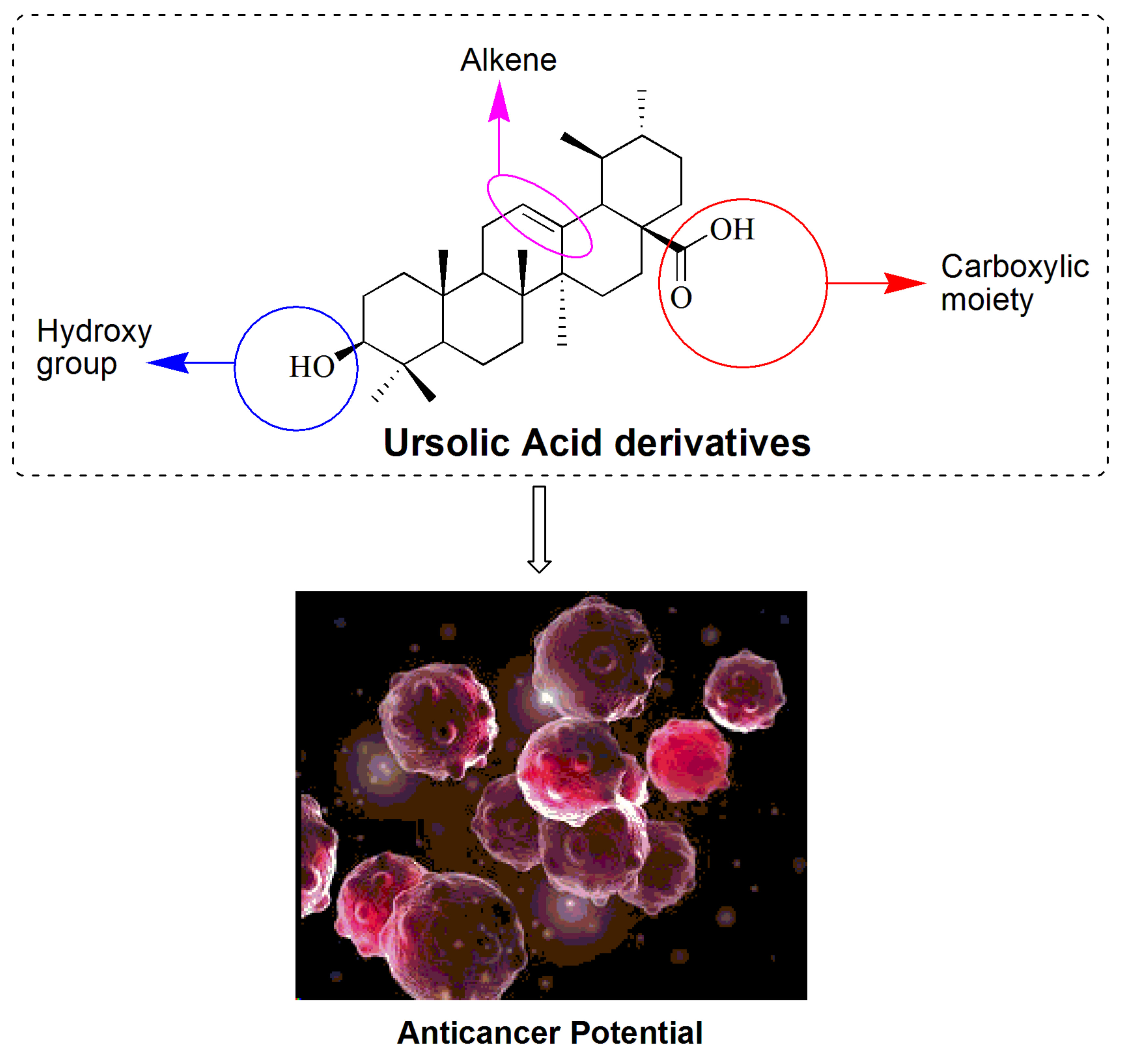
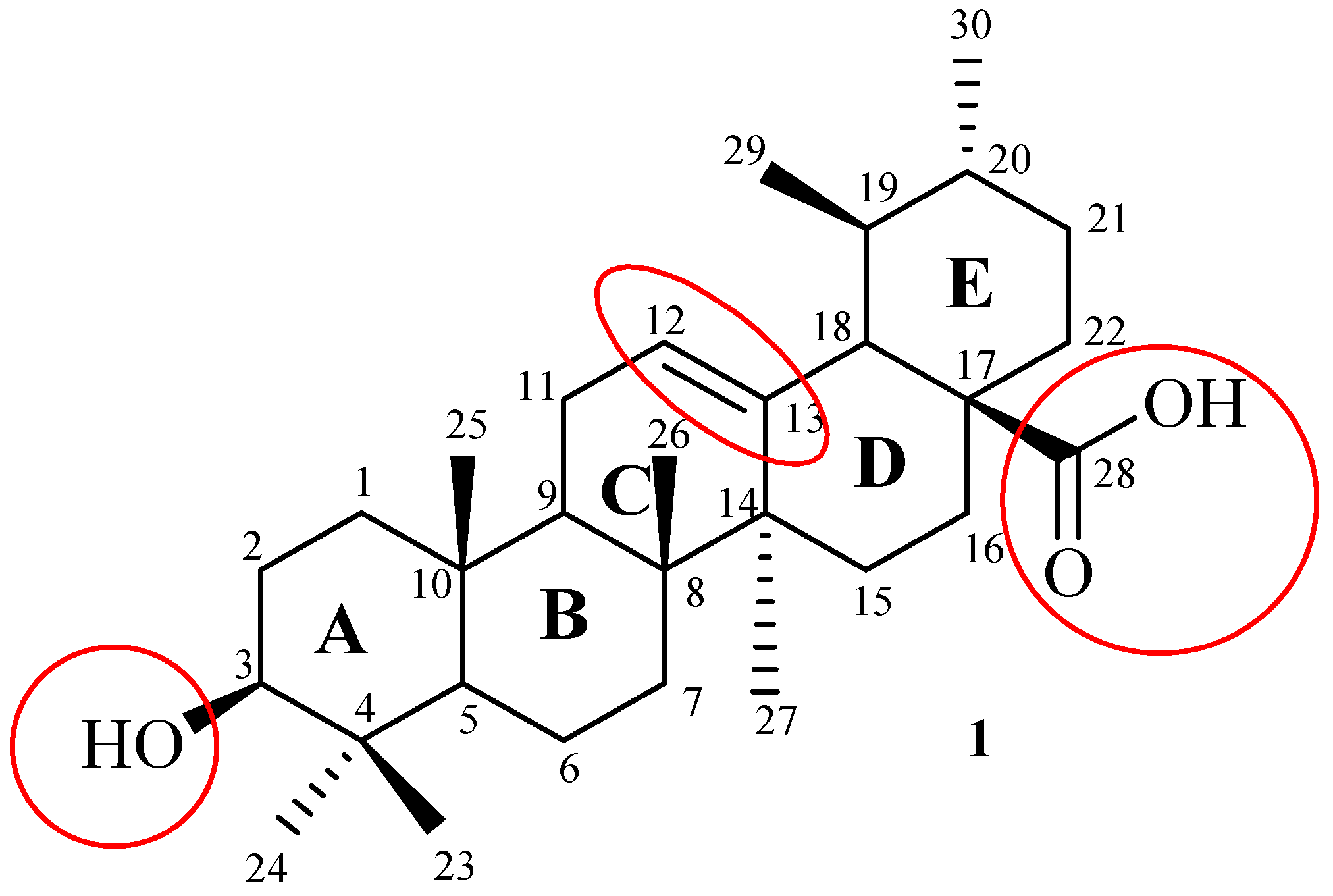
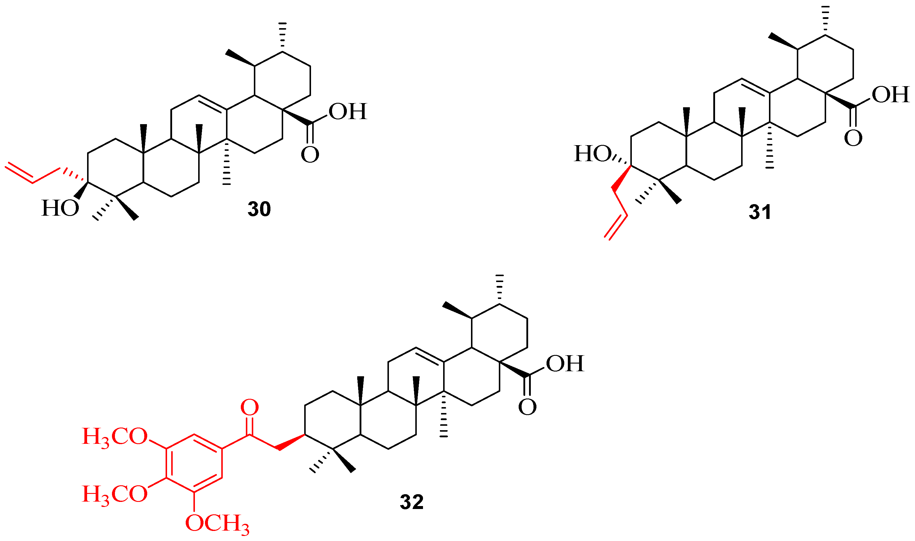
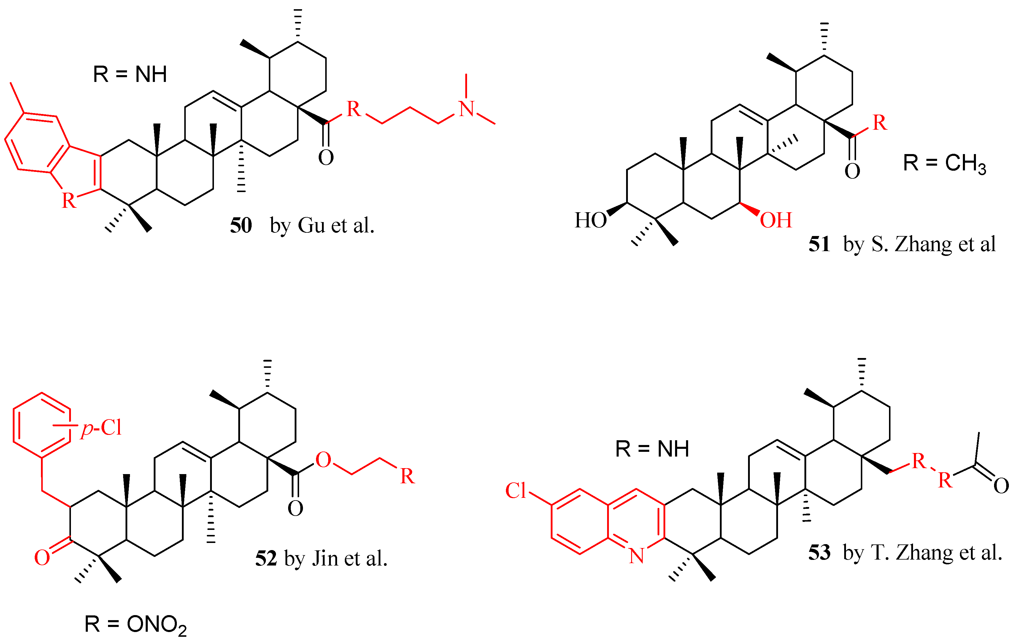
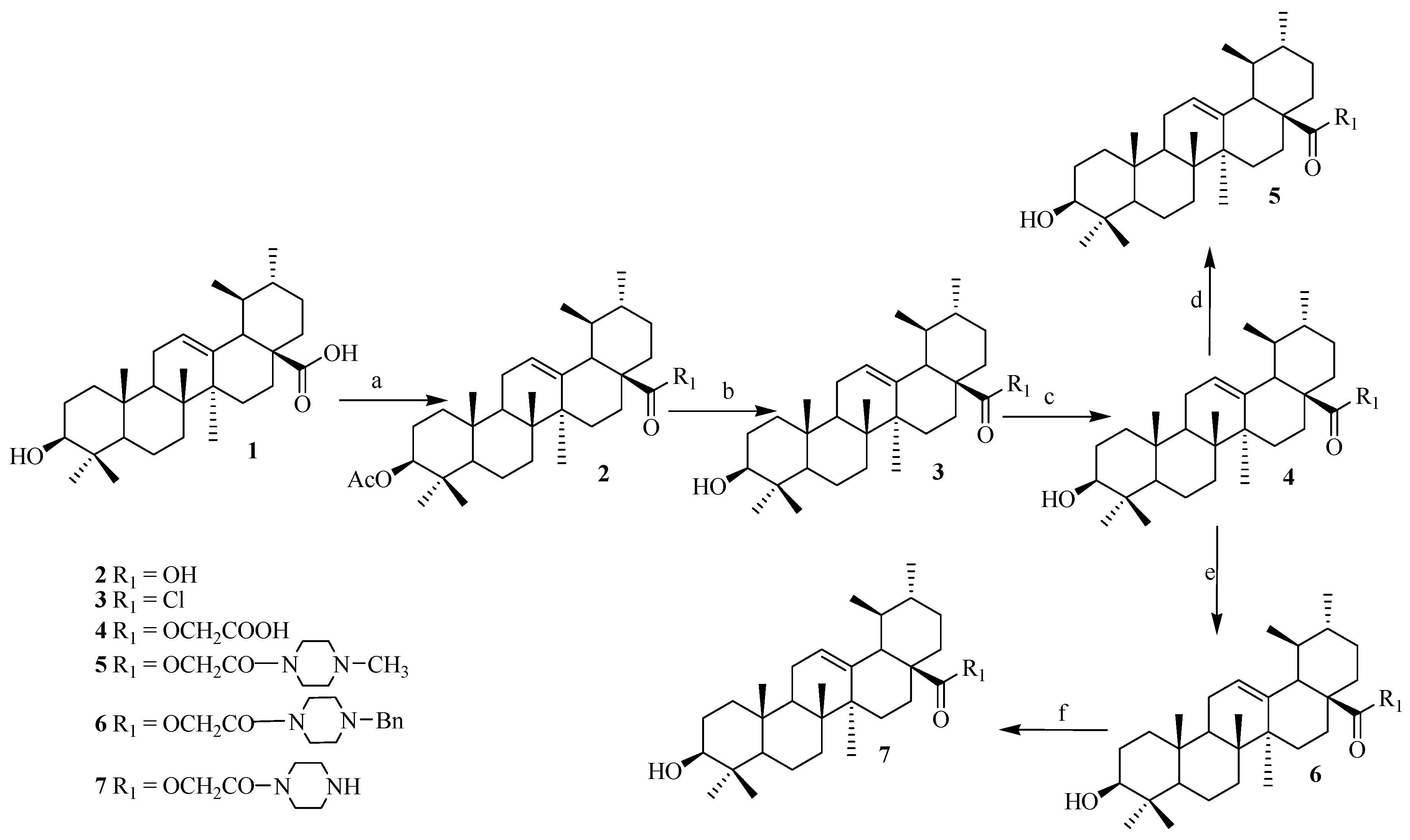
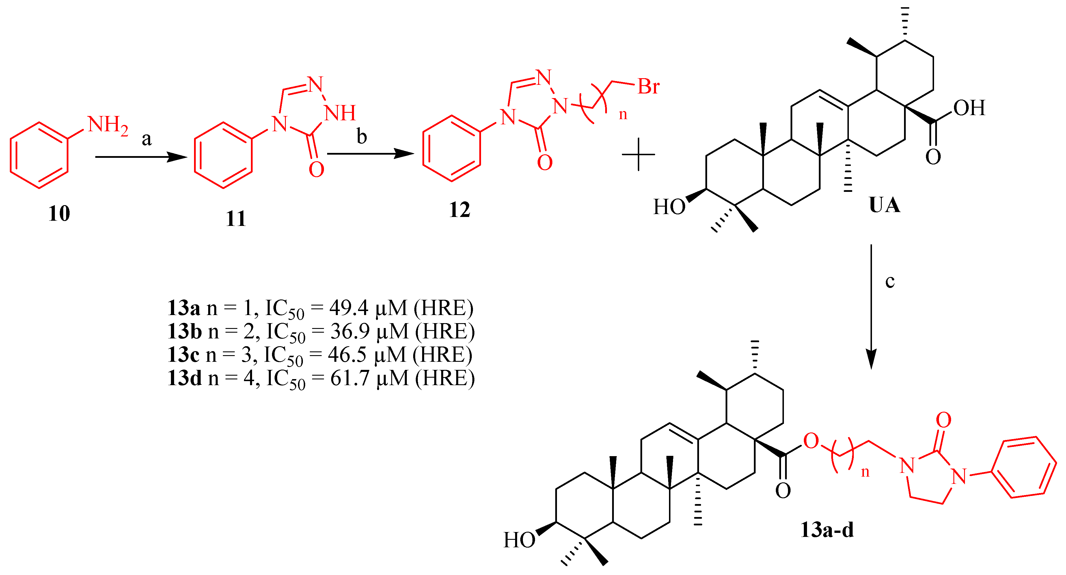
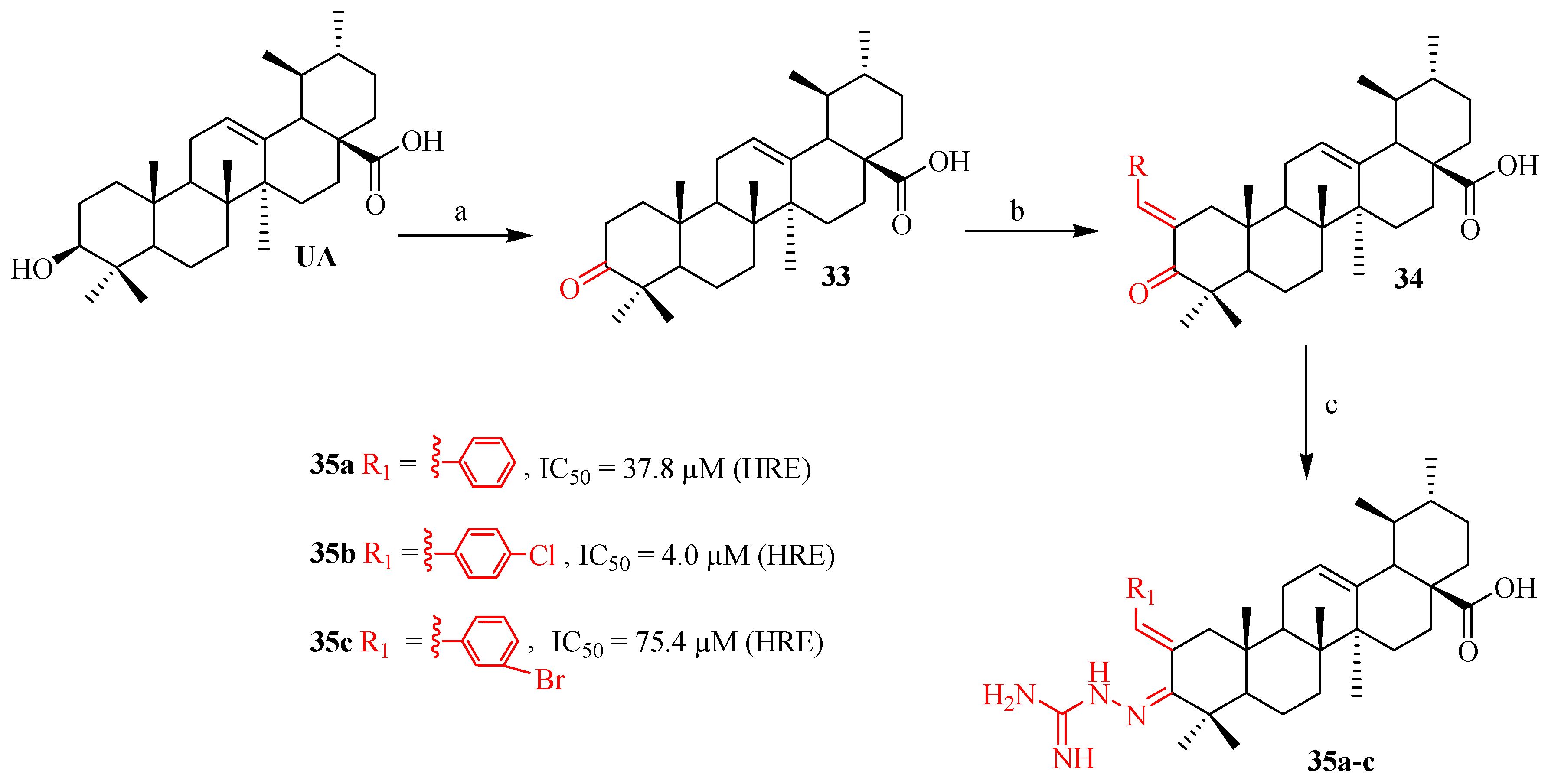
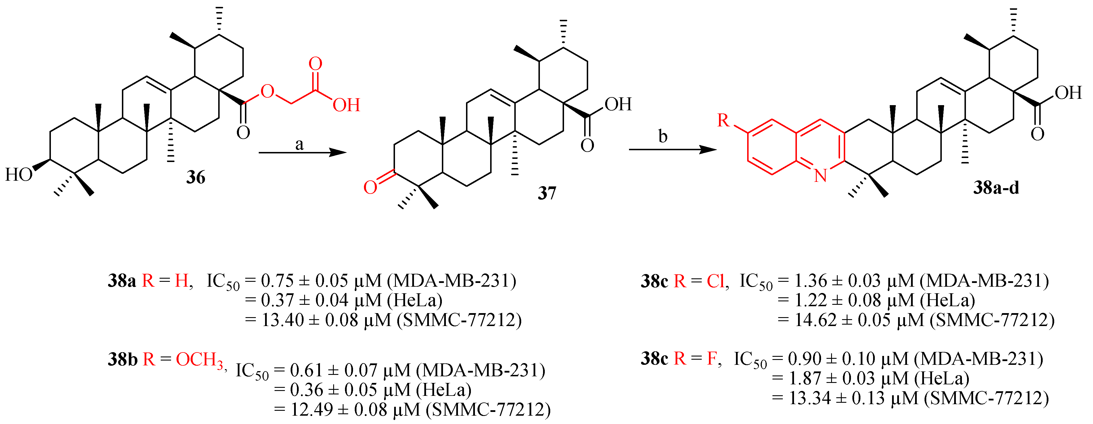
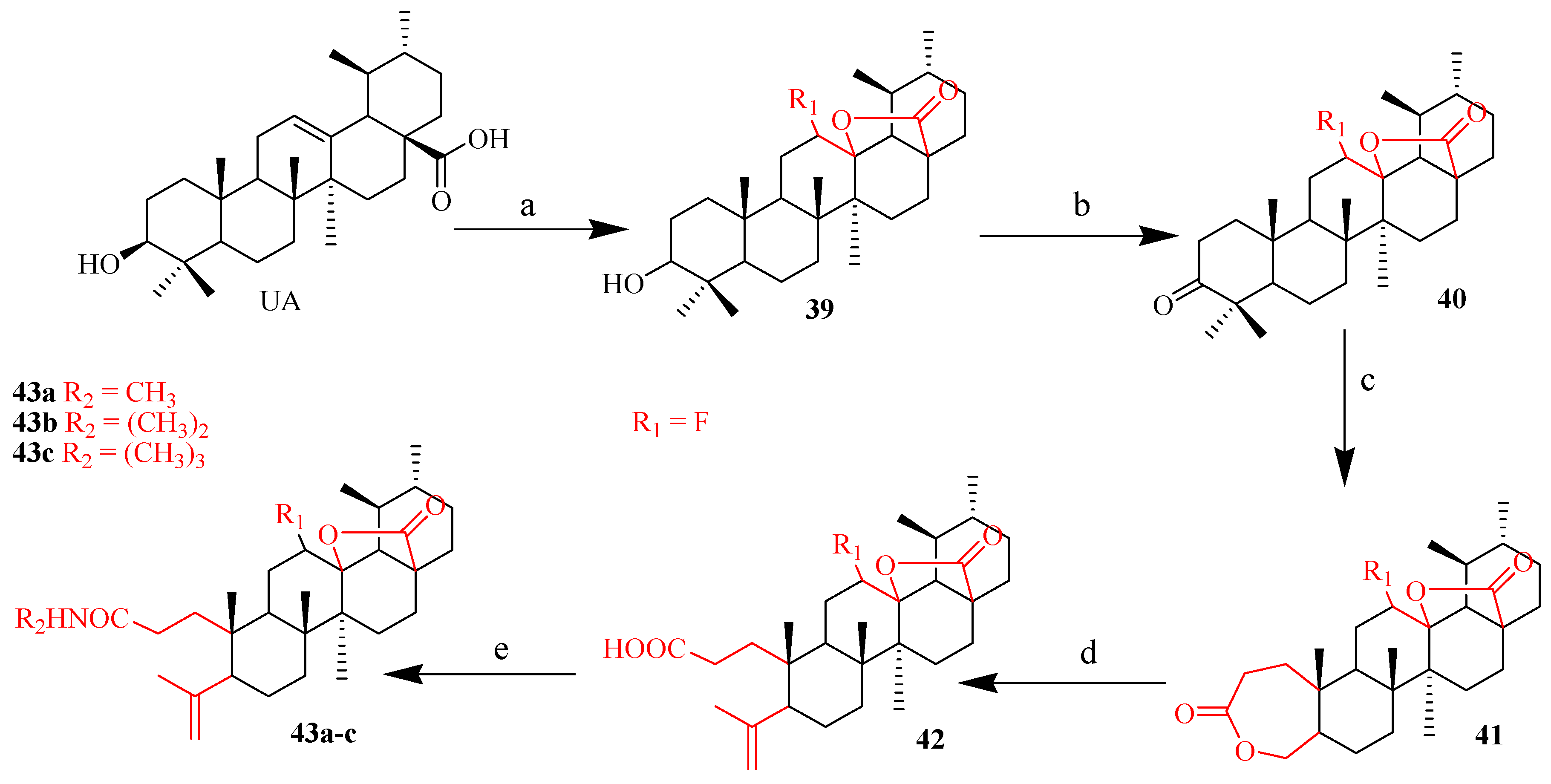
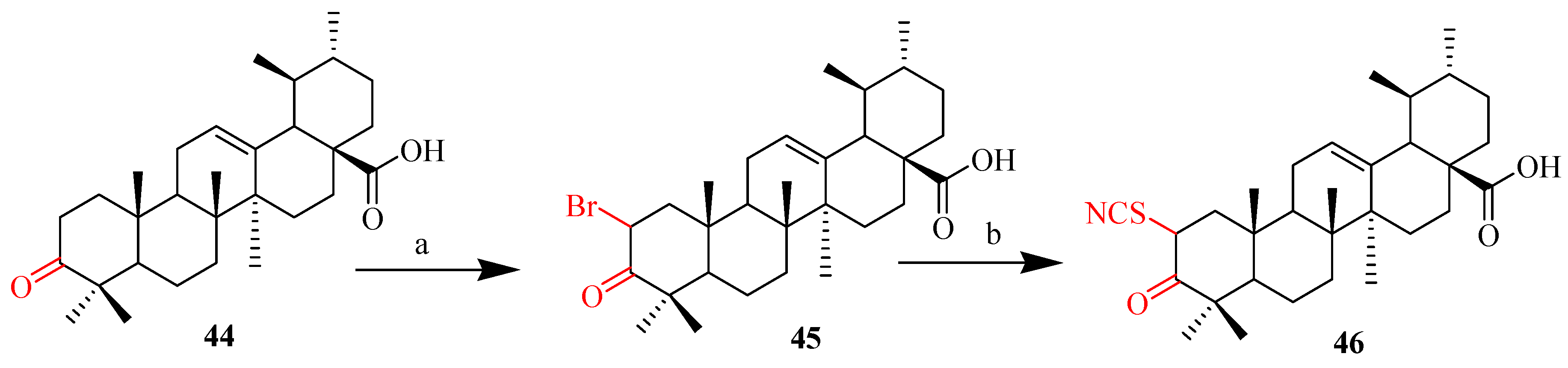
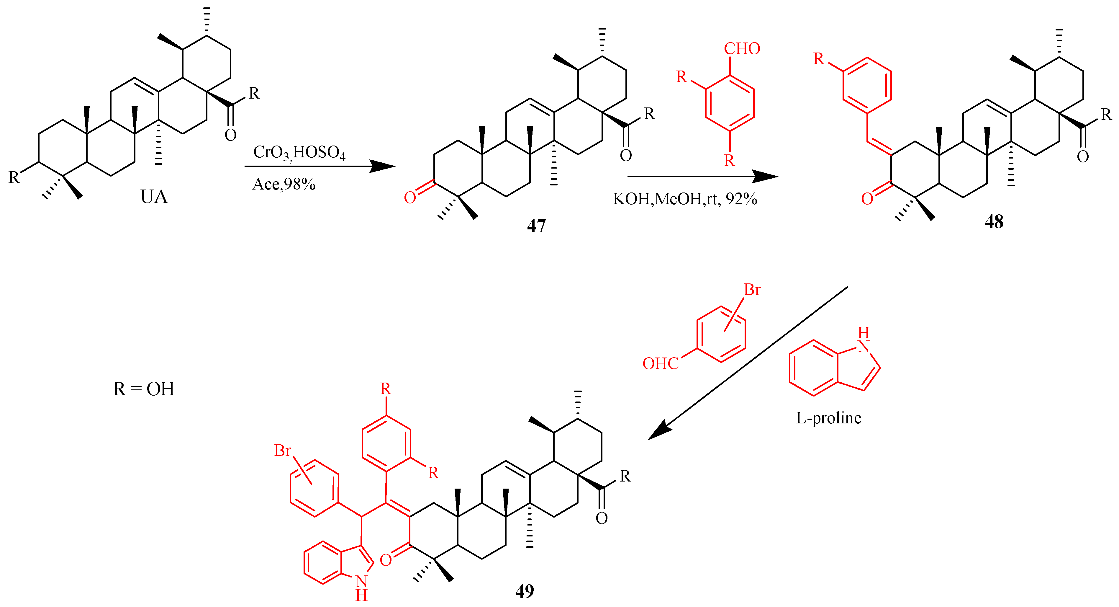
Table 1.
Several plant species with (ursolic acid) UA constituent.
| Plant Species (Family) | Plant Parts Used | Bioactivities | Bibliography |
|---|---|---|---|
| Arctostaphylos uva-ursi (L.) Spreng (Ericaceae) | Leaves | Antitumor, antibacterial | [15,16] |
| Argania spinosa (L.) Skeels (Sapotaceae) | Fruits, leaves | Antibacterial, antifungal | [17,18] |
| Bouvardia ternifolia (Cav.) Schltdl. (Rubiaceae) | Aerial parts | Anti-Alzheimer | [19] |
| Bursera cuneata (Schldl.) Engl (Burseraceae) | Aerial parts (stems and leaves) | Anti-inflammatory, antihistaminic | [20] |
| Catharanthus roseus (L.) G. Don (Apocynaceae) | Leaves | Anticancer | [21] |
| Cornus mas (L.) (Cornaceae) | Fruits | Antitumor | [22] |
| Eriobotrya japonica (Thunb.) Lindl (Rosaceae) | Leaves | Anti-cancer, anti-osteoclastic, skin disorder anti-inflammatory, and anti-arthritic | [23,24,25,26,27] |
| Eucalyptus globulus (Labill.) (Myrtaceae) | Leaves, bark | Antioxidant, neuroprotective | [28,29] |
| Fragraefragrans (Roxb.) (Gentianaceae) | Leaves, fruits, bark | Antimycobacterial | [30] |
| Ilex aquifolium (L.) (Aquifoliaceae) | Leaves | Anticancer, antimalarial, antibacterial | [31] |
| Lamium album (L.) (Lamiaceae) | Flowers | Antioxidant and anti-inflammatory | [32] |
| Lantana Camara (L.) (Verbenaceae) | Leaves | Antifungal, antiproliferative, anti-diabetes, anxiolytic | [33,34,35] |
| Lepidozia chordulifera (Dumort.) (Porellaceae) | Leaves | Antibacterial | [36] |
| Ligustrum lucidum (Ait.) (Oleaceae) | Fruits | Coronary heart disease and diabetes | [37,38] |
| Malus domestica (sp.) (Rosaceae) | Fruits, leaves | Antioxidant | [39] |
| Malus pumila (Mill.) (Rosaceae) | fruits | Antitumor | [40] |
| Ocimum forskolei (Benth.) (Lamiaceae) | Aerial parts (leaves and stems) | Antiulcer | [41] |
| Ocimum sanctum (L.) (Lamiaceae) | leaves | Induced arthritis, antiproliferative, anti-stress | [42,43,44] |
| Panax ginseng (C.A. Mey.) Baill. (Araliaceae) | Roots and rhizomes | Anticancer, antiviral | [45] |
| Paulownia tomentosa (Thunb.) Steud. (Scrophulariaceae) | Leaves | Anticancer | [46] |
| Prunella vulgaris (L.) (Lamiaceae) | Aerial parts | Antiviral, antiestrogenic | [47,48] |
| Psidium guajava (L.) (Myrtaceae) | Leaves | Hypoglycaemic, antimicrobial | [49] |
| Rabdosia rubescens (Linn.) (Lamiaceae) | Anti-tumour | Antitumor | [50] |
| Rosmarinus officinalis (L.) (Lamiaceae) | Stems and leaves | Antidepressant | [51] |
| Sambucus australis (Cham. & Schltdl.) (Adoxaceae) | Aerial parts | Antibacterial and Antioxidant | [52] |
| Saurauja roxburghii (Wall.) (Dilleniaceae) | Leaves | Anticancer | [53] |
| Thymus vulgaris (L.) (Lamiaceae) | Aerial parts (stems and leaves) | Anticancer, cardiovascular, antihyperlipidemic, antioxidant, antifungal | [54,55,56] |
| Tribulus arabicus (Hosni.) (Zygophyllaceae) | Aerial parts | Antihyperuricemic, antioxidant | [57,58] |
| Paulownia tomentos (Thunb.) Steud. (Scrophulariaceae) | Fruits | Anticancer | [46] |
| Punica granatum (Linn.) (Punicaceae) | Flowers | Antioxidant, antidiabetic | [59,60] |
| Uncaria rhynchophylla (Gouteng.) (Rubiaceae) | Stems and hooks | Anticancer | [61,62] |
| Vitex negundo (L.) (Lamiaceae) | Leaves | Antibacterial, antifeedant against the larvae | [63] |
| Ziziphus jujuba (Mill.) (Rhamnaceae) | Leaves | Anticancer, anti-obesity, and antioxidant | [64] |
Table 2.
Reports of ursolic acid’s significant pharmacological activities.
| Sr. No. | Pharmacological Activities | Bibliography |
|---|---|---|
| 1 | Antioxidant | [102,103,104] |
| 2 | Antibacterial | [52,67,105,106,107,108,109,110] |
| 3 | Antifungal | [111,112,113,114,115,116,117] |
| 4 | Anticancer | [13,118,119,120,121,122,123] |
| 5 | Antidiabetic | [34,124,125,126,127,128] |
| 6 | Anti-inflammatory | [129,130,131,132,133] |
| 7 | Antiviral | [134,135,136] |
Table 3.
IC50 values of UA derivatives modified on the carboxylic moiety (C-28).
 | |||||
|---|---|---|---|---|---|
| Compound | R | Biological Activity | Cell Lines Tested IC50 (µM) | Reference Molecules IC50 (µM) | Bibliography |
| 9a |  | Antiproliferative | MCF-7 (8.45 ± 0.26) Hela (8.37 ± 0.11) A549 (10.06 ± 1.39) | MCF-7 (Gefitinib) 17.83 ± 7.85 Hela (Gefitinib) 15.40 ± 4.65 A549 (Gefitinib) 11.02 ± 3.27 | [155] |
| 13b |  | Anticancer activity | HRE (36.9) | n.d | [156] |
| 14 |  | Anticancer activity | MGC-803 (4.99 ± 0.40) Bcap-37 (8.56 ± 0.44) | MGC-803(UA) 26.51 ± 1.1 Bcap-37(UA) 31.39 ± 0.85 | [157] |
Table 4.
Modification of C-3 and C-28 positions.
 | ||||||
|---|---|---|---|---|---|---|
| Compound | R1 | R2 | Biological Activities | Cell Lines Tested(IC50µM) | Reference Molecules (IC50µM) | Bibliography |
| 15 |  |  | Cytotoxicity | MGC-803 (9.82 ± 0.29) HCT-116 (18.97 ± 0.53) T24 (19.60 ± 0.43) HepG2 (15.72 ± 0.84) A549 (20.79 ± 0.54) HL-7702 (˃100) | MGC-803(UA) 27.08 ± 0.29 HCT-116(UA) 38.78 ± 0.16 T24(UA) 29.29 ± 0.80 HepG2(UA) 30.21± 0.58 A549(UA) 35.79 ± 0.37 HL-7702(˃100) | [139] |
| 16 |  |  | Cytotoxicity | 518A2 (3.6 ± 0.1) A2780 (2.7 ± 0.1) A549 (3.9 ± 0.1) FaDu (6.4 ± 0.4) HT29 (3.5 ± 0.3) MCF-7 (3.3 ± 0.2) NIH 3T3 (2.5 ± 0.6) | 518A2 (UA) 14.7 ± 0.1 A2780 (UA) 11.7 ± 0.6 A549 (UA) 15.5 ± 1.3 FaDu (UA) 14.2 ± 2.0 HT29 (UA) 10.6 ± 0.3 MCF-7 (UA) 12.7 ± 0.1 NIH 3T3 (UA)18.7 ± 1.6 | [158] |
| 17 |  |  | Cytotoxicity | TET21N (0.81 ± 0.08) MCF-7(1.59 ± 0.11) | TET21N (˃10) MCF-7(˃25) | [159] |
| 18 |  |  | T24(6.01 ± 0.87) A549(5.22 ± 0.65) HepG2(6.82 ± 1.07) SKOV3(8.95± 1.26) | T24(UA)37.88 ± 1.12 A549(UA) HepG2(UA) SKOV3(UA) | [94] | |
| 19 |  |  | Anti-cancer | HRE(0.8 ± 0.2) | HRE (UA) > 100 | [160] |
| 20 |  |  | Cytotoxicity | 518A2(2.7 ± 0.10) A2780(2.3 ± 0.10) HT29(1.8 ± 0.10) MCF-7(2.0 ± 0.10) 8505C(4.1 ± 0.40) NIH3T3(2.6 ± 0.30) | 518A2(UA) 14.7 ± 0.1 A2780(UA) 11.7 ± 0.6 HT29(UA) 10.6 ± 0.7 MCF-7(UA) 12.7 ± 0.1 8505C(UA) 13.5 ± 1.5 NIH 3T3(UA) 18.7 ± 1.6 | [161] |
| 21 |  |  | 518A2(3.2 ± 0.10) A2780(2.4 ±0.10) HT29(1.8 ± 0.20) MCF-7(2.7 ± 0.30) 8505C(5.4 ± 0.40) NIH 3T3(2.2 ± 0.10) | 518A2(UA) 14.7 ± 0.1 A2780(UA) 11.7 ± 0.6 HT29(UA) 10.6 ± 0.7 MCF-7(UA) 12.7 ± 0.1 8505C(UA) 13.5 ± 1.5 NIH 3T3(UA) 18.7 ± 1.6 | ||
| 22 |  |  | 518A2(2.7 ± 0.10) A2780(2.6 ± 0.10) HT29(1.7 ± 0.10) MCF-7(1.7 ± 0.10) 8505C(3.2 ± 0.01) NIH 3T3 (1.3 ± 0.20) | 518A2(UA) 14.7 ± 0.1 A2780(UA) 11.7 ± 0.6 HT29(UA) 10.6 ± 0.7 MCF-7(UA) 12.7 ± 0.1 8505C(UA) 13.5 ± 1.5 NIH 3T3(UA) 18.7 ± 1.6 | ||
| 23 |  |  | Cytotoxicity | Hela (2.6 ± 1.1) MKN45(2.1 ± 0.3) | Hela (Cisplatin) 15.1 ± 0.9 MKN45(Cisplatin) 2.8 ± 0.1 | [162] |
| 24 |  |  | Cytotoxicity | BEL-7402 (4.49) SGC-7901(7.01) | BEL-7402 (UA) >50 SGC-7901(UA) >50 | [163] |
| 25 |  |  | Cytotoxicity | A375(0.51 ± 0.05) A2780(0.45 ± 0.03) HT29(0.50 ± 0.07) MCF7(0.39 ± 0.04) NiH3T3(0.40 ± 0.03) SW1736 (n.d) | A375(BA) A2780(BA) HT29(BA) MCF7(BA) NiH3T3(BA) SW1736(BA) | [164] |
| 26 |  |  | Cytotoxicity | A375(1.5 ± 0.4) A2780(1.9 ± 0.3) HT29 (5.7 ± 0.5) MCF-7(4.4 ± 0.7) FaDu (3.7 ± 0.6) NIH 3T3(4.6 ± 1.0) | A375(UA) n.d. A2780(UA) 11.7 ± 0.6 HT29 (UA) 10.6 ± 0.7 MCF-7(UA) 12.7 ± 0.1 FaDu (UA) n.d NIH 3T3(UA) 13.1 ± 1.1 | [165] |
| 27 |  |  | A375(2.0 ± 0.1) A2780(1.7 ± 0.1) HT29 (2.3 ± 0.3) MCF-7(1.8 ± 0.1) FaDu (2.0 ± 0.2) NIH 3T3(1.4 ± 0.1) | A375(UA) n.d. A2780(UA) 11.7 ± 0.6 HT29 (UA) 10.6 ± 0.7 MCF-7(UA) 12.7 ± 0.1 FaDu (UA) n.d NIH 3T3(UA) 13.1 ± 1.1 | ||
| 28 |  |  | cytotoxicity | HeLa(9.25) HepG2(21.2) BGC-823(8.06) | HeLa(Gefitinib) 17.1 HepG2(Gefitinib) 20.7 BGC-823 Gefitinib 19.3 | [166] |
| 29 |  |  | HeLa(13.8) HepG2(23.7) BGC-823(9.15) | |||
n.d = not determined. BA = Betulinic acid.
Table 5.
Modification of β-hydroxy (C-3).
| Compound | Biological Activity | Cell Lines Tested (IC50µM) | Reference Molecules (IC50µM) | Bibliography |
|---|---|---|---|---|
| 30 | Cytotoxicity | HA22T/VGH(31.0 ± 1.5) HepG2(28.0 ± 2.0) Hep3B(32.5 ± 2.5) | HA22T/VGH(UA) ˃ 100 HepG2(UA) ˃ 100 Hep3B(UA) ˃ 100 | [167] |
| 31 | Cytotoxicity | HA22T/VGH(31.0 ±1.5) HepG2(28.0 ± 2.0) Hep3B(32.5 ± 2.5) | HA22T/VGH(UA) ˃100 HepG2(UA) ˃ 100 Hep3B(UA) ˃ 100 | [167] |
| 32 | Cytotoxicity | A549 (6.07 ± 0.91) H1975(10.64 ± 1.94) MCF-7(22.27± 3.51) BGC-823 (17.10 ± 1.04) t-HSC/Cl-6 (29.12 ± 3.71) | A549 (mitomycin C) 28.14 ± 3.41 H1975(mitomycin C) 34.51 ± 3.06 MCF-7(mitomycin C) 44.08 ± 4.01 BGC-823 (mitomycin C) 37.94 ± 2.88 t-HSC/Cl-6 (mitomycin C) n.d | [140] |
n.d = not determined.
Table 6.
Modification of miscellaneous groups.
| Compound | Activity | Cell Lines Tested (IC50µM) | Reference Molecules (IC50µM) | Bibliography |
|---|---|---|---|---|
| 35b | Cytotoxicity | HRE (4.0) | HRE(UA) >100 | [168] |
| 38b | Cytotoxicity | MDA-MB-231 (0.61 ± 0.07) HeLa (0.36 ± 0.05) SMMC-7721(12.49± 0.08) QSG-7701( > 40) | MDA-MB-231(UA) > 40 HeLa (UA) >40 SMMC-7721(UA) >40 QSG-7701(UA) n.d | [169] |
| 43a | Cytotoxicity | H460(4.5 ± 0.4) H322(6.8 ± 1.5) H460(6.7 ± 0.5) | H460(UA) 14.8 ± 0.6 H322(UA) 15.3 ± 2.8 H460 LKB1+/+(UA) 21.1 ± 1.6 | [170] |
| 43b | H460(5.3 ± 0.3) H322(7.3 ± 1.0) H460 LKB1+/+(7.8 ± 1.1) | |||
| 43c | H460(2.6 ± 0.9) H322(3.3 ± 0.9) H460 LKB1+/+(4.4 ± 0.6) | |||
| 45 | Cytotoxicity | CCRF-CEM (3.6) CEM-DNR (21.8) HCT116 (28.4) HCT116 p53−/−(29.8) K562 (38.8) K562-TAX (25.1) A549 (27.6) U2OS (20.7) BJ (49.5) MRC-5(29.3) | CCRF-CEM14 (44) 10.4 CEM-DNR (44) 34.0 HCT116(44) 34.0 HCT116 p53−/−(44) 49.7 K562 (44) >50 K562-TAX (44) 35.5 A549 (44) >50 U2OS (44) >50 BJ (44) >50 MRC-5(44) >50 | [171] |
| 46 | CCRF-CEM (4.7) CEM-DNR (28.2) HCT116 (32.1) HCT116 p53−/−(32.3) K562 (34.7) K562-TAX (29.0) A549 (42.5) U2OS (33.1) BJ (˃ 50) MRC-5(39.8) | |||
| 50 | Cytotoxicity | SMMC-7721 (1.08 ± 0.22) HepG2 (1.26 ± 0.17) | SMMC-7721(Doxorubicin) 0.62 ± 0.16 HepG2 (Doxorubicin) 0.77 ± 0.12) | [174] |
| 51 | Cytotoxicity | Hela (1.06) K562 (28.7) KB(35.6) | Hela (UA) 14.2 K562 (UA) 52.7 KB 3(UA) 42.9 | [175] |
| 52 | Cytotoxicity | HepG-2(65.8 ± 6.3) MCF-7(> 100) HT-29(4.28 ± 3.5) A549(78.39 ± 5.6) | HepG-2 (UA) 44.35 ± 4.9 MCF-7 (UA) > 100 HT-29 (UA) > 100 A549 (UA) > 100 | [176] |
| 53 | MEK inhibitors | MDA-MB-231 (1.84 ± 0.13) HeLa (1.18 ± 0.03) SMMC-7721 (17.48 ± 0.10) QSG-7701(40.59 ± 2.89) | MDA-MB-231 (Etoposide) 5.26 ± 1.21 HeLa (Etoposide) 2.98 ± 0.42 SMMC-7721 (Etoposide) 3.48 ± 0.35 QSG-7701(Etoposide) 28.75 ± 3.28 | [177] |
n.d = not determined.
© 2020 by the authors. Licensee MDPI, Basel, Switzerland. This article is an open access article distributed under the terms and conditions of the Creative Commons Attribution (CC BY) license (http://creativecommons.org/licenses/by/4.0/).
Share and Cite
MDPI and ACS Style
Khwaza, V.; Oyedeji, O.O.; Aderibigbe, B.A. Ursolic Acid-Based Derivatives as Potential Anti-Cancer Agents: An Update. Int. J. Mol. Sci. 2020, 21, 5920. https://0-doi-org.brum.beds.ac.uk/10.3390/ijms21165920
AMA Style
Khwaza V, Oyedeji OO, Aderibigbe BA. Ursolic Acid-Based Derivatives as Potential Anti-Cancer Agents: An Update. International Journal of Molecular Sciences. 2020; 21(16):5920. https://0-doi-org.brum.beds.ac.uk/10.3390/ijms21165920
Chicago/Turabian StyleKhwaza, Vuyolwethu, Opeoluwa O. Oyedeji, and Blessing A. Aderibigbe. 2020. "Ursolic Acid-Based Derivatives as Potential Anti-Cancer Agents: An Update" International Journal of Molecular Sciences 21, no. 16: 5920. https://0-doi-org.brum.beds.ac.uk/10.3390/ijms21165920
Note that from the first issue of 2016, this journal uses article numbers instead of page numbers. See further details here.