Article

Regulation of Fetal Genes by Transitions among RNA-Binding Proteins during Liver Development

1
Center for Integrative Medical Sciences, Laboratory for Immunogenetics, RIKEN, Kanagawa 230-0045, Japan
2
Molecular Profiling Research Center for Drug Discovery, National Institute of Advanced Industrial Science and Technology, Tokyo 135-0064, Japan
3
Department of Physiology, Keio University School of Medicine, Tokyo 160-8582, Japan
4
Cell Signal Unit, Okinawa Institute of Science and Technology Graduate University, Okinawa 904-0495, Japan
5
Center for Integrative Medical Sciences, Laboratory for Developmental Genetics, RIKEN, Kanagawa 230-0045, Japan
*
Author to whom correspondence should be addressed.
Int. J. Mol. Sci. 2020, 21(23), 9319; https://0-doi-org.brum.beds.ac.uk/10.3390/ijms21239319
Submission received: 23 November 2020 / Revised: 5 December 2020 / Accepted: 5 December 2020 / Published: 7 December 2020
(This article belongs to the Section Molecular Biology)

Abstract

:
Transcripts of alpha-fetoprotein (Afp), H19, and insulin-like growth factor 2 (Igf2) genes are highly expressed in mouse fetal liver, but decrease drastically during maturation. While transcriptional regulation of these genes has been well studied, the post-transcriptional regulation of their developmental decrease is poorly understood. Here, we show that shortening of poly(A) tails and subsequent RNA decay are largely responsible for the postnatal decrease of Afp, H19, and Igf2 transcripts in mouse liver. IGF2 mRNA binding protein 1 (IMP1), which regulates stability and translation efficiency of target mRNAs, binds to these fetal liver transcripts. When IMP1 is exogenously expressed in mouse adult liver, fetal liver transcripts show higher expression and possess longer poly(A) tails, suggesting that IMP1 stabilizes them. IMP1 declines concomitantly with fetal liver transcripts as liver matures. Instead, RNA-binding proteins (RBPs) that promote RNA decay, such as cold shock domain containing protein E1 (CSDE1), K-homology domain splicing regulatory protein (KSRP), and CUG-BP1 and ETR3-like factors 1 (CELF1), bind to 3′ regions of fetal liver transcripts. These data suggest that transitions among RBPs associated with fetal liver transcripts shift regulation from stabilization to decay, leading to a postnatal decrease in those fetal transcripts.

1. Introduction

Genes that are expressed in fetal and neonatal stages, but inactivated in adults, participate in early developmental processes and maintenance of immature tissue characteristics. Some of these genes are reactivated during tumorigenesis, tissue injury, or regeneration; hence, they are called oncofetal genes [1]. Afp and H19 genes show an oncofetal expression pattern in mammalian liver. The Igf2 gene also shows an oncofetal expression pattern in mice, but not in humans [2].
AFP is an abundant serum protein in mammalian fetuses; however, reactivation of Afp expression is observed in ~80% of hepatocellular carcinoma (HCC) patients [3]. H19 is a long non-coding RNA responsible for several liver functions and pathogenesis [4], whereas Igf2 encodes a growth factor for a transmembrane receptor, transducing signals for cell growth, survival, and migration during tissue development [5]. Epigenetic abnormalities in both genes are detected in HCC [6]. Moreover, an increase of these genes is associated with other types of liver diseases, such as hepatitis and alcohol-dependent liver injury [7].
The postnatal decrease of fetal liver transcripts has been analyzed mainly at the transcriptional level. FOXA1 is a critical transcriptional activator of Afp and H19 in liver [8,9]. Direct binding of p53 to the Afp promoter induces histone modification for transcriptional repression, resulting in suppression of FOXA1-mediated Afp transcription [8,10]. In addition, a developmental decrease of FOXA1 binding to the H19 enhancer is observed [9]. Insufficient postnatal reduction of AFP observed in BALB/cJ mice is due to a natural hypomorph mutation in the Zhx2 gene locus [11,12]. ZHX2 functions as a transcriptional repressor [13], and exogenous expression of the Zhx2 gene significantly reduces Afp and H19 transcripts [12]. ZHX2 level increases after birth, partly accounting for postnatal decrease of Afp and H19 transcripts. Liver-specific suppression of the zinc finger protein, ZBTB20, leads to persistent Afp expression in liver throughout adult life, indicating that ZBTB20 also contributes to postnatal Afp repression [14]. However, it seems unlikely that these transcriptional mechanisms fully account for postnatal repression of fetal liver transcripts. Indeed, involvement of post-transcriptional mechanisms is suggested in ZHX2-mediated reduction of Afp and H19 expression [15].
Among post-transcriptional mechanisms, translational repression and RNA decay are relevant to decreased mRNA levels. MicroRNAs (miRNAs) are major translational repressors, which form RNA-induced silencing complexes (RISC) and bind mainly to 3′ untranslated regions (3′UTRs) [16]. Following translational repression, mRNAs usually undergo degradation in a manner dependent upon ribonucleases, including the CCR4-NOT complex. The CCR4-NOT complex initiates decay of mRNAs by shortening their poly(A) tails. To recognize target transcripts, the CCR4-NOT complex uses various RBPs, such as CSDE1, bicaudal-C, RISC, tristetraproline, butyrate response factors 1 and 2 (BRF1/2), Nanos2, Roquin, and dead end 1 [17,18,19,20,21,22,23,24].
RBPs compete with one another for binding and regulation of common target RNAs during various biological events. While Hu antigen D (HuD) stabilizes and promotes translation of GAP-43 mRNA, KSRP binding to GAP-43 mRNA leads to destabilization of the mRNA [25,26]. Both HuD and KSRP recognize AU-rich element (ARE) in GAP-43 mRNA, and KSRP can displace HuD from GAP-43 mRNA [25]. CELF1 competes with HuR or calreticulin for translation of occluding or p21 mRNAs, respectively [27,28]. Importantly, the effect of competitive binding was clearly reflected in axonal outgrowth, proliferation, or other cellular functions [26,27,28].
When mice lack expression of microRNA-122 (an abundant liver miRNA), Dicer, which produces mature miRNAs, or core subunits of the CCR4-NOT complex in liver, enhanced expression of Afp, H19, and Igf2 transcripts is detected in adult livers compared to corresponding controls [29,30,31,32,33], suggesting that post-transcriptional mechanisms are responsible for the postnatal decrease of fetal liver transcripts. On the other hand, deletion of miR-122, Dicer, or the CCR4-NOT complex results in severe liver damage [29,30,31,32,33]. More detailed analyses are required to determine whether abundant expression of Afp, H19, and Igf2 in adult livers of these mouse strains is due to loss of post-transcriptional control mechanisms or to secondary effects of liver damage.
In this study, we provide evidence that CCR4-NOT complex-mediated RNA decay is involved in the postnatal decrease of Afp, H19, and Igf2 transcripts. Our analyses also show that different RBPs bind to 3′ regions of Afp, H19, and Igf2 transcripts, depending on liver developmental stages. Based on our findings, we propose a model for developmental repression of oncofetal genes involving interactions between RBPs and RNAs.

2. Results

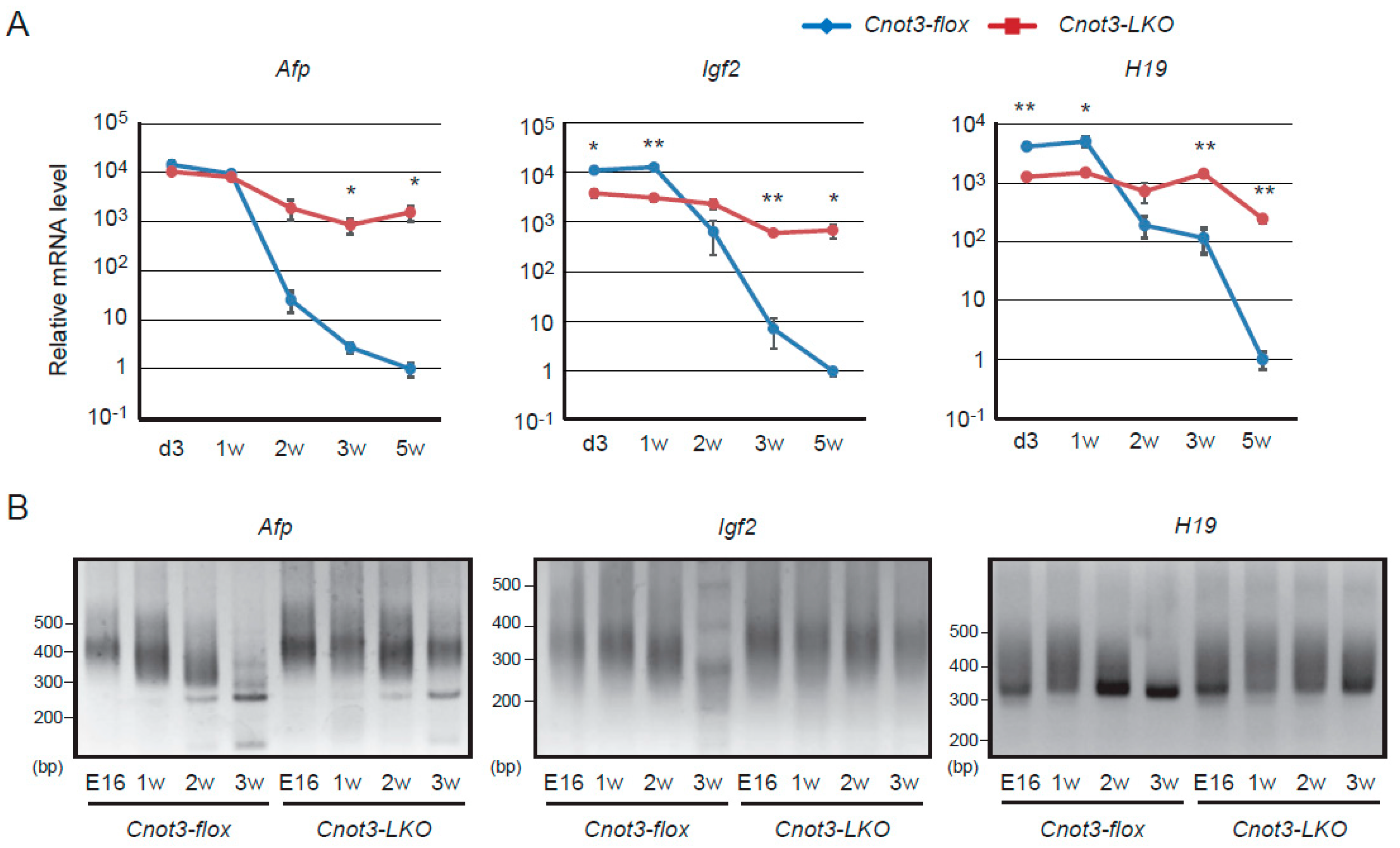
2.1. Afp, H19, and Igf2 Transcripts Decrease in a CNOT3-Dependent Manner as Liver Matures

We have previously shown that Afp, H19, and Igf2 transcripts are more highly expressed and had longer poly(A) tails in livers from adult mice lacking Cnot3 specifically in liver (Cnot3-LKO mice) than in those from control mice possessing floxed alleles in the Cnot3 gene locus (called control mice hereafter) [32]. In this report, we use Afp, H19, and Igf2 transcripts as representative fetal liver transcripts in mice. We examined expression and poly(A) tail lengths of fetal liver transcripts in mouse liver at various postnatal days. At 3 days and 1 week after birth, Afp transcripts were expressed at similar levels in livers from control and Cnot3-LKO mice. Igf2 and H19 transcripts showed higher expression in livers from control mice than those from Cnot3-LKO mice (Figure 1A). In control mouse livers, levels of these three transcripts started to decrease 2 weeks after birth, and declined steadily thereafter (Figure 1A). Concomitantly, poly(A) tails of Afp and Igf2 transcripts became shortened (Figure 1B). Poly(A) tails of H19 transcripts were elongated 1 week after birth compared to embryonic day 16, and were gradually shortened, as were those of Afp and Igf2 transcripts (Figure 1B). The decrease of fetal liver transcripts in livers from Cnot3-LKO mice was significantly less than that observed in livers from control mice (Figure 1A). Consistent with this, fetal liver RNAs had longer poly(A) tails in livers from Cnot3-LKO mice than those from control mice (Figure 1B). These results suggest that CCR4-NOT complex-mediated poly(A) shortening and subsequent RNA decay is involved in postnatal decrease of fetal liver transcripts.

2.2. IMP1 Decreases Concomitantly with Fetal Liver Transcripts during Liver Development

As mice grew from 1 week to 5 weeks, subunits of the CCR4-NOT complex were expressed at more or less constant levels in the liver (Figure 2A). Furthermore, immunoprecipitation analyses showed that a comparable amount of the CCR4-NOT complex was continuously present during this developmental period (Figure 2B). Therefore, the postnatal decrease of fetal liver transcripts was not determined by the abundance of the CCR4-NOT complex.
Then, we examined expression of molecules that bind to and stabilize RNAs. IMP family proteins regulate RNA at various post-transcriptional levels, determining localization, translation, or stability [34,35]. Among them, IMP1 and IMP3 are highly expressed in mouse embryonic stages, but show very low expression in most adult tissues [34,36]. We detected a decrease of IMP1 protein in liver as mice grew from 1 week to 5 weeks after birth both in the presence or the absence of CNOT3 (Figure 2C). Phosphorylation state of IMP1 was not influenced by CNOT3 suppression, either (Figure 2C). IMP1 and AFP started to decrease at about 2 weeks after birth, and both IMP1 and AFP were barely detectable 3 weeks after birth and thereafter (Figure 2C). The decrease of fetal liver transcripts and shortening of their poly(A) tails were particularly noticeable at around 2 weeks after birth (Figure 1). These data suggest that IMP1 maintains expression and translation of fetal liver transcripts during fetal and perinatal days, and that a decrease of IMP1 enables the CCR4-NOT complex to degrade mRNAs at a later developmental stage. RNA immunoprecipitation experiments demonstrated binding of IMP1 to fetal liver transcripts, further supporting the idea (Figure 2D). Like IMP1, IMP3 decreased during liver maturation, though the magnitude of the decrease was less prominent than that of IMP1 (Figure 2C). However, IMP3 did not decrease in livers from Cnot3-LKO mice in contrast to IMP1 (Figure 2C). We confirmed that both IMP3 mRNA and protein were more highly expressed in livers from Cnot3-LKO mice than those from control mice at 4 weeks of age (Figure 3A,B), raising the possibility that high expression of fetal liver transcripts is due to persistent expression of IMP3.

2.3. High Expression of Fetal Liver Transcripts in Adult Cnot3-LKO Mice Is not Affected by the Absence of IMP3

We first examined whether increased IMP3 is responsible for continuous expression of fetal liver transcripts in adult livers from Cnot3-LKO mice. We generated mice lacking the Imp3 gene (Imp3−/− mice) and crossed them with Cnot3-LKO mice to obtain mouse strains lacking both Imp3 and Cnot3 genes in liver (Imp3−/−; Cnot3-LKO mice). Even in the absence of IMP3 in all tissues, mice were born at Mendelian frequency (Imp3+/+:Imp3+/−:Imp3−/− = 9:14:7 in 4 crossings) and grew without any obvious abnormalities (Supplementary Figure S1A–C). Imp3−/− mice indeed showed normal liver histology (Supplementary Figure S1C). When we examined AFP protein expression in livers from 4-week-old mice, AFP was detected in livers from Cnot3-LKO and Imp3−/−; Cnot3-LKO mice, but not those from control and Imp3−/− mice (Figure 3C). Moreover, fetal liver transcripts were more highly expressed and had longer poly(A) tails in livers from Imp3−/−; Cnot3-LKO mice compared to those from Imp3−/− mice (Figure 3D,E). These results suggested that an increase of IMP3 in livers from Cnot3-LKO mice is dispensable or irrelevant to postnatal expression of fetal liver transcripts. It should be also noted that IMP3 increase has little effect on phenotypes of Cnot3-LKO mice, because we observed comparable abnormalities in Imp3−/−; Cnot3-LKO mice to those we have previously observed in Cnot3-LKO mice, such as decreased liver and body weights, misaligned sinusoids, and inflammation (Supplementary Figure S1, and [32]).

2.4. Poly(A) Tail Elongation and Increased Levels of Fetal Liver Transcripts in Imp1-Transgenic Mice

Next, to investigate the relationship between the developmental decrease of IMP1 and expression of fetal liver transcripts, we generated mice possessing an Imp1 transgene under control of the Albumin enhancer and promoter (Imp1-transgenic mice), so that IMP1 was expressed in liver even after it matured. We confirmed that IMP1 was expressed in livers from Imp1-transgenic mice, but not in livers from wild-type mice at 4 weeks of age (Figure 4A). Imp1-transgenic mice were almost normal and showed comparable body and liver weights to those of wild-type mice (Supplementary Figure S2A). Liver histology was also normal at the ages we examined (Supplementary Figure S2B). In spite of normal appearance, Afp, Igf2 and H19 transcripts showed significantly higher expression in livers from Imp1-transgenic mice than those from wild-type mice at 4 weeks of age (Figure 4B). Furthermore, Afp and Igf2 transcripts had longer poly(A) tails in liver from Imp1-transgenic mice (Figure 4C). Whereas expression of unspliced immature transcripts of Afp and Igf2 genes was comparable in livers between Imp1-transgenic mice and wild-type mice, the mature/immature ratios of transcripts increased significantly in Imp1-transgenic mice, suggesting that those transcripts were stabilized (Figure 4D,E). Poly(A) tail elongation of H19 transcripts was less prominent than of Afp and Igf2 transcripts (Figure 4C). H19 transcripts undergo splicing like that of protein-coding RNAs [37], leading us to examine the level of unspliced immature H19 transcripts. Immature H19 transcripts increased significantly in Imp1-transgenic mice (Figure 4D); therefore, IMP1 expressed from the transgene appeared to influence mainly transcription of the H19 gene. These data suggest that IMP1, at least in part, is responsible for developmental expression of fetal liver genes.

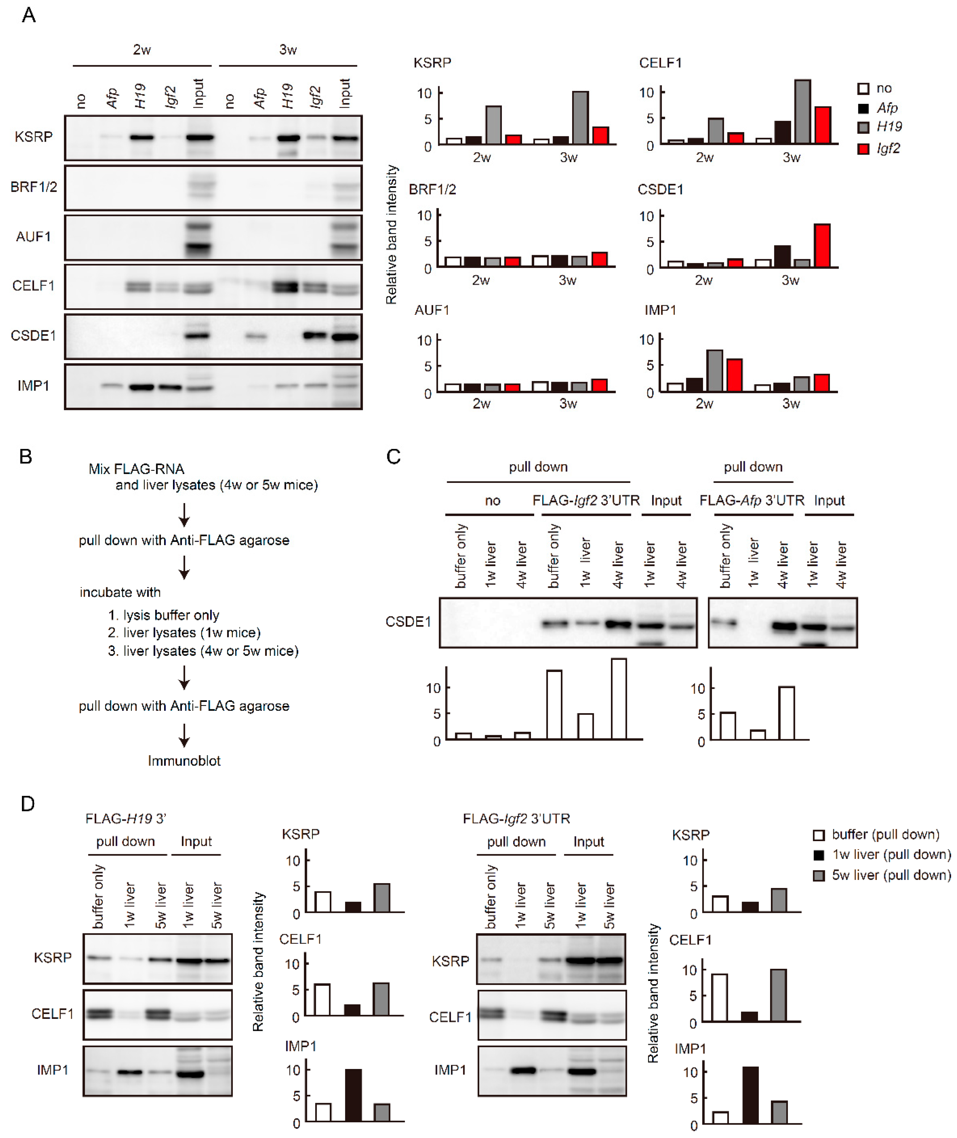
2.5. Binding of RBPs to Fetal Liver Transcripts Changes during Liver Maturation
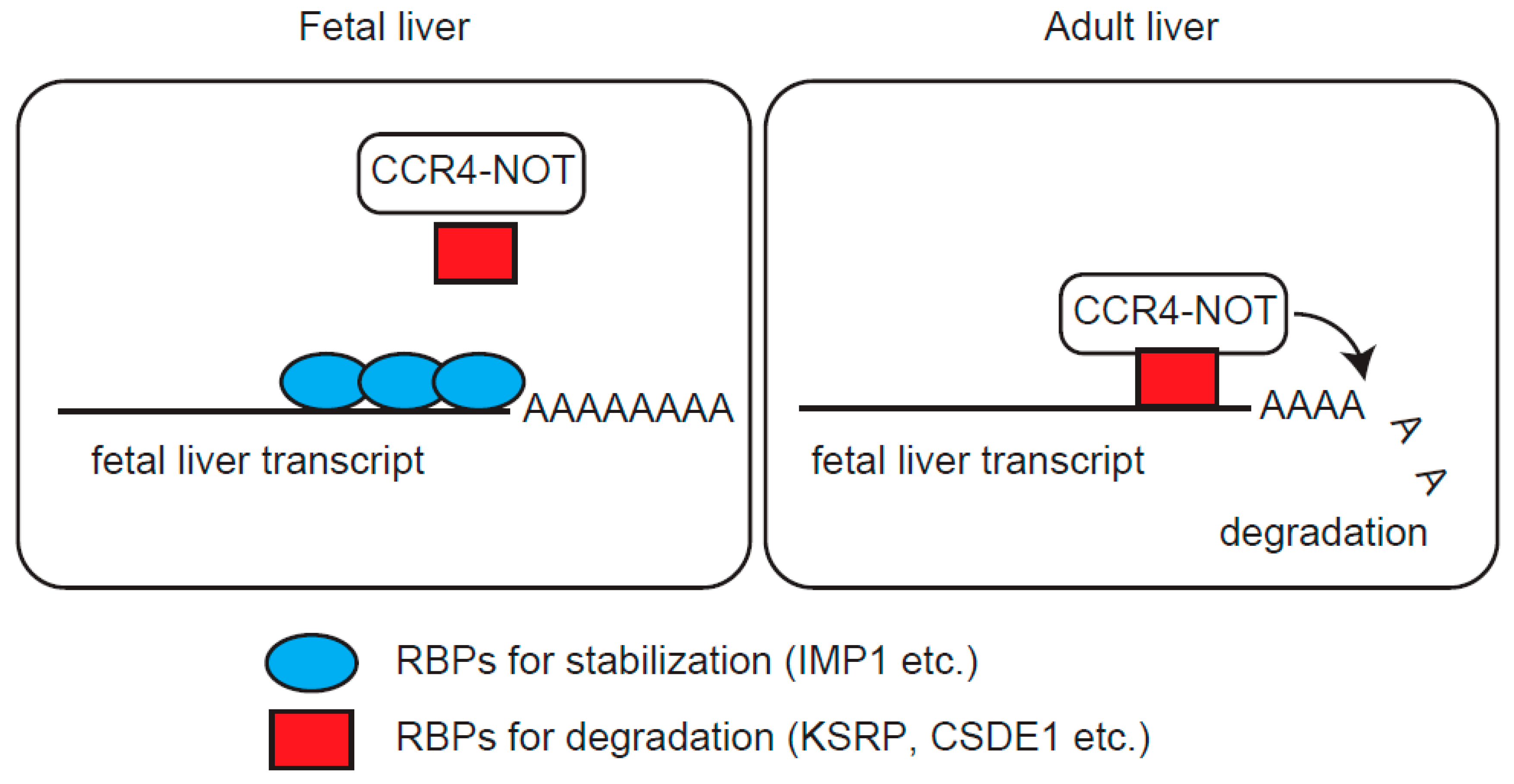
Increased expression and poly(A) tail elongation of fetal liver transcripts upon exogenous IMP1 expression led us to speculate that RBPs responsible for RNA decay, contribute to the postnatal decrease of fetal liver transcripts. We performed an in vitro RNA binding experiment that we previously established [20]. We prepared FLAG-peptides conjugated with approximately 500 bases of 3′ regions of fetal liver transcripts and examined binding proteins following incubation of FLAG-RNAs with liver lysates from 2 or 3-week-old mice.
We first focused on RBPs that recognize AREs, because the nucleotide sequences are usually in the 3′ UTRs of their mRNAs and function critically as cis-acting elements in mRNA decay [38,39]. Immunoblot analysis showed that KSRP bound to 3′ regions of fetal liver transcripts (Figure 5A, top). Binding was enhanced in lysates from 3-week-old mice compared to those from 2-week-old mice, especially in Igf2 and H19 transcripts. In contrast, BRF1/2 and AU-rich element RNA binding factor 1 (AUF1) failed to bind to those transcripts (Figure 5A). CELF1, which recognize GU-rich elements (GREs) or U-rich elements in addition to AREs [40,41], bound to 3′ regions in H19 and Igf2 transcripts, and binding similarly increased in livers from 3-week-old mice (Figure 5A). CSDE1 interacts with the CCR4-NOT complex and promotes mRNA decay [17]. CSDE1 bound predominantly to 3′ regions of Afp and Igf2 transcripts in livers from 3-week-old mice (Figure 5A). IMP1 bound to all the three transcripts and the amount of FLAG-RNA-bound IMP1 was reduced as the level of IMP1 protein decreased in livers from 2-week-old to 3-week-old mice (Figure 5A).
We confirmed that binding of RNA decay-related RBPs to fetal liver transcripts was enhanced as mice matured using competition experiments. We first incubated FLAG-RNAs with liver lysates from adult mice (4~5 weeks of age) and collected complexes of FLAG-RNAs and associated proteins using anti-FLAG antibody. Subsequently, anti-FLAG immunoprecipitates were incubated with liver lysates from 1-week-old mice. Proteins binding to FLAG-RNAs after the second incubation were analyzed by immunoblot (Figure 5B). CSDE1 bound to the 3′ regions of Afp and Igf2 transcripts in liver lysates from 4-week-old mice, and binding was reduced following incubation with liver lysates from 1-week-old mice (Figure 5C). Similar results were obtained when we examined binding of KSRP or CELF1 to 3′regions of H19 or Igf2 transcripts (Figure 5C). Instead of reduced binding of those proteins to FLAG-RNAs, binding of IMP1 was evident after incubation with liver lysates from 1-week-old mice (Figure 5C).

3. Discussion

Liver maturation requires an increase of liver function-related genes, as well as a decrease of genes related to immature liver. A strictly controlled network of transcription factors largely governs liver maturation and function through transcriptional activation or repression [42,43]. This study reveals contributions of post-transcriptional mechanisms to postnatal repression of fetal liver genes. While one of the major RNA decay molecules, the CCR4-NOT complex, is continually present during liver development, different RBPs bind to 3′ regions of fetal liver transcripts, depending on the developmental stage. RNA decay-related RBPs, such as KSRP, CELF1, and CSDE1 bound more abundantly to fetal liver transcripts at later developmental stages (Figure 5A). This increased binding paralleled the developmental decrease of IMP1 protein, identifying IMP1 as one of the candidate RBPs that competitively regulate fetal liver transcript levels. This was supported by results using transgenic mice and competitive experiments (Figure 4 and Figure 5C). Thus, we provide a model in which regulation of fetal liver transcripts shifts from stabilization to decay through a liver maturation-dependent switch of associated RBPs (Figure 6).
Exogenous IMP1 expression led to increased expression and poly(A) tail elongation in fetal liver transcripts (Figure 4). The IMP1-dependent increase of those transcripts was much less than the difference in their expression levels between neonatal and adult livers (Figure 1 and Figure 4). Similarly, poly(A) tail elongation of those transcripts in Imp1-transgenic was less pronounced than that in Cnot3-LKO mice (Figure 4, [32]). Given that IMP1 is mainly involved in post-transcriptional mechanisms [34,35], transcriptional repression of fetal liver genes should function normally in Imp1-transgenic mice. It is also possible that IMP1 requires cooperation of other RBPs to efficiently stabilize fetal liver transcripts, as IMP1 and HuD cooperate in binding to target mRNAs in neuronal cells [44,45]. Liver development was apparently normal even despite sustained IMP1 expression and a subsequent increase of fetal liver transcripts (Figure 4). That would be also explained by partial effects of IMP1 expression.
CELF1 is involved in many post-transcriptional processes including splicing, stabilization/translation, or degradation of transcripts [41,46]. IMP1 mediates destabilization of long non-coding RNA in a CCR4-NOT complex-dependent manner [47]. While HuR stabilizes MyoD, Myogenin, and p21 mRNAs in muscle cells, HuR destabilizes nucleophosmin mRNA in cooperation with KSRP [48]. These data indicate that RBPs can have opposite effects, stabilization or destabilization of target RNAs, depending on the context, including associating partners and biological events. Further analyses are necessary to define the biological significance of competitive binding of IMP1 with KSRP, CELF1, and CSDE1 during liver development. In addition, many other RBPs except for those examined in this study, probably contribute to developmental regulation of fetal liver transcripts. Mass spectrometry analysis of FLAG-RNA bound proteins is useful to identify critical RBP groups critical for regulation [20].
RBPs antagonize miRNA-mediated post-transcriptional regulation. For example, HuR competes with various miRNAs, including miR-122, miR-548c-3p, miR-494, miR-331-3p and miR-16 for binding to 3′UTRs of target mRNAs [49,50,51,52,53,54]. IMP1 prevents the AGO2-miR-183 complex from binding to the mRNA that encodes beta-transducin repeat-containing protein 1, though the competition occurs in the coding region [55]. We have previously detected AGO2 as a FLAG-RNA-bound protein, suggesting that miRNAs associate with FLAG-RNAs [56]. Whether competition between RBPs and miRNAs is involved in regulation of fetal liver transcripts will be examined using the FLAG-RNA system in a future study.
Transcriptional activation largely contributed to an increase of H19 transcripts in livers from Imp1-transgenic mice and Cnot3-LKO mice (Figure 4, [32]). These results are likely to be relevant to roles of the CCR4-NOT complex in transcription, in addition to its function as a deadenylase. The CCR4-NOT complex is involved in direct and indirect transcriptional regulation. In S. cerevisiae, the CCR4-NOT complex interacts with TATA-binding protein (TBP), TBP-associated factor, and RNA polymerase II complexes to control transcription initiation and elongation [43,44,45]. The CCR4-NOT complex regulates transcription in mammals through association with various transcriptional cofactors such as histone deacetylase, retinoid X receptor alpha, estrogen receptor, and NRC-interacting factor [24,32,46,47,48]. Our previous studies provided evidence that suppression of the CCR4-NOT complex in liver and pancreatic-cells promotes stabilization of mRNAs encoding transcription factors, leading to transcriptional activation of their target genes [23,50].
The effect of IMP1 on poly(A) tail elongation was less pronounced in H19 transcripts than in Afp and Igf2 mRNAs (Figure 4C). Given that H19 functions as an RNA in biological events, such as proliferation, drug-resistance, or tumorgenicity [4,57,58], interactions of H19 with RBPs are not only relevant to stability, but also to functions of H19 transcripts. H19 regulates transcription of miR-200 through interaction with the hnRNP U/PCAF/RNA Pol II complex [59]. In particular, KSRP interacts with H19 transcripts to destabilize Myogenin mRNA during myogenic differentiation [60], suggesting that the KSRP-H19 interaction we observed in liver, is involved in regulation of other transcripts.
Upregulation of fetal liver transcripts is frequently observed in injured livers, hepatocellular carcinoma, and regenerating livers [3,61,62]. Both transcriptional and post-transcriptional mechanisms are responsible for the increase. We found that different RBPs bound to fetal liver transcripts, depending on the state of liver maturation, allowing their postnatal decrease. This facilitates studies examining whether a change in associated RBPs contributes to upregulation of fetal liver transcripts and other disease-related transcripts in various liver diseases. Manipulation of associating RBPs may provide useful information for developing novel therapeutic strategies.

4. Materials and Methods

4.1. Vectors

Imp1 cDNA was isolated from a mouse liver cDNA pool by polymerase chain reaction (PCR) using Phusion (Thermo Fisher Scientific, Wilmington, MA, USA). The nucleotide sequence for a FLAG tag was included in the PCR primer. The amplified cDNA fragment was inserted into BamHI and XhoI sites in multicloning sites of pLIVE vector (MIR5420, Mirus Bio, Madison, WI, USA). Oligonucleotides containing loxP sequence were inserted into both BglII and XhoI sites. To construct an Imp3-targeted vector, a 4.0 kb genomic fragment in front of exon 1 and a 3.3 kb genomic fragment behind exon 2 were subcloned for the 5′ homology arm and 3′ homology arm, respectively. A lacZ fragment following a splicing acceptor and an internal ribosome entry sequence, and neo cassette flanked by two loxP sites were inserted between 5′ homology arm and 3′ homology arm. A thymidine kinase expression cassette was included in front of the 5′ arm for negative selection.

4.2. Mice

Mice carrying the floxed allele of Cnot3 crossed with Albumin-Cre mice (#003574, The Jackson Laboratory, Bar Harbor, ME, USA) have been described previously [32]. To generate Imp1-transgenic mice, the BglII-PstI fragment (around 3.6 kb) containing mouse AFP enhancer II, mouse minimal Albumin promoter and FLAG-Imp1 cDNA (see Vectors), was injected into pronuclei of fertilized eggs obtained from C57BL/6J females (CLEA Japan Inc., Tokyo, Japan). Microinjected eggs were implanted into the oviducts of plugged pseudopregnant ICR females. To generate mice lacking the Imp3 gene, exons 1 and 2 were replaced with the LacZ-neomycin resistant gene cassette by homologous recombination (Supplementary Figure S1A). The linearized targeting vector was electroporated into ES cells (129/Sv). The cells were cultured in the presence of G418. We injected positive clones into C57BL/6J blastocysts and mated chimeric offspring with C57BL/6J mice (SLC Japan Inc., Shizuoka, Japan). After backcrossing heterozygous mutants with C57BL/6J mice, the F7-8 progeny of heterozygous intercrosses were used for this study. Experiments were performed according to animal use guidelines issued by the Committee of Animal Experiments at Okinawa Institute of Science and Technology Graduate University and Keio University, and Institutional Animal Care and Use Committee of RIKEN Yokohama Branch.

4.3. Histology

Dissected livers were fixed with 10% formaldehyde. Fixed livers were processed as paraffin embedded sections (4 μm) for hematoxylin and eosin staining. Hematoxylin 3G (8656) and eosin (8659) were from Sakura Finetek Japan (Tokyo, Japan). We captured images using a BZ X-700 microscope (Keyence, Osaka, Japan).

4.4. Antibodies and Reagents

Antibodies against CNOT3, CNOT6L, and CNOT8 have been described previously [56]. Anti-CNOT1 antibody (14276-1-AP) was purchased from Proteintech (Rosemont, IL, USA). Antibodies against CNOT7 (H00029883-M01) were from Abnova (Taipei, Taiwan). Antibodies against CNOT2 (#34214) and BRF1/2 (#2119) were purchased from Cell Signaling Technology (Danvers, MA, USA). Antibodies against IMP1 (RN007P) and IMP3 (RN009P) were from MBL (Nagoya, Japan). Antibodies against AFP (ab46799), AUF1 (ab61193), CELF1 (ab9549), and CSDE1 (ab201688) were obtained from Abcam (Cambridge, UK). Anti-Albumin antibody (sc-46291) was from SantaCruz (Dallas, TX, USA). Anti-KSRP antibody (A302-021A) was from Bethyl Laboratories (Montgomery, TX, USA). Antibody against FLAG-tag was from Sigma (St. Louis, MO, USA, F1804). Normal rabbit immunoglobulin G (IgG) or mouse IgG were from WAKO (Osaka, Japan, 148-09551 or 140-09511, respectively). Polyclonal antiserum against Ser181-phosphorylated IMP1 was generated by immunizing a rabbit with the synthetic peptide CQPRQGS(PO4)PVAAG coupled to KLH by their N-terminal cysteine. After depleting antibodies reactive with the corresponding non-phosphorylated peptide, phospho-specific antibodies were affinity-purified with the immunizing peptide-conjugated column. Immunizations, enzyme-linked immuno sorbent assays, and serum collection were performed at Oriental Yeast. Co., Ltd. (Tokyo, Japan).

4.5. Immunoprecipitation and Immunoblot Analysis

Livers were homogenized with a glass homogenizer in lysis buffer A (1% NP-40, 50 mM Tris–HCl [pH 7.5], 150 mM NaCl, 1 mM EDTA, 1 mM phenylmethylsulfonylfluoride, 10 mM NaF, 10 mM -glycerophosphate). After centrifugation at 15,000 rpm for 10 min, supernatants were filtered with a sterile filter unit (Millex-HV, 0.45 μm, Millipore, Burlington, MA, USA). Liver lysates (2 mg) were subjected to immunoprecipitation using anti-CNOT3 antibody (1 μg) and 12 microL of protein G Sepharose (GE Healthcare, Chicago, IL, USA). Immunoprecipitates or equal amounts of proteins were resolved on 8.5% SDS-polyacrylamide gels and transferred to Immobilon-p (Millipore). Membranes were blocked with 3% nonfat dry milk (Morinaga, Tokyo, Japan) in Tris-buffered saline containing 0.05% Tween 20 (TBST) for 2 h at RT. Membranes were incubated with the indicated primary antibodies overnight at 4 °C. We used Can Get Signal® Immunoreaction Enhancer Solution (TOYOBO, Osaka, Japan) for antibody dilution. Membranes were washed with TBST, 3 times for 10 min at RT, and then incubated with secondary antibodies for 1 h at RT. Again, membranes were washed with TBST, 3 times for 10 min at RT. We used a Western Lightning Plus ECL (PerkinElmer, Waltham, MA, USA) for developing membranes and images were detected on an Amersham Imager 600 (GE Healthcare). We used Image J (https://imagej.net/) to measure band intensities. We performed at least two independent experiments using different samples.

4.6. Binding Analysis between 3′UTRs of MRNAs and Protein

cDNA was synthesized from total liver RNA. 3′ regions of Afp, Igf2, and H19 RNA were isolated by PCR. The regions are as follows. Afp: 434 bases (1581-2014 in NM_007423.4), Igf2: 416 bases (3602-4017 in NM_010514.3), H19: 437 bases (2160-2596 in NR_001592.1). FLAG peptide-conjugated RNA was generated as previously described [20]. Livers were lysed with lysis buffer A. Lysates (2 mg) were incubated with FLAG-RNA (10 pmol) for 2 h. Binding proteins were purified with anti-FLAG antibody (M2)-conjugated agarose (A2220, Sigma) and analyzed by immunoblot. In competition experiments, liver lysates (2 mg) from 4 or 5-week-old mice were incubated with FLAG-RNA (10 pmol) for 2 h. Binding proteins were purified with anti-FLAG antibody-conjugated agarose. Purified FLAG-RNA-bound protein complexes were further incubated with (1) liver lysates (2 mg) from 1-week-old mice, (2) those (2 mg) from 5-week-old mice, or (3) lysis buffer only. Binding proteins were purified again with anti-FLAG antibody (M2)-conjugated agarose and analyzed by immunoblot. We performed two independent experiments using different samples.

4.7. RNA Analysis

Total RNA (1 μg) was used for reverse transcription with oligo(dT)12-18 primers (Thermo Fisher Scientific) using the SuperScript III First-Strand Synthesis System (Thermo Fisher Scientific). Quantitative PCR (qPCR) reactions were carried out using TB Green Premix Ex Taq II (Takara, Shiga, Japan) and the StepOnePlus Real-Time PCR System (Thermo Fisher Scientific). Gapdh mRNA level was used for normalization. To compare poly(A) tail lengths, we used a Poly(A) Tail-Length Assay Kit (Thermo Fisher Scientific) according to the manufacturer’s protocol. For RNA immunoprecipitation-RT-PCR (or qPCR) analysis, liver lysates were immunoprecipitated with anti-IMP, or normal rabbit IgG antibodies. RNAs in immunoprecipitates were isolated using ISOGENII (Nippon Gene, Tokyo, Japan). Primers for RT-PCR, qPCR reactions and poly(A) tail analyses are listed in Supplementary Table.

4.8. Statistical Analyses

Differences between groups were examined for statistical significance using Student’s t-test (two-tailed distribution with two-sample equal variance). Values represent means ± standard error of means (sem) and are represented as error bars. A p-value of <0.05 was considered statistically significant. No asterisk or “ns” mean not significant.

Supplementary Materials

Author Contributions

T.S., C.K., S.A., and S.N. performed experiments. S.S., H.O., Y.K., Y.I., and H.K. generated mouse strains. S.A. and T.N. prepared essential materials. T.S. and T.Y. designed the study, and wrote the manuscript. All authors have read and agreed to the published version of the manuscript.

Funding

This work was supported by a Grant-in-Aid for Scientific Research (C) (18K07079) from Japan Society for the Promotion of Science, and for Scientific Research on Innovative Areas (17H05610) from the Japan Ministry of Education, Culture, Sports, Science and Technology.

Acknowledgments

We thank Okinawa Institute of Science and Technology Graduate University for generous support to the Cell Signal Unit.

Conflicts of Interest

The authors declare no conflict of interest.

References

  1. Coggin, J.H. The implications of embryonic gene expression in neoplasia. Crit. Rev. Oncol. Hematol. 1986, 5, 37–55. [Google Scholar] [CrossRef]
  2. Holly, J.M.P.; Biernacka, K.; Perks, C.M. The Neglected Insulin: IGF-II, a Metabolic Regulator with Implications for Diabetes, Obesity, and Cancer. Cells 2019, 8, 1207. [Google Scholar] [CrossRef] [PubMed] [Green Version]
  3. Tilghman, S.M. The structure and regulation of the alpha-fetoprotein and albumin genes. Oxf. Surv. Eukaryot. Genes 1985, 2, 160–206. [Google Scholar] [PubMed]
  4. Pope, C.; Mishra, S.; Russell, J.; Zhou, Q.; Zhong, X.B. Targeting H19, an Imprinted Long Non-Coding RNA, in Hepatic Functions and Liver Diseases. Diseases 2017, 5, 11. [Google Scholar] [CrossRef] [PubMed]
  5. O’Dell, S.D.; Day, I.N. Insulin-like growth factor II (IGF-II). Int. J. Biochem. Cell Biol. 1998, 30, 767–771. [Google Scholar] [CrossRef]
  6. Wu, J.; Qin, Y.; Li, B.; He, W.Z.; Sun, Z.L. Hypomethylated and hypermethylated profiles of H19DMR are associated with the aberrant imprinting of IGF2 and H19 in human hepatocellular carcinoma. Genomics 2008, 91, 443–450. [Google Scholar] [CrossRef] [Green Version]
  7. Peterson, M.L.; Ma, C.; Spear, B.T. Zhx2 and Zbtb20: Novel regulators of postnatal alpha-fetoprotein repression and their potential role in gene reactivation during liver cancer. Semin. Cancer Biol. 2011, 21, 21–27. [Google Scholar] [CrossRef] [Green Version]
  8. Lee, K.C.; Crowe, A.J.; Barton, M.C. p53-mediated repression of alpha-fetoprotein gene expression by specific DNA binding. Mol. Cell. Biol. 1999, 19, 1279–1288. [Google Scholar] [CrossRef] [Green Version]
  9. Long, L.; Spear, B.T. FoxA proteins regulate H19 endoderm enhancer E1 and exhibit developmental changes in enhancer binding in vivo. Mol. Cell. Biol. 2004, 24, 9601–9609. [Google Scholar] [CrossRef] [Green Version]
  10. Nguyen, T.T.; Cho, K.; Stratton, S.A.; Barton, M.C. Transcription factor interactions and chromatin modifications associated with p53-mediated, developmental repression of the alpha-fetoprotein gene. Mol. Cell. Biol. 2005, 25, 2147–2157. [Google Scholar] [CrossRef] [Green Version]
  11. Belayew, A.; Tilghman, S.M. Genetic analysis of alpha-fetoprotein synthesis in mice. Mol. Cell. Biol. 1982, 2, 1427–1435. [Google Scholar] [CrossRef] [PubMed] [Green Version]
  12. Perincheri, S.; Dingle, R.W.; Peterson, M.L.; Spear, B.T. Hereditary persistence of alpha-fetoprotein and H19 expression in liver of BALB/cJ mice is due to a retrovirus insertion in the Zhx2 gene. Proc. Natl. Acad. Sci. USA 2005, 102, 396–401. [Google Scholar] [CrossRef] [PubMed] [Green Version]
  13. Kawata, H.; Yamada, K.; Shou, Z.; Mizutani, T.; Yazawa, T.; Yoshino, M.; Sekiguchi, T.; Kajitani, T.; Miyamoto, K. Zinc-fingers and homeoboxes (ZHX) 2, a novel member of the ZHX family, functions as a transcriptional repressor. Biochem. J. 2003, 373, 747–757. [Google Scholar] [CrossRef] [PubMed] [Green Version]
  14. Xie, Z.; Zhang, H.; Tsai, W.; Zhang, Y.; Du, Y.; Zhong, J.; Szpirer, C.; Zhu, M.; Cao, X.; Barton, M.C.; et al. Zinc finger protein ZBTB20 is a key repressor of alpha-fetoprotein gene transcription in liver. Proc. Natl. Acad. Sci. USA 2008, 105, 10859–10864. [Google Scholar] [CrossRef] [Green Version]
  15. Vacher, J.; Camper, S.A.; Krumlauf, R.; Compton, R.S.; Tilghman, S.M. raf regulates the postnatal repression of the mouse alpha-fetoprotein gene at the posttranscriptional level. Mol. Cell. Biol. 1992, 12, 856–864. [Google Scholar] [CrossRef] [PubMed] [Green Version]
  16. Bartel, D.P. MicroRNAs: Target recognition and regulatory functions. Cell 2009, 136, 215–233. [Google Scholar] [CrossRef] [Green Version]
  17. Chang, T.C.; Yamashita, A.; Chen, C.Y.; Yamashita, Y.; Zhu, W.; Durdan, S.; Kahvejian, A.; Sonenberg, N.; Shyu, A.B. UNR, a new partner of poly(A)-binding protein, plays a key role in translationally coupled mRNA turnover mediated by the c-fos major coding-region determinant. Genes Dev. 2004, 18, 2010–2023. [Google Scholar] [CrossRef] [Green Version]
  18. Fabian, M.R.; Mathonnet, G.; Sundermeier, T.; Mathys, H.; Zipprich, J.T.; Svitkin, Y.V.; Rivas, F.; Jinek, M.; Wohlschlegel, J.; Doudna, J.A.; et al. Mammalian miRNA RISC recruits CAF1 and PABP to affect PABP-dependent deadenylation. Mol. Cell 2009, 35, 868–880. [Google Scholar] [CrossRef] [Green Version]
  19. Fabian, M.R.; Frank, F.; Rouya, C.; Siddiqui, N.; Lai, W.S.; Karetnikov, A.; Blackshear, P.J.; Nagar, B.; Sonenberg, N. Structural basis for the recruitment of the human CCR4-NOT deadenylase complex by tristetraprolin. Nat. Struct. Mol. Biol. 2013, 20, 735–739. [Google Scholar] [CrossRef] [PubMed] [Green Version]
  20. Adachi, S.; Homoto, M.; Tanaka, R.; Hioki, Y.; Murakami, H.; Suga, H.; Matsumoto, M.; Nakayama, K.I.; Hatta, T.; Iemura, S.; et al. ZFP36L1 and ZFP36L2 control LDLR mRNA stability via the ERK-RSK pathway. Nucleic Acids Res. 2014, 42, 10037–10049. [Google Scholar] [CrossRef]
  21. Chicoine, J.; Benoit, P.; Gamberi, C.; Paliouras, M.; Simonelig, M.; Lasko, P. Bicaudal-C recruits CCR4-NOT deadenylase to target mRNAs and regulates oogenesis, cytoskeletal organization, and its own expression. Dev. Cell 2007, 13, 691–704. [Google Scholar] [CrossRef] [PubMed] [Green Version]
  22. Yamaji, M.; Jishage, M.; Meyer, C.; Suryawanshi, H.; Der, E.; Garzia, A.; Morozov, P.; Manickavel, S.; McFarland, H.L.; Roeder, R.G.; et al. DND1 maintains germline stem cells via recruitment of the CCR4-NOT complex to target mRNAs. Nature 2017, 543, 568–572. [Google Scholar] [CrossRef] [PubMed]
  23. Leppek, K.; Schott, J.; Reitter, S.; Poetz, F.; Hammond, M.C.; Stoecklin, G. Roquin promotes constitutive mRNA decay via a conserved class of stem-loop recognition motifs. Cell 2013, 153, 869–881. [Google Scholar] [CrossRef] [PubMed] [Green Version]
  24. Suzuki, A.; Igarashi, K.; Aisaki, K.; Kanno, J.; Saga, Y. NANOS2 interacts with the CCR4-NOT deadenylation complex and leads to suppression of specific RNAs. Proc. Natl. Acad. Sci. USA 2010, 107, 3594–3599. [Google Scholar] [CrossRef] [PubMed] [Green Version]
  25. Lu, J.Y.; Schneider, R.J. Tissue distribution of AU-rich mRNA-binding proteins involved in regulation of mRNA decay. J. Biol. Chem. 2004, 279, 12974–12979. [Google Scholar] [CrossRef] [PubMed] [Green Version]
  26. Bird, C.W.; Gardiner, A.S.; Bolognani, F.; Tanner, D.C.; Chen, C.Y.; Lin, W.J.; Yoo, S.; Twiss, J.L.; Perrone-Bizzozero, N. KSRP modulation of GAP-43 mRNA stability restricts axonal outgrowth in embryonic hippocampal neurons. PLoS ONE 2013, 8, e79255. [Google Scholar] [CrossRef] [Green Version]
  27. Yu, T.X.; Rao, J.N.; Zou, T.; Liu, L.; Xiao, L.; Ouyang, M.; Cao, S.; Gorospe, M.; Wang, J.Y. Competitive binding of CUGBP1 and HuR to occludin mRNA controls its translation and modulates epithelial barrier function. Mol. Biol. Cell 2013, 24, 85–99. [Google Scholar] [CrossRef]
  28. Iakova, P.; Wang, G.L.; Timchenko, L.; Michalak, M.; Pereira-Smith, O.M.; Smith, J.R.; Timchenko, N.A. Competition of CUGBP1 and calreticulin for the regulation of p21 translation determines cell fate. EMBO J. 2004, 23, 406–417. [Google Scholar] [CrossRef] [Green Version]
  29. Sekine, S.; Ogawa, R.; Ito, R.; Hiraoka, N.; McManus, M.T.; Kanai, Y.; Hebrok, M. Disruption of Dicer1 induces dysregulated fetal gene expression and promotes hepatocarcinogenesis. Gastroenterology 2009, 136, 2304–2315. [Google Scholar] [CrossRef] [Green Version]
  30. Hsu, S.H.; Wang, B.; Kota, J.; Yu, J.; Costinean, S.; Kutay, H.; Yu, L.; Bai, S.; La Perle, K.; Chivukula, R.R.; et al. Essential metabolic, anti-inflammatory, and anti-tumorigenic functions of miR-122 in liver. J. Clin. Investig. 2012, 122, 2871–2883. [Google Scholar] [CrossRef] [Green Version]
  31. Tsai, W.C.; Hsu, S.D.; Hsu, C.S.; Lai, T.C.; Chen, S.J.; Shen, R.; Huang, Y.; Chen, H.C.; Lee, C.H.; Tsai, T.F.; et al. MicroRNA-122 plays a critical role in liver homeostasis and hepatocarcinogenesis. J. Clin. Investig. 2012, 122, 2884–2897. [Google Scholar] [CrossRef] [PubMed] [Green Version]
  32. Suzuki, T.; Kikuguchi, C.; Nishijima, S.; Nagashima, T.; Takahashi, A.; Okada, M.; Yamamoto, T. Postnatal liver functional maturation requires Cnot complex-mediated decay of mRNAs encoding cell cycle and immature liver genes. Development 2019, 146. [Google Scholar] [CrossRef] [PubMed] [Green Version]
  33. Takahashi, A.; Suzuki, T.; Soeda, S.; Takaoka, S.; Kobori, S.; Yamaguchi, T.; Mohamed, H.M.A.; Yanagiya, A.; Abe, T.; Shigeta, M.; et al. The CCR4-NOT complex maintains liver homeostasis through mRNA deadenylation. Life Sci. Alliance 2020, 3. [Google Scholar] [CrossRef]
  34. Bell, J.L.; Wächter, K.; Mühleck, B.; Pazaitis, N.; Köhn, M.; Lederer, M.; Hüttelmaier, S. Insulin-like growth factor 2 mRNA-binding proteins (IGF2BPs): Post-transcriptional drivers of cancer progression? Cell. Mol. Life Sci. 2013, 70, 2657–2675. [Google Scholar] [CrossRef] [PubMed] [Green Version]
  35. Degrauwe, N.; Suvà, M.L.; Janiszewska, M.; Riggi, N.; Stamenkovic, I. IMPs: An RNA-binding protein family that provides a link between stem cell maintenance in normal development and cancer. Genes Dev. 2016, 30, 2459–2474. [Google Scholar] [CrossRef] [PubMed] [Green Version]
  36. Mori, H.; Sakakibara, S.; Imai, T.; Nakamura, Y.; Iijima, T.; Suzuki, A.; Yuasa, Y.; Takeda, M.; Okano, H. Expression of mouse igf2 mRNA-binding protein 3 and its implications for the developing central nervous system. J. Neurosci. Res. 2001, 64, 132–143. [Google Scholar] [CrossRef] [PubMed]
  37. Pachnis, V.; Brannan, C.I.; Tilghman, S.M. The structure and expression of a novel gene activated in early mouse embryogenesis. EMBO J. 1988, 7, 673–681. [Google Scholar] [CrossRef] [PubMed]
  38. Vlasova-St Louis, I.; Bohjanen, P.R. Feedback Regulation of Kinase Signaling Pathways by AREs and GREs. Cells 2016, 5, 4. [Google Scholar] [CrossRef] [Green Version]
  39. Beiter, T.; Hoene, M.; Prenzler, F.; Mooren, F.C.; Steinacker, J.M.; Weigert, C.; Nieß, A.M.; Munz, B. Exercise, skeletal muscle and inflammation: ARE-binding proteins as key regulators in inflammatory and adaptive networks. Exerc. Immunol. Rev. 2015, 21, 42–57. [Google Scholar]
  40. Marquis, J.; Paillard, L.; Audic, Y.; Cosson, B.; Danos, O.; Le Bec, C.; Osborne, H.B. CUG-BP1/CELF1 requires UGU-rich sequences for high-affinity binding. Biochem. J. 2006, 400, 291–301. [Google Scholar] [CrossRef] [Green Version]
  41. Rattenbacher, B.; Beisang, D.; Wiesner, D.L.; Jeschke, J.C.; von Hohenberg, M.; St Louis-Vlasova, I.A.; Bohjanen, P.R. Analysis of CUGBP1 targets identifies GU-repeat sequences that mediate rapid mRNA decay. Mol. Cell. Biol. 2010, 30, 3970–3980. [Google Scholar] [CrossRef] [PubMed] [Green Version]
  42. Spear, B.T.; Jin, L.; Ramasamy, S.; Dobierzewska, A. Transcriptional control in the mammalian liver: Liver development, perinatal repression, and zonal gene regulation. Cell. Mol. Life Sci. 2006, 63, 2922–2938. [Google Scholar] [CrossRef] [PubMed]
  43. Berasain, C.; Avila, M.A. Regulation of hepatocyte identity and quiescence. Cell. Mol. Life Sci. 2015, 72, 3831–3851. [Google Scholar] [CrossRef] [PubMed]
  44. Yoo, S.; Kim, H.H.; Kim, P.; Donnelly, C.J.; Kalinski, A.L.; Vuppalanchi, D.; Park, M.; Lee, S.J.; Merianda, T.T.; Perrone-Bizzozero, N.I.; et al. A HuD-ZBP1 ribonucleoprotein complex localizes GAP-43 mRNA into axons through its 3′ untranslated region AU-rich regulatory element. J. Neurochem. 2013, 126, 792–804. [Google Scholar] [CrossRef] [Green Version]
  45. Atlas, R.; Behar, L.; Elliott, E.; Ginzburg, I. The insulin-like growth factor mRNA binding-protein IMP-1 and the Ras-regulatory protein G3BP associate with tau mRNA and HuD protein in differentiated P19 neuronal cells. J. Neurochem. 2004, 89, 613–626. [Google Scholar] [CrossRef]
  46. Schoser, B.; Timchenko, L. Myotonic dystrophies 1 and 2: Complex diseases with complex mechanisms. Curr. Genom. 2010, 11, 77–90. [Google Scholar] [CrossRef] [Green Version]
  47. Hämmerle, M.; Gutschner, T.; Uckelmann, H.; Ozgur, S.; Fiskin, E.; Gross, M.; Skawran, B.; Geffers, R.; Longerich, T.; Breuhahn, K.; et al. Posttranscriptional destabilization of the liver-specific long noncoding RNA HULC by the IGF2 mRNA-binding protein 1 (IGF2BP1). Hepatology 2013, 58, 1703–1712. [Google Scholar] [CrossRef]
  48. Cammas, A.; Sanchez, B.J.; Lian, X.J.; Dormoy-Raclet, V.; van der Giessen, K.; López de Silanes, I.; Ma, J.; Wilusz, C.; Richardson, J.; Gorospe, M.; et al. Destabilization of nucleophosmin mRNA by the HuR/KSRP complex is required for muscle fibre formation. Nat. Commun. 2014, 5, 4190. [Google Scholar] [CrossRef] [Green Version]
  49. Bhattacharyya, S.N.; Habermacher, R.; Martine, U.; Closs, E.I.; Filipowicz, W. Relief of microRNA-mediated translational repression in human cells subjected to stress. Cell 2006, 125, 1111–1124. [Google Scholar] [CrossRef] [Green Version]
  50. Srikantan, S.; Abdelmohsen, K.; Lee, E.K.; Tominaga, K.; Subaran, S.S.; Kuwano, Y.; Kulshrestha, R.; Panchakshari, R.; Kim, H.H.; Yang, X.; et al. Translational control of TOP2A influences doxorubicin efficacy. Mol. Cell. Biol. 2011, 31, 3790–3801. [Google Scholar] [CrossRef] [Green Version]
  51. Tominaga, K.; Srikantan, S.; Lee, E.K.; Subaran, S.S.; Martindale, J.L.; Abdelmohsen, K.; Gorospe, M. Competitive regulation of nucleolin expression by HuR and miR-494. Mol. Cell. Biol. 2011, 31, 4219–4231. [Google Scholar] [CrossRef] [PubMed] [Green Version]
  52. Epis, M.R.; Barker, A.; Giles, K.M.; Beveridge, D.J.; Leedman, P.J. The RNA-binding protein HuR opposes the repression of ERBB-2 gene expression by microRNA miR-331-3p in prostate cancer cells. J. Biol. Chem. 2011, 286, 41442–41454. [Google Scholar] [CrossRef] [PubMed] [Green Version]
  53. Young, L.E.; Moore, A.E.; Sokol, L.; Meisner-Kober, N.; Dixon, D.A. The mRNA stability factor HuR inhibits microRNA-16 targeting of COX-2. Mol. Cancer Res. 2012, 10, 167–180. [Google Scholar] [CrossRef] [PubMed] [Green Version]
  54. Guo, X.; Connick, M.C.; Vanderhoof, J.; Ishak, M.A.; Hartley, R.S. MicroRNA-16 modulates HuR regulation of cyclin E1 in breast cancer cells. Int. J. Mol. Sci. 2015, 16, 7112–7132. [Google Scholar] [CrossRef] [PubMed]
  55. Elcheva, I.; Goswami, S.; Noubissi, F.K.; Spiegelman, V.S. CRD-BP protects the coding region of betaTrCP1 mRNA from miR-183-mediated degradation. Mol. Cell 2009, 35, 240–246. [Google Scholar] [CrossRef] [PubMed] [Green Version]
  56. Suzuki, T.; Kikuguchi, C.; Sharma, S.; Sasaki, T.; Tokumasu, M.; Adachi, S.; Natsume, T.; Kanegae, Y.; Yamamoto, T. CNOT3 suppression promotes necroptosis by stabilizing mRNAs for cell death-inducing proteins. Sci. Rep. 2015, 5. [Google Scholar] [CrossRef] [Green Version]
  57. Matouk, I.J.; DeGroot, N.; Mezan, S.; Ayesh, S.; Abu-lail, R.; Hochberg, A.; Galun, E. The H19 non-coding RNA is essential for human tumor growth. PLoS ONE 2007, 2, e845. [Google Scholar] [CrossRef] [Green Version]
  58. Tsang, W.P.; Kwok, T.T. Riboregulator H19 induction of MDR1-associated drug resistance in human hepatocellular carcinoma cells. Oncogene 2007, 26, 4877–4881. [Google Scholar] [CrossRef] [PubMed] [Green Version]
  59. Zhang, L.; Yang, F.; Yuan, J.H.; Yuan, S.X.; Zhou, W.P.; Huo, X.S.; Xu, D.; Bi, H.S.; Wang, F.; Sun, S.H. Epigenetic activation of the MiR-200 family contributes to H19-mediated metastasis suppression in hepatocellular carcinoma. Carcinogenesis 2013, 34, 577–586. [Google Scholar] [CrossRef] [Green Version]
  60. Giovarelli, M.; Bucci, G.; Ramos, A.; Bordo, D.; Wilusz, C.J.; Chen, C.Y.; Puppo, M.; Briata, P.; Gherzi, R. H19 long noncoding RNA controls the mRNA decay promoting function of KSRP. Proc. Natl. Acad. Sci. USA 2014, 111, E5023–E5028. [Google Scholar] [CrossRef] [Green Version]
  61. Li, X.; Liu, R. Long non-coding RNA H19 in the liver-gut axis: A diagnostic marker and therapeutic target for liver diseases. Exp. Mol. Pathol. 2020, 115, 104472. [Google Scholar] [CrossRef] [PubMed]
  62. Adamek, A.; Kasprzak, A. Insulin-Like Growth Factor (IGF) System in Liver Diseases. Int. J. Mol. Sci. 2018, 19, 1308. [Google Scholar] [CrossRef] [PubMed] [Green Version]
Figure 1. Afp, H19 and Igf2 transcripts maintain long poly(A) tails and fail to undergo postnatal decrease in the absence of CNOT3. (A) qPCR analysis of Afp, H19, and Igf2 transcripts in livers from control and Cnot3-LKO mice at the indicated postnatal days. Gapdh mRNA levels were used for normalization. Levels in 5-week-old (5 w) control mice are set to 1 (n = 3). Values in graphs represent means ± sem. * p < 0.05, ** p < 0.01. (B) Comparison of poly(A) tail lengths of liver transcripts from control and Cnot3-LKO mice at the indicated postnatal days. Total RNA was subjected to PCR-based analysis (see Methods).
Ijms 21 09319 g001
Figure 2. IMP1 abundance, but not that of the CCR4-NOT complex decreases as liver matures. (A,B) Immunoblot of mouse liver lysates (A) and anti-CNOT3 immunoprecipitates from mouse liver lysates (B) at the indicated postnatal days. Antibodies used are indicated on the left. (C) Immunoblot of liver lysates from control and Cnot3-LKO mice at the indicated postnatal days. Antibodies used are indicated on the left. (D,E) Liver lysates from 2 w mice were immunoprecipitated with control rabbit immunoglobulin (rIgG) or antibodies against IMP1. RNAs in immunoprecipitates were analyzed by RT-PCR (D) or qPCR (n = 3) (E) for the indicated genes. Values in graphs represent means ± sem. * p < 0.05, ** p < 0.01.
Ijms 21 09319 g002
Figure 3. Increased levels and elongated poly(A) tails of Afp, H19 and Igf2 transcripts in Cnot3-LKO mice are not affected by an absence of IMP3. (A) qPCR of Imp3 mRNA in livers from 4 w control and Cnot3-LKO mice. Gapdh mRNA levels were used for normalization. The Imp3 level in control mice was set to 1 (n = 3). Values in graphs represent means ± sem. * p < 0.05. (B) Immunoblot of indicated molecules in livers from 4 w control and Cnot3-LKO mice (two representatives of three different samples). Right graphs show quantification of the immunoblot data. Relative band intensities normalized to GAPDH levels are calculated (n = 3). Values in control mice are set to 1. Data are presented as means ± sem. * p < 0.05, *** p < 0.001. (C) Immunoblot of indicated molecules in livers from control, Imp3−/−; Cnot3-flox, Cnot3-LKO, and Imp3−/−; Cnot3-LKO mice. (D) qPCR analysis of the indicated transcripts in livers from 4 w Imp3−/−; Cnot3-flox and Imp3−/−; Cnot3-LKO mice. Gapdh mRNA levels were used for normalization. Levels in Imp3−/−; Cnot3-flox mice are set to 1 (n = 3). Values in graphs represent means ± sem. ** p < 0.01. (E) Comparison of poly(A) tail lengths of the indicated transcripts in livers from 4 w Imp3−/−; Cnot3-flox and Imp3−/−; Cnot3-LKO mice.
Ijms 21 09319 g003
Figure 4. Mice exogenously expressing IMP1 in adult liver show higher expression and longer poly(A) tails of fetal liver transcripts. (A) Immunoblot of the indicated molecules in tissues from 4 w wild-type (WT) and Imp1-transgenic (TG) mice. (B) qPCR analysis of the indicated transcripts in livers from 4 w wild-type and Imp1-TG mice (n = 8). Gapdh mRNA levels were used for normalization. (C) Comparison of poly(A) tail lengths of the indicated transcripts in livers from 4 w WT and Imp1-TG mice. (D) qPCR analysis of unspliced immature transcripts of fetal liver genes in livers from 4 w WT and Imp1-TG mice (n = 8). Gapdh mRNA levels were used for normalization. (E) Mature/unspliced immature transcript ratios were calculated using the results in (B,D). Levels in WT mice are set to 1. Values in graphs represent means ± sem. * p < 0.05, *** p < 0.001. ns (not significant).
Ijms 21 09319 g004
Figure 5. RBPs bind to 3′ regions of Afp, H19, and Igf2 transcripts in a developmental stage-specific manner. (A) FLAG-RNAs were incubated with liver lysates from 2 w or 3 w mice. FLAG-RNA-protein complexes were purified and analyzed by immunoblot analyses. Right graphs show quantification of immunoblot data. Relative band intensities to inputs (2 w or 3 w) are calculated (n = 2). (B) Flow of competition experiments. FLAG-RNAs were incubated with liver lysates from 4 w or 5 w mice. FLAG-RNA-protein complexes were purified and then mixed with lysis buffer, liver lysates from 1 w mice, or those from 4 w or 5 w mice. After incubation, FLAG-RNA-protein complexes were purified again. (C,D) Final purified proteins as in (B) as well as input lysates were analyzed by immunoblot using the indicated antibodies. Graphs below blots (C) or on the right of blots (D) show quantification of immunoblot data. Relative band intensities to inputs (4 w or 5 w livers) are calculated (n = 2).
Ijms 21 09319 g005
Figure 6. Schematic model of the postnatal decrease of fetal liver transcripts through mRNA decay. In fetal liver, RBPs for RNA stabilization (blue), such as IMP1 protect fetal liver transcripts from degradation. Expression of stabilization-related RBPs decreases during liver maturation, and instead different RBPs (red) that associate with the CCR4-NOT complex, bind to fetal liver transcripts. Consequently, fetal liver transcripts undergo CCR4-NOT complex-mediated degradation.
Ijms 21 09319 g006
Publisher’s Note: MDPI stays neutral with regard to jurisdictional claims in published maps and institutional affiliations.

