Review

Intracellular Calcium Dysregulation by the Alzheimer’s Disease-Linked Protein Presenilin 2

1
Department of Biomedical Sciences, University of Padua, 35131 Padua, Italy
2
Neuroscience Institute, National Research Council (CNR), 35131 Padua, Italy
3
Venetian Institute of Molecular Medicine (VIMM), 35131 Padua, Italy
*
Author to whom correspondence should be addressed.
These authors contributed equally to this work.
Int. J. Mol. Sci. 2020, 21(3), 770; https://0-doi-org.brum.beds.ac.uk/10.3390/ijms21030770
Submission received: 17 December 2019 / Revised: 17 January 2020 / Accepted: 21 January 2020 / Published: 24 January 2020

Abstract

:
Alzheimer’s disease (AD) is the most common form of dementia. Even though most AD cases are sporadic, a small percentage is familial due to autosomal dominant mutations in amyloid precursor protein (APP), presenilin-1 (PSEN1), and presenilin-2 (PSEN2) genes. AD mutations contribute to the generation of toxic amyloid β (Aβ) peptides and the formation of cerebral plaques, leading to the formulation of the amyloid cascade hypothesis for AD pathogenesis. Many drugs have been developed to inhibit this pathway but all these approaches currently failed, raising the need to find additional pathogenic mechanisms. Alterations in cellular calcium (Ca2+) signaling have also been reported as causative of neurodegeneration. Interestingly, Aβ peptides, mutated presenilin-1 (PS1), and presenilin-2 (PS2) variously lead to modifications in Ca2+ homeostasis. In this contribution, we focus on PS2, summarizing how AD-linked PS2 mutants alter multiple Ca2+ pathways and the functional consequences of this Ca2+ dysregulation in AD pathogenesis.

1. APP, PS1, and PS2 Physiopathology: Focus on Alzheimer’s Disease

Alzheimer’s disease (AD) is an age-related neurodegenerative disorder, considered the most common cause of dementia. According to the original 1984 diagnostic criteria, and the revised clinical criteria evaluated by the National Institute on Aging-Alzheimer’s Association (NIA-AA) in 2011 [1], AD is characterized by a cognitive decline (i.e., memory impairment, language, and visuospatial function deterioration) that culminates in the loss of abilities necessary to perform basic daily life activities [2,3,4]. Even if new clinical criteria can help to perform AD diagnosis in living patients [5,6], most commonly, this is performed on post-mortem brain tissues, by evaluating the presence of senile plaques, formed by extracellular deposits of amyloid β (Aβ) peptides, and intracellular neurofibrillary tangles (NFT) of the microtubule-binding protein tau [7]. Based on the time of onset, AD is sub-classified in Early-Onset AD (EOAD), indicating patients with an onset before 65 years of age, or Late-Onset AD (LOAD), when the disease begins after 65 years of age [8]. From a genetic point of view, AD can be divided in Sporadic AD (SAD) and Familial AD (FAD) [8,9,10,11], depending on the absence or presence of specific genetic lesions, respectively.

2. Sporadic Alzheimer’s Disease (SAD)

The most common form of AD (with more than 90% of cases) is sporadic, without any specific familial link. Genetic factors, as well as environment and lifestyle, may contribute to its onset. It is usually related to aging as it begins after the age of 60–65 and its frequency increases with age. Among the risk factors, APOE gene polymorphisms are the best characterized. APOE encodes an apolipoprotein that has a prominent role in cholesterol homeostasis. It exists in different polymorphic forms, and the Epsilon 4 (ε4) form correlates with a major risk of AD, both in homozygosis and heterozygosis [11]. Moreover, APOE ε4 increases AD risk in combination with type 2 diabetes [12]. Recently, the P86L polymorphism in CALHM1 (codifying for a newly discovered Ca2+ channel [13]) has been included as a risk factor for SAD [13]. Moreover, it has been suggested that the CALHM1 P86L polymorphism may modulate AD onset in conjunction with that of APOE [14]. Importantly, CALMH1 polymorphism is reported to impact on the Ca2+ permeability of CALHM1 channels [13], suggesting a key role for Ca2+ signaling in AD pathogenesis. However, different studies have also argued against this proposal [15,16].
Vascular diseases, traumatic brain injuries, epilepsy, depression, hyperlipidemia have also been considered comorbidity factors for AD. Among lifestyle factors, physical activity, sleep disturbance, and diet could modify AD development and progression [17]. In this context, the Latent Early-life Associated Regulation (LEARn) model hypothesized that exposure to stressors, such as heavy metals, early in life perturbs gene regulation in a long-term fashion, leading to the pathology later in life [18,19,20,21]. Other studies proposed the “mitochondrial cascade hypothesis” [22,23] that correlates age-associated mitochondrial defects with AD histological changes (i.e., Aβ production), placing mitochondrial dysfunctions, in particular, an increased mitochondrial ROS production, at the basis of SAD onset [24].

3. Familial Alzheimer’s Disease (FAD)

FAD contributes to 2–3% of all AD cases and is characterized by the early onset of symptoms (age < 65 years) and a positive familial history of dementia in the last three generations, with an autosomal dominant transmission [25]. The genetic factors involved include mutations in amyloid precursor protein (APP), presenilin-1 (PSEN1), and presenilin-2 (PSEN2) genes [10,26]. Even if FAD has some unique characteristics with respect to SAD (i.e., earlier age of onset of symptoms and a positive familial story for dementia), they present common clinical features, including neuronal loss and brain Aβ deposition [26]. The overlapping clinical phenotype of SAD and FAD, and the lack of reliable animal models of SAD, justifies the use of FAD-based mouse models as experimental tools to investigate AD pathogenesis.

3.1. Amyloid Precursor Protein

As its discovery in 1987, the functions of APP and its cleavage products have been subjected to intense investigations stimulated by the seminal finding that 40/42 amino acid fragments of APP, called Aβ peptides, are abundant in brain amyloid plaques of AD patients. Human APP is a member of the APP family of conserved type I membrane proteins, located at the plasma membrane (PM), which also includes APP-like proteins 1 and 2 (APLP1 and APLP2). The human APP gene is located on chromosome 21 (21q21.3), contains at least 18 exons [27,28], and undergoes tissue-specific splicing, generating different isoforms that encode proteins of different length, ranging from 365 to 770 amino acids [29]. The functional significance of this apparent tissue-specific alternative splicing is still poorly understood [30].
In mammals, important insights into APP functions have come from genetic analyses. APP knockout (KO) mice are viable, but they have reduced body weight and locomotor activity, disturbed forelimb strength and gliosis, reduced spine numbers, brain mass, and altered long-term potentiation (LTP) response [31]. Instead, APP overexpression in transgenic (tg) mice increases spine density, alters LTP responses and performance in the Morris water maze, reduces brain weight, induces defects in both axonal growth/white matter and axonal transport [32]. Taken together, these data support a role for APP in cellular growth and apoptosis, synapse formation, and maintenance and in neuronal migration in early embryogenesis, by itself or in association with other intracellular proteins (reviewed in [33,34]).
The coverage of such a wide range of functions is partially guaranteed by both post-translational modifications and intracellular proteolytic cleavages that APP undergoes in the constitutive secretory pathway [35,36]. Of note for AD pathogenesis, APP is proteolytically cleaved by α-, β-, and γ-secretases, a class of membrane-bound aspartyl proteinases. APP processing can follow two different pathways: the non-amyloidogenic and the amyloidogenic ones (for reviews, see [37,38]). In the non-amyloidogenic pathway, APP is first cleaved by α-secretase, which yields the soluble sAPPα and leaves the C-terminal fragment (CTFα) in the PM. CTFα is cleaved by γ-secretase producing a soluble extracellular peptide (p3) and the APP intracellular domain (AICD). In the amyloidogenic pathway, BACE1 (β-secretase) cleaves APP to release a soluble fragment (sAPPβ) and generates a membrane-inserted C-terminal peptide, called CTFβ. CTFβ is further cleaved by γ-secretase, resulting in the production of AICD and, extracellularly, Aβ peptides [39]. The γ-secretase cleavage activity is not precise and can produce Aβ peptides of different lengths, ranging from 49 to 38 amino acids. Aβ40 is the most abundant product, while Aβ42, more hydrophobic and more prone to oligomerization, represents only about 10% of total products.
It is worth mentioning that FAD-APP mutations are mainly localized near the sites of secretase cleavage, promoting the amyloidogenic pathway. These considerations, together with the fact that mutations inside the Aβ peptide sequence favor its aggregation, and that APP overexpression (e.g., in Down Syndrome, due to trisomy 21) is associated with EOAD symptoms, give strong genetic support to the amyloid cascade hypothesis for AD pathogenesis. This proposes that AD-related neurodegeneration is due to a sequence of events generated by the altered processing of APP and subsequent production of Aβ42 peptides that, oligomerizing, form the initial nucleus for subsequent Aβ40 deposition, leading to the formation of fibrils found in brain amyloid plaques [40,41]. A crucial player in this pathway is γ-secretase that releases Aβ peptides from the membrane. Different mechanisms have been proposed to explain the pathogenic activity of Aβ, such as alterations in Ca2+ homeostasis, mitochondrial, and energy metabolism dysfunctions, formation of cationic channels in the PM, oxidative stress due to ROS overproduction, cytoskeleton, and axonal transport alterations, inflammatory processes, increased susceptibility to pro-apoptotic and pro-necrotic stimuli [42,43,44,45,46]. Of note, it has been proposed that Aβ soluble oligomers, and not the insoluble fibrils, are the toxic forms causing synaptic dysfunctions [47].

3.2. Presenilins

PSENs were first identified in 1995 looking for genes responsible for early-onset, autosomal dominant forms of FAD [48,49]. A few years after their discovery, it was shown that PSENs encode proteins that, acting as aspartyl proteases, sustain γ-secretase cleavage of APP to produce Aβ peptides [50,51]. PSEN genes are highly conserved during evolution having homologs in organisms as distant as Caenorhabditis elegans [52], Drosophila melanogaster [53], and lower chordates [54].
PSEN1 and PSEN2 mRNA are ubiquitously and equally expressed in different human and mouse tissues, including brain regions [55], with the highest expression levels in the hippocampus and cerebellum. They are mainly expressed in neurons, but detectable also in glial cells [56,57]. PSEN1, on chromosome 14 (14q24.3) in humans, encodes presenilin-1 (PS1) while PSEN2, on chromosome 1 (1q42.2), encodes presenilin-2 (PS2). Both PS1 and PS2 are 50-kDa polytopic transmembrane (TM) proteins that share an identity of about 65% and are mainly localized at the endoplasmic reticulum (ER) and Golgi Apparatus (GA) membranes but also, although less abundantly, at PM and endosomes [58]. More recently, PSs have been reported to be enriched at mitochondria-associated membranes (MAM; see below for details; [59,60,61]). PS2 has two isoforms: isoform 1 is found in placenta, skeletal muscle and heart, while isoform 2, which lacks amino acids 263–296, is found in brain, heart, placenta, liver, pancreas, skeletal muscle, and kidney [56].
PSs have nine helical TM domains arranged with the hydrophilic, flexible N-terminus in the cytosol and the C-terminus protruding into the lumen of the extracellular space [62,63,64]. PSs form the catalytic core of γ-secretase [51,65,66,67]. This enzyme is indeed a high molecular weight heterotetrameric complex consisting of PS (PS1 or PS2), nicastrin, anterior pharynx defective 1 (APH1), and presenilin enhancer (PEN-2) (reviewed in [68,69]) that promotes intramembranous proteolysis of a variety of type I membrane proteins. Among them, there are APP, Notch, Delta1, E- and N-cadherins, CD44, Nectina-1α, ErbB4 and the β2 subunit of the voltage-dependent Na+ channel [70]. During the assembly and maturation of γ-secretase, the PS1, or PS2, subunit undergoes endoproteolytic cleavage into N- and C-terminal fragments (NTF and CTF, respectively), which then remain stably associated with each other [71,72,73]. NTF, of about 30 kDa, and CTF, of about 20 kDa, are generated from the immature holoprotein by an autocatalytic cleavage within the 7th hydrophobic domain (which is part of a large cytosolic loop) that occurs once PS is incorporated into the enzymatic complex forming a dimer. Although the cleavage of PSs is autocatalytic, PS maturation needs other molecular partners and represents a saturable process. The accumulation of non-mature form of PS, called full length (FL), leads to its proteasomal degradation. Because of this degradation, FL-PS has a very short half-life, around 1.5 hours, compared to the half-life of 24 hours of the mature form [74].
More than 150 autosomal dominant mutations in PS1 and 14 in PS2 are associated with FAD onset [75]. In agreement with the finding that PSs form the catalytic core of the γ-secretase, first studies showed that FAD-linked PS mutants are associated with increased activity, generating more Aβ peptides, in different cell models and tg mice carrying FAD-PS mutations [76,77,78]. This original idea has been challenged by other studies reporting, in FAD-PS expressing models, a lower total enzymatic activity, with an increase only in the Aβ42/Aβ40 ratio, due mainly to a drop in the production of Aβ40 [79,80,81]. Thus, although an intense debate is still ongoing on this topic, the first hypothesis of gain-of-function FAD-PS mutations has been revised into a loss-of-function view: FAD-PSs decrease total production of Aβ, due to the less precise cleavage of APP, altering the physiological balance of the process and causing an increased Aβ42 and a decreased Aβ40 generation. Recently, another mechanism has been proposed in which mutations in PS1 cause a deficiency in the carboxypeptidase function of γ-secretase affecting its trimming ability and thus favoring the production of Aβ42 [78,82].
In addition to their well-defined catalytic role within the γ-secretase complex, PSs are known to have pleiotropic γ-secretase-independent functions, mainly in regulating Ca2+ homeostasis, but also in protein trafficking, cell adhesion, and autophagy [73,83]. Multiple works by different groups showed that FAD-PSs, although by distinct mechanisms, alter some pathways of cellular Ca2+ homeostasis, contributing to the early phases of AD pathogenesis (see below). Moreover, the observation that SAD cases are also characterized by alterations in Ca2+ homeostasis led to the formulation of the “Ca2+ hypothesis” for AD, proposing a precocious and central role for Ca2+ in the pathogenesis of the disease.
As mentioned above, PS2 is expressed not only in the brain but also in different tissues, indicating that the protein may have a role in non-neuronal cell processes. In particular, PS2 expression in the heart is needed for correct cardiac development, and the modulatory effects of the protein on calcium signaling have a role in cardiac systolic function ([84,85]).
Importantly, mutations of PS genes have been described in Dilated Cardiomyopathy (DCM) and heart failure [86]. Over the past decades, intense efforts have been made to understand the relationship between cardiac defects and AD, but the issue remains still unclear [87,88]. An “heart-brain continuum” hypothesis has been made based on the fact that AD and some cardiac pathologies share many features, including common risk factors, similar protein aggregates and genetic mutations [87]. Indeed, protein aggregates found in the myocardium of idiopathic DCM patients are biochemically similar to brain AD deposits, and soluble pre-amyloid oligomers are commonly found in heart failure patients [89]. Importantly, abnormal protein oligomers/aggregates have been reported to disturb Ca2+ homeostasis and contribute to the cardiac pathology [90]. Thus, the common regulatory mechanism behind the convergence between AD and cardiac diseases could be the Ca2+ dysfunction [87] but the field is open to new hypotheses [91].

4. Ca2+ Molecular Toolkit and Signalling: A General Overview

Ca2+ plays a pivotal role in the regulation of multiple neuronal and astrocytic functions, from neurotransmitter release to synaptic plasticity [92], from membrane excitability to gene transcription, from proliferation to cell death [93]. All these different functions are orchestrated by a precise spatial and temporal regulation of Ca2+ concentration ([Ca2+]) that is ensured by a highly complex molecular Ca2+ toolkit composed of cell surface receptors, channels, pumps, antiporters, Ca2+ buffers, and Ca2+ sensors. They have specific distributions and roles within the cell, cooperating in the maintenance of cell Ca2+ homeostasis [94] (Figure 1). These components allow cells to maintain a large [Ca2+] gradient between the cytosol ([Ca2+]c ~ 100 nM) and the extracellular medium ([Ca2+]e ~ 1.2–2 mM), in resting conditions, and to increase the [Ca2+]c up to 1–3 μM, upon cell stimulation. Indeed, Ca2+ can cross the PM and/or can be released from intracellular stores [95]. Different types of Ca2+ channels are present in the PM with a different distribution, resulting in a highly regulated connection between the intracellular and extracellular space: Receptor-Operated Ca2+ Channels (ROCCs); Second Messenger-Operated Ca2+ Channels (SMOCCs); Store-Operated Ca2+ Channels (SOCCs; see also below) or Voltage-Operated Ca2+ Channels (VOCCs) (Figure 1). VOCCs are particularly crucial in excitable cells as their opening is regulated by membrane depolarization. VOCCs have been classified into three families according to their voltage/inhibitor sensitivity: CaV1 (L-type), CaV2 (N-, P/Q- and R-type), and CaV3 (T-type). The N- and P/Q- types are present in synapses and control the Ca2+-dependent release of neurotransmitters; L-type are present in dendrites and soma and can mediate Ca2+-dependent gene transcription [96].
The other major source for the intracellular Ca2+ signal is the Ca2+ release from internal stores, mainly the ER. The extensive ER network presents two key intracellular Ca2+ releasing channels, the Ryanodine Receptor (RyR, see [97] for a recent review) and the Inositol 1,4,5-trisPhosphate (IP3) receptor (IP3R; see [98] for a recent review) families (Figure 1). By activation of phospholipase C (PLC), the Ca2+-mobilizing second messenger IP3 and diacylglycerol (DAG) are generated. IP3 interacts with IP3Rs, causing their opening and the release of Ca2+ from the lumen of the ER to the cytosol. Released cytosolic Ca2+ modulates, in a precise concentration-dependent manner, the IP3R opening and the activation, by direct binding, of RyRs, causing a further release of Ca2+ from the ER (Ca2+-induced Ca2+ release; CICR; Figure 1). Another important intracellular Ca2+ store is represented by the GA that partially shares the molecular Ca2+ toolkit with the ER [99,100], despite the fact that sub-compartments of the organelle, i.e., medial- and trans-GA, also express specific Ca2+ handling proteins (see below). Finally, secretory granules and lysosomes have been also proposed to be Ca2+-releasing organelles, possibly though Cyclic ADP-ribose (cADPR)-sensitive channels, but these mechanisms are still under debate (reviewed in [101]).
Once Ca2+ is released from the ER, the subsequent Ca2+ depletion induces a Ca2+ influx through highly specific PM channels, triggering a phenomenon called Capacitative Ca2+ Entry (CCE) or Store-Operated Ca2+ Entry (SOCE, reviewed in [102]). Crucial players of SOCE are STromal Interaction Molecule 1 (STIM1) and Orai1. Orai1 is the Ca2+ permeable channel at the PM, while STIM1 can “sense” the [Ca2+] in the ER lumen. After ER depletion, STIM1 changes its distribution forming clusters on ER membranes that localize close to the PM (puncta), interacting with, and opening, the PM-located Orai1 channels. This allows Ca2+ entry from the extracellular medium, increases in cytosolic [Ca2+] and refilling of empty stores [102] (Figure 1). Of note, despite STIM1 is the only protein required for SOCE activation, a similar protein called STIM2 plays a role in SOCE, maintaining the correct resting [Ca2+] in the ER lumen [103]. The ER-located Sarco/Endoplasmic Reticulum Ca2+ ATPase (SERCA) is responsible for the Ca2+ uptake of ER and cis/medial-GA, by pumping two Ca2+ ions in the organelle lumen for one ATP molecule consumed. There are three major paralogs, SERCA1-3, differentially expressed in different cell types, and additional post-translational isoforms of both SERCA2 and SERCA3 [104]. The most abundant form in the brain is SERCA2 [105]. The GA, in addition to SERCA, presents another pump for Ca2+ refilling, the Secretory Pathway Ca2+-ATPase 1 (SPCA1; [106]) (Figure 1). This pump is the only responsible for Ca2+ uptake in the trans-GA [106] and in part in medial-GA [107]. Importantly, Ca2+ store overfilling is prevented by the ER transmembrane protein TMCO1, acting as a Ca2+ Load Activated Ca2+ (CLAC) channel [108].
To maintain resting [Ca2+], and to restore cytosolic Ca2+ levels after Ca2+ rises, extrusion mechanisms, that transport Ca2+ out of the cell or back into intracellular stores, are necessary. Ca2+ is extruded from the cell by the PM Ca2+ ATPase (PMCA, reviewed in [109]) and the Na+/Ca2+ exchanger (NCX, [110]) (Figure 1). On the one hand, NCX represents the main Ca2+ extrusion system activated in excitable cells after a rise in cytosolic [Ca2+], due to its higher capacity and transport rate compared to PMCA [111]. These features enable NCX to function since the beginning of the recovery process, to rapidly remove large amounts of cytosolic Ca2+. On the other, PMCA, as well as SERCA, have lower capacity compared to NCX, but higher Ca2+ affinity, meaning that the pump plays a role later in the Ca2+ extrusion phase, completing the recovery process, to restore low resting cytosolic Ca2+ levels.
The innumerable functions controlled by Ca2+ are guaranteed also by Ca2+-binding proteins [112] that can shape the Ca2+ signal in time and space through the rapid binding of the cation. Different Ca2+-binding proteins are present in both the cytoplasm and the organelle lumen. There are cytosolic Ca2+-binding proteins, such as calmodulin, parvalbumin, calbindin D-28k, and calretinin, as well as ER-located proteins, such as calsequestrin, calreticulin, glucose-regulated protein (GRP) 78, and GRP94 [93,113]. Other Ca2+ binding proteins are found in the lumens of GA and secretory vesicles [100].
Finally, mitochondria also play an important role in shaping cytosolic Ca2+ transients. Indeed, mitochondria are endowed with both Ca2+ uptake (the Mitochondrial Ca2+ Uniporter Complex, MCUC, [114,115]) and release (the Ca2+/Na+ antiporter, named NCLX [116], and the Ca2+/H+ antiporter [117]; Figure 1) mechanisms. Under resting conditions, the mitochondrial Ca2+ uptake and release mechanisms are in perfect kinetic equilibrium, which maintains the matrix [Ca2+] at levels comparable to those of the cytosol. When cytosolic Ca2+ levels increase, thanks to MCUC [114,115], Ca2+ is rapidly accumulated within the matrix. Ca2+ is only transiently sequestered into mitochondria as, when cytosolic Ca2+ levels start to decrease, the cation is released back to the cytosol by the Na+/Ca2+ and the H+/Ca2+ exchangers (Figure 1). It is worth noting that, the MCUC Ca2+ affinity (~20 µM) largely exceeds the [Ca2+] reachable in the cytosol upon cell stimulation (2–3 µM). This implies that mitochondria can take up Ca2+ very rapidly only when microdomains of high [Ca2+] occur close to their surface. These Ca2+ microdomains are typically formed near the mouth of channels and receptors permeable to Ca2+. Thus, the mitochondria that are able to rapidly take up Ca2+, during cytosolic Ca2+ rises, are those in close proximity to PM Ca2+ channels or ER Ca2+ releasing channels [118]. It needs to be stressed that increases in intra-mitochondrial Ca2+ levels occur for any rise of cytosolic [Ca2+], but these increases are slow and small for organelles distant from the microdomains. In particular, sites of proximity between ER and mitochondria constitute specific subcellular regions, called MAM (for a recent review see [119]; Figure 1), in which Ca2+ microdomains can easily form. These ER membrane domains are highly dynamic structures that can change in number and extension upon specific cell stimulations and needs. ER-mitochondria juxtapositions are particularly important in cell pathophysiology as they are believed to be not only the main sites of constitutive Ca2+ shuttling between the two organelles but hosting also crucial players in lipid biosynthesis, inflammation, autophagy and apoptosis. Indeed, various alterations in MAM domains have been linked to pathological conditions, such as cancer, neurodegenerative diseases, and metabolic syndromes (reviewed by [120]).

5. Presenilin 2 and Ca2+ Homeostasis

In 1988, Khachaturian firstly proposed that sustained alterations in Ca2+ homeostasis could be responsible for AD neurodegeneration [121]. Altered cytosolic Ca2+ responses have been found in peripheral cells from SAD or FAD patients [122,123]. Lately, dysregulation of Ca2+ handling has been reported in different AD mouse models (reviewed in [124]), as well as in aged and diseased brains (reviewed in [125]). mRNA levels of several genes involved in the maintenance of Ca2+ homeostasis have been found to be altered in cerebral tissues from AD patients [126], further sustaining a link between Ca2+ signaling defects and the pathology. Finally, an imbalance in Ca2+ handling has been proposed as an early event in the pathogenesis of FAD [127,128,129], although the mechanisms through which FAD-PS mutants affect Ca2+ dynamics are still controversial.
Originally, studies in fibroblasts from asymptomatic FAD-PS patients reported an increased IP3-mediated Ca2+ release from the ER [127,128]. These studies lay the groundwork for the formulation of the “Ca2+ overload” hypothesis for AD that sustains that FAD-linked PS mutations, by increasing ER Ca2+ content, cause excessive Ca2+ release in the cytosol, altering APP processing, increasing neuronal sensitization to Aβ and producing cytotoxic stimuli that eventually lead to a Ca2+-dependent cell death [130]. This hypothesis has been further supported by evidence of an increased activated ER Ca2+ release in different FAD-PS1 cell models and neurons from FAD-PS1 tg mice [131,132,133]. Furthermore, other data showed that FAD-PS1 and FAD-PS2 mutations induce the potentiation of ER Ca2+ release from both RyRs [132,134,135,136,137,138,139] and IP3Rs [140,141,142]. Finally, it has been reported that wild type (wt) PS holoproteins form passive Ca2+ leak channels in planar lipid bilayers and FAD-PS mutants impair this leak activity, leading to an increased ER Ca2+ content [143,144,145].
The initially proposed “Ca2+ overload” hypothesis, however, has been later challenged by several groups, reporting conflicting data on ER Ca2+ overload in the presence of FAD-PSs, suggesting alternative explanations for the old data and opening a new scenario. In particular, an increase in ER Ca2+ release has been reported in the absence of an ER Ca2+ overload, due to the hyperactivity of IP3Rs [146,147]. Moreover, multiple FAD-linked PS1 mutations showed no effect on [148], or even attenuated [149], ER Ca2+ levels. Importantly, additional studies did not confirm the hypothesis that PS1 holoprotein functions as an ER Ca2+ leak channel [150]. On the same line, several FAD-linked PS2 mutations have been shown to reduce, instead of increasing, the cytosolic Ca2+ rise induced by an IP3-generating agonist (or by inhibiting SERCA pumps), in both fibroblasts from FAD patients and cell lines stably or transiently expressing the PS2 mutant [60,129,151,152,153] (see also below and Figure 1). Thus, the more recent view on Ca2+ dysregulation in AD agrees with a Ca2+-based neuronal hyperexcitability, yet in the presence of unmodified or reduced ER Ca2+ content. It is worth noting that the above described discording results on ER Ca2+ content obtained in multiple FAD-PS models could be due to the different specificity of the probes used to measure Ca2+ dynamics (see Box 1). Indeed, most of the recent studies questioning the Ca2+ overload hypothesis has been performed employing Genetically Encoded Ca2+ Indicators (GECIs), specifically targeted to the lumen of different intracellular Ca2+ stores (see Box 1), thus measuring directly their Ca2+ content. Instead, classical cytosolic chemical dyes give an indirect (and sometimes misleading) estimation of stored Ca2+ by measuring the amplitude of the cytosolic Ca2+ rises upon cell stimulations.
The involvement of PSs in regulating ER Ca2+ homeostasis is further supported by studies that showed a physical interaction of PSs with sorcin [154], a protein regulating RyR activity, calsenilin [155], and calbindin D28K [156], two Ca2+ buffer proteins, SERCA2b [157], IP3Rs [146], and RyRs [137]. Furthermore, PSs result enriched in MAM, the specific ER membrane domains in close apposition with mitochondria [59,60,61,158]. However, only PS2, and not PS1, acts as a positive modulator of ER-mitochondria tethering and Ca2+ transfer [60,61,158], with FAD-PS2 mutants more effective in this function compared to the wt molecule, suggesting a pathogenic effect of mutants on this signaling axis (Figure 1). An alternative explanation is that the increased number of contact sites between ER and mitochondria, observed in FAD-PS2 cells, compensates for the decrease in ER [Ca2+] caused by mutated PS2, augmenting the number of Ca2+ microdomains on organelle surface, thus increasing the efficiency of ER to mitochondria Ca2+ transfer [60,152,159]. Interestingly, an increased ER-mitochondria coupling has been observed in several AD cellular and animal models, in cells acutely expose to Aβ oligomers, as well as in diseased human brains [160], reinforcing the idea that an alteration in ER-mitochondria connection represents a common feature in neurodegeneration [161].
As mentioned above, FAD-PS2 mutants reduce ER Ca2+ content and the cytosolic Ca2+ rise induced by stimuli that cause Ca2+ store depletion [60,129,148,151,152,153,159,162,163] (Figure 1). This effect is due to the capacity of the protein to interfere with SERCA activity, partially blocking it [162]. More recently, the effect of FAD-PS2 on Ca2+ handling has been tested not only in the ER, the main intracellular Ca2+ store, but also in other Ca2+ storing organelles, such as the GA. In particular, Ca2+ homeostasis was investigated in the medial- and trans-GA by two specifically targeted FRET-based Ca2+ probes (see Box 1). The analysis indicates that FAD-linked PS2 mutations decrease the Ca2+ content of the medial-GA, without affecting the trans-GA [153] (Figure 1), extending the results obtained earlier employing an aequorin Ca2+ probe targeted to the whole GA [148]. The ineffectiveness of PS2 on trans-GA is due to the fact that this sub-compartment does not express, as Ca2+ refilling mechanism, SERCA pumps, whose activity is inhibited by FAD-PS2 [162], but only SPCA1 [106]. The medial-GA, instead, being endowed with both pumps [107], results partially depleted in its Ca2+ content by FAD-PS2 expression [153]. Of note, FAD-linked PS1 mutants do not cause significant changes in ER and GA Ca2+ levels [148,153], as well as in ER-mitochondria tethering [60,152,158].
The data summarized so far suggest a differential role of PS1 and PS2 in Ca2+ homeostasis regulation. However, both FAD-PS1 and FAD-PS2 mutants present also similar features related to Ca2+ handling: they both increase the expression of RyRs [159,164,165] and induce hyperactivity of IP3Rs [146] and they both reduce SOCE. The decreased SOCE has been found in different cell models expressing PS1 or PS2 mutants [129,133,148,151,153,166,167], as well as in human fibroblasts from both PS1 and PS2 FAD-patients [129,153] (Figure 1). This effect of PSs on SOCE is explained, at least partially, by a significant reduction in STIM1 levels, found in mutant PS-expressing cells, human FAD fibroblasts, and in the brains of SAD patients [153,168]. Importantly, a SOCE attenuation has been reported also in mushroom spines of hippocampal neurons from FAD-PS1 M146 V knock-in mice, where STIM2 seems to be the essential SOCE component responsible for the degeneration of neuronal spines [169,170]. If the PS-mediated SOCE effect is dependent or not on γ-secretase activity is still under debate [151,153,166,171,172]. On the other hand, the effects of mutants PSs on intracellular Ca2+ stores appear to be independent of γ-secretase activity [151]. Indeed, in cells KO for both PSs, the expression of the PS2-D366A mutant (a loss-of-γ-secretase-function mutant of PS2 that precludes its endoproteolysis, preventing its incorporation in the γ-secretase complex) has been shown to cause effects on Ca2+ handling similar to those reported in FAD-PS2 mutant-expressing cells [151]. Moreover, in the same cells, the co-expression of PS2 NTF and CTF, which recovered the γ-secretase activity in these cells, failed to mimic the effect of wt or mutant PS2 on Ca2+ handling [162], indicating that the PS2 active form in Ca2+ homeostasis is the holoprotein [162].
Of note, Ca2+ dysregulation caused by FAD-PS2 was also found in primary neurons, the most suitable AD cell model. Indeed, these cells represent the primary target of the disease and neuronal Ca2+ homeostasis is fundamental in sustaining neuronal activity. Taking advantage of PS2.30H and B6.152H PS2-based tg mice (see Box 2), expressing the PS2-N141I mutation in the absence, or presence, of the APP Swedish (APPswe) mutation, respectively, it was confirmed that FAD-PS2 causes a decrease in ER Ca2+ content in both cortical neuronal cultures and hippocampal brain slices. The altered Ca2+ phenotype has been found in both tg lines, indicating that PS2 mutations are sufficient per se to cause Ca2+ dysregulation (in particular, to reduce ER Ca2+ content [159], as revealed by employing a FRET-based Ca2+ probe targeted to the ER lumen, the D4ER, [173]; see Box 1). The effect of FAD-PS2 on ER-mitochondria tethering and Ca2+ transfer was confirmed in these neurons. Furthermore, neurons and hippocampal slices from both tg lines show an increase in Ca2+ excitability, confirming that the PS2-N141I mutation alone (without the additional APP mutation-linked effect, i.e., a substantial increase in Aβ peptide production) is sufficient to cause Ca2+ hyperactivity and ER/mitochondria Ca2+ dysregulation [159].
Alterations in Ca2+ homeostasis due to FAD-linked PS2 mutations have multiple consequences on cell physiology, including impaired autophagy [174,175] and defective mitochondrial activity [176,177]. Moreover, in primary neurons from B6.152H tg mice, cell bioenergetics was also significantly affected [178]. Finally, the Ca2+ hyperactivity described in both FAD-PS2 mouse models likely contributes to the hyper-excitability found at the brain network level in adult, anesthetized animals, in the presence or absence of Aβ accumulation [163,179,180].
Ultimately, a possible link between the “amyloid cascade” and the “Ca2+ hypothesis” in AD pathogenesis emerged from studies showing that, firstly, Ca2+ dysregulation modulates amyloid plaque formation and secondly, amyloid plaques and soluble Aβ aggregates increase cytosolic [Ca2+], by different mechanisms (reviewed in [124]).
Importantly, often the overexpression of the wt PS2 mimics the effect of FAD-PS2 mutants on Ca2+ homeostasis [153]. However, Ca2+ dysregulation seems to correlate with PS2 protein expression, and higher levels of wt PS2, compared to those of FAD mutants, are required to obtain similar effects on Ca2+ handling [129]. Thus, it can be speculated that the effect on Ca2+ signaling could be due to an accumulation of PS2 (wt and FAD) holoprotein (see above). Importantly, higher levels of wt PS2 have been found also in several SAD brain samples, due to the loss of the transcription factor REST (Repressor Element 1-Silencing Transcription factor). REST is a neuronal repressor of transcription that modulates different genes, including PSEN2, promoting cell death and AD [181]. Thus, the loss of REST causes an up-regulation of wt PS2 transcript/protein that in SAD could mimic the Ca2+ dysregulation effects of FAD-PS2 mutants.

6. Concluding Remarks

PS1 and PS2 mutations account for more than 80% of the genetic lesions responsible for FAD. The two proteins represent the catalytic core of the γ-secretase complex that, by cleaving APP in concert with β-secretase, produces neurotoxic Aβ peptides involved in AD pathogenesis, in accordance with the so-called amyloid cascade hypothesis. However, the mechanism linking these mutations to neuronal dysfunction, and eventually cell death, is still largely obscure and the pharmacological targeting of Aβ production/accumulation currently failed to halt AD.
Many studies performed in different FAD models indicate alterations in Ca2+ signaling as early events in the pathology, despite contrasting results on how Ca2+ handling is modified have been reported. Nowadays, it is well known that Ca2+ homeostasis controls different processes required for normal brain function, such as neurotransmitter release and memory and learning processes, as well as different detrimental processes (e.g., cell death and degeneration [93,182]). The current and common idea is that in AD an altered neuronal Ca2+ handling, due to excessive Ca2+ release from intracellular stores (mainly ER through its Ca2+ releasing channels IP3Rs and RyRs), causes neuronal Ca2+ hyper-excitability, independently on the Ca2+ content of the ER. On the other hand, however, ER (and GA) Ca2+ depletion can cause, in the long term, additional problems. For example, as the ER and mitochondria are physically and functionally coupled, ER Ca2+ alterations affect mitochondrial Ca2+ signaling, possibly leading to metabolic defects that culminate in neuronal death activation. In this view, FAD-PS2 mutants, by reducing ER and medial-GA Ca2+ content and increasing ER-mitochondria apposition, play a key role in AD-linked altered Ca2+ homeostasis. Moreover, PS2 and PS1 have also been shown to share some Ca2+ related phenotypes, such as the negative impact on SOCE and the increased expression levels of RyRs, linked to a potentiated Ca2+ release from the ER. Based on this evidence, a mitochondrial Ca2+ dysregulation can be proposed as a convergent pathological pathway between PS1- and PS2-dependent AD, linking defective global Ca2+ signals, organelle dysfunction and neurodegeneration. Indeed, either a mitochondrial Ca2+ overload, caused by an excessive release of Ca2+ from ER or by neuronal Ca2+ hyperexcitability, or a reduced mitochondrial Ca2+ signal, unavoidably linked to impaired ATP production, caused by a decreased ER Ca2+ content or a reduced SOCE, could be no-return signals that trigger neuronal cell death.

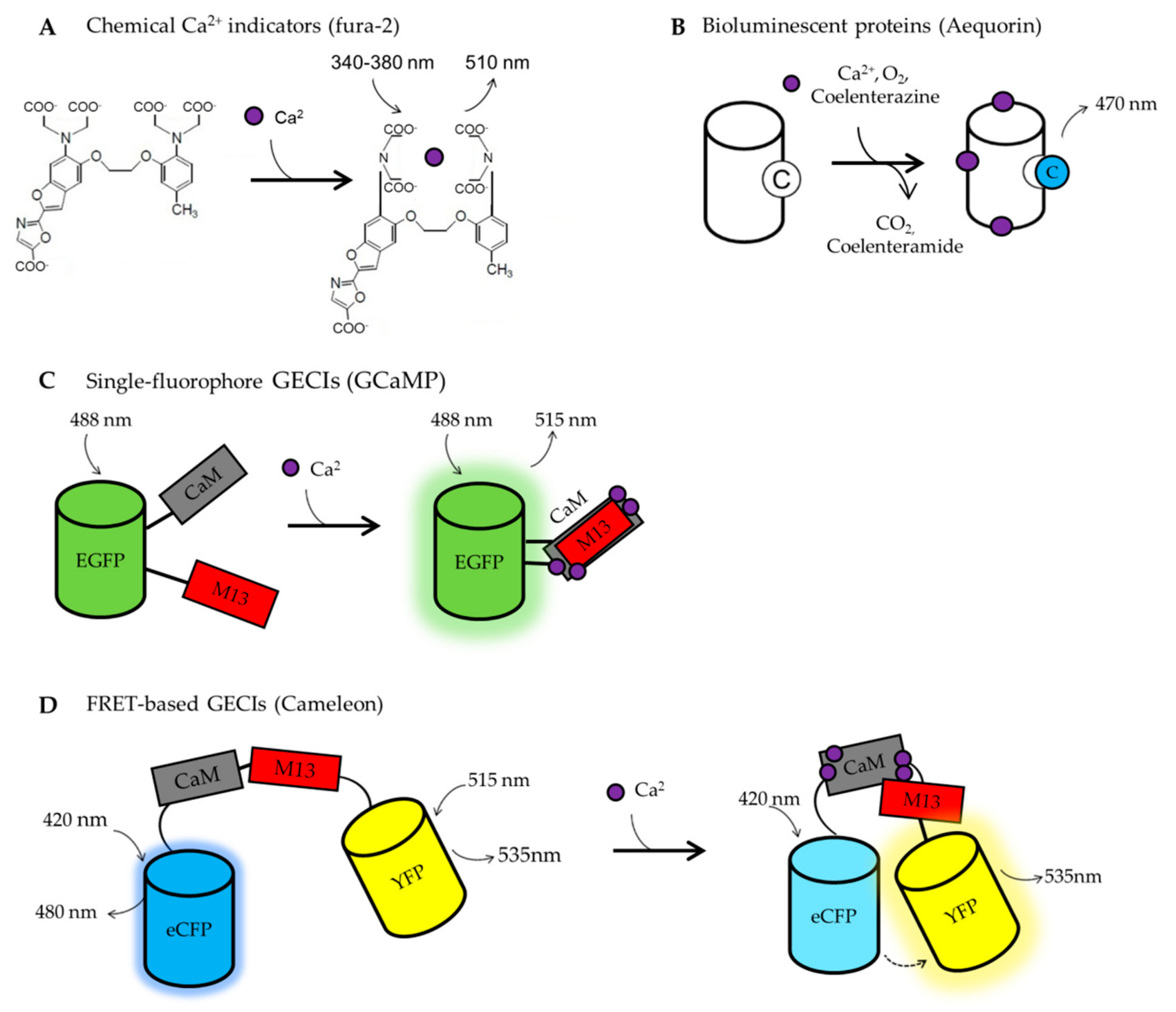
6.1. BOX1. Calcium Imaging Tools

Ca2+ indicators are fluorescent or luminescent molecules that undergo spectral modifications following Ca2+ binding (Figure 2). These changes permit the recording of free [Ca2+] in living cells, in real-time. Tsien and colleagues created the first generation of synthetic, chemical probes [183], fusing a fluorescent dye with Ca2+-selective chelators (EGTA or BAPTA). Among these chemical probes, fura-2 [184], and in particular its cell-permeant version containing the acetoxymethylated ester (AM) group, is the most used to monitor [Ca2+]c [185]. Fura-2 is a ratiometric indicator with a dissociation constant (Kd) of 140 nM in vitro. It is excited at 340 nm and 380 nm and it emits at 510 nm (Figure 2A). The emission ratio is dependent on [Ca2+]. Indeed, upon Ca2+ binding, the emission at 340 nm of excitation increases, whereas the emission at 380 nm of excitation decreases. The ratiometric properties of this dye allow accurate measurements of Ca2+ dynamics in living cells, being independent on dye leakage, photobleaching, differential dye loading, focal plane changes or cell movement artifacts. These parameters cannot be controlled using non-ratiometric probes, such as Fluo-4. A limitation of the classic fura-2, and of chemical probes in general, is that it cannot be specifically targeted to intracellular organelles. However, recently, this limitation has been overcome by the development of a mitochondria-targeted version of fura-2, the mito-fura, with a Kd of 1.5 μM in vitro and 6 μM in living cells [186]. The different affinities for Ca2+, displayed by mito-fura in vitro and in cells, indicate that, for quantitative measurements, a precise probe calibration in the biological system under investigation is required.
A revolution in the field of Ca2+ imaging is represented by the development of Genetically Encoded Calcium Indicators (GECIs) (Figure 2B–D). Being genetically encoded, GECIs can be targeted to different subcellular compartments and their expression can be controlled both spatially and temporally. The first developed GECI was based on aequorin (Aeq, [187]) (Figure 2B) and only later GECIs based on the Green Fluorescent Protein (GFP) have been generated [188] (Figure 2C,D).
Aeq is a Ca2+ sensitive bioluminescent protein extracted from the jellyfish, Aequoria victoria [189]. It consists of an apoprotein, named apoaequorin, that requires the luminescent substratum coelenterazine to function. Upon Ca2+ binding, coelenterazine is irreversibly oxidized to coelenteramide, emitting a photon at 470 nm. The rate of photon emission is proportional to [Ca2+] (Figure 2B). The large dynamic range, the low interference with endogenous Ca2+ buffering proteins and the low sensibility to pH make Aeq-based indicators good sensors for cell population analysis. However, Aeq suffers from some disadvantages, i.e., a low emitted light and the consumption of the probe during the experiment, making this GECI mainly suitable for relatively short (tens of minutes) experiments in cell populations [190]. Nowadays, several modified Aeq-based probes, with different Ca2+ affinities and targeted to almost all the subcellular compartments, are available (see [191] for a complete list). The wt Aeq can report [Ca2+] dynamics between 1–10 μM, the mutated form (D119A, [192]) can sense variation from 10 to 100 μM and the ER-targeted versions (N28L, [193,194]) are able to work in the mM [Ca2+] range. Being able to sense Ca2+ in the μM range, Aeq does not suffer from buffering capacity problems, meaning that its usage does not interfere with the physiology of Ca2+ signaling.
The GFP-based GECIs are the most used as they are easy to use and allow a single-cell analysis of real-time Ca2+ dynamics. Generally, they consist of a Ca2+ binding domain fused to a fluorescent protein (FP). When the Ca2+ ions bind the specific responsive domain, the FP changes its fluorescent properties. The use of fluorescent GECIs presents numerous advantages over chemical and bioluminescent Ca2+ sensors, such as the possibility of monitoring changes in [Ca2+] in real-time within specific organelles, restricting the analysis only to co-transfected cells expressing also the protein under investigation. They can be divided into two main groups: single fluorophore-based GECIs (Figure 2C) and Förster resonance energy transfer (FRET)-based GECIs (Figure 2D).
The first group includes camgaroos, pericam, and GCaMPs family. They are characterized by the presence of an FP, typically circularly permuted, and a Ca2+ binding domain inserted within the FP sequence, nearby the chromophore. Thus, changes in intensity or emission wavelength occur upon Ca2+ binding (Figure 2C). Among the single fluorescence-based GECI, GCaMPs [195] are the most used. They have been generated from the fusion of a circularly permuted GFP with two Ca2+ responsive elements: calmodulin (CaM) and a peptide sequence from the myosin light chain kinase (M13) (Figure 2C). These sensors are highly sensitive to Ca2+, with a Kds from 0.15 to 2 μM depending on the variants in use. These Kds have been evaluated in vitro and, as far as we know, in situ calibrations are missing. Of note, non-ratiometric sensors are difficult to calibrate being their fluorescence affected by changes in both cell morphology and focal plane. One of the last versions, i.e., GCaMP6 [196], has been widely used in the neuroscience field to study Ca2+ dynamics in different cell sub-compartments in living organisms. Indeed, many tg mouse lines expressing these specifically targeted probes have been created [197], allowing researchers to extend their investigation in vivo [198,199]. However, the recently observed neurotoxicity upon chronic expression of GCaM6P [200,201] imposes caution in the usage of such tg mouse lines, suggesting that GECI viral delivery or inducible/tissue-specific expression are preferable over their ubiquitous and constitutive expression. This toxic side effect could be partially explained by the fact that being these probes highly sensitive to Ca2+, they have a high Ca2+ buffering capacity that can interfere with various physiological processes when chronically expressed.
The second group of GFP-based GECIs are represented by those based on FRET, such as Cameleon probes [202,203], or their variants based on Troponin C, as Ca2+ responsive element [204]. Among the FRET-based GECIs, Cameleons are the first generated and the most used (Figure 2D). They have been targeted to many intracellular organelles, allowing researchers to analyze organelle Ca2+ dynamics in living cells. They are based on cyan (CFP) and yellow (YFP) variants of GFP and they exploit M13 and CaM for sensing Ca2+. Briefly, Ca2+ ions bind to CaM causing a conformational change that allows M13 to wrap around CaM, forcing the two FPs closed by. These changes in the molecule’s structure allow FRET to occur between the two fluorophores (Figure 2D). Thus, upon Ca2+ binding, the CFP fluorescence decreases, whereas the YFP fluorescence increases. Therefore, the YFP/CFP emission ratio changes dependently on [Ca2+]. Cameleon probes have all the advantages of other ratiometric probes [205]. Nowadays, many variants are available with Kds from 0.1 to almost 70 μM in vitro. Many Cameleon Kds have been also calculated in living cells and are reported in [206].

6.2. BOX2. PS2-Based Mouse AD Models

Numerous tg mouse lines expressing wt or mutated human APP and/or PS have been generated to provide animal models of AD. The expression of human FAD mutant of PS1 or PS2 in mice does not cause the characteristic histological hallmarks found in AD patients as amyloid plaques or neurofibrillary tangles are absent. This is because the mouse APP is not-amyloidogenic. Generally, PSEN mutations accelerate plaque formation in double tg lines expressing both FAD PSEN (PSEN1 or PSEN2) and human FAD APP mutated genes (mainly APPswe, carrying two amino acidic mutations in position 670 and 671) [207,208,209]. A complete list of the available AD mouse models is provided in [210].
The first-generation mouse models, based on APP or APP/PS overexpression, present however some limitations, i.e., overproduction of APP fragments in addition to Aβ [211] and lack of a clear phenotype standardization of the different models [212]. To overcome these limitations, second-generation mouse models were created. These new mice have been obtained by an APP knock-in strategy and present overproduction of Aβ42 without APP overexpression. Like the first-generation mouse models, also these mice present limitations, i.e., they do not exhibit tau pathology or neurodegeneration (see [211] for further details).
Focusing on PS2 and APP, the homozygous tg lines PS2-N141I (PS2.30H) and PS2APP are commonly used. The PS2-N141I (PS2.30H) mouse line expresses the human PSEN2 gene carrying the N141I mutation under the control of the mouse prion protein promoter [213]. This model is still poorly characterized since it has been created only to generate double tg PS2APP mice. Two PS2APP mouse models are present in literature: the first refers to the model generated by co-injecting two constructs into C57/BL/6 zygotes, known as B6.152H [214]. The other PS2APP line was generated by crossing two single tg animals (PS2N141I x APPswe) [213]. In this latter model, the human APPswe expression is controlled by the Thy1.2 promoter. Both lines of double tg animals express the same transgenes and both develop a similar level of cerebral pathology [215], but only B6.152H mice show enhanced LTP (see below). B6.152H mice have higher expression of tg human APP mRNA (1.45-fold) compared to PS2APP mice, and approximately 30-fold higher levels of PSEN2 mRNA. They also have about 2-fold higher Aβ levels than PS2APP mice [216]. The B6.152H line has been used also to generate the triple tg line, TauPS2APP [217], in which a mutation in Tau (P301L) is also present.
In B6.152H mice, amyloid plaques are absent at 3 months of age. The overt Aβ deposition in the brain appears only at approximately 6 months, with heavy plaque load in the hippocampus, frontal cortex, and subiculum at 10 months. Aβ deposits in blood vessels were observed sporadically and mainly in large vessels. The amount of cerebral amyloid deposits correlated with levels of the human APP transcript at 12 months [214,215]. Decreased survival of new-born neurons in the dentate gyrus, at about 4 months compared to wt, has also been observed [216]. Furthermore, a strong increase in LTP and post-tetanic potentiation (PTP) in hippocampal slices of 10-month-old animals, compared to wt mice, has been reported [216]. Finally, a decreased perfusion of the occipital cortex occurs from 10 to 17 months of age in these tg mice [215].
In general, despite several AD mouse models have been generated, none of them fully recapitulates the human pathology [211,218], strongly indicating that further efforts should be devoted to generating a model that can better mimic both SAD and FAD disease progression.

Funding

This work was funded by grants from the University of Padova, Italy (SID 2019), the Italian Ministry of University and Scientific Research (PRIN 2017) and EU Joint Programme in Neurodegenerative Disease, 2015–2018 (grant CeBioND) to P.P.; Euro-BioImaging–Roadmap/ESFRI from European Commission to T.P; Fondazione Cassa di Risparmio di Padova e Rovigo (CARIPARO Foundation, Progetti di Eccellenza 2017 to T.P.); the UNIPD Funds for Research Equipment-2015. L.G. and N.R. are fellows from Telethon (n. GGP16029 to T.P.) and Fondazione Cassa di Risparmio di Padova e Rovigo (CARIPARO Foundation, Starting Grant 2015 to P.P.) grant, respectively.

Conflicts of Interest

The authors declare no conflicts of interest.

References

  1. Albert, M.S.; DeKosky, S.T.; Dickson, D.; Dubois, B.; Feldman, H.H.; Fox, N.C.; Gamst, A.; Holtzman, D.M.; Jagust, W.J.; Petersen, R.C.; et al. The diagnosis of mild cognitive impairment due to Alzheimer’s disease: Recommendations from the National Institute on Aging-Alzheimer’s Association workgroups on diagnostic guidelines for Alzheimer’s disease. Alzheimer’s Dement. 2011, 7, 270–279. [Google Scholar] [CrossRef] [PubMed] [Green Version]
  2. Rossor, M.N.; Fox, N.C.; Freeborough, P.A.; Harvey, R.J. Clinical features of sporadic and familial Alzheimer’s disease. Neurodegeneration 1996, 5, 393–397. [Google Scholar] [CrossRef] [PubMed]
  3. Schachter, A.S.; Davis, K.L. Alzheimer’s disease. Dialogues Clin. Neurosci. 2000, 2, 91–100. [Google Scholar] [CrossRef] [PubMed]
  4. Bondi, M.W.; Edmonds, E.C.; Salmon, D.P. Alzheimer’s disease: Past, present, and future. J. Int. Neuropsychol. Soc. 2017, 23, 818–831. [Google Scholar] [CrossRef] [PubMed] [Green Version]
  5. Weller, J.; Budson, A. Current understanding of Alzheimer’s disease diagnosis and treatment. F1000Research 2018, 7. [Google Scholar] [CrossRef] [PubMed] [Green Version]
  6. Padovani, A.; Benussi, A.; Cantoni, V.; Dell’Era, V.; Cotelli, M.S.; Caratozzolo, S.; Turrone, R.; Rozzini, L.; Alberici, A.; Altomare, D.; et al. Diagnosis of mild cognitive impairment due to Alzheimer’s disease with transcranial magnetic stimulation. J. Alzheimer’s Dis. 2018, 65, 221–230. [Google Scholar] [CrossRef]
  7. Murphy, M.P.; Levine, H. Alzheimer’s disease and the amyloid-β peptide. J. Alzheimer’s Dis. 2010, 19, 311–323. [Google Scholar] [CrossRef] [Green Version]
  8. Dorszewska, J.; Prendecki, M.; Oczkowska, A.; Dezor, M.; Kozubski, W. Molecular Basis of Familial and Sporadic Alzheimer’s Disease. Curr. Alzheimer Res. 2016, 13, 952–963. [Google Scholar] [CrossRef]
  9. Duara, R.; Lopez-Alberola, R.F.; Barker, W.W.; Loewenstein, D.A.; Zatinsky, M.; Eisdorfer, C.E.; Weinberg, G.B. A comparison of familial and sporadic alzheimer’s disease. Neurology 1993, 43, 1377–1384. [Google Scholar] [CrossRef]
  10. Ray, W.J.; Ashall, F.; Goate, A.M. Molecular pathogenesis of sporadic and familial forms of Alzheimer’s disease. Mol. Med. Today 1998, 4, 151–157. [Google Scholar] [CrossRef]
  11. Piaceri, I.; Nacmias, B.; Sorbi, S. Genetics of familial and sporadic Alzheimer’s disease. Front. Biosci. - Elit. 2013, 5 E, 167–177. [Google Scholar] [CrossRef] [Green Version]
  12. Peila, R.; Rodriguez, B.L.; Launer, L.J. Type 2 diabetes, APOE gene, and the risk for dementia and related pathologies: The Honolulu-Asia Aging Study. Diabetes 2002, 51, 1256–1262. [Google Scholar] [CrossRef] [PubMed] [Green Version]
  13. Dreses-Werringloer, U.; Lambert, J.C.; Vingtdeux, V.; Zhao, H.; Vais, H.; Siebert, A.; Jain, A.; Koppel, J.; Rovelet-Lecrux, A.; Hannequin, D.; et al. A Polymorphism in CALHM1 Influences Ca2+ Homeostasis, Aβ Levels, and Alzheimer’s Disease Risk. Cell 2008, 133, 1149–1161. [Google Scholar] [CrossRef] [PubMed] [Green Version]
  14. Lambert, J.C.; Sleegers, K.; González-Pérez, A.; Ingelsson, M.; Beecham, G.W.; Hiltunen, M.; Combarros, O.; Bullido, M.J.; Brouwers, N.; Bettens, K.; et al. The CALHM1 P86L polymorphism is a genetic modifier of age at onset in Alzheimer’s disease: A meta-analysis study. J. Alzheimer’s Dis. 2010, 22, 247–255. [Google Scholar] [CrossRef] [PubMed] [Green Version]
  15. Minster, R.L.; Demirci, F.Y.; Dekosky, S.T.; Kamboh, M.I. No association between CALHM1 variation and risk of alzheimer disease. Hum. Mutat. 2009, 30. [Google Scholar] [CrossRef] [Green Version]
  16. Tan, E.K.; Ho, P.; Cheng, S.Y.; Yih, Y.; Li, H.H.; Fook-Chong, S.; Lee, W.L.; Zhao, Y. CALHM1 variant is not associated with Alzheimer’s disease among Asians. Neurobiol. Aging 2011, 32, 546.e11–546.e12. [Google Scholar] [CrossRef]
  17. Edwards, G.A.; Gamez, N.; Escobedo, G.; Calderon, O.; Moreno-Gonzalez, I. Modifiable risk factors for Alzheimer’s disease. Front. Aging Neurosci. 2019, 11. [Google Scholar] [CrossRef] [PubMed] [Green Version]
  18. Lahiri, D.K.; Maloney, B.; Basha, M.R.; Ge, Y.W.; Zawia, N.H. How and When Environmental Agents and Dietary Factors Affect the Course of Alzheimers Disease: The “LEARn” Model (Latent Early-Life Associated Regulation) May Explain the Triggering of AD. Curr. Alzheimer Res. 2007, 4, 219–228. [Google Scholar] [CrossRef]
  19. Lahiri, D.K.; Zawia, N.H.; Greig, N.H.; Sambamurti, K.; Maloney, B. Early-life events may trigger biochemical pathways for Alzheimer’s disease: The “LEARn” model. Biogerontology 2008, 9, 375–379. [Google Scholar] [CrossRef] [Green Version]
  20. Lahiri, D.K.; Maloney, B. The “LEARn” (Latent Early-life Associated Regulation) model integrates environmental risk factors and the developmental basis of Alzheimer’s disease, and proposes remedial steps. Exp. Gerontol. 2010, 45, 291–296. [Google Scholar] [CrossRef] [Green Version]
  21. Chakrabarti, S.; Khemka, V.K.; Banerjee, A.; Chatterjee, G.; Ganguly, A.; Biswas, A. Metabolic risk factors of sporadic Alzheimer’s disease: Implications in the pathology, pathogenesis and treatment. Aging Dis. 2015, 6, 282–299. [Google Scholar] [CrossRef] [PubMed] [Green Version]
  22. Swerdlow, R.H.; Khan, S.M. A “mitochondrial cascade hypothesis” for sporadic Alzheimer’s disease. Med. Hypotheses 2004, 63, 8–20. [Google Scholar] [CrossRef] [PubMed]
  23. Swerdlow, R.H.; Burns, J.M.; Khan, S.M. The Alzheimer’s disease mitochondrial cascade hypothesis: Progress and perspectives. Biochim. Biophys. Acta - Mol. Basis Dis. 2014, 1842, 1219–1231. [Google Scholar] [CrossRef] [PubMed] [Green Version]
  24. Ridge, P.G.; Kauwe, J.S.K. Mitochondria and Alzheimer’s Disease: the Role of Mitochondrial Genetic Variation. Curr. Genet. Med. Rep. 2018, 6, 1–10. [Google Scholar] [CrossRef] [Green Version]
  25. Wu, L.; Rosa-Neto, P.; Hsiung, G.Y.R.; Sadovnick, A.D.; Masellis, M.; Black, S.E.; Jia, J.; Gauthier, S. Early-onset familial alzheimer’s disease (EOFAD). Can. J. Neurol. Sci. 2012, 39, 436–445. [Google Scholar] [CrossRef] [Green Version]
  26. Lippa, C.F.; Saunders, A.M.; Smith, T.W.; Swearer, J.M.; Drachman, D.A.; Ghetti, B.; Nee, L.; Pulaski-Salo, D.; Dickson, D.; Robitaille, Y.; et al. Familial and sporadic Alzheimer’s disease: Neuropathology cannot exclude a final common pathway. Neurology 1996, 46, 406–412. [Google Scholar] [CrossRef]
  27. Yoshikai, S.i.; Sasaki, H.; Doh-ura, K.; Furuya, H.; Sakaki, Y. Genomic organization of the human amyloid beta-protein precursor gene. Gene 1990, 87, 257–263. [Google Scholar] [CrossRef]
  28. Lamb, B.T.; Sisodia, S.S.; Lawler, A.M.; Slunt, H.H.; Kitt, C.A.; Kearns, W.G.; Pearson, P.L.; Price, D.L.; Gearhart, J.D. Introduction and expression of the 400 kilobase precursor amyloid protein gene in transgenic mice. Nat. Genet. 1993, 5, 22–30. [Google Scholar] [CrossRef]
  29. Matsui, T.; Ingelsson, M.; Fukumoto, H.; Ramasamy, K.; Kowa, H.; Frosch, M.P.; Irizarry, M.C.; Hyman, B.T. Expression of APP pathway mRNAs and proteins in Alzheimer’s disease. Brain Res. 2007, 1161, 116–123. [Google Scholar] [CrossRef]
  30. Nguyen, K.V. β-Amyloid precursor protein (APP) and the human diseases. AIMS Neurosci. 2019, 6, 273–281. [Google Scholar] [CrossRef]
  31. Van der Kant, R.; Goldstein, L.S.B. Cellular Functions of the Amyloid Precursor Protein from Development to Dementia. Dev. Cell 2015, 32, 502–515. [Google Scholar] [CrossRef] [PubMed] [Green Version]
  32. Lee, K.J.; Moussa, C.E.H.; Lee, Y.; Sung, Y.; Howell, B.W.; Turner, R.S.; Pak, D.T.S.; Hoe, H.S. Beta amyloid-independent role of amyloid precursor protein in generation and maintenance of dendritic spines. Neuroscience 2010, 169, 344–356. [Google Scholar] [CrossRef] [PubMed] [Green Version]
  33. Müller, U.C.; Deller, T.; Korte, M. Not just amyloid: Physiological functions of the amyloid precursor protein family. Nat. Rev. Neurosci. 2017, 18, 281–298. [Google Scholar] [CrossRef] [PubMed]
  34. Chen, G.F.; Xu, T.H.; Yan, Y.; Zhou, Y.R.; Jiang, Y.; Melcher, K.; Xu, H.E. Amyloid beta: Structure, biology and structure-based therapeutic development. Acta Pharmacol. Sin. 2017, 38, 1205–1235. [Google Scholar] [CrossRef]
  35. Wang, X.; Zhou, X.; Li, G.; Zhang, Y.; Wu, Y.; Song, W. Modifications and trafficking of APP in the pathogenesis of alzheimer’s disease. Front. Mol. Neurosci. 2017, 10. [Google Scholar] [CrossRef]
  36. Hooper, N.M.; Walter, J.; Haass, C. Posttranslational Modifications of Amyloid Precursor Protein: Ectodomain Phosphorylation and Sulfation. In Alzheimer’s Disease; Humana Press Inc.: Totowa, NJ, USA, 2003; pp. 149–168. [Google Scholar]
  37. Zheng, H.; Koo, E.H. Biology and pathophysiology of the amyloid precursor protein. Mol. Neurodegener. 2011, 6. [Google Scholar] [CrossRef] [Green Version]
  38. Thinakaran, G.; Koo, E.H. Amyloid precursor protein trafficking, processing, and function. J. Biol. Chem. 2008, 283, 29615–29619. [Google Scholar] [CrossRef] [Green Version]
  39. Zhou, Y.; Sun, Y.; Ma, Q.-H.; Liu, Y. Alzheimer’s disease: Amyloid-based pathogenesis and potential therapies. Cell Stress 2018, 2, 150–161. [Google Scholar] [CrossRef]
  40. Hardy, J.; Selkoe, D.J. The amyloid hypothesis of Alzheimer’s disease: Progress and problems on the road to therapeutics. Science 2002, 297, 353–356. [Google Scholar] [CrossRef] [Green Version]
  41. Chen, Y.R.; Glabe, C.G. Distinct early folding and aggregation properties of Alzheimer amyloid-β peptides Aβ40 and Aβ42: Stable trimer or tetramer formation by Aβ42. J. Biol. Chem. 2006, 281, 24414–24422. [Google Scholar] [CrossRef] [Green Version]
  42. Parihar, M.S.; Hemnani, T. Alzheimer’s disease pathogenesis and therapeutic interventions. J. Clin. Neurosci. 2004, 11, 456–467. [Google Scholar] [CrossRef] [PubMed]
  43. Fiala, M.; Liu, P.T.; Espinosa-Jeffrey, A.; Rosenthal, M.J.; Bernard, G.; Ringman, J.M.; Sayre, J.; Zhang, L.; Zaghi, J.; Dejbakhsh, S.; et al. Innate immunity and transcription of MGAT-III and Toll-like receptors in Alzheimer’s disease patients are improved by bisdemethoxycurcumin. Proc. Natl. Acad. Sci. USA 2007, 104, 12849–12854. [Google Scholar] [CrossRef] [PubMed] [Green Version]
  44. LaFerla, F.M.; Green, K.N.; Oddo, S. Intracellular amyloid-β in Alzheimer’s disease. Nat. Rev. Neurosci. 2007, 8, 499–509. [Google Scholar] [CrossRef] [PubMed]
  45. Gandy, S. The role of cerebral amyloid β accumulation in common forms of Alzheimer disease. J. Clin. Invest. 2005, 115, 1121–1129. [Google Scholar] [PubMed]
  46. Querfurth, H.W.; LaFerla, F.M. Alzheimer’s disease. N. Engl. J. Med. 2010, 362, 329–344. [Google Scholar] [CrossRef] [PubMed] [Green Version]
  47. Haass, C.; Selkoe, D.J. Soluble protein oligomers in neurodegeneration: Lessons from the Alzheimer’s amyloid β-peptide. Nat. Rev. Mol. Cell Biol. 2007, 8, 101–112. [Google Scholar] [CrossRef]
  48. Rogaev, E.I.; Sherrington, R.; Rogaeva, E.A.; Levesque, G.; Ikeda, M.; Liang, Y.; Chi, H.; Lin, C.; Holman, K.; Tsuda, T.; et al. Familial Alzheimer’s disease in kindreds with missense mutations in a gene on chromosome 1 related to the Alzheimer’s disease type 3 gene. Nature 1995, 376, 775–778. [Google Scholar] [CrossRef]
  49. Sherrington, R.; Rogaev, E.I.; Liang, Y.; Rogaeva, E.A.; Levesque, G.; Ikeda, M.; Chi, H.; Lin, C.; Li, G.; Holman, K.; et al. Cloning of a gene bearing missense mutations in early-onset familial Alzheimer’s disease. Nature 1995, 375, 754–760. [Google Scholar] [CrossRef]
  50. De Strooper, B.; Saftig, P.; Craessaerts, K.; Vanderstichele, H.; Guhde, G.; Annaert, W.; Von Figura, K.; Van Leuven, F. Deficiency of presenilin-1 inhibits the normal cleavage of amyloid precursor protein. Nature 1998, 391, 387–390. [Google Scholar] [CrossRef]
  51. Wolfe, M.S.; Xia, W.; Ostaszewski, B.L.; Diehl, T.S.; Kimberly, W.T.; Selkoe, D.J. Two transmembrane aspartates in presenilin-1 required for presenilin endoproteolysis and γ-secretase activity. Nature 1999, 398, 513–517. [Google Scholar] [CrossRef]
  52. Levitan, D.; Greenwald, I. Facilitation of lin-12-mediated signalling by sel-12, a Caenorhabditis elegans S182 Alzheimer’s disease gene. Nature 1995, 377, 351–354. [Google Scholar] [CrossRef] [PubMed]
  53. Boulianne, G.L.; Livne-Bar, I.; Humphreys, J.M.; Liang, Y.; Lin, C.; Rogaev, E.; George-Hyslop, P.S. Cloning and characterization of the Drosophila presenilin homologue. Neuroreport 1997, 8, 1025–1029. [Google Scholar] [CrossRef] [PubMed]
  54. Martínez-Mir, A.; Cañestro, C.; Gonzàlez-Duarte, R.; Albalat, R. Characterization of the amphioxus presenilin gene in a high gene-density genomic region illustrates duplication during the vertebrate lineage. Gene 2001, 279, 157–164. [Google Scholar] [CrossRef]
  55. Lee, M.K.; Slunt, H.H.; Martin, L.J.; Thinakaran, G.; Kim, G.; Gandy, S.E.; Seeger, M.; Koo, E.; Price, D.L.; Sisodia, S.S. Expression of presenilin 1 and 2 (PS1 and PS2) in human and murine tissues. J. Neurosci. 1996, 16, 7513–7525. [Google Scholar] [CrossRef] [Green Version]
  56. Kovacs, D.M.; Fausett, H.J.; Page, K.J.; Kim, T.W.; Moir, R.D.; Merriam, D.E.; Hollister, R.D.; Hallmark’, O.G.; Mancini, R.; Felsenstein, K.M.; et al. Alzheimer-associated presenilins 1 and 2: Neuronal expression in brain and localization to intracellular membranes in mammalian cells. Nat. Med. 1996, 2, 224–229. [Google Scholar] [CrossRef]
  57. Suzuki, T.; Nishiyama, K.; Murayama, S.; Yamamoto, A.; Sato, S.; Kanazawa, I.; Sakaki, Y. Regional and cellular Presenilin 1 gene expression in human and rat tissues. Biochem. Biophys. Res. Commun. 1996, 219, 708–713. [Google Scholar] [CrossRef]
  58. Brunkan, A.L.; Goate, A.M. Presenilin function and γ-secretase activity. J. Neurochem. 2005, 93, 769–792. [Google Scholar] [CrossRef]
  59. Area-Gomez, E.; De Groof, A.J.C.; Boldogh, I.; Bird, T.D.; Gibson, G.E.; Koehler, C.M.; Yu, W.H.; Duff, K.E.; Yaffe, M.P.; Pon, L.A.; et al. Presenilins are enriched in endoplasmic reticulum membranes associated with mitochondria. Am. J. Pathol. 2009, 175, 1810–1816. [Google Scholar] [CrossRef] [Green Version]
  60. Filadi, R.; Greotti, E.; Turacchio, G.; Luini, A.; Pozzan, T.; Pizzo, P. Presenilin 2 Modulates Endoplasmic Reticulum-Mitochondria Coupling by Tuning the Antagonistic Effect of Mitofusin 2. Cell Rep. 2016, 15, 2226–2238. [Google Scholar] [CrossRef] [Green Version]
  61. Leal, N.S.; Schreiner, B.; Pinho, C.M.; Filadi, R.; Wiehager, B.; Karlström, H.; Pizzo, P.; Ankarcrona, M. Mitofusin-2 knockdown increases ER–mitochondria contact and decreases amyloid β-peptide production. J. Cell. Mol. Med. 2016, 20, 1686–1695. [Google Scholar] [CrossRef]
  62. Doan, A.; Thinakaran, G.; Borchelt, D.R.; Slunt, H.H.; Ratovitsky, T.; Podlisny, M.; Selkoe, D.J.; Seeger, M.; Gandy, S.E.; Price, D.L.; et al. Protein topology of presenilin 1. Neuron 1996, 17, 1023–1030. [Google Scholar] [CrossRef] [Green Version]
  63. Laudon, H.; Hansson, E.M.; Melén, K.; Bergman, A.; Farmery, M.R.; Winblad, B.; Lendahl, U.; Von Heijne, G.; Näslund, J. A nine-transmembrane domain topology for presenilin 1. J. Biol. Chem. 2005, 280, 35352–35360. [Google Scholar] [CrossRef] [PubMed] [Green Version]
  64. Wolfe, M.S. Toward the structure of presenilin/γ-secretase and presenilin homologs. Biochim. Biophys. Acta - Biomembr. 2013, 1828, 2886–2897. [Google Scholar] [CrossRef] [PubMed] [Green Version]
  65. Li, Y.M.; Lai, M.T.; Xu, M.; Huang, Q.; DiMuzio-Mower, J.; Sardana, M.K.; Shi, X.P.; Yin, K.C.; Shafer, J.A.; Gardell, S.J. Presenilin 1 is linked with γ-secretase activity in the detergent solubilized state. Proc. Natl. Acad. Sci. USA 2000, 97, 6138–6143. [Google Scholar] [CrossRef] [PubMed] [Green Version]
  66. Chen, F.; Hasegawa, H.; Schmitt-Ulms, G.; Kawarai, T.; Bohm, C.; Katayama, T.; Gu, Y.; Sanjo, N.; Glista, M.; Rogaeva, E.; et al. TMP21 is a presenilin complex component that modulates γ-secretase but not ε-secretase activity. Nature 2006, 440, 1208–1212. [Google Scholar] [CrossRef] [PubMed]
  67. Ahn, K.; Shelton, C.C.; Tian, Y.; Zhang, X.; Gilchrist, M.L.; Sisodia, S.S.; Li, Y.M. Activation and intrinsic γ-secretase activity of presenilin 1. Proc. Natl. Acad. Sci. USA 2010, 107, 21435–21440. [Google Scholar] [CrossRef] [Green Version]
  68. Edbauer, D.; Winkler, E.; Regula, J.T.; Pesold, B.; Steiner, H.; Haass, C. Reconstitution of gamma-secretase activity. Nat. Cell Biol. 2003. [Google Scholar] [CrossRef]
  69. Iwatsubo, T. The γ-secretase complex: Machinery for intramembrane proteolysis. Curr. Opin. Neurobiol. 2004, 14, 379–383. [Google Scholar] [CrossRef]
  70. McCarthy, J.V.; Twomey, C.; Wujek, P. Presenilin-dependent regulated intramembrane proteolysis and γ-secretase activity. Cell. Mol. Life Sci. 2009, 66, 1534–1555. [Google Scholar] [CrossRef]
  71. Thinakaran, G.; Borchelt, D.R.; Lee, M.K.; Slunt, H.H.; Spitzer, L.; Kim, G.; Ratovitsky, T.; Davenport, F.; Nordstedt, C.; Seeger, M.; et al. Endoproteolysis of presenilin 1 and accumulation of processed derivatives in vivo. Neuron 1996, 17, 181–190. [Google Scholar] [CrossRef] [Green Version]
  72. Steiner, H.; Fluhrer, R.; Haass, C. Intramembrane proteolysis by γ-secretase. J. Biol. Chem. 2008, 283, 29627–29631. [Google Scholar] [CrossRef] [PubMed] [Green Version]
  73. De Strooper, B.; Annaert, W. Novel Research Horizons for Presenilins and γ-Secretases in Cell Biology and Disease. Annu. Rev. Cell Dev. Biol. 2010, 26, 235–260. [Google Scholar] [CrossRef] [PubMed]
  74. Kim, T.W.; Pettingell, W.H.; Hallmark, O.G.; Moir, R.D.; Wasco, W.; Tanzi, R.E. Endoproteolytic cleavage and proteasomal degradation of presenilin 2 in transfected cells. J. Biol. Chem. 1997, 272, 11006–11010. [Google Scholar] [CrossRef] [PubMed] [Green Version]
  75. Center for Molecular Neurology. Available online: https://uantwerpen.vib.be/ (accessed on 17 December 2019).
  76. Scheuner, D.; Eckman, C.; Jensen, M.; Song, X.; Citron, M.; Suzuki, N.; Bird, T.D.; Hardy, J.; Hutton, M.; Kukull, W.; et al. Secreted amyloid β-protein similar to that in the senile plaques of Alzheimer’s disease is increased in vivo by the presenilin 1 and 2 and APP mutations linked to familial Alzheimer’s disease. Nat. Med. 1996, 2, 864–870. [Google Scholar] [CrossRef] [PubMed]
  77. Citron, M.; Westaway, D.; Xia, W.; Carlson, G.; Diehl, T.; Levesque, G.; Johnson-Wood, K.; Lee, M.; Seubert, P.; Davis, A.; et al. Mutant presenilins of Alzheimer’s disease increase production of 42-residue amyloid β-protein in both transfected cells and transgenic mice. Nat. Med. 1997, 3, 67–72. [Google Scholar] [CrossRef] [PubMed]
  78. Chévez-Gutiérrez, L.; Bammens, L.; Benilova, I.; Vandersteen, A.; Benurwar, M.; Borgers, M.; Lismont, S.; Zhou, L.; Van Cleynenbreugel, S.; Esselmann, H.; et al. The mechanism of γ-Secretase dysfunction in familial Alzheimer disease. EMBO J. 2012, 31, 2261–2274. [Google Scholar] [CrossRef]
  79. Florean, C.; Zampese, E.; Zanese, M.; Brunello, L.; Ichas, F.; De Giorgi, F.; Pizzo, P. High content analysis of γ-secretase activity reveals variable dominance of presenilin mutations linked to familial Alzheimer’s disease. Biochim. Biophys. Acta - Mol. Cell Res. 2008, 1783, 1551–1560. [Google Scholar] [CrossRef]
  80. Shimojo, M.; Sahara, N.; Murayama, M.; Ichinose, H.; Takashima, A. Decreased Aβ secretion by cells expressing familial Alzheimer’s disease-linked mutant presenilin 1. Neurosci. Res. 2007, 57, 446–453. [Google Scholar] [CrossRef]
  81. Walker, E.S.; Martinez, M.; Brunkan, A.L.; Goate, A. Presenilin 2 familial Alzheimer’s disease mutations result in partial loss of function and dramatic changes in Aβ 42/40 ratios. J. Neurochem. 2005, 92, 294–301. [Google Scholar] [CrossRef]
  82. Quintero-Monzon, O.; Martin, M.M.; Fernandez, M.A.; Cappello, C.A.; Krzysiak, A.J.; Osenkowski, P.; Wolfe, M.S. Dissociation between the processivity and total activity of γ-secretase: Implications for the mechanism of Alzheimer’s disease-causing presenilin mutations. Biochemistry 2011, 50, 9023–9035. [Google Scholar] [CrossRef] [Green Version]
  83. De Strooper, B.; Iwatsubo, T.; Wolfe, M.S. Presenilins and γ-secretase: Structure, function, and role in Alzheimer disease. Cold Spring Harb. Perspect. Med. 2012, 2. [Google Scholar] [CrossRef] [PubMed]
  84. Takeda, T.; Asahi, M.; Yamaguchi, O.; Hikoso, S.; Nakayama, H.; Kusakari, Y.; Kawai, M.; Hongo, K.; Higuchi, Y.; Kashiwase, K.; et al. Presenilin 2 regulates the systolic function of heart by modulating Ca2+ signaling. FASEB J. 2005, 19, 2069–2071. [Google Scholar] [CrossRef] [PubMed]
  85. Donoviel, D.B.; Hadjantonakis, A.-K.; Ikeda, M.; Zheng, H.; Hyslop, P.S.G.; Bernstein, A. Mice lacking both presenilin genes exhibitearlyembryonic patterningdefects. Genes Dev. 1999, 13, 2801–2810. [Google Scholar] [CrossRef] [PubMed] [Green Version]
  86. Li, D.; Parks, S.B.; Kushner, J.D.; Nauman, D.; Burgess, D.; Ludwigsen, S.; Partain, J.; Nixon, R.R.; Allen, C.N.; Irwin, R.P.; et al. Mutations of Presenilin Genes in Dilated Cardiomyopathy and Heart Failure. Am. J. Hum. Genet. 2006, 79, 1030–1039. [Google Scholar] [CrossRef] [Green Version]
  87. Yang, M.; Li, C.; Zhang, Y.; Ren, J. Interrelationship between Alzheimer’s disease and cardiac dysfunction: the brain–heart continuum? Acta Biochim. Biophys. Sin. 2020. [Google Scholar] [CrossRef] [Green Version]
  88. An, S.S.; Cai, Y.; Kim, S. Mutations in presenilin 2 and its implications in Alzheimer’s disease and other dementia-associated disorders. Clin. Interv. Aging 2015, 1163. [Google Scholar] [CrossRef] [Green Version]
  89. Subramanian, K.; Gianni, D.; Balla, C.; Assenza, G.E.; Joshi, M.; Semigran, M.J.; Macgillivray, T.E.; Van Eyk, J.E.; Agnetti, G.; Paolocci, N.; et al. Cofilin-2 Phosphorylation and Sequestration in Myocardial Aggregates. J. Am. Coll. Cardiol. 2015, 65, 1199–1214. [Google Scholar] [CrossRef] [Green Version]
  90. Gianni, D.; Li, A.; Tesco, G.; McKay, K.M.; Moore, J.; Raygor, K.; Rota, M.; Gwathmey, J.K.; Dec, G.W.; Aretz, T.; et al. Protein Aggregates and Novel Presenilin Gene Variants in Idiopathic Dilated Cardiomyopathy. Circulation 2010, 121, 1216–1226. [Google Scholar] [CrossRef] [Green Version]
  91. Tublin, J.M.; Adelstein, J.M.; del Monte, F.; Combs, C.K.; Wold, L.E. Getting to the Heart of Alzheimer Disease. Circ. Res. 2019, 124, 142–149. [Google Scholar] [CrossRef]
  92. Berridge, M.J. Neuronal calcium signaling. Neuron 1998, 21, 13–26. [Google Scholar] [CrossRef] [Green Version]
  93. Berridge, M.J.; Lipp, P.; Bootman, M.D. The versatility and universality of calcium signalling. Nat. Rev. Mol. Cell Biol. 2000, 1, 11–21. [Google Scholar] [CrossRef] [PubMed]
  94. Zampese, E.; Pizzo, P. Intracellular organelles in the saga of Ca2+ homeostasis: Different molecules for different purposes? Cell. Mol. Life Sci. 2012, 69, 1077–1104. [Google Scholar] [CrossRef] [PubMed]
  95. Clapham, D.E. Calcium Signaling. Cell 2007, 131, 1047–1058. [Google Scholar] [CrossRef] [PubMed] [Green Version]
  96. Berridge, M.J.; Bootman, M.D.; Roderick, H.L. Calcium signalling: Dynamics, homeostasis and remodelling. Nat. Rev. Mol. Cell Biol. 2003, 4, 517–529. [Google Scholar] [CrossRef] [PubMed] [Green Version]
  97. Santulli, G.; Nakashima, R.; Yuan, Q.; Marks, A.R. Intracellular calcium release channels: An update. J. Physiol. 2017, 595, 3041–3051. [Google Scholar] [CrossRef] [PubMed] [Green Version]
  98. Prole, D.L.; Taylor, C.W. Structure and function of ip3 receptors. Cold Spring Harb. Perspect. Biol. 2019, 11. [Google Scholar] [CrossRef] [Green Version]
  99. Pinton, P.; Pozzan, T.; Rizzuto, R. The Golgi apparatus is an inositol 1,4,5-trisphosphate-sensitive Ca2+ store, with functional properties distinct from those of the endoplasmic reticulum. EMBO J. 1998, 17, 5298–5308. [Google Scholar] [CrossRef] [Green Version]
  100. Pizzo, P.; Lissandron, V.; Capitanio, P.; Pozzan, T. Ca2+ signalling in the Golgi apparatus. Cell Calcium 2011, 50, 184–192. [Google Scholar] [CrossRef]
  101. Morgan, A.J. Ca2+ dialogue between acidic vesicles and ER. Biochem. Soc. Trans. 2016, 44, 546–553. [Google Scholar] [CrossRef]
  102. Putney, J.W.; Steinckwich-Besançon, N.; Numaga-Tomita, T.; Davis, F.M.; Desai, P.N.; D’Agostin, D.M.; Wu, S.; Bird, G.S. The functions of store-operated calcium channels. Biochim. Biophys. Acta Mol. Cell Res. 2017, 1864, 900–906. [Google Scholar] [CrossRef]
  103. Kar, P.; Bakowski, D.; Di Capite, J.; Nelson, C.; Parekh, A.B. Different agonists recruit different stromal interaction molecule proteins to support cytoplasmic Ca2+ oscillations and gene expression. Proc. Natl. Acad. Sci. USA 2012, 109, 6969–6974. [Google Scholar] [CrossRef] [PubMed] [Green Version]
  104. Chemaly, E.R.; Troncone, L.; Lebeche, D. SERCA control of cell death and survival. Cell Calcium 2018, 69, 46–61. [Google Scholar] [CrossRef] [PubMed]
  105. Baba-Aissa, F.; Raeymaekers, L.; Wuytack, F.; Dode, L.; Casteels, R. Distribution and isoform diversity of the organellar Ca2+ pumps in the brain. Mol. Chem. Neuropathol. 1998, 33, 199–208. [Google Scholar] [CrossRef] [PubMed]
  106. Lissandron, V.; Podini, P.; Pizzo, P.; Pozzan, T. Unique characteristics of Ca2+ homeostasis of the trans-Golgi compartment. Proc. Natl. Acad. Sci. USA 2010, 107, 9198–9203. [Google Scholar] [CrossRef] [PubMed] [Green Version]
  107. Wong, A.K.C.; Capitanio, P.; Lissandron, V.; Bortolozzi, M.; Pozzan, T.; Pizzo, P. Heterogeneity of Ca2+ handling among and within Golgi compartments. J. Mol. Cell Biol. 2013, 5, 266–276. [Google Scholar] [CrossRef] [PubMed]
  108. Wang, Q.C.; Zheng, Q.; Tan, H.; Zhang, B.; Li, X.; Yang, Y.; Yu, J.; Liu, Y.; Chai, H.; Wang, X.; et al. TMCO1 is an ER Ca2+ load-activated Ca2+ channel. Cell 2016, 165, 1454–1466. [Google Scholar] [CrossRef] [Green Version]
  109. Stafford, N.; Wilson, C.; Oceandy, D.; Neyses, L.; Cartwright, E.J. The plasma membrane calcium ATPases and their role as major new players in human disease. Physiol. Rev. 2017, 97, 1089–1125. [Google Scholar] [CrossRef] [Green Version]
  110. DiPolo, R.; Beaugé, L. Sodium/calcium exchanger: Influence of metabolic regulation on ion carrier interactions. Physiol. Rev. 2006, 86, 155–203. [Google Scholar] [CrossRef] [Green Version]
  111. Rizzuto, R.; Pozzan, T. Microdomains of intracellular Ca2+: Molecular determinants and functional consequences. Physiol. Rev. 2006, 86, 369–408. [Google Scholar] [CrossRef]
  112. Schwaller, B. The continuing disappearance of “pure” Ca2+ buffers. Cell. Mol. Life Sci. 2009, 66, 275–300. [Google Scholar] [CrossRef]
  113. Giorgi, C.; Danese, A.; Missiroli, S.; Patergnani, S.; Pinton, P. Calcium Dynamics as a Machine for Decoding Signals. Trends Cell Biol. 2018, 28, 258–273. [Google Scholar] [CrossRef] [PubMed]
  114. Baughman, J.M.; Perocchi, F.; Girgis, H.S.; Plovanich, M.; Belcher-Timme, C.A.; Sancak, Y.; Bao, X.R.; Strittmatter, L.; Goldberger, O.; Bogorad, R.L.; et al. Integrative genomics identifies MCU as an essential component of the mitochondrial calcium uniporter. Nature 2011, 476, 341–345. [Google Scholar] [CrossRef] [PubMed] [Green Version]
  115. De Stefani, D.; Raffaello, A.; Teardo, E.; Szabó, I.; Rizzuto, R. A forty-kilodalton protein of the inner membrane is the mitochondrial calcium uniporter. Nature 2011, 476, 336–340. [Google Scholar] [CrossRef] [PubMed]
  116. Palty, R.; Silverman, W.F.; Hershfinkel, M.; Caporale, T.; Sensi, S.L.; Parnis, J.; Nolte, C.; Fishman, D.; Shoshan-Barmatz, V.; Herrmann, S.; et al. NCLX is an essential component of mitochondrial Na+/Ca2+ exchange. Proc. Natl. Acad. Sci. USA 2010, 107, 436–441. [Google Scholar] [CrossRef] [Green Version]
  117. Jiang, D.; Zhao, L.; Clapham, D.E. Genome-wide RNAi screen identifies Letm1 as a mitochondrial Ca2+/H+ antiporter. Science 2009, 326, 144–147. [Google Scholar] [CrossRef] [Green Version]
  118. Rizzuto, R. Close Contacts with the Endoplasmic Reticulum as Determinants of Mitochondrial Ca2+ Responses. Science 1998, 280, 1763–1766. [Google Scholar] [CrossRef]
  119. Filadi, R.; Greotti, E.; Pizzo, P. Highlighting the endoplasmic reticulum-mitochondria connection: Focus on Mitofusin 2. Pharmacol. Res. 2018, 128, 42–51. [Google Scholar] [CrossRef]
  120. Pinton, P. Mitochondria-associated membranes (MAMs) and pathologies editorial. Cell Death Dis. 2018, 9. [Google Scholar] [CrossRef] [Green Version]
  121. Khachaturian, Z.S.; Radebaugh, T.S. Alzheimer disease: Where are we now? Where are we going? Alzheimer Dis Assoc Disord 1988, 12 (Suppl. 3), S24–S28. [Google Scholar]
  122. Peterson, C.; Ratan, R.R.; Shelanski, M.L.; Goldman, J.E. Altered response of fibroblasts from aged and Alzheimer donors to drugs that elevate cytosolic free calcium. Neurobiol. Aging 1988, 9, 261–266. [Google Scholar] [CrossRef]
  123. Khachaturian, Z.S. Calcium Hypothesis of Alzheimer’s Disease and Brain Aging. Ann. N. Y. Acad. Sci. 2006, 747, 1–11. [Google Scholar] [CrossRef] [PubMed]
  124. Agostini, M.; Fasolato, C. When, where and how? Focus on neuronal calcium dysfunctions in Alzheimer’s Disease. Cell Calcium 2016, 60, 289–298. [Google Scholar] [CrossRef] [PubMed]
  125. Kumar, A. Calcium Signaling During Brain Aging and Its Influence on the Hippocampal Synaptic Plasticity. In Advances in Experimental Medicine and Biology; Springer: Basel, Switzerland, 2020; Volume 1131, pp. 985–1012. [Google Scholar]
  126. Emilsson, L.; Saetre, P.; Jazin, E. Alzheimer’s disease: MRNA expression profiles of multiple patients show alterations of genes involved with calcium signaling. Neurobiol. Dis. 2006, 21, 618–625. [Google Scholar] [CrossRef] [PubMed]
  127. Ito, E.; Oka, K.; Etcheberrigaray, R.; Nelson, T.J.; McPhie, D.L.; Tofel-Grehl, B.; Gibson, G.E.; Alkon, D.L. Internal Ca2+ mobilization is altered in fibroblasts from patients with Alzheimer disease. Proc. Natl. Acad. Sci. USA 1994, 91, 534–538. [Google Scholar] [CrossRef] [PubMed] [Green Version]
  128. Etcheberrigaray, R.; Hirashima, N.; Nee, L.; Prince, J.; Govoni, S.; Racchi, M.; Tanzi, R.E.; Alkon, D.L. Calcium responses in fibroblasts from asymptomatic members of Alzheimer’s disease families. Neurobiol. Dis. 1998, 5, 37–45. [Google Scholar] [CrossRef] [Green Version]
  129. Giacomello, M.; Barbiero, L.; Zatti, G.; Squitti, R.; Binetti, G.; Pozzan, T.; Fasolato, C.; Ghidoni, R.; Pizzo, P. Reduction of Ca2+ stores and capacitative Ca2+ entry is associated with the familial Alzheimer’s disease presenilin-2 T122R mutation and anticipates the onset of dementia. Neurobiol. Dis. 2005, 18, 638–648. [Google Scholar] [CrossRef]
  130. LaFerla, F.M. Calcium dyshomeostasis and intracellular signalling in alzheimer’s disease. Nat. Rev. Neurosci. 2002, 3, 862–872. [Google Scholar] [CrossRef]
  131. Leissring, M.A.; Paul, B.A.; Parker, I.; Cotman, C.W.; Laferla, F.M. Alzheimer’s presenilin-1 mutation potentiates inositol 1,4,5- trisphosphate-mediated calcium signaling in Xenopus oocytes. J. Neurochem. 1999, 72, 1061–1068. [Google Scholar] [CrossRef]
  132. Chan, S.L.; Mayne, M.; Holden, C.P.; Geiger, J.D.; Mattson, M.P. Presenilin-1 mutations increase levels of ryanodine receptors and calcium release in PC12 cells and cortical neurons. J. Biol. Chem. 2000, 275, 18195–18200. [Google Scholar] [CrossRef] [Green Version]
  133. Leissring, M.A.; Akbari, Y.; Fanger, C.M.; Cahalan, M.D.; Mattson, M.P.; LaFerla, F.M. Capacitative calcium entry deficits and elevated luminal calcium content in mutant presenilin-1 knockin mice. J. Cell Biol. 2000, 149, 793–797. [Google Scholar] [CrossRef] [Green Version]
  134. Smith, I.F.; Boyle, J.P.; Vaughan, P.F.T.; Pearson, H.A.; Peers, C. Effects of chronic hypoxia on Ca2+ stores and capacitative Ca2+ entry in human neuroblastoma (SH-SY5Y) cells. J. Neurochem. 2001, 79, 877–884. [Google Scholar] [CrossRef] [PubMed]
  135. Stutzmann, G.E.; Caccamo, A.; LaFerla, F.M.; Parker, I. Dysregulated IP3 Signaling in Cortical Neurons of Knock-In Mice Expressing an Alzheimer’s-Linked Mutation in Presenilin1 Results in Exaggerated Ca2+ Signals and Altered Membrane Excitability. J. Neurosci. 2004, 24, 508–513. [Google Scholar] [CrossRef] [PubMed] [Green Version]
  136. Stutzmann, G.E.; Smith, I.; Caccamo, A.; Oddo, S.; LaFerla, F.M.; Parker, I. Enhanced ryanodine receptor recruitment contributes to Ca2+ disruptions in young, adult, and aged Alzheimer’s disease mice. J. Neurosci. 2006, 26, 5180–5189. [Google Scholar] [CrossRef] [PubMed] [Green Version]
  137. Hayrapetyan, V.; Rybalchenko, V.; Rybalchenko, N.; Koulen, P. The N-terminus of presenilin-2 increases single channel activity of brain ryanodine receptors through direct protein-protein interaction. Cell Calcium 2008, 44, 507–518. [Google Scholar] [CrossRef] [PubMed]
  138. Rybalchenko, V.; Hwang, S.Y.; Rybalchenko, N.; Koulen, P. The cytosolic N-terminus of presenilin-1 potentiates mouse ryanodine receptor single channel activity. Int. J. Biochem. Cell Biol. 2008, 40, 84–97. [Google Scholar] [CrossRef]
  139. Chakroborty, S.; Goussakov, I.; Miller, M.B.; Stutzmann, G.E. Deviant ryanodine receptor-mediated calcium release resets synaptic homeostasis in presymptomatic 3xTg-AD mice. J. Neurosci. 2009, 29, 9458–9470. [Google Scholar] [CrossRef]
  140. Leissring, M.A.; Parker, I.; LaFerla, F.M. Presenilin-2 mutations modulate amplitude and kinetics of inositol 1,4,5-trisphosphate-mediated calcium signals. J. Biol. Chem. 1999, 274, 32535–32538. [Google Scholar] [CrossRef] [Green Version]
  141. Schneider, I.; Reversé, D.; Dewachter, I.; Ris, L.; Caluwaerts, N.; Kuipér, C.; Gilis, M.; Geerts, H.; Kretzschmar, H.; Godaux, E.; et al. Mutant Presenilins Disturb Neuronal Calcium Homeostasis in the Brain of Transgenic Mice, Decreasing the Threshold for Excitotoxicity and Facilitating Long-term Potentiation. J. Biol. Chem. 2001, 276, 11539–11544. [Google Scholar] [CrossRef] [Green Version]
  142. Cai, D.; Netzer, W.J.; Zhong, M.; Lin, Y.; Du, G.; Frohman, M.; Foster, D.A.; Sisodia, S.S.; Xu, H.; Gorelick, F.S.; et al. Presenilin-1 uses phospholipase D1 as a negative regulator of β-amyloid formation. Proc. Natl. Acad. Sci. USA 2006, 103, 1941–1946. [Google Scholar] [CrossRef] [Green Version]
  143. Tu, H.; Nelson, O.; Bezprozvanny, A.; Wang, Z.; Lee, S.F.; Hao, Y.H.; Serneels, L.; De Strooper, B.; Yu, G.; Bezprozvanny, I. Presenilins Form ER Ca2+ Leak Channels, a Function Disrupted by Familial Alzheimer’s Disease-Linked Mutations. Cell 2006, 126, 981–993. [Google Scholar] [CrossRef] [Green Version]
  144. Nelson, O.; Supnet, C.; Liu, H.; Bezprozvanny, I. Familial Alzheimer’s disease mutations in presenilins: Effects on endoplasmic reticulum calcium homeostasis and correlation with clinical phenotypes. J. Alzheimer’s Dis. 2010, 21, 781–793. [Google Scholar] [CrossRef] [PubMed] [Green Version]
  145. Nelson, O.; Supnet, C.; Tolia, A.; Horre, K.; De Strooper, B.; Bezprozvanny, I. Mutagenesis mapping of the presenilin 1 calcium leak conductance pore. J. Biol. Chem. 2011, 286, 22339–22347. [Google Scholar] [CrossRef] [PubMed] [Green Version]
  146. Cheung, K.H.; Shineman, D.; Müller, M.; Cárdenas, C.; Mei, L.; Yang, J.; Tomita, T.; Iwatsubo, T.; Lee, V.M.Y.; Foskett, J.K. Mechanism of Ca2+ Disruption in Alzheimer’s Disease by Presenilin Regulation of InsP3 Receptor Channel Gating. Neuron 2008, 58, 871–883. [Google Scholar] [CrossRef] [PubMed] [Green Version]
  147. Cheung, K.H.; Mei, L.; Mak, D.O.D.; Hayashi, I.; Iwatsubo, T.; Kang, D.E.; Foskett, J.K. Gain-of-function enhancement of IP3 receptor modal gating by familial Alzheimer’s disease-linked presenilin mutants in human cells and mouse neurons. Sci. Signal. 2010, 3. [Google Scholar] [CrossRef] [Green Version]
  148. Zatti, G.; Burgo, A.; Giacomello, M.; Barbiero, L.; Ghidoni, R.; Sinigaglia, G.; Florean, C.; Bagnoli, S.; Binetti, G.; Sorbi, S.; et al. Presenilin mutations linked to familial Alzheimer’s disease reduce endoplasmic reticulum and Golgi apparatus calcium levels. Cell Calcium 2006, 39, 539–550. [Google Scholar] [CrossRef] [PubMed]
  149. McCombs, J.E.; Gibson, E.A.; Palmer, A.E. Using a genetically targeted sensor to investigate the role of presenilin-1 in ER Ca2+ levels and dynamics. Mol. Biosyst. 2010, 6, 1640–1649. [Google Scholar] [CrossRef] [PubMed] [Green Version]
  150. Shilling, D.; Mak, D.O.D.; Kang, D.E.; Foskett, J.K. Lack of evidence for presenilins as endoplasmic reticulum Ca2+ leak channels. J. Biol. Chem. 2012, 287, 10933–10944. [Google Scholar] [CrossRef] [Green Version]
  151. Zatti, G.; Ghidoni, R.; Barbiero, L.; Binetti, G.; Pozzan, T.; Fasolato, C.; Pizzo, P. The presenilin 2 M239I mutation associated with familial Alzheimer’s disease reduces Ca2+ release from intracellular stores. Neurobiol. Dis. 2004, 15, 269–278. [Google Scholar] [CrossRef]
  152. Zampese, E.; Fasolato, C.; Kipanyula, M.J.; Bortolozzi, M.; Pozzan, T.; Pizzo, P. Presenilin 2 modulates endoplasmic reticulum (ER)-mitochondria interactions and Ca2+ cross-talk. Proc. Natl. Acad. Sci. USA 2011, 108, 2777–2782. [Google Scholar] [CrossRef] [Green Version]
  153. Greotti, E.; Capitanio, P.; Wong, A.; Pozzan, T.; Pizzo, P.; Pendin, D. Familial Alzheimer’s disease-linked presenilin mutants and intracellular Ca2+ handling: A single-organelle, FRET-based analysis. Cell Calcium 2019, 79, 44–56. [Google Scholar] [CrossRef]
  154. Pack-Chung, E.; Meyers, M.B.; Pettingell, W.P.; Moir, R.D.; Brownawell, A.M.; Cheng, I.; Tanzi, R.E.; Kim, T.W. Presenilin 2 interacts with sorcin, a modulator of the ryanodine receptor. J. Biol. Chem. 2000, 275, 14440–14445. [Google Scholar] [CrossRef] [PubMed] [Green Version]
  155. Buxbaum, J.D.; Choi, E.K.; Luo, Y.; Lilliehook, C.; Crowley, A.C.; Merriam, D.E.; Wasco, W. Calsenilin: A calcium-binding protein that interacts with the presenilins and regulates the levels of a presenilin fragment. Nat. Med. 1998, 4, 1177–1181. [Google Scholar] [CrossRef] [PubMed]
  156. Guo, Q.; Christakos, S.; Robinson, N.; Mattson, M.P. Calbindin D28K blocks the proapoptotic actions of mutant presenilin 1: Reduced oxidative stress and preserved mitochondrial function. Proc. Natl. Acad. Sci. USA 1998. [Google Scholar] [CrossRef] [PubMed] [Green Version]
  157. Green, K.N.; Demuro, A.; Akbari, Y.; Hitt, B.D.; Smith, I.F.; Parker, I.; LaFerla, F.M. SERCA pump activity is physiologically regulated by presenilin and regulates amyloid β production. J. Cell Biol. 2008, 181, 1107–1116. [Google Scholar] [CrossRef] [Green Version]
  158. Filadi, R.; Theurey, P.; Pizzo, P. The endoplasmic reticulum-mitochondria coupling in health and disease: Molecules, functions and significance. Cell Calcium 2017, 62, 1–15. [Google Scholar] [CrossRef]
  159. Kipanyula, M.J.; Contreras, L.; Zampese, E.; Lazzari, C.; Wong, A.K.C.; Pizzo, P.; Fasolato, C.; Pozzan, T. Ca2+ dysregulation in neurons from transgenic mice expressing mutant presenilin 2. Aging Cell 2012, 11, 885–893. [Google Scholar] [CrossRef] [Green Version]
  160. Hedskog, L.; Pinho, C.M.; Filadi, R.; Rönnbäck, A.; Hertwig, L.; Wiehager, B.; Larssen, P.; Gellhaar, S.; Sandebring, A.; Westerlund, M.; et al. Modulation of the endoplasmic reticulum-mitochondria interface in Alzheimer’s disease and related models. Proc. Natl. Acad. Sci. USA 2013, 110, 7916–7921. [Google Scholar] [CrossRef] [Green Version]
  161. Liu, Y.; Zhu, X. Endoplasmic reticulum-mitochondria tethering in neurodegenerative diseases. Transl. Neurodegener. 2017, 6. [Google Scholar] [CrossRef] [Green Version]
  162. Brunello, L.; Zampese, E.; Florean, C.; Pozzan, T.; Pizzo, P.; Fasolato, C. Presenilin-2 dampens intracellular Ca2+ stores by increasing Ca2+ leakage and reducing Ca2+ uptake. J. Cell. Mol. Med. 2009, 13, 3358–3369. [Google Scholar] [CrossRef] [Green Version]
  163. Pendin, D.; Fasolato, C.; Basso, E.; Filadi, R.; Greotti, E.; Galla, L.; Gomiero, C.; Leparulo, A.; Redolfi, N.; Scremin, E.; et al. Familial Alzheimer’s disease presenilin-2 mutants affect Ca2+ homeostasis and brain network excitability. Aging Clin. Exp. Res. 2019. [Google Scholar] [CrossRef]
  164. Lee, S.Y.; Hwang, D.Y.; Kim, Y.K.; Lee, J.W.; Shin, I.C.; Oh, K.W.; Lee, M.K.; Lim, J.S.; Yoon, D.Y.; Hwang, S.J.; et al. PS2 mutation increases neuronal cell vulnerability to neurotoxicants through activation of caspase-3 by enhancing of ryanodine receptor-mediated calcium release. FASEB J. 2006, 20, 151–153. [Google Scholar] [CrossRef]
  165. Stutzmann, G.E.; Smith, I.; Caccamo, A.; Oddo, S.; Laferla, F.M.; Parker, I. Enhanced Ryanodine-Mediated Calcium Release in Mutant PS1-Expressing Alzheimer’s Mouse Models. Ann. N. Y. Acad. Sci. 2007, 1097, 265–277. [Google Scholar] [CrossRef]
  166. Tong, B.C.K.; Lee, C.S.K.; Cheng, W.H.; Lai, K.O.; Foskett, J.K.; Cheung, K.H. Familial Alzheimer’s disease-associated presenilin 1 mutants promote γ-secretase cleavage of STIM1 to impair store-operated Ca2+ entry. Sci. Signal. 2016, 9. [Google Scholar] [CrossRef] [PubMed] [Green Version]
  167. Smith, I.F.; Boyle, J.P.; Vaughan, P.F.T.; Pearson, H.A.; Cowburn, R.F.; Peers, C.S. Ca2+ stores and capacitative Ca2+ entry in human neuroblastoma (SH-SY5Y) cells expressing a familial Alzheimer’s disease presenilin-1 mutation. Brain Res. 2002, 949, 105–111. [Google Scholar] [CrossRef]
  168. Pascual-Caro, C.; Berrocal, M.; Lopez-Guerrero, A.M.; Alvarez-Barrientos, A.; Pozo-Guisado, E.; Gutierrez-Merino, C.; Mata, A.M.; Martin-Romero, F.J. STIM1 deficiency is linked to Alzheimer’s disease and triggers cell death in SH-SY5Y cells by upregulation of L-type voltage-operated Ca2+ entry. J. Mol. Med. 2018, 96, 1061–1079. [Google Scholar] [CrossRef] [Green Version]
  169. Sun, Y.; Chauhan, A.; Sukumaran, P.; Sharma, J.; Singh, B.B.; Mishra, B.B. Inhibition of store-operated calcium entry in microglia by helminth factors: Implications for immune suppression in neurocysticercosis. J. Neuroinflammation 2014, 11. [Google Scholar] [CrossRef] [PubMed] [Green Version]
  170. Zhang, H.; Wu, L.; Pchitskaya, E.; Zakharova, O.; Saito, T.; Saido, T.; Bezprozvanny, I. Neuronal store-operated calcium entry and mushroom spine loss in amyloid precursor protein knock-in mouse model of Alzheimer’s disease. J. Neurosci. 2015, 35, 13275–13286. [Google Scholar] [CrossRef] [PubMed] [Green Version]
  171. Akbari, Y.; Hitt, B.D.; Murphy, M.P.; Dagher, N.N.; Tseng, B.P.; Green, K.N.; Golde, T.E.; LaFerla, F.M. Presenilin regulates capacitative calcium entry dependently and independently of γ-secretase activity. Biochem. Biophys. Res. Commun. 2004, 322, 1145–1152. [Google Scholar] [CrossRef]
  172. Shideman, C.R.; Reinardy, J.L.; Thayer, S.A. γ-Secretase activity modulates store-operated Ca2+ entry into rat sensory neurons. Neurosci. Lett. 2009, 451, 124–128. [Google Scholar] [CrossRef] [Green Version]
  173. Greotti, E.; Wong, A.; Pozzan, T.; Pendin, D.; Pizzo, P. Characterization of the ER-targeted low affinity Ca2+ probe D4ER. Sensors 2016, 16, 1419. [Google Scholar] [CrossRef] [Green Version]
  174. Fedeli, C.; Filadi, R.; Rossi, A.; Mammucari, C.; Pizzo, P. PSEN2 (presenilin 2) mutants linked to familial Alzheimer disease impair autophagy by altering Ca2+ homeostasis. Autophagy 2019, 15, 2044–2062. [Google Scholar] [CrossRef] [PubMed]
  175. Filadi, R.; Pizzo, P. Defective autophagy and Alzheimer’s disease: Is calcium the key? Neural Regen. Res. 2019, 14, 2081–2082. [Google Scholar] [CrossRef] [PubMed]
  176. Rossi, A.; Pizzo, P.; Filadi, R. Calcium, mitochondria and cell metabolism: A functional triangle in bioenergetics. Biochim. Biophys. Acta - Mol. Cell Res. 2019, 1866, 1068–1078. [Google Scholar] [CrossRef] [PubMed]
  177. Rossi, A.; Rigotto, G.; Valente, G.; Giorgio, V.; Basso, E.; Filadi, R.; Pizzo, P. Defective mitochondrial pyruvate flux affects cell bioenergetics in Alzheimer’s disease related models. Cell Rep. 2020. In press. [Google Scholar]
  178. Theurey, P.; Connolly, N.M.C.; Fortunati, I.; Basso, E.; Lauwen, S.; Ferrante, C.; Moreira Pinho, C.; Joselin, A.; Gioran, A.; Bano, D.; et al. Systems biology identifies preserved integrity but impaired metabolism of mitochondria due to a glycolytic defect in Alzheimer’s disease neurons. Aging Cell 2019, 18. [Google Scholar] [CrossRef] [PubMed] [Green Version]
  179. Fontana, R.; Agostini, M.; Murana, E.; Mahmud, M.; Scremin, E.; Rubega, M.; Sparacino, G.; Vassanelli, S.; Fasolato, C. Early hippocampal hyperexcitability in PS2APP mice: Role of mutant PS2 and APP. Neurobiol. Aging 2017, 50, 64–76. [Google Scholar] [CrossRef] [PubMed]
  180. Leparulo, A.; Mahmud, M.; Scremin, E.; Pozzan, T.; Vassanelli, S.; Fasolato, C. Dampened Slow Oscillation Connectivity Anticipates Amyloid Deposition in the PS2APP Mouse Model of Alzheimer’s Disease. Cells 2019, 9, 54. [Google Scholar] [CrossRef] [Green Version]
  181. Lu, T.; Aron, L.; Zullo, J.; Pan, Y.; Kim, H.; Chen, Y.; Yang, T.H.; Kim, H.M.; Drake, D.; Liu, X.S.; et al. REST and stress resistance in ageing and Alzheimer’s disease. Nature 2014, 507, 448–454. [Google Scholar] [CrossRef] [Green Version]
  182. Pinton, P.; Romagnoli, A.; Rizzuto, R.; Giorgi, C. Ca2+ Signaling, Mitochondria and Cell Death. Curr. Mol. Med. 2008, 8, 119–130. [Google Scholar] [CrossRef]
  183. Tsien, R.Y. New Calcium Indicators and Buffers with High Selectivity Against Magnesium and Protons: Design, Synthesis, and Properties of Prototype Structures. Biochemistry 1980, 19, 2396–2404. [Google Scholar] [CrossRef]
  184. Grynkiewicz, G.; Poenie, M.; Tsien, R.Y. A new generation of Ca2+ indicators with greatly improved fluorescence properties. J. Biol. Chem. 1985, 260, 3440–3450. [Google Scholar] [PubMed]
  185. Tsien, R.Y. A non-disruptive technique for loading calcium buffers and indicators into cells. Nature 1981, 290, 527–528. [Google Scholar] [CrossRef] [PubMed]
  186. Pendin, D.; Norante, R.; De Nadai, A.; Gherardi, G.; Vajente, N.; Basso, E.; Kaludercic, N.; Mammucari, C.; Paradisi, C.; Pozzan, T.; et al. A Synthetic Fluorescent Mitochondria-Targeted Sensor for Ratiometric Imaging of Calcium in Live Cells. Angew. Chemie Int. Ed. 2019, 58, 9917–9922. [Google Scholar] [CrossRef] [PubMed]
  187. Shimomura, O.; Johnson, F.H.; Saiga, Y. Further data on the bioluminescent protein, aequorin. J. Cell. Comp. Physiol. 1963, 62, 1–8. [Google Scholar] [CrossRef]
  188. Tsien, R.Y. The Green Fluorescent Protein. Annu. Rev. Biochem. 1998, 67, 509–544. [Google Scholar] [CrossRef]
  189. Shimomura, O.; Johnson, F.H.; Saiga, Y. Extraction, purification and properties of aequorin, a bioluminescent. J. Cell. Comp. Physiol. 1962, 59, 223–239. [Google Scholar] [CrossRef]
  190. Pendin, D.; Greotti, E.; Filadi, R.; Pozzan, T. Spying on organelle Ca2+ in living cells: The mitochondrial point of view. J. Endocrinol. Invest. 2015, 38, 39–45. [Google Scholar] [CrossRef]
  191. Bonora, M.; Giorgi, C.; Bononi, A.; Marchi, S.; Patergnani, S.; Rimessi, A.; Rizzuto, R.; Pinton, P. Subcellular calcium measurements in mammalian cells using jellyfish photoprotein aequorin-based probes. Nat. Protoc. 2013, 8, 2105–2118. [Google Scholar] [CrossRef]
  192. Kendall, J.M.; Sala-Newby, G.; Ghalaut, V.; Dormer, R.L.; Cambell, A.K. Engineering the Ca(2+)-activated photoprotein aequorin with reduced affinity for calcium. Biochem. Biophys. Res. Commun. 1992, 187, 1091–1097. [Google Scholar] [CrossRef]
  193. de la Fuente, S.; Fonteriz, R.I.; de la Cruz, P.J.; Montero, M.; Alvarez, J. Mitochondrial free [Ca2+] dynamics measured with a novel low-Ca2+ affinity aequorin probe. Biochem. J. 2012, 445, 371–376. [Google Scholar] [CrossRef] [Green Version]
  194. De la Fuente, S.; Fonteriz, R.I.; Montero, M.; Alvarez, J. Ca2+ homeostasis in the endoplasmic reticulum measured with a new low-Ca2+-affinity targeted aequorin. Cell Calcium 2013, 54, 37–45. [Google Scholar] [CrossRef] [PubMed]
  195. Nakai, J.; Ohkura, M.; Imoto, K. A high signal-to-noise Ca2+ probe composed of a single green fluorescent protein. Nat. Biotechnol. 2001, 19, 137–141. [Google Scholar] [CrossRef] [PubMed]
  196. Ding, J.J.; Luo, A.F.; Hu, L.Y.; Wang, D.C.; Shao, F. Structural basis of the ultrasensitive calcium indicator GCaMP6. Sci. China Life Sci. 2014, 57, 269–274. [Google Scholar] [CrossRef] [PubMed] [Green Version]
  197. The Jackson Laboratory. Available online: https://www.jax.org (accessed on 17 December 2019).
  198. Dana, H.; Chen, T.W.; Hu, A.; Shields, B.C.; Guo, C.; Looger, L.L.; Kim, D.S.; Svoboda, K. Thy1-GCaMP6 transgenic mice for neuronal population imaging in vivo. PLoS ONE 2014, 9. [Google Scholar] [CrossRef] [PubMed]
  199. Dana, H.; Sun, Y.; Mohar, B.; Hulse, B.K.; Kerlin, A.M.; Hasseman, J.P.; Tsegaye, G.; Tsang, A.; Wong, A.; Patel, R.; et al. High-performance calcium sensors for imaging activity in neuronal populations and microcompartments. Nat. Methods 2019, 16, 649–657. [Google Scholar] [CrossRef] [PubMed]
  200. Steinmetz, N.A.; Buetfering, C.; Lecoq, J.; Lee, C.R.; Peters, A.J.; Jacobs, E.A.K.; Coen, P.; Ollerenshaw, D.R.; Valley, M.T.; De Vries, S.E.J.; et al. Aberrant cortical activity in multiple GCaMP6-expressing transgenic mouse lines. eNeuro 2017, 4. [Google Scholar] [CrossRef] [Green Version]
  201. Yang, Y.; Liu, N.; He, Y.; Liu, Y.; Ge, L.; Zou, L.; Song, S.; Xiong, W.; Liu, X. Improved calcium sensor GCaMP-X overcomes the calcium channel perturbations induced by the calmodulin in GCaMP. Nat. Commun. 2018, 9. [Google Scholar] [CrossRef]
  202. Miyawaki, A.; Llopis, J.; Heim, R.; Michael McCaffery, J.; Adams, J.A.; Ikura, M.; Tsien, R.Y. Fluorescent indicators for Ca2+ based on green fluorescent proteins and calmodulin. Nature 1997, 388, 882–887. [Google Scholar] [CrossRef]
  203. Persechini, A.; Lynch, J.A.; Romoser, V.A. Novel fluorescent indicator proteins for monitoring free intracellular Ca2+. Cell Calcium 1997, 22, 209–216. [Google Scholar] [CrossRef]
  204. Heim, N.; Griesbeck, O. Genetically Encoded Indicators of Cellular Calcium Dynamics Based on Troponin C and Green Fluorescent Protein. J. Biol. Chem. 2004, 279, 14280–14286. [Google Scholar] [CrossRef] [Green Version]
  205. Palmer, A.E.; Tsien, R.Y. Measuring calcium signaling using genetically targetable fluorescent indicators. Nat. Protoc. 2006, 1, 1057–1065. [Google Scholar] [CrossRef] [PubMed]
  206. Galla, L.; Pizzo, P.; Greotti, E. Exploiting Cameleon Probes to Investigate Organelles Ca2+ Handling. In Methods in Molecular Biology; Humana Press: Totowa, NJ, USA, 2019; pp. 15–30. [Google Scholar]
  207. Huang, X.G.; Yee, B.K.; Nag, S.; Chan, S.T.H.; Tang, F. Behavioral and neurochemical characterization of transgenic mice carrying the human presenilin-1 gene with or without the leucine-to-proline mutation at codon 235. Exp. Neurol. 2003, 183, 673–681. [Google Scholar] [CrossRef]
  208. Sun, X.; Beglopoulos, V.; Mattson, M.P.; Shen, J. Hippocampal spatial memory impairments caused by the familial Alzheimer’s disease-linked presenilin 1 M146V mutation. Neurodegener. Dis. 2005, 2, 6–15. [Google Scholar] [CrossRef] [PubMed]
  209. Wen, P.H.; Hof, P.R.; Chen, X.; Gluck, K.; Austin, G.; Younkin, S.G.; Younkin, L.H.; DeGasperi, R.; Gama Sosa, M.A.; Robakis, N.K.; et al. The presenilin-1 familial Alzheimer disease mutant P117L impairs neurogenesis in the hippocampus of adult mice. Exp. Neurol. 2004, 188, 224–237. [Google Scholar] [CrossRef]
  210. Alzforum Networking for a Cure. Available online: www.alzforum.org/research-models/alzheimers-disease (accessed on 17 December 2019).
  211. Sasaguri, H.; Nilsson, P.; Hashimoto, S.; Nagata, K.; Saito, T.; De Strooper, B.; Hardy, J.; Vassar, R.; Winblad, B.; Saido, T.C. APP mouse models for Alzheimer’s disease preclinical studies. EMBO J. 2017, 36, 2473–2487. [Google Scholar] [CrossRef]
  212. Webster, S.J.; Bachstetter, A.D.; Nelson, P.T.; Schmitt, F.A.; Van Eldik, L.J. Using mice to model Alzheimer’s dementia: An overview of the clinical disease and the preclinical behavioral changes in 10 mouse models. Front. Genet. 2014, 5. [Google Scholar] [CrossRef] [Green Version]
  213. Richards, J.G.; Higgins, G.A.; Ouagazzal, A.M.; Ozmen, L.; Kew, J.N.C.; Bohrmann, B.; Malherbe, P.; Brockhaus, M.; Loetscher, H.; Czech, C.; et al. PS2APP transgenic mice, coexpressing hPS2mut and hAPPswe, show age-related cognitive deficits associated with discrete brain amyloid deposition and inflammation. J. Neurosci. 2003, 23, 8989–9003. [Google Scholar] [CrossRef] [Green Version]
  214. Ozmen, L.; Albientz, A.; Czech, C.; Jacobsen, H. Expression of transgenic APP mRNA is the key determinant for beta-amyloid deposition in PS2APP transgenic mice. Neurodegener. Dis. 2008, 6, 29–36. [Google Scholar] [CrossRef]
  215. Weidensteiner, C.; Metzger, F.; Bruns, A.; Bohrmann, B.; Kuennecke, B.; Von Kienlin, M. Cortical hypoperfusion in the B6.PS2APP mouse model for Alzheimer’s disease: Comprehensive phenotyping of vascular and tissular parameters by MRI. Magn. Reson. Med. 2009, 62, 35–45. [Google Scholar] [CrossRef]
  216. Poirier, R.; Veltman, I.; Pflimlin, M.C.; Knoflach, F.; Metzger, F. Enhanced dentate gyrus synaptic plasticity but reduced neurogenesis in a mouse model of amyloidosis. Neurobiol. Dis. 2010, 40, 386–393. [Google Scholar] [CrossRef]
  217. Grueninger, F.; Bohrmann, B.; Czech, C.; Ballard, T.M.; Frey, J.R.; Weidensteiner, C.; von Kienlin, M.; Ozmen, L. Phosphorylation of Tau at S422 is enhanced by Aβ in TauPS2APP triple transgenic mice. Neurobiol. Dis. 2010, 37, 294–306. [Google Scholar] [CrossRef] [PubMed]
  218. Esquerda-Canals, G.; Montoliu-Gaya, L.; Güell-Bosch, J.; Villegas, S. Mouse Models of Alzheimer’s Disease. J. Alzheimer’s Dis. 2017, 57, 1171–1183. [Google Scholar] [CrossRef] [PubMed]
Figure 1. Ca2+ signaling pathways and molecular toolkit and their dysregulation by FAD-PS2. Cytosolic [Ca2+] ([Ca2+]c) can increase up to 1-3 μM upon cell stimulation thanks to a highly complex molecular toolkit that cooperates to maintain cell Ca2+ homeostasis. Ca2+ can cross the PM through different types of Ca2+ channels (ROCCs, SMOCCs, SOCCs, VOCCs) and/or it can be released from intracellular stores, primarily the endoplasmic reticulum (ER). Once the stores are depleted, a refilling process, called SOCE, occurs to restore their [Ca2+]. Extrusion mechanisms, i.e., PMCA and NCX in the PM, and re-uptake processes, such as that mediated by SERCA pumps, restore the resting [Ca2+]c. Finally, cytosolic Ca2+ rises can be modulated by mitochondria that can transiently and rapidly take up Ca2+ when Ca2+ hotspots are generated close to their surface, e.g., at MAM. PS2 can interact with many important elements of the Ca2+ toolkit thanks to its localization mainly in ER, GA (cis-, medial- and trans-GA) membranes and specific domains such as MAM. Boxes show specific FAD-PS-modulated Ca2+ pathways. See text for details.
Ijms 21 00770 g001
Figure 2. Indicators. (A) Chemical calcium indicators: fura-2. Following the binding of Ca2+ to fura-2, the probe emission, at 340 nm of excitation, increases, whereas that at 380 nm decreases. The 340/380 ratio is dependent on Ca2+ concentration. (B) Bioluminescent proteins: Aequorin. When Ca2+ binds aequorin, the prosthetic group of coelenterazine (C, left side) oxidates to coelenteramide (C, right side), emitting a photon at 470 nm. (C) Single-fluorophore-based GECIs: GCaMP. Binding of Ca2+ to GCaMP leads to conformational changes between the two Ca2+ responsive elements that increase the emitted fluorescence. (D) FRET-based GECIs: Cameleon. Binding of Ca2+ to CaM, induces a conformational change between CaM and M13 that enables FRET between donor and acceptor fluorophores.
Ijms 21 00770 g002

