Molecular Imaging of Pulmonary Inflammation and Infection

 , ,
, ,
Abstract
:1. Introduction
2. SPET and SPET/CT
2.1. Radiolabeled WBC
2.2. Gallium-67 Citrate
2.3. Other SPET/CT Tracers
3. PET and PET/CT
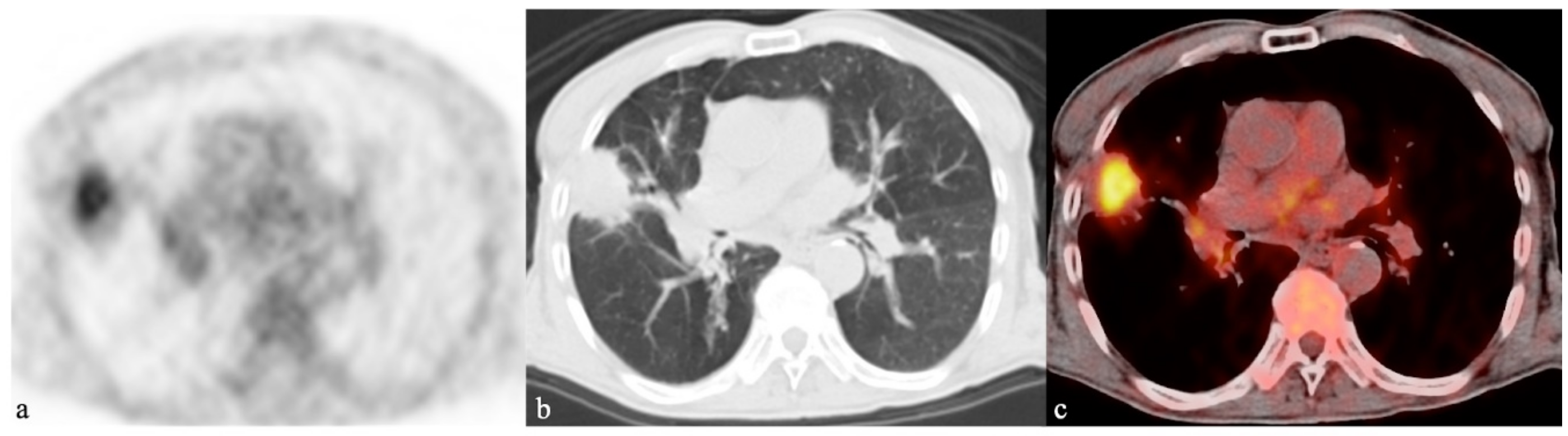
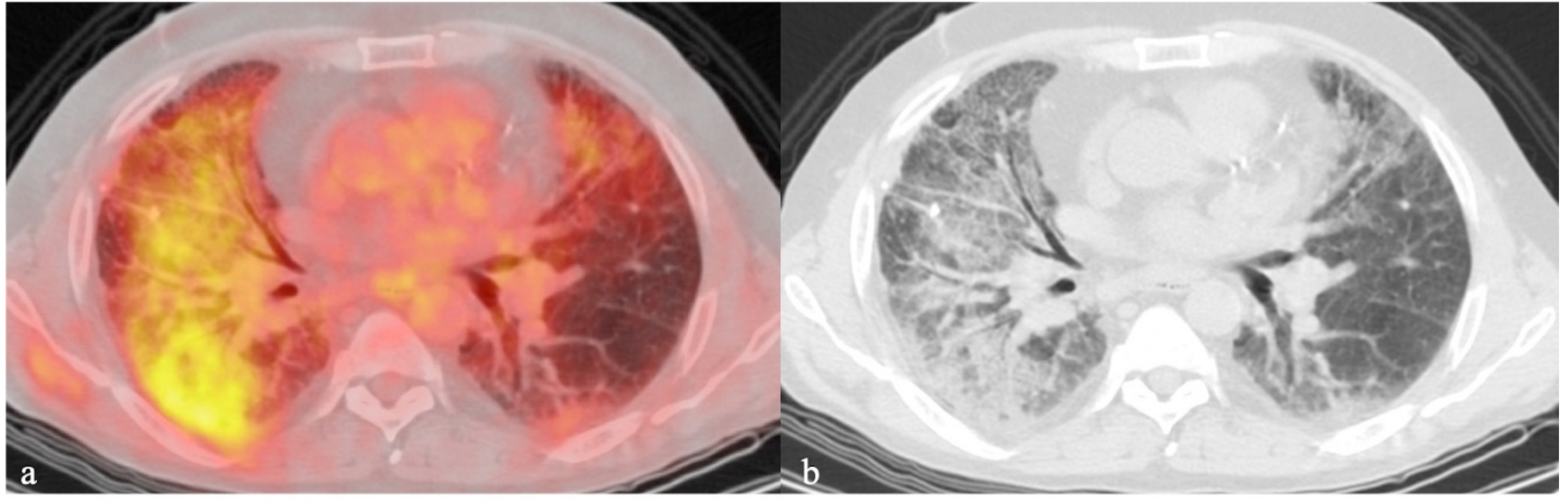
3.1. 18F-FDG
3.2. Other PET/CT Tracers
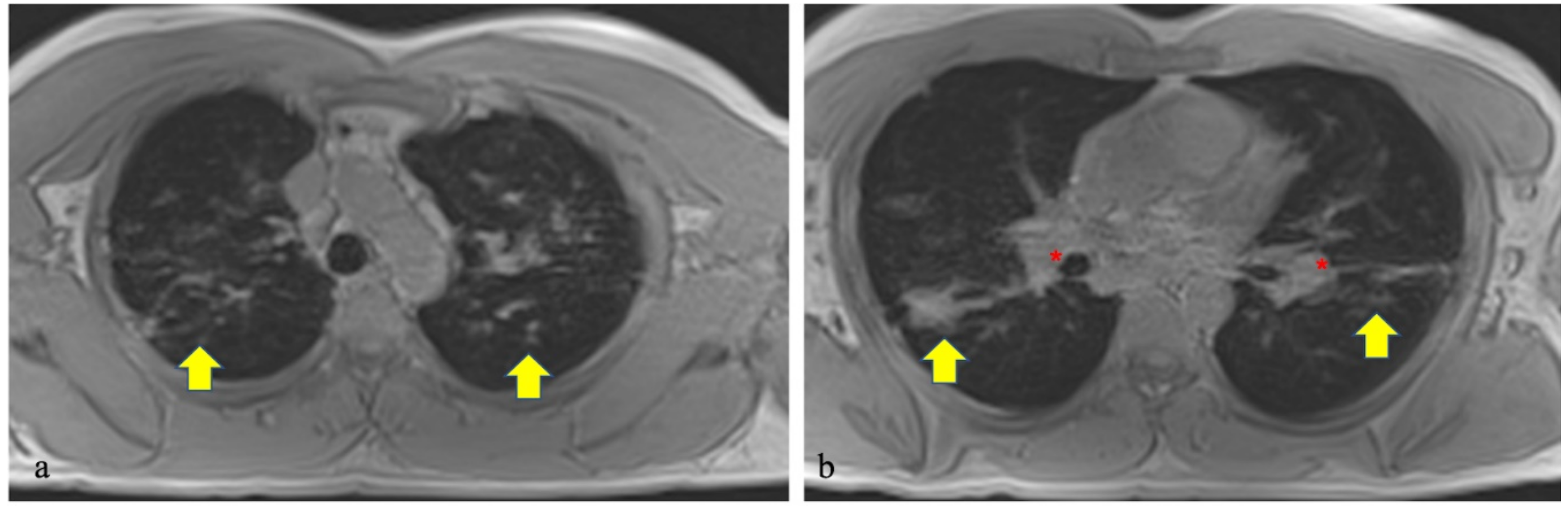
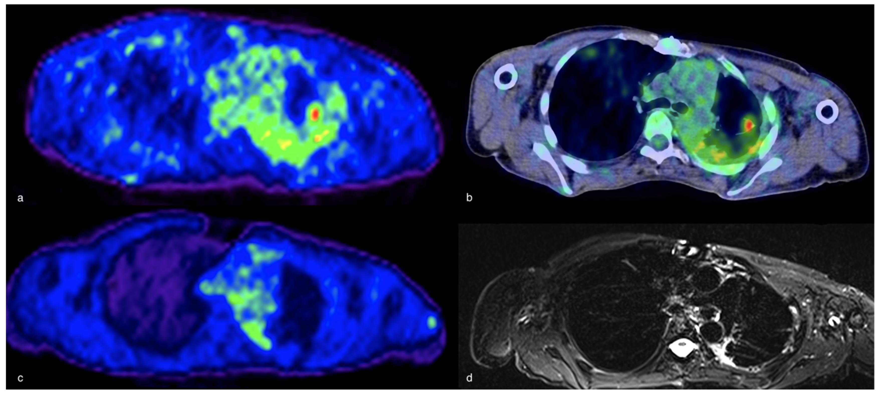
3.3. Magnetic Resonance Imaging (MRI) and PET/MRI
4. Future Perspectives
5. Conclusions
Author Contributions
Funding
Acknowledgments
Conflicts of Interest
References
- Marrie, T.J.; Huang, J.D. Epidemiology of community-acquired pneumonia in Edmonton, Alberta: an emergency department-based study. Can. Respir. J. 2005, 12, 139–142. [Google Scholar] [CrossRef] [PubMed] [Green Version]
- Ramirez, J.A.; Wiemken, T.L.; Peyrani, P.; Arnold, F.W.; Kelley, R.; Mattingly, W.A.; Nakamatsu, R.; Pena, S.; Guinn, B.E.; Furmanek, S.P.; et al. Adults Hospitalized With Pneumonia in the United States: Incidence, Epidemiology, and Mortality. Clin. Infect. Dis. 2017, 65, 1806–1812. [Google Scholar] [CrossRef] [PubMed] [Green Version]
- Torres, A.; Peetermans, W.E.; Viegi, G.; Blasi, F. Risk factors for community-acquired pneumonia in adults in Europe: a literature review. Thorax 2013, 68, 1057–1065. [Google Scholar] [CrossRef] [PubMed] [Green Version]
- Trotter, C.L.; Stuart, J.M.; George, R.; Miller, E. Increasing hospital admissions for pneumonia, England. Emerg. Infect. Dis. 2008, 14, 727–733. [Google Scholar] [CrossRef] [PubMed]
- Navaratnam, V.; Fleming, K.M.; West, J.; Smith, C.J.; Jenkins, R.G.; Fogarty, A.; Hubbars, R.B. The rising incidence of idiopathic pulmonary fibrosis in the UK. Thorax 2011, 66, 462–467. [Google Scholar] [CrossRef] [Green Version]
- Dimastromatteo, J.; Charles, E.J.; Laubach, L.E. Molecular imaging of pulmonary diseases. Resp. Res. 2018, 19, 1–17. [Google Scholar] [CrossRef] [Green Version]
- Kao, C.H.; Wang, S.J.; Liao, S.Q.; Lin, W.Y.; Hsu, C.Y. Usefulness of gallium-67-citrate scans in patients with acute disseminated tuberculosis and comparison with chest x-rays. J. Nucl. Med. 1993, 34, 1918–1921. [Google Scholar]
- Forstrom, L.A.; Loken, M.K.; Cook, A.; Chandler, R.; McCullough, J. In-111-labeled leukocytes in the diagnosis of rejection and cytomegalovirus infection in renal transplant patients. Clin. Nucl. Med. 1981, 6, 146–149. [Google Scholar] [CrossRef]
- Bar-Shalom, R.; Yefremov, N.; Guralnik, L.; Keidar, Z.; Engel, A.; Nitecki, S.; Israel, O. SPET/CT using 67Ga and 111In-labeled leukocyte scintigraphy for diagnosis of infection. J. Nucl. Med. 2006, 47, 587–594. [Google Scholar]
- Ingui, C.J.; Shah, N.P.; Oates, M.E. Infection scintigraphy: added value of single-photon emission computed tomography/computed tomography fusion compared with traditional analysis. J. Comput. Assist. Tomogr. 2007, 31, 375–380. [Google Scholar] [CrossRef]
- Grubstein, A.; Bernstine, H.; Steinmetz, A.P. Chest CT and gallium-67 SPET scintigraphy scan co-registration in a post-heart transplantation patient with unresolved fever. Isr. Med. Assoc. J. 2007, 9, 827–828. [Google Scholar] [PubMed]
- Bajaj, S.K.; Seitz, J.P.; Qing, F. Diagnosis of acute bacterial prostatitis by Ga-67 scintigraphy and SPET-CT. Clin. Nucl. Med. 2008, 33, 813–815. [Google Scholar] [CrossRef] [PubMed]
- Schuster, D.M.; Alazraki, N. Gallium and other agents in diseases of the lung. Semin. Nucl. Med. 2002, 32, 193–211. [Google Scholar] [CrossRef] [PubMed]
- Segall, G.M.; McDougall, I.R. Diagnostic vale of lung uptake of indium-111 oxine-labeled white blood cells. Am. J. Roentgenol. 1986, 147, 601–606. [Google Scholar] [CrossRef] [PubMed] [Green Version]
- Love, C.; Opoku-Agyemang, P.; Tomas, M.B.; Pugliese, P.V.; Bhargava, K.K.; Palestro, C.J. Pulmonary Activity on Labeled Leukocyte Images: Physiologic, Pathologic, and Imaging Correlation. Radiographics 2002, 22, 1385–1393. [Google Scholar] [CrossRef] [PubMed] [Green Version]
- Signore, A.; Jamar, F.; Israel, O.; Buscombe, J.; Martin-Comin, J.; Lazzeri, E. Clinical indications, image acquisition and data interpretation for white blood cells and anti-granulocyte monoclonal antibody scintigraphy: An EANM procedural guideline. Eur. J. Nucl. Med. Mol. Imaging. 2018, 10, 1816–1831. [Google Scholar] [CrossRef] [PubMed] [Green Version]
- McAfee, J.G.; Samin, A. In-111 labeled leukocytes: A review of problems in image interpretation. Radiology 1985, 155, 221–229. [Google Scholar] [CrossRef]
- Cook, P.S.; Datz, F.L.; Disbro, M.A.; Alazraki, N.P.; Taylor, A.T. Pulmonary uptake in indium-111 leukocyte imaging: clinical significance in patients with suSPETed occult infections. Radiology 1984, 150, 557–561. [Google Scholar] [CrossRef]
- Love, C.; Tomas, M.B.; Palestro, C.J. Pulmonary activity on labelled leukocyte images: Patterns of uptake and their significance. Nucl. Med. Commun. 2002, 23, 559–563. [Google Scholar] [CrossRef]
- Audi, S.H.; Clough, A.V.; Haworth, S.T.; Medhora, M.; Ranji, M.; Densmore, J.C.; Jacobs, E.R. 99MTc-Hexamethylpropyleneamine Oxime Imaging for Early Detection of Acute Lung Injury in Rats Exposed to Hyperoxia or Lipopolysaccharide Treatment. Shock 2016, 46, 420–430. [Google Scholar] [CrossRef]
- Altiay, G.; Çermik, T.F. 99mTc-HMPAO labelled white blood cell scintigraphy in the diagnosis and monitoring of response of the therapy in patients with active bronchiectasis. Rev. Esp. Med. Nucl. Imagen. Mol. 2012, 31, 9–14. [Google Scholar] [PubMed]
- Fineman, D.; Palestro, C.J.; Kim, C.K.; Needle, L.B.; Vallabhajosula, S.; Solomon, R.W.; Goldsmith, S.J. Detection of abnormalities in febrile AIDS patients with In-111-labeled leukocyte and GA-67 scintigraphy. Radiology 1989, 170, 677–680. [Google Scholar] [CrossRef] [PubMed]
- Palestro, C.J.; Schultz, B.L.; Horowitz, M.; Swyer, A.J. Indium-111-leukocyte and gallium-67 imaging in acute sarcoidosis: Report of two patients. J. Nucl. Med. 1992, 33, 2027–2029. [Google Scholar] [PubMed]
- Lavender, J.P.; Lowe, J.; Barker, J.R.; Burn, J.I.; Chaudhri, M.A. Gallium 67 citrate scanning in neoplastic and inflammatory lesions. Br. J. Radiol. 1971, 44, 361–366. [Google Scholar] [CrossRef] [PubMed]
- Hoffer, P.B.; Huberty, J.; Khazan-Bushi, H. The association of 67Ga and lactoferrin. J. Nucl. Med. 1977, 18, 713–717. [Google Scholar] [PubMed]
- Tsan, M.F.; Chen, W.Y.; Scheffel, U.; Menon, S.; Wagner, H. Mechanism of gallium localization in inflammatory lesions. J. Nucl. Med. 1977, 18, 619. [Google Scholar]
- Higashi, T.; Kobayashi, M.; Wakao, H.; Jinbu, Y. The relationship between 67Ga accumulation and ATP metabolism in tumor cells in vitro. Eur. J. Nucl. Med. 1989, 15, 152–156. [Google Scholar] [CrossRef] [PubMed]
- Turoglu, T.H.; Akisik, M.F.; Naddag, S.Y.; Omar, W.S.; Kempf, J.S.; Abdel-Dayem, H.M. Tumor and Infection Localization in AIDS Patients: Ga-67 and Tl-201 Findings. Clin. Nucl. Med. 1998, 23, 446–459. [Google Scholar] [CrossRef]
- Gambhir, S.; Ravina, M.; Rangan, K.; Dixit, M.; Barai, S.; Bomanji, J.; International Atomic Energy Agency Extra-pulmonary TB Consortium. Imaging in extrapulmonary tuberculosis. Int. J. Infect. Dis. 2017, 56, 237–247. [Google Scholar] [CrossRef] [Green Version]
- Siemsen, J.K.; Grebe, S.F.; Sargent, E.N.; Wentz, D. Gallium-67 scintigraphy of pulmonary diseases as a complement to radiography. Radiology 1976, 118, 371–375. [Google Scholar] [CrossRef]
- Siemsen, J.K.; Grebe, S.F.; Waxman, A.D. The use of gallium-67 in pulmonary disorders. Semin. Nucl. Med. 1978, 8, 235–249. [Google Scholar] [CrossRef]
- Walsh, T.J.; Bekerman, C.; Chausow, A.; Szidon, P. The value of gallium-67 scanning in pulmonary tuberculosis. Am. Rev. Respir. Dis. 1985, 132, 746–747. [Google Scholar] [PubMed]
- Santin, M.; Podzamczer, D.; Ricart, I.; Mascaro, J.; Ramon, J.M.; Dominguez, A.; Rufi, G.; Gudiol, F. Utility of the gallium-67 citrate scan for the early diagnosis of tuberculosis in patients infected with the human immunodeficiency virus. Clin. Infect. Dis. 1995, 20, 652–656. [Google Scholar] [CrossRef] [PubMed]
- Goldfarb, C.R.; Colp, C.; Ongseng, F.; Finestone, H.; Havas, J. Gallium scanning in the ‘new’ tuberculosis. Clin. Nucl. Med. 1997, 22, 470–474. [Google Scholar] [CrossRef] [PubMed]
- Lai, F.M.; Liam, C.K.; Paramsothy, M.; George, J. The role of 67gallium scintigraphy and high resolution computed tomography as predictors of disease activity in sputum smear-negative pulmonary tuberculosis. Int. J. Tuberc. Lung. Dis. 1997, 1, 563–569. [Google Scholar]
- Utsunomiya, K.; Narabayashi, I.; Nishigaki, H.; Tsujimoto, K.; Kariyone, S.; Ohnishi, S. Clinical significance of thallium-201 and gallium-67 scintigraphy in pulmonary tuberculosis. Eur. J. Nucl. Med. 1997, 24, 252–257. [Google Scholar]
- Ergün, P.; Turay, U.Y.; Ortapamuk, H.; Biber, C.; Keyf, I.A.; Çayan, C.; Erdoğan, Y. The role of gallium-67 scintigraphy and high resolution computed tomography as predictors of disease activity in sputum smear negative pulmonary tuberculosis. Turk. Respir. J. 2003, 4, 123–126. [Google Scholar]
- Sultana, S.; Buscombe, J.R.; Hilson, A.J. In pyrexia resulting from occult tuberculosis Ga-67 citrate is still more sensitive than newer agents such as Tc-99m selusomab. Clin. Nucl. Med. 2005, 30, 414–415. [Google Scholar] [CrossRef]
- Goswami, G.K.; Jana, S.; Santiago, J.F.; Buyukdereli, G.; Salem, S.S.; Heiba, B.; Abdel-Dayem, H.M. Discrepancy between Ga-67 citrate and F-I8 fluorodeoxyglucose positron emission tomographic scans in pulmonary infection. Clin. Nucl. Med. 2000, 25, 490–491. [Google Scholar] [CrossRef]
- Hsu, C.C.; Huang, Y.C.; Chuang, Y.W.; Lee, T.L.; Yeh, J.J. Association between gallium-67 uptake by lung foci and sputum smear status in patients with pulmonary tuberculosis. Nucl. Med. Commun. 2012, 33, 941–946. [Google Scholar] [CrossRef]
- Thakur, A.; Rodriguez-Rodriguez, C.; Saatchi, K.; Rose, F.; Esposito, T.; Nosrati, Z.; Andersen, P.; Christensen, D.; Hafeli, U.O.; Foged, C. Dual-Isotope SPET/CT Imaging of the Tuberculosis Subunit Vaccine H56/CAF01: Induction of Strong Systemic and Mucosal IgA and T-Cell Responses in Mice Upon Subcutaneous Prime and Intrapulmonary Boost Immunization. Front Immunol. 2018, 30, 2825. [Google Scholar]
- Rizzato, G.; Blasi, A. A European survey on the usefulness of 67Ga lung scans in assessing sarcoidosis. Experience in 14 research centers in seven different countries. Ann. N. Y. Acad. Sci. 1986, 465, 463–478. [Google Scholar] [CrossRef] [PubMed]
- Israel, H.L.; Albertine, K.H.; Park, C.H. Whole-body gallium 67 scans. Role in diagnosis of sarcoidosis. Am. Rev. Respir. Dis. 1991, 144, 1182–1186. [Google Scholar] [CrossRef] [PubMed]
- Sulavik, S.B.; Spencer, R.P.; Weed, D.A.; Shapiro, H.R.; Shiue, H.R.; Castriotta, R.J. Recognition of distinctive patterns of gallium-67 distribution in sarcoidosis. J. Nucl. Med. 1990, 31, 1909–1914. [Google Scholar] [PubMed]
- Sulavik, S.B.; Spencer, R.P.; Palestro, C.J.; Swyer, A.J.; Teirstein, A.S.; Goldsmith, S.J. Specificity and sensitivity of distinctive chest radiographic and/or 67Ga images in the noninvasive diagnosis of sarcoidosis. Chest 1993, 103, 403–409. [Google Scholar] [CrossRef] [PubMed]
- Gelb, A.F.; Dreisen, R.B.; Epstein, J.D.; Silverthorne, J.D.; Bickel, Y.; Fields, M.; Border, W.A.; Taylor, C.R. Immune complexes, gallium lung scans, and bronchoalveolar lavage in idiopathic interstitial pneumonitis-fibrosis. Chest 1983, 84, 148–153. [Google Scholar] [CrossRef]
- Baughman, R.P.; Fernandez, M. Radionuclide imaging in interstitial lung disease. Curr. Opin. Pulm. Med. 1996, 2, 376–379. [Google Scholar] [CrossRef]
- Schiff, R.G.; Kabat, L.; Kamani, N. Gallium scanning in lymphoid interstitial pneumonitis of children with AIDS. J. Nucl. Med. 1987, 28, 1915–1919. [Google Scholar]
- Nimkin, K.; Oates, E. Gallium-67 lung uptake in extrinsic hypersensitivity pneumonitis. Clin. Nucl. Med. 1989, 14, 451–452. [Google Scholar] [CrossRef]
- Brown, D.G.; Aguirre, A.; Weaver, A. 67Gallium scanning in talc-induced pulmonary granulomatosis. Chest 1980, 77, 561–565. [Google Scholar] [CrossRef]
- Hayes, A.A.; Thickbroom, G.W.; Guelfi, G.R.; Musk, A.W.; van der Schaaf, A.A. Computer quantitation of gallium 67 lung uptake in crocidolite (blue asbestos) workers of Western Australia. Eur. J. Nucl. Med. 1990, 16, 855–858. [Google Scholar] [CrossRef] [PubMed]
- Deseran, M.W.; Colletti, P.M.; Ratto, D.; Ansari, A.N.; Siegel, M.E. Chronic berylliosis. Demonstration by gallium-67 imaging and magnetic resonance imaging. Clin. Nucl. Med. 1988, 13, 509–511. [Google Scholar] [CrossRef] [PubMed]
- Kanner, R.E.; Barkman, H.W.; Rom, W.N.; Taylor, A.T., Jr. Gallium-67 citrate imaging in underground coal miners. Am. J. Ind. Med. 1985, 8, 49–55. [Google Scholar] [CrossRef] [PubMed]
- Lin, R.Y. Severe spirometric defects in systemic lupus erythematosus. A possible role for bronchoalveolar lavage and gallium scanning. Clin. Rheumatol. 1987, 6, 276–281. [Google Scholar] [CrossRef] [PubMed]
- Baron, M.; Feiglin, D.; Hyland, R.; Urowitz, M.B.; Shiff, B. 67Gallium scans in progressive systemic sclerosis. Arthritis Rheum. 1983, 26, 969–974. [Google Scholar] [CrossRef]
- Yeh, S.D.; White, D.A.; Stuver-Pepe, D.E.; Caravelli, J.F.; Van Uitert, C.; Benua, R.S. Abnormal gallium scintigraphy in pulmonary alveolar proteinosis (PAP). Clin. Nucl. Med. 1987, 12, 294–297. [Google Scholar] [CrossRef]
- Morals, J.; Carrier, L.; Gariepy, G.; Le Bel, L.; Chartrand, R.; Picard, D. Gallium-67 pulmonary uptake in eosinophilic pneumonia. Clin. Nucl. Med. 1988, 13, 41–43. [Google Scholar]
- Alpert, L.I. Pulmonary uptake of gallium-67 in Wegener’s granulomatusis. Clin. Nucl. Med. 1980, 5, 53–54. [Google Scholar] [CrossRef]
- Javaheri, S.; Levine, B.W.; McKusick, K.A. Serial 67Ga lung scanning in pulmonary eosinophilic granuloma. Thorax 1979, 34, 822–823. [Google Scholar] [CrossRef] [Green Version]
- Seabold, J.E.; Palestro, C.J.; Brown, M.L.; Datz, F.L.; Forstrom, L.A.; Greenspan, B.S.; McAfee, J.G.; Schauwecker, D.S.; Royal, H.D. Procedure guideline for gallium scintigraphy in inflammation. Society of Nuclear Medicine. J. Nucl. Med. 1997, 38, 994–997. [Google Scholar]
- Raziei, G.; Masjedi, M.R.; Fotouhi, F.; Asli, N.I.; Shafiei, B.; Javadi, H.; Assadi, M. The role of 99mTc-MIBI scintigraphy in the management of patients with pulmonary tuberculosis. Eur. Rev. Med. Pharm. Sci. 2012, 16, 622–629. [Google Scholar]
- Ahmadihosseini, H.; Sadeghi, R.; Zakavi, R.; Kakhki, V.R.; Kakhki, A.H. Application of technetium-99m-sestamibi in differentiation of active from inactive pulmonary tuberculosis using a single photon emission computed tomography method. Nucl. Med. Commun. 2008, 29, 690–694. [Google Scholar] [CrossRef] [PubMed]
- Sun, S.S.; Kao, C.H. Demonstration of skeletal tuberculosis with Tc-99m (V) DMSA scintigraphy and a negative Ga-67 scan. Clin. Nucl. Med. 2002, 27, 539. [Google Scholar] [CrossRef] [PubMed]
- Gulaldi, N.C.; Bayhan, H.; Ercan, M.T.; Kiber, M.; Oztürk, B.; Oğretensoy, M.; Bekdik, C.F. The visualization of pulmonary tuberculosis with Tc-99m (V) DMSA and Tc-99m citrate in comparison to Ga-67 citrate. Clin. Nucl. Med. 1995, 20, 1012–1014. [Google Scholar] [CrossRef]
- Ahmadihosseini, H.; Abedib, J.; Ghodsi Rad, M.A.; Zakavi, S.R.; Knoll, P.; Mirzaeic, S.; Sadeghi, R. Diagnostic utility of 99mTc-EDDA-tricine-HYNIC-Tyr3-octreotate SPET for differentiation of active from inactive pulmonary tuberculosis. Nucl. Med. Commun. 2014, 35, 1262–1267. [Google Scholar] [CrossRef]
- Monteiro, P.H.S.; de Souza, T.F.; Moretti, M.L.; Resende, M.R.; Mengatti, J.; de Lima, M.D.C.L.; Santos, A.O.; Ramos, C.D. SPET/CT with radiolabeled somatostatin analogues in the evaluation of systemic granulomatous infections. Radiol. Bras. 2017, 50, 378–382. [Google Scholar] [CrossRef] [Green Version]
- Kwekkeboom, D.J.; Krenning, E.P.; Kho, G.S.; Breman, W.A.; Van Hagen, P.M. Somatostatin receptor imaging in patients with sarcoidosis. Eur. J. Nucl. Med. 1998, 25, 1284–1292. [Google Scholar] [CrossRef]
- Lebtahi, R.; Crestani, B.; Belmatoug, N.; Daou, D.; Genin, R.; Dombret, M.C.; Palazzo, E.; Faraggi, M.; Aubier, M.; Le Guludec, D. Somatostatin receptor scintigraphy and gallium scintigraphy in patients with sarcoidosis. J. Nucl. Med. 2001, 42, 21–26. [Google Scholar]
- Nishiyama, Y.; Yamamoto, Y.; Fukunaga, K.; Takinami, H.; Iwado, Y.; Satoh, K.; Ohkawa, M. Comparative evaluation of 18F-FDG PET and 67Ga scintigraphy in patients with sarcoidosis. J. Nucl. Med. 2006, 47, 1571–1576. [Google Scholar]
- Prager, E.; Wehrschuetz, M.; Bisail, B.; Woltsche, M.; Schwarz, T.; Lanz, H.; Sorantin, E.; Aigner, R.M. Comparison of 18F-FDG and 67Ga-citrate in sarcoidosis imaging. Nuklearmedizin 2008, 47, 18–23. [Google Scholar]
- Keijsers, R.G.; Grutters, J.C.; Thomeer, M.; Du Bois, R.M.; Van Buul, M.M.; Lavalaye, J.; Van Den Bosch, J.M.; Verzijlbergen, F.J. Imaging the inflammatory activity of sarcoidosis: sensitivity and inter observer agreement of 67Ga imaging and 18F-FDG PET. Q. J. Nucl. Med. Mol. Imaging 2011, 55, 66–71. [Google Scholar] [PubMed]
- Kartamihardja, A.H.S.; Kurniawati, Y.; Gunawan, R. Diagnostic value of 99mTc-ethambutol scintigraphy in tuberculosis: compared to microbiological and histopathological tests. Ann. Nucl. Med. 2018, 32, 60–68. [Google Scholar] [CrossRef] [PubMed] [Green Version]
- Foss, C.A.; Kulik, L.; Ordonez, A.A.; Jain, S.K.; Michael Holers, V.; Thurman, J.M.; Pomper, M.G. SPET/CT Imaging of Mycobacterium tuberculosis Infection with [125I]anti-C3d mAb. Mol. Imaging Biol. 2019, 21, 473–481. [Google Scholar] [CrossRef] [PubMed]
- Zheng, L.; Ding, X.; Liu, K.; Feng, S.; Tang, B.; Li, Q.; Huang, D.; Yang, S. Molecular imaging of fibrosis using a novel collagen-binding peptide labelled with 99mTc on SPET/CT. Amino Acids 2017, 49, 89–101. [Google Scholar] [CrossRef]
- Britton, K.E.; Wareham, D.W.; Das, S.S.; Solanki, K.K.; Amaral, H.; Bhatnagar, A.; Katamihardja, A.H.S.; Malamitsi, J.; Moustafa, H.M.; Soroa, V.E.; et al. Imaging bacterial infection with (99m)Tc-ciprofloxacin (Infecton). J. Clin. Pathol. 2002, 55, 817–823. [Google Scholar] [CrossRef] [Green Version]
- Vinjamuri, S.; Hall, A.V.; Solanki, K.K.; Bomanji, J.B.; Siraj, Q.; O’Shaughnessy, E.M.; Das, S.S.; Britton, K.E. Comparison of99mTc-Infecton imaging with radiolabelled white-cell imaging in the evaluation of bacterial infection. Lancet 1996, 347, 233–235. [Google Scholar] [CrossRef]
- Auletta, S.; Varani, M.; Horvat, R.; Galli, F.; Signore, A.; Hess, S. PET Radiopharmaceuticals for Specific Bacteria Imaging: A Systematic Review. J. Clin. Med. 2019, 8, 197. [Google Scholar] [CrossRef] [Green Version]
- Akhtar, M.S.; Imran, M.B.; Nadeem, M.A.; Shahid, A. Antimicrobial Peptides as Infection Imaging Agents: Better Than Radiolabeled Antibiotics. Int. J. Pept. 2012, 2012, 1–19. [Google Scholar] [CrossRef] [Green Version]
- King, G.G.; Eberl, S.; Salome, C.M.; Meikle, S.R.; Woolcock, A.J. Airway closure measured by a technegas bolus and SPECT. Am. J. Respir. Crit. Care Med. 1997, 155, 682–688. [Google Scholar] [CrossRef]
- Farrow, C.E.; Salome, C.M.; Harris, B.E.; Bailey, D.L.; Bailey, E.; Berend, N.; Young, I.H.; King, G.G. Airway closure on imaging relates to airway hyperresponsiveness and peripheral airway disease in asthma. J. Appl. Physiol. 2012, 113, 958–966. [Google Scholar] [CrossRef] [Green Version]
- Farrow, C.E.; Salome, C.M.; Harris, B.E.; Bailey, D.L.; Berend, N.; King, G.G. Peripheral ventilation heterogeneity determines the extent of bronchoconstriction in asthma. J. Appl. Physiol. 2017, 123, 1188–1194. [Google Scholar] [CrossRef] [PubMed]
- Bajc, M.; Chen, Y.; Wang, J.; Li, X.Y.; Shen, W.M.; Wang, C.Z.; Huang, H.; Lindqvist, A.; He, X.Y. Identifying the heterogeneity of COPD by V/P SPECT: a new tool for improving the diagnosis of parenchymal defects and grading the severity of small airways disease. Int. J. Chron. Obs. Pulmon. Dis. 2017, 12, 1579–1587. [Google Scholar] [CrossRef] [PubMed] [Green Version]
- Karacavus, S.; Intepe, Y.S. The role of Tc-99m DTPA aerosol scintigraphy in the differential diagnosis of COPD and asthma. Clin. Respir. J. 2015, 9, 189–195. [Google Scholar] [CrossRef] [PubMed]
- Mochizuki, T.; Tsukamoto, E.; Kuge, Y.; Kanegae, K.; Zhao, S.; Hikosaka, K.; Hikosaka, M.; Hikosaka, M.; Tamaki, N. FDG uptake and glucose transporter subtype expressions in experimental tumor and inflammation models. J. Nucl. Med. 2001, 42, 1551–1555. [Google Scholar]
- Kubota, R.; Yamada, S.; Kubota, K.; Ishiwata, K.; Tamahashi, N.; Ido, T. Intratumoral distribution of fluorine-18-fluorodeoxyglucose in vivo: high accumulation in macrophages and granulation tissues studied by microautoradiography. J. Nucl. Med. 1992, 33, 1972–1980. [Google Scholar]
- Bell, G.I.; Burant, C.F.; Takeda, J.; Gould, G.W. Structure and function of mammalian facilitative sugar transporters. J. Biol. Chem. 1993, 268, 19161–19164. [Google Scholar]
- Pauwels, E.K.; Ribeiro, M.J.; Stoot, J.H.; McCready, V.R.; Bourguignon, M.; Mazière, B. FDG accumulation and tumor biology. Nucl. Med. Biol. 1998, 25, 317–322. [Google Scholar] [CrossRef] [Green Version]
- Zhuang, H.; Alavi, A. 18-fluorodeoxyglucose positron emission tomographic imaging in the detection and monitoring of infection and inflammation. Semin. Nucl. Med. 2002, 32, 47–59. [Google Scholar] [CrossRef]
- Paik, J.Y.; Lee, K.H.; Choe, Y.S.; Choi, Y.; Kim, B.T. Augmented 18F-FDG uptake in activated monocytes occurs during the priming process and involves tyrosine kinases and protein kinase C. J. Nucl. Med. 2004, 45, 124–128. [Google Scholar]
- Chang, J.M.; Lee, H.J.; Goo, J.M.; Lee, H.Y.; Lee, J.J.; Chung, J.K.; Im, J.G. False positive and false negative FDG-PET scans in various thoracic diseases. Korean J. Radiol. 2006, 7, 57–69. [Google Scholar] [CrossRef] [Green Version]
- Sathekge, M.M.; Maes, A.; Pottel, H.; Stoltz, A.; van de Wiele, C. Dual time-point FDG PET-CT for differentiating benign from malignant solitary pulmonary nodules in a TB endemic area. S. Afr. Med. J. 2010, 100, 598–601. [Google Scholar] [CrossRef] [PubMed] [Green Version]
- Chen, C.J.; Lee, B.F.; Yao, W.J.; Cheng, L.; Wu, P.S.; Chu, C.L.; Chiu, N.T. Dual-phase 18F-FDG PET in the diagnosis of pulmonary nodules with an initial standard uptake value less than 2.5. Am. J. Roentgenol. 2008, 191, 475–479. [Google Scholar] [CrossRef] [PubMed]
- Kaneko, K.; Sadashima, E.; Irie, K.; Hayashi, A.; Masunari, S.; Yoshida, T.; Omagari, J. Assessment of FDG retention differences between the FDG-avid benign pulmonary lesion and primary lung cancer using dual-time-point FDG-PET imaging. Ann. Nucl. Med. 2013, 27, 329–339. [Google Scholar] [CrossRef] [PubMed]
- Heysell, S.K.; Thomas, T.A.; Sifri, C.D.; Rehm, P.K.; Houpt, E.R. 18-Fluorodeoxyglucose positron emission tomography for tuberculosis diagnosis and management: A case series. BMC Pulm. Med. 2013, 21, 13–14. [Google Scholar] [CrossRef] [Green Version]
- Franzius, C.; Biermann, M.; Hülskamp, G.; Frosch, M.; Roth, J.; Sciuk, J.; Schober, O. Therapy monitoring in aspergillosis using F-18 FDG positron emission tomography. Clin. Nucl. Med. 2001, 26, 232–233. [Google Scholar] [CrossRef]
- Reuter, S.; Schirrmeister, H.; Kratzer, W.; Dreweck, C.; Reske, S.N.; Kern, P. Pericystic metabolic activity in alveolar echinococcosis: assessment and follow-up by positron emission tomography. Clin. Infect. Dis. 1999, 29, 1157–1163. [Google Scholar] [CrossRef]
- Reuter, S.; Buck, A.; Manfras, B.; Kratzer, W.; Seitz, H.M.; Darge, K.; Reske, S.N.; Kern, P. Structured treatment interruption in patients with alveolar echinococcosis. Hepatology 2004, 39, 509–517. [Google Scholar] [CrossRef]
- Stumpe, K.D.; Renner-Schneiter, E.C.; Kuenzle, A.K.; Grimm, F.; Kadry, Z.; Clavein, P.A.; Deplazes, P.; von Schulthess, G.K.; Muellhaupt, B.; Amman, R.W.; et al. F-18-fluorodeoxyglucose (FDG) positron-emission tomography of Echinococcus multilocularis liver lesions: proSPETive evaluation of its value for diagnosis and follow-up during benzimidazole therapy. Infect. 2007, 35, 11–18. [Google Scholar] [CrossRef]
- Bleeker-Rovers, C.P.; Warris, A.; Drenth, J.P.; Corstens, F.H.; Oyen, W.J.; Kullberg, B.J. Diagnosis of Candida lung abscesses by 18F-fluorodeoxyglucose positron emission tomography. Clin. Microbiol. Infect. 2005, 11, 493–495. [Google Scholar] [CrossRef] [Green Version]
- Glaudemans, A.W.; de Vries, E.F.; Galli, F.; Dierckx, R.A.; Slart, R.H.; Signore, A. The use of (18)F-FDG-PET/CT for diagnosis and treatment monitoring of inflammatory and infectious diseases. Clin. Dev. Immunol. 2013, 2013, 623036. [Google Scholar] [CrossRef] [Green Version]
- Mostard, R.L.; van Kroonenburgh, M.J.; Drent, M. The role of the PET scan in the management of sarcoidosis. Curr. Opin. Pulm. Med. 2013, 19, 538–544. [Google Scholar] [CrossRef] [PubMed]
- Teirstein, A.S.; Machac, J.; Almeida, O.; Lu, P.; Padilla, M.L.; Iannuzzi, M.C. Results of 188 whole-body fluorodeoxyglucose positron emission tomography scans in 137 patients with sarcoidosis. Chest 2007, 132, 1949–1953. [Google Scholar] [CrossRef] [PubMed]
- Coker, R.K. Management strategies for pulmonary sarcoidosis. Clin. Risk. Manag. 2009, 5, 575–584. [Google Scholar] [CrossRef] [PubMed] [Green Version]
- Klein, M.; Cohen-Cymberknoh, M.; Armoni, S.; Shoseyov, D.; Chisin, R.; Orevi, M.; Freedman, N.; Kerem, E. 18F-fluorodeoxyglucose-PET/CT imaging of lungs in patients with cystic fibrosis. Chest 2009, 136, 1220–1228. [Google Scholar] [CrossRef]
- Amin, R.; Charron, M.; Grinblat, L.; Shammas, A.; Grasemann, H.; Graniel, K.; Ciet, P.; Tiddens, H.; Ratjen, F. Cystic fibrosis: Detecting changes in airway inflammation with FDG PET/CT. Radiology 2012, 264, 868–875. [Google Scholar] [CrossRef]
- Cremers, J.P.; Van Kroonenburgh, M.J.; Mostard, R.L.; Vöö, S.A.; Wijnen, P.A.; Koek, G.H.; Drent, M. Extent of disease activity assessed by 18F-FDG PET/CT in a Dutch sarcoidosis population. Sarcoidosis Vasc. Diffus. Lung Dis. 2014, 31, 37–45. [Google Scholar]
- Dolovich, M.B.; Schuster, D.P. Positron emission tomography and computed tomography versus positron emission tomography computed tomography: tools for imaging the lung. Proc. Am. Thorax Soc. 2007, 4, 328–333. [Google Scholar] [CrossRef]
- Groves, A.M.; Win, T.; Screaton, N.J.; Berovic, M.; Endozo, R.; Booth, H.; Kayani, I.; Menezes, L.J.; Dickson, J.C.; Ell, P.J. Idiopathic pulmonary fibrosis and diffuse parenchymal lung disease: implications from initial experience with 18F-FDG PET/CT. J. Nucl. Med. 2009, 50, 538–545. [Google Scholar] [CrossRef] [Green Version]
- Chen, D.L.; Wang, X.; Yamamoto, S.; Carpenter, D.; Engle, J.T.; Li, W.; Lin, X.; Kreisel, D.; Krupnick, A.S.; Huang, H.J.; et al. Increased T cell glucose uptake reflects acute rejection in lung grafts. Am. J. Transpl. 2013, 13, 2540–2549. [Google Scholar] [CrossRef] [Green Version]
- Kothekar, E.; Borja, A.J.; Gerke, O.; Werner, T.J.; Alavi, A.; Revheim, M.E. Assessing respiratory muscle activity with 18F-FDG-PET/CT in patients with COPD. Am. J. Nucl. Med. Mol. Imaging. 2019, 9, 309–315. [Google Scholar]
- Braune, J.; Hofheinz, F.; Bluth, T.; Kiss, T.; Wittenstein, J.; Scharffenberg, M.; Kotzerke, J.; Gama de Abreu, M. Comparison of static 18F-FDG-PET/CT (SUV, SUR) and dynamic 18F-FDG-PET/CT (Ki) for quantification of pulmonary inflammation in acute lung injury. J. Nucl. Med. 2019, 60, 1629–1634. [Google Scholar] [CrossRef] [PubMed]
- Verleden, S.E.; Gheysens, O.; Goffin, K.E.; Vanaudenaerde, B.M.; Verbeken, E.K.; Weynand, B.; Van Raemdonck, D.E.; Verleden, G.M.; Vos, R. Role of 18F-FDG PET/CT in restrictive allograft syndrome after lung transplantation. Transplantation 2019, 103, 823–831. [Google Scholar] [CrossRef]
- Hara, T.; Kosaka, N.; Suzuki, T.; Kudo, K.; Niino, H. Uptake rates of 18F-fluorodeoxyglucose and 11C-choline in lung cancer and pulmonary tuberculosis: A positron tomography study. Chest 2003, 124, 893–901. [Google Scholar] [CrossRef] [PubMed]
- Liu, Q.; Peng, Z.M.; Liu, Q.W.; Yao, S.Z.; Zhang, L.; Meng, L.; Chen, J.H. The role of 11C-choline positron emission tomography– computed tomography and videomediastinoscopy in the evaluation of diseases of middle mediastinum. Chin. Med. J. 2006, 119, 634–639. [Google Scholar] [CrossRef] [PubMed]
- Hara, T.; Inagaki, K.; Kosaka, N.; Morita, T. Sensitive detection of mediastinal lymph node metastasis of lung cancer with 11C-choline PET. J. Nucl. Med. 2000, 41, 1507–1513. [Google Scholar]
- Norikane, T.; Yamamoto, Y.; Maeda, Y.; Noma, T.; Dobashi, H.; Nishiyama, Y. Comparative evaluation of 18F-FLT and 18F-FDG for detecting cardiac and extra-cardiac thoracic involvement in patients with newly diagnosed sarcoidosis. Ejnmmi Res. 2017, 7, 69. [Google Scholar] [CrossRef]
- Win, T.; Screaton, N.J.; Porter, J.; Endozo, R.; Wild, D.; Kayani, I.; Dickson, J.; Shortman, R.I.; Reubi, J.C.; Ell, P.J.; et al. Novel positron emission tomography/computed tomography of diffuse parenchymal lung disease combining a labeled somatostatin receptor analogue and 2-deoxy-2[18F]fluoro-D-glucose. Mol. Imaging 2012, 11, 91–98. [Google Scholar] [CrossRef]
- Ordenez, A.A.; DeMarco, V.P.; Klunk, M.H.; Pokkali, S.; Jain, S.K. Imaging chronic tuberculosis lesions using sodium [18F] Fluoride positron emission tomography in mice. Mol. Imaging. Biol. 2015, 17, 609–614. [Google Scholar] [CrossRef] [Green Version]
- Mirsadraee, S.; Marin, A.; Jenkins, W.; Connell, M.; Tavares, A.; Flecher, A.; Hirani, N.; van Beek, E.J. The identification of systemic integrin activation in idiopathic and systemic sclerosis pulmonary fibrosis using 18F-fluciclatide positron emission tomography. Insights Imaging 2016, 7, S424. [Google Scholar]
- Knudsen, L.; Atochina-Vasserman, E.N.; Massa, C.B.; Birkelbach, B.; Guo, C.J.; Scott, P.; Haenni, B.; Beers, M.F.; Ochs, M.; Gow, A.J. The role of inducible nitric oxide synthase for interstitial remodeling of alveolar septa in surfactant protein D-deficient mice. Am. J. Physiol. Lung Cell. Mol. Physiol. 2015, 309, 959–969. [Google Scholar] [CrossRef] [Green Version]
- Zhou, D.; Lee, H.; Rothfuss, J.M.; Chen, D.L.; Ponde, D.E.; Welch, M.J.; March, R.H. Design and synthesis of 2-amino-4-methylpyridine analogues as inhibitors for inducible nitric oxide synthase and in vivo evaluation of [18F]6-(2-fluoropropyl)-4-methyl-pyridin-2-amine as a potential PET tracer for inducible nitric oxide synthase. J. Med. Chem. 2009, 52, 2443–2453. [Google Scholar] [CrossRef] [PubMed] [Green Version]
- Huang, H.L.; Isakow, W.; Byers, D.E.; Engle, J.T.; Griffin, E.A.; Kemp, D.; Brody, S.L.; Gropler, R.J.; Miller, J.P.; Chu, W.; et al. Imaging pulmonary inducible nitric oxide synthase expression with PET. J. Nucl. Med. 2015, 56, 76–81. [Google Scholar] [CrossRef] [PubMed] [Green Version]
- Ebenhan., T.; Mokaleng, B.; Venter, J.; Kruger, H.; Zeevaart, J.; Sathekge, M. Preclinical assessment of a 68Ga-DOTA-functionalized depsipeptide as a radiodiagnostic infection imaging agent. Molecules 2017, 22, 1403. [Google Scholar] [CrossRef] [PubMed] [Green Version]
- Mukherjee, A.; Bhatt, J.; Shinto, A.; Korde, A.; Kumar, M.; Kamaleshwaran, K.; Joseph, J.; Sarma, H.D.; Dash, A. 68Ga-NOTA-ubiquicidin fragment for PET imaging of infection: From bench to bedside. J. Pharm. Biomed. Anal. 2018, 159, 245–251. [Google Scholar] [CrossRef] [PubMed]
- Ebenhan, T.; Zeevaart, J.R.; Venter, J.D.; Govender, T.; Kruger, G.H.; Jarvis, N.V.; Sathekge, M. Preclinical evaluation of 68Ga-labeled 1,4,7-Triazacyclononane-1,4,7-Triacetic acid-ubiquicidin as a radioligand for PET infection imaging. J. Nucl. Med. 2014, 55, 308–314. [Google Scholar] [CrossRef] [PubMed] [Green Version]
- Kapur, S.; Bhalla, A.S.; Jana, M. Pediatric Chest MRI: A Review. Indian J. Pediatr. 2019, 86, 842–853. [Google Scholar] [CrossRef]
- Richard, J.-C.; Chen, D.L.; Ferkol, T.; Schuster, D.P. Molecular imaging for pediatric lung diseases. Pediatr. Pulmonol. 2004, 37, 286–296. [Google Scholar] [CrossRef]
- Syrjala, H.; Broas, M.; Ohtonen, P.; Jartti, A.; Pääkkö, E. Chest magnetic resonance imaging for pneumonia diagnosis in outpatients with lower respiratory tract infection. Eur. Respir. J. 2016, 49, 1601303. [Google Scholar] [CrossRef] [Green Version]
- Hirsch, W.; Sorge, I.; Krohmer, S.; Weber, D.; Meier, K.; Till, H. MRI of the lungs in children. Eur. J. Radiol. 2008, 68, 278–288. [Google Scholar] [CrossRef]
- Poelmans, J.; Hillen, A.; Vanherp, L.; Govaerts, K.; Maertens, J.; Dresselaers, T.; Himmelreich, U.; Lagrou, K.; Vande Velde, G. Longitudinal, in vivo assessment of invasive pulmonary aspergillosis in mice by computed tomography and magnetic resonance imaging. Lab. Investig. 2016, 96, 692–704. [Google Scholar] [CrossRef]
- Tournebize, R.; Doan, B.-T.; Dillies, M.-A.; Maurin, S.; Beloeil, J.-C.; Sansonetti, P.J. Magnetic resonance imaging of Klebsiella pneumoniae-induced pneumonia in mice. Cell. Microbiol. 2006, 8, 33–43. [Google Scholar] [CrossRef] [PubMed]
- Marzola, P.; Lanzoni, A.; Nicolato, E.; Di Modugno, V.; Cristofori, P.; Osculati, F.; Sbarbati, A. 1H MRI of pneumococcal pneumonia in a murine model. J. Magn. Reson. Imaging 2005, 22, 170–174. [Google Scholar] [CrossRef] [PubMed]
- Hoerr, V.; Faber, C. Magnetic resonance imaging characterization of microbial infections. J. Pharm. Biomed. Anal. 2014, 93, 136–146. [Google Scholar] [CrossRef] [PubMed] [Green Version]
- Asanuma, T.; Kawahara, T.; Inanami, O.; Nakao, M.; Nakaya, K.; Ito, A.; Takiguchi, M.; Hashimoto, A.; Kuwabara, M. Magnetic resonance imaging of alveolar echinococcosis experimentally induced in the rat lung. J. Vet. Med. Sci. 2006, 68, 15–20. [Google Scholar] [CrossRef] [Green Version]
- Mc Fadden, R.G.; Carr, T.J.; Mackie, I.D. Thoracic magnetic resonance imaging in the evaluation of HIV-1/AIDS pneumonitis. Chest 1992, 101, 371–374. [Google Scholar] [CrossRef]
- Kuhlman, J.E. Pneumocystic infections: the radiologist’s perSPETive. Radiology 1996, 198, 623–635. [Google Scholar] [CrossRef]
- Nishimura, A.; Yamaguchi, H.; Ito, Y.; Tokumoto, S.; Toyoshima, D.; Kasai, M.; Maruyama, A. Empyema necessitatis due to Pseudomonas aeruginosa in a child with cerebral palsy. J. Infect. Public Health 2019, 13, 140–142. [Google Scholar] [CrossRef]
- Liszewski, M.C.; Görkem, S.; Sodhi, K.S.; Lee, E.Y. Lung magnetic resonance imaging for pneumonia in children. Pediatr. Radiol. 2017, 47, 1420–1430. [Google Scholar] [CrossRef]
- Ekinci, A.; Yucel Ucarkus, T.; Okur, A.; Ozturk, M.; Dogan, S. MRI of pneumonia in immunocompromised patients: comparison with CT. Diagn. Interv. Radiol. 2017, 23, 22–28. [Google Scholar] [CrossRef]
- Ozcan, H.N.; Gormez, A.; Ozsurekci, Y.; Karakaya, J.; Oguz, B.; Unal, S.; Haliloglu, M. Magnetic resonance imaging of pulmonary infection in immunocompromised children: Comparison with multidetector computed tomography. Pediatric Radiol. 2016, 47, 146–153. [Google Scholar] [CrossRef]
- Yan, C.; Tan, X.; Wei, Q.; Feng, R.; Li, C.; Wu, Y.; Xu, Y. Lung MRI of invasive fungal infection at 3 Tesla: Evaluation of five different pulse sequences and comparison with multidetector computed tomography (MDCT). Eur. Radiol. 2014, 25, 550–557. [Google Scholar] [CrossRef] [PubMed]
- Yan, C.; Xu, J.; Xiong, W.; Wei, Q.; Feng, R.; Wu, Y.; Xu, Y. Use of intravoxel incoherent motion diffusion-weighted MR imaging for assessment of treatment response to invasive fungal infection in the lung. Eur. Radiol. 2016, 27, 212–221. [Google Scholar] [CrossRef] [PubMed]
- Ohno, Y.; Hatabu, H.; Takenaka, D.; Adachi, S.; Kono, M.; Sugimura, K. Solitary Pulmonary Nodules: Potential Role of Dynamic MR Imaging in Management—Initial Experience. Radiology 2002, 224, 503–511. [Google Scholar] [CrossRef] [PubMed]
- Chung, M.H.; Lee, H.G.; Kwon, S.S.; Park, S.H. MR imaging of solitary pulmonary lesion: Emphasis on tuberculomas and comparison with tumors. J. Magn. Reson. Imaging 2000, 11, 629–637. [Google Scholar] [CrossRef]
- Chung, J.H.; Huitt, G.; Yagihashi, K.; Hobbs, S.B.; Faino, A.V.; Bolster, B.D.; Lynch, D.A. Proton Magnetic Resonance Imaging for Initial Assessment of Isolated Mycobacterium avium Complex Pneumonia. Ann. Am. Thorac. Soc. 2016, 13, 49–57. [Google Scholar] [CrossRef]
- Blum, U.; Windfuhr, M.; Buitrago-Tellez, C.; Sigmund, G.; Herbst, E.W.; Langer, M. Invasive Pulmonary Aspergillosis. Chest 1994, 106, 1156–1161. [Google Scholar] [CrossRef] [Green Version]
- Sodhi, K.S.; Gupta, P.; Shrivastav, A.; Saxena, A.K.; Mathew, J.L.; Singh, M.; Agarwal, R. Evaluation of 3 T lung magnetic resonance imaging in children with allergic bronchopulmonary aspergillosis: Pilot study. Eur. J. Radiol. 2019, 111, 88–92. [Google Scholar] [CrossRef]
- Moore, E.; Webb, W.; Muller, N.; Sollitto, R. MRI of pulmonary airspace disease: experimental model and preliminary clinical results. Am. J. Roentgenol. 1986, 146, 1123–1128. [Google Scholar] [CrossRef] [Green Version]
- Cohen, M.D.; Scales, R.L.; Eigen, H.; Scott, P.; Tepper, R.; Cory, D.A.; Smith, J.A. Evaluation of pulmonary parenchymal disease by magnetic resonance imaging. Br. J. Radiol. 1987, 60, 223–230. [Google Scholar] [CrossRef]
- Montella, S.; Maglione, M.; Bruzzese, D.; Mollica, C.; Pignata, C.; Aloj, G.; Santamaria, F. Magnetic resonance imaging is an accurate and reliable method to evaluate non-cystic fibrosis paediatric lung disease. Respirology 2011, 17, 87–91. [Google Scholar] [CrossRef]
- Owrangi, A.M.; Parraga, G. Chest MRI in children: Why bother? Respirology 2011, 17, 3–4. [Google Scholar] [CrossRef] [PubMed]
- Jacob, R.E.; Amidan, B.G.; Soelberg, J.; Minard, K.R. In vivo MRI of altered proton signal intensity and T2 relaxation in a bleomycin model of pulmonary inflammation and fibrosis. J. Magn. Reson. Imaging 2010, 31, 1091–1099. [Google Scholar] [CrossRef] [PubMed]
- Yi, C.A.; Lee, K.S.; Han, J.; Chung, M.P.; Chung, M.J.; Shin, K.M. 3-T MRI for Differentiating Inflammation- and Fibrosis-Predominant Lesions of Usual and Nonspecific Interstitial Pneumonia: Comparison Study with Pathologic Correlation. Am. J. Roentgenol. 2008, 190, 878–885. [Google Scholar] [CrossRef] [PubMed]
- Wallace, W.A.; Fitch, P.M.; Simpson, A.J.; Howie, S.E. Inflammation-associated remodelling and fibrosis in the lung – a process and an end point. Int. J. Exp. Pathol. 2007, 88, 103–110. [Google Scholar] [CrossRef] [PubMed] [Green Version]
- Romei, C.; Turturici, L.; Tavanti, L.; Miedema, J.; Fiorini, S.; Marletta, M.; Ciet, P. The use of chest magnetic resonance imaging in interstitial lung disease: a systematic review. Eur. Respir. Rev. 2018, 27, 180062. [Google Scholar] [CrossRef] [Green Version]
- Lutterbey, G.; Grohé, C.; Gieseke, J.; von Falkenhausen, M.; Morakkabati, N.; Wattjes, M.P.; Schild, H.H. Initial experience with lung-MRI at 3.0T: Comparison with CT and clinical data in the evaluation of interstitial lung disease activity. Eur. J. Radiol. 2007, 61, 256–261. [Google Scholar] [CrossRef]
- Cutillo, A.G.; Morris, A.H.; Ailion, D.C.; Durney, C.H. Clinical Implications of Nuclear Magnetic Resonance Lung Research. Chest 1989, 96, 643–652. [Google Scholar] [CrossRef]
- Buzan, M.T.A.; Eichinger, M.; Kreuter, M.; Kauczor, H.-U.; Herth, F.J.; Warth, A.; Dinkel, J. T2 mapping of CT remodelling patterns in interstitial lung disease. Eur. Radiol. 2015, 25, 3167–3174. [Google Scholar] [CrossRef]
- Müller, C.d.S.; Warszawiak, D.; Paiva, E.d.S.; Escuissato, D.L. Pulmonary magnetic resonance imaging is similar to chest tomography in detecting inflammation in patients with systemic sclerosis. Rev. Bras. Reumatol. 2017, 57, 419–424. [Google Scholar] [CrossRef]
- Buzan, M.T.A.; Wetscherek, A.; Heussel, C.P.; Kreuter, M.; Herth, F.J.; Warth, A.; Dinkel, J. Texture analysis using proton density and T2 relaxation in patients with histological usual interstitial pneumonia (UIP) or nonspecific interstitial pneumonia (NSIP). PloS ONE 2017, 12, e0177689. [Google Scholar] [CrossRef]
- Gaeta, M.; Blandino, A.; Scribano, E.; Minutoli, F.; Barone, M.; Andò, F.; Pandolfo, I. Chronic Infiltrative Lung Diseases. Chest 2000, 117, 1173–1178. [Google Scholar] [CrossRef] [PubMed] [Green Version]
- Bianchi, A.; Ozier, A.; Ousova, O.; Raffard, G.; Crémillieux, Y. Ultrashort-TE MRI longitudinal study and characterization of a chronic model of asthma in mice: Inflammation and bronchial remodeling assessment. NMR Biomed. 2013, 26, 1451–1459. [Google Scholar] [CrossRef] [PubMed]
- Caravan, P.; Yang, Y.; Zachariah, R.; Schmitt, A.; Mino-Kenudson, M.; Chen, H.H.; Lanuti, M. Molecular Magnetic Resonance Imaging of Pulmonary Fibrosis in Mice. Am. J. Respir. Cell Mol. Biol. 2013, 49, 1120–1126. [Google Scholar] [CrossRef] [PubMed] [Green Version]
- Vogel-Claussen, J.; Renne, J.; Hinrichs, J.; Schönfeld, C.; Gutberlet, M.; Schaumann, F.; Hohlfeld, J.M. Quantification of Pulmonary Inflammation after Segmental Allergen Challenge Using Turbo-Inversion Recovery-Magnitude Magnetic Resonance Imaging. Am. J. Respir. Crit. Care Med. 2014, 189, 650–657. [Google Scholar] [CrossRef]
- Evans, R.L.; Changani, K.K.; Hotee, S.; Pindoria, K.; Campbell, S.; Nials, A.T.; Kidd, E.J. Pulmonary edema measured by MRI correlates with late-phase response to allergen challenge. Exp. Lung Res. 2015, 41, 189–198. [Google Scholar] [CrossRef]
- Quintana, H.K.; Cannet, C.; Schaeublin, E.; Zurbruegg, S.; Sugar, R.; Mazzoni, L.; Beckmann, N. Identification with MRI of the pleura as a major site of the acute inflammatory effects induced by ovalbumin and endotoxin challenge in the airways of the rat. Am. J. Physiol. Lung Cell. Mol. Physiol. 2006, 291, L651–L657. [Google Scholar] [CrossRef] [Green Version]
- Renne, J.; Hinrichs, J.; Schönfeld, C.; Gutberlet, M.; Winkler, C.; Faulenbach, C.; Vogel-Claussen, J. Noninvasive Quantification of Airway Inflammation Following Segmental Allergen Challenge with Functional MR Imaging: A Proof of Concept Study. Radiology 2015, 274, 267–275. [Google Scholar] [CrossRef] [Green Version]
- Castro, M.; Fain, S.B.; Hoffman, E.A.; Gierada, D.S.; Erzurum, S.C.; Wenzel, S. Lung imaging in asthmatic patients: The picture is clearer. J. Allergy Clin. Immunol. 2011, 128, 467–478. [Google Scholar] [CrossRef] [Green Version]
- Aysola, R.; de Lange, E.E.; Castro, M.; Altes, T.A. Demonstration of the heterogeneous distribution of asthma in the lungs using CT and hyperpolarized helium-3 MRI. J. Magn. Reson. Imaging 2010, 32, 1379–1387. [Google Scholar] [CrossRef]
- Haczku, A.; Emami, K.; Fischer, M.C.; Kadlecek, S.; Ishii, M.; Panettieri, R.A.; Rizi, R.R. Hyperpolarized 3He MRI in Asthma. Acad. Radiol. 2005, 12, 1362–1370. [Google Scholar] [CrossRef]
- Fiel, S.B.; Friedman, A.C.; Caroline, D.F.; Radecki, P.D.; Faerber, E.; Grumbach, K. Magnetic Resonance Imaging in Young Adults with Cystic Fibrosis. Chest 1987, 91, 181–184. [Google Scholar] [CrossRef] [PubMed]
- Biederer, J.; Mirsadraee, S.; Beer, M.; Molinari, F.; Hintze, C.; Bauman, G.; Puderbach, M. MRI of the lung (3/3)—current applications and future perSPETives. Insights Imaging 2012, 3, 373–386. [Google Scholar] [CrossRef] [PubMed] [Green Version]
- Wielpütz, M.O.; Heußel, C.P.; Herth, F.J.F.; Kauczor, H.-U. Radiological Diagnosis in Lung Disease. Dtsch. Aerzteblatt Online 2014, 111, 181–187. [Google Scholar] [CrossRef] [PubMed] [Green Version]
- Ciet, P.; Serra, G.; Andrinopoulou, E.R.; Bertolo, S.; Ros, M.; Catalano, C.; Morana, G. Diffusion weighted imaging in cystic fibrosis disease: beyond morphological imaging. Eur. Radiol. 2016, 26, 3830–3839. [Google Scholar] [CrossRef] [PubMed]
- Ciet, P.; Bertolo, S.; Ros, M.; Andrinopoulou, E.R.; Tavano, V.; Lucca, F.; Morana, G. Detection and monitoring of lung inflammation in cystic fibrosis during respiratory tract exacerbation using diffusion-weighted magnetic resonance imaging. Eur. Respir. J. 2017, 50, 1601437. [Google Scholar] [CrossRef] [Green Version]
- Tiddens, H.A.W.M.; Stick, S.M.; Wild, J.M.; Ciet, P.; Parker, G.J.M.; Koch, A.; Vogel-Claussen, J. Respiratory tract exacerbations revisited: Ventilation, inflammation, perfusion, and structure (VIPS) monitoring to redefine treatment. Pediatr. Pulmonol. 2015, 50, S57–S65. [Google Scholar] [CrossRef] [Green Version]
- Baues, M.; Dasgupta, A.; Ehling, J.; Prakash, J.; Boor, P.; Tacke, F.; Kiessling, F.; Lammers, T. Fibrosis imaging: Current concepts and future directions. Adv. Drug. Deliv. Rev. 2017, 121, 9–26. [Google Scholar] [CrossRef]
- Toma, P.; Bertaina, A.; Castagnola, E.; Colafati, G.S.; D’Andrea, M.L.; Finocchi, A.; Granata, C. Fungal infections of the lung in children. Pediatr. Radiol. 2016, 46, 1856–1865. [Google Scholar] [CrossRef]
- Sodhi, K.S.; Khandelwal, N.; Saxena, A.K.; Singh, M.; Agarwal, R.; Bhatia, A.; Lee, E.Y. Rapid lung MRI in children with pulmonary infections: Time to change our diagnostic algorithms. J. Magn. Reson. Imaging 2016, 43, 1196–1206. [Google Scholar] [CrossRef]
- Bray, M.; Lawler, J.; Paragas, J.; Jahrling, P.B.; Mollura, D.J. Molecular Imaging of Influenza and Other Emerging Respiratory Viral Infections. J. Infect. Dis. 2011, 203, 1348–1359. [Google Scholar] [CrossRef] [Green Version]
- Peltola, V.; Ruuskanen, O.; Svedström, E. Magnetic resonance imaging of lung infections in children. Pediatr. Radiol. 2008, 38, 1225–1231. [Google Scholar] [CrossRef] [PubMed]
- Rieger, C.; Herzog, P.; Eibel, R.; Fiegl, M.; Ostermann, H. Pulmonary MRI—a new approach for the evaluation of febrile neutropenic patients with malignancies. Support. Care Cancer 2007, 16, 599–606. [Google Scholar] [CrossRef] [PubMed]
- Sodhi, K.S.; Bhatia, A.; Khandelwal, N. Rapid lung magnetic resonance imaging in children with pulmonary infection. Pediatr. Radiol. 2017, 47, 764–765. [Google Scholar] [CrossRef] [PubMed]
- Serra, G.; Milito, C.; Mitrevski, M.; Granata, G.; Martini, H.; Pesce, A.M.; Quinti, I. Lung MRI as a Possible Alternative to CT Scan for Patients With Primary Immune Deficiencies and Increased Radiosensitivity. Chest 2011, 140, 1581–1589. [Google Scholar] [CrossRef] [PubMed]
- Heusch, P.; Köhler, J.; Wittsack, H.J.; Heusner, T.A.; Buchbender, C.; Poeppel, T.D.; Nensa, F.; Wetter, A.; Gauler, T.; Hartung, V.; et al. Hybrid [18F]-FDG PET/MRI including non-Gaussian diffusion-weighted imaging (DWI): Preliminary results in non-small cell lung cancer (NSCLC). Eur. J. Radiol. 2013, 82, 2055–2060. [Google Scholar] [CrossRef]
- Raad, R.A.; Friedman, K.P.; Heacock, L.; Ponzo, F.; Melsaether, A.; Chandarana, H. Outcome of small lung nodules missed on hybrid PET/MRI in patients with primary malignancy. J. Magn. Reson. Imaging 2016, 43, 504–511. [Google Scholar] [CrossRef]
- Heusch, P.; Buchbender, C.; Köhler, J.; Nensa, F.; Gauler, T.; Gomez, B.; Reis, H.; Stamatis, G.; Kühl, H.; Hartung, V.; et al. Thoracic staging in lung cancer: prospective comparison of 18F-FDG PET/MR imaging and 18F-FDG PET/CT. J. Nucl. Med. 2014, 55, 373–378. [Google Scholar] [CrossRef] [Green Version]
- Lee, S.M.; Goo, J.M.; Park, C.M.; Yoon, S.H.; Paeng, J.C.; Cheon, G.J.; Kim, Y.T.; Park, Y.S. Preoperative staging of non-small cell lung cancer: prospective comparison of PET/MR and PET/CT. Eur. Radiol. 2016, 26, 3850–3857. [Google Scholar] [CrossRef]
- Ohno, Y.; Koyama, H.; Yoshikawa, T.; Takenaka, D.; Seki, S.; Yui, M.; Yamagata, H.; Aoyagi, K.; Matsumoto, S.; Sugimura, K. Three-way Comparison of Whole-Body MR, Coregistered Whole-Body FDG PET/MR, and Integrated Whole-Body FDG PET/CT Imaging: TNM and Stage Assessment Capability for Non-Small Cell Lung Cancer Patients. Radiology 2015, 275, 849–861. [Google Scholar] [CrossRef]
- Schaarschmidt, B.M.; Buchbender, C.; Nensa, F.; Grueneisen, J.; Gomez, B.; Köhler, J.; Reis, H.; Ruhlmann, V.; Umutlu, L.; Heusch, P. Correlation of the apparent diffusion coefficient (ADC) with the standardized uptake value (SUV) in lymph node metastases of non-small cell lung cancer (NSCLC) patients using hybrid 18F-FDG PET/MRI. PloS ONE 2015, 10, e0120606. [Google Scholar] [CrossRef]
- Thomas, B.A.; Molton, J.S.; Leek, F.; Pang, Y.; Totman, J.J.; Paton, N.I.; Townsend, D.W. A comparison of 18F-FDG PET/MR with PET/CT in pulmonary tuberculosis. Nucl. Med. Commun. 2017, 38, 971–978. [Google Scholar] [CrossRef] [PubMed] [Green Version]
- Ebner, B.; Behm, P.; Jacoby, C.; Burghoff, S.; French, B.A.; Schrader, J.; Flogel, U. Early Assessment of Pulmonary Inflammation by 19F MRI In Vivo. Circ. Cardiovasc. Imaging 2010, 3, 202–210. [Google Scholar] [CrossRef] [PubMed] [Green Version]
- Stoll, G.; Basse-Lüsebrink, T.; Weise, G.; Jakob, P. Visualization of inflammation using 19F-magnetic resonance imaging and perfluorocarbons. Wiley Interdiscip. Rev. Nanomed. Nanobiotechnol. 2012, 4, 438–447. [Google Scholar] [CrossRef] [PubMed]
- Schuster, D.P.; Kovacs, A.; Garbow, J.; Piwnica-Worms, D. Recent Advances in Imaging the Lungs of Intact Small Animals. Am. J. Respir. Cell Mol. Biol. 2004, 30, 129–138. [Google Scholar] [CrossRef] [PubMed]
- Kashefi, A.; Kuo, J.; Shelton, D.K. Molecular Imaging in Pulmonary Diseases. Am. J. Roentgenol. 2011, 197, 295–307. [Google Scholar] [CrossRef] [PubMed]


| Tracer | Technique | Target |
|---|---|---|
| 18F-FDG | PET | Neutrophils, lymphocytes, eosinophils and macrophages |
| 67Ga/68Ga citrate | SPECT/PET | Neutrophils and leukocytes |
| 99mTc-leukocites | SPECT | Leukocytes |
| 111In/68Ga-Somatostatic receptors | SPECT/PET | Lymphocytes, monocytes and macrophages (also fibroblasts) |
| 11C-PK11195 | PET | Monocities and neutrophils |
| Tracer | Disease | Clinical Indication |
|---|---|---|
| Radiolabeled WBC | Pneumonia | - |
| Pleural and lung infections | Diagnosis | |
| Mycobacterial infections | Diagnosis | |
| 67Ga-citrate | Pulmonary infections | - |
| Pneumonia* | - | |
| Tuberculosis | Diagnosis, evaluation of response to therapy, active vs. nonactive disease | |
| Sarcoidosis | Diagnosis | |
| Pulmonary fibrosis | - | |
| FDG | Tuberculosis | - |
| Sarcoidosis and extrapulmonary involvement | Diagnosis, evaluation of response to therapy, biopsy guide |
© 2020 by the authors. Licensee MDPI, Basel, Switzerland. This article is an open access article distributed under the terms and conditions of the Creative Commons Attribution (CC BY) license (http://creativecommons.org/licenses/by/4.0/).
Share and Cite
Giraudo, C.; Evangelista, L.; Fraia, A.S.; Lupi, A.; Quaia, E.; Cecchin, D.; Casali, M. Molecular Imaging of Pulmonary Inflammation and Infection. Int. J. Mol. Sci. 2020, 21, 894. https://0-doi-org.brum.beds.ac.uk/10.3390/ijms21030894
Giraudo C, Evangelista L, Fraia AS, Lupi A, Quaia E, Cecchin D, Casali M. Molecular Imaging of Pulmonary Inflammation and Infection. International Journal of Molecular Sciences. 2020; 21(3):894. https://0-doi-org.brum.beds.ac.uk/10.3390/ijms21030894
Chicago/Turabian StyleGiraudo, Chiara, Laura Evangelista, Anna Sara Fraia, Amalia Lupi, Emilio Quaia, Diego Cecchin, and Massimiliano Casali. 2020. "Molecular Imaging of Pulmonary Inflammation and Infection" International Journal of Molecular Sciences 21, no. 3: 894. https://0-doi-org.brum.beds.ac.uk/10.3390/ijms21030894