The Role of the Cutaneous Microbiome in Hidradenitis Suppurativa—Light at the End of the Microbiological Tunnel

Abstract
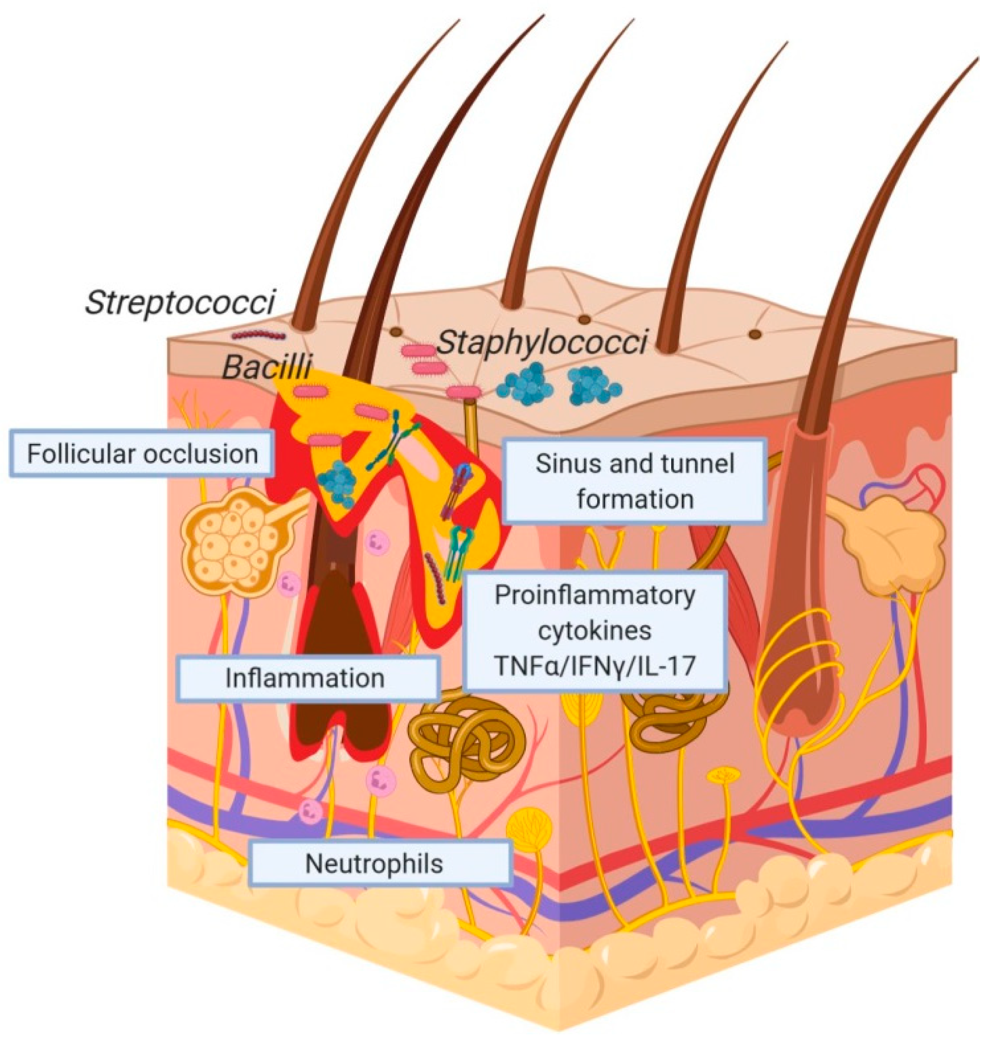
:1. Background
2. Characterising the Cutaneous Microbiota in HS
3. The benefits of 16S rRNA Sequencing and Metagenomic Approaches
4. Environmental Factors
5. Limitations of the Current Metagenomic Evidence Base
6. The Cutaneous and/or Gastrointestinal Microbiomes as a Therapeutic Targets
7. Conclusions
Funding
Conflicts of Interest
References
- Kolli, S.S.; Senthilnathan, A.; Cardwell, L.A.; Richardson, I.M.; Feldman, S.R.; Pichardo, R.O. Hidradenitis suppurativa has an enormous impact on patients’ lives. J. Am. Acad. Dermatol. 2019. [Google Scholar] [CrossRef] [PubMed] [Green Version]
- Garg, A.; Neuren, E.; Cha, D.; Kirby, J.S.; Ingram, J.R.; Jemec, G.B.E.; Esmann, S.; Thorlacius, L.; Villumsen, B.; Marmol, V.D.; et al. Evaluating Patients’ Unmet Needs in Hidradenitis Suppurativa: results from the Global VOICE project. J. Am. Acad. Dermatol. 2019. [Google Scholar] [CrossRef] [Green Version]
- Keary, E.; Hevey, D.; Tobin, A.M. A qualitative analysis of psychological distress in hidradenitis suppurativa. Br. J. Dermatol. 2019. [Google Scholar] [CrossRef] [PubMed]
- Marvel, J.; Vlahiotis, A.; Sainski-Nguyen, A.; Willson, T.; Kimball, A. Disease burden and cost of hidradenitis suppurativa: a retrospective examination of US administrative claims data. BMJ Open 2019, 9, e030579. [Google Scholar] [CrossRef] [Green Version]
- Desai, N.; Shah, P. High burden of hospital resource utilization in patients with hidradenitis suppurativa in England: a retrospective cohort study using hospital episode statistics. Br. J. Dermatol. 2017, 176, 1048–1055. [Google Scholar] [CrossRef]
- Kirby, J.S.; Miller, J.J.; Adams, D.R.; Leslie, D. Health care utilization patterns and costs for patients with hidradenitis suppurativa. JAMA Dermatol. 2014, 150, 937–944. [Google Scholar] [CrossRef] [Green Version]
- Vekic, D.A.; Frew, J.; Cains, G.D. Hidradenitis suppurativa, a review of pathogenesis, associations and management. Part 1. Australas J. Dermatol. 2018, 59, 267–277. [Google Scholar] [CrossRef]
- Bettoli, V.; Cazzaniga, S.; Scuderi, V.; Zedde, P.; Di Landro, A.; Naldi, L.; Group, I. Hidradenitis suppurativa epidemiology: from the first Italian registry in 2009 to the most recent epidemiology updates—Italian Registry Hidradenitis Suppurativa project 2. J. Eur. Acad. Dermatol. Venereol. 2019, 33 Suppl. 6, 4–6. [Google Scholar] [CrossRef]
- Goldburg, S.R.; Strober, B.E.; Payette, M.J. Part I. Hidradenitis Suppurativa: Epidemiology, clinical presentation and pathogenesis. J. Am. Acad. Dermatol. 2019. [Google Scholar] [CrossRef]
- Garg, A.; Kirby, J.S.; Lavian, J.; Lin, G.; Strunk, A. Sex- and Age-Adjusted Population Analysis of Prevalence Estimates for Hidradenitis Suppurativa in the United States. JAMA Dermatol. 2017, 153, 760–764. [Google Scholar] [CrossRef]
- Vekic, D.A.; Cains, G.D. Hidradenitis suppurativa, a review of pathogenesis, associations and management. Part 2. Australas J. Dermatol. 2018, 59, 261–266. [Google Scholar] [CrossRef] [PubMed]
- Mendonca, C.O.; Griffiths, C.E. Clindamycin and rifampicin combination therapy for hidradenitis suppurativa. Br. J. Dermatol. 2006, 154, 977–978. [Google Scholar] [CrossRef] [PubMed]
- Bettoli, V.; Zauli, S.; Borghi, A.; Toni, G.; Minghetti, S.; Ricci, M.; Virgili, A. Oral clindamycin and rifampicin in the treatment of hidradenitis suppurativa-acne inversa: a prospective study on 23 patients. J. Eur. Acad. Dermatol. Venereol. 2014, 28, 125–126. [Google Scholar] [CrossRef] [PubMed]
- Dessinioti, C.; Zisimou, C.; Tzanetakou, V.; Stratigos, A.; Antoniou, C. Oral clindamycin and rifampicin combination therapy for hidradenitis suppurativa: a prospective study and 1-year follow-up. Clin. Exp. Dermatol. 2016, 41, 852–857. [Google Scholar] [CrossRef]
- Gener, G.; Canoui-Poitrine, F.; Revuz, J.E.; Faye, O.; Poli, F.; Gabison, G.; Pouget, F.; Viallette, C.; Wolkenstein, P.; Bastuji-Garin, S. Combination therapy with clindamycin and rifampicin for hidradenitis suppurativa: a series of 116 consecutive patients. Dermatology 2009, 219, 148–154. [Google Scholar] [CrossRef]
- van der Zee, H.H.; Boer, J.; Prens, E.P.; Jemec, G.B. The effect of combined treatment with oral clindamycin and oral rifampicin in patients with hidradenitis suppurativa. Dermatology 2009, 219, 143–147. [Google Scholar] [CrossRef]
- Albrecht, J.; Baine, P.A.; Ladizinski, B.; Jemec, G.B.; Bigby, M. Long-term clinical safety of clindamycin and rifampicin combination for the treatment of hidradenitis suppurativa. A Critically Appraised Topic. Br. J. Dermatol. 2019, 180, 749–755. [Google Scholar] [CrossRef]
- Marasca, C.; Masara, A.; Annunziata, M.C.; Bettoli, V.; Luciano, M.A.; Fabbrocini, G. Long-term clinical safety of clindamycin and rifampicin combination for the treatment of hidradenitis suppurativa: a strategy to reduce side-effects, improving patients’ compliance. Br. J. Dermatol. 2019, 180, 949. [Google Scholar] [CrossRef]
- Albrecht, J.; Barbaric, J.; Nast, A. Rifampicin alone may be enough: is it time to abandon the classic oral clindamycin-rifampicin combination for hidradenitis suppurativa? Br. J. Dermatol. 2019, 180, 949–950. [Google Scholar] [CrossRef]
- Hambly, R.; Kirby, B. Prolonged clindamycin and rifampicin for hidradenitis suppurativa: resist to prevent resistance. Br. J. Dermatol. 2019, 180, 702–703. [Google Scholar] [CrossRef]
- Sayed, C.; Alikhan, A. Response to “Rifampin and Clindamycin are safe long term”. J. Am. Acad. Dermatol. 2019. [Google Scholar] [CrossRef] [PubMed]
- Schneller-Pavelescu, L.; Vergara-de Caso, E.; Martorell, A.; Romani, J.; Lazaro, M.; Vilarrasa, E.; Diaz-Ley, B.; Vazquez-Osorio, I.; Segura Palacios, J.M.; Azana, J.M.; et al. Reply to “Comment on ‘Interruption of oral clindamycin plus rifampicin therapy in patients with hidradenitis suppurativa: an observational study to assess prevalence and causes’ ”. J. Am. Acad Dermatol 2019. [Google Scholar] [CrossRef] [PubMed]
- Albrecht, J.; Bigby, M. Rifampin and Clindamycin are safe long term Response to North American clinical management guidelines for hidradenitis suppurativa: A publication from the United States and Canadian Hidradenitis Suppurativa Foundations Part II: Topical, intralesional and systemic medical management. J. Am. Acad Dermatol 2019. [Google Scholar] [CrossRef]
- Kimball, A.B.; Okun, M.M.; Williams, D.A.; Gottlieb, A.B.; Papp, K.A.; Zouboulis, C.C.; Armstrong, A.W.; Kerdel, F.; Gold, M.H.; Forman, S.B.; et al. Two Phase 3 Trials of Adalimumab for Hidradenitis Suppurativa. N Engl. J. Med. 2016, 375, 422–434. [Google Scholar] [CrossRef]
- Sand, F.L.; Thomsen, S.F. Off-label use of TNF-alpha inhibitors in a dermatological university department: retrospective evaluation of 118 patients. Dermatol. Ther. 2015, 28, 158–165. [Google Scholar] [CrossRef]
- Grant, A.; Gonzalez, T.; Montgomery, M.O.; Cardenas, V.; Kerdel, F.A. Infliximab therapy for patients with moderate to severe hidradenitis suppurativa: a randomized, double-blind, placebo-controlled crossover trial. J. Am. Acad. Dermatol. 2010, 62, 205–217. [Google Scholar] [CrossRef]
- Maarouf, M.; Clark, A.K.; Lee, D.E.; Shi, V.Y. Targeted treatments for hidradenitis suppurativa: a review of the current literature and ongoing clinical trials. J. Dermatolog. Treat. 2018, 29, 441–449. [Google Scholar] [CrossRef]
- Kerdel, F.R.; Azevedo, F.A.; Kerdel Don, C.; Don, F.A.; Fabbrocini, G.; Kerdel, F.A. Apremilast for the Treatment of Mild-to-Moderate Hidradenitis Suppurativa in a Prospective, Open-Label, Phase 2 Study. J. Drugs Dermatol. 2019, 18, 170–176. [Google Scholar]
- Vossen, A.; van Doorn, M.B.A.; van der Zee, H.H.; Prens, E.P. Apremilast for moderate hidradenitis suppurativa: Results of a randomized controlled trial. J. Am. Acad. Dermatol. 2019, 80, 80–88. [Google Scholar] [CrossRef]
- Alikhan, A.; Sayed, C.; Alavi, A.; Alhusayen, R.; Brassard, A.; Burkhart, C.; Crowell, K.; Eisen, D.B.; Gottlieb, A.B.; Hamzavi, I.; et al. North American clinical management guidelines for hidradenitis suppurativa: A publication from the United States and Canadian Hidradenitis Suppurativa Foundations: Part II: Topical, intralesional and systemic medical management. J. Am. Acad. Dermatol. 2019, 81, 91–101. [Google Scholar] [CrossRef] [Green Version]
- McPhie, M.L.; Bridgman, A.C.; Kirchhof, M.G. Combination Therapies for Hidradenitis Suppurativa: A Retrospective Chart Review of 31 Patients. J. Cutan. Med. Surg. 2019, 23, 270–276. [Google Scholar] [CrossRef] [PubMed]
- Cline, A.; Pichardo, R.O. Successful treatment of hidradenitis suppurativa in the setting of Crohn disease with combination adalimumab and ustekinumab. Dermatol. Online J. 2019, 25. [Google Scholar]
- Molina-Leyva, A. Adalimumab every other week combined with dexamethasone pulses for the treatment of refractory hidradenitis suppurativa. Dermatol. Ther. 2019, 32, e12885. [Google Scholar] [CrossRef] [PubMed]
- DeFazio, M.V.; Economides, J.M.; King, K.S.; Han, K.D.; Shanmugam, V.K.; Attinger, C.E.; Evans, K.K. Outcomes After Combined Radical Resection and Targeted Biologic Therapy for the Management of Recalcitrant Hidradenitis Suppurativa. Ann. Plast Surg. 2016, 77, 217–222. [Google Scholar] [CrossRef] [PubMed] [Green Version]
- Highet, A.S.; Warren, R.E.; Staughton, R.C.; Roberts, S.O. Streptococcus milleri causing treatable infection in perineal hidradenitis suppurativa. Br. J. Dermatol. 1980, 103, 375–382. [Google Scholar] [CrossRef]
- Brook, I.; Frazier, E.H. Aerobic and anaerobic microbiology of axillary hidradenitis suppurativa. J. Med. Microbiol. 1999, 48, 103–105. [Google Scholar] [CrossRef]
- Benzecry, V.; Grancini, A.; Guanziroli, E.; Nazzaro, G.; Barbareschi, M.; Marzano, A.V.; Muratori, S.; Veraldi, S. Hidradenitis suppurativa/acne inversa: a prospective bacteriological study of 46 patients and review of the literature. G Ital. Dermatol. Venereol. 2018. [Google Scholar] [CrossRef]
- Lapins, J.; Jarstrand, C.; Emtestam, L. Coagulase-negative staphylococci are the most common bacteria found in cultures from the deep portions of hidradenitis suppurativa lesions, as obtained by carbon dioxide laser surgery. Br. J. Dermatol. 1999, 140, 90–95. [Google Scholar] [CrossRef] [Green Version]
- Sartorius, K.; Killasli, H.; Oprica, C.; Sullivan, A.; Lapins, J. Bacteriology of hidradenitis suppurativa exacerbations and deep tissue cultures obtained during carbon dioxide laser treatment. Br. J. Dermatol. 2012, 166, 879–883. [Google Scholar] [CrossRef]
- Ring, H.C.; Bay, L.; Kallenbach, K.; Miller, I.M.; Prens, E.; Saunte, D.M.; Bjarnsholt, T.; Jemec, G.B. Normal Skin Microbiota is Altered in Pre-clinical Hidradenitis Suppurativa. Acta Derm Venereol. 2017, 97, 208–213. [Google Scholar] [CrossRef] [Green Version]
- Ring, H.C.; Riis Mikkelsen, P.; Miller, I.M.; Jenssen, H.; Fuursted, K.; Saunte, D.M.; Jemec, G.B. The bacteriology of hidradenitis suppurativa: a systematic review. Exp. Dermatol. 2015, 24, 727–731. [Google Scholar] [CrossRef]
- Jemec, G.B.; Faber, M.; Gutschik, E.; Wendelboe, P. The bacteriology of hidradenitis suppurativa. Dermatology 1996, 193, 203–206. [Google Scholar] [CrossRef] [PubMed]
- Matusiak, L.; Bieniek, A.; Szepietowski, J.C. Bacteriology of hidradenitis suppurativa - which antibiotics are the treatment of choice? Acta Derm. Venereol. 2014, 94, 699–702. [Google Scholar] [CrossRef] [PubMed] [Green Version]
- Nikolakis, G.; Join-Lambert, O.; Karagiannidis, I.; Guet-Revillet, H.; Zouboulis, C.C.; Nassif, A. Bacteriology of hidradenitis suppurativa/acne inversa: A review. J. Am. Acad. Dermatol. 2015, 73, S12–S18. [Google Scholar] [CrossRef] [PubMed]
- Grice, E.A.; Kong, H.H.; Renaud, G.; Young, A.C.; Program, N.C.S.; Bouffard, G.G.; Blakesley, R.W.; Wolfsberg, T.G.; Turner, M.L.; Segre, J.A. A diversity profile of the human skin microbiota. Genome Res. 2008, 18, 1043–1050. [Google Scholar] [CrossRef] [Green Version]
- Grice, E.A.; Kong, H.H.; Conlan, S.; Deming, C.B.; Davis, J.; Young, A.C.; Program, N.C.S.; Bouffard, G.G.; Blakesley, R.W.; Murray, P.R.; et al. Topographical and temporal diversity of the human skin microbiome. Science 2009, 324, 1190–1192. [Google Scholar] [CrossRef] [Green Version]
- Grogan, M.D.; Bartow-McKenney, C.; Flowers, L.; Knight, S.A.B.; Uberoi, A.; Grice, E.A. Research Techniques Made Simple: Profiling the Skin Microbiota. J. Invest. Dermatol. 2019, 139, 747–752 e741. [Google Scholar] [CrossRef] [Green Version]
- Oliveira, M.; Amorim, A. Microbial forensics: new breakthroughs and future prospects. Appl. Microbiol. Biotechnol. 2018, 102, 10377–10391. [Google Scholar] [CrossRef]
- Alekseyenko, A.V.; Perez-Perez, G.I.; De Souza, A.; Strober, B.; Gao, Z.; Bihan, M.; Li, K.; Methe, B.A.; Blaser, M.J. Community differentiation of the cutaneous microbiota in psoriasis. Microbiome 2013, 1, 31. [Google Scholar] [CrossRef] [Green Version]
- Fry, L.; Baker, B.S.; Powles, A.V.; Fahlen, A.; Engstrand, L. Is chronic plaque psoriasis triggered by microbiota in the skin? Br. J. Dermatol. 2013, 169, 47–52. [Google Scholar] [CrossRef]
- Gao, Z.; Tseng, C.H.; Strober, B.E.; Pei, Z.; Blaser, M.J. Substantial alterations of the cutaneous bacterial biota in psoriatic lesions. PLoS ONE 2008, 3, e2719. [Google Scholar] [CrossRef] [PubMed]
- Tobin, A.M. Unravelling the microbiome in psoriasis. Br. J. Dermatol. 2019. [Google Scholar] [CrossRef] [PubMed]
- Kong, H.H.; Oh, J.; Deming, C.; Conlan, S.; Grice, E.A.; Beatson, M.A.; Nomicos, E.; Polley, E.C.; Komarow, H.D.; Program, N.C.S.; et al. Temporal shifts in the skin microbiome associated with disease flares and treatment in children with atopic dermatitis. Genome Res. 2012, 22, 850–859. [Google Scholar] [CrossRef] [PubMed] [Green Version]
- Paller, A.S.; Kong, H.H.; Seed, P.; Naik, S.; Scharschmidt, T.C.; Gallo, R.L.; Luger, T.; Irvine, A.D. The microbiome in patients with atopic dermatitis. J. Allergy Clin. Immunol. 2019, 143, 26–35. [Google Scholar] [CrossRef] [PubMed] [Green Version]
- Fyhrquist, N.; Muirhead, G.; Prast-Nielsen, S.; Jeanmougin, M.; Olah, P.; Skoog, T.; Jules-Clement, G.; Feld, M.; Barrientos-Somarribas, M.; Sinkko, H.; et al. Microbe-host interplay in atopic dermatitis and psoriasis. Nat. Commun. 2019, 10, 4703. [Google Scholar] [CrossRef] [Green Version]
- Miodovnik, M.; Kunstner, A.; Langan, E.A.; Zillikens, D.; Glaser, R.; Sprecher, E.; Baines, J.F.; Schmidt, E.; Ibrahim, S.M. A distinct cutaneous microbiota profile in autoimmune bullous disease patients. Exp. Dermatol. 2017, 26, 1221–1227. [Google Scholar] [CrossRef]
- Scaglione, G.L.; Fania, L.; De Paolis, E.; De Bonis, M.; Mazzanti, C.; Di Zenzo, G.; Lechiancole, S.; Messinese, S.; Capoluongo, E. Evaluation of cutaneous, oral and intestinal microbiota in patients affected by pemphigus and bullous pemphigoid: A pilot study. Exp. Mol. Pathol. 2019, 112, 104331. [Google Scholar] [CrossRef]
- Ramasamy, S.; Barnard, E.; Dawson, T.L., Jr.; Li, H. The role of the skin microbiota in acne pathophysiology. Br. J. Dermatol. 2019, 181, 691–699. [Google Scholar] [CrossRef]
- Guet-Revillet, H.; Coignard-Biehler, H.; Jais, J.P.; Quesne, G.; Frapy, E.; Poiree, S.; Le Guern, A.S.; Le Fleche-Mateos, A.; Hovnanian, A.; Consigny, P.H.; et al. Bacterial pathogens associated with hidradenitis suppurativa, France. Emerg Infect. Dis 2014, 20, 1990–1998. [Google Scholar] [CrossRef]
- Guet-Revillet, H.; Jais, J.P.; Ungeheuer, M.N.; Coignard-Biehler, H.; Duchatelet, S.; Delage, M.; Lam, T.; Hovnanian, A.; Lortholary, O.; Nassif, X.; et al. The Microbiological Landscape of Anaerobic Infections in Hidradenitis Suppurativa: A Prospective Metagenomic Study. Clin. Infect. Dis. 2017, 65, 282–291. [Google Scholar] [CrossRef] [Green Version]
- Ring, H.C.; Sigsgaard, V.; Thorsen, J.; Fuursted, K.; Fabricius, S.; Saunte, D.M.; Jemec, G.B. The microbiome of tunnels in hidradenitis suppurativa patients. J. Eur. Acad Dermatol. Venereol. 2019, 33, 1775–1780. [Google Scholar] [CrossRef] [PubMed]
- Naik, H.B.; Jo, J.H.; Paul, M.; Kong, H.H. Skin microbiota perturbations are distinct and disease severity-dependent in hidradenitis suppurativa. J. Invest. Dermatol. 2019. [Google Scholar] [CrossRef]
- Meisel, J.S.; Hannigan, G.D.; Tyldsley, A.S.; SanMiguel, A.J.; Hodkinson, B.P.; Zheng, Q.; Grice, E.A. Skin Microbiome Surveys Are Strongly Influenced by Experimental Design. J. Invest. Dermatol. 2016, 136, 947–956. [Google Scholar] [CrossRef] [PubMed]
- Schneider, A.M.; Cook, L.C.; Zhan, X.; Banerjee, K.; Cong, Z.; Imamura-Kawasawa, Y.; Gettle, S.L.; Longenecker, A.L.; Kirby, J.S.; Nelson, A.M. Loss of skin microbial diversity and alteration of bacterial metabolic function in Hidradenitis Suppurativa. J. Invest. Dermatol. 2019. [Google Scholar] [CrossRef] [PubMed]
- Fahlen, A.; Engstrand, L.; Baker, B.S.; Powles, A.; Fry, L. Comparison of bacterial microbiota in skin biopsies from normal and psoriatic skin. Arch. Dermatol. Res. 2012, 304, 15–22. [Google Scholar] [CrossRef] [PubMed]
- Prast-Nielsen, S.; Tobin, A.M.; Adamzik, K.; Powles, A.; Hugerth, L.W.; Sweeney, C.; Kirby, B.; Engstrand, L.; Fry, L. Investigation of the skin microbiome: swabs vs. biopsies. Br. J. Dermatol. 2019, 181, 572–579. [Google Scholar] [CrossRef]
- Acharya, P.; Mathur, M. Hidradenitis suppurativa and smoking: a systematic review and meta-analysis. J. Am. Acad. Dermatol. 2019. [Google Scholar] [CrossRef] [Green Version]
- Yu, G.; Gail, M.H.; Consonni, D.; Carugno, M.; Humphrys, M.; Pesatori, A.C.; Caporaso, N.E.; Goedert, J.J.; Ravel, J.; Landi, M.T. Characterizing human lung tissue microbiota and its relationship to epidemiological and clinical features. Genome Biol. 2016, 17, 163. [Google Scholar] [CrossRef] [Green Version]
- Savin, Z.; Kivity, S.; Yonath, H.; Yehuda, S. Smoking and the intestinal microbiome. Arch. Microbiol. 2018, 200, 677–684. [Google Scholar] [CrossRef]
- Eppinga, H.; Sperna Weiland, C.J.; Thio, H.B.; van der Woude, C.J.; Nijsten, T.E.; Peppelenbosch, M.P.; Konstantinov, S.R. Similar Depletion of Protective Faecalibacterium prausnitzii in Psoriasis and Inflammatory Bowel Disease but not in Hidradenitis Suppurativa. J. Crohns Colitis 2016, 10, 1067–1075. [Google Scholar] [CrossRef] [Green Version]
- Szanto, M.; Dozsa, A.; Antal, D.; Szabo, K.; Kemeny, L.; Bai, P. Targeting the gut-skin axis-Probiotics as new tools for skin disorder management? Exp. Dermatol. 2019. [Google Scholar] [CrossRef] [PubMed] [Green Version]
- Phan, K.; Tatian, A.; Woods, J.; Cains, G.; Frew, J.W. Prevalence of inflammatory bowel disease (IBD) in hidradenitis suppurativa (HS): systematic review and adjusted meta-analysis. Int. J. Dermatol. 2019. [Google Scholar] [CrossRef] [PubMed]
- Gopalakrishnan, V.; Spencer, C.N.; Nezi, L.; Reuben, A.; Andrews, M.C.; Karpinets, T.V.; Prieto, P.A.; Vicente, D.; Hoffman, K.; Wei, S.C.; et al. Gut microbiome modulates response to anti-PD-1 immunotherapy in melanoma patients. Science 2018, 359, 97–103. [Google Scholar] [CrossRef] [Green Version]
- SanMiguel, A.J.; Meisel, J.S.; Horwinski, J.; Zheng, Q.; Bradley, C.W.; Grice, E.A. Antiseptic Agents Elicit Short-Term, Personalized and Body Site-Specific Shifts in Resident Skin Bacterial Communities. J. Invest. Dermatol. 2018, 138, 2234–2243. [Google Scholar] [CrossRef]
- Kelhala, H.L.; Aho, V.T.E.; Fyhrquist, N.; Pereira, P.A.B.; Kubin, M.E.; Paulin, L.; Palatsi, R.; Auvinen, P.; Tasanen, K.; Lauerma, A. Isotretinoin and lymecycline treatments modify the skin microbiota in acne. Exp. Dermatol. 2018, 27, 30–36. [Google Scholar] [CrossRef] [PubMed]
- Hendricks, A.J.; Hsiao, J.L.; Lowes, M.A.; Shi, V.Y. A Comparison of International Management Guidelines for Hidradenitis Suppurativa. Dermatology 2019, 1–16. [Google Scholar] [CrossRef]
- Chng, K.R.; Tay, A.S.; Li, C.; Ng, A.H.; Wang, J.; Suri, B.K.; Matta, S.A.; McGovern, N.; Janela, B.; Wong, X.F.; et al. Whole metagenome profiling reveals skin microbiome-dependent susceptibility to atopic dermatitis flare. Nat. Microbiol. 2016, 1, 16106. [Google Scholar] [CrossRef]
- Langan, E.A.; Kunstner, A.; Miodovnik, M.; Zillikens, D.; Thaci, D.; Baines, J.F.; Ibrahim, S.M.; Solbach, W.; Knobloch, J.K. Combined culture and metagenomic analyses reveal significant shifts in the composition of the cutaneous microbiome in psoriasis. Br. J. Dermatol. 2019. [Google Scholar] [CrossRef] [Green Version]
- Flood, K.S.; Porter, M.L.; Kimball, A.B. Biologic Treatment for Hidradenitis Suppurativa. Am. J. Clin. Dermatol. 2019, 20, 625–638. [Google Scholar] [CrossRef]
- Lee, R.A.; Eisen, D.B. Treatment of hidradenitis suppurativa with biologic medications. J. Am. Acad. Dermatol. 2015, 73, S82–S88. [Google Scholar] [CrossRef]
- Ingram, J.R.; Collier, F.; Brown, D.; Burton, T.; Burton, J.; Chin, M.F.; Desai, N.; Goodacre, T.E.E.; Piguet, V.; Pink, A.E.; et al. British Association of Dermatologists guidelines for the management of hidradenitis suppurativa (acne inversa) 2018. Br. J. Dermatol. 2019, 180, 1009–1017. [Google Scholar] [CrossRef] [PubMed] [Green Version]
- Ghias, M.H.; Johnston, A.D.; Kutner, A.J.; Micheletti, R.G.; Hosgood, H.D.; Cohen, S.R. High-dose, high-frequency infliximab: A novel treatment paradigm for hidradenitis suppurativa. J. Am. Acad. Dermatol. 2019. [Google Scholar] [CrossRef] [PubMed]
- Takeda, K.; Kikuchi, K.; Kanazawa, Y.; Yamasaki, K.; Aiba, S. Ustekinumab treatment for hidradenitis suppurativa. J. Dermatol. 2019, 46, 1215–1218. [Google Scholar] [CrossRef]
- Prussick, L.; Rothstein, B.; Joshipura, D.; Saraiya, A.; Turkowski, Y.; Abdat, R.; Alomran, A.; Zancanaro, P.; Kachuk, C.; Dumont, N.; et al. Open-label, investigator-initiated, single-site exploratory trial evaluating secukinumab, an anti-interleukin-17A monoclonal antibody, for patients with moderate-to-severe hidradenitis suppurativa. Br. J. Dermatol. 2019, 181, 609–611. [Google Scholar] [CrossRef] [PubMed]
- Loesche, M.A.; Farahi, K.; Capone, K.; Fakharzadeh, S.; Blauvelt, A.; Duffin, K.C.; DePrimo, S.E.; Munoz-Elias, E.J.; Brodmerkel, C.; Dasgupta, B.; et al. Longitudinal Study of the Psoriasis-Associated Skin Microbiome during Therapy with Ustekinumab in a Randomized Phase 3b Clinical Trial. J. Invest. Dermatol. 2018, 138, 1973–1981. [Google Scholar] [CrossRef] [PubMed] [Green Version]
- Yeh, N.L.; Hsu, C.Y.; Tsai, T.F.; Chiu, H.Y. Gut Microbiome in Psoriasis is Perturbed Differently During Secukinumab and Ustekinumab Therapy and Associated with Response to Treatment. Clin. Drug Investig. 2019, 39, 1195–1203. [Google Scholar] [CrossRef] [PubMed]
- Salem, I.; Ramser, A.; Isham, N.; Ghannoum, M.A. The Gut Microbiome as a Major Regulator of the Gut-Skin Axis. Front. Microbiol. 2018, 9, 1459. [Google Scholar] [CrossRef] [Green Version]
- Bosman, E.S.; Albert, A.Y.; Lui, H.; Dutz, J.P.; Vallance, B.A. Skin Exposure to Narrow Band Ultraviolet (UVB) Light Modulates the Human Intestinal Microbiome. Front. Microbiol. 2019, 10, 2410. [Google Scholar] [CrossRef]
- Emelianov, V.U.; Bechara, F.G.; Glaser, R.; Langan, E.A.; Taungjaruwinai, W.M.; Schroder, J.M.; Meyer, K.C.; Paus, R. Immunohistological pointers to a possible role for excessive cathelicidin (LL-37) expression by apocrine sweat glands in the pathogenesis of hidradenitis suppurativa/acne inversa. Br. J. Dermatol. 2012, 166, 1023–1034. [Google Scholar] [CrossRef]
- Kelly, G.; Hughes, R.; McGarry, T.; van den Born, M.; Adamzik, K.; Fitzgerald, R.; Lawlor, C.; Tobin, A.M.; Sweeney, C.M.; Kirby, B. Dysregulated cytokine expression in lesional and nonlesional skin in hidradenitis suppurativa. Br. J. Dermatol. 2015, 173, 1431–1439. [Google Scholar] [CrossRef]
- Vossen, A.; van der Zee, H.H.; Prens, E.P. Hidradenitis Suppurativa: A Systematic Review Integrating Inflammatory Pathways Into a Cohesive Pathogenic Model. Front. Immunol. 2018, 9, 2965. [Google Scholar] [CrossRef] [PubMed] [Green Version]
- Thorlacius, L.; Cohen, A.D.; Gislason, G.H.; Jemec, G.B.E.; Egeberg, A. Increased Suicide Risk in Patients with Hidradenitis Suppurativa. J. Invest. Dermatol. 2018, 138, 52–57. [Google Scholar] [CrossRef] [PubMed] [Green Version]
- Machado, M.O.; Stergiopoulos, V.; Maes, M.; Kurdyak, P.A.; Lin, P.Y.; Wang, L.J.; Shyu, Y.C.; Firth, J.; Koyanagi, A.; Solmi, M.; et al. Depression and Anxiety in Adults With Hidradenitis Suppurativa: A Systematic Review and Meta-analysis. JAMA Dermatol. 2019. [Google Scholar] [CrossRef] [PubMed]
- Frew, J.W.; Navrazhina, K.; Byrd, A.S.; Garg, A.; Ingram, J.R.; Kirby, J.S.; Lowes, M.A.; Naik, H.; Piguet, V.; Prens, E.P. Defining lesional, perilesional and unaffected skin in hidradenitis suppurativa: proposed recommendations for clinical trials and translational research studies. Br. J. Dermatol. 2019, 181, 1339–1341. [Google Scholar] [CrossRef]
- Alavi, A.; Piguet, V. Genotype-phenotype correlation in inherited hidradenitis suppurativa: one step forward, one step back. Br. J. Dermatol. 2019, 181, 443–444. [Google Scholar] [CrossRef]


© 2020 by the authors. Licensee MDPI, Basel, Switzerland. This article is an open access article distributed under the terms and conditions of the Creative Commons Attribution (CC BY) license (http://creativecommons.org/licenses/by/4.0/).
Share and Cite
Langan, E.A.; Recke, A.; Bokor-Billmann, T.; Billmann, F.; Kahle, B.K.; Zillikens, D. The Role of the Cutaneous Microbiome in Hidradenitis Suppurativa—Light at the End of the Microbiological Tunnel. Int. J. Mol. Sci. 2020, 21, 1205. https://0-doi-org.brum.beds.ac.uk/10.3390/ijms21041205
Langan EA, Recke A, Bokor-Billmann T, Billmann F, Kahle BK, Zillikens D. The Role of the Cutaneous Microbiome in Hidradenitis Suppurativa—Light at the End of the Microbiological Tunnel. International Journal of Molecular Sciences. 2020; 21(4):1205. https://0-doi-org.brum.beds.ac.uk/10.3390/ijms21041205
Chicago/Turabian StyleLangan, Ewan A., Andreas Recke, Therezia Bokor-Billmann, Franck Billmann, Birgit K. Kahle, and Detlef Zillikens. 2020. "The Role of the Cutaneous Microbiome in Hidradenitis Suppurativa—Light at the End of the Microbiological Tunnel" International Journal of Molecular Sciences 21, no. 4: 1205. https://0-doi-org.brum.beds.ac.uk/10.3390/ijms21041205