Article

Sexual Dimorphism in Doxorubicin-induced Systemic Inflammation: Implications for Hepatic Cytochrome P450 Regulation

by
Marianne K.O. Grant
,
Ibrahim Y. Abdelgawad
,
Christine A. Lewis
and
Beshay N. Zordoky
*
Department of Experimental and Clinical Pharmacology, University of Minnesota College of Pharmacy, Minneapolis, MN 55455, USA
*
Author to whom correspondence should be addressed.
Int. J. Mol. Sci. 2020, 21(4), 1279; https://0-doi-org.brum.beds.ac.uk/10.3390/ijms21041279
Submission received: 9 January 2020 / Revised: 3 February 2020 / Accepted: 11 February 2020 / Published: 14 February 2020
(This article belongs to the Section Molecular Toxicology)

Abstract

:
Doxorubicin (DOX) is an effective chemotherapeutic agent used to treat a wide variety of malignancies. In addition to its multi-organ toxicity, DOX treatment has been shown to induce systemic inflammation in patients and experimental animals. Inflammation alters the expression of hepatic cytochrome P450 (CYP) enzymes, which play important roles in drug metabolism and DOX-induced toxicity. Significant sex differences have been reported in DOX-induced toxicity; however, sex differences in DOX-induced systemic inflammation and the potential effects on hepatic CYP expression have not been determined. In the current work, male and female C57Bl/6 mice were administered DOX (20 mg/kg by intraperitoneal injection), and groups of mice were sacrificed 24 and 72 h after DOX administration. DOX elicited a systemic inflammatory response in both male and female mice, but the inflammatory response was stronger in male mice. DOX altered the expression of hepatic CYP isoforms in a sex-dependent manner. Most notably, inhibition of Cyp2c29 and Cyp2e1 was stronger in male than in female mice, which paralleled the sex differences in systemic inflammation. Therefore, sex differences in DOX-induced systemic inflammation may lead to sexually dimorphic drug interactions, in addition to contributing to the previously reported sexual dimorphism in specific DOX-induced organ toxicity.

1. Introduction

Doxorubicin (DOX) is an effective chemotherapeutic agent used to treat a wide variety of solid and hematological malignancies. Nevertheless, the clinical utility of DOX is limited by its multi-organ toxicity. DOX is known for its cardiotoxic effects that may lead to cardiac dysfunction and heart failure [1]. In addition to cardiotoxicity, DOX has also been shown to cause hepatotoxicity [2,3,4,5,6], gastro-intestinal toxicity [7], and nephrotoxicity [8,9]. Although DOX-induced toxicity is likely to be multi-factorial, inflammation has been shown to be a central player in mediating DOX-induced toxicity [10,11,12,13,14]. In addition to organ-specific inflammatory changes, DOX treatment has been shown to cause systemic inflammation in cancer patients [10], piglets [7], and rodents [15].
We and others have shown that inflammation plays a major role in the regulation of hepatic cytochrome P450 (CYP) enzymes [16,17,18]. Since hepatic CYP enzymes are the main drug-metabolizing enzymes, DOX-induced inflammation may lead to significant changes in drug metabolism and drug interactions. Indeed, we and others have previously demonstrated that acute DOX toxicity alters the expression of several hepatic CYP isoforms in male rats [5,19], which resulted in altered metabolism of clinically relevant drugs [19,20]. Importantly, DOX/cyclophosphamide chemotherapy caused a differential effect on drug-metabolizing enzyme activities in breast cancer patients with a notable reduction of CYP2C9 activity by 315% [21].
In addition to their important role in drug metabolism, CYP enzymes play important toxicological roles. For instance, CYP enzymes metabolize arachidonic acid to different hydroxyeicosatetraenoic acid and epoxyeicosatrienoic acid metabolites [22]. These CYP-mediated endogenous metabolites, among others, play important roles in modulating DOX-induced toxicity [5,23]. Therefore, determining the effect of DOX administration on hepatic CYP enzymes will have a paramount impact on our understanding of potential DOX-induced drug interactions as well as DOX-induced toxicity.
We have recently shown that acute DOX administration caused a sex-dependent alteration in the gene expression of several CYP isoforms in the heart of C57Bl/6 mice [11]. Since all previous studies reporting the effect of DOX on hepatic CYP expression and activity were performed using male experimental animals [5,19], sex-related differences in the hepatic effects of DOX have not been identified. Therefore, in the current study, we determined the effect of acute DOX administration on the expression of hepatic CYP enzymes in male and female C57Bl/6 mice to reveal potential sex-related differences.

2. Results

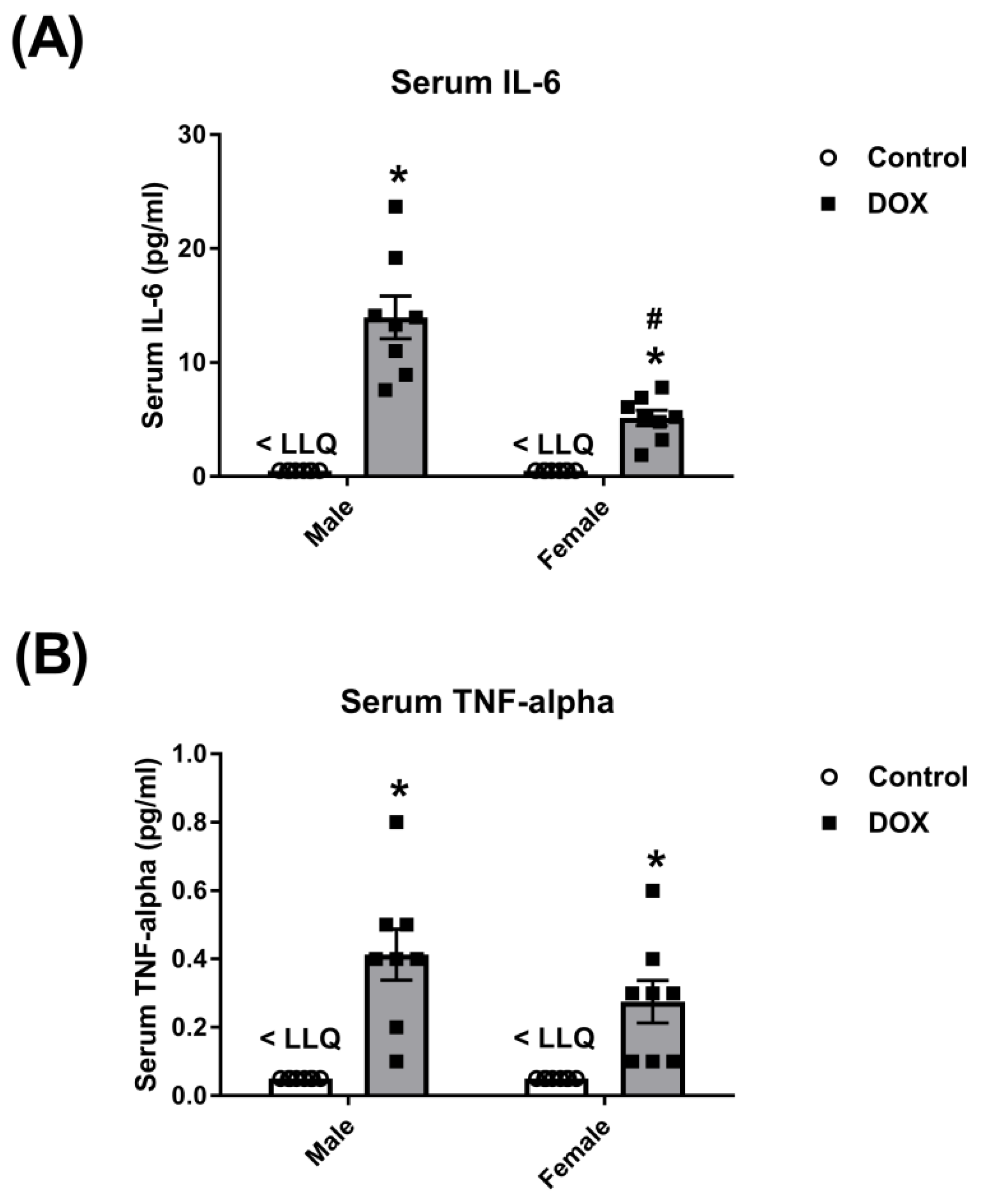
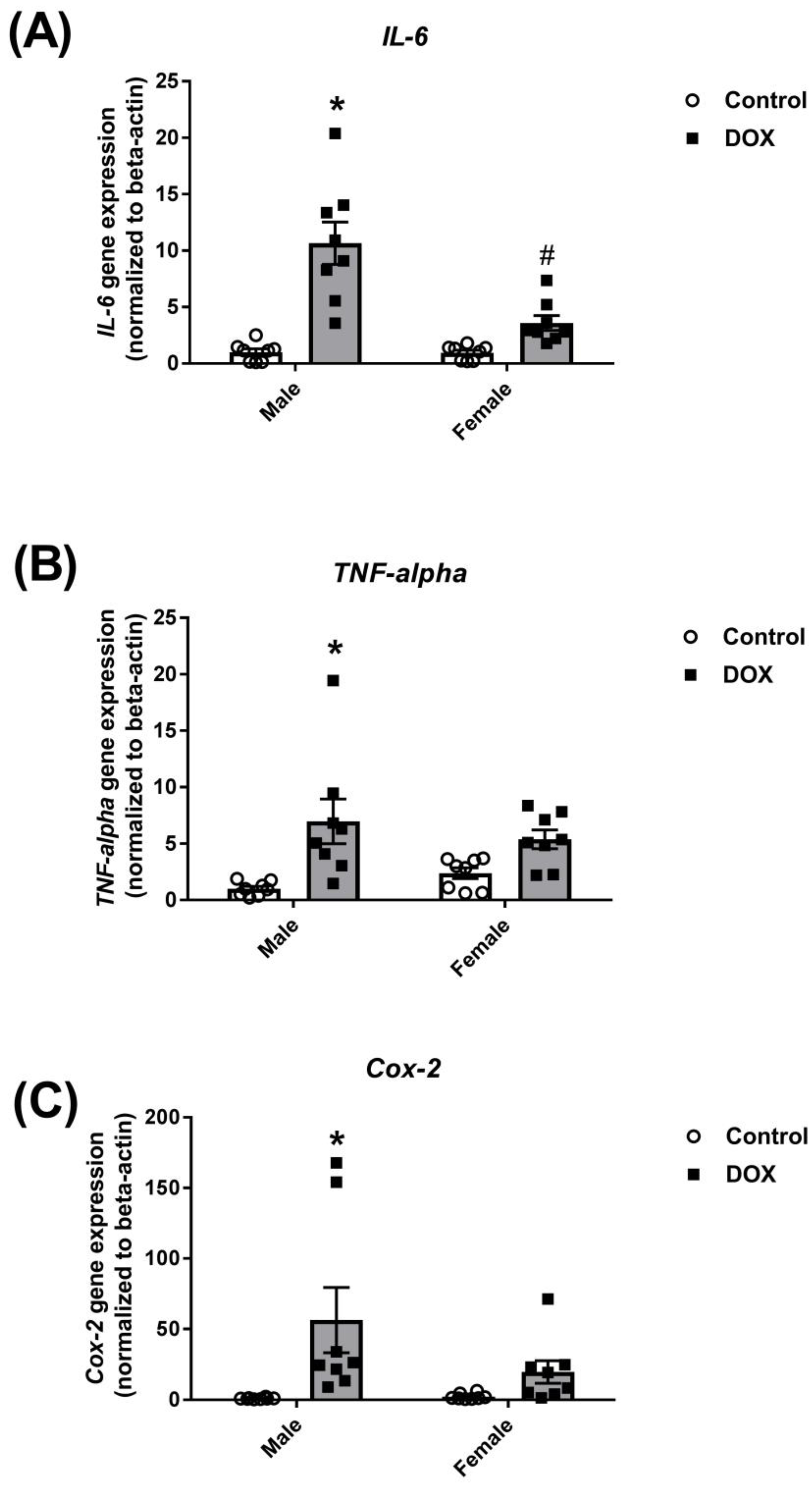
2.1. Effect of DOX Treatment on Inflammatory Markers

We and others have demonstrated significant sex-related differences in DOX-induced cardiotoxicity and nephrotoxicity [11,22,24,25]. Nevertheless, sex-related differences in DOX-induced systemic inflammation have not been previously reported. Therefore, we determined the effect of acute DOX administration on serum level of key inflammatory mediators, interleukin 6 (IL-6) and tumor necrosis factor-alpha (TNF-alpha), in adult male and female C57Bl/6 mice 24 h post-treatment. Acute DOX administration (20 mg/kg) elicited a systemic inflammatory response and caused a significantly higher increase in the serum level of IL-6 in male than in female mice (14 pg/mL in DOX-treated males vs. 5 pg/mL in DOX-treated females). Although the TNF-alpha serum level was higher in DOX-treated male than female mice (0.4 pg/mL in DOX-treated males vs. 0.3 pg/mL in DOX-treated females), this difference was not statistically significant. The serum levels of IL-6 and TNF-alpha were below the lower limit of quantification (LLQ) in control male and female mice (Figure 1).
Corroborating these findings, acute DOX administration caused a significant 10-, 7-, and 60-fold induction of the inflammatory markers IL-6, TNF-alpha, and cyclooxygenase-2 (Cox-2) gene expression, respectively, in the liver of male mice (Figure 2). There was a modest induction of these markers in the liver of female mice, but the differences between the female-control and female-DOX groups did not reach statistical significance (Figure 2).

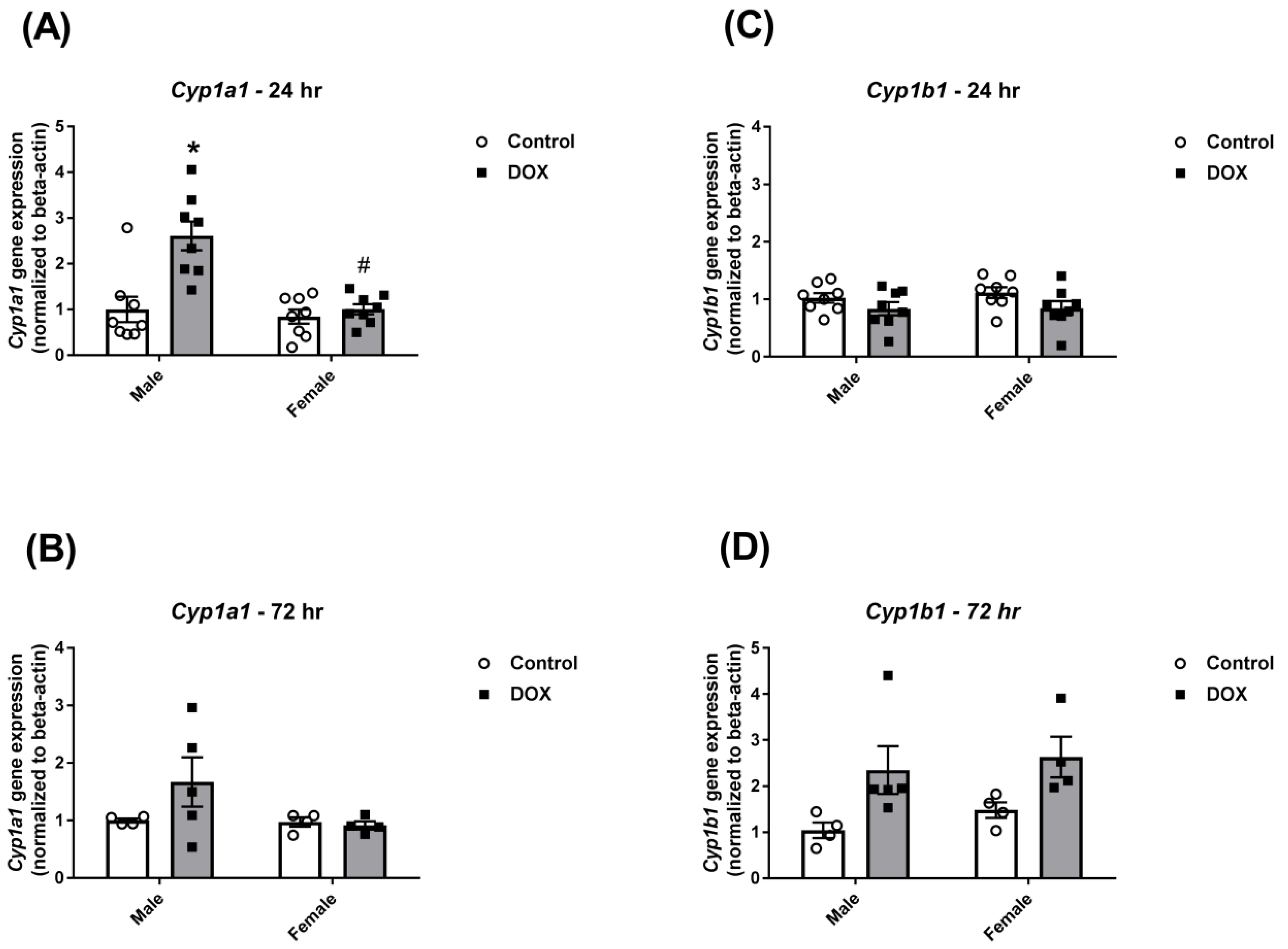
2.2. Effect of DOX Treatment on Cyp1 Family Expression

Twenty-four hours following DOX administration, DOX caused a significant 2.6-fold induction of hepatic Cyp1a1 gene expression in male mice only (Figure 3A). Two-way ANOVA showed significant DOX and sex effects as well as a significant interaction between DOX exposure and sex (Table 1). Seventy-two hours following DOX administration, DOX-induced Cyp1a1 upregulation in male mice was not statistically significant (Figure 3B). On the other hand, there was no significant change in Cyp1b1 gene expression 24 h following DOX administration in male or female mice (Figure 3C). Nevertheless, there was a significant DOX effect on Cyp1b1 induction after 72 h (Figure 3D and Table 1).

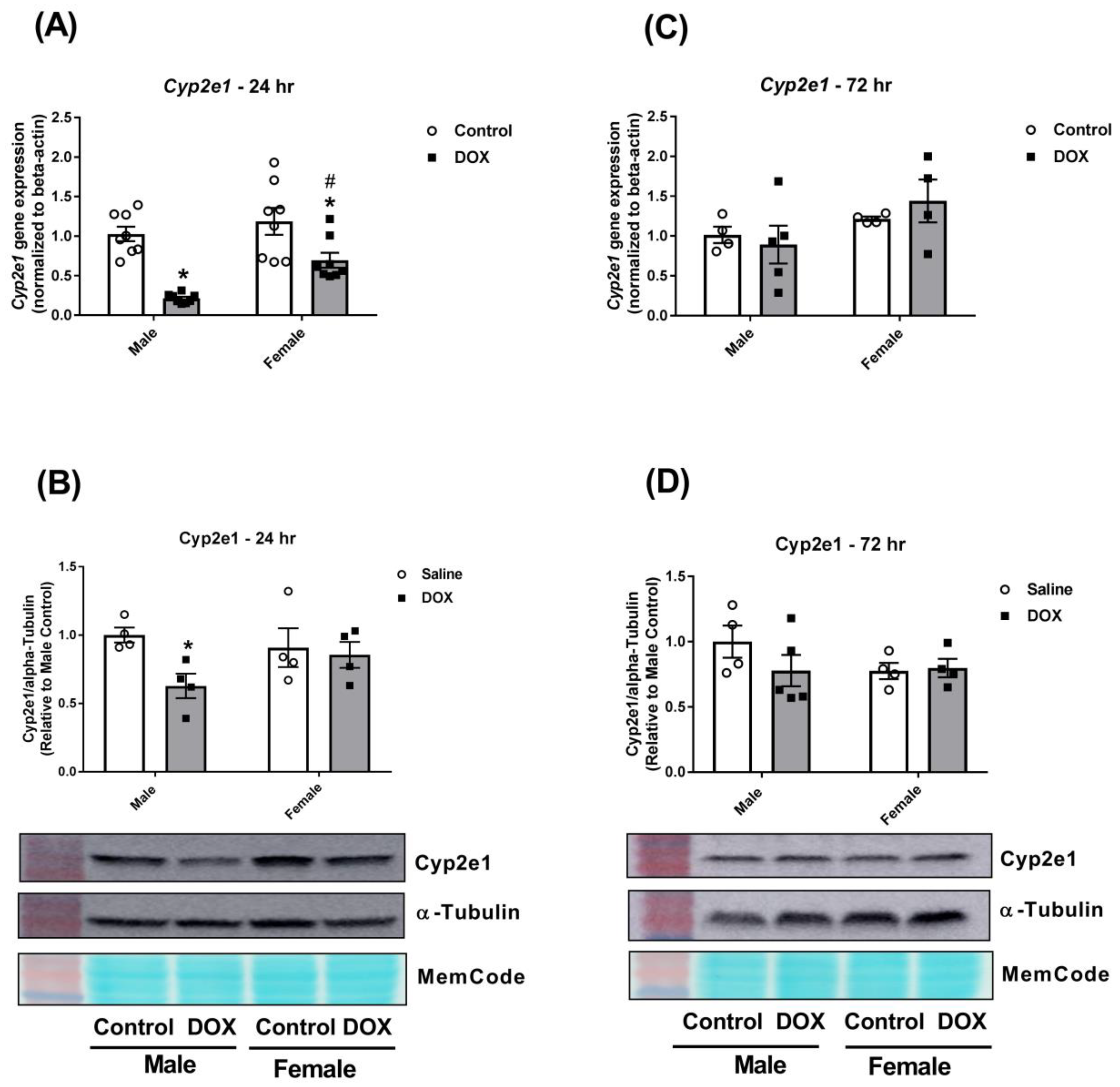
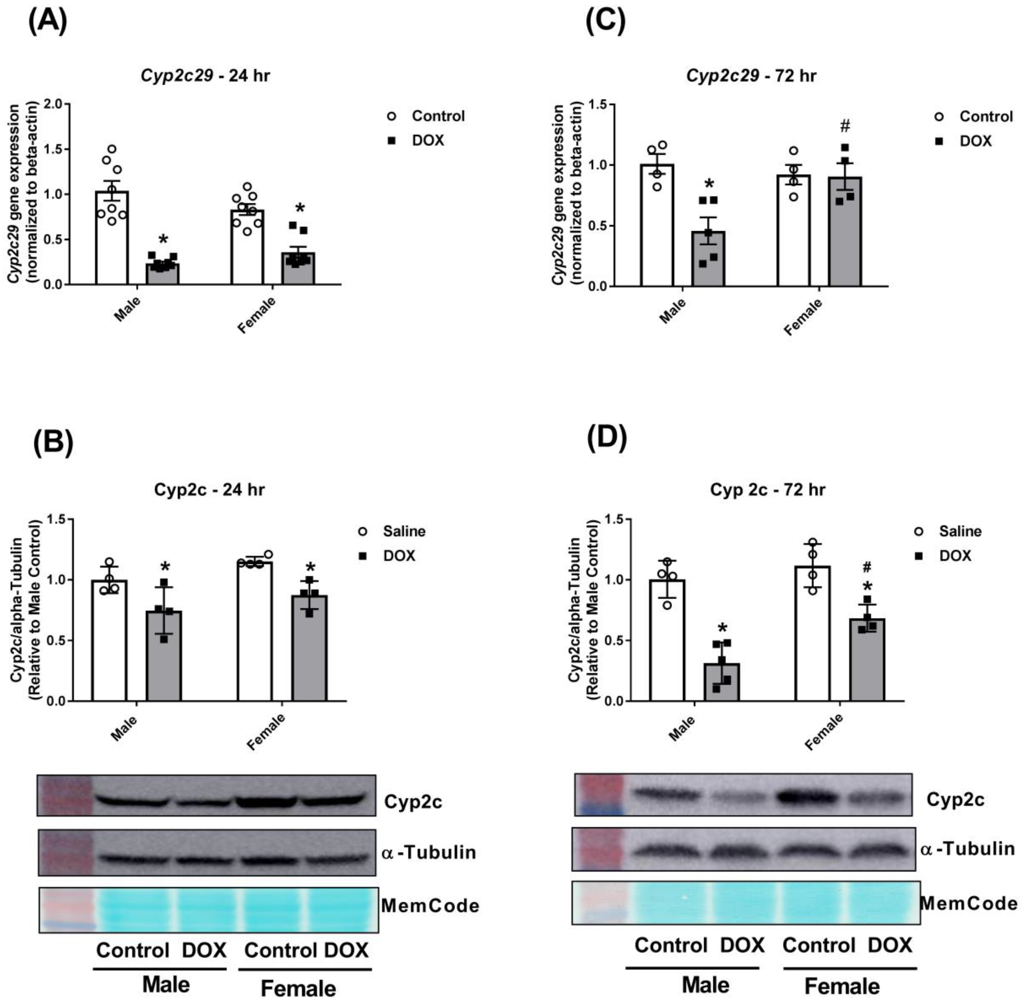
2.3. Effect of DOX Treatment on Cyp2 Family Expression

DOX caused a significant 75% and 50% inhibition in Cyp2c29 gene expression in male and female mice 24 h after administration, respectively (Figure 4A). Measurement of Cyp2c protein expression corroborated the gene expression results. There was a significant 25% and 20% inhibition of Cyp2c protein expression 24 h after DOX administration in male and female mice, respectively (Figure 4B). Interestingly, Cyp2c29 gene expression returned to the control value in females, but not in male mice, 72 h after DOX administration (Figure 4C). Two-way ANOVA showed a significant DOX effect as well as a significant interaction between DOX exposure and sex (Table 1). At the protein expression level, DOX caused 70% inhibition of Cyp2c protein expression in male mice, but only 30% inhibition in female mice, 72 h after DOX administration (Figure 4D). There was a significant difference in Cyp2c protein expression between DOX-treated male and female mice (Figure 4D).
DOX inhibited Cyp2e1 gene expression in both male and female mice 24 h after administration. However, there was significantly more inhibition in male (80% inhibition) than in female mice (30% inhibition) (Figure 5A). The marked inhibition of Cyp2e1 gene expression in male mice resulted in a significant 38% inhibition in Cyp2e1 protein expression in male mice only (Figure 5B). There were no significant changes in Cyp2e1 gene or protein expressions 72 h after DOX administration (Figure 5C,D).

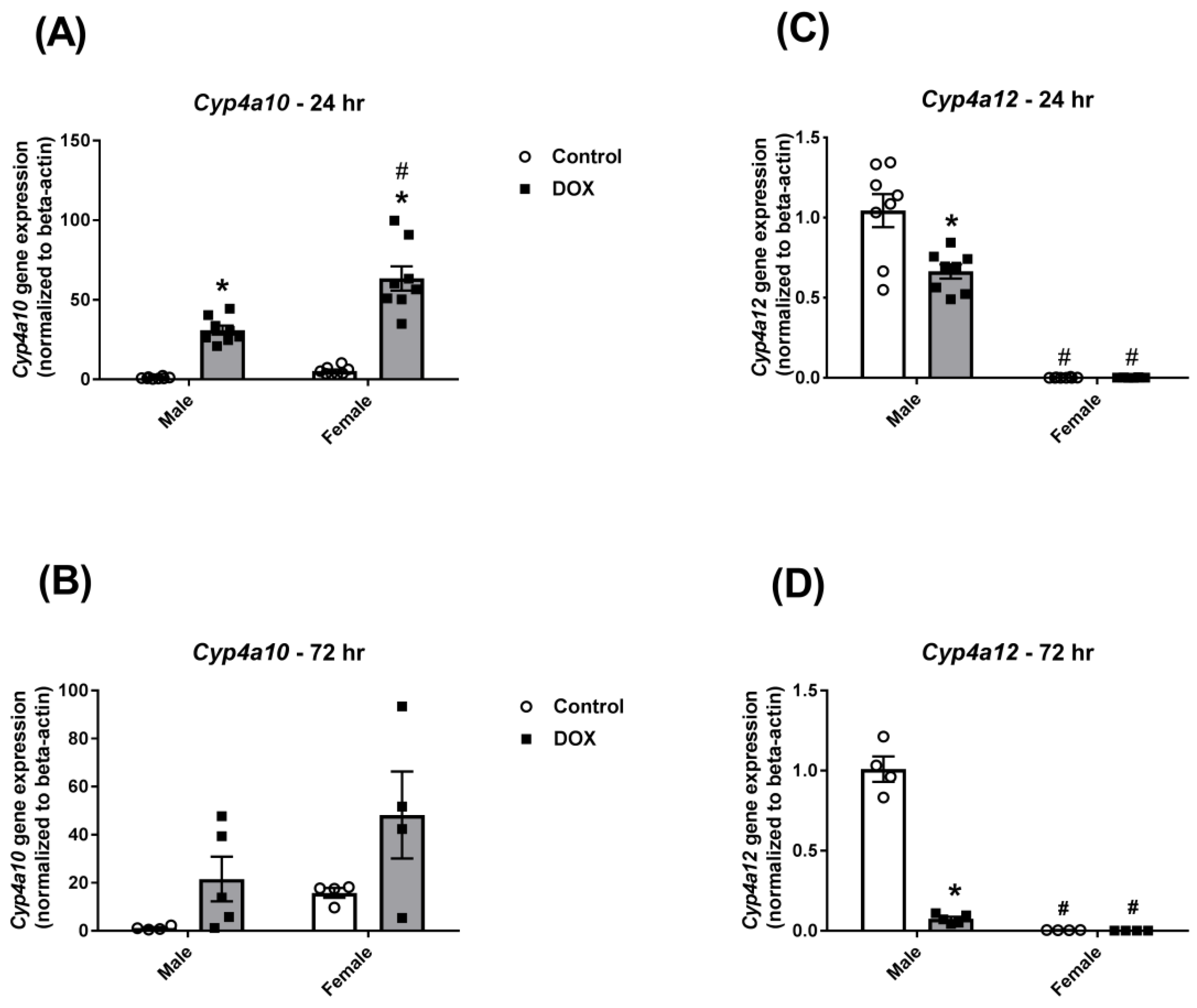
2.4. Effect of DOX Treatment on Cyp4a Sub-family Expression

DOX caused a significant 30-fold induction in Cyp4a10 gene expression in male mice 24 h after administration (Figure 6A). Basal Cyp4a10 gene expression was 5 times higher in females than males and was further induced 12-fold by DOX administration to be 60 times higher than its value in the control-male group (Figure 6A). Similar results were observed 72 h following acute DOX administration; nevertheless, the differences did not reach statistical significance (Figure 6B). Cyp4a12 was expressed in male mice at much higher levels than in female mice (Figure 6C,D), confirming that Cyp4a12 is a male-specific isoform as previously reported [24]. Interestingly, DOX caused a significant 35% and 90% inhibition of Cyp4a12 gene expression after 24 h and 72 h of administration, respectively, in male mice (Figure 6C,D).

3. Discussion

The current study demonstrates, for the first time, sex-related differences in DOX-induced systemic inflammation and DOX-induced alteration of hepatic CYP expression in mice. DOX-induced systemic inflammation is a clinically relevant adverse effect of DOX treatment since it has been reported in patients receiving DOX-based chemotherapy [10]. Furthermore, systemic inflammatory reaction is associated with poor outcomes in cancer patients [25], which may be attributed to the role of interleukins in mediating progression, metastatic processes, and drug resistance of cancer cells [26]. Indeed, nutraceuticals with anti-inflammatory properties such as lycopene, curcumin, and resveratrol have been shown to reduce the toxicity and improve the efficacy of chemotherapeutic agents [14,27]. We and others have reported significant sex-related differences in acute and chronic DOX-induced cardiotoxicity and nephrotoxicity in experimental animals [11,28,29,30,31,32]. Sexual dimorphism of acute DOX-induced cardiotoxicity is associated with higher expression of inflammatory genes in the heart of male than female mice [11]. Similarly, sexual dimorphism of chronic DOX-induced cardiotoxicity is associated with higher inflammatory cell infiltration in male than in female hearts [30,32]. In the current study, we demonstrate significant sexual dimorphism in DOX-induced systemic inflammation. In agreement with previous studies [10], acute DOX administration elicited a systemic inflammatory response in both male and female mice; however, the inflammatory reaction was stronger in male mice. The observed sexual dimorphism in DOX-induced systemic inflammation may contribute to the previously reported sex differences in DOX-induced organ-specific toxicity.
In addition to a potential role in mediating sexual dimorphism of DOX-induced toxicity, sex differences in DOX-induced systemic inflammation may also lead to sex differences in altering the expression of hepatic CYP enzymes by DOX. We previously demonstrated significant sexual dimorphism in DOX-induced alteration of CYP gene expression in the heart of C57Bl/6 mice [11]. In the current work, we observed a male-specific induction of Cyp1a1 gene expression 24 h after DOX administration. We previously reported that DOX upregulated CYP1A1 gene expression in H9c2 cardiomyoblasts [33] and in hearts of male Sprague Dawley rats [23], an observation that was later confirmed by other investigators [34,35]. Since inflammation has been shown to downregulate Cyp1a1 gene expression [16,36,37], the observed DOX-induced upregulation of the Cyp1a1 gene in the current work is likely mediated by mechanisms other than inflammation. Indeed, DOX has been shown to activate the aryl hydrocarbon receptor (AhR), the main transcription factor regulating Cyp1 family gene expression [34]. We also observed a significant DOX effect to upregulate Cyp1b1 gene expression 72 h following DOX administration in both male and female mice. In contrast to Cyp1a1, inflammation has been shown to upregulate Cyp1b1 [16,38]. Therefore, the observed DOX-induced upregulation of the Cyp1b1 gene can likely be attributed to both inflammation and AhR activation.
With regard to the Cyp2 family, acute DOX administration caused a marked inhibition of Cyp2c29 isoform gene expression and total Cyp2c protein expression in the liver of male and female mice. Nevertheless, DOX caused a stronger inhibition in male than in female mice 72 h following DOX administration. Importantly, CYP2C9 enzyme activity was reduced by 319% in breast cancer patients who were treated by DOX/cyclophosphamide therapy. Cyp2c enzymes are known to be down-regulated by inflammation [16,17,18,37]. Therefore, the observed sexually dimorphic downregulation is likely to be attributed to the sex difference in the DOX-induced inflammatory reaction. Since Cyp2c enzymes are known to metabolize arachidonic acid to epoxyeicosatrienoic acids [39], inhibition of Cyp2c enzymes may lead to a reduction in the level of these anti-inflammatory molecules, thus worsening DOX-induced inflammation via a positive feedback loop. Interestingly, inhibition of Cyp2c by DOX was more marked 72 h after administration than at the 24-hour time-point, demonstrating an exaggeration of the DOX effect over time. In contrast, DOX caused male-specific inhibition of the Cyp2e1 enzyme that was completely recovered at the 72-hour time-point. Downregulation of Cyp2e1 by DOX can also be attributed to DOX-induced inflammation since inflammation has been shown to downregulate hepatic Cyp2e1 [16].
Cyp4a is a family of enzymes that play an important role in the metabolism of endogenous compounds, e.g., arachidonic acid [22]. We previously demonstrated that acute DOX toxicity induced CYP4A enzymes in the liver of male Sprague Dawley rats [5]. In agreement with this study, DOX caused a marked induction of Cyp4a10 gene expression in both male and female mice in the current work. Lipopolysaccharide-induced inflammation has also been shown to upregulate hepatic Cyp4a enzymes in male rats [16]. In the current study, DOX caused a significant inhibition in Cyp4a12 gene expression in male mice. Since Cyp4a12 is a male-specific isoform [24], its inhibition may be attributed to DOX-induced perturbation in testosterone levels secondary to DOX-induced gonadotoxicity as we previously demonstrated [28]. In support of this argument, castration has been shown to inhibit Cyp4a protein expression and activity in male mice, an effect that was reversed by testosterone supplementation [40]. Further research is warranted to delineate the potential impact of DOX-induced gonadotoxicity on the regulation of sexually dimorphic CYP enzymes.

4. Materials and Methods

4.1. Animals

All experimental procedures involving animals were approved by the University of Minnesota Institutional Animal Care and Use Committee (IACUC number A3456-01; Protocol number 1508-32853A approved on 10/02/2015). Male (n = 26) and female (n = 24) C57Bl/6 mice were purchased from Charles River Laboratories (Raleigh, NC, USA) at twelve weeks of age. After an acclimation period of one week, mice were administered either 20 mg/kg DOX by intraperitoneal (IP) injection (DOX group) or an equivalent volume of sterile normal saline (control group) as we previously described [11,28]. Thereafter, mice were humanely euthanized at 24 h (8 male-control, 8 male-DOX, 8 female-control, and 8 female-DOX) or at 72 h (4 male-control, 5 male-DOX, 4 female-control, and 4 female-DOX) following DOX or saline administration. Mortality was observed in the male-DOX group followed for 72 h (1 out of 6 male-DOX mice) as previously reported in our earlier studies utilizing this model [11,28]. At the experimental end point, mice were euthanized by decapitation under isoflurane anesthesia. Terminal blood was collected, and livers were harvested, washed in ice-cold phosphate buffered saline solution, flash frozen in liquid nitrogen, and stored at −80 °C until further analysis.

4.2. Measurement of Inflammatory Markers

Terminal blood was collected from animals euthanized 24 h following DOX or saline administration and incubated at room temperature for 20 min to allow blood to clot. Samples were centrifuged at 4000 rpm for 30 min at 4 °C; serum was collected and stored at −80 °C until use. Serum samples were analyzed by the Cytokine Reference Laboratory (University of Minnesota) for mouse-specific IL-6 and TNF-alpha using the Luminex platform and done as a multiplex. The magnetic bead set (catalog LXSAMSM-03) was purchased from R&D Systems (Minneapolis, MN, USA). Samples were assayed according to manufacturer’s instructions. Briefly, fluorescent color-coded beads coated with a specific capture antibody were added to each sample. After incubation and washing, biotinylated detection antibody was added followed by phycoerythrin-conjugated streptavidin. The beads were read on a Luminex instrument (Bioplex 200, Bio-Rad Laboratories, Inc., Hercules, CA, USA), which is a dual-laser fluidics-based instrument. Samples were run in duplicate, and values were interpolated from 5-parameter fitted standard curves.

4.3. RNA Extraction and Real-time PCR

Total RNA from the frozen liver tissues was isolated using TRIzol® reagent (Life Technologies, Carlsbad, CA, USA), according to the manufacturer’s instructions, and quantified by using a Nanodrop 8000 spectrophotometer (Thermo Fisher Scientific, Wilmington, DE, USA). Thereafter, 1.5 μg of total RNA from each sample was used to synthesize first-strand cDNA by the high-capacity cDNA reverse transcription kit (Life Technologies, Carlsbad, CA, USA) according to the manufacturer’s instructions. Real time-polymerase chain reaction (PCR) was used to measure specific mRNA expression by subjecting the resulting cDNA to PCR amplification using 384-well optical reaction plates in an ABI 7900HT instrument (Applied Biosystems, Foster City, CA, USA). The 20 μL reaction mix contained 1 μL of cDNA sample, 0.025 μL of 30 μM forward primer and 0.025 μL of 30 μM reverse primer (40 nM final concentration of each primer), 10 μL of SYBR Green Universal Mastermix (Life Technologies, Carlsbad, CA, USA), and 8.95 μL of nuclease-free water as we described previously. Since we previously reported sex-dependent changes of certain CYP genes by acute DOX exposure in hearts of C57Bl/6 mice [11], these CYP genes were selected for the current study. The primers used in the current study were chosen from previously published studies, checked with the Primer-BLAST on-line tool, and are listed in Table 2. Thermocycling conditions were initiated at 95 °C for 10 min, followed by 40 PCR cycles of denaturation at 95 °C for 15 sec, and annealing/extension at 60 °C for 1 min. To ensure the specificity of the primers and the purity of the final PCR product, melting curve analysis was performed at the end of each cycle.

4.4. Protein Extraction and Western Blotting

Frozen liver tissues were homogenized as described previously, and protein concentration was determined using the Pierce bicinchoninic acid (BCA) protein assay kit according to manufacturer’s instructions (Pierce, Thermo Fisher Scientific, Rockford, IL, USA). Thereafter, 50 μg of protein per sample was separated by sodium dodecyl sulfate-polyacrylamide gel electrophoresis (SDS-PAGE) and electro-transferred onto nitrocellulose membranes as we previously described [41]. Primary rabbit antibodies against CYP2C and CYP2E1 (catalog ab137015 and ab151544; 1:1000 dilution) were purchased from Abcam (Cambridge, MA, USA). Primary rabbit antibody against alpha-tubulin (catalog 2144; 1:1000 dilution) was purchased from Cell Signaling Technology (Danvers, MA, USA). Secondary anti-rabbit conjugated to HRP was purchased from Jackson ImmunoResearch (catalog 111-035-144; 1:10,000 dilution; West Grove, PA, USA). Band intensities were quantified using ImageJ software (National Institutes of Health, Bethesda, MD, USA). Alpha-tubulin protein levels were used as loading controls to normalize the band intensities.

4.5. Statistical Analysis

Statistical analysis and data presentation were performed using GraphPad Prism software (version 7.04) for Windows, La Jolla California USA, www.graphpad.com. Data are presented as the mean ± SEM. Comparisons among different sex and treatment groups were performed by 2-way analysis of variance (ANOVA), followed by Tukey’s multiple comparison tests. A p value of < 0.05 was taken to indicate statistical significance.

5. Conclusions

Acute DOX administration elicits a systemic inflammatory response in a sex-dependent manner. Male mice are more susceptible to DOX-induced inflammation, which may contribute to the previously reported sexual dimorphism of DOX-induced cardiotoxicity and nephrotoxicity. Importantly, sex differences in DOX-induced inflammation may also cause sex-dependent alteration of hepatic CYP enzymes. Nevertheless, other mechanisms may also contribute to the alteration of hepatic CYP by acute DOX administration, including AhR activation and perturbation in sex hormones. Further research is needed to delineate the possible contribution of these factors to the sexual dimorphism of DOX-induced alteration of hepatic CYP expression.

Author Contributions

Conceptualization, B.N.Z.; formal analysis, M.K.O.G. and B.N.Z.; data curation, M.K.O.G., I.Y.A., and C.A.L.; writing—original draft preparation, B.N.Z.; writing—review and editing, M.K.O.G., I.Y.A., C.A.L., and B.N.Z.; supervision, B.N.Z.; project administration, M.K.O.G. and B.N.Z.; funding acquisition, B.N.Z. All authors have read and agreed to the published version of the manuscript.

Funding

Beshay N. Zordoky is a Masonic Cancer Center Women’s Health Scholar, sponsored by the Masonic Cancer Center, a comprehensive cancer center designated by the National Cancer Institute and administered by the University of Minnesota Women’s Health Research Program. This research was supported by a research grant from the Rally Foundation for Childhood Cancer (Award ID 582420) and by the National Institutes of Health’s National Center for Advancing Translational Sciences, grant UL1TR002494. The content is solely the responsibility of the authors and does not necessarily represent the official views of the National Institutes of Health’s National Center for Advancing Translational Sciences.

Acknowledgments

Experiments using the NanoDrop 8000, ABI 7900 HT, and Agilent 2100 Bioanalyzer were done with staff support at the University of Minnesota Genomics Center. Experiments using the Amersham Imager 600UV were done with staff support at the University of Minnesota Imaging Center. Experiments using the Luminex platform were done with staff support at the University of Minnesota Cytokine Reference Laboratory.

Conflicts of Interest

The authors declare no conflict of interest. The funders had no role in the design of the study; in the collection, analyses, or interpretation of data; in the writing of the manuscript, or in the decision to publish the results.

Abbreviations

AhRAryl hydrocarbon Receptor
Cox-2Cyclooxygenase-2
CYPCytochrome P450
DOXDoxorubicin
IL-6Interleukin-6
TNF-alphaTumor Necrosis Factor-alpha

References

  1. Lipshultz, S.E.; Giantris, A.L.; Lipsitz, S.R.; Kimball Dalton, V.; Asselin, B.L.; Barr, R.D.; Clavell, L.A.; Hurwitz, C.A.; Moghrabi, A.; Samson, Y.; et al. Doxorubicin administration by continuous infusion is not cardioprotective: The Dana-Farber 91-01 Acute Lymphoblastic Leukemia protocol. J. Clin. Oncol. 2002, 20, 1677–1682. [Google Scholar] [CrossRef] [PubMed]
  2. Damodar, G.; Smitha, T.; Gopinath, S.; Vijayakumar, S.; Rao, Y. An evaluation of hepatotoxicity in breast cancer patients receiving injection Doxorubicin. Ann. Med. Health Sci. Res. 2014, 4, 74–79. [Google Scholar] [CrossRef] [PubMed]
  3. Barakat, B.M.; Ahmed, H.I.; Bahr, H.I.; Elbahaie, A.M. Protective Effect of Boswellic Acids against Doxorubicin-Induced Hepatotoxicity: Impact on Nrf2/HO-1 Defense Pathway. Oxidative Med. Cell. Longev. 2018, 2018, 8296451. [Google Scholar] [CrossRef] [PubMed] [Green Version]
  4. Jacevic, V.; Dragojevic-Simic, V.; Tatomirovic, Z.; Dobric, S.; Bokonjic, D.; Kovacevic, A.; Nepovimova, E.; Valis, M.; Kuca, K. The Efficacy of Amifostine against Multiple-Dose Doxorubicin-Induced Toxicity in Rats. Int. J. Mol. Sci. 2018, 19, 2370. [Google Scholar] [CrossRef] [PubMed] [Green Version]
  5. Zordoky, B.N.; Anwar-Mohamed, A.; Aboutabl, M.E.; El-Kadi, A.O.S. Acute doxorubicin toxicity differentially alters cytochrome P450 expression and arachidonic acid metabolism in rat kidney and liver. Drug Metab. Dispos. 2011, 39, 1440–1450. [Google Scholar] [CrossRef] [PubMed] [Green Version]
  6. Ali, K.; Sial, A.A.; Baig, M.T.; Ansari, S.H.; Adil, S.O.; Shamsi, T.S. Detection of the Incidence of Infections and Acute Biochemical Changes in Diffused Large B-Cell Lymphoma Patients Treated with Cyclophosphamide, Doxorubicin, Vincristine and Prednisone (CHOP) with and without Rituximab. Curr. Drug Saf. 2018, 13, 102–106. [Google Scholar] [CrossRef]
  7. Shen, R.L.; Pontoppidan, P.E.; Rathe, M.; Jiang, P.; Hansen, C.F.; Buddington, R.K.; Heegaard, P.M.; Muller, K.; Sangild, P.T. Milk diets influence doxorubicin-induced intestinal toxicity in piglets. Am. J. Physiol. Gastrointest. Liver Physiol. 2016, 311, G324–G333. [Google Scholar] [CrossRef] [Green Version]
  8. Carron, P.L.; Padilla, M.; Maurizi Balzan, J. Nephrotic syndrome and acute renal failure during pegylated liposomal doxorubicin treatment. Hemodial. Int. 2014, 18, 846–847. [Google Scholar] [CrossRef]
  9. Mohamed, N.; Goldstein, J.; Schiff, J.; John, R. Collapsing glomerulopathy following anthracycline therapy. Am. J. Kidney Dis. 2013, 61, 778–781. [Google Scholar] [CrossRef]
  10. Wang, L.; Chen, Q.; Qi, H.; Wang, C.; Wang, C.; Zhang, J.; Dong, L. Doxorubicin-Induced Systemic Inflammation Is Driven by Upregulation of Toll-Like Receptor TLR4 and Endotoxin Leakage. Cancer Res. 2016, 76, 6631–6642. [Google Scholar] [CrossRef] [Green Version]
  11. Grant, M.K.; Seelig, D.M.; Sharkey, L.C.; Zordoky, B.N. Sex-dependent alteration of cardiac cytochrome P450 gene expression by doxorubicin in C57Bl/6 mice. Biol. Sex Differ. 2017, 8, 1. [Google Scholar] [CrossRef] [PubMed] [Green Version]
  12. Zhang, Q.L.; Yang, J.J.; Zhang, H.S. Carvedilol (CAR) combined with carnosic acid (CAA) attenuates doxorubicin-induced cardiotoxicity by suppressing excessive oxidative stress, inflammation, apoptosis and autophagy. Biomed. Pharmacother. 2019, 109, 71–83. [Google Scholar] [CrossRef] [PubMed]
  13. Pecoraro, M.; Del Pizzo, M.; Marzocco, S.; Sorrentino, R.; Ciccarelli, M.; Iaccarino, G.; Pinto, A.; Popolo, A. Inflammatory mediators in a short-time mouse model of doxorubicin-induced cardiotoxicity. Toxicol. Appl. Pharmacol. 2016, 293, 44–52. [Google Scholar] [CrossRef] [PubMed]
  14. Quagliariello, V.; Vecchione, R.; Coppola, C.; Di Cicco, C.; De Capua, A.; Piscopo, G.; Paciello, R.; Narciso, V.; Formisano, C.; Taglialatela-Scafati, O.; et al. Cardioprotective Effects of Nanoemulsions Loaded with Anti-Inflammatory Nutraceuticals against Doxorubicin-Induced Cardiotoxicity. Nutrients 2018, 10, 1304. [Google Scholar] [CrossRef] [PubMed] [Green Version]
  15. Sauter, K.A.; Wood, L.J.; Wong, J.; Iordanov, M.; Magun, B.E. Doxorubicin and daunorubicin induce processing and release of interleukin-1beta through activation of the NLRP3 inflammasome. Cancer Biol. Ther. 2011, 11, 1008–1016. [Google Scholar] [CrossRef] [PubMed] [Green Version]
  16. Anwar-mohamed, A.; Zordoky, B.N.; Aboutabl, M.E.; El-Kadi, A.O. Alteration of cardiac cytochrome P450-mediated arachidonic acid metabolism in response to lipopolysaccharide-induced acute systemic inflammation. Pharmacol. Res. 2010, 61, 410–418. [Google Scholar] [CrossRef]
  17. Aitken, A.E.; Morgan, E.T. Gene-specific effects of inflammatory cytokines on cytochrome P450 2C, 2B6 and 3A4 mRNA levels in human hepatocytes. Drug Metab. Dispos. 2007, 35, 1687–1693. [Google Scholar] [CrossRef]
  18. Barclay, T.B.; Peters, J.M.; Sewer, M.B.; Ferrari, L.; Gonzalez, F.J.; Morgan, E.T. Modulation of cytochrome P-450 gene expression in endotoxemic mice is tissue specific and peroxisome proliferator-activated receptor-alpha dependent. J. Pharmacol. Exp. Ther. 1999, 290, 1250–1257. [Google Scholar]
  19. Fukuno, S.; Nagai, K.; Yamamoto, K.; Tanimura, T.; Nabe, T.; Konishi, H. Pharmacokinetic interference of doxorubicin with tolbutamide due to reduced metabolic clearance with increased serum unbound fraction in rats. Biopharm. Drug Dispos. 2019, 40, 225–233. [Google Scholar] [CrossRef]
  20. Chen, Z.B.; Zhi, A.Y.; Lin, F.Y.; Li, D.; Yu, X.G.; Chen, W.H.; Hu, L.F. Pharmacokinetic of four probe drugs in adriamycin-induced nephropathy rat. Eur. Rev. Med. Pharmacol. Sci. 2014, 18, 1439–1447. [Google Scholar]
  21. Elkiran, T.; Harputluoglu, H.; Yasar, U.; Babaoglu, M.O.; Dincel, A.K.; Altundag, K.; Ozisik, Y.; Guler, N.; Bozkurt, A. Differential alteration of drug-metabolizing enzyme activities after cyclophosphamide/adriamycin administration in breast cancer patients. Methods Find. Exp. Clin. Pharmacol. 2007, 29, 27–32. [Google Scholar] [CrossRef] [PubMed]
  22. Zordoky, B.N.; El-Kadi, A.O. Effect of cytochrome P450 polymorphism on arachidonic acid metabolism and their impact on cardiovascular diseases. Pharmacol. Ther. 2010, 125, 446–463. [Google Scholar] [CrossRef] [PubMed]
  23. Zordoky, B.N.; Anwar-Mohamed, A.; Aboutabl, M.E.; El-Kadi, A.O. Acute doxorubicin cardiotoxicity alters cardiac cytochrome P450 expression and arachidonic acid metabolism in rats. Toxicol. Appl. Pharmacol. 2010, 242, 38–46. [Google Scholar] [CrossRef] [PubMed]
  24. Jeffery, B.; Choudhury, A.I.; Horley, N.; Bruce, M.; Tomlinson, S.R.; Roberts, R.A.; Gray, T.J.; Barrett, D.A.; Shaw, P.N.; Kendall, D.; et al. Peroxisome proliferator activated receptor alpha regulates a male-specific cytochrome P450 in mouse liver. Arch. Biochem. Biophys. 2004, 429, 231–236. [Google Scholar] [CrossRef]
  25. Sun, F.; Zhu, J.; Lu, S.; Zhen, Z.; Wang, J.; Huang, J.; Ding, Z.; Zeng, M.; Sun, X. An inflammation-based cumulative prognostic score system in patients with diffuse large B cell lymphoma in rituximab era. BMC Cancer 2018, 18, 5. [Google Scholar] [CrossRef] [Green Version]
  26. Serri, C.; Quagliariello, V.; Iaffaioli, R.V.; Fusco, S.; Botti, G.; Mayol, L.; Biondi, M. Combination therapy for the treatment of pancreatic cancer through hyaluronic acid-decorated nanoparticles loaded with quercetin and gemcitabine: A preliminary in vitro study. J. Cell. Physiol. 2019, 234, 4959–4969. [Google Scholar] [CrossRef]
  27. Abdelgawad, I.Y.; Grant, M.K.O.; Zordoky, B.N. Leveraging the Cardio-Protective and Anticancer Properties of Resveratrol in Cardio-Oncology. Nutrients 2019, 11, 627. [Google Scholar] [CrossRef] [Green Version]
  28. Grant, M.K.O.; Seelig, D.M.; Sharkey, L.C.; Choi, W.S.V.; Abdelgawad, I.Y.; Zordoky, B.N. Sexual dimorphism of acute doxorubicin-induced nephrotoxicity in C57Bl/6 mice. PLoS ONE 2019, 14, e0212486. [Google Scholar] [CrossRef]
  29. Zordoky, B.N.; Radin, M.J.; Heller, L.; Tobias, A.; Matise, I.; Apple, F.S.; McCune, S.A.; Sharkey, L.C. The interplay between genetic background and sexual dimorphism of doxorubicin-induced cardiotoxicity. Cardiooncology 2016, 2, 4. [Google Scholar] [CrossRef] [Green Version]
  30. Jenkins, G.R.; Lee, T.; Moland, C.L.; Vijay, V.; Herman, E.H.; Lewis, S.M.; Davis, K.J.; Muskhelishvili, L.; Kerr, S.; Fuscoe, J.C.; et al. Sex-related differential susceptibility to doxorubicin-induced cardiotoxicity in B6C3F1 mice. Toxicol. Appl. Pharmacol. 2016, 310, 159–174. [Google Scholar] [CrossRef] [PubMed]
  31. Meiners, B.; Shenoy, C.; Zordoky, B.N. Clinical and preclinical evidence of sex-related differences in anthracycline-induced cardiotoxicity. Biol. Sex Differ. 2018, 9, 38. [Google Scholar] [CrossRef] [PubMed]
  32. Zhang, J.; Knapton, A.; Lipshultz, S.E.; Cochran, T.R.; Hiraragi, H.; Herman, E.H. Sex-related differences in mast cell activity and doxorubicin toxicity: A study in spontaneously hypertensive rats. Toxicol. Pathol. 2014, 42, 361–375. [Google Scholar] [CrossRef] [PubMed]
  33. Zordoky, B.N.; El-Kadi, A.O. Induction of several cytochrome P450 genes by doxorubicin in H9c2 cells. Vasc. Pharmacol. 2008, 49, 166–172. [Google Scholar] [CrossRef] [PubMed]
  34. Volkova, M.; Palmeri, M.; Russell, K.S.; Russell, R.R. Activation of the aryl hydrocarbon receptor by doxorubicin mediates cytoprotective effects in the heart. Cardiovasc. Res. 2011, 90, 305–314. [Google Scholar] [CrossRef] [PubMed] [Green Version]
  35. Asnani, A.; Zheng, B.; Liu, Y.; Wang, Y.; Chen, H.H.; Vohra, A.; Chi, A.; Cornella-Taracido, I.; Wang, H.; Johns, D.G.; et al. Highly potent visnagin derivatives inhibit Cyp1 and prevent doxorubicin cardiotoxicity. JCI Insight 2018, 3, e96753. [Google Scholar] [CrossRef] [PubMed] [Green Version]
  36. Gharavi, N.; El-Kadi, A.O. Down-regulation of aryl hydrocarbon receptor-regulated genes by tumor necrosis factor-alpha and lipopolysaccharide in murine hepatoma Hepa 1c1c7 cells. J. Pharm. Sci. 2005, 94, 493–506. [Google Scholar] [CrossRef] [PubMed]
  37. Zordoky, B.N.; El-Kadi, A.O. Role of NF-kappaB in the regulation of cytochrome P450 enzymes. Curr. Drug Metab. 2009, 10, 164–178. [Google Scholar] [CrossRef]
  38. Malaplate-Armand, C.; Ferrari, L.; Masson, C.; Siest, G.; Batt, A.M. Astroglial CYP1B1 up-regulation in inflammatory/oxidative toxic conditions: IL-1beta effect and protection by N-acetylcysteine. Toxicol. Lett. 2003, 138, 243–251. [Google Scholar] [CrossRef]
  39. Deng, Y.; Theken, K.N.; Lee, C.R. Cytochrome P450 epoxygenases, soluble epoxide hydrolase, and the regulation of cardiovascular inflammation. J. Mol. Cell. Cardiol. 2010, 48, 331–341. [Google Scholar] [CrossRef] [Green Version]
  40. Hiratsuka, M.; Matsuura, T.; Watanabe, E.; Sato, M.; Suzuki, Y. Sex and strain differences in constitutive expression of fatty acid omega-hydroxylase (CYP4A-related proteins) in mice. J. Biochem. 1996, 119, 340–345. [Google Scholar] [CrossRef]
  41. Carlson, A.; Alderete, K.S.; Grant, M.K.O.; Seelig, D.M.; Sharkey, L.C.; Zordoky, B.N.M. Anticancer effects of resveratrol in canine hemangiosarcoma cell lines. Vet. Comp. Oncol. 2018, 16, 253–261. [Google Scholar] [CrossRef] [PubMed]
Figure 1. Effect of DOX treatment on the serum level of inflammatory markers. Serum was collected from male or female C57Bl/6 mice 24 h following the administration of a single intraperitoneal injection of 20 mg/kg DOX or saline (n = 8 per group). Inflammatory markers (A) IL-6 and (B) TNF-alpha were measured using the Luminex platform. Data are presented as the mean ± SEM. * p < 0.05, compared to saline-treated mice of the same sex; # p < 0.05, compared to male DOX-treated mice.
Ijms 21 01279 g001
Figure 2. Effect of DOX treatment on the gene expression of inflammatory markers in the liver. Livers were collected from male or female C57Bl/6 mice 24 h following the administration of a single intraperitoneal injection of 20 mg/kg DOX or saline, and total RNA was isolated (n = 8 per group). Gene expression of (A) IL-6, (B) TNF-alpha, or (C) Cox-2 was determined by real-time PCR. Results were normalized to beta-actin and are expressed relative to the male control. Data are presented as the mean ± SEM. * p < 0.05, compared to control mice of the same sex; # p < 0.05, compared to male DOX-treated mice.
Ijms 21 01279 g002
Figure 3. Effect of DOX treatment on the gene expression of the Cyp1 family in the liver. Livers were collected from male or female C57Bl/6 mice 24 h (n = 8 per group) or 72 h (n = 4–5 per group) following the administration of a single intraperitoneal injection of 20 mg/kg DOX or saline, and total RNA was isolated. Gene expression of Cyp1a1 was determined by real-time PCR in samples collected (A) 24 or (B) 72 h following DOX treatment. Gene expression of Cyp1b1 in samples collected (C) 24 or (D) 72 h following DOX treatment. Results were normalized to beta-actin and are expressed relative to the male control. Data are presented as the mean ± SEM. * p < 0.05, compared to control mice of the same sex; # p < 0.05, compared to male DOX-treated mice.
Ijms 21 01279 g003
Figure 4. Effect of DOX treatment on the expression of the Cyp2c sub-family in the liver. Livers were collected from male or female C57Bl/6 mice 24 or 72 h following the administration of a single intraperitoneal injection of 20 mg/kg DOX or saline, and total proteins and RNA were isolated. (A) Gene expression of Cyp2c29 was determined by real-time PCR (n = 8 per group) and (B) protein levels of total Cyp2c were determined by western blotting (n = 4 per group) in samples collected 24 h following DOX treatment. (C) Gene expression of Cyp2c29 (n = 4–5 per group) and (D) protein levels of Cyp2c (n = 4–5 per group) were determined in samples collected 72 h following DOX treatment. PCR results were normalized to beta-actin, and protein expression was normalized to alpha-tubulin. Results of all groups are expressed relative to the male control. Data are presented as the mean ± SEM. * p < 0.05, compared to control mice of the same sex; # p < 0.05, compared to male DOX-treated mice.
Ijms 21 01279 g004
Figure 5. Effect of DOX treatment on the expression of Cyp2e1 in the liver. Livers were collected from male or female C57Bl/6 mice 24 or 72 h following the administration of a single intraperitoneal injection of 20 mg/kg DOX or saline, and total proteins and RNA were isolated. (A) Gene expression of Cyp2e1 was determined by real-time PCR (n = 8 per group), and (B) protein levels of Cyp2e1 were determined by western blotting (n = 4 per group) in samples collected 24 h following DOX treatment. (C) Gene expression of Cyp2e1 (n = 4–5 per group) and (D) protein levels of Cyp2e1 (n = 4–5 per group) were determined in samples collected 72 h following DOX treatment. PCR results were normalized to beta-actin, and protein expression was normalized to alpha-tubulin. Results of all groups are expressed relative to the male control. Data are presented as the mean ± SEM. * p < 0.05, compared to control mice of the same sex; # p < 0.05, compared to male DOX-treated mice.
Ijms 21 01279 g005
Figure 6. Effect of DOX treatment on the gene expression of the Cyp4a sub-family in the liver. Livers were collected from male or female C57Bl/6 mice 24 h (n = 8 per group) or 72 h (n = 4–5 per group) following the administration of a single intraperitoneal injection of 20 mg/kg DOX or saline, and total RNA was isolated. Gene expression of Cyp4a10 was determined by real-time PCR in samples collected (A) 24 or (B) 72 h following DOX treatment. Gene expression of Cyp4a12 in samples collected (C) 24 or (D) 72 h following DOX treatment. Results were normalized to beta-actin and are expressed relative to the male control. Data are presented as the mean ± SEM. * p < 0.05, compared to control mice of the same sex; # p < 0.05, compared to male mice of the same treatment.
Ijms 21 01279 g006
Table 1. Two-Way ANOVA for CYP Gene Expression Data.
24 h Post-DOX
DOX EffectSex EffectInteraction
F (1, 28)p ValueF (1, 28)p ValueF (1, 28)p Value
Cyp1a114.750.000614.610.00079.8550.004
Cyp1b1nsnsns
Cyp2c2982.54<0.0001ns5.580.0254
Cyp2e136.63<0.00018.8170.0061ns
Cyp4a10114.9<0.000120.010.000111.670.002
Cyp4a1211.420.0022229.9<0.000111.450.0021
72 h Post-DOX
DOX EffectSex EffectInteraction
F (1, 13)p ValueF (1, 13)p ValueF (1, 13)p Value
Cyp1a1nsnsns
Cyp1b19.8280.0079nsns
Cyp2c298.0050.0142ns7.1360.0192
Cyp2e1nsnsns
Cyp4a106.7110.0224nsns
Cyp4a12153.1<0.0001202.6<0.0001151.4<0.0001
Table 2. Primers used in this study.
GeneForward Primer (5′–3′)Reverse Primer (5′–3’)
IL-6CCA GAG ATA CAA AGA AAT GAT GGACT CCA GAA GAC CAG AGG AAA T
TNF-alphaCCA GAC CCT CAC ACT CAG ATC ACAC TTG GTG GTT TGC TAC GAC
Cox-2CTG GTG CCT GGT CTG ATG ATGGGC AAT GCG GTT CTG ATA CTG
Cyp1a1GGT TAA CCA TGA CCG GGA ACTTGC CCA AAC CAA AGA GAG TGA
Cyp1b1AAT GAG GAG TTC GGG CGC ACAGGC GTG TGG AAT GGT GAC AGG
Cyp2c29TGG TCC ACC CAA AAG AAA TTG AGCA GAG AGG CAA ATC CAT TCA
Cyp2e1CCC AAG TCT TTA ACC AAG TTG GCCTT CCA TGT GGG TCC ATT ATT GA
Cyp4a10GTG CTG AGG TGG ACA CAT TCA TTGT GGC CAG AGC ATA GAA GAT C
Cyp4a12TGA CCC CAG CTT TCC ACT ATGTTG TTC AGG TCC TCA ACT GCC
Beta-actinTAT TGG CAA CGA GCG GTT CCGGC ATA GAG GTC TTT ACG GAT GTC

