Article

A Coil-to-Helix Transition Serves as a Binding Motif for hSNF5 and BAF155 Interaction

1
Structural Biochemistry & Molecular Biophysics Laboratory, Department of Biochemistry, College of Life Science and Biotechnology, Yonsei University, Seoul 120-740, Korea
2
Department of Agricultural Biotechnology and Research Institute of Agriculture and Life Sciences, Seoul National University, 1 Gwanak-ro, Gwanak-gu, Seoul 08826, Korea
3
Center for In Silico Protein Science and Center for Advanced Computation, Korea Institute for Advanced Study, Seoul 130-722, Korea
4
Division of Magnetic Resonance Research, Korea Basic Science Institute, Yangcheong-Ri 804-1, Ochang-Eup, Cheongwon-Gun, Chungcheongbuk-Do 363-883, Korea
5
National Center for Inter-University Research Facilities, Seoul National University, 1 Gwanak-ro, Gwanak-gu, Seoul 08826, Korea
6
Drug Design Laboratory, Graduate School of Medical Life Science, Yokohama City University, Tsurumi, Yokohama 230-0045, Japan
7
Department of Biological Sciences, Institute of Molecular Biology and Genetics, Research Center for Functional Cellulomics, Seoul National University, Seoul 151-742, Korea
8
Center for In Silico Protein Science and School of Computational Sciences, Korea Institute for Advanced Study, Seoul 130-722, Korea
*
Authors to whom correspondence should be addressed.
These authors contributed equally.
Int. J. Mol. Sci. 2020, 21(7), 2452; https://0-doi-org.brum.beds.ac.uk/10.3390/ijms21072452
Submission received: 13 March 2020 / Revised: 27 March 2020 / Accepted: 30 March 2020 / Published: 1 April 2020

Abstract

:
Human SNF5 and BAF155 constitute the core subunit of multi-protein SWI/SNF chromatin-remodeling complexes that are required for ATP-dependent nucleosome mobility and transcriptional control. Human SNF5 (hSNF5) utilizes its repeat 1 (RPT1) domain to associate with the SWIRM domain of BAF155. Here, we employed X-ray crystallography, nuclear magnetic resonance (NMR) spectroscopy, and various biophysical methods in order to investigate the detailed binding mechanism between hSNF5 and BAF155. Multi-angle light scattering data clearly indicate that hSNF5171–258 and BAF155SWIRM are both monomeric in solution and they form a heterodimer. NMR data and crystal structure of the hSNF5171–258/BAF155SWIRM complex further reveal a unique binding interface, which involves a coil-to-helix transition upon protein binding. The newly formed αN helix of hSNF5171–258 interacts with the β2–α1 loop of hSNF5 via hydrogen bonds and it also displays a hydrophobic interaction with BAF155SWIRM. Therefore, the N-terminal region of hSNF5171–258 plays an important role in tumorigenesis and our data will provide a structural clue for the pathogenesis of Rhabdoid tumors and malignant melanomas that originate from mutations in the N-terminal loop region of hSNF5.

Graphical Abstract

1. Introduction

Chromatin remodeling is a process that changes the chromatin structure between a condensed state and a transcriptionally accessible state, which is essential for the control of gene expression [1,2,3,4]. The chromatin structures are mediated by various covalent modifications in histones and nucleic acids, along with a set of multi-protein complexes that regulate and respond to these modifications [5]. SWI/SNF (SWItch/Sucrose Non-Fermentable) is an evolutionarily conserved chromatin-remodeling complex that couples ATP hydrolysis with chromatin structure rearrangement. The eukaryotic SWI/SNF complex consists of 12–15 subunits, with an overall molecular weight of ~2 MDa, and it controls diverse cellular processes, such as transcriptional regulation, differentiation, apoptosis, and tumorigenesis [6,7,8,9,10,11,12]. SWI/SNF is comprised of invariable core components, including BRG1/hBRM, SNF5/INI1/BAF47, BAF155/SRG3, and BAF170, as well as a number of variable components that confer functional diversity via combinatorial assembly [13,14,15,16]. The core proteins, SNF5, BAF155, and BAF170, are evolutionarily conserved among yeast, flies, plants, and mammals [17,18].
Human SNF5 (hSNF5) regulates the cell cycle at various stages of the mitotic checkpoint and it interacts with a variety of proteins, such as HIV-1 integrase, c-Myc, p53, Epstein-Barr virus nuclear protein 2, and human papillomavirus E1 protein [19,20,21,22]. The interaction with HIV-1 integrase affects viral integration and infectivity [23,24]. hSNF5 is involved in tumor proliferation and progression in he p16-RB pathway, WNT signaling pathway, sonic hedgehog signaling pathway and Polycomb pathway [25]. Additionally, truncating mutations of hSNF5 gene lead to aggressive pediatric atypical teratoid and malignant rhabdoid tumors [25,26]. BAF155 (a human homologue of yeast SWI3) is another core component of the SWI/SNF complex that displays ATPase activity [27,28,29,30]. This protein protects the SWI/SNF complex from proteasomal degradation and it also directs the nuclear localization of the complex [29,31,32]. BAF155 is ubiquitously expressed, being similar to other members of the SWI/SNF complex [33].
Recently, it has been reported that hSNF5 and BAF155 interact via their RPT1 and SWIRM domains, respectively [32,33,34,35]. Here, we investigated the detailed interaction between an N- and C-terminal elongated hSNF5RPT1 (hSNF5171–258) domain and the BAF155SWIRM domain while using solution nuclear magnetic resonance (NMR) spectroscopy and X-ray crystallography. Notably, we detected a discrepancy between the binding interface that was identified by NMR-titration experiments and the previous crystal structure (Protein Data Bank (PDB) code 5GJK) [34]. In addition, we found that the crystal structure of the hSNF5171–258/BAF155SWIRM complex is dramatically different from that reported in a previous study [34]. Our structure reveals that hSNF5171–258 employs its N-terminal disordered region as a binding motif for the BAF155SWIRM domain via a coil-to-helix transition. The truncation of the BAF155-binding region from hSNF5 results in a seven-fold decrease in binding affinity, demonstrating the importance of this conformational switch for the hSNF5–BAF155 interaction. Our structure reveals the detailed binding interface between hSNF5 and BAF155 and it highlights a novel folding-upon-binding mechanism in the assembly of this chromatin-remodeling complex.

2. Results

2.1. hSNF5171–258 and BAF155SWIRM form a Heterodimer

hSNF5 is comprised of a winged-helix DNA-binding domain (residues 11–115) and two tandem-repeat domains, repeat 1 (RPT1; residues 186–245) and repeat 2 (RPT2; residues 256–319), which have 39% sequence identity with one another (Figure 1A). Human BAF155 contains five structural domains, including—BRCT, SANT, SWIRM, Glu-rich, and Pro-rich (Figure 1B). Pairwise sequence alignment identity matrices show that the consensus sequence of hSNF5RPT1 is highly conserved, whereas the region that extends from the N-terminus of hSNF5RPT1 exhibits significant sequence variation in distant orthologs (Figure 1C,D). BAF155SWIRM is also well conserved (Figure 1D). We generated BAF155SWIRM (residues 449–546) and eight constructs of hSNF5, including hSNF5171–258, hSNF5171–253, hSNF5174–253, hSNF5179–253, hSNF5181–253, hSNF5183–253, hSNF5186–253, and N-terminal mutant of hSNF171–253 (hSNF5SFH1/171–253, a chimeric mutant that contains the N-terminal tail region (residues 171–184) from the yeast protein (SFH1)) in this study, to dissect the importance of the region N-terminal elongated hSNF5RPT1 domain for BAF155 binding (Figure 1A).
A previous report describing the crystal structure of the hSNF5169–252/BAF155SWIRM complex found that these proteins form a heterotetramer [34], whereas the size exclusion chromatography (SEC) and multi-angle light scattering (MALS) data have indicated that hSNF5184–249 is a monomer [35]. We performed SEC and MALS experiments with different constructs of hSNF5 in complex with BAF155SWIRM to investigate this discrepancy. The calculated molecular weights of hSNF5171–258 and BAF155SWIRM were 10.0 kDa and 12.1 kDa, respectively. Using SEC, we found that hSNF5171–258 elutes at a higher molecular weight than the monomer fraction, whereas BAF155SWIRM elutes in the monomer fraction (Figure 2A,B). However, MALS data clearly indicate that both hSNF5171–258 and BAF155SWIRM are monomeric in solution, with absolute molar masses of 10.0 ± 0.9 kDa and 12.6 ± 1.4 kDa, respectively (Figure 2C,D). The SEC elution profile of hSNF5171–258 reflects the effect of a long flexible N-terminal loop, as shown in our NMR structure (Figure 2E,F, Table 1). The N-terminal region of hSNF5171–258 from His171 to Val185 lacks medium- and long-range nuclear Overhauser effects (NOEs) and it remains unstructured in the solution structure of free hSNF5171–258. Except for the N-terminal loop regions, our solution structure of hSNF5171-258 is similar to the previously reported hSNF5RPT1 structures covering 184–249 residues (PDB code 5L7A) [35], forming a β1β2α1α2 fold, with a Cα RMSD of 2.0 Å for the 64 atoms between Glu184 and Tyr248 (Figure 2E,F). The hSNF5171–258/BAF155SWIRM complex also eluted at an even higher molecular weight than the heterodimer from SEC (Figure 2G), but the absolute molar mass of the hSNF5171–258/BAF155SWIRM complex was found to be 22.5 ± 1.6 kDa from MALS data, which confirmed that hSNF5171–258 and BAF155SWIRM form a heterodimer (calculated m.w. 22,067 Da) (Figure 2H). Therefore, we clearly defined that hSNF5171–258 is a monomer and it binds to BAF155SWIRM in a molar ratio of 1:1, even though their complex eluted in higher molecular weight in the SEC.

2.2. An N-Terminal Loop near the hSNF5RPT1 Domain Serves as a Binding Motif of BAF155SWIRM

Backbone 1H, 15N, and 13C resonances of hSNF5171–258 and BAF155SWIRM were assigned while using a suite of three-dimensional (3D) heteronuclear correlation NMR spectroscopy. NMR data for hSNF5171–258 and BAF155SWIRM reveal their detailed intermolecular interaction in solution. The chemical-shift perturbation (CSP) of the backbone amide resonances indicates a slow-to-intermediate exchange regime of binding. We assigned backbone resonances of the hSNF5171–258/BAF155SWIRM complex from 3D NMR experiments with 13C,15N-hSNF5171–258: BAF155SWIRM and 13C,15N-BAF155SWIRM:hSNF5171–258 samples (Figure 3A,B). The heteronuclear single quantum coherence (HSQC) spectra superimposed between the free and complex states of both hSNF5171–258 and BAF155SWIRM show that a number of backbone amide resonances are shifted upon interaction. We exploited the backbone chemical shifts of free and complexed proteins in order to obtain CSP diagrams for both hSNF5171–258 and BAF155SWIRM upon complex formation (Figure 3C,D). The free form of hSNF5171–258 has two β-strands, followed by two α-helices, and BAF155SWIRM is composed of five α-helices, respectively.
Interestingly, our NMR data show that the N-terminal region, β2 strand, and β2–α1 loop of hSNF5171–258 exhibit significant CSPs, revealing the detailed interaction with BAF155SWIRM (Figure 3C). In particular, the N-terminal loop region (His171–Val185) that is near the hSNF5RPT1 domain shows a dramatic CSP upon BAF155SWIRM binding. In addition, line widths of the amide resonances of His171, Glu178, Asn179, Ser181, and Gln182 residues in hSNF5171–258 are unusually broadened upon BAF155SWIRM binding, which suggested that the N-terminal loop region of hSNF5171–258 plays an important role in this interaction (Figure 3C). The BAF155SWIRM domain also exhibits CSP in the α4 and α5 helices, as well as in the α4–α5 loop, a finding that is consistent with the previous report [34] (Figure 3D). Interestingly, our data show large CSP in E473 and Q538 in the α2 helix and the C-terminal loop of BAF155SWIRM, which does not exhibit with the previous structure [34]. Therefore, we expect that the major binding site of hSNF5171–258 and BAF155SWIRM during the complex formation differs from the previously reported site. We next determined the X-ray crystal structure of the hSNF5171–258/BAF155SWIRM complex to confirm our findings in the solution state for the binding interface between hSNF5171–258 and BAF155SWIRM.

2.3. N-Terminal Loop of hSNF5RPT1 Reveals Conformational Change upon BAF155SWIRM Binding

We determined the high-resolution structure of the hSNF5171–258/BAF155SWIRM complex using X-ray crystallography for a better understanding of the major binding force between hSNF5171–258 and BAF155SWIRM. We obtained a viable crystal at conditions of neutral pH and lower ionic strength and then solved the crystal structure of the complex at 2.28 Å resolution (Figure 4A, Table 2). In overall complex structure, hSNF5171–258 has an antiparallel β-sheet (β1, 186–195; β2, 198–208), flanked by three α-helices (αN, 172–181; α1, 216–226; α2, 230–248), and BAF155SWIRM has five α-helices (α1, 465–470; α2, 472–475; α3, 484–500; α4, 508–514; and, α5, 519–532) (Figure 4A). Although our crystal structure exhibits a similar fold to the previously reported structure (PDB code 5GJK), the additional N-terminal helix (αN) of hSNF5171–258 is only observed in our complex structure (Figure 4A). The electron density map of a newly formed αN helix is clearly observed in our crystal structure and it provides a detailed orientation of the bound conformation (Figure 4B). The αN helix and β2–α1 loop in hSNF5171–258 intramolecularly interacts via hydrogen bonds and hydrophobic interaction following the residues, such as Val175, Asn179, Lys211, and Leu212 (Figure 4C). Strikingly, in our complex structure, their secondary structure of the N-terminal loop of hSNF5171–258, which shows the large CSP change during BAF155SWIRM binding, dramatically differs from its free state structure showing the random-coil fold (Figure 2E,F). We note that the conformational change mainly occurs in the N-terminal loop region, otherwise hSNF5171–258 in complex maintains the same structure as free hSNF5171–258 in solution, yielding a Cα RMSD of 2.2 Å for secondary structural regions. Therefore, we found that there is a conformational transition from the coil to the helix in the N-terminal loop of hSNF5171–258 during the complex state with BAF155SWIRM (Figure 4D).
1H-15N heteronuclear NOE (XNOE) data for hSNF5171-258 in solution indicates the overall structural flexibility. The compactly folded secondary structure parts, β1β2α1α2, reveals high XNOE values, whereas the N-terminal and C-terminal regions exhibited reduced XNOE values, which indicated a flexible and disordered conformation (Figure 4E). We could not observe the XNOE values of all the participating residues binding with BAF155SWIRM because of the line broadening. However, we find a valid increment of the XNOE values in the residues from Asp172 to His177 locating the N-terminal helix after it binds with BAF155SWIRM. It should also be noted that the C-terminal region (residues 249–258) of hSNF5171–258 lacks a regular secondary structure in both the free state and the complex (Figure 4D,E).
The combined data from the NMR spectroscopy and X-ray crystallography uncovered that the N-terminal region of hSNF5171–258 undergoes a coil-to-helix transition upon its binding with BAF155SWIRM, illustrating a coupled folding and binding mechanism.

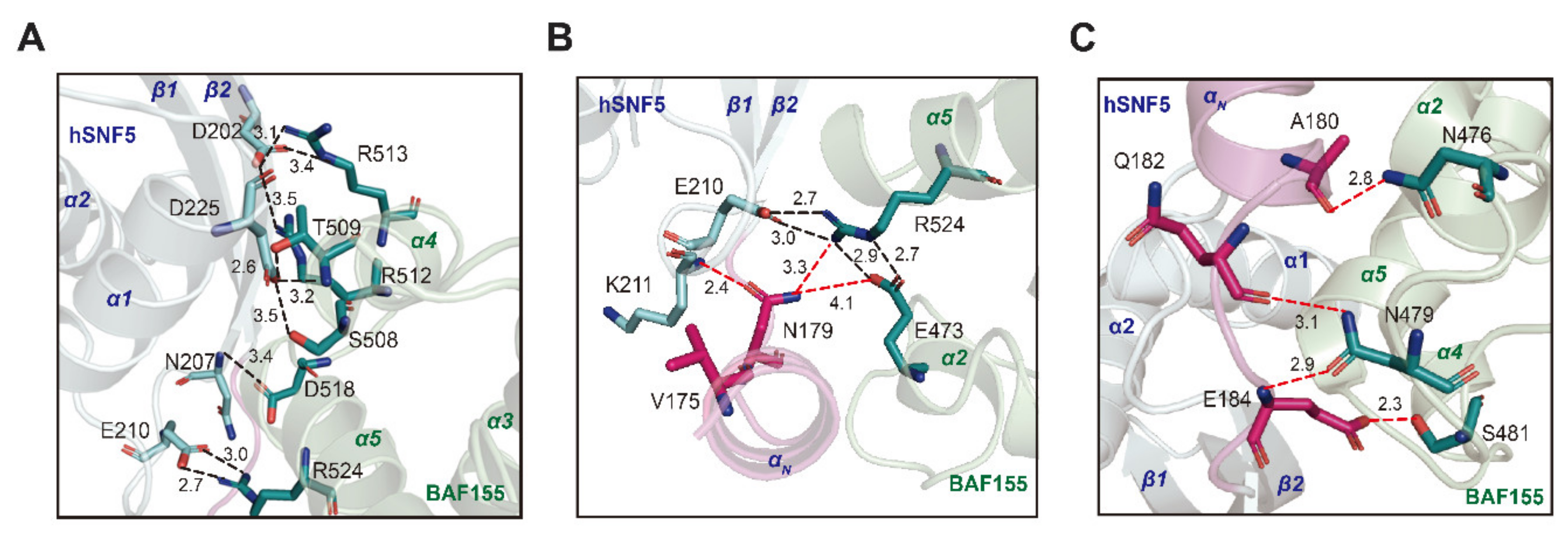
2.4. The Interface between hSNF5171–258 and BAF155SWIRM Features Charge Complementarity

hSNF5171–258 is a highly acidic protein (pI = 4.1), whereas BAF155SWIRM is basic (pI = 7.9). The inspection of surface charges at the binding interface reveals charge complementarity, such that positively charged surfaces of BAF155SWIRM (Figure S1A) interact with negatively charged surfaces of hSNF5171–258 (Figure S1B). Complex formation between hSNF5171–258 and BAF155SWIRM buries 1684 Å2 of solvent-accessible surface area, which is ~30% larger than the buried surface area of 1308 Å2 detected in the same complex without the αN helix. The interaction surface features key salt bridges and hydrogen bonds between hSNF5171–258 and BAF155SWIRM (Asp202—Arg512/Arg513 and Glu210—Arg524) (Figure 5A). In addition, the Asn207 of hSNF5171–258 forms a hydrogen bond to Asp518 of BAF155SWIRM, and Asp225 of hSNF5171–258 forms a hydrogen-bonding network with Ser508/Thr509 of BAF155SWIRM. The binding interface with hSNF5171–258 and BAF155SWIRM is similar to that of the previous structure [34], but the binding distance between the residues are quite different (Figure 5A). Specifically, the salt bridge distance between Asp202 and Arg512 in our hSNF5171–258/BAF155SWIRM complex structure is about 0.7Å further apart when compared with that of the previous structure [34]. On the other hand, the salt bridge distance between Glu210 and Arg524 in our hSNF5171–258/BAF155SWIRM complex structure is approximately 0.3Å closer than that of the previous structure (Figure 5A). Moreover, we also found an additional hydrogen bond network in the presence of the αN helix of hSNF5171–258. A hydrogen bond network formed between the Asn179 of hSNF5171–258 and Arg524 and the Glu473 of BAF155SWIRM. The Asn179 located in the αN helix of hSNF5171–258, showing a high CSP value, also interacts directly with Lys211 (Figure 3C and Figure 5B). Therefore, it is considered that the newly observed hydrogen bond network that is mediated by the Asn179 in the αN helix of hSNF5171–258 is a very important binding force for the hSNF5171–258/BAF155SWIRM complex as well as hydrogen and hydrophobic interactions that were observed on the charged surface (Figure 5B). In the NMR titration result, the Glu473 of BAF155SWIRM represents a high CSP value, which is consistent with our hSNF5171–258/BAF155SWIRM complex structure (Figure 3D and Figure 5B). In addition, hydrogen bond pairs (Ala180—Asn476, Gln182—Asn479, and Glu184—Ans479/Ser481) form in a complex formation of hSNF5171–258/BAF155SWIRM, which makes hydrogen bonding networks in the N-terminal region of hSNF5171–258. Backbone carbonyl groups of Ala180 and Gln182 from hSNF5171–258 form hydrogen bonds with side chains of Asn476 and Asn479, respectively, from BAF155SWIRM. The carboxyl side chain of Glu184 from hSNF5171–258 forms hydrogen bonds and salt bridges with the side chains of Ser481 and Lys482 of BAF155SWIRM (Figure 5C). The αN helix also induces some hydrophobic interactions. Three residues, Ile176, His177, and Ala180, provide the hydrophobic interfaces for Pro472 of BAF155SWIRM.
The crystal structure in this study is fully consistent with the CSP analysis from the NMR-titration experiments. Finally, we identified that the N-terminal region of hSNF5171–258 has a conformational change and it induces a tight binding pocket during the complex state.

2.5. The N-Terminal Binding Motif of hSNF5171–258 Enhances the Molecular Interaction for Stabilizing hSNF5171–258/BAF155SWIRM Complex

We further investigated how the N-terminal region of hSNF5171–258 is important for the target binding and stability of the protein complex. Data from isothermal titration calorimetry (ITC) experiments support the importance of the αN helix for BAF155SWIRM binding in a quantitative manner. We observed that hSNF5171–258 and hSNF5171–253 exhibit the same binding affinity to BAF155SWIRM, which indicates that the five residues in the C-terminal tail do not affect protein binding. We also prepared a series of N-terminal truncation hSNF5171–253 mutants and measured how these truncations impact BAF155SWIRM binding (Table 3). Our data show that hSNF5171–253 binds to BAF155SWIRM, with a dissociation constant (KD) of 110 ± 20 nM and a 1:1 stoichiometry (Figure 6A).
The binding affinity reduced by 2.6-fold when the whole αN helix was removed (hSNF5183–253) (KD ~290 ± 80 nM). The moderate decrease in affinity suggests that the αN helix might engage with the BAF155SWIRM via transient interactions. We note that the αN helix exhibited relatively low heteronuclear NOE (XNOE) values, even in complex with the BAF155SWIRM, as shown in Figure 4E. Therefore, the αN helix in the hSNF5171-258/BAF155SWIRM complex in solution could retain a degree of conformational mobility, providing favorable interactions for stabilizing the complex.
Progressive truncations of the αN helix show that hSNF5174–253 maintains the same binding affinity, with a KD of ~110 ± 30 nM, whereas hSNF5179–253 and hSNF5183–253 show decreased binding affinities, with KD values of ~300 ± 50 nM and ~290 ± 80 nM, respectively, similar to that observed for the whole αN helix-deletion mutant. The truncation of the N-terminal loop preceding the β1 strand results in a ~7-fold reduction in binding affinity, such that hSNF5186–253 exhibits a KD of ~758 ± 80.3 nM for BAF155SWIRM binding (Fig. 6B). This large reduction results from the loss of hydrogen bonding from Glu184 of hSNF5171-253, as well as the αN-helix deletion. We note that a single mutation of Glu184 reduces the binding affinity by 3.4-fold, yielding a KD of ~370 ± 100 nM for the E184A mutation of hSNF5171–253 (Figure 6C). We further found that alanine mutations of Asn476 and Asn479 in BAF155SWIRM, which make hydrogen bonds to Ala180 and Gln182, respectively, in hSNF5171–253 have a negligible impact on binding. The N476A/N479A mutant of BAF155SWIRM only shows a modest decrease in binding affinity, with a KD of ~140 ± 30 nM, indicating a minor role or replaceable nature for their interactions. Finally, the N-terminal mutant of hSNF171–253 (hSNF5SFH1/171–253), followed by the human sequence (residues 185–253), exhibits a 4.4-fold lower binding affinity. The N-terminal tail region that extends 15 amino acids in the RPT1 sequence, is not conserved between the human and yeast sequences, so this reduced binding results from divergent sequences in the αN helix and its surrounding region.
Lastly, we investigated whether the αN helix formation significantly contributes to the stability of the hSNF5171–253/BAF155SWIRM complex by measuring the melting temperature (Tm) with circular dichroism (CD) spectroscopy. Although hSNF5171–253 and hSNF5186-253 both exhibit the same Tm of 49 °C in the free state, hSNF5171–253 in complex has a higher Tm (~53 °C) than hSNF5186–253 (Tm ~48 °C), demonstrating that the αN helix is a determinant of protein stability for the hSNF5171–253/BAF155SWIRM complex (Figure 6D,E).

3. Discussion

The core complex that is composed of SNF5, BAF155, and BAF170 was found to show a remodeling activity that resembles that of the entire mammalian SWI/SNF complex [38,39]. An important step toward understanding how specific subunits function is to elucidate how they interact within the complex. To date, an atomic-resolution structure of the human SWI/SNF complex has not been obtained, although several individual subunits and domains have been described. Here, we solved the structure of the hSNF5171–258/BAF155SWIRM complex at atomic resolution, providing biophysically and functionally important information. Our data indicate that the N-terminal loop preceding hSNF5RPT1 undergoes a conformational change upon interaction with BAF155SWIRM, which enhances the binding strength and stability of the complex state. The yeast Cryo-EM structure showed the presence of αN helix in the SWI/SNF complex [40]. Further, the hydrogen bond network and hydrophobic interactions between the αN helix and the SWIRM domain in our structure recapitulated, illustrating that the αN helix presents the bona fide interface for the SWIRM domain.
Interestingly, a number of studies report that mutations in the N-terminal residues Pro173 and Glu184 of hSNF5 lead to different cancers, such as Rhabdoid tumors and malignant melanoma [41,42]. The Glu184 residue of hSNF5171–258 plays an important role in binding between the N-terminal region of hSNF5171–258 and BAF155SWIRM, and it is involved in tumor suppression [41]. We speculate that the mutation of Glu184, where the N-terminal region of hSNF5171–258 binds to BAF155SWIRM, interrupts the formation of the hSNF5 and BAF155 complex, making it difficult to form chromatin-remodeling complexes and chromatin, and thereby resulting in tumor genesis. When considering that mutations of Glu184 and Val185 abolish the interaction with HIV-1 integrase (IN) [43], we infer that residues on the non-structured loop following the N-terminal tail play an important role in the interaction with IN to a greater extent than with BAF155SWIRM.
Intrinsically disordered regions can perform various biological functions via coupled folding and binding mechanisms [44,45,46,47,48]. The complex structure of hSNF5171–258 and BAF155SWIRM illustrates that the interaction between hSNF5 and BAF155 also involves a coupled folding and binding mechanism. The coil-to-helix transition of hSNF5171–258 upon BAF155SWIRM binding might help to distinguish between various targets, thereby fine-tuning individual interactions. The structural transition of hSNF5171–258 can alter chromatin states and remodeling activity in vivo for the temporal and spatial control of biological processes, such as the recruitment of chromatin remodelers or transcriptional activation.
In summary, we have determined the crystal structure of the hSNF5171–258/BAF155SWIRM complex as a heterodimer by 1:1 molar ratio, which reveals the detailed binding mode. The N-terminal disordered loop of hSNF5171–258 undergoes a coil-to-helix transition upon binding to BAF155SWIRM. In the N-terminal region of hSNF5171–258, residues between Asn179 and Glu184 were identified as core regions that bind to BAF155SWIRM, which is critical for protein binding and it enhances the stability of the protein complex. In addition, our data will provide a structural clue to understanding the mechanism of the pathogenesis of diseases that are related to the chromatin remodeling complex and originated from mutations in the N-terminal region of hSNF5.

4. Materials and Methods

4.1. Cloning, Protein Expression, and Purification

The cDNA fragments encoding residues 171–258 of hSNF5 (hSNF5171–258) and the various N-terminal truncation mutants and residues 449–546 of human BAF155 (BAF155SWIRM) and the BAF155SWIRM mutants were amplified by PCR. The amplified cDNA fragments were inserted into the modified expression vector pET21b (Novagen, Darmstadt, Germany) for hSNF5171–258 and the N-terminal truncation mutants and pMAL-C2X (Novagen, Darmstadt, Germany) for BAF155SWIRM and the BAF155SWIRM mutants as fusions with N-terminal His6-affinity tags and tobacco etch virus (TEV) protease cleavage sites. The plasmids were transformed into Escherichia coli Strain BL21 (DE3) (Invitrogen, Waltham, MA, USA). Cells expressing hSNF5171-258 and BAF155SWIRM were grown in LB or M9 minimal media with 15NH4Cl and/or 13C6-glucose as the sole nitrogen and carbon sources, respectively, at 37 °C to an OD600 of ~0.6. Protein expression was induced with 1 mM isopropyl-β-d-thiogalactopyranoside (IPTG) at 25 °C and the cells were harvested by centrifugation after 16 h of induction and then stored frozen at −70 °C. Cell pellets were resuspended and disrupted by sonication in lysis buffer containing 25 mM sodium phosphate, pH 7.0, 300 mM NaCl, 5 mM β-mercaptoethanol, and protease inhibitor cocktail (Roche, Basel, Switzerland). The His6-tagged fusion proteins were purified by immobilized metal affinity chromatography on a Ni-NTA column (QIAGEN, Hilden, Germany) and then cleaved by TEV protease for 12 h. The digestion reactions were loaded onto a Ni-NTA column, and the flow-through was collected and loaded onto a Superdex 200 16/60 column (GE Healthcare, Chicago, IL, USA), equilibrated with 10 mM HEPES, pH 7.0, 100 mM NaCl, and 2 mM dithiothreitol (DTT).

4.2. Size Exclusion Chromatography and Multi-Angle Light Scattering Analysis

SEC analysis was performed on a Superdex 200 16/60 column (GE Healthcare, Chicago, IL, USA) at 25 °C. The column was equilibrated with buffer containing 10 mM HEPES, pH 7.0, 100 mM NaCl, and 2 mM DTT. Purified hSNF5171-258 (0.2 mM) and BAF155SWIRM (0.3 mM) were loaded onto the column, individually or together. The following molecular weight marker proteins were used: aldolase (158 kDa), conalbumin (75 kDa), ovalbumin (44 kDa), carbonic anhydrase (29 kDa), ribonuclease A (13.7 kDa), and aprotinin (6.5 kDa). The molecular weights of free protein and protein-protein complexes were calculated while using the following equation: Y = −0.162X + 2.2157, where R2 = 0.9955, and X = Ve/Vo (Vo = 45.15 mL, Ve = elution volume, Y = logMW). Sodium dodecyl sulfate-polyacrylamide gel electrophoresis (SDS-PAGE) confirmed the eluted samples.
SEC multi-angle light scattering (SEC-MALS) experiments were performed using the Agilent 1200 HPLC system (Agilent Technologies, Santa Clara, CA, USA), combined with a Wyatt DAWN HELEOS-II MALS instrument and a Wyatt OPtilab rEX differential refractometer (Wyatt Technology, Santa Barbara, CA, USA). For chromatographic separation, a WTC-030S5 column (Wyatt Technology, Santa Barbara, CA, USA) was used at a flow rate of 0.5 mL/min. in the same buffer as was used for SEC experiments. The same concentrations of hSNF5171-258, BAF155SWIRM, and hSNF5171–258/BAF155SWIRM complex that were used for SEC analysis were also used in the SEC-MALS experiments. The results were normalized and analyzed while using ASTRA software (Wyatt Technology, Santa Barbara, CA, USA).

4.3. NMR Spectroscopy

The NMR samples contained 0.5 mM 13C,15N-hSNF5171–258 or 13C,15N-BAF155SWIRM in 10 mM HEPES, pH 7.0, 100 mM NaCl, and 2 mM DTT. The NMR spectra were recorded at 25 °C on a Bruker 500 and 800 MHz spectrometer, which was equipped with a z-shielded gradient triple resonance probe. Sequential assignments of 1H, 15N, and 13C resonance were performed by 3D triple resonance through-bond scalar correlation experiments (CBCACONH, HNCACB, HNCO, HNCACO, HNHA, (H)CCC(CO)NH, and H(CCCO)NH). For structure calculation purposes, 15N-edited NOESY (τm = 150 ms) and 13C-edited NOESY-HSQC (τm = 120 ms) experiments were acquired. Dihedral backbone restraints were derived from 1Ha, 13Ca, 13Cb, and 13CO chemical shifts with the TALOS program [49]. The residual 1DNH dipolar couplings were obtained by taking the difference in the J splitting values measured in oriented (12 mg/mL of pf1 alignment media) and isotropic (water) hSNF5171-258 using 2D in-phase/antiphase 1H-15N HSQC spectra [49]. Heteronuclear 1H-15N NOE was measured using pulse schemes, as described previously [50]. The NMR spectra were processed and analyzed using the NMRPipe [51] and NMRFAM-SPARKY [52] programs. 15N-HSQC titration experiments were performed at 1:1molar ratios of 15N-labeled proteins to unlabeled counter proteins between hSNF5171-258 and BAF155SWIRM. Chemical-shift perturbations were calculated using the equation ΔδAV = ((Δδ1H)2 + (Δδ15N/5)2)1/2, where ΔδAV, Δδ1H, and Δδ15N are the average, proton, and 15N chemical-shift changes, respectively.

4.4. NMR Structure Determination and Analysis

The structure calculations for BAF155SWIRM and hSNF5171–258 in the free state were performed using semi-automated CYANA 2.1 [53]. The initial fold was obtained by manual NOE assignment, and secondary topology was determined by peak picking. NOE cross-peak assignments were obtained using manual procedures. An initial fold of the protein was calculated on the basis of manually assigned NOEs, which is essential for secondary topology determination, with subsequent use of the program CYANA 2.1. Seven cycles of the CYANA routine were calculated; each cycle had 10,000 steps of torsion-angle dynamics, with a simulated annealing protocol. A total of 100 structures were calculated, and the 20 structures with the lowest target function values were chosen for analysis. Table 2 shows a summary of the NMR-derived restraints and structures of hSNF5171–258. The final structures with the lowest NOE energies were retained and validated with the PROCHECK program [54]. The structures were analyzed and visualized using PyMOL (Delano Scientific LLC, San Carlos, CA, USA, https://pymol.org/) and MOLMOL(https://sourceforge.net/projects/molmol/) [55].

4.5. Crystallization and Structure Determination

Crystals of the hSNF5171–258/BAF155SWIRM complex were grown under oil at 15 °C in a 1.5 µL micro-batch, containing equal volumes of protein solution and mother liquor (15% v/v Tacsimate, pH 7.0, 0.1 M HEPES, pH 7.0, 2% (w/v) polyethylene glycol 3350). Purified hSNF5171–258 was mixed with BAF155SWIRM at a 1:1 molar ratio. The crystals were flash-cooled in liquid nitrogen, with 25% glycerol being used as a cryoprotectant. The diffraction datasets were collected using a synchrotron radiation source at beamline 4A at the Pohang Accelerator Laboratory (Pohang, Korea) at 100 K. Collected data were integrated and scaled while using the HKL-2000 program (HKL Research Inc., Charlottesville, VA, USA). The crystal structure of the hSNF5171–258/BAF155SWIRM complex was solved by the molecular replacement method with the MOLREP [56] and Phaser programs, using a high-accuracy template-based modeling method as the search model. The final structure was refined to the maximum resolution of 2.28 Å by iterative cycles of the manual building in COOT and then restrained refinement with Refmac5 and Phenix [57]. Table 1 summarizes data collection and refinement for the complex structure. The final structures were analyzed while using MolProbity [58].

4.6. Crystallization and Structure Determination

The ITC experiments were performed using a VP-ITC system (MicroCal Inc. Northampton, MA, USA) at 25 °C in a buffer containing 25 mM sodium phosphate, 100 mM NaCl, and 1 mM Tris(2-carboxyethyl)-phosphine (TCEP), pH 7.0. Typically, 160 µM hSNF5171–253 was injected 35 times in 7 µL aliquots into the 1.4 mL sample cell, containing either the BAF155SWIRM domain or BAF155SWIRM mutants at a concentration of 10 μM. The data were fit with a non-linear least-squares best-fit curve, while using a single-site binding model with Origin for ITC (Microcal Inc.), by varying the stoichiometry (n), the enthalpy of the reaction (ΔH), and the dissociation constant (KD).

4.7. Circular Dichroism Spectroscopy

CD experiments to measure the melting temperatures were performed in buffer containing 25 mM sodium phosphate, 100 mM NaCl, and 1 mM Tris(2-carboxyethyl)-phosphine (TCEP), pH 7.0, as described previously [59,60]. Samples containing 10 μM hSNF5171–253 or hSNF5186–253 alone or with 10 µM BAF155SWIRM were monitored at temperatures between 25 °C and 95 °C, with an interval of 5 °C. The parameters for far-ultraviolet UV CD measurements used a cell with a path length of 0.2 cm for scanning between 250–200 nm, with a 1 nm bandwidth and scan speed of 200 nm/min−1. The values were baseline corrected by subtracting a buffer spectrum, and an average of five scans was recorded for each experiment. The apparent melting temperature at the midpoint of the transition was obtained by fitting the experimental data points (CD signal at 222 nm vs. temperature) with a Boltzmann sigmoidal function.

Supplementary Materials

Supplementary materials can be found at https://0-www-mdpi-com.brum.beds.ac.uk/1422-0067/21/7/2452/s1.; Figure S1: The electrostatic surface charged models of hSNF5171-258 and BAF155SWIR.

Author Contributions

J.H., I.T., J.-Y.S., and W.L. conceptualized and designed the experiments; R.H.S. provided hSNF5 and BAF155 genes and contributed to the project initiation; J.H., T.K., and G.-Y.P. cloned, expressed, and purified protein; J.H., I.K., J.-H.Y., and K.M. performed formal data analysis.; J.H., I.K., K.-S.R., and Y.-J.K. conducted NMR experiments and data analysis.; J.H., J.-H.P., and S.-Y.P. performed crystal analysis.; K.J. and J.L. performed computer modeling; J.H., I.K., J.L., J.-Y.S., and W.L. wrote the manuscript. All authors have read and agreed to the published version of the manuscript.

Funding

This work was supported by several grants (NRF-2017R1A2B2008483, 2019M3A9F6021810, NRF-2017M3A9F6029753, NRF-2019M3E5D6063903 to W. Lee, NRF-20161A6A3A04010213 to J.-H. Yun, NRF-2018R1D1A1B07049312 to K.J. and NRF-2017R1E1A1A01077717 to J.L.), through the National Research Foundation of Korea (NRF) and Brain Korea Plus (BK+) program (J. Han). This research was also supported by the Cooperative Research Program for Agriculture Science & Technology Development, Rural Development Administration, Republic of Korea (PJ011112, PJ01495901), and the Creative-Pioneering Researchers Program, through Seoul National University (SNU).

Acknowledgments

We thank the Research Institute of Agriculture and Life Sciences, the high-field NMR facility at the Korea Institute of Science and Technology, the Korea Basic Science Institute, and the National Center for Inter-University Research Facilities. We would like to express our gratitude to the staff scientists at the NCI station of the Pohang Accelerator Laboratory X-ray Free-Electron Laser, 5C and 7C beamline of the Pohang Accelerator Laboratory.

Conflicts of Interest

The authors declare no conflict of interest.

Accession codes

Atomic coordinates of the crystal structure of the hSNF5171–258/BAF155SWIRM complex have been deposited in the PDB under the accession code, 6KZ7, and the chemical shifts of hSNF5171–258/BAF155SWIRM complex and hSNF5171–258 free have been deposited in Biological Magnetic Resonance Bank (BMRB) under the accession codes: 28062, 28063, and 28064. The NMR structure of hSNF5171–258 has deposited in the PDB under the accession code, 6LZP.

Abbreviations

SWI/SNFSWItch/Sucrose Non-Fermentable
RPT1repeat 1
RPT2repeat2
NMRnuclear magnetic resonance
HSQCheteronuclear single quantum coherence
CSPchemical-shift perturbation
PDBProtein Data Bank
NOEsOverhauser effects
XNOE1H–15N heteronuclear NOE
ITCisothermal titration calorimetry
INHIV-1 integrase
TEVtobacco etch virus
IPTGisopropyl-β-D-thiogalactopyranoside
TCEPTris(2-carboxyethyl)-phosphine
BMRBBiological Magnetic Resonance Bank

References

  1. Papamichos-Chronakis, E.; Peterson, C.L. Chromatin and the genome integrity network. Nat. Rev. Genet. 2013, 14, 62–75. [Google Scholar] [CrossRef] [PubMed] [Green Version]
  2. Cosgrove, M.S.; Boeke, J.D.; Wolberger, C. Regulated nucleosome mobility and the histone code. Nat. Struct. Mol. Biol. 2004, 11, 1037–1043. [Google Scholar] [CrossRef] [PubMed]
  3. Workman, J.L.; Kingston, R.E. Alteration of Nucleosome Structure As a Mechanism of Transcriptional Regulation. Annu. Rev. Biochem. 1998, 67, 545–579. [Google Scholar] [CrossRef] [PubMed] [Green Version]
  4. Barkess, G. Chromatin remodeling and genome stability. Genome Biol. 2006, 7, 6–7. [Google Scholar] [CrossRef] [PubMed] [Green Version]
  5. Roberts, C.W.M.; Orkin, S.H. The SWI/SNF complex-Chromatin and cancer. Nat. Rev. Cancer 2004, 4, 133–142. [Google Scholar] [CrossRef] [PubMed]
  6. Wang, W.; Côté, J.; Xue, Y.; Zhou, S.; Khavari, P.A.; Biggar, S.R.; Muchardt, C.; Kalpana, G.V.; Goff, S.P.; Yaniv, M.; et al. Purification and biochemical heterogeneity of the mammalian SWI-SNF complex. EMBO J. 1996, 15, 5370–5382. [Google Scholar] [CrossRef] [PubMed]
  7. Crabtree, G.R. Diversity and specialization of mammalian SWI / SNF complexes. 2000, 4, 2117–2130. [Google Scholar]
  8. Kadoch, C.; Hargreaves, D.C.; Hodges, C.; Elias, L.; Ho, L.; Ranish, J.; Crabtree, G.R. Proteomic and bioinformatic analysis of mammalian SWI/SNF complexes identifies extensive roles in human malignancy. Nat. Genet. 2013, 45, 592–601. [Google Scholar] [CrossRef]
  9. Khavari, P.A.; Peterson, C.L.; Tamkun, J.W.; Mendel, D.B.; Crabtree, G.R. BRG1 contains a conserved domain of the SWI2/SNF2 family necessary for normal mitotic growth and transcription. Nature 1993, 366, 170–174. [Google Scholar] [CrossRef]
  10. Vignali, M.; Hassan, A.H.; Neely, K.E.; Workman, J.L. MINIREVIEW ATP-Dependent Chromatin-Remodeling Complexes. Mol. Cell. Biol. 2000, 20, 1899–1910. [Google Scholar] [CrossRef] [Green Version]
  11. Reisman, D.; Glaros, S.; Thompson, E.A. The SWI/SNF complex and cancer. Oncogene 2009, 28, 1653–1668. [Google Scholar] [CrossRef] [PubMed] [Green Version]
  12. Wu, J.I. Diverse functions of ATP-dependent chromatin remodeling complexes in development and cancer Epigenetic Regulation of Transcription during Development Brg1 / Brm-Associated Factors (BAF), A Mammalian SWI / SNF-like ATP-dependent. Breast 2012, 44, 54–69. [Google Scholar]
  13. Kadoch, C.; Crabtree, G.R. Mammalian SWI/SNF chromatin remodeling complexes and cancer: Mechanistic insights gained from human genomics. Rev. Clin. Esp. 2015, 197, 804–809. [Google Scholar] [CrossRef] [PubMed] [Green Version]
  14. Clapier, C.R.; Cairns, B.R. The Biology of Chromatin Remodeling Complexes. Annu. Rev. Biochem. 2009, 78, 273–304. [Google Scholar] [CrossRef]
  15. Sarnowska, E.; Gratkowska, D.M.; Sacharowski, S.P.; Cwiek, P.; Tohge, T.; Fernie, A.R.; Siedlecki, J.A.; Koncz, C.; Sarnowski, T.J. The Role of SWI/SNF Chromatin Remodeling Complexes in Hormone Crosstalk. Trends Plant Sci. 2016, 21, 594–608. [Google Scholar] [CrossRef] [PubMed]
  16. Lessard, J.A.; Crabtree, G.R. Chromatin Regulatory Mechanisms in Pluripotency. Annu. Rev. Cell Dev. Biol. 2010, 26, 503–532. [Google Scholar] [CrossRef] [Green Version]
  17. Hughes, A.L.; Owen-Hughes, T. Deciphering Subunit-Specific Functions within SWI/SNF Complexes. Cell Rep. 2017, 18, 2075–2076. [Google Scholar] [CrossRef] [Green Version]
  18. Wu, J.I.; Lessard, J.; Crabtree, G.R. Understanding the Words of Chromatin Regulation. Cell 2009. [Google Scholar] [CrossRef] [Green Version]
  19. Kalpana, G.V.; Marmon, S.; Wang, W.; Crabtree, G.R.; Goff, S.P. Binding and stimulation of HIV-1 integrase by a human homolog of yeast transcription factor SNF5. Science (80-) 1994, 266, 2002–2006. [Google Scholar] [CrossRef]
  20. Morozov, A.; Yung, E.; Kalpana, G.V. Structure-function analysis of integrase interactor 1/hSNF5L1 reveals differential properties of two repeat motifs present in the highly conserved region. Proc. Natl. Acad. Sci. USA 1998, 95, 1120–1125. [Google Scholar] [CrossRef] [Green Version]
  21. Cheng, S.W.G.; Davies, K.P.; Yung, E.; Beltran, R.J.; Yu, J.; Kalpana, G.V. c-MYC interacts with INI1/hSNF5 and requires the SWI/SNF complex for transactivation function. Nat. Genet. 1999, 22, 102–105. [Google Scholar] [CrossRef] [PubMed]
  22. Lee, D.; Kim, J.W.; Seo, T.; Hwang, S.G.; Choi, E.J.; Choe, J. SWI/SNF complex interacts with tumor suppressor p53 and is necessary for the activation of p53-mediated transcription. J. Biol. Chem. 2002, 277, 22330–22337. [Google Scholar] [CrossRef] [PubMed] [Green Version]
  23. Maillot, B.; Lévy, N.; Eiler, S.; Crucifix, C.; Granger, F.; Richert, L.; Didier, P.; Godet, J.; Pradeau-Aubreton, K.; Emiliani, S.; et al. Structural and Functional Role of INI1 and LEDGF in the HIV-1 Preintegration Complex. PLoS ONE 2013, 8, e60734. [Google Scholar] [CrossRef] [PubMed] [Green Version]
  24. Mathew, S.; Nguyen, M.; Wu, X.; Pal, A.; Shah, V.B.; Prasad, V.R.; Aiken, C.; Kalpana, G.V. INI1/hSNF5-interaction defective HIV-1 IN mutants exhibit impaired particle morphology, reverse transcription and integration in vivo. Retrovirology 2013, 10, 1. [Google Scholar] [CrossRef] [Green Version]
  25. Kohashi, K.; Oda, Y. Oncogenic roles of SMARCB1/INI1 and its deficient tumors. Cancer Sci. 2017, 108, 547–552. [Google Scholar] [CrossRef] [Green Version]
  26. Versteege, I.; Sévenet, N.; Lange, J.; Rousseau-Merck, M.F.; Ambros, P.; Handgretinger, R.; Aurias, A.; Delattre, O. Truncatingmutations of hSNF5/INI1 inaggressive paediatric cancer. 1998, 394, 203–206. [Google Scholar]
  27. Phelan, M.L.; Sif, S.; Narlikar, G.J.; Kingston, R.E. Reconstitution of a Core Chromatin Remodeling Complex from SWI/SNF Subunits. Mol. Cell 1999, 3, 247–253. [Google Scholar] [CrossRef]
  28. Alpsoy, A.; Dykhuizen, E.C. Glioma tumor suppressor candidate region gene 1 (GLTSCR1) and its paralog GLTSCR1-like form SWI/SNF chromatin remodeling subcomplexes. J. Biol. Chem. 2018, 293, 3892–3903. [Google Scholar] [CrossRef] [Green Version]
  29. Sohn, D.H.; Lee, K.Y.; Lee, C.; Oh, J.; Chung, H.; Jeon, S.H.; Seong, R.H. SRG3 interacts directly with the major components of the SWI/SNF chromatin remodeling complex and protects them from proteasomal degradation. J. Biol. Chem. 2007, 282, 10614–10624. [Google Scholar] [CrossRef] [Green Version]
  30. Kim, J.K.; Huh, S.-O.; Choi, H.; Lee, K.-S.; Shin, D.; Lee, C.; Nam, J.-S.; Kim, H.; Chung, H.; Lee, H.W.; et al. Srg3, a Mouse Homolog of Yeast SWI3, Is Essential for Early Embryogenesis and Involved in Brain Development. Mol. Cell. Biol. 2001, 21, 7787–7795. [Google Scholar] [CrossRef] [Green Version]
  31. Salbaum, J.M.; Niswander, L. A Unique Missense Allele of BAF155, a Core BAF Chromatin Remodeling Complex Protein, Causes Neural Tube Closure Defects in Mice. Dev. Neurobiol. 2015, 74, 483–497. [Google Scholar]
  32. Chen, J.; Archer, T.K. Regulating SWI / SNF Subunit Levels via Protein-Protein Interactions and Proteasomal Degradation: BAF155 and BAF170 Limit Expression of BAF57 Regulating SWI / SNF Subunit Levels via Protein-Protein Interactions and Proteasomal Degradation: BAF155 and BA. Mol. Cell. Biol. 2005, 25, 9016–9027. [Google Scholar] [CrossRef] [PubMed] [Green Version]
  33. Del Bove, J.; Rosson, G.; Strobeck, M.; Chen, J.; Archer, T.K.; Wang, W.; Knudsen, E.S.; Weissman, B.E. Identification of a core member of the SWI/SNF complex, BAF155/SMARCC1, as a human tumor suppressor gene. Epigenetics 2011, 6, 1444–1453. [Google Scholar] [CrossRef] [PubMed] [Green Version]
  34. Yan, L.; Xie, S.; Du, Y.; Qian, C. Structural Insights into BAF47 and BAF155 Complex Formation. J. Mol. Biol. 2017, 429, 1650–1660. [Google Scholar] [CrossRef] [PubMed]
  35. Sammak, S.; Allen, M.D.; Hamdani, N.; Bycroft, M.; Zinzalla, G. The structure of INI1/hSNF5 RPT1 and its interactions with the c-MYC:MAX heterodimer provide insights into the interplay between MYC and the SWI/SNF chromatin remodeling complex. FEBS J. 2018, 285, 4165–4180. [Google Scholar] [CrossRef]
  36. Notredame, C.; Higgins, D.G.; Heringa, J. T-coffee: A novel method for fast and accurate multiple sequence alignment. J. Mol. Biol. 2000, 302, 205–217. [Google Scholar] [CrossRef] [Green Version]
  37. Robert, X.; Gouet, P. Deciphering key features in protein structures with the new ENDscript server. Nucleic Acids Res. 2014, 42, 320–324. [Google Scholar] [CrossRef] [Green Version]
  38. Ruijtenberg, S.; van den Heuvel, S. Coordinating cell proliferation and differentiation: Antagonism between cell cycle regulators and cell type-specific gene expression. Cell Cycle 2016, 15, 196–212. [Google Scholar] [CrossRef] [Green Version]
  39. Hohmann, A.F.; Vakoc, C.R. A rationale to target the SWI/SNF complex for cancer therapy. Trends Genet. 2014, 30, 356–363. [Google Scholar] [CrossRef] [Green Version]
  40. Han, Y.; Reyes, A.A.; Malik, S.; He, Y. Cryo-EM structure of SWI/SNF complex bound to a nucleosome. Nature 2020, 579, 452–455. [Google Scholar] [CrossRef]
  41. Pinto, E.M.; Hamideh, D.; Bahrami, A.; Orr, B.A.; Lin, T.; Pounds, S.; Zambetti, G.P.; Pappo, A.S.; Gajjar, A.; Agnihotri, S.; et al. Malignant rhabdoid tumors originating within and outside the central nervous system are clinically and molecularly heterogeneous. Acta Neuropathol. 2018, 136, 315–326. [Google Scholar] [CrossRef] [PubMed]
  42. Uno, K.; Takita, J.; Yokomori, K.; Tanaka, Y.; Ohta, S.; Shimada, H.; Gilles, F.H.; Sugita, K.; Abe, S.; Sako, M.; et al. Aberrations of the hSNF5/INII gene are restricted to malignant rhabdoid tumors or atypical teratoid/rhabdoid tumors in pediatric solid tumors. Genes Chromosom. Cancer 2002, 34, 33–41. [Google Scholar] [CrossRef] [PubMed]
  43. Das, S.; Cano, J.; Kalpana, G.V. Multimerization and DNA binding properties of INI1/hSNF5 and its functional significance. J. Biol. Chem. 2009, 284, 19903–19914. [Google Scholar] [CrossRef] [PubMed] [Green Version]
  44. Iakoucheva, L.M.; Brown, C.J.; Lawson, J.D.; Obradović, Z.; Dunker, A.K. Intrinsic disorder in cell-signaling and cancer-associated proteins. J. Mol. Biol. 2002, 323, 573–584. [Google Scholar] [CrossRef] [Green Version]
  45. Galea, C.A.; Wang, Y.; Sivakolundu, S.G.; Kriwacki, R.W. Regulation of cell division by intrinsically unstructured proteins: Intrinsic flexibility, modularity, and signaling conduits. Biochemistry 2008, 47, 7598–7609. [Google Scholar] [CrossRef] [PubMed] [Green Version]
  46. Liu, J.; Perumal, N.B.; Oldfield, C.J.; Su, E.W.; Uversky, V.N.; Dunker, A.K. Intrinsic Disorder in Transcription Factors†. Biochem. 2006, 45, 6873–6888. [Google Scholar] [CrossRef] [Green Version]
  47. Sugase, K.; Dyson, H.J.; Wright, P.E. Mechanism of coupled folding and binding of an intrinsically disordered protein. Nature 2007, 447, 1021–1025. [Google Scholar] [CrossRef]
  48. Wright, P.E.; Dyson, H.J. Intrinsically unstructured proteins: Re-assessing the protein structure-function paradigm. J. Mol. Biol. 1999, 293, 321–331. [Google Scholar] [CrossRef] [Green Version]
  49. Shen, Y.; Bax, A. Protein backbone chemical shifts predicted from searching a database for torsion angle and sequence homology. J. Biomol. NMR 2007, 38, 289–302. [Google Scholar] [CrossRef]
  50. Tjandra, N.; Wingfield, P.; Stahl, S.; Bax, A. Anisotropic rotational diffusion of perdeuterated HIV protease from 15N NMR relaxation measurements at two magnetic fields. J. Biomol. NMR 1996, 8, 273–284. [Google Scholar] [CrossRef]
  51. Delaglio, F.; Grzesiek, S.; Vuister, G.W.; Zhu, G.; Pfeifer, J.; Bax, A. NMRPipe: A multidimensional spectral processing system based on UNIX pipes. J. Biomol. NMR 1995, 6, 277–293. [Google Scholar] [CrossRef] [PubMed]
  52. Lee, W.; Tonelli, M.; Markley, J.L. Nmrfam-Sparky: Enhanced software for biomolecular NMR spectroscopy. Bioinformatics 2015, 31, 1325–1327. [Google Scholar] [CrossRef] [PubMed] [Green Version]
  53. Güntert, P. Automated NMR structure calculation with CYANA. Methods Mol. Biol. 2004, 278, 353–378. [Google Scholar] [PubMed]
  54. Morris, A.L.; MacArthur, M.W.; Hutchinson, E.G.; Thornton, J.M. Stereochemical quality of protein structure coordinates. Proteins Struct. Funct. Bioinforma. 1992, 12, 345–364. [Google Scholar] [CrossRef] [PubMed]
  55. Koradi, R.; Billeter, M.; Wüthrich, K. MOLMOL: A program for display and analysis of macromolecular structures. J. Mol. Graph. 1996, 14, 51–55. [Google Scholar] [CrossRef]
  56. Vagin, A.; Teplyakov, A. MOLREP: An Automated Program for Molecular Replacement. J. Appl. Crystallogr. 1997, 30, 1022–1025. [Google Scholar] [CrossRef]
  57. Emsley, P.; Lohkamp, B.; Scott, W.G.; Cowtan, K. Features and development of Coot. Acta Crystallogr. Sect. D Biol. Crystallogr. 2010, 66, 486–501. [Google Scholar] [CrossRef] [Green Version]
  58. Williams, C.J.; Headd, J.J.; Moriarty, N.W.; Prisant, M.G.; Videau, L.L.; Deis, L.N.; Verma, V.; Keedy, D.A.; Hintze, B.J.; Chen, V.B.; et al. MolProbity: More and better reference data for improved all-atom structure validation. Protein Sci. 2018, 27, 293–315. [Google Scholar] [CrossRef]
  59. Williams, J.C.; Zeelen, J.P.; Neubauer, G.; Vriend, G.; Backmann, J.; Michels, P.A.M.; Lambeir, A.M.; Wierenga, R.K. Structural and mutagenesis studies of leishmania triosephosphate isomerase: A point mutation can convert a mesophilic enzyme into a superstable enzyme without losing catalytic power. Protein Eng. 1999, 12, 243–250. [Google Scholar] [CrossRef] [Green Version]
  60. Greenfield, N.J. Using circular dichroism collected as a function of temperature to determine the thermodynamics of protein unfolding and binding interactions. Nat. Protoc. 2007, 1, 2527–2535. [Google Scholar] [CrossRef]
Figure 1. Domain structures of hSNF5 and BAF155. (A) Schematic representation of full-length hSNF5 and eight hSNF5 constructs containing the hSNF5RPT1 domain. (B) Domain structure of full-length BAF155. The hSNF5RPT1 (residues 186–245) domain (red) and the BAF155SWIRM domain (blue); red dotted box indicates the interacting region. (C) Multiple sequence alignment of the hSNF5171–258 region with homologs in mouse, fruit fly, nematode, and yeast. Sequence alignment was performed using T-coffee software [36] and rendered further with ESPript software [37]. (D) Pairwise sequence alignment identity matrices for hSNF5171–258 (left) and BAF155SWIRM (right) and their homologs and orthologs. Hs, Homo sapiens; Mm, Mus musculus; Dm, Drosophila melanogaster; Ce, Caenorhabditis elegans; and Sc, Saccharomyces cerevisiae. For both hSNF5171–258 and BAF155SWIRM, the sequence in Hs is identical to that in Mm.
Ijms 21 02452 g001
Figure 2. Analysis of the complex formation of hSNF5171–258, binding with BAF155SWIRM, and its solution structure. Size exclusion chromatography (SEC) analysis using a Superdex 200 16/60 column for (A) hSNF5171–258 (~13.7 kDa) and (B) BAF155SWIRM (~13 kDa). SEC-multi-angle light scattering (MALS) elution profiles from a Wyatt WTC-030S5 column for (C) hSNF5171–258 and (D) BAF155SWIRM. (E) Superposition of the backbone atoms of the final 20 lowest-energy calculated structure and (F) a ribbon model of hSNF5171–258. The N-terminal region of hSNF5171–258 is shown in the flexible loop (red). (G) SEC profile of the hSNF5171–258/BAF155SWIRM complex (~29 kDa). Elution profiles of standard marker protein are shown at the top of the chromatograms as a reference. (H) SEC-MALS elution profiles of the hSNF5171–258/BAF155SWIRM complex. The molecular masses obtained from light scattering and refractive index measurements correspond to the molecular weights of free hSNF5171–258, free BAF155SWIRM, and the hSNF5171–258/BAF155SWIRM complex.
Ijms 21 02452 g002
Figure 3. The interaction site mapping on hSNF5171–258/BAF155SWIRM complex using nuclear magnetic resonance (NMR) titration. (A) 1H-15N heteronuclear single quantum coherence (HSQC) spectrum of free 15N-hSNF5171–258 (black) was superimposed with the spectrum of 15N-hSNF5171–258/BAF155SWIRM complex (red). (B) 1H-15N HSQC spectrum of free 15N-BAF155SWIRM (black) was superimposed with the spectrum of the 15N-BAF155SWIRM/hSNF5171–258 complex (red). For these experiments, 0.3 mM 15N-hSNF5171–258 or 15N-BAF155SWIRM was stoichiometrically titrated with an equivalent amount of unlabeled BAF155SWIRM or hSNF5171–258, respectively. The residues that show a large chemical-shift perturbation (CSP) are annotated. CSP plots for (C) hSNF5171–258 and (D) BAF155SWIRM, as a function of the residue number obtained from the NMR titration. Average chemical-shift changes were calculated using the following formula: ΔδAV = ((Δδ1H)2 + (Δδ15N/5)2)1/2. Secondary structures are shown above the CSP plots. Residues with broadened line widths are indicated with stars (*), and the secondary structure shown in transparent red represents the α-helix in the N-terminal region of hSNF5171–258N) formed upon binding with BAF155SWIRM.
Ijms 21 02452 g003
Figure 4. Conformational changes of the N-terminal region of hSNF5171–258 during the complex formation with BAF155SWIRM. (A) The high-resolution crystal structure of hSNF5171–258/BAF155SWIRM complex is shown as a cartoon diagram. Both molecules, hSNF5171–258 and BAF155SWIRM, are colored cyan and green, respectively. Secondary structures are annotated, and the N-terminal region of hSNF5171–258 is shown in pink. (B) The N-terminal helix region of the hSNF5171–258 is plotted while using an omit map at the contour level 1.2 σ as a gray mesh and it shows both the backbone (left) and the side chain (right) electron density map. (C) The intermolecular hydrophobic and hydrogen bond interactions between αN helix and β2–α1 loop in hSNF5171–258 are depicted as sticks and blue mesh. Hydrogen bonds and water molecules are shown in the red dashed lines and red sphere, respectively. (D) The solution structure of hSNF5171–258 in a free state (purple ribbon model) is superimposed with the crystal structure of hSNF5171-258/BAF155SWIRM in a complex state (cyan ribbon model). The N-terminal region of hSNF5171–258 in both structures, that experience the conformational change from the coil to helix, are marked by red colors, respectively. (E) Backbone dynamics profile of hSNF5171–258 in a free state (black dot) and the BAF155SWIRM bound state (red dot). The XNOE data were plotted against the residue number and the corresponding secondary structure of hSNF5171–258. A red box indicates the N-terminal region of hSNF5171–258, forming helix upon binding with BAF155SWIRM, and a red transparent helix represents the secondary structure.
Ijms 21 02452 g004
Figure 5. Detailed view of residual networks against with the N-terminal helix of hSNF5171–258 complexed with BAF155SWIRM. (A) The hydrogen bond network in the binding interface between hSNF5171–258 (cyan) and BAF155SWIRM (green). (B,C) The additional hydrogen bond network mediated by the N-terminal region (magenta) of hSNF5171–258 in complex with BAF155SWIRM is shown in stick model. The residual networks newly represented by the N-terminal loop are marked with a red dash to distinguish it from the known interacting network.
Ijms 21 02452 g005
Figure 6. N-terminal disordered regions of hSNF5171–253 contribute to the enhancement of the interaction with BAF155SWIRM. Isothermal titration calorimetry (ITC) for the interaction between (A) hSNF5171–253 and BAF155SWIRM, (B) hSNF5186–253 and BAF155SWIRM, and (C) hSNF5171-253 with the E184A mutation and BAF155SWIRM. The integrated heat of injection is presented, and the experimental data are shown as solid squares. The least-squares best-fit curves derived from a simple one-site binding model are shown as a black line. Thermodynamic parameters are shown in Table 3. Thermal stability of hSNF5171–253 and hSNF5186–253 (D) alone and (E) with BAF155SWIRM. The melting temperatures (Tm) were calculated by the decrease in normalized molar ellipticity at 222 nm in the circular dichroism (CD) spectrum.
Ijms 21 02452 g006
Table 1. Restraints and structural statistics for hSNF5171–258.
Experimental Restraints<SA> *
Non-redundant NOEs1891
Dihedral angles, φ/ψ62/62
Hydrogen bonds25
Residual dipolar coupling, 1DNH77
Total number of restraints1782 (24.3 per residue)
RMSD from experimental restraints
 Distances (Å) (1891)0.034 ± 0.002
 Torsion angles (°) (124)2.34 ± 0.14
Residual dipolar coupling R-factor (%)
1DNH (%) (77)2.9 ± 0.4
RMSD from idealized covalent geometry
 Bonds (Å)0.003 ± 0
 Angles (°)0.59 ± 0.01
 Impropers (°)0.67 ± 0.02
Coordinate precision (Å) *
 Backbone0.48 ± 0.15
 Heavy atoms1.29 ± 0.17
Ramachandran statistics (%) ‡§
 Most favorable regions80.5 ± 1.1
 Allowed regions19.5 ± 1.1
* For the ensemble of the final 20 simulated annealing structures. The magnitudes of the axial and rhombic components of the alignment tensor were 5.4 Hz and 0.65, respectively. Regions with secondary structures (residues 186−195, 198−207, 218−225, and 233−247). § Calculated using the program PROCHECK.
Table 2. Data collection and refinement statistics for the hSNF5171–258/BAF155SWIRM complex.
hSNF5171–258/BAF155SWIRM Complex
Data Collection
Space groupH3
Cell dimensions
a, b, c (Å)77.23, 77.23, 207.24
α, β, γ (°)90.00, 90.00, 120.00
Resolution (Å)a28.1–2.28 (2.236–2.28)
I/σI20.16 (3.01)
Rmerge (%) a8.5 (40.5)
Completeness (%) a99.87 (99.9)
Redundancy a5.43 (4.8)
Refinement
Resolution (Å)28.1–2.28 (2.236–2.28)
No. reflections20987
Rwork/Rfree0.1671/0.2003
No. atoms
 Protein2876
 water223
B-factors
 Protein33.21
 Water32.54
RMSD
 Bond lengths (Å)0.003
 Bond angles (°)0.58
Ramachandran plot (%) b
 Most favored regions98.55%
 Allowed regions1.45%
 Disallowed regions0%
a Values in parentheses are for the highest-resolution shell. b Ramachandran plot was calculated using the PROCHECK program.
Table 3. Thermodynamic parameters for the interaction between various hSNF5171–253 and BAF155SWIRM constructs that were obtained by isothermal titration calorimetry at 25 °C.
DescriptionKD
(nM)
ΔG
(kcal/mol)
ΔH
(kcal/mol)
−TΔS
(kcal/mol)
hSNF5BAF155SWIRM
171–253 aWild-type110 ± 20−9.5 ± 0.1−19.7 ± 0.310.2 ± 0.3
174–253 aWild-type110 ± 30−9.5 ± 0.2−15.8 ± 0.56.3 ± 0.3
179–253 aWild-type300 ± 50−8.9 ± 0.1−21.0 ± 0.612.1 ± 0.6
181–253 aWild-type300 ± 70−8.9 ± 0.1−12.9 ± 0.44.0 ± 0.5
183–253 aWild-type290 ± 80−8.9 ± 0.2−18.7 ± 0.89.8 ± 0.8
186–253 aWild-type760 ± 80−8.4 ± 0.1−16.0 ± 0.37.6 ± 0.3
171–253/E184AbWild-type370 ± 100−8.8 ± 0.2−11.2 ± 0.42.4 ± 0.4
171–253N476A/N479Ad140 ± 30−9.3 ± 0.1−17.7 ± 0.78.3 ± 0.3
SFH1/171–253cWild-type480 ± 140 −8.6 ± 0.2−13.4 ± 0.64.8 ± 0.6
All the data in this table are derived from ITC experiments. Total free binding energy change ΔG, enthalpy change ΔH, entropy factor change ΔTS and dissociation constant KD. a N-terminal trucation of hSNF5 mutants. b E184A mutation of hSNF5171–253. c A chimeric mutant that contains the N-terminal tail region from the yeast protein (SFH1). d N476A and N479A mutations of BAF155SWIRM.

