Developmental Exposure to the Flame Retardant Mixture Firemaster 550 Compromises Adult Bone Integrity in Male but not Female Rats

 , ,
, , 
Abstract
:1. Introduction
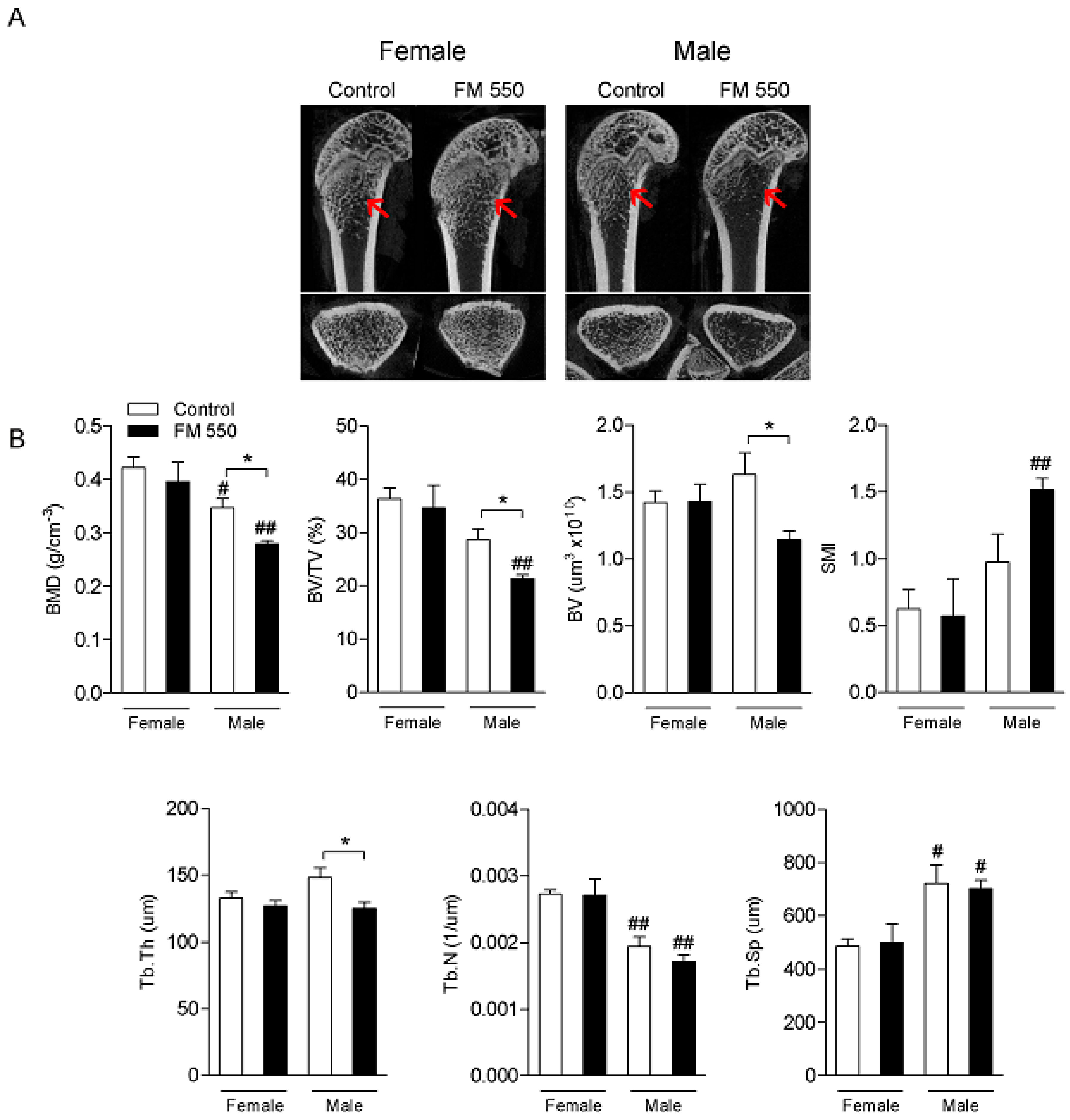
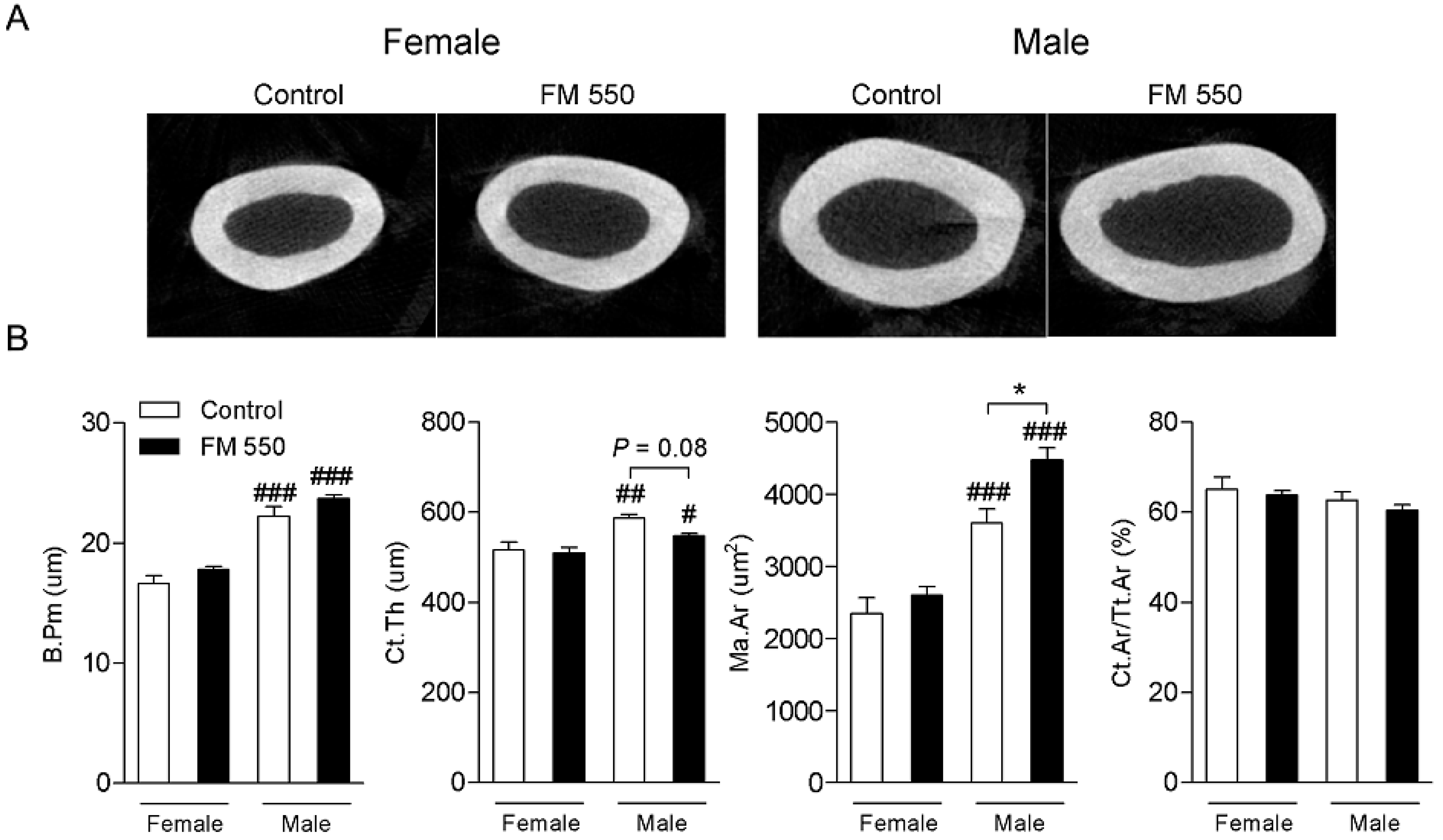
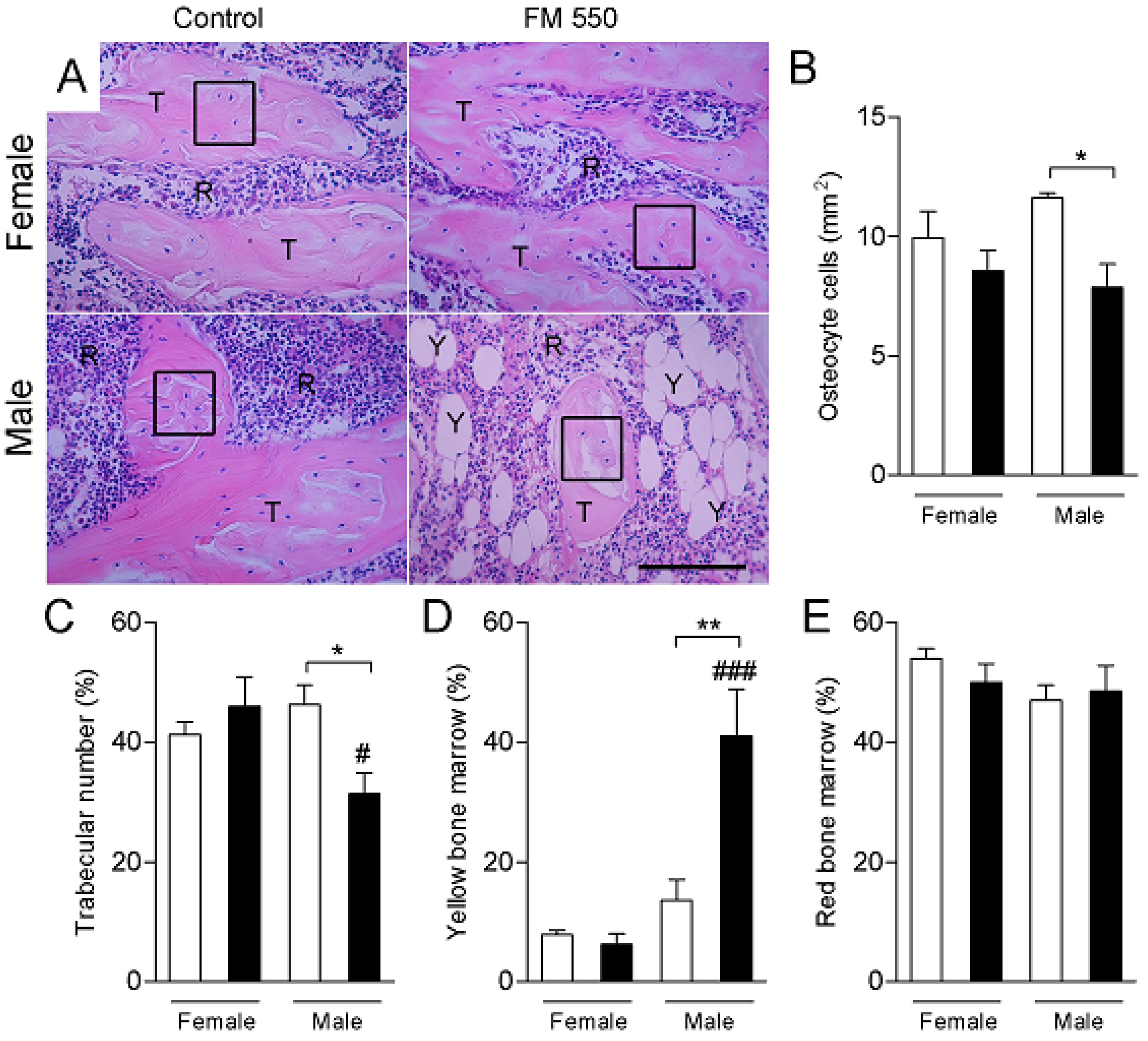
2. Results
3. Discussion
4. Materials and Methods
4.1. Micro Computed-Tomography (microCT) Imaging
4.2. Bone Histomorphometry
4.3. Statistical Analysis
5. Conclusions
Author Contributions
Funding
Acknowledgments
Conflicts of Interest
References
- Stapleton, H.M.; Allen, J.G.; Kelly, S.M.; Konstantinov, A.; Klosterhaus, S.; Watkins, D.; McClean, M.D.; Webster, T.F. Alternate and new brominated flame retardants detected in US house dust. Environ. Sci. Technol. 2008, 42, 6910–6916. [Google Scholar] [CrossRef]
- Dishaw, L.V.; Macaulay, L.J.; Roberts, S.C.; Stapleton, H.M. Exposures, mechanisms, and impacts of endocrine-active flame retardants. Curr. Opin. Pharmacol. 2014, 19, 125–133. [Google Scholar] [CrossRef] [Green Version]
- Stapleton, H.M.; Klosterhaus, S.; Eagle, S.; Fuh, J.; Meeker, J.D.; Blum, A.; Webster, T.F. Detection of organophosphate flame retardants in furniture foam and U.S. house dust. Environ. Sci. Technol. 2009, 43, 7490–7495. [Google Scholar] [CrossRef] [Green Version]
- Hoffman, K.; Butt, C.M.; Webster, T.F.; Preston, E.V.; Hammel, S.C.; Makey, C.; Lorenzo, A.M.; Cooper, E.M.; Carignan, C.; Meeker, J.D.; et al. Temporal Trends in Exposure to Organophosphate Flame Retardants in the United States. Environ. Sci. Technol. Lett. 2017, 4, 112–118. [Google Scholar] [CrossRef] [PubMed]
- Dishaw, L.V.; Powers, C.M.; Ryde, I.T.; Roberts, S.C.; Seidler, F.J.; Slotkin, T.A.; Stapleton, H.M. Is the PentaBDE replacement, tris (1,3-dichloro-2-propyl) phosphate (TDCPP), a developmental neurotoxicant? Studies in PC12 cells. Toxicol. Appl. Pharmacol. 2011, 256, 281–289. [Google Scholar] [CrossRef] [PubMed] [Green Version]
- Patisaul, H.B.; Roberts, S.C.; Mabrey, N.; McCaffrey, K.A.; Gear, R.B.; Braun, J.; Belcher, S.M.; Stapleton, H.M. Accumulation and endocrine disrupting effects of the flame retardant mixture firemaster((R)) 550 in rats: An exploratory assessment. J. Biochem. Mol. Toxicol. 2013, 27, 124–136. [Google Scholar] [CrossRef] [PubMed] [Green Version]
- Rock, K.D.; Horman, B.; Phillips, A.L.; McRitchie, S.L.; Watson, S.; Deese-Spruill, J.; Jima, D.; Sumner, S.; Stapleton, H.M.; Patisaul, H.B. EDC IMPACT: Molecular effects of developmental FM 550 exposure in Wistar rat placenta and fetal forebrain. Endocr. Connect. 2018, 7, 305–324. [Google Scholar] [CrossRef]
- Baldwin, K.R.; Phillips, A.L.; Horman, B.; Arambula, S.E.; Rebuli, M.E.; Stapleton, H.M.; Patisaul, H.B. Sex Specific Placental Accumulation and Behavioral Effects of Developmental Firemaster 550 Exposure in Wistar Rats. Sci. Rep. 2017, 7, 7118. [Google Scholar] [CrossRef]
- Belcher, S.M.; Cookman, C.J.; Patisaul, H.B.; Stapleton, H.M. In vitro assessment of human nuclear hormone receptor activity and cytotoxicity of the flame retardant mixture FM 550 and its triarylphosphate and brominated components. Toxicol. Lett. 2014, 228, 93–102. [Google Scholar] [CrossRef] [Green Version]
- Pillai, H.K.; Fang, M.; Beglov, D.; Kozakov, D.; Vajda, S.; Stapleton, H.M.; Webster, T.F.; Schlezinger, J.J. Ligand binding and activation of PPARgamma by Firemaster(R) 550: Effects on adipogenesis and osteogenesis in vitro. Environ. Health Perspect. 2014, 122, 1225–1232. [Google Scholar] [CrossRef] [Green Version]
- McGee, S.P.; Konstantinov, A.; Stapleton, H.M.; Volz, D.C. Aryl phosphate esters within a major PentaBDE replacement product induce cardiotoxicity in developing zebrafish embryos: Potential role of the aryl hydrocarbon receptor. Toxicol. Sci. 2013, 133, 144–156. [Google Scholar] [CrossRef] [PubMed] [Green Version]
- Fang, M.; Webster, T.F.; Ferguson, P.L.; Stapleton, H.M. Characterizing the peroxisome proliferator-activated receptor (PPARgamma) ligand binding potential of several major flame retardants, their metabolites, and chemical mixtures in house dust. Environ. Health Perspect. 2015, 123, 166–172. [Google Scholar] [CrossRef] [PubMed]
- Springer, C.; Dere, E.; Hall, S.J.; McDonnell, E.V.; Roberts, S.C.; Butt, C.M.; Stapleton, H.M.; Watkins, D.J.; McClean, M.D.; Webster, T.F.; et al. Rodent thyroid, liver, and fetal testis toxicity of the monoester metabolite of bis-(2-ethylhexyl) tetrabromophthalate (tbph), a novel brominated flame retardant present in indoor dust. Environ. Health Perspect. 2012, 120, 1711–1719. [Google Scholar] [CrossRef] [PubMed]
- Hoffman, K.; Fang, M.; Horman, B.; Patisaul, H.B.; Garantziotis, S.; Birnbaum, L.S.; Stapleton, H.M. Urinary Tetrabromobenzoic Acid (TBBA) as a Biomarker of Exposure to the Flame Retardant Mixture Firemaster 550. Environ. Health Perspect. 2014, 122, 963–969. [Google Scholar] [CrossRef] [Green Version]
- Phillips, A.L.; Chen, A.; Rock, K.D.; Horman, B.; Patisaul, H.B.; Stapleton, H.M. Editor’s Highlight: Transplacental and Lactational Transfer of Firemaster(R) 550 Components in Dosed Wistar Rats. Toxicol. Sci. 2016, 153, 246–257. [Google Scholar] [CrossRef] [Green Version]
- Hoffman, K.; Butt, C.M.; Chen, A.; Limkakeng, A.T., Jr.; Stapleton, H.M. High Exposure to Organophosphate Flame Retardants in Infants: Associations with Baby Products. Environ. Sci. Technol. 2015, 49, 14554–14559. [Google Scholar] [CrossRef]
- Castorina, R.; Butt, C.; Stapleton, H.M.; Avery, D.; Harley, K.G.; Holland, N.; Eskenazi, B.; Bradman, A. Flame retardants and their metabolites in the homes and urine of pregnant women residing in California (the CHAMACOS cohort). Chemosphere 2017, 179, 159–166. [Google Scholar] [CrossRef] [Green Version]
- Mendelsohn, E.; Hagopian, A.; Hoffman, K.; Butt, C.M.; Lorenzo, A.; Congleton, J.; Webster, T.F.; Stapleton, H.M. Nail polish as a source of exposure to triphenyl phosphate. Environ. Int. 2016, 86, 45–51. [Google Scholar] [CrossRef] [Green Version]
- Tajima, S.; Araki, A.; Kawai, T.; Tsuboi, T.; Ait Bamai, Y.; Yoshioka, E.; Kanazawa, A.; Cong, S.; Kishi, R. Detection and intake assessment of organophosphate flame retardants in house dust in Japanese dwellings. Sci. Total Environ. 2014, 478, 190–199. [Google Scholar] [CrossRef] [PubMed] [Green Version]
- Abdallah, M.A.; Covaci, A. Organophosphate flame retardants in indoor dust from Egypt: Implications for human exposure. Environ. Sci. Technol. 2014, 48, 4782–4789. [Google Scholar] [CrossRef]
- Brommer, S.; Harrad, S.; Van den Eede, N.; Covaci, A. Concentrations of organophosphate esters and brominated flame retardants in German indoor dust samples. J. Environ. Monit. 2012, 14, 2482–2487. [Google Scholar] [CrossRef] [PubMed]
- Coelho, S.D.; Sousa, A.C.A.; Isobe, T.; Kim, J.W.; Kunisue, T.; Nogueira, A.J.A.; Tanabe, S. Brominated, chlorinated and phosphate organic contaminants in house dust from Portugal. Sci. Total Environ. 2016, 569–570, 442–449. [Google Scholar] [CrossRef] [PubMed]
- Hoffman, K.; Daniels, J.L.; Stapleton, H.M. Urinary metabolites of organophosphate flame retardants and their variability in pregnant women. Environ. Int. 2014, 63, 169–172. [Google Scholar] [CrossRef] [PubMed] [Green Version]
- Meeker, J.D.; Cooper, E.M.; Stapleton, H.M.; Hauser, R. Urinary metabolites of organophosphate flame retardants: Temporal variability and correlations with house dust concentrations. Environ. Health Perspect. 2013, 121, 580–585. [Google Scholar] [CrossRef] [PubMed] [Green Version]
- Butt, C.M.; Congleton, J.; Hoffman, K.; Fang, M.; Stapleton, H.M. Metabolites of organophosphate flame retardants and 2-ethylhexyl tetrabromobenzoate in urine from paired mothers and toddlers. Environ. Sci Technol. 2014, 48, 10432–10438. [Google Scholar] [CrossRef] [PubMed]
- Van den Eede, N.; Heffernan, A.L.; Aylward, L.L.; Hobson, P.; Neels, H.; Mueller, J.F.; Covaci, A. Age as a determinant of phosphate flame retardant exposure of the Australian population and identification of novel urinary PFR metabolites. Environ. Int. 2015, 74, 1–8. [Google Scholar] [CrossRef]
- Cequier, E.; Sakhi, A.K.; Marce, R.M.; Becher, G.; Thomsen, C. Human exposure pathways to organophosphate triesters—A biomonitoring study of mother-child pairs. Environ. Int. 2015, 75, 159–165. [Google Scholar] [CrossRef]
- Dodson, R.E.; Van den Eede, N.; Covaci, A.; Perovich, L.J.; Brody, J.G.; Rudel, R.A. Urinary biomonitoring of phosphate flame retardants: Levels in California adults and recommendations for future studies. Environ. Sci. Technol. 2014, 48, 13625–13633. [Google Scholar] [CrossRef] [Green Version]
- Van den Eede, N.; Neels, H.; Jorens, P.G.; Covaci, A. Analysis of organophosphate flame retardant diester metabolites in human urine by liquid chromatography electrospray ionisation tandem mass spectrometry. J. Chromatogr. A 2013, 1303, 48–53. [Google Scholar] [CrossRef]
- Tontonoz, P.; Spiegelman, B.M. Fat and beyond: The diverse biology of PPARgamma. Annu. Rev. Biochem. 2008, 77, 289–312. [Google Scholar] [CrossRef]
- Yan, H.; Hales, B.F. Effects of Organophosphate Ester Flame Retardants on Endochondral Ossification in Ex Vivo Murine Limb Bud Cultures. Toxicol. Sci. 2019, 168, 420–429. [Google Scholar] [CrossRef] [PubMed]
- Berger, R.G.; Lefevre, P.L.; Ernest, S.R.; Wade, M.G.; Ma, Y.Q.; Rawn, D.F.; Gaertner, D.W.; Robaire, B.; Hales, B.F. Exposure to an environmentally relevant mixture of brominated flame retardants affects fetal development in Sprague-Dawley rats. Toxicology 2014, 320, 56–66. [Google Scholar] [CrossRef] [PubMed] [Green Version]
- Tung, E.W.; Yan, H.; Lefevre, P.L.; Berger, R.G.; Rawn, D.F.; Gaertner, D.W.; Kawata, A.; Rigden, M.; Robaire, B.; Hales, B.F.; et al. Gestational and Early Postnatal Exposure to an Environmentally Relevant Mixture of Brominated Flame Retardants: General Toxicity and Skeletal Variations. Birth Defects Res. B Dev. Reprod. Toxicol. 2016, 107, 157–168. [Google Scholar] [CrossRef] [PubMed]
- Pierce, J.L.; Begun, D.L.; Westendorf, J.J.; McGee-Lawrence, M.E. Defining osteoblast and adipocyte lineages in the bone marrow. Bone 2019, 118, 2–7. [Google Scholar] [CrossRef]
- Tung, E.W.Y.; Peshdary, V.; Gagne, R.; Rowan-Carroll, A.; Yauk, C.L.; Boudreau, A.; Atlas, E. Adipogenic Effects and Gene Expression Profiling of Firemaster(R) 550 Components in Human Primary Preadipocytes. Environ. Health Perspect. 2017, 125, 097013. [Google Scholar] [CrossRef] [Green Version]
- Behl, M.; Hsieh, J.H.; Shafer, T.J.; Mundy, W.R.; Rice, J.R.; Boyd, W.A.; Freedman, J.H.; Hunter, E.S., 3rd; Jarema, K.A.; Padilla, S.; et al. Use of alternative assays to identify and prioritize organophosphorus flame retardants for potential developmental and neurotoxicity. Neurotoxicol. Teratol. 2015, 52, 181–193. [Google Scholar] [CrossRef]
- Compston, J.E.; Vedi, S.; Stephen, A.B.; Bord, S.; Lyons, A.R.; Hodges, S.J.; Scammell, B.E. Reduced bone formation after exposure to organophosphates. Lancet 1999, 354, 1791–1792. [Google Scholar] [CrossRef]
- Martiniakova, M.; Bobonova, I.; Omelka, R.; Grosskopf, B.; Chovancova, H.; Spankova, J.; Toman, R. Simultaneous subchronic exposure to selenium and diazinon as possible risk factor for osteoporosis in adult male rats. Acta Vet. Scand. 2013, 55, 81. [Google Scholar] [CrossRef] [Green Version]
- Hoogduijn, M.J.; Rakonczay, Z.; Genever, P.G. The effects of anticholinergic insecticides on human mesenchymal stem cells. Toxicol. Sci. 2006, 94, 342–350. [Google Scholar] [CrossRef] [Green Version]
- Thigpen, J.E.; Setchell, K.D.R.; Ahlmark, K.B.; Locklear, J.; Spahr, T.; Caviness, G.F.; Goelz, M.F.; Haseman, J.K.; Newbold, R.R.; Forsythe, D.B. Phytoestrogen content of purified open- and closed-formula laboratory animal diets. Lab. Anim. Sci. 1999, 49, 530–536. [Google Scholar]
- Howdeshell, K.L.; Peterman, P.H.; Judy, B.M.; Taylor, J.A.; Orazio, C.E.; Ruhlen, R.L.; vom Saal, F.S.; Welshons, W.V. Bisphenol A is released from used polycarbonate animal cages into water at room temperature. Environ. Health Perspect. 2003, 111, 1180–1187. [Google Scholar] [CrossRef] [PubMed] [Green Version]
- McDonnell-Dowling, K.; Kleefeld, S.; Kelly, J.P. Consequences of oral gavage during gestation and lactation on rat dams and the neurodevelopment and behavior of their offspring. J. Am. Assoc. Lab. Anim. Sci. 2017, 56, 79–83. [Google Scholar] [PubMed]
- Brown, A.P.; Dinger, N.; Levine, B.S. Stress produced by gavage administration in the rat. Contemp. Top. Lab. Anim. Sci. 2000, 39, 17–21. [Google Scholar] [PubMed]
- Patisaul, H.B. Achieving CLARITY on bisphenol A, brain and behaviour. J. Neuroendocrinol. 2020, 32, e12730. [Google Scholar] [CrossRef] [PubMed]
- Macari, S.; Sharma, L.A.; Wyatt, A.; da Silva, J.M.; Dias, G.J.; Silva, T.A.; Szawka, R.E.; Grattan, D.R. Lactation induces increases in the RANK/RANKL/OPG system in maxillary bone. Bone 2018, 110, 160–169. [Google Scholar] [CrossRef]
- Bouxsein, M.L.; Boyd, S.K.; Christiansen, B.A.; Guldberg, R.E.; Jepsen, K.J.; Muller, R. Guidelines for assessment of bone microstructure in rodents using micro-computed tomography. J. Bone Miner. Res. 2010, 25, 1468–1486. [Google Scholar] [CrossRef]


© 2020 by the authors. Licensee MDPI, Basel, Switzerland. This article is an open access article distributed under the terms and conditions of the Creative Commons Attribution (CC BY) license (http://creativecommons.org/licenses/by/4.0/).
Share and Cite
Macari, S.; Rock, K.D.; Santos, M.S.; Lima, V.T.M.; Szawka, R.E.; Moss, J.; Horman, B.; Patisaul, H.B. Developmental Exposure to the Flame Retardant Mixture Firemaster 550 Compromises Adult Bone Integrity in Male but not Female Rats. Int. J. Mol. Sci. 2020, 21, 2553. https://0-doi-org.brum.beds.ac.uk/10.3390/ijms21072553
Macari S, Rock KD, Santos MS, Lima VTM, Szawka RE, Moss J, Horman B, Patisaul HB. Developmental Exposure to the Flame Retardant Mixture Firemaster 550 Compromises Adult Bone Integrity in Male but not Female Rats. International Journal of Molecular Sciences. 2020; 21(7):2553. https://0-doi-org.brum.beds.ac.uk/10.3390/ijms21072553
Chicago/Turabian StyleMacari, Soraia, Kylie D. Rock, Mariana S. Santos, Virgínia T. M. Lima, Raphael E. Szawka, Jamal Moss, Brian Horman, and Heather B. Patisaul. 2020. "Developmental Exposure to the Flame Retardant Mixture Firemaster 550 Compromises Adult Bone Integrity in Male but not Female Rats" International Journal of Molecular Sciences 21, no. 7: 2553. https://0-doi-org.brum.beds.ac.uk/10.3390/ijms21072553