Communication

Human NANOS1 Represses Apoptosis by Downregulating Pro-Apoptotic Genes in the Male Germ Cell Line

by
Damian M. Janecki
1,2,*,
Erkut Ilaslan
1,†,
Maciej J. Smialek
1,†,
Marcin P. Sajek
1,
Maciej Kotecki
3,
Barbara Ginter-Matuszewska
4,
Patryk Krainski
5,
Jadwiga Jaruzelska
1 and
Kamila Kusz-Zamelczyk
1,*
1
Institute of Human Genetics, Polish Academy of Sciences, Strzeszynska 32, 60-479 Poznan, Poland
2
Institute of Bioorganic Chemistry, Polish Academy of Sciences, Noskowskiego 12/14, 61-704 Poznan, Poland
3
Department of Developmental, Molecular and Chemical Biology, Tufts University Medical School, 136 Harrison Ave, Boston, MA 02111, USA
4
Department of Histology and Embryology, Poznan University of Medical Sciences, Swiecickiego 6, 60-781 Poznan, Poland
5
Department of Clinical Pathology, Poznan University of Medical Sciences, Przybyszewskiego 49, 60-355 Poznan, Poland
*
Authors to whom correspondence should be addressed.
These authors contributed equally to this work.
Int. J. Mol. Sci. 2020, 21(8), 3009; https://0-doi-org.brum.beds.ac.uk/10.3390/ijms21083009
Submission received: 28 February 2020 / Revised: 17 April 2020 / Accepted: 22 April 2020 / Published: 24 April 2020

Abstract

:
While two mouse NANOS paralogues, NANOS2 and NANOS3, are crucial for maintenance of germ cells by suppression of apoptosis, the mouse NANOS1 paralogue does not seem to regulate these processes. Previously, we described a human NANOS1 p.[(Pro34Thr);(Ser83del)] mutation associated with the absence of germ cells in seminiferous tubules of infertile patients, which might suggest an anti-apoptotic role of human NANOS1. In this study, we aimed to determine a potential influence of human NANOS1 on the maintenance of TCam-2 model germ cells by investigating proliferation, cell cycle, and apoptosis. Constructs encoding wild-type or mutated human NANOS1 were used for transfection of TCam-2 cells, in order to investigate the effect of NANOS1 on cell proliferation, which was studied using a colorimetric assay, as well as apoptosis and the cell cycle, which were measured by flow cytometry. RNA-Seq (RNA sequencing) analysis followed by RT-qPCR (reverse transcription and quantitative polymerase chain reaction) was conducted for identifying pro-apoptotic genes repressed by NANOS1. Here, we show that overexpression of NANOS1 downregulates apoptosis in TCam-2 cells. Moreover, we found that NANOS1 represses a set of pro-apoptotic genes at the mRNA level. We also found that the infertility-associated p.[(Pro34Thr);(Ser83del)] mutation causes NANOS1 to functionally switch from being anti-apoptotic to pro-apoptotic in the human male germ cell line. Thus, this report is the first to show an anti-apoptotic role of NANOS1 exerted by negative regulation of mRNAs of pro-apoptotic genes.

1. Introduction

Apoptosis is an essential process in male germ cell development. This kind of cell death controls the correctness and number of germ cells and occurs during both the migration of primordial germ cells (PGCs) to primary gonads and spermatogenesis. The final number of germ cells in the testes is determined by a tightly regulated balance between cell proliferation and apoptosis. Consequently as many as 75% of germ cells are eliminated by apoptosis in healthy vertebrate testes. Imbalance between proliferation and apoptosis can lead to a decreased number of germ cells manifested clinically as infertility, while an uncontrolled increase in the number of these cells may lead to testicular germ cell tumors (TGCT); for a review, see [1]. Thus, expanding the knowledge of molecular factors influencing germ cell apoptosis is crucial for the development of novel therapeutic strategies for both male infertility and TGCT.
Nanos proteins are widespread within the animal kingdom, and their critical function for apoptosis regulation in germ cells, e.g., an anti-apoptotic role in Drosophila germ cells, is well established. Nanos proteins act as post-transcriptional repressors of specific mRNAs by binding to them using highly conserved zinc-finger domain; for a review, see [2]. In particular, Drosophila Nanos was shown to repress caspase activators such as hid (head involution defective) and skl (sickle) to inhibit apoptosis in PGCs. This inhibition is crucial for the survival of PGCs during migration to the primary gonads [3]. Nanos knockout in Drosophila leads to infertility in both sexes caused by the lack of germ cells [4]. An anti-apoptotic property of Nanos is conserved in evolution at least from Drosophila to mice. There are three NANOS paralogues in mammals and two of them, NANOS2 and NANOS3, play anti-apoptotic roles in different stages of germ cell lineage development in mice [5,6,7]. Apoptosis of PGCs is suppressed by NANOS3 [5], and knockout of the murine gene causes infertility in both sexes [6]. In turn, NANOS2 represses apoptosis, specifically of male gonocytes, and its knockout in mice causes infertility restricted to male sex [6,7]. By contrast to Drosophila, to the best of our knowledge, specific mRNAs encoding apoptotic factors controlled by NANOS2 or NANOS3 in mammals have not been revealed. Unlike Nanos2 and Nanos3 knockout resulting in mice infertility, Nanos1 knockout mice are viable and fertile, indicating that the NANOS1 protein is dispensable for mouse development and fertility [8].
While NANOS proteins have also been implicated in human germ cell development [9,10,11,12], their functions identified in other species, such as regulation of apoptosis, have hardly been characterized in humans. So far, only the expression profile of human NANOS paralogues and the association between NANOS mutations and infertility have been investigated. Similar to the mouse orthologue, human NANOS2 was shown to be expressed specifically in male germ cells, indicating a potential association between NANOS2 mutations and male infertility. However, the detected mutations in infertile patients seem not to be causative [12]. In turn, human NANOS3 expression was shown in fetal and adult gonads as well as in the adult brain [10,11]. NANOS3 gene mutations were found in a group of infertile men, but no causation was detected [11]. On the other hand, two out of four NANOS3 mutations were detected to be linked to premature ovarian insufficiency (POI) in infertile women [13,14,15,16]. Moreover, Santos et al. demonstrated that one NANOS3 mutation linked to POI causes increased apoptosis of cultured cells, suggesting an anti-apoptotic role of human NANOS3 [14], as was shown for a mouse orthologue.
Although the mouse Nanos1 orthologue seems not to be critical for germ cell development and human NANOS1 expression is more ubiquitous than NANOS2 and NANOS3 [10], one out of NANOS1 mutations was found to be potentially causative among a group of infertile men. Namely, a NM_199461.4(NANOS1_v001):c.[100C>A;240_242del];NM_199461.4(NANOS1_i001):p.[(Pro34Thr);(Ser83del)] double mutation (in this report referred to as p.[(Pro34Thr);(Ser83del)]) was identified in two infertile male patients manifesting in the absence of germ cells in semen and seminiferous tubules (Sertoli cell only syndrome–SCOS) [17]. This mutation encompasses the N-terminal conserved NIM (NOT1 interacting motif) region (Figure 1A), which is necessary for recruitment of the deadenylase complex to deadenylate, and leads to degradation of mRNA targets [18]. Interestingly, the p.[(Pro34Thr);(Ser83del)] double mutation is located in a NANOS1 region which is not present in the mouse orthologue [17]. The difference in the structures of the mouse and human NANOS1 protein could reflect a distinct significance of these NANOS1 orthologues for germ cell development. The SCOS associated with the NANOS1 mutation suggests the critical role of this gene in maintenance of the germline in men, but specific biological processes controlled by NANOS1 have remained elusive.
The aim of this work was to determine the potential influence, and its mechanism, of human NANOS1 on maintenance of germ cells. We show that the NANOS1 protein suppresses apoptosis of germ cells in culture through repression, at the mRNA level, of a group of pro-apoptotic genes. Moreover, we demonstrate that the aforementioned p.[(Pro34Thr);(Ser83del)] mutation reverses NANOS1 activity from anti-apoptotic to pro-apoptotic by disrupting NANOS1 repression of some pro-apoptotic genes.

2. Results

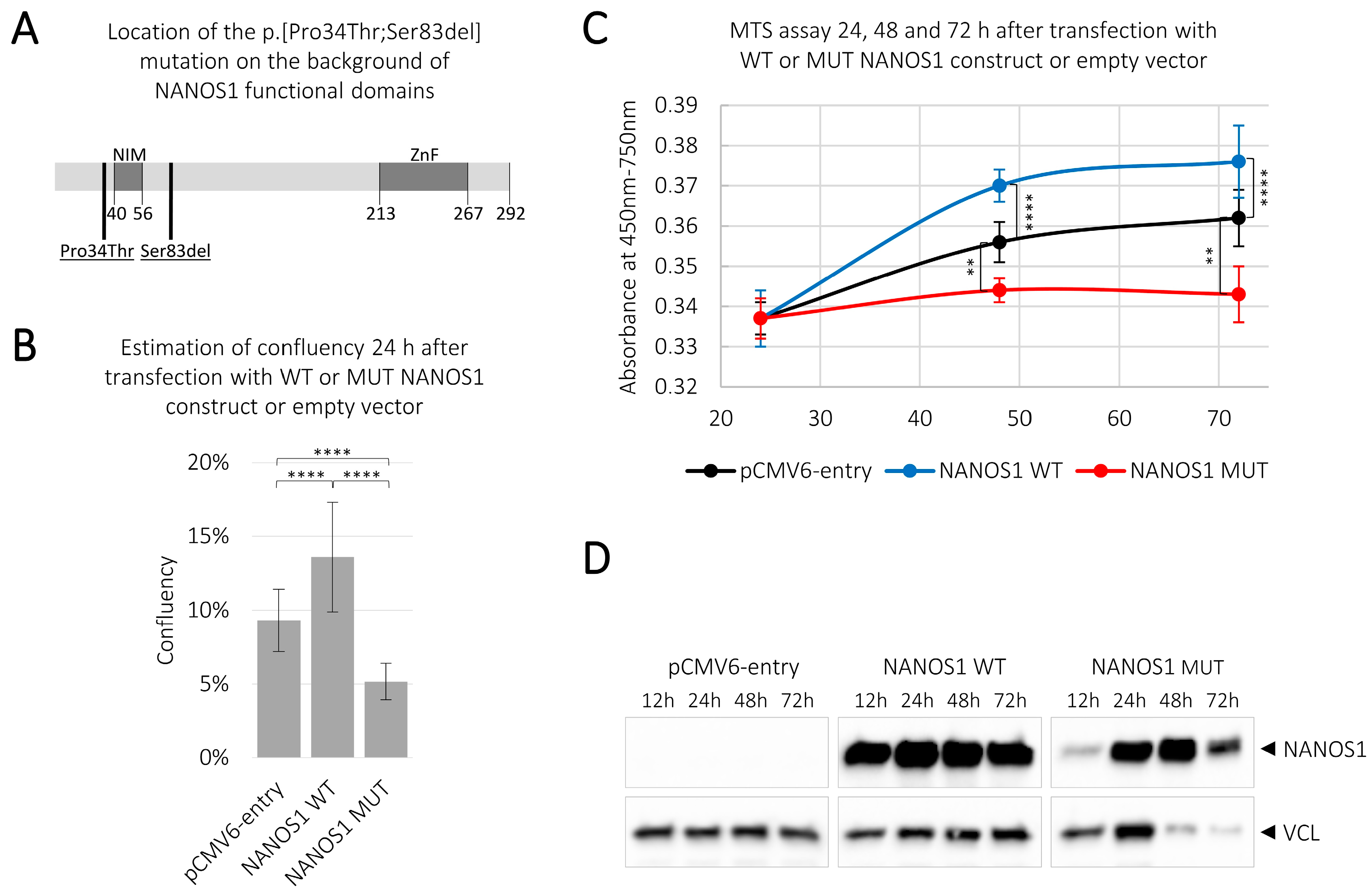
2.1. Overexpression of the Wild-Type NANOS1 Increases, While the Mutated NANOS1 p.[(Pro34Thr);(Ser83del)] Decreases, the Proliferation of TCam-2 cells

In order to examine the potential influence of NANOS1 on the viability of germ cells as well as the functional significance of the NANOS1 double mutation (Figure 1A), we studied the wild-type and the mutated NANOS1 effects on the proliferation status of the TCam-2 cell line. This cell line originates from human seminoma and represents male germ cells at an early stage of development as PGCs/gonocytes [19]. Our transcriptome analysis revealed that NANOS1 expression is very low at the mRNA level (1.76 FPKM–fragments per kilobase of transcript per million mapped reads) in TCam-2 cells [20]. For that reason, instead of using a knock-down strategy, we decided to overexpress the wild-type and the mutated NANOS1 in TCam-2 cells in order to markedly alter the expression level and to investigate the function of this protein in human germ cells. While overexpression of the wild-type NANOS1 caused a significant increase (of nearly 50%) in cell confluency compared to the empty vector, overexpression of the mutated NANOS1 caused a significant decrease (of nearly 45%) in the cell confluency, as compared to the empty vector (Figure 1B). To check whether the above effects on the cell confluency reflect the influence of NANOS1 on cell proliferation, we overexpressed the wild-type and the mutated NANOS1 in TCam-2 cells and performed a colorimetric assay (MTS assay). We found that the wild-type NANOS1 stimulated cell proliferation, whereas the mutated NANOS1 caused a significant decrease in cell proliferation, as compared to the empty vector (Figure 1C). Both the cell confluency and the MTS assay consistently showed that NANOS1 may maintain germ cells by pro-proliferative activity, whereas its mutation reverts this ability. Expression of both the NANOS1 constructs after transfection is shown in Figure 1D.

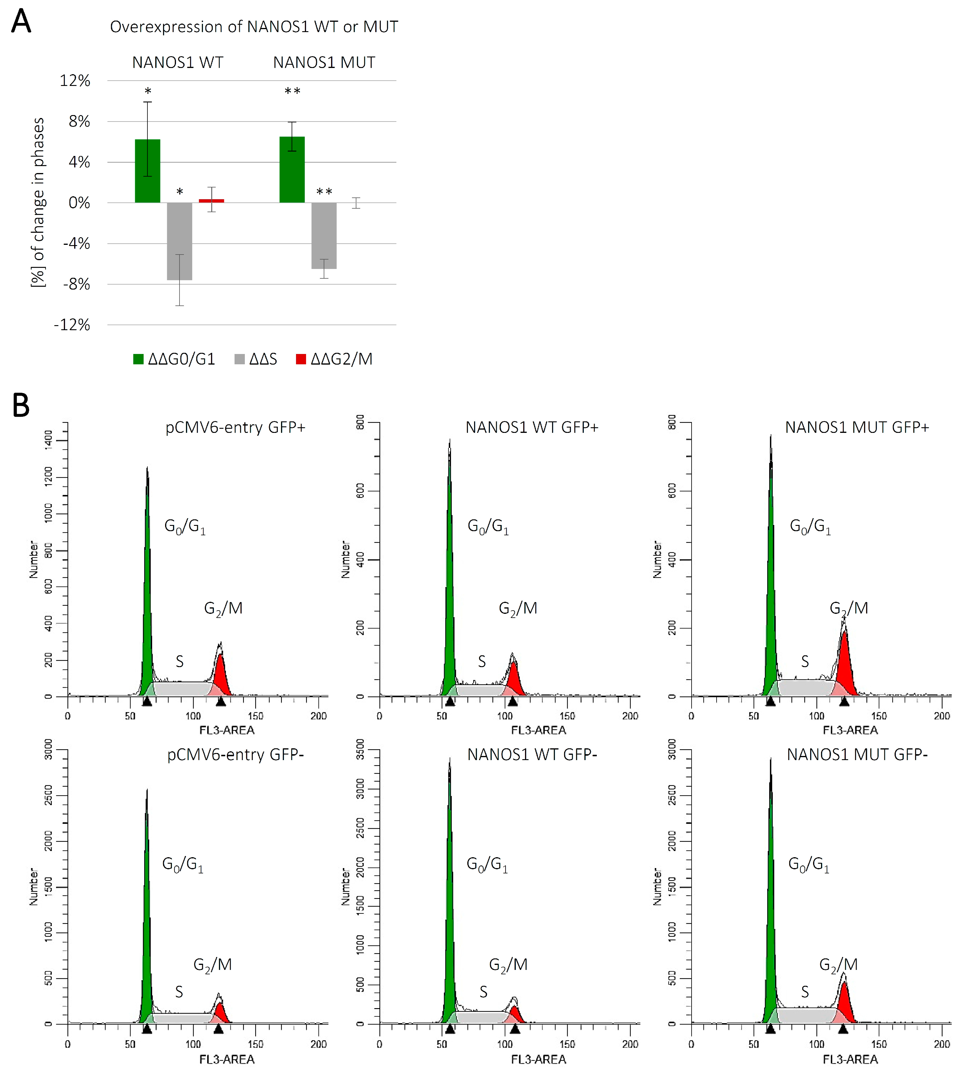
2.2. Overexpression of Both the Wild-Type and the Mutated NANOS1 Inhibits the Cell Cycle of TCam-2 cells

Since we observed increased proliferation upon overexpression of the wild-type NANOS1 (Figure 1C), we addressed the question whether that effect was due to modulation of the cell cycle and, if so, whether the p.[(Pro34Thr);(Ser83del)] mutation affected such a modulation. We performed flow cytometric analysis of the cell cycle distribution in unsynchronized TCam-2 cells overexpressing the wild-type or the mutated NANOS1, in comparison with the empty vector (Figure 2A). The separation of cell populations in each cell cycle phase was performed using ModFit LT 5.0 software (Figure 2B). Upon overexpression of the wild-type NANOS1, we observed an increase in the proportion of cells in the G0/G1 phase, with a concordant decrease in the proportion of cells in the S phase, as compared to the empty vector (Figure 2A), indicating that NANOS1 inhibits the TCam-2 cell cycle. We show, for the first time, the effect of any Nanos protein on cell cycle distribution. However, the inhibition of the cell cycle by NANOS1 does not explain its pro-proliferative role. Moreover, no significant difference in cell cycle effects was observed between cells expressing the wild-type and the mutated NANOS1 (Figure 2A).

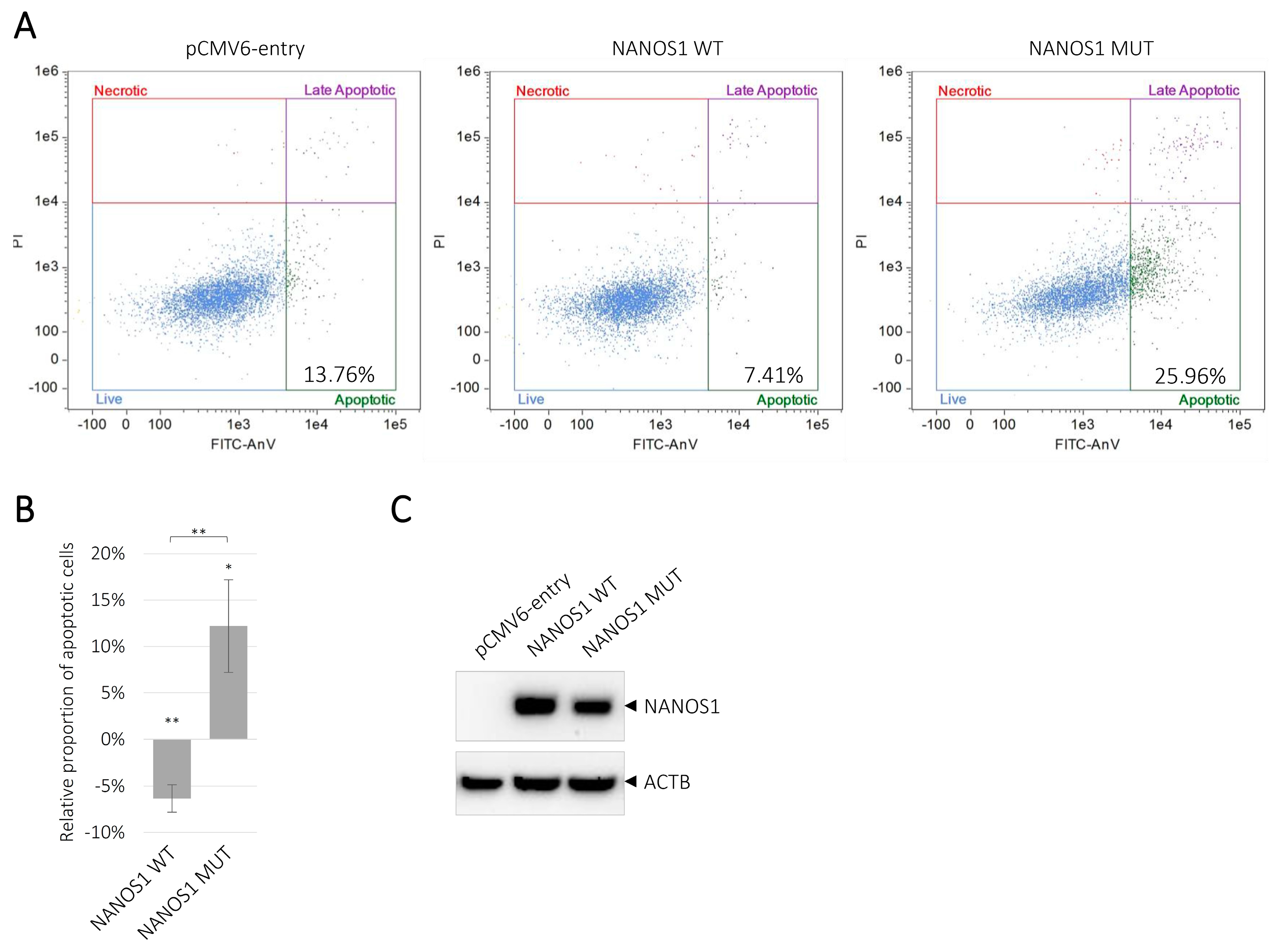
2.3. Overexpression of the Wild-Type NANOS1 Decreases, Whereas the Mutated NANOS1 p.[(Pro34Thr);(Ser83del)] Increases Apoptosis of TCam-2 cells

We investigated the function of NANOS1 in the maintenance of germ cells by studying the NANOS1 effect on apoptosis. To determine whether the divergent effects of the wild-type and the mutated NANOS1 on cell proliferation (Figure 1C) were associated with a different influence on apoptosis, we performed flow cytometry analysis of Annexin V-stained TCam-2 cells transfected with the wild-type or the mutated NANOS1, as compared to the empty vector. We showed that the wild-type NANOS1 reduced the apoptosis of TCam-2 cells. To the best of our knowledge, this is the first evidence for any human NANOS to suppress apoptosis. Moreover, we found that the mutated NANOS1 induced apoptosis (Figure 3A). Thus, we showed that the NANOS1 anti-apoptotic activity was reversed to pro-apoptotic activity by the mutation. A quantification of the flow cytometric data is presented on the graph in Figure 3B, while the expression of the wild-type and the mutated NANOS1 proteins in transfected cells is shown in Figure 3C.

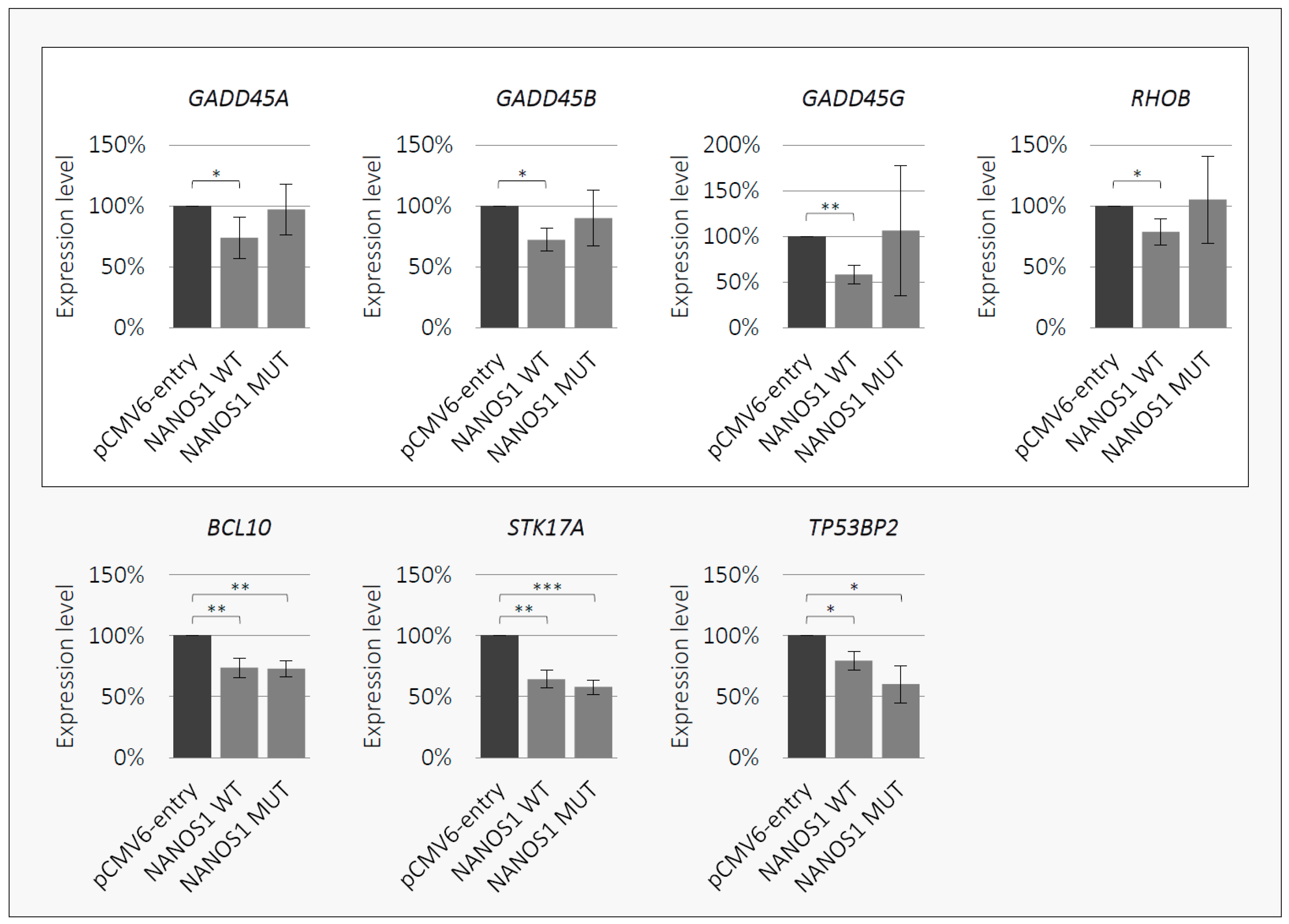
2.4. NANOS1 Negatively Regulates Pro-Apoptotic Genes and This Regulation is Disrupted by the p.[(Pro34Thr);(Ser83del)] Mutation

Given that the NANOS1 protein is a posttranscriptional regulator of gene expression [18], we performed a search for apoptosis-related transcripts regulated by NANOS1 in order to elucidate its anti-apoptotic property. Therefore, we carried out RNA-Seq analysis of the wild-type NANOS1 overexpression in TCam-2 cells (RNA-Seq data are available from the Gene Expression Omnibus, accession GSE134802). In this way, we identified 10 mRNAs encoding pro-apoptotic factors (GADD45A, GADD45B, GADD45G, RHOB, BCL10, STK17A, TP53BP2, RIPK1, SIAH1, JUN), which were downregulated upon NANOS1 overexpression (Table 1). We confirmed downregulation of seven of them (GADD45A, GADD45B, GADD45G, RHOB, BCL10, STK17A, TP53BP2) using RT-qPCR (Figure 4). Thus, for the first time, we identified genes encoding pro-apoptotic factors to be downregulated by NANOS1. To the best of our knowledge, this is also the first identification of any apoptosis-related mRNAs regulated by any Nanos protein among vertebrates. Finally, we investigated whether the p.[(Pro34Thr);(Ser83del)] mutation affects NANOS1-mediated regulation by performing RT-qPCR upon overexpression of the mutated NANOS1. We found that the regulation of four mRNAs (GADD45A, GADD45B, GADD45G, RHOB) was abrogated by the p.[(Pro34Thr);(Ser83del)] NANOS1 mutation (Figure 4, upper panel). Interestingly, all of these mRNAs encode proteins of closely related functions, namely, factors activating DNA damage-induced apoptosis.

3. Discussion

Regulation of germ cell number is critical for both fertility and prevention of TGCT. Some Nanos proteins are known to play a critical role in this regulation by suppression of germ cell apoptosis. In the mouse model, NANOS2 and NANOS3 paralogues were shown as apoptosis suppressors, but the molecular background of this activity has not been explained [5,6,7]. In turn, mouse NANOS1 seems to be unnecessary for regulation of germ cell number since Nanos1 knockout animals were fertile and healthy [8]. Recently, we described a NANOS1 gene p.[(Pro34Thr);(Ser83del)] mutation associated with the lack of germ cells in seminiferous tubules in infertile SCOS patients, suggesting a possible anti-apoptotic role of the human NANOS1 homologue [17].
Here, we investigated the potential influence of human NANOS1 on the maintenance of germ cells showing that NANOS1 overexpression increases the number of viable male germ cells in culture. In order to shed light on this phenomenon, we studied the NANOS1 influence on the cell cycle and apoptosis. We showed that overexpression of NANOS1 caused a slight increase in the proportion of cells in the G0/G1 phase, with a concordant decrease of cells in the S phase. This is the first report, to the best of our knowledge, demonstrating quantification of cells in various phases of the cell cycle in response to NANOS proteins. Our result indicates that slight NANOS1-mediated cell cycle inhibition is accompanied by stronger NANOS1 inhibition of apoptosis, which accounts for the overall NANOS1 positive role in cell proliferation. It is the first report showing an anti-apoptotic and pro-proliferative role of any human NANOS paralogue and, more broadly, any NANOS1 mammalian orthologue. We hypothesized that NANOS1 negatively regulates apoptosis by repressing mRNAs encoding pro-apoptotic factors. Indeed, we identified seven mRNAs encoding well-established pro-apoptotic factors to be downregulated by NANOS1: GADD45A, GADD45B, GADD45G, RHOB, BCL10, STK17A, and TP53BP2. Downregulation of these mRNAs by NANOS1 may account for its strong anti-apoptotic properties in human male germ cells. Regulation of mRNAs encoding apoptosis-promoting factors by any mammalian NANOS homologue has never been reported. Although we observed downregulation of the above-mentioned pro-apoptotic mRNAs caused by NANOS1 overexpression, we did not explore whether NANOS1 regulates them directly or indirectly. Therefore, it requires further investigation to establish whether these mRNAs are direct targets of NANOS1. Importantly, we observed that the NANOS1 p.[(Pro34Thr);(Ser83del)] mutation increased apoptosis of human male germline cells in culture. This suggests that this mutation provoked a functional reversal of NANOS1 from anti-apoptotic to pro-apoptotic. We also found that this mutation disrupted NANOS1 inhibition of four pro-apoptotic mRNAs, namely, GADD45A, GADD45B, GADD45G, and RHOB. These genes seem to be the most specifically responsible for the NANOS1 anti-apoptotic function. Interestingly, each of these four mRNAs encodes a protein activating DNA damage-induced apoptosis. Their transcription is upregulated in response to genotoxic stress [21,22,23]. The three GADD45 (growth arrest and DNA damage inducible 45) protein family members induce apoptosis by binding and activating MAP3K4 kinase, which in turn positively regulates the p38/JNK-mediated apoptotic pathway [21,24]. RHOB (ras homologue family member B), in turn, activates apoptosis by positive regulation of JNK that triggers the activation of pro-apoptotic BCL2L11 [25,26]. We propose that abrogated inhibition of the above-mentioned four pro-apoptotic mRNAs upon expression of mutated NANOS1 accounts for increased apoptosis of TCam-2 cells. The functional switch of NANOS1 from anti-apoptotic to pro-apoptotic may represent the mechanism underlying the infertility in patients with the NANOS1 p.[(Pro34Thr);(Ser83del)] mutation. In patients, this mutation could affect the regulation of GADD45A, GADD45B, GADD45G, and RHOB at the post-transcriptional level, resulting in upregulated expression of encoded pro-apoptotic factors, thereby causing increased apoptosis of germ cells and finally, the absence of germ cells from seminiferous tubules and infertility.
It is important to note that the human male germ cell line TCam-2 originates from seminoma, which is a type of TGCT [19]. In this context, it is worth noting that almost all NANOS1-repressed pro-apoptotic factors identified in this study (GADD45A, GADD45B, GADD45G, RHOB, and TP53BP2) are well-established tumor suppressors [27,28,29,30,31,32,33,34,35]. This is in line with NANOS1 itself being proposed to promote carcinogenesis [36]. Therefore, it is likely that in addition to playing a role in germ cell development, NANOS1 downregulates the expression of tumor-suppressing factors, as has already been suggested for other RNA-binding proteins [37]. This finding represents an interesting issue for future studies aimed at investigating the role of NANOS1 in carcinogenesis.

4. Materials and Methods

4.1. Cell Culture and Transfection

TCam-2 cells were obtained from Dr. Sohei Kitazawa and were maintained in RPMI with GlutaMAX medium (Gibco, Life Technologies, 61870044, Paisley, UK) supplemented with 10% (v/v) foetal bovine serum (GE Healthcare HyClone, SH30071, Logan, Utah, USA) and 1% (v/v) antibiotic antimycotic solution (Lonza, EE17-602E, Germany). The cells were transfected with plasmid constructs using the Neon Transfection System (Thermo Fisher Scientific, Waltham, Massachusetts, USA) according to the manufacturer’s protocol.

4.2. Estimation of Cell Confluency

TCam-2 cells (2 × 10⁵) were transfected with 1.5 µg of constructs encoding the wild-type, the mutated NANOS1 p.[(Pro34Thr);(Ser83del)] or the empty pCMV6-entry vector (OriGene Technologies, PS100001, Rockville, Maryland, USA) in three biological replicates (three independent transfections). Cell confluency was measured 24 h after transfection in five random areas of each 6-well plate using the JuLI™ FL Fluorescence Cell History Recorder (NanoEnTek, Seoul, South Korea).

4.3. MTS Assay

TCam-2 cells (2 × 10⁵) were transfected with 1.5 µg of constructs encoding the wild-type, the mutated NANOS1 p.[(Pro34Thr);(Ser83del)] or the empty pCMV6-entry vector (OriGene Technologies, PS100001, Rockville, Maryland, USA) in three biological replicates (three independent transfections; five technical repeats in each of five different wells of a 96-well plate) and were cultured for 24, 48, and 72 h after transfection. In order to obtain a similar number per well of viable cells expressing the different constructs for the MTS assay, viable cells were counted 12 h post-transfection using Trypan Blue staining. Similar numbers of cells were seeded in five different wells of a 96-well plate. The cell viability was measured using GloMax®-Multi Detection System (Promega, Madison, Wisconsin, USA) at a 450 nm (and 750 nm for the cell background) wavelength 24, 48, and 72 h after transfection and 4 h after adding 20 µl of CellTiter 96® AQ One Solution Reagent (Promega, Madison, Wisconsin, USA) to each well containing transfected TCam-2 cells. The overexpression of the wild-type and the mutated NANOS1 was measured 12, 24, 48, and 72 h after transfection by western blot using an anti-DDK antibody (OriGene Technologies, TA50011, Rockville, Maryland, USA). VCL (vinculin) protein was used as a loading control.

4.4. Western Blotting

Overexpression efficiency was measured by western blot analysis under standard conditions, using a nitrocellulose membrane and horseradish peroxidase (HRP)-conjugated secondary antibodies. Semi-quantitative measurement of protein levels was performed using ImageLab 5.1 software (Bio-Rad, Hercules, California, USA). The chemiluminescent signal was detected using the Clarity™ Western ECL Substrate for HRP (Bio-Rad, 1705060, Hercules, California, USA).

4.5. Antibodies

The primary antibody anti-DDK (FLAG®) (OriGene Technologies, TA50011, 1:2500, Rockville, Maryland, USA) was used for detection of proteins expressed with the pCMV6-entry vector. Anti-ACTB (actin beta) (Sigma-Aldrich, A2066, 1:10000, Saint Louis, Missouri, USA), and anti-VCL (vinculin) (Abcam, ab129002, 1:20000, Cambridge, UK) antibodies were used as loading controls. Secondary antibodies, i.e., goat anti-rabbit IgG-HRP (Sigma-Aldrich, A6154, 1:250000, Saint Louis, Missouri, USA) and goat anti-mouse IgG-HRP (Santa Cruz Biotechnology, sc-2005, 1:10000, Dallas, Texas, USA), were used.

4.6. Flow Cytometry

Detection of apoptotic TCam-2 cells was performed 48 h after transfection of 3 × 10⁵ TCam-2 cells with 2 µg of constructs encoding the wild-type, the mutated NANOS1 p.[(Pro34Thr);(Ser83del)] or the empty pCMV6-entry vector (OriGene Technologies, PS100001, Rockville, Maryland, USA). Cells were stained using the Annexin V-FITC Apoptosis Detection Kit (Beckman Coulter, Brea, California, USA) according to the manufacturer’s protocol and were measured by flow cytometry using a FlowSight instrument (Amnis, Seattle, Washington, USA). The results were analyzed using Image Data Exploration and Analysis Software version 6.0 (IDEAS® v6.0, Amnis, Seattle, Washington, USA). The experiment was performed in three biological replicates. Cell cycle analysis was performed 48 h after transfection of 2 × 10⁶ TCam-2 cells with 20 µg of constructs encoding the wild-type, the mutated NANOS1 p.[(Pro34Thr);(Ser83del)] or the empty pCMV6-entry vector co-transfected with GFP-F at a proportion of 9:1. For this purpose, TCam-2 cells were washed with PBS and fixed in a cold 100% methanol on ice for 10 min. After incubation at 37 °C for 15 min in 50 µg/mL propidium iodide (PI) (Sigma-Aldrich, P4170, Saint Louis, Missouri, USA) containing 330 µg/mL RNase A (Sigma-Aldrich, R6513, Saint Louis, Missouri, USA), cells were incubated for 1 h on ice and finally measured using the S3e™ Cell Sorter (Bio-Rad, Hercules, California, USA) apparatus. Data files were analyzed using ModFit LT™ 5.0 (Verity Software House, Topsham, Maine, USA). The experiment was performed in four biological replicates.

4.7. cDNA Library Preparation and RNA-Seq

TCam-2 cells (2 × 10⁵) were transfected with 1.5 µg of constructs encoding the wild-type NANOS1 or the empty pCMV6-entry vector in three biological replicates. Transcription was stopped by Actinomycin D (5 µg/mL)(Sigma-Aldrich, A1410, Saint Louis, Missouri, USA) 24 h after transfection for 4 h. Total RNA was extracted using an RNeasy Plus Micro Kit (Qiagen, Hilden, Germany) according to the manufacturer’s protocol. RNA quality was checked on a Bioanalyzer (Agilent Technologies, Santa Clara, California, USA) using the RNA 6000 Nano Kit (Agilent Technologies, Santa Clara, California, USA). Total RNA samples with RIN value > 7 were used for library preparation. cDNA libraries were prepared using the TruSeq RNA Sample Prep v2 (Illumina, San Diego, Santa Clara, California, USA), and subsequent next-generation sequencing (PE:100 nt) was performed on an Illumina HiSeq 4000 platform by Macrogen (Seoul, South Korea). RNA-Seq data described in this study are available from the Gene Expression Omnibus (accession GSE134802).

4.8. Bioinformatic Analysis

Differential expression analysis was performed on the Galaxy platform (Pennsylvania State University, State College, Pennsylvania, USA; Johns Hopkins University, Baltimore, Maryland, USA; Oregon Health and Science University, Portland, Oregon, USA) by the following pipeline: Paired-End sequences obtained from the HiSeq 4000 platform were trimmed using Trimmomatic v0.36.3 [38]. Sequence reads that passed quality filters were mapped to the human reference genome hg38 using Bowtie 2 v2.3.4.1 [39]. Mapped reads from three biological repetitions of NANOS1 and empty vector overexpression libraries were counted using featureCounts v1.6.0.3 [40] followed by calculation of the differential gene expression with DESeq2 v2.11.40.1 [41]. Differentially expressed transcripts were filtered according to the parameters log2FC ≤ −0.3 and adjusted p value ≤ 0.05.

4.9. Quantitative RT-PCR

TCam-2 cells (2 × 10⁵) were transfected with 1.5 µg of constructs encoding the wild-type, the mutated NANOS1 p.[(Pro34Thr);(Ser83del)] or the empty pCMV6-entry vector in three biological replicates. Transcription was stopped by Actinomycin D (5 µg/mL) (Sigma-Aldrich, A1410, Saint Louis, Missouri, USA) 24 h after transfection for 4 h. Total RNA isolation, template preparation, and RT-qPCR reactions conditions were described previously [42]. Sequences of the primers are listed in Supplementary Table S1. ARNT, GAPDH, RPL13, and UBC genes were used for normalization.

4.10. Constructs

For the wild-type and the mutated NANOS1 p.[(Pro34Thr);(Ser83del)] overexpression, we used constructs previously described [43].

4.11. Accession Numbers

NANOS1 NM_199461.4, GADD45A NM_001924.4, GADD45B NM_015675.4, GADD45G NM_006705.4, RHOB NM_004040.4, BCL10 NM_003921.5, STK17A NM_004760.3, TP53BP2 NM_001031685.3, RIPK1 NM_003804.6, SIAH1 NM_003031.4, JUN NM_002228.4.

4.12. Statistical Analysis

A one-way unpaired t-test was used to estimate statistical significance. A p value < 0.05 (*) was considered statistically significant.

5. Conclusions

We found that the human NANOS1 protein plays an anti-apoptotic role in germ cells by negatively regulating a set of pro-apoptotic genes (GADD45A, GADD45B, GADD45G, RHOB). Release of this negative regulation by the p.[(Pro34Thr);(Ser83del)] mutation could account for switching of NANOS1 activity from an anti-apoptotic to a pro-apoptotic state. Such a mechanism may explain the lack of germ cells in infertile patients carrying that mutation.

Supplementary Materials

Supplementary materials can be found at https://0-www-mdpi-com.brum.beds.ac.uk/1422-0067/21/8/3009/s1. Supplementary Table S1. List of primers used for RT-qPCR.

Author Contributions

Conceptualization, D.M.J., J.J., K.K.-Z.; Methodology, D.M.J., E.I., M.J.S.; Investigation D.M.J., E.I., M.J.S., M.P.S., M.K., B.G.-M., P.K.; Writing—Original Draft Preparation, K.K.-Z.; Writing—Review & Editing, D.M.J., E.I., M.J.S., M.P.S., M.K., B.G.-M.; Visualization, D.M.J., M.J.S.; Supervision, J.J., K.K.-Z.; Funding Acquisition, K.K.-Z. All authors have read and agreed to the published version of the manuscript.

Funding

This research was funded by the National Science Centre, Poland, grant number 2014/15/B/NZ1/03384 awarded to Kamila Kusz-Zamelczyk.

Acknowledgments

We thank Sohei Kitazawa for providing us with the TCam-2 cell line and Riko Kitazawa for the maintenance of this cell line for us.

Conflicts of Interest

The authors declare no conflict of interest.

Abbreviations

ACTBactin beta
cDNAcomplementary DNA
FPKMfragments per kilobase of transcript per million mapped reads
GADD45growth arrest and DNA damage inducible 45
hidhead involution defective
HRPhorseradish peroxidase
mRNAmessenger RNA
MTS(3-(4,5-dimethylthiazol-2-yl)-5-(3-carboxymethoxyphenyl)-2-(4-sulfophenyl)-2H-tetrazolium)
MUTmutated
NIMNOT1 interacting motif
PGCsprimordial germ cells
POIpremature ovarian insufficiency
RHOBras homologue family member B
RNA-SeqRNA sequencing
RT-qPCRreverse transcription and quantitative polymerase chain reaction
SCOSSertoli cell-only syndrome
sklsickle
TGCTtesticular germ cell tumors
VCLvinculin
WTwild-type
ZnFzinc-finger domain

References

  1. Shaha, C.; Tripathi, R.; Mishra, D.P. Male germ cell apoptosis: Regulation and biology. Philos. Trans. R. Soc. Lond. B Biol. Sci. 2010, 365, 1501–1515. [Google Scholar] [CrossRef] [Green Version]
  2. De Keuckelaere, E.; Hulpiau, P.; Saeys, Y.; Berx, G.; Van Roy, F. Nanos genes and their role in development and beyond. Cell. Mol. Life Sci. 2018, 75, 1929–1946. [Google Scholar] [CrossRef] [PubMed]
  3. Sato, K.; Hayashi, Y.; Ninomiya, Y.; Shigenobu, S.; Arita, K.; Mukai, M.; Kobayashi, S. Maternal Nanos represses hid/skl-dependent apoptosis to maintain the germ line in Drosophila embryos. Proc. Natl. Acad. Sci. USA 2007, 104, 7455–7460. [Google Scholar] [CrossRef] [PubMed] [Green Version]
  4. Bhat, K.M. The posterior determinant gene nanos is required for the maintenance of the adult germline stem cells during Drosophila oogenesis. Genetics 1999, 151, 1479–1492. [Google Scholar] [PubMed]
  5. Suzuki, A.; Saga, Y. Nanos2 suppresses meiosis and promotes male germ cell differentiation. Genes Dev. 2008, 22, 430–435. [Google Scholar] [CrossRef] [Green Version]
  6. Tsuda, M.; Sasaoka, Y.; Kiso, M.; Abe, K.; Haraguchi, S.; Kobayashi, S.; Saga, Y. Conserved role of nanos proteins in germ cell development. Science 2003, 301, 1239–1241. [Google Scholar] [CrossRef]
  7. Suzuki, A.; Tsuda, M.; Saga, Y. Functional redundancy among Nanos proteins and a distinct role of Nanos2 during male germ cell development. Development 2007, 134, 77–83. [Google Scholar] [CrossRef] [Green Version]
  8. Haraguchi, S.; Tsuda, M.; Kitajima, S.; Sasaoka, Y.; Nomura-Kitabayashid, A.; Kurokawa, K.; Saga, Y. nanos1: A mouse nanos gene expressed in the central nervous system is dispensable for normal development. Mech. Dev. 2003, 120, 721–731. [Google Scholar] [CrossRef]
  9. Jaruzelska, J.; Kotecki, M.; Kusz, K.; Spik, A.; Firpo, M.; Reijo Pera, R.A. Conservation of a Pumilio-Nanos complex from Drosophila germ plasm to human germ cells. Dev. Genes Evol. 2003, 213, 120–126. [Google Scholar] [CrossRef]
  10. Julaton, V.T.; Reijo Pera, R.A. NANOS3 function in human germ cell development. Hum. Mol. Genet. 2011, 20, 2238–2250. [Google Scholar] [CrossRef] [Green Version]
  11. Kusz, K.; Tomczyk, L.; Spik, A.; Latos-Bielenska, A.; Jedrzejczak, P.; Pawelczyk, L.; Jaruzelska, J. NANOS3 gene mutations in men with isolated sterility phenotype. Mol. Reprod. Dev. 2009, 76, 804. [Google Scholar] [CrossRef] [PubMed]
  12. Kusz, K.M.; Tomczyk, L.; Sajek, M.; Spik, A.; Latos-Bielenska, A.; Jedrzejczak, P.; Pawelczyk, L.; Jaruzelska, J. The highly conserved NANOS2 protein: Testis-specific expression and significance for the human male reproduction. Mol. Hum. Reprod. 2009, 15, 165–171. [Google Scholar] [CrossRef] [PubMed] [Green Version]
  13. Qin, Y.; Zhao, H.; Kovanci, E.; Simpson, J.L.; Chen, Z.J.; Rajkovic, A. Mutation analysis of NANOS3 in 80 Chinese and 88 Caucasian women with premature ovarian failure. Fertil. Steril. 2007, 88, 1465–1467. [Google Scholar] [CrossRef] [PubMed] [Green Version]
  14. Santos, M.G.; Machado, A.Z.; Martins, C.N.; Domenice, S.; Costa, E.M.; Nishi, M.Y.; Ferraz-de-Souza, B.; Jorge, S.A.; Pereira, C.A.; Soardi, F.C.; et al. Homozygous inactivating mutation in NANOS3 in two sisters with primary ovarian insufficiency. BioMed Res. Int. 2014, 2014, 787465. [Google Scholar] [CrossRef] [PubMed] [Green Version]
  15. Sousa, B.L.; Nishi, M.Y.; Santos, M.G.; Brito, V.N.; Domenice, S.; Mendonca, B.B. Mutation analysis of NANOS3 in Brazilian women with primary ovarian failure. Clinics 2016, 71, 695–698. [Google Scholar] [CrossRef]
  16. Wu, X.; Wang, B.; Dong, Z.; Zhou, S.; Liu, Z.; Shi, G.; Cao, Y.; Xu, Y. A NANOS3 mutation linked to protein degradation causes premature ovarian insufficiency. Cell Death Dis. 2013, 4, e825. [Google Scholar] [CrossRef] [Green Version]
  17. Kusz-Zamelczyk, K.; Sajek, M.; Spik, A.; Glazar, R.; Jedrzejczak, P.; Latos-Bielenska, A.; Kotecki, M.; Pawelczyk, L.; Jaruzelska, J. Mutations of NANOS1, a human homologue of the Drosophila morphogen, are associated with a lack of germ cells in testes or severe oligo-astheno-teratozoospermia. J. Med. Genet. 2013, 50, 187–193. [Google Scholar] [CrossRef]
  18. Bhandari, D.; Raisch, T.; Weichenrieder, O.; Jonas, S.; Izaurralde, E. Structural basis for the Nanos-mediated recruitment of the CCR4-NOT complex and translational repression. Genes Dev. 2014, 28, 888–901. [Google Scholar] [CrossRef]
  19. De Jong, J.; Stoop, H.; Gillis, A.J.; Hersmus, R.; Van Gurp, R.J.; Van de Geijn, G.J.; Van Drunen, E.; Beverloo, H.B.; Schneider, D.T.; Sherlock, J.K.; et al. Further characterization of the first seminoma cell line TCam-2. Genes Chromosomes Cancer 2008, 47, 185–196. [Google Scholar] [CrossRef]
  20. Smialek, M.J. Human PUM1 and PUM2 exhibit regulation of divergent mRNA targets in male germ cells. Biorxiv 2019. [Google Scholar] [CrossRef] [Green Version]
  21. Takekawa, M.; Saito, H. A family of stress-inducible GADD45-like proteins mediate activation of the stress-responsive MTK1/MEKK4 MAPKKK. Cell 1998, 95, 521–530. [Google Scholar] [CrossRef] [Green Version]
  22. Fritz, G.; Kaina, B.; Aktories, K. The ras-related small GTP-binding protein RhoB is immediate-early inducible by DNA damaging treatments. J. Biol. Chem. 1995, 270, 25172–25177. [Google Scholar] [CrossRef] [PubMed] [Green Version]
  23. Trapp, T.; Olah, L.; Holker, I.; Besselmann, M.; Tiesler, C.; Maeda, K.; Hossmann, K.A. GTPase RhoB: An early predictor of neuronal death after transient focal ischemia in mice. Mol. Cell. Neurosci. 2001, 17, 883–894. [Google Scholar] [CrossRef] [PubMed]
  24. Harkin, D.P.; Bean, J.M.; Miklos, D.; Song, Y.H.; Truong, V.B.; Englert, C.; Christians, F.C.; Ellisen, L.W.; Maheswaran, S.; Oliner, J.D.; et al. Induction of GADD45 and JNK/SAPK-dependent apoptosis following inducible expression of BRCA1. Cell 1999, 97, 575–586. [Google Scholar] [CrossRef] [Green Version]
  25. Marlow, L.A.; Bok, I.; Smallridge, R.C.; Copland, J.A. RhoB upregulation leads to either apoptosis or cytostasis through differential target selection. Endocr. Relat. Cancer 2015, 22, 777–792. [Google Scholar] [CrossRef] [Green Version]
  26. Srougi, M.C.; Burridge, K. The nuclear guanine nucleotide exchange factors Ect2 and Net1 regulate RhoB-mediated cell death after DNA damage. PLoS ONE 2011, 6, e17108. [Google Scholar] [CrossRef]
  27. Adnane, J.; Muro-Cacho, C.; Mathews, L.; Sebti, S.M.; Munoz-Antonia, T. Suppression of rho B expression in invasive carcinoma from head and neck cancer patients. Clin. Cancer Res. 2002, 8, 2225–2232. [Google Scholar]
  28. Liu, B.; Yang, L.; Li, X.J.; Li, R.; Sun, W.; Chen, X.Y.; Liu, J.C. Expression and significance of ASPP2 in squamous carcinoma of esophagus. Kaohsiung J. Med. Sci. 2018, 34, 321–329. [Google Scholar] [CrossRef]
  29. Mazieres, J.; Antonia, T.; Daste, G.; Muro-Cacho, C.; Berchery, D.; Tillement, V.; Pradines, A.; Sebti, S.; Favre, G. Loss of RhoB expression in human lung cancer progression. Clin. Cancer Res. 2004, 10, 2742–2750. [Google Scholar] [CrossRef] [Green Version]
  30. Mukherjee, K.; Sha, X.; Magimaidas, A.; Maifrede, S.; Skorski, T.; Bhatia, R.; Hoffman, B.; Liebermann, D.A. Gadd45a deficiency accelerates BCR-ABL driven chronic myelogenous leukemia. Oncotarget 2017, 8, 10809–10821. [Google Scholar] [CrossRef]
  31. Prendergast, G.C. Farnesyltransferase inhibitors define a role for RhoB in controlling neoplastic pathophysiology. Histol. Histopathol. 2001, 16, 269–275. [Google Scholar] [PubMed]
  32. Sha, X.; Hoffman, B.; Liebermann, D.A. Loss of Gadd45b accelerates BCR-ABL-driven CML. Oncotarget 2018, 9, 33360–33367. [Google Scholar] [CrossRef] [PubMed]
  33. Song, B.; Bian, Q.; Zhang, Y.J.; Shao, C.H.; Li, G.; Liu, A.A.; Jing, W.; Liu, R.; Zhou, Y.Q.; Jin, G.; et al. Downregulation of ASPP2 in pancreatic cancer cells contributes to increased resistance to gemcitabine through autophagy activation. Mol. Cancer 2015, 14, 177. [Google Scholar] [CrossRef] [PubMed] [Green Version]
  34. Tian, L.; Deng, Z.; Xu, L.; Yang, T.; Yao, W.; Ji, L.; Lu, Y.; Zhang, J.; Liu, Y.; Wang, J. Downregulation of ASPP2 promotes gallbladder cancer metastasis and macrophage recruitment via aPKC-iota/GLI1 pathway. Cell Death Dis. 2018, 9, 1115. [Google Scholar] [CrossRef]
  35. Wu, T.; Song, H.; Xie, D.; Zhao, B.; Xu, H.; Wu, C.; Hua, K.; Deng, Y.; Ji, C.; Hu, J.; et al. Silencing of ASPP2 promotes the proliferation, migration and invasion of triple-negative breast cancer cells via the PI3K/AKT pathway. Int. J. Oncol. 2018, 52, 2001–2010. [Google Scholar] [CrossRef] [Green Version]
  36. Bonnomet, A.; Polette, M.; Strumane, K.; Gilles, C.; Dalstein, V.; Kileztky, C.; Berx, G.; Van Roy, F.; Birembaut, P.; Nawrocki-Raby, B. The E-cadherin-repressed hNanos1 gene induces tumor cell invasion by upregulating MT1-MMP expression. Oncogene 2008, 27, 3692–3699. [Google Scholar] [CrossRef] [Green Version]
  37. Blackinton, J.G.; Keene, J.D. Post-transcriptional RNA regulons affecting cell cycle and proliferation. Semin. Cell Dev. Biol. 2014, 34, 44–54. [Google Scholar] [CrossRef] [Green Version]
  38. Bolger, A.M.; Lohse, M.; Usadel, B. Trimmomatic: A flexible trimmer for Illumina sequence data. Bioinformatics 2014, 30, 2114–2120. [Google Scholar] [CrossRef] [Green Version]
  39. Langmead, B.; Salzberg, S.L. Fast gapped-read alignment with Bowtie 2. Nat. Methods 2012, 9, 357–359. [Google Scholar] [CrossRef] [Green Version]
  40. Liao, Y.; Smyth, G.K.; Shi, W. featureCounts: An efficient general purpose program for assigning sequence reads to genomic features. Bioinformatics 2014, 30, 923–930. [Google Scholar] [CrossRef] [Green Version]
  41. Love, M.I.; Huber, W.; Anders, S. Moderated estimation of fold change and dispersion for RNA-seq data with DESeq2. Genome Biol. 2014, 15, 550. [Google Scholar] [CrossRef] [PubMed] [Green Version]
  42. Janecki, D.M.; Sajek, M.; Smialek, M.J.; Kotecki, M.; Ginter-Matuszewska, B.; Kuczynska, B.; Spik, A.; Kolanowski, T.; Kitazawa, R.; Kurpisz, M.; et al. SPIN1 is a proto-oncogene and SPIN3 is a tumor suppressor in human seminoma. Oncotarget 2018, 9, 32466–32477. [Google Scholar] [CrossRef] [PubMed] [Green Version]
  43. Sajek, M.; Janecki, D.M.; Smialek, M.J.; Ginter-Matuszewska, B.; Spik, A.; Oczkowski, S.; Ilaslan, E.; Kusz-Zamelczyk, K.; Kotecki, M.; Blazewicz, J.; et al. PUM1 and PUM2 exhibit different modes of regulation for SIAH1 that involve cooperativity with NANOS paralogues. Cell. Mol. Life Sci. 2019, 76, 147–161. [Google Scholar] [CrossRef] [PubMed]
Figure 1. Influence of the wild-type and the mutated NANOS1 p.[(Pro34Thr);(Ser83del)] on cell confluency and proliferation. TCam-2 cells were transfected with plasmids encoding the wild-type (WT) NANOS1, the mutated (MUT) NANOS1 or the empty pCMV6-entry vector. (A) Scheme of the NANOS1 protein. NOT1 interacting motif (NIM) domain, zinc-finger (ZnF) domain, and amino acid position of the double mutation p.[(Pro34Thr);(Ser83del)] are indicated. (B) Confluency measurement 24 h after transfection using JuLI™ FL Fluorescence Cell History Recorder. (C) The MTS assay was used to analyze cell proliferation of TCam-2 cells 24, 48, and 72 h after transfection. A similar number of cells counted 12 h after transfection was used for normalization of each experiment. This experiment was performed in three biological replicates. (D) The expression of the wild-type and the mutated NANOS1 constructs after transfection compared to the negative control was analyzed by western blot analysis using an anti-DDK antibody. As a loading control, VCL (vinculin) protein was used, in each sample shown in the lower panel. A p value < 0.05 was considered statistically significant, a p value < 0.005 was marked by two asterisks (**), and a p value < 0.00005 by four asterisks (****).
Ijms 21 03009 g001
Figure 2. Cell cycle analysis of propidium iodide-stained TCam-2 cells by flow cytometry. (A) The graph represents distribution at G0/G1, S, and G2/M cell cycle phases of unsynchronized TCam-2 cells overexpressing the wild-type (WT) NANOS1 or the mutated (MUT) NANOS1 p.[(Pro34Thr);(Ser83del)] compared to the empty pCMV6-entry control vector (baseline). Flow cytometry measurement of the distribution was performed 48 h after transfection with the construct encoding the wild-type, the mutated NANOS1 or the empty pCMV6-entry vector co-transfected with GFP-F. Percentages of cells in each cell cycle phase were calculated using ModFit LT 5.0 software. This experiment was repeated in four independent replicates. A p value < 0.05 was considered statistically significant and was marked by one asterisk (*), and a p value < 0.005 was marked by two asterisks (**). (B) Histogram representation of the TCam-2 cell distribution at G0/G1, S, and G2/M phases of the cell cycle in unsynchronized culture upon transfection with the construct encoding the wild-type (WT) or the mutated (MUT) NANOS1 or the empty pCMV6-entry vector co-transfected with GFP-F. Histograms were generated by ModFit LT 5.0 software and show one representative example from four independent experiments.
Ijms 21 03009 g002
Figure 3. Influence of the wild-type and the mutated NANOS1 p.[(Pro34Thr);(Ser83del)] on apoptosis of TCam-2 cells. The apoptosis was measured by using Annexin V staining and flow cytometry. (A) The representation of dot plots generated by flow cytometric analysis of the apoptosis in TCam-2 cells upon transfection of the wild-type (WT), the mutated (MUT) NANOS1 or the empty pCMV6-entry plasmid. The percentages of apoptotic cells are shown in each plot. (B) The dot plots data quantitation that represents the proportion of apoptotic cells in TCam-2 cells transfected with the wild-type or the mutated NANOS1, relative to cells transfected with the empty vector (baseline). (C) Western blot analysis after transfection of TCam-2 cells with the pCMV6-entry vector, the wild-type or the mutated NANOS1. As a loading control, we used ACTB (actin beta) protein, in each sample shown in the lower panel. These experiments were performed in three biological replicates. A p value < 0.05 was considered statistically significant and was marked by one asterisk (*), and a p value < 0.005 was marked by two asterisks (**).
Ijms 21 03009 g003
Figure 4. RT-qPCR analysis of selected pro-apoptotic genes in TCam-2 cells overexpressing the empty pCMV6-entry vector, the wild-type (WT) or the mutated (MUT) NANOS1 p.[(Pro34Thr);(Ser83del)]. Repression of seven mRNAs by the wild-type NANOS1 is shown in the grey box, while disruption of the repression of four mRNAs by the mutated NANOS1 is shown in the white box. A p value < 0.05 was considered statistically significant and was marked by one asterisk (*), a p value < 0.005 was marked by two asterisks (**), and a p value < 0.0005 by three asterisks (***).
Ijms 21 03009 g004
Table 1. List of pro-apoptotic genes selected from RNA-Seq analysis as downregulated by NANOS1. Expressions of these genes were reduced in TCam-2 cells transfected with the wild-type NANOS1 construct in comparison to cells transfected with the empty vector.
HGNCBase Meanlog2 Fold ChangeAdjusted
p Value
p ValueStandard Error
SIAH126.98−0.789.31 × 10−31.11 × 10−30.24
GADD45B311.44−0.656.91 × 10−43.08 × 10−50.16
GADD45G334.60−0.644.47 × 10−33.97 × 10−40.18
BCL1032.64−0.643.32 × 10−26.68 × 10−30.24
STK17A392.07−0.627.51 × 10−43.48 × 10−50.15
JUN234.83−0.621.63 × 10−31.00 × 10−40.16
GADD45A1051.99−0.558.22 × 10−43.93 × 10−50.13
TP53BP2894.20−0.514.09 × 10−41.57 × 10−50.12
RHOB516.17−0.423.00 × 10−25.78 × 10−30.15
RIPK1294.70−0.413.69 × 10−27.70 × 10−30.15

