P2 Receptors in Cardiac Myocyte Pathophysiology and Mechanotransduction


Laboratory of Pathophysiology, College of Pharmacy, Chungnam National University, 99 Daehak-ro, Yuseong-gu, Daejeon 34134, Korea
*
Author to whom correspondence should be addressed.
Int. J. Mol. Sci. 2021, 22(1), 251; https://0-doi-org.brum.beds.ac.uk/10.3390/ijms22010251
Submission received: 4 December 2020
/
Revised: 22 December 2020
/
Accepted: 22 December 2020
/
Published: 29 December 2020
(This article belongs to the Special Issue Dissecting the Purinergic Signaling Puzzle)
Abstract
:ATP is a major energy source in the mammalian cells, but it is an extracellular chemical messenger acting on P2 purinergic receptors. A line of evidence has shown that ATP is released from many different types of cells including neurons, endothelial cells, and muscle cells. In this review, we described the distribution of P2 receptor subtypes in the cardiac cells and their physiological and pathological roles in the heart. So far, the effects of external application of ATP or its analogues, and those of UTP on cardiac contractility and rhythm have been reported. In addition, specific genetic alterations and pharmacological agonists and antagonists have been adopted to discover specific roles of P2 receptor subtypes including P2X4-, P2X7-, P2Y2- and P2Y6-receptors in cardiac cells under physiological and pathological conditions. Accumulated data suggest that P2X4 receptors may play a beneficial role in cardiac muscle function, and that P2Y2- and P2Y6-receptors can induce cardiac fibrosis. Recent evidence further demonstrates P2Y1 receptor and P2X4 receptor as important mechanical signaling molecules to alter membrane potential and Ca2+ signaling in atrial myocytes and their uneven expression profile between right and left atrium.
1. Introduction
ATP has long been recognized as an intracellular energy source. ATP is now widely accepted as a key extracellular chemical messenger released from many cell types including neuronal cells, endothelial cells, muscle cells, and it significantly regulates different cell functions via P2 purinergic receptors [1]. Extracellular ATP exerts several important effects in cardiac myocytes, such as negative and positive inotropic effects, negative or positive chronotropic effects as well as antihypertrophic effects [2]. It is also known that ATP inhibits glucose transport in the heart [3]. Cardiac cells from different heart regions and from different species have different contexts of purinergic receptor subtypes. Such context of P2 receptor subtypes appears to determine ATP-mediated cellular responses, such as inotropy and chronotrophy. Use of transgenic and knock-out animals as well as pharmacological agonists and antagonists enabled understanding of specific function of each P2 receptor subtype in the heart under physiological and pathological conditions. The present review concentrates on the effects of ATP on cardiac functions at the cellular levels and whole heart under physiological and pathological conditions and recent advances in discovering role of certain P2 receptors in atrial myocytes in mechanotransduction and Ca2+ regulation.
2. ATP as an Extracellular Chemical Messenger
Early studies have demonstrated ATP exocytosis using new bioluminescence methods with cell surface attached firefly luciferase [4,5], or atomic force microscopy [6]. Cellular ATP release can be detected by luciferin-luciferase assay at the multicellular levels [7,8,9], and also by reporter cells at the single cell level [10,11]. Using a reporter cell expressing a P2X receptor or P2Y receptor one can measure ATP release from nearby single target cells in real-time as P2X receptor currents or as P2Y receptor-mediated cytosolic Ca2+ increase that are activated by ATP [10,11,12,13]. In the mammalian cells there are multiple pathways of ATP release from intracellular space to external space. They include gap junction channels, connexins [9,11,14] and pannexins [8], cystic fibrosis transmembrane conductance regulator-linked pathway [15,16,17,18,19,20,21], maxi anion channels [12], volume-regulated Cl− channel [22], and exocytosis [23,24]. The ATP release processes in the cells are regulated by different types of stimuli including mechanical stimuli [9,25,26]. A line of evidence strongly support that ATP is a co-neurotransmitter in sympathetic nerves around the blood vessel [27]. In the atrial myocardium of the human heart, the nerve terminal varicosities form a dense network innervating the cardiac muscle, coming into close apposition with the cardiac myocytes [28]. In addition to neuron [29,30,31], cardiac myocytes [8,9,11,32,33,34,35], endothelial cells [36,37,38], smooth muscle cells [36,39,40], platelets [41,42,43], and other cell types have been shown to release ATP from cytosol to extracellular space under a stimulus via one or two type(s) of ATP release pathways.
Extracellular ATP concentrations are thought to be about 1–40 nM and intracellular ATP concentrations are about 10 mM. In the coronary artery in the heart, the levels of ATP are physiologically very low (1 nM; [44]), mainly because ATP is rapidly degraded to ADP, AMP and adenosine by soluble and membrane bound ectonucleotidases (ecto-ATPases) [45,46]. However, in the interstitial fluid in the heart higher levels (40 nM) of ATP can be measured [47]. ATP level in the coronary artery in the heart significantly increases under electrical stimulation, application of cardiotonic agents [44,48,49,50], mechanical stretch [51], higher blood flow [48,52], high workload [53] and hypoxia/ischemia [32,33,34,35,41,47,50]. In addition, a line of evidence shows significant ATP release from cardiac ventricular and atrial myocytes under mechanical stresses. Stretch has been demonstrated using luciferin-luciferase assay to induce ATP release from ventricular myocytes of mouse heart through the pannexin-1 [7] and from atrial cells via pannexin-2 gap junction channels [8]. Recently, it has been shown using P2X7 receptor-expressing human embryonic kidney (HEK) 293 cells as a reporter that shear stress elicits immediate ATP release from isolated adult rat atrial myocytes, thereby inducing two different types of global Ca2+ waves [11]. It has also been shown that left atrial (LA) cells release ATP more than right atrial (RA) cells under the same shear force, and that the generations of different types of Ca2+ waves depend on P2 receptor subtypes (see below; [9,11]). Atrial ATP release under shear stress is known to be mediated by connexin 43 (Cx43) hemichannels [9].
3. P2 Receptors in Cardiac Muscle and Their Pharmacological Properties
Extracellular ATP and its analogs initiate large effects via cell surface P2 purinergic receptors in the cardiovascular system [1,2,53,54,55,56,57]. The P2 purinergic receptors are further divided in P2X ionotropic receptors and metabotropic P2Y receptors [58,59,60,61,62,63]. The P2X receptors are ligand-gated channels made of proteins with 379–472 amino acids and have two transmembrane domains with a large extracellular loop [64]. These receptors share a trimer topology, and they can assemble as both homomeric and heteromeric trimers of two transmembrane domain subunits that form non-selective cation channels [65]. P2X receptors open in response to micromolar ATP binding, resulting in the flow of cations such as Na+, K+, and Ca2+ across the cell membrane (see review [2]). There are seven P2X receptor subtypes (P2X receptor-1, -2, -3, -4, -5, -6, and -7) expressed in mammalian tissues [56,57]. Specialized functions are achieved by different P2X receptor subtypes in different cell types depending on the subtype expression profile, subcellular distributions, and their biophysical properties [2,56,57].
The P2Y receptors are G-protein coupled receptors, which, in turn, activates intracellular second messenger systems to modulate the physiological function of the cells. In addition to ATP and ADP, P2Y receptors bind to the pyrimidines UTP and UDP. A group of P2Y receptor subtypes (P2Y receptor-1, -2, -4, -6, and -11) are metabotropic receptors mainly coupled with Gq proteins to stimulate phospholipase C (PLC)-β followed by inositol 1,4,5-trisphosphate (IP3) generation from phosphatidylinositol 4,5-bisphosphate (PIP2) and Ca2+ mobilization from intracellular stores [63,66]. In particular, the P2Y11 receptor only additionally activate adenylate cyclase [63]. Remaining P2Y receptors, P2Y receptor-12, -13, and -14 are coupled to Gi proteins that inhibit adenylate cyclase followed by a reduction of cAMP level in the cytosol [63,67].
Studies have demonstrated several subtypes of P2X receptors in the heart [61,68,69,70]. Early study has shown using immunohistochemical methods that P2X1 receptors in the heart localized to cardiac myocytes [71]. Among the seven subtypes of P2X receptors P2X4 receptors have been shown to be highly expressed in cardiac ventricular myocytes using immunoblotting [72,73] and immunocytochemistry [72,74]. Quantitative polymerase chain reaction (PCR) and in situ hybridization have demonstrated that expression of mRNA of P2X receptors varied in different regions of the heart as well as in different species [75]. In the rat hearts, P2X5 receptor mRNA was the most abundant of the P2X receptors in left ventricle (LV), right atrium and sinoatrial node (SAN) [75]. In human the same methods revealed that mRNA of P2X4 receptor and P2X7 receptor were the highest among P2X receptors in RA cells and SAN [75]. The same method by these authors has shown that, in myocardial infarction (MI)-induced heart failure rats, P2X4 receptor mRNA was up-regulated in the RA cells and SAN [75]. mRNA for P2X1 receptor was specifically expressed in the human SAN, but not in human RA cells [75]. In addition, mRNA for P2X2- and P2X3-receptor and P2Y11 receptor were not detected in human RA cells and SAN [75]. Somewhat consistent observation has been reported in isolated atrial myocytes from rats and mouse atrial cell line HL-1. The levels of P2X4 receptor mRNA have been found to be highest among seven P2X receptor subtypes in these atrial cells [11]. Interestingly, the P2X4 receptor protein level has been shown to be significantly higher in the RA myocytes than LA myocytes from adult rats [11]. P2X5 receptor and P2X7 receptor are also expressed in rat atrial myocytes, and the level of the P2X7 receptor is also higher in the RA myocytes compared with LA myocytes [11]. However, it should be noted that HL-1 cells have more abundant P2X7 receptor proteins compared with intact ventricular and atrial myocytes [11]. This may be because of existence of nodal cells in the HL-1 cell preparation [76] and/or its mouse origin [77]. In mouse atrial cells, P2X7 receptors have been detected and they have been shown to be co-localized with caveolin 1 and 3 [78]. High abundance of P2X7 receptor has also been observed in most of cancer cells [79], which may be one reason for the high P2X7 receptor expression in immortalized HL-1 atrial cells.
The P2X4 receptor is structurally similar to others in the P2X receptor family and binds to ATP with similar EC50 to P2X3-, P2X5-, and P2X6-receptors. However, they have higher EC50 compared with P2X1- and P2X3-receptors. P2X4 receptor has 1 magnitude lower EC50 for ATP compared with P2X7 receptor. Unique property of P2X4 receptor is its resistance to suramin and PPADS, the well-known P2 receptor antagonists [37,54]. In adult ventricular myocytes, 2-methylthio-ATP (2-MeS-ATP), the P2X receptor agonist, causes an increase in a nonselective cation current that is partly resistant to suramin. This current has been shown to be significantly bigger in P2X4 receptor transgenic myocytes [80]. This suramin-resistant current turned out to be mediated by P2X4 receptor [80]. This P2X subtype is selectively potentiated by ivermectin, the P2X4 receptor-specific allosteric enhancer. These pharmacological properties permit distinction of P2X4 receptor from other P2X receptors.
Abundant P2Y receptor subtypes expressed in cardiac tissues include P2Y receptor-1, -2, and -6 (Table 1). P2Y1 receptors are expressed in many types of tissues including heart. Only purines can activate P2Y1 receptors, while UTP and its derivatives are not active at this receptor type [81]. ADP and 2-MeS-ADP are potent full agonist for P2Y1 receptor. P2Y2 receptor is also expressed in a wide variety of tissue including heart and it is activated by UTP and ATP with equal potency and efficacy [82]. However, ATPγS is less potent and α,β-methyl-ATP and 2-MeS-ATP are week partial agonist for P2Y2 receptor [82]. P2Y4 receptor has an agonist selectivity similar to that of P2Y2 receptor. P2Y6 receptor is known to be expressed in the heart and it is activated by UDP and UTP with higher affinity to UDP than UTP, ATP, and ADP [63].
It has been found that there are differences in the expression profiles of P2Y receptor subtypes within the heart and among the species. It has been reported by Musa et al. (2009) [75] that the mRNA level for P2Y receptor-1, -2, and -14 were highest for P2Y receptor in LV, while in rat RA and SAN, P2Y2 receptor and P2Y14 receptor levels are highest. P2Y1- and P2Y2-receptor mRNA have been shown to be abundant for P2Y receptor in the RA, while P2Y1-, 2-, and 14-receptor are abundant P2Y receptor in human SAN [75]. In the adult rat ventricular myocytes, P2Y1-, P2Y2- and P2Y6-receptor mRNA have been detected with higher P2Y1 receptor expression, while in neonatal rat heart, mRNA of P2Y1-, P2Y2-, P2Y4- and P2Y6-receptors have been detected [83]. In the neonatal fibroblast, P2Y1 and P2Y6 appears to be expressed at higher levels than P2Y2- and P2Y4-receptor [83]. The P2Y1 receptor expression and cell membrane immunofluorescence have been found in pacemaker cells of toad hearts [84]. Another paper has reported using the PCR analysis significant expressions of P2Y1-, P2Y2-, and P2Y6-receptors in mouse heart [7], with high abundance of P2Y1- and P2Y6-receptor mRNA. Recently, it has been reported in isolated rat atrial myocytes that LA myocytes have two-fold higher P2Y1 receptor protein levels compared with RA myocytes [11].
4. Regulation of Cardiac Contractility by ATP and Roles of P2 Receptors
In the heart, extracellular ATP exerts both negative and positive inotropic effects (For review see [2]). Extracellular ATP changes cardiac contraction biphasically and the effects are different among different species (Table 2). There are some controversies among the observations on the effects of purinergic receptor antagonists on the ATP-induced negative and positive inotropy (Table 2). In rat and human atrium, ATP first decreases contraction, which is followed by a positive inotropic effect [136,137]. It has been demonstrated in electrically driven rat LA tissue that, ATP, ADP, AMP, adenosine and UTP causes a dual inotropic effect: first a rapid decrease in contractility, and second an increase in contractile tension [136]. The P2X receptor agonist 2-MeS-ATP has only induced a negative inotropic effect in the rat LA tissue [136]. The A1 receptor antagonist, 1,3-dipropyl-8-cyclopentylxanthine (DPCPX), has inhibited the negative effects of ATP and adenosine [136]. In contrast, in human cardiac atrium, it has been shown that ATP has biphasic effects like those seen in rat atrium, but that A1 receptor antagonist DPCPX or suramin does not suppress negative inotropy by ATP [137]. PLC blockade has not affected ATP-induced biphasic effects [137]. In this paper, they have shown that 2-MeS-ATP increases contraction and does not induce negative inotropy in human atrium. However, ATPγS has shown biphasic inotropy, which means that the effects are not caused by metabolite of ATP and suggests possible role of P2X receptor in the positive inotropy. UTP, however, induces a positive inotropic effect mediated by suramin-sensitive receptors in human, rat and mouse atrium [136,137,138]. This UTP-induced positive inotropic effect has been suppressed by PLC inhibition (U73122) or protein kinase A inhibition, and suggested to be mediated by P2Y2- or P2Y4-receptors [138].
There are some controversies among the previous reports on the role of P2X receptor on ATP-mediated negative or positive inotropy among the species and heart regions (Table 2). In rat and guinea pig atrium the positive inotropic effect of ATP has been shown to be sensitive to suramin or reactive blue, while the negative inotropic effect of ATP in rat and guinea pig has been blocked by DPCPX [136,139]. In rat P2X receptor agonist 2-MeS-ATP has decreased contraction, but in mouse and chicken cardiac cells it has increased contractility [136]. In rat ventricle it has been shown that ATP only increases contraction via enhancement of Ca2+ current and Ca2+ transient [140]. The ATP-mediated positive inotropic effect in cardiac muscle is also mediated by cytosolic alkalinization, but not by sensitization of myofilament [149,150]. In human atrium, the positive inotropic effect of ATP has been suggested to be mediated by P2X4-like receptors because it was not blocked by suramin, the non-specific P2 receptor antagonist, or by PLC blocker or adenylate cyclase inhibitor [137]. In mice ventricular myocytes, evidence has further shown a role of P2X4 receptor on ATP-induced positive inotropy. In this regard, treatment of P2X agonist (2-MeS-ATP) or ivermectin has increased cell shortening in mice ventricle cells [141]. In addition, these agonists failed to show positive inotropy in P2X4 receptor knock-out mouse cardiac cells. In human P2X4 receptor-overexpressed mice ventricular myocytes, 2-MeS-ATP induced greater increase of myocyte contraction than in wild-type myocytes [72]. Consistently, in cardiac myocytes from cardiac-specific P2X4 receptor overexpression showed mild enhancement of cardiac contractility without having hypertrophy or cardiomyopathy [72].
5. Regulation of Heart Rate by P2 Receptors
In clinic, ATP is used to treat supraventricular arrhythmias mainly in children, because adenosine degraded from ATP in serum activates P1 receptors [151]. In frog atria, it has been also shown that the P1 agonist adenosine mimicked the negative chronotropic effect of ATP [61]. In mammalian SAN cells, adenosine activates acetylcholine-activated K+ channels (KACh) [152,153]. However, in toad pacemaker cells, adenosine (1–1000 μM) has not shown any effect on either firing rate or intracellular Ca2+ concentrations. In these cells, Ju et al. (2003) [84] have shown that ATP (100 μM) application still transiently increases beating rate and Ca2+ transient amplitudes, which is followed by decrease in the rate of beating [84]. They also have shown that this effect is well-mimicked by P2Y1 receptor agonist 2-MeS-ADP (1–5 μM), but not by P2X1- or -3 receptor agonist (α,β-mATP), and that it is suppressed by P2Y1 receptor inhibitor, the bisphosphate derivative, 2’-deoxy-N6-methyladenosine-3’,5’-bisphosphate (MRS 2179) [154] or by the PLC inhibitor (U73122). The large Ca2+ increase by 2-MeS-ADP in toad SAN cells seems to be similar to caffeine-induced Ca2+ release. The secondary negative chronotropy by ATP in these cells has been suggested to be associated with partial sarcoplasmic reticulum (SR) Ca2+ depletion [84].
In SAN-containing beating atrial strip from rat, ATP has decreased heart rate and contractility [142]. The negative chronotropy induced by ATP in this preparation has been suggested to be due to activation of P2X4 receptors [142]. Such role of P2X4 receptor in the ATP-mediated negative chronotropy has also been suggested by other group [72]. The proposed functions of P2X4 receptors, negative chronotropy and positive inotropy, are thought to be somewhat similar to the negative chronotropy and positive inotropy by digitalis [155,156]. Electrophysiological investigation in HEK293 and Xenopus oocytes have provided evidence that activation of P2X4 receptors leads to permeation of various cations (mainly Na+) through the cell membrane [157,158]. Therefore, inhibition of Na+-Ca2+ exchanger (NCX) by activation of P2X4 receptor similar to the action of digitalis via Na+-K+ pump inhibition has been suggested to suppress SAN beating rate [142]. Further electrophysiological investigation on this mechanism involving crosstalk between P2X4 receptor and NCX in the heart warrants further investigations.
6. Role of P2 Receptors in Cardiac Stress Responses
Extracellular ATP has been thought to have beneficial effects on the heart via its metabolic product adenosine [2,159,160]. However, a line of evidence also suggests that ATP itself exerts cardioprotective effects via P2X4 receptors (Table 2). Cardiac overexpression of P2X4 receptor does not produce any hypertrophy or failure, but it only modestly increases basal cardiac contraction [72]. However, enhanced in vivo contractility is not associated with enhanced contraction in single cardiac myocytes, supporting the notion that extracellular ATP activates overexpressed P2X4 receptor to induce an increased in vivo contractile function [72]. In P2X4 receptor overexpressing cardiomyocyte the P2X4 receptor agonist has enhanced contraction, but has not modulated L-type Ca2+ channel [161]. It has been suggested that the entry of Na+ through P2X4 receptors can increase intracellular Ca2+ concentration via affecting the NCX [161]. In fact, P2X receptor agonist increases Ca2+ transient and SR Ca2+ loading, of which effects were larger in P2X4 receptor transgenic myocytes, providing a mechanism for P2X4 receptor-mediated increase in contractility in this mice ventricle [161].
It has been shown that the P2X4 receptor knock-out mice develop worse heart failure phenotype after coronary artery ligation or pressure overload by transverse aortic constriction, such that it depresses contractile function faster and more significantly in pressure overload or MI-induced heart failure in mice [141]. The cardioprotective role of P2X4 receptors has been thought to be partly mediated by endothelial NO synthase (eNOS). In fact, P2X4 receptors are co-immunoprecipitated and colocalized with eNOS in mouse ventricular myocytes [141]. Cardiac specific overexpression of P2X4 receptors in cardiac myocytes increased S-nitrosylation, cGMP, and NO formation, and protected heart from pressure overload and infarction induced heart failure [141].
Another pathway of beneficial effect exerted by ATP itself is P2X7 receptor. It has been reported that activation of P2X7 receptors by ATP can also protect cardiac muscle under ischemia-reperfusion injury in the heart. The cardiac ischemia-reperfusion injury is prevented with appropriate treatments initiated either before (preconditioning) or immediately after (postconditioning) the index ischemia. Ischemia preconditioning or postconditioning has been shown to induce release of endogenous cardioprotectants from cardiomyocytes via the opening of a channel formed by the interaction of a P2X7 receptor with a pannexin 1 hemichannel [143,162]. It has been demonstrated that P2X7 receptor opening by ATP makes coupling between P2X7 receptor and pannexin-1, thereby opening the pannexin-1 [163]. In fact, ATP is released from ischemic cardiac tissues, and ATP as well as P2X7 receptor agonist (benzoyl benzoyl-ATP, BzATP) has also been suggested as cardioprotectants to activate this pathway [144]. Taken together, one may think that these effects by ATP though P2X receptors (P2X4- or P2X7-receptor) may involve a crosstalk between ATP release pathway and P2X receptors in a microdomain since ATP can be easily broken down by enzymes once they are released from cells.
In mouse atrial myocytes, it has been shown that caveolin 1 and 3 are co-localized with PX7 receptors [78]. The absence of any component of the caveolin and PX7 receptor complex in these preparations has caused compensatory up-regulation of PX7 receptor or caveolins [78]. The complex of PX7 receptors and caveolins are predominantly localized in buoyant membrane fractions (lipid rafts/caveolae) prepared from hearts using detergent-free sucrose gradient centrifugation. It has been shown that the PX7 receptor can accelerate caspase-1 activation [164]. In fact, caspase-1 acts as a potent proapoptotic caspase in isolated cardiac myocytes [165]. Since caveolins may be binding partners for intracellular caspases [166], it may be possible that PX7 receptor regulates inflammation in the heart. However, the link between caveolins and PX7 receptors and its role in caspase activation and inflammation in the heart remain unknown.
Prolonged exposure of cardiac muscle to neurohumoral factors, such as norepinephrine and endothelin-1, induces muscle hypertrophy [167,168,169]. However, extracellular ATP does not seem to induce ventricular hypertrophy, since it has not caused hypertrophy in neonatal ventricular myocytes [145]. UTP, on the other hand, produces cardiac myocyte hypertrophy [145], suggesting a role of P2Y receptors in this muscle remodeling. Consistently, it has also been shown that ATP inhibits norepinephrine- or phenylephrine-induced increase in the size of neonatal ventricular myocytes, and that it reduces hypertrophy marker gene (ex. ANP, MLC-2) expression in the norepinephrine- or phenylephrine-treated cells [170]. The UTP-dependent hypertrophy in neonatal cardiac myocytes has been shown to be mediated by ERK activation [145]. Interestingly, however, in adult atrial HL-1 cell line, there is an evidence that ATP induces hypertrophic growth similar to endothelin-1 or norepinephrine [9]. In addition, this ATP effect has been suggested to be mediated by type 1 IP3Rs localized in the perinuclear region [9]. In fact, the IP3R1 is not expressed in ventricular myocytes. These previous findings suggest distinct ATP signaling and/or receptor context and distinct role of ATP in hypertrophic growth of atrial and ventricular myocytes.
It has been suggested that inhibition of P2Y2 receptors may diminish fibrotic remodeling and turnover of extracellular matrix in the heart, because the nucleotide, UTP, induces a profibrotic response via P2Y2 receptor in cardiac fibroblast [146]. P2Y6 receptor-Gα12,13 signaling has been shown to mediate pressure-overload induced cardiac fibrosis [7]. Transgenic expression of inhibitory polypeptides of the heterotrimeric G12 family G protein (Gα12/13) in cardiomyocytes suppressed pressure overload-induced fibrosis without affecting hypertrophy. The mRNA for P2Y6 receptors increases in pressure-overloaded mice having decreased ejection fraction and Gα12,13 signaling [7]. This signaling is thought to be associated with cardiac fibrosis, not hypertrophy, and associated with Gα13 and stimulated by upstream ATP and UTP releases through pannexin-1 in this pressure-overloaded mice ventricles [7]. Regarding the role of P2Y6 receptor in the cardiac pathogenesis the previous reports have shown contradictory findings. Deletion of P2Y6 receptor, in fact, promoted pressure overload-induced sudden death, as well as cardiac remodeling and dysfunction. Mice with cardiomyocyte-specific overexpression of P2Y6 receptor also exhibited cardiac dysfunction and severe fibrosis. In contrast, P2Y6 receptor deletion had little impact on oxidative stress-mediated cardiac dysfunction induced by doxorubicin treatment [171].
Pressure overload and volume overload in the heart are associated with cardiac myocyte remodeling and dysfunction, leading to arrhythmogeneis and failure. Such mechanical forces are clinically related to hypertension, heart failure, and valvular heart diseases and include stretch, shear stress, and afterload increase. Enlarged cardiac chamber has been observed under heart failure and stretch signaling has been thought to play an important role in the pathogenesis of such congestive heart failure and subsequent arrhythmias [172,173]. Stretch and shear stress can induce ATP release from cardiac myocytes, and therefore, they could activate P2 receptors. Role of P2 receptors in endothelial cell shear stress responses has been relatively well understood. For example, endothelial P2X4 receptor channels are crucial to flow-sensitive mechanisms that regulate blood pressure and vascular remodeling. It has been shown by Yamamoto et al. (2006) [147] that P2X4 receptor knock-out mice have higher blood pressure and do not have normal endothelial cell responses to flow, such as influx of Ca2+ and subsequent production of the potent vasodilator NO. Blood vessel dilation induced by acute increases in blood flow is markedly suppressed in P2X4 receptor knock-out mice.
Role of P2 receptors in cardiac myocytes in mechanotransduction and their involvement in cardiac cell pathogenesis, however, are poorly understood. In this regard, there are recent evidence on the role of P2 receptors in shear stress-induced two different types of global Ca2+ waves. In fact, the same shear force elicits action potential (AP)-involved transverse Ca2+ wave in most of RA myocytes, but it induces slow longitudinal Ca2+ wave in a majority of LA myocytes [11,174]. Interestingly, the different types of shear-stress-induced Ca2+ waves in atrial myocytes have been discovered to be dependent on distinctly distributed P2 receptor subtypes between LA myocytes and RA myocytes [11]. In this regard, shear stress-induced spontaneous action potential in RA myocytes has been suppressed by specific inhibition of P2X4 receptors ([9]; Figure 1), while longitudinal Ca2+ wave in LA myocytes under shear stress has been known to be due to activation of P2Y1 receptor-PLC-IP3R type 2 signaling with subsequent Ca2+-induced Ca2+ release via ryanodine receptor type 2 (RyR2) ([174]; Figure 2). Consistently, higher P2Y1 receptor levels in LA myocytes than RA myocytes and more abundance of P2X4 receptors in RA myocytes versus LA myocytes have been demonstrated [11]. The P2Y1 receptor-mediated slow Ca2+ wave propagation can disturb normal Ca2+ signaling having Ca2+ propagation in a transverse direction ([148]; Figure 2). Note that atrial cells lack transverse-tubules [175,176], such that action potential triggers L-type Ca2+ current-induced Ca2+ release in the peripheral domain first. This peripheral Ca2+ increase, then, propagates into the cell interior via diffusion-dependent sequential RyR2 activations in a transverse direction [148,177,178,179]. Finally, the shear-induced P2Y1 receptor signaling results in significant attenuation of regular Ca2+ transients ([174]; Figure 2b), thereby causing LA contractile dysfunction. This can increase thrombus formation in atrial chamber and decrease of ventricular ejection. Shear stress-mediated P2X4 receptor signaling in resting atrial cells is associated with AP generation and depolarization ([9]; Figure 1), which may also alter rhythmic Ca2+ release process mostly in RA cells. Specific roles of P2X4 receptors and P2Y1 receptors in atrial myocytes under shear stress and their role in volume- or pressure-overload-mediated atrial remodeling and arrhythmogenesis remain to be uncovered.
7. Concluding Remarks
Cardiac myocytes express several types of P2X- and P2Y-receptor subtypes and themselves release ATP under various stimuli. So far, the effects of ATP or UTP on cardiac contraction and rhythm have been studied. There is some consensus on the role of P2X4 receptors in positive inotropy in ventricular tissue and negative chronotropy in beating atrial tissue based on a line of evidence with pharmacological and genetic interventions. A role of P2Y1 receptor subtype, one abundant P2Y receptor in cardiac myocytes, has been found in the positive regualtion of rhythm in SAN preparation and in pathologic alteration in LA cell Ca2+ signaling with shear stress. UTP signaling appears to be involved in cardiac remodeling and fibrosis via P2Y6 receptor, and also modulate atrial contraction although there are still contradictory findings with regard to P2Y6 receptor. Accumulated findings strongly suggest that ATP release and subsequent activation of P2 receptors are major signaling pathway activated by mechanical stimulus in the ventricular and atrial muscles, and that different responses between differnt sides of cardiac chamber can be achived by adopting distict P2X- or P2Y-receptor subtypes. In the pathologic hearts a signaling between ATP release pathway and purinergic receptor activation may occur in a compartmentalized microdomains in cardiac myocytes more significantly because of larger mechanical stresses and they could play a role in myocytes remodeling, functional alterations, and fibrosis. Role of P2 receptor subtypes in different cardiac pathogenesis with distict environmental changes needs to be further discovered considering cardiac regions.
Author Contributions
Conceptualization, S.-H.W.; writing—review and editing, S.-H.W. and T.N.T. All authors have read and agreed to the published version of the manuscript.
Funding
The work was supported by National Research Foundation of Korea (NRF) grants funded by the Korean Government (MEST) (2017R1E1A1A01074504).
Institutional Review Board Statement
Not applicable.
Informed Consent Statement
Not applicable.
Data Availability Statement
Data available in a publicly accessible repository.
Conflicts of Interest
The authors declare no conflict of interest.
Abbreviations
| 2-Cl-ATP | 2-chloro-ATP |
| 2-MeS-ADP | 2-methylthio-ADP |
| 2-MeS-ATP | 2-methylthio-ATP |
| 5-Br-UDP | 5-bromo-UDP |
| ADP | Adenosine diphosphate |
| AMP | Adenosine monophosphate |
| ANP | Atrial natriuretic peptide |
| AP | Action potential |
| Ap4A | Diadenosine tetraphosphate |
| ARC67085 | 2-propylthio-β,γ-dichloromethylene-D-ATP |
| ATP | Adenosine triphosphate |
| ATPγS | Adenosine-(O-3-thiotriphosphate) |
| BzATP | Benzoyl–benzoyl–ATP |
| cAMP | Cyclic AMP |
| cGMP | Cyclic guanosine monophosphate |
| CICR | Ca2+-induced Ca2+ release |
| Cx43 | Connexin 43 |
| DPCPX | 1,3-dipropyl-8-cyclopentylxanthine |
| eNOS | Endothelial nitric oxide synthase |
| ERK | Extracellular signal-regulated kinase |
| INS37217 | P1-(uridine 5′)-P4-(2′-deoxycytidine-5′)-tetraphosphate |
| IP3R | Inositol 1,4,5-trisphosphate receptor |
| KAch | Acetylcholine activated K+ channels |
| LA | Left atrial |
| LV | Left ventricle |
| MI | Myocardial infarction |
| MLC-2 | Myosin light chain-2 |
| MRS 2179 | 2’-deoxy-N6-methyladenosine-3’,5’-bisphosphate |
| (N)-mc-2-MeSADP | (N)-methanocarba-2-methylthio-ADP |
| NO | Nitric oxide |
| PCR | Polymerase chain reaction |
| PLC | Phospholipase C |
| PPADS | Pyridoxal phosphate-6-azo(bensene-2,4-disulfonic acid) tetrasodium |
| RA | Right atrial |
| ROI | Region-of-interest |
| RyR2 | Ryanodine receptor type 2 |
| SAN | Sinoatrial node |
| SR | Sarcoplasmic reticulum |
| UDP | Uridine diphosphate |
| UTP | Uridine triphosphate |
| UTPγS | Uridine-(O-3-thiotriphosphate) |
References
- Burnstock, G. Historical review: ATP as a neurotransmitter. Trends Pharmacol. Sci. 2006, 27, 166–176. [Google Scholar] [CrossRef] [PubMed]
- Vassort, G. Adenosine 5′-triphosphate: A P2-Purinergic agonist in the myocardium. Physiol. Rev. 2001, 81, 767–806. [Google Scholar] [CrossRef] [PubMed]
- Fischer, Y.; Becker, C.; Löken, C. Purinergic inhibition of glucose transport in cardiomyocytes. J. Biol. Chem. 1999, 274, 755–761. [Google Scholar] [CrossRef] [PubMed] [Green Version]
- Taylor, A.L.; Kudlow, B.A.; Marrs, K.L.; Gruenert, D.C.; Guggino, W.B.; Schwiebert, E.M. Bioluminescence detection of ATP release mechanisms in epithelia. Am. J. Physiol. Cell Physiol. 1998, 275, C1391–C1406. [Google Scholar] [CrossRef] [PubMed]
- Beigi, R.; Kobatake, E.; Aizawa, M.; Dubyak, G.R. Detection of local ATP release from activated platelets using cell surface-attached firefly luciferase. Am. J. Physiol. Cell Physiol. 1999, 276, C267–C278. [Google Scholar] [CrossRef]
- Schneider, S.W.; Egan, M.E.; Jena, B.P.; Guggino, W.B.; Oberleithner, H.; Geibel, J.P. Continuous detection of extracellular ATP on living cells by using atomic force microscopy. Proc. Natl. Acad. Sci. USA 1999, 96, 12180–12185. [Google Scholar] [CrossRef] [Green Version]
- Nishida, M.; Sato, Y.; Uemura, A.; Narita, Y.; Tozaki-Saitoh, H.; Nakaya, M.; Ide, T.; Suzuki, K.; Inoue, K.; Nagao, T.; et al. P2Y6 receptor-Gα12/13 signalling in cardiomyocytes triggers pressure overload-induced cardiac fibrosis. EMBO J. 2008, 27, 3104–3115. [Google Scholar] [CrossRef]
- Oishi, S.; Sasano, T.; Tateishi, Y.; Tamura, N.; Isobe, M.; Furukawa, T. Stretch of atrial myocytes stimulates recruitment of macrophages via ATP released through gap-junction channels. J. Pharmacol. Sci. 2012, 120, 296–304. [Google Scholar] [CrossRef] [Green Version]
- Kim, J.C.; Son, M.J.; Woo, S.H. Ca2+ Signaling Triggered by Shear-Autocrine P2X Receptor Pathway in Rat Atrial Myocytes. Cell. Physiol. Biochem. 2018, 50, 2296–2313. [Google Scholar] [CrossRef]
- Allen, T.G. The ‘sniffer-patch’ technique for detection of neurotransmitter release. Trends Neurosci. 1997, 20, 192–197. [Google Scholar] [CrossRef]
- Le, Q.A.; Kim, J.C.; Kim, K.H.; Van Vu, A.T.; Woo, S.H. Distinct shear-induced Ca2+ signaling in the left and right atrial myocytes: Role of P2 receptor context. J. Mol. Cell. Cardiol. 2020, 143, 38–50. [Google Scholar] [CrossRef] [PubMed]
- Bell, P.D.; Lapointe, J.Y.; Sabirov, R.; Hayashi, S.; Peti-Peterdi, J.; Manabe, K.; Kovacs, G.; Okada, Y. Macula densa cell signaling involves ATP release through a maxi anion channel. Proc. Natl. Acad. Sci. USA 2003, 100, 4322–4327. [Google Scholar] [CrossRef] [PubMed] [Green Version]
- Hayashi, S.; Hazama, A.; Dutta, A.K.; Sabirov, R.Z.; Okada, Y. Detecting ATP release by a biosensor method. Sci. Signal. 2004, 2004, pl14. [Google Scholar] [CrossRef] [PubMed]
- Stout, C.E.; Costantin, J.L.; Naus, C.C.; Charles, A.C. Intercellular calcium signaling in astrocytes via ATP signaling in astrocytes. Anal. Chem. 2000, 72, 10482–10488. [Google Scholar]
- Abraham, E.H.; Prat, A.G.; Gerweck, L.; Seneveratne, T.; Arceci, R.J.; Kramer, R.; Guidotti, G.; Cantiello, H.F. The multidrug resistance (mdr1) gene product functions as an ATP channel. Proc. Natl. Acad. Sci. USA 1993, 90, 312–316. [Google Scholar] [CrossRef] [Green Version]
- Abraham, E.H.; Okunieff, P.; Scala, S.; Vos, P.; Oosterveld, M.J.; Chen, A.Y.; Shrivastav, B. Cystic fibrosis transmembrane conductance regulator and adenosine triphosphate. Science 1997, 275, 1324–1326. [Google Scholar] [CrossRef] [Green Version]
- Al-Awqati, Q. Regulation of ion channels by ABC transporters that secrete ATP. Science 1995, 269, 805–806. [Google Scholar] [CrossRef]
- Pasyk, E.A.; Foskett, J.K. Cystic fibrosis transmembrane conductance regulator-associated ATP and adenosine 3′-phosphate 5′-phosphosulfate channels in endoplasmic reticulum and plasma membranes. J. Biol. Chem. 1997, 272, 7746–7751. [Google Scholar] [CrossRef] [Green Version]
- Schwiebert, E.M.; Egan, M.E.; Hwang, T.H.; Fulmer, S.B.; Allen, S.S.; Cutting, G.R.; Guggino, W.B. CFTR regulates outwardly rectifying chloride channels through an autocrine mechanism involving ATP. Cell 1995, 81, 1063–1073. [Google Scholar] [CrossRef] [Green Version]
- Wang, Y.; Roman, R.; Lidofsky, S.D.; Fitz, J.G. Autocrine signaling through ATP release represents a novel mechanism for cell volume regulation. Proc. Natl. Acad. Sci. USA 1996, 93, 12020–12025. [Google Scholar] [CrossRef] [Green Version]
- Sugita, M.; Yue, Y.; Foskett, J.K. CFTR Cl- channel and CFTR-associated ATP channel: Distinct pores regulated by common gates. EMBO J. 1998, 17, 898–908. [Google Scholar] [CrossRef] [PubMed] [Green Version]
- Sabirov, R.Z.; Okada, Y. Wide nanoscopic pore of maxi-anion channel suits its function as an ATP-conductive pathway. Biophys. J. 2004, 87, 1672–1685. [Google Scholar] [CrossRef] [PubMed] [Green Version]
- Barzu, T.; Huerta, F.; Pourrias, B. The chronotropic effect of adenosine and ATP in dogs. The antagonism by theophylline. J. Pharmacol. 1985, 16, 197–211. [Google Scholar] [PubMed]
- Gordon, J.L. Extracellular ATP: Effects, sources and fate. Biochem. J. 1986, 233, 309–319. [Google Scholar] [CrossRef]
- Yamamoto, K.; Sokabe, T.; Ohura, N.; Nakatsuka, H.; Kamiya, A.; Ando, J. Endogenously released ATP mediates shear stress-induced Ca2+ influx into pulmonary artery endothelial cells. Am. J. Physiol. Heart Circ. Physiol. 2003, 285, H793–H803. [Google Scholar] [CrossRef] [Green Version]
- Yamamoto, K.; Furuya, K.; Nakamura, M.; Kobatake, E.; Sokabe, M.; Ando, J. Visualization of flow-induced ATP release and triggering of Ca2+ waves at caveolae in vascular endothelial cells. J. Cell Sci. 2011, 124, 3477–3483. [Google Scholar] [CrossRef] [Green Version]
- Burnstock, G. Purinergic nerves. Pharmacol. Rev. 1972, 24, 509–581. [Google Scholar]
- Kyösola, K.; Partanen, S.; Korkala, O.; Merikallio, E.; Penttilä, O.; Siltanen, P. Fluorescence histochemical and electron-microscopical observations on the innervation of the atrial myocardium of the adult human heart. Virchows Arch. 1976, 371, 101–119. [Google Scholar] [CrossRef]
- Burnstock, G. Noradrenaline and ATP as co-transmitters in sympathetic nerves. Neurochem. Int. 1990, 17, 357–368. [Google Scholar] [CrossRef]
- Holton, P. The liberation of adenosine triphosphate on antidromic stimulation of sensory nerves. J. Physiol. 1959, 145, 494–504. [Google Scholar] [CrossRef]
- Richardson, P.J.; Brown, S.J. ATP release from affinity-purified rat cholinergic nerve terminals. J. Neurochem. 1987, 48, 622–630. [Google Scholar] [CrossRef] [PubMed]
- Berne, R.M. Cardiac nucleotides in hypoxia: Possible role in regulation of coronary blood flow. Am. J. Physiol. 1963, 204, 317–322. [Google Scholar] [CrossRef] [PubMed]
- Forrester, T.; Williams, C.A. Release of adenosine triphosphate from isolated adult heart cells in response to hypoxia. J. Physiol. 1977, 268, 371–390. [Google Scholar] [CrossRef] [PubMed]
- Paddle, B.M.; Burnstock, G. Release of ATP from perfused heart during coronary vasodilatation. J. Vasc. Res. 1974, 11, 110–119. [Google Scholar] [CrossRef]
- Williams, C.A.; Forrester, T. Possible source of adenosine triphosphate released from rat myocytes in response to hypoxia and acidosis. Cardiovasc. Res. 1983, 17, 301–312. [Google Scholar] [CrossRef]
- Bodin, P.; Bailey, D.; Burnstock, G. Increased flow-induced ATP release from isolated vascular endothelial cells but not smooth muscle cells. Br. J. Pharmacol. 1991, 103, 1203–1205. [Google Scholar] [CrossRef] [Green Version]
- Ralevic, V.; Burnstock, G. Receptors for purrines and pyrimidines. Pharmacol. Rev. 1998, 50, 413–492. [Google Scholar]
- Yang, S.; Cheek, D.J.; Westfall, D.P.; Buxton, I.L. Purinergic axis in cardiac blood vessels. Agonist-mediated release of ATP from cardiac endothelial cells. Circ. Res. 1994, 74, 401–417. [Google Scholar] [CrossRef] [Green Version]
- Katsuragi, T.; Tokunaga, T.; Usune, S.; Sato, C.; Furukawa, T. Neurotransmitter-mediated ATP release from smooth muscles. In Role of Adenosine and Adenine Nucleotides in the Biological System; Imai, S., Nakazawa, M., Eds.; Elsevier Science: Amsterdam, The Netherlands, 1991; pp. 407–414. [Google Scholar]
- Pearson, J.D.; Gordon, J.L. Vascular endothelial and smooth muscle cells in culture selectively release adenine nucleotides. Nature 1979, 281, 384–386. [Google Scholar] [CrossRef]
- Day, H.J.; Holmsen, H. Concepts of the blood platelet release reaction. Ser. Hematol. 1971, 4, 3–27. [Google Scholar]
- Holmsen, H. Platelet metabolism and activation. Semin. Hematol. 1985, 22, 219–240. [Google Scholar] [PubMed]
- Mills, D.C.; Robb, I.A.; Roberts, G.C. The release of nucleotides, 5-hydroxytryptamine and enzymes from human blood platelets during aggregation. J. Physiol. 1968, 195, 715–729. [Google Scholar] [CrossRef] [PubMed] [Green Version]
- Borst, M.M.; Schrader, J. Adenine nucleotide release from isolated perfused guinea pig hearts and extracellular formation of adenosine. Circ. Res. 1991, 68, 797–806. [Google Scholar] [CrossRef] [PubMed] [Green Version]
- Jorgensen, S. Breakdown of adenine and hypoxanthine nucleotides and nucleosides in human plasma. Acta Pharmacol. Toxicol. 1956, 12, 294–302. [Google Scholar] [CrossRef]
- Welford, L.A.; Cusack, N.J.; Hourani, S.M. The structure-activity relationships of ectonucleotidases and of excitatory P2-purinoceptors: Evidence that dephosphorylation of ATP analogues reduces pharmacological potency. Eur. J. Pharmacol. 1987, 141, 123–130. [Google Scholar] [CrossRef]
- Kuzmin, A.I.; Lakomkin, V.L.; Kapelko, V.I.; Vassort, G. Interstitial ATP level and degradation in control and postmyocardial infarcted rats. Am. J. Physiol. 1998, 275, C766–C771. [Google Scholar] [CrossRef]
- Darius, H.; Stahl, G.L.; Lefer, A.M. Pharmacologic modulation of ATP release from isolated rat hearts in response to vasoconstrictor stimuli using a continuous flow technique. J. Pharmacol. Exp. Ther. 1987, 240, 542–547. [Google Scholar]
- Katsuragi, T.; Tokunaga, T.; Ohba, M.; Sato, C.; Furukawa, T. Implication of ATP released from atrial, but not papillary, muscle segments of guinea pig by isoproterenol and forskolin. Life Sci. 1993, 53, 961–967. [Google Scholar] [CrossRef]
- Vial, C.; Owen, P.; Opie, L.H.; Posel, D. Significance of release of adenosine triphosphate and adenosine induced by hypoxia or adrenaline in perfused rat heart. J. Mol. Cell. Cardiol. 1987, 19, 187–197. [Google Scholar] [CrossRef]
- Uozumi, H.; Kudoh, S.; Zou, Y.; Harada, K.; Yamazaki, T.; Komuro, I. Autocrine release of ATP mediates mechanical stress-induced cardiomyocyte hypertrophy. Circulation 1998, 98, I-624. [Google Scholar]
- Vials, A.J.; Burnstock, G. ATP release from the isolated perfused guinea pig heart in response to increased flow. J. Vasc. Res. 1996, 33, 1–4. [Google Scholar] [CrossRef] [PubMed]
- Doyle, T.B.; Forrester, T. Appearance of adenosine triphosphate in the perfusate from working frog heart. Pflüg. Arch. 1985, 405, 80–82. [Google Scholar] [CrossRef] [PubMed]
- Kunapuli, S.P.; Daniel, J.L. P2 receptor subtypes in the cardiovascular system. Biochem. J. 1998, 336, 513–523. [Google Scholar] [CrossRef] [PubMed]
- Burnstock, G.; Kennedy, C. P2X Receptors in Health and Disease. In Advances in Pharmacology; Kenneth, A.J., Joel, J., Eds.; Academic Press: Cambridge, MA, USA, 2011; Volume 61, pp. 333–372. [Google Scholar]
- North, R.A. Molecular physiology of P2X receptors. Physiol. Rev. 2002, 82, 1013–1067. [Google Scholar] [CrossRef] [PubMed]
- Khakh, B.S.; North, R.A. Neuromodulation by extracellular ATP and P2X receptors in the CNS. Neuron 2012, 76, 51–69. [Google Scholar] [CrossRef] [PubMed] [Green Version]
- Barnard, E.A. Receptor classes and the transmitter-gated ion channels. Trends Biochem. Sci. 1992, 17, 368–374. [Google Scholar] [CrossRef]
- Brake, A.J.; Julius, D. Signaling by extracellular nucleotides. Annu. Rev. Cell Dev. Biol. 1996, 12, 519–541. [Google Scholar] [CrossRef]
- Buell, G.; Collo, G.; Rassendren, F. P2X receptors: An emerging channel family. Eur. J. Neurosci. 1996, 8, 2221–2228. [Google Scholar] [CrossRef]
- Burnstock, G.; Meghji, P. Distribution of P1- and P2-purinoceptors in the guinea-pig and frog heart. Br. J. Pharmacol. 1981, 73, 879–885. [Google Scholar] [CrossRef] [Green Version]
- Fredholm, B.B.; Abbracchio, M.P.; Burnstock, G.; Daly, J.W.; Harden, T.K.; Jacobson, K.A.; Leff, P.; Williams, M. Nomenclature and classification of purinoceptors. Pharmacol. Rev. 1994, 46, 143–156. [Google Scholar]
- Kügelgen, I. Pharmacological profiles of cloned mammalian P2Y-receptor subtypes. Pharmacol. Ther. 2006, 110, 415–432. [Google Scholar] [CrossRef] [PubMed]
- Brake, A.J.; Wagenbach, M.J.; Julius, D. New structural motif for ligand-gated ion channels defined by an ionotropic ATP receptor. Nature 1994, 371, 519–523. [Google Scholar] [CrossRef]
- Hattori, M.; Gouaux, E. Molecular mechanism of ATP binding and ion channel activation in P2X receptors. Nature 2012, 485, 207–212. [Google Scholar] [CrossRef] [PubMed] [Green Version]
- Waldo, G.L.; Harden, T.K. Agonist binding and Gq-stimulating activities of the purified human P2Y1 receptor. Mol. Pharmacol. 2004, 65, 426–436. [Google Scholar] [CrossRef] [PubMed] [Green Version]
- Bodor, E.T.; Waldo, G.L.; Hooks, S.B.; Corbitt, J.; Boyer, J.L.; Harden, T.K. Purification and functional reconstitution of the human P2Y12 receptor. Mol. Pharmacol. 2003, 64, 1210–1216. [Google Scholar] [CrossRef]
- Michel, A.D.; Humphrey, P.P. Distribution and characterisation of [3H]alpha,beta-methylene ATP binding sites in the rat. Naunyn-Schmiedeberg’s Arch. Pharmacol. 1993, 348, 608–617. [Google Scholar] [CrossRef]
- Froldi, G.; Varani, K.; Chinellato, A.; Ragazzi, E.; Caparrotta, L.; Borea, P.A. P2X-purinoceptors in the heart: Actions of ATP and UTP. Life Sci. 1997, 60, 1419–1430. [Google Scholar] [CrossRef]
- Dhulipala, P.D.; Wang, Y.X.; Kotlikoff, M.I. The human P2X4 receptor gene is alternatively spliced. Gene 1998, 207, 259–266. [Google Scholar] [CrossRef]
- Vulchanova, L.; Arvidsson, U.; Riedl, M.; Wang, J.; Buell, G.; Surprenant, A.; North, R.A.; Elde, R. Differential distribution of two ATP-gated channels (P2X receptors) determined by immunocytochemistry. Proc. Natl. Acad. Sci. USA 1996, 93, 8063–8067. [Google Scholar] [CrossRef] [Green Version]
- Hu, B.; Mei, Q.B.; Yao, X.J.; Smith, E.; Barry, W.H.; Liang, B.T. A novel contractile phenotype with cardiac transgenic expression of the human P2X4 receptor. FASEB J. 2001, 15, 2739–2741. [Google Scholar] [CrossRef] [Green Version]
- Hu, B.; Senkler, C.; Yang, A.; Soto, F.; Liang, B.T. P2X4 receptor is a glycosylated cardiac receptor mediating a positive inotropic respense to ATP. J. Biol. Chem. 2002, 277, 15752–15757. [Google Scholar] [CrossRef] [PubMed] [Green Version]
- Bo, X.; Kim, M.; Nori, S.L.; Schoepfer, R.; Burnstock, G.; North, R.A. Tissue distribution of P2X4 receptors studied with an ectodomain antibody. Cell Tissue Res. 2003, 313, 159–165. [Google Scholar] [CrossRef] [PubMed]
- Musa, H.; Tellez, J.O.; Chandler, N.J.; Greener, I.D.; Maczewski, M.; Mackiewicz, U.; Beresewicz, A.; Molenaar, P.; Boyett, M.R.; Dobrzynski, H. P2 purinergic receptor mRNA in rat and human sinoatrial node and other heart regions. Naunyn-Schmiedeberg’s Arch. Pharmacol. 2009, 379, 541–549. [Google Scholar] [CrossRef] [PubMed]
- Sartiani, L.; Bochet, P.; Cerbai, E.; Mugelli, A.; Fischmeister, R. Functional expression of the hyperpolarization-activated, non-selective cation current If in immortalized HL-1 cardiomyocytes. J. Physiol. 2002, 545, 81–92. [Google Scholar] [CrossRef] [PubMed]
- Claycomb, W.C.; Lanson, N.A., Jr.; Stallworth, B.S.; Egeland, D.B.; Delcarpio, J.B.; Bahinski, A.; Izzo, N.J., Jr. HL-1 cells: A cardiac muscle cell line that contracts and retains phenotypic characteristics of the adult cardiomyocyte. Proc. Natl. Acad. Sci. USA 1998, 95, 2979–2984. [Google Scholar] [CrossRef] [Green Version]
- Pfleger, C.; Ebeling, G.; Bläsche, R.; Patton, M.; Patel, H.H.; Kasper, M.; Barth, K. Detection of caveolin-3/caveolin-1/P2X7R complexes in mice atrial cardiomyocytes in vivo and in vitro. Histochem. Cell Biol. 2012, 138, 231–241. [Google Scholar] [CrossRef] [Green Version]
- Adinolfi, E.; Amoroso, F.; Giuliani, A.L. P2X7 Receptor Function in Bone-Related Cancer. J. Osteoporos. 2012, 2012, 637863. [Google Scholar] [CrossRef] [Green Version]
- Shen, J.B.; Pappano, A.J.; Liang, B.T. Extracellular ATP-stimulated current in wild-type and P2X4 receptor transgenic mouse ventricular myocytes: Implications for a cardiac physiologic role of P2X4 receptors. FASEB J. 2006, 20, 277–284. [Google Scholar] [CrossRef]
- Simon, J.; Webb, T.E.; King, B.F.; Burnstock, G.; Barnard, E.A. Characterisation of a recombinant P2Y purinoceptor. Eur. J. Pharmacol. 1995, 291, 281–289. [Google Scholar] [CrossRef]
- Lustig, K.D.; Shiau, A.K.; Brake, A.J.; Julius, D. Expression cloning of an ATP receptor from mouse neuroblastoma cells. Proc. Natl. Acad. Sci. USA 1993, 90, 5113–5117. [Google Scholar] [CrossRef] [Green Version]
- Webb, T.E.; Boluyt, M.O.; Barnard, E.A. Molecular biology of P2Y purinoceptors: Expression in rat heart. J. Auton. Pharmacol. 1996, 16, 303–307. [Google Scholar] [CrossRef] [PubMed]
- Ju, Y.K.; Huang, W.; Jiang, L.; Barden, J.A.; Allen, D.G. ATP modulates intracellular Ca2+ and firing rate through a P2Y1 purinoceptor in cane toad pacemaker cells. J. Physiol. 2003, 552, 777–787. [Google Scholar] [CrossRef] [PubMed]
- Tokuyama, Y.; Hara, M.; Jones, E.M.; Fan, Z.; Bell, G.I. Cloning of rat and mouse P2Y purinoceptors. Biochem. Biophys. Res. Commun. 1995, 211, 211–218. [Google Scholar] [CrossRef] [PubMed]
- Vöhringer, C.; Schäfer, R.; Reiser, G. A chimeric rat brain P2Y1 receptor tagged with green-fluorescent protein: High-affinity ligand recognition of adenosine diphosphates and triphosphates and selectivity identical to that of the wild-type receptor. Biochem. Pharmacol. 2000, 59, 791–800. [Google Scholar] [CrossRef]
- Henderson, D.J.; Elliot, D.G.; Smith, G.M.; Webb, T.E.; Dainty, I.A. Cloning and characterisation of a bovine P2Y receptor. Biochem. Biophys. Res. Commun. 1995, 212, 648–656. [Google Scholar] [CrossRef]
- Ayyanathan, K.; Webb, T.E.; Sandhu, A.K.; Athwal, R.S.; Barnard, E.A.; Kunapuli, S.P. Cloning and chromosomal localization of the human P2Y1 purinoceptor. Biochem. Biophys. Res. Commun. 1996, 218, 783–788. [Google Scholar] [CrossRef]
- Janssens, R.; Communi, D.; Pirotton, S.; Samson, M.; Parmentier, M.; Boeynaems, J.M. Cloning and tissue distribution of the human P2Y1 receptor. Biochem. Biophys. Res. Commun. 1996, 221, 588–593. [Google Scholar] [CrossRef]
- Leon, C.; Vial, C.; Cazenave, J.P.; Gachet, C. Cloning and sequencing of a human cDNA encoding endothelial P2Y1 purinoceptor. Gene 1996, 171, 295–297. [Google Scholar] [CrossRef]
- Leon, C.; Hechler, B.; Vial, C.; Leray, C.; Cazenave, J.P.; Gachet, C. The P2Y1 receptor is an ADP receptor antagonized by ATP and expressed in platelets and megakaryoblastic cells. FEBS Lett. 1997, 403, 26–30. [Google Scholar] [CrossRef] [Green Version]
- Palmer, R.K.; Boyer, J.L.; Schachter, J.B.; Nicholas, R.A.; Harden, T.K. Agonist action of adenosine triphosphates at the human P2Y1 receptor. Mol. Pharmacol. 1998, 54, 1118–1123. [Google Scholar] [CrossRef] [Green Version]
- Chhatriwala, M.; Ravi, R.G.; Patel, R.I.; Boyer, J.L.; Jacobson, K.A.; Harden, T.K. Induction of novel agonist selectivity for the ADPactivated P2Y1 receptor versus the ADP-activated P2Y12 and P2Y13 receptors by conformational constraint of an ADP analog. J. Pharmacol. Exp. Ther. 2004, 311, 1038–1043. [Google Scholar] [CrossRef] [PubMed] [Green Version]
- Rice, W.R.; Burton, F.M.; Fiedeldey, D.T. Cloning and expression of the alveolar type II cell P2u-purinergic receptor. Am. J. Respir. Cell Mol. Biol. 1995, 12, 27–32. [Google Scholar] [CrossRef] [PubMed]
- Chen, Z.P.; Krull, N.; Xu, S.; Levy, A.; Lightman, S.L. Molecular cloning and functional characterization of a rat pituitary G protein-coupled adenosine triphosphate (ATP) receptor. Endocrinology 1996, 137, 1833–1840. [Google Scholar] [CrossRef] [PubMed] [Green Version]
- Wildman, S.S.; Unwin, R.J.; King, B.F. Extended pharmacological profiles of rat P2Y2 and rat P2Y4 receptors and their sensitivity to extracellular H+ and Zn2+ ions. Br. J. Pharmacol. 2003, 140, 1177–1186. [Google Scholar] [CrossRef] [Green Version]
- Zambon, A.C.; Hughes, R.J.; Meszaros, J.G.; Wu, J.J.; Torres, B.; Brunton, L.L.; Insel, P.A. P2Y(2) receptor of MDCK cells: Cloning, expression, and cell-specific signaling. Am. J. Physiol. Ren. Physiol. 2000, 279, F1045–F1052. [Google Scholar] [CrossRef]
- Shen, J.; Seye, C.I.; Wang, M.; Weisman, G.A.; Wilden, P.A.; Sturek, M. Cloning, up-regulation, and mitogenic role of porcine P2Y2 receptor in coronary artery smooth muscle cells. Mol. Pharmacol. 2004, 66, 1265–1274. [Google Scholar] [CrossRef] [Green Version]
- Parr, C.E.; Sullivan, D.M.; Paradiso, A.M.; Lazarowski, E.R.; Burch, L.H.; Olsen, J.C.; Erb, L.; Weisman, G.A.; Boucher, R.C.; Turner, J.T. Cloning and expression of a human P2U nucleotide receptor, a target for cystic fibrosis pharmacotherapy. Proc. Natl. Acad. Sci. USA 1994, 91, 3275–3279. [Google Scholar] [CrossRef] [Green Version]
- Lazarowski, E.R.; Watt, W.C.; Stutts, M.J.; Boucher, R.C.; Harden, T.K. Pharmacological selectivity of the cloned human P2U-purinoceptor: Potent activation by diadenosine tetraphosphate. Br. J. Pharmacol. 1995, 116, 1619–1627. [Google Scholar] [CrossRef] [Green Version]
- Nicholas, R.A.; Watt, W.C.; Lazarowski, E.R.; Li, Q.; Harden, K. Uridine nucleotide selectivity of three phospholipase C-activating P2 receptors: Identification of a UDP-selective, a UTP-selective, and an ATP- and UTP-specific receptor. Mol. Pharmacol. 1996, 50, 224–229. [Google Scholar]
- Yerxa, B.R.; Sabater, J.R.; Davis, C.W.; Stutts, M.J.; Lang-Furr, M.; Picher, M.; Jones, A.C.; Cowlen, M.; Dougherty, R.; Boyer, J.; et al. Pharmacology of INS37217 [P1-(uridine 5V)-P4-(2′-deoxycytidine 5′) tetraphosphate, tetrasodium salt], a next-generation P2Y2 receptor agonist for the treatment of cystic fibrosis. J. Pharmacol. Exp. Ther. 2002, 302, 871–880. [Google Scholar] [CrossRef]
- Bogdanov, Y.D.; Wildman, S.S.; Clements, M.P.; King, B.F.; Burnstock, G. Molecular cloning and characterization of rat P2Y4 nucleotide receptor. Br. J. Pharmacol. 1998, 124, 428–430. [Google Scholar] [CrossRef] [PubMed] [Green Version]
- Webb, T.E.; Henderson, D.J.; Roberts, J.A.; Barnard, E.A. Molecular cloning and characterization of the rat P2Y4 receptor. J. Neurochem. 1998, 71, 1348–1357. [Google Scholar] [CrossRef] [PubMed]
- Kennedy, C.; Qi, A.D.; Herold, C.L.; Harden, T.K.; Nicholas, R.A. ATP, an agonist at the rat P2Y4 receptor, is an antagonist at the human P2Y4 receptor. Mol. Pharmacol. 2000, 57, 926–931. [Google Scholar] [PubMed]
- Suarez-Huerta, N.; Pouillon, V.; Boeynaems, J.; Robaye, B. Molecular cloning and characterization of the mouse P2Y4 nucleotide receptor. Eur. J. Pharmacol. 2000, 416, 197–202. [Google Scholar] [CrossRef]
- Communi, D.; Pirotton, S.; Parmentier, M.; Boeynaems, J.M. Cloning and functional expression of a human uridine nucleotide receptor. J. Biol. Chem. 1995, 270, 30849–30852. [Google Scholar] [CrossRef] [PubMed] [Green Version]
- Communi, D.; Motte, S.; Boeynaems, J.M.; Pirotton, S. Pharmacological characterization of the human P2Y4 receptor. Eur. J. Pharmacol. 1996, 317, 383–389. [Google Scholar] [CrossRef] [Green Version]
- Nguyen, T.; Erb, L.; Weisman, G.A.; Marchese, A.; Heng, H.H.; Garrad, R.C.; George, S.R.; Turner, J.T.; O’Dowd, B.F. Cloning, expression, and chromosomal localization of the human uridine nucleotide receptor gene. J. Biol. Chem. 1995, 270, 30845–30848. [Google Scholar] [CrossRef] [Green Version]
- Herold, C.L.; Qi, A.D.; Harden, T.K.; Nicholas, R.A. Agonist versus antagonist action of ATP at the P2Y4 receptor is determined by the second extracellular loop. J. Biol. Chem. 2004, 279, 11456–11464. [Google Scholar] [CrossRef] [Green Version]
- Chang, K.; Hanaoka, K.; Kumada, M.; Takuwa, Y. Molecular cloning and functional analysis of a novel P2 nucleotide receptor. J. Biol. Chem. 1995, 270, 26152–26158. [Google Scholar] [CrossRef] [Green Version]
- Lazarowski, E.R.; Rochelle, L.G.; O’Neal, W.K.; Ribeiro, C.M.; Grubb, B.R.; Zhang, V.; Harden, T.K.; Boucher, R.C. Cloning and functional characterization of two murine uridine nucleotide receptors reveal a potential target for correcting ion transport deficiency in cystic fibrosis gallbladder. J. Pharmacol. Exp. Ther. 2001, 297, 43–49. [Google Scholar]
- Communi, D.; Parmentier, M.; Boeynaems, J.M. Cloning, functional expression and tissue distribution of the human P2Y6 receptor. Biochem. Biophys. Res. Commun. 1996, 222, 303–308. [Google Scholar] [CrossRef] [PubMed]
- Southey, M.C.; Hammet, F.; Hutchins, A.M.; Paidhungat, M.; Somers, G.R.; Venter, D.J. Molecular cloning and sequencing of a novel human P2 nucleotide receptor. Biochim. Biophys. Acta 1996, 1309, 77–80. [Google Scholar] [CrossRef]
- Maier, R.; Glatz, A.; Mosbacher, J.; Bilbe, G. Cloning of P2Y6 cDNAs and identification of a pseudogene: Comparison of P2Y receptor subtype expression in bone and brain tissues. Biochem. Biophys. Res. Commun. 1997, 240, 298–302. [Google Scholar] [CrossRef] [PubMed]
- Qi, A.D.; Zambon, A.C.; Insel, P.A.; Nicholas, R.A. An arginine/glutamine difference at the juxtaposition of transmembrane domain 6 and the third extracellular loop contributes to the markedly different nucleotide selectivities of human and canine P2Y11 receptors. Mol. Pharmacol. 2001, 60, 1375–1382. [Google Scholar] [CrossRef]
- Zambon, A.C.; Brunton, L.L.; Barrett, K.E.; Hughes, R.J.; Torres, B.; Insel, P.A. Cloning, expression, signaling mechanisms, and membrane targeting of P2Y11 receptors in Madin Darby canine kidney cells. Mol. Pharmacol. 2001, 60, 26–35. [Google Scholar] [CrossRef]
- Communi, D.; Govaerts, C.; Parmentier, M.; Boeynaems, J.M. Cloning of a human purinergic P2Y receptor coupled to phospholipase C and adenylyl cyclase. J. Biol. Chem. 1997, 272, 31969–31973. [Google Scholar] [CrossRef] [Green Version]
- Communi, D.; Robaye, B.; Boeynaems, J.M. Pharmacological characterization of the human P2Y11 receptor. Br. J. Pharmacol. 1999, 128, 1199–1206. [Google Scholar] [CrossRef]
- White, P.J.; Webb, T.E.; Boarder, M.R. Characterization of a Ca2+ response to both UTP and ATP at human P2Y11 receptors: Evidence for agonist-specific signaling. Mol. Pharmacol. 2003, 63, 1356–1363. [Google Scholar] [CrossRef]
- Hollopeter, G.; Jantzen, H.M.; Vincent, D.; Li, G.; England, L.; Ramakrishnan, V.; Yang, R.B.; Nurden, P.; Nurden, A.; Julius, D.; et al. Identification of the platelet ADP receptor targeted by antithrombotic drugs. Nature 2001, 409, 202–207. [Google Scholar] [CrossRef] [Green Version]
- Simon, J.; Filippov, A.K.; Goransson, S.; Wong, Y.H.; Frelin, C.; Michel, A.D.; Brown, D.A.; Barnard, E.A. Characterization and channel coupling of the P2Y(12) nucleotide receptor of brain capillary endothelial cells. J. Biol. Chem. 2002, 277, 31390–31400. [Google Scholar] [CrossRef] [Green Version]
- Foster, C.J.; Prosser, D.M.; Agans, J.M.; Zhai, Y.; Smith, M.D.; Lachowicz, J.E.; Zhang, F.L.; Gustafson, E.; Monsma, F.J., Jr.; Wiekowski, M.T.; et al. Molecular identification and characterization of the platelet ADP receptor targeted by thienopyridine antithrombotic drugs. J. Clin. Investig. 2001, 107, 1591–1598. [Google Scholar] [CrossRef] [PubMed]
- Von Kügelgen, I.; Kulick, M.; Bönisch, H.; Göthert, M.; Brüss, M. Cloning of the rat and mouse P2Y12-receptor from neuronal cells or tissues. Naunyn-Schmiedeberg’s Arch. Pharmacol. 2001, 364, R30. [Google Scholar]
- Pausch, M.H.; Lai, M.; Tseng, E.; Paulsen, J.; Bates, B.; Kwak, S. Functional expression of human and mouse P2Y12 receptors in Saccharomyces cerevisiae. Biochem. Biophys. Res. Commun. 2004, 324, 171–177. [Google Scholar] [CrossRef] [PubMed]
- Ennion, S.J.; Powell, A.D.; Seward, E.P. Identification of the P2Y12 receptor in nucleotide inhibition of exocytosis from bovine chromaffin cells. Mol. Pharmacol. 2004, 66, 601–611. [Google Scholar] [CrossRef] [PubMed]
- Takasaki, J.; Kamohara, M.; Saito, T.; Matsumoto, M.; Matsumoto, S.; Ohishi, T.; Soga, T.; Matsushime, H.; Furuichi, K. Molecular cloning of the platelet P2T(AC) ADP receptor: Pharmacological comparison with another ADP receptor, the P2Y(1) receptor. Mol. Pharmacol. 2001, 60, 432–439. [Google Scholar] [PubMed]
- Zhang, F.L.; Luo, L.; Gustafson, E.; Lachowicz, J.; Smith, M.; Qiao, X.; Liu, Y.H.; Chen, G.; Pramanik, B.; Laz, T.M.; et al. ADP is the cognate ligand for the orphan G protein-coupled receptor SP1999. J. Biol. Chem. 2001, 276, 8608–8615. [Google Scholar] [CrossRef] [Green Version]
- Fumagalli, M.; Trincavelli, L.; Lecca, D.; Martini, C.; Ciana, P.; Abbracchio, M.P. Cloning, pharmacological characterisation and distribution of the rat G-protein-coupled P2Y(13) receptor. Biochem. Pharmacol. 2004, 68, 113–124. [Google Scholar] [CrossRef]
- Zhang, F.L.; Luo, L.; Gustafson, E.; Palmer, K.; Qiao, X.; Fan, X.; Yang, S.; Laz, T.M.; Bayne, M.; Monsma, F., Jr. P2Y13: Identification and characterization of a novel Galphaicoupled ADP receptor from human and mouse. J. Pharmacol. Exp. Ther. 2002, 301, 705–713. [Google Scholar] [CrossRef]
- Communi, D.; Gonzalez, N.S.; Detheux, M.; Brezillon, S.; Lannoy, V.; Parmentier, M.; Boeynaems, J.M. Identification of a novel human ADP receptor coupled to Gi. J. Biol. Chem. 2001, 276, 41479–41485. [Google Scholar] [CrossRef] [Green Version]
- Marteau, F.; Le Poul, E.; Communi, D.; Communi, D.; Labouret, C.; Savi, P.; Boeynaems, J.; Gonzalez, N.S. Pharmacological characterization of the human P2Y13 receptor. Mol. Pharmacol. 2003, 64, 104–112. [Google Scholar] [CrossRef] [Green Version]
- Charlton, M.E.; Williams, A.S.; Fogliano, M.; Sweetnam, P.M.; Duman, R.S. The isolation and characterization of a novel G protein-coupled receptor regulated by immunologic challenge. Brain Res. 1997, 764, 141–148. [Google Scholar] [CrossRef]
- Freeman, K.; Tsui, P.; Moore, D.; Emson, P.C.; Vawter, L.; Naheed, S.; Lane, P.; Bawagan, H.; Herrity, N.; Murphy, K.; et al. Cloning, pharmacology, and tissue distribution of G-protein coupled receptor GPR105 (KIAA0001) rodent orthologs. Genomics 2001, 78, 124–128. [Google Scholar] [CrossRef] [PubMed]
- Chambers, J.K.; Macdonald, L.E.; Sarau, H.M.; Ames, R.S.; Freeman, K.; Foley, J.J.; Zhu, Y.; McLaughlin, M.M.; Murdock, P.; McMillan, L.; et al. A G protein-coupled receptor for UDP-glucose. J. Biol. Chem. 2000, 275, 10767–10771. [Google Scholar] [CrossRef] [PubMed] [Green Version]
- Froldi, G.; Pandolfo, L.; Chinellato, A.; Ragazzi, E.; Caparrotta, L.; Fassina, G. Dual effect of ATP and UTP on rat atria: Which types of receptors are involved? Naunyn-Schmiedeberg’s Arch. Pharmacol. 1994, 349, 381–386. [Google Scholar] [CrossRef]
- Gergs, U.; Boknik, P.; Schmitz, W.; Simm, A.; Silber, R.E.; Neumann, J. A positive inotropic effect of ATP in the human cardiac atrium. Am. J. Physiol. Heart Circ. Physiol. 2008, 294, H1716–H1723. [Google Scholar] [CrossRef] [Green Version]
- Gergs, U.; Simm, A.; Bushnaq, H.; Silber, R.E.; Neumann, J. A positive inotropic effect of UTP in the human cardiac atrium. Eur. J. Pharmacol. 2014, 724, 24–30. [Google Scholar] [CrossRef]
- Mantelli, L.; Amerini, S.; Filippi, S.; Ledda, F. Blockade of adenosine receptors unmasks a stimulatory effect of ATP on cardiac contractility. Br. J. Pharmacol. 1993, 109, 1268–1271. [Google Scholar] [CrossRef]
- Scamps, F.; Legssyer, A.; Mayoux, E.; Vassort, G. The mechanism of positive inotropy induced by adenosine triphosphate in rat heart. Circ. Res. 1990, 67, 1007–1016. [Google Scholar] [CrossRef] [Green Version]
- Yang, S.M.; Liu, J.; Li, C.X. Intermedin protects against myocardial ischemia-reperfusion injury in hyperlipidemia rats. Genet. Mol. Res. 2014, 13, 8309–8319. [Google Scholar] [CrossRef]
- Bragança, B.; Nogueira-Marques, S.; Ferreirinha, F.; Fontes-Sousa, A.P.; Correia-de-Sá, P. The Ionotropic P2X4 Receptor has Unique Properties in the Heart by Mediating the Negative Chronotropic Effect of ATP While Increasing the Ventricular Inotropy. Front. Pharmacol. 2019, 10, 1103. [Google Scholar] [CrossRef]
- Vessey, D.A.; Li, L.; Kelley, M. Pannexin-I/P2X 7 purinergic receptor channels mediate the release of cardioprotectants induced by ischemic pre- and postconditioning. J. Cardiovasc. Pharmacol. Ther. 2010, 15, 190–195. [Google Scholar] [CrossRef] [PubMed]
- Vessey, D.A.; Li, L.; Kelley, M. P2X7 receptor agonists pre- and postcondition the heart against ischemia-reperfusion injury by opening pannexin-1/P2X7 channels. Am. J. Physiol. Heart Circ. Physiol. 2011, 301, H881–H887. [Google Scholar] [CrossRef] [PubMed]
- Pham, T. UTP but not ATP causes hypertrophic growth in neonatal rat cardiomyocytes. J. Mol. Cell. Cardiol. 2003, 35, 287–292. [Google Scholar] [CrossRef]
- Braun, O.O.; Jagroop, A.; Wang, L.; Mikhailidis, D.P.; Burnstock, G.; Erlinge, D. Increased platelet purinergic sensitivity in peripheral arterial disease-a pilot study. Platelets 2005, 16, 261–267. [Google Scholar] [CrossRef]
- Yamamoto, K.; Sokabe, T.; Matsumoto, T.; Yoshimura, K.; Shibata, M.; Ohura, N.; Fukuda, T.; Sato, T.; Sekine, K.; Kato, S.; et al. Impaired flow-dependent control of vascular tone and remodeling in P2X4-deficient mice. Nat. Med. 2006, 12, 133–137. [Google Scholar] [CrossRef]
- Woo, S.H.; Cleemann, L.; Morad, M. Ca2+ current-gated focal and local Ca2+ release in rat atrial myocytes: Evidence from rapid 2-D confocal imaging. J. Physiol. 2002, 543, 439–453. [Google Scholar] [CrossRef]
- Fabiato, A.; Fabiato, F. Effects of pH on the myofilaments and the sarcoplasmic reticulum of skinned cells from cardiace and skeletal muscles. J. Physiol. 1978, 276, 233–255. [Google Scholar] [CrossRef]
- Pucéat, M.; Clément-Chomienne, O.; Terzic, A.; Vassort, G. Alpha 1-adrenoceptor and purinoceptor agonists modulate Na-H antiport in single cardiac cells. Am. J. Physiol. 1993, 264, H310–H319. [Google Scholar]
- Saito, D.; Ueeda, M.; Abe, Y.; Tani, H.; Nakatsu, T.; Yoshida, H.; Haraoka, S.; Nagashima, H. Treatment of paroxysmal supraventricular tachycardia with intravenous injection of adenosine triphosphate. Br. Heart J. 1986, 55, 291–294. [Google Scholar] [CrossRef] [Green Version]
- Pfaffinger, P.J.; Martin, J.M.; Hunter, D.D.; Nathanson, N.M.; Hille, B. GTP-binding proteins couple cardiac muscarinic receptors to a K channel. Nature 1985, 317, 536–538. [Google Scholar] [CrossRef]
- Belardinelli, L.; Giles, W.R.; West, A. Ionic mechanisms of adenosine actions in pacemarker cells from rabbit heart. J. Physiol. 1988, 405, 615–633. [Google Scholar] [CrossRef] [PubMed] [Green Version]
- Von Kügelgen, I.; Wetter, A. Molecular pharmacology of P2Y-receptors. Naunyn-Schmiedeberg’s Arch. Pharmacol. 2000, 362, 310–323. [Google Scholar] [CrossRef] [PubMed]
- Blomström-Lundqvist, C.; Scheinman, M.M.; Aliot, E.M.; Alpert, J.S.; Calkins, H.; Camm, A.J.; Campbell, W.B.; Haines, D.E.; Kuck, K.H.; Lerman, B.B.; et al. ACC/AHA/ESC guidelines for the management of patients with supraventricular arrhythmias--executive summary: A report of the American College of Cardiology/American Heart Association Task Force on Practice Guidelines and the European Society of Cardiology Committee for Practice Guidelines (Writing Committee to Develop Guidelines for the Management of Patients with Supraventricular Arrhythmias). Circulation 2003, 108, 1871–1909. [Google Scholar]
- Ponikowski, P.; Voors, A.A.; Anker, S.D.; Bueno, H.; Cleland, J.G.F.; Coats, A.J.S.; Falk, V.; González-Juanatey, J.R.; Harjola, V.P.; Jankowska, E.A.; et al. 2016 ESC Guidelines for the diagnosis and treatment of acute and chronic heart failure: The Task Force for the diagnosis and treatment of acute and chronic heart failure of the European Society of Cardiology (ESC)Developed with the special contribution of the Heart Failure Association (HFA) of the ESC. Eur. Heart J. 2016, 37, 2129–2200. [Google Scholar] [PubMed]
- Soto, F.; Garcia-Guznam, M.; Gomez-Hernandez, J.M.; Hollmann, M.; Karschin, C.; Stuhmer, W. P2X4: An ATP-activated ionotropic receptor cloned from rat brain. Proc. Natl. Acad. Sci. USA 1996, 93, 3684–3688. [Google Scholar] [CrossRef] [PubMed] [Green Version]
- Jones, C.A.; Chessell, I.P.; Simon, J.; Barnard, E.A.; Miller, K.J.; Michel, A.D.; Humphrey, P.P.A. Functional characterization of the P2X4 receptor orthologues. Br. J. Pharmacol. 2000, 129, 388–394. [Google Scholar] [CrossRef] [PubMed] [Green Version]
- Cohen, M.V.; Downey, J.M. Adenosine: Trigger and mediator of cardioprotection. Basic Res. Cardiol. 2007, 103, 203–215. [Google Scholar] [CrossRef]
- Peart, J.N.; Headrick, J.P. Adenosinergic cardioprotection: Multiple receptors, multiple pathways. Pharmacol. Ther. 2007, 114, 208–221. [Google Scholar] [CrossRef]
- Shen, J.B.; Shutt, R.; Pappano, A.; Liang, B.T. Characterization and mechanism of P2X receptor-mediated increase in cardiac myocyte contractility. Am. J. Physiol. Heart Circ. Physiol. 2007, 293, H3056–H3062. [Google Scholar] [CrossRef]
- Vessey, D.A.; Li, L.; Honbo, N.; Karliner, J.S. Sphingosine 1-phosphate is an important endogenous cardioprotectant released by ischemic pre- and postconditioning. Am. J. Physiol. Heart Circ. Physiol. 2009, 297, H1429–H1435. [Google Scholar] [CrossRef]
- Scemes, E.; Suadicani, S.O.; Dahl, G.; Spray, D.C. Connexin and pannexin mediated cell-cell communication. Neuron Glia Biol. 2007, 3, 199–208. [Google Scholar] [CrossRef] [PubMed] [Green Version]
- Kahlenberg, J.M.; Lundberg, K.C.; Kertesy, S.B.; Qu, Y.; Dubyak, G.R. Potentiation of caspase-1 activation by the P2X7 receptor is dependent on TLR signals and requires NF-kappaB-driven protein synthesis. J. Immunol. 2005, 175, 7611–7622. [Google Scholar] [CrossRef] [PubMed] [Green Version]
- Merkle, S.; Frantz, S.; Schon, M.P.; Bauersachs, J.; Buitrago, M.; Frost, A.J.; Schmitteckert, E.M.; Lohse, M.J.; Engelhardt, S. A role for caspase-1 in heart failure. Circ. Res. 2007, 100, 645–653. [Google Scholar] [CrossRef] [PubMed] [Green Version]
- Sharma, V.; Sharma, A.; Saran, V.; Bernatchez, P.N.; Allard, M.F.; McNeill, J.H. β-receptor antagonist treatment prevents activation of cell death signaling in the diabetic heart independent of its metabolic actions. Eur. J. Pharmacol. 2011, 657, 117–125. [Google Scholar] [CrossRef]
- Wu, X.; Zhang, T.; Bossuyt, J.; Li, X.; McKinsey, T.A.; Dedman, J.R.; Olson, E.N.; Chen, J.; Brown, J.H.; Bers, D.M. Local InsP3-dependent perinuclear Ca2+ signaling in cardiac myocyte excitation-transcription coupling. J. Clin. Investig. 2006, 116, 675–682. [Google Scholar] [CrossRef]
- Subedi, K.P.; Son, M.J.; Chidipi, B.; Kim, S.W.; Wang, J.; Kim, K.H.; Woo, S.H.; Kim, J.C. Signaling Pathway for endothelin-1- and phenylephrine-induced cAMP response element binding protein activation in rat ventricular myocytes: Role of inositol 1,4,5-trisphosphate receptors and CaMKII. Cell. Physiol. Biochem. 2017, 41, 399–412. [Google Scholar] [CrossRef]
- Sugden, P.; Clerk, A. Cellular mechanisms of cardiac hypertrophy. J. Mol. Med. 1980, 76, 725–742. [Google Scholar] [CrossRef] [Green Version]
- Zheng, J.; Boluyt, M.; Long, X.; O’Neill, L.; Lakatta, E.; Crow, M. Extracellular ATP inhibits adrenergic agonist-induced hypertrophy of neonatal cardiac myocytes. Circ. Res. 1996, 78, 525–535. [Google Scholar] [CrossRef]
- Shimoda, K.; Nishimura, A.; Sunggip, C.; Ito, T.; Nishiyama, K.; Kato, Y.; Tanaka, T.; Tozaki-Saitoh, H.; Tsuda, M.; Nishida, M. Modulation of P2Y6R expression exacerbates pressure overload-induced cardiac remodeling in mice. Sci. Rep. 2020, 10, 13926. [Google Scholar] [CrossRef]
- Nazir, S.A.; Lab, M.J. Mechanoelectric feedback in the atrium of the isolated guinea-pig heart. Cardiovasc. Res. 1996, 32, 112–119. [Google Scholar] [CrossRef]
- Nattel, S. New ideas about atrial fibrillation 50 years on. Nature 2002, 415, 219–226. [Google Scholar] [CrossRef] [PubMed]
- Kim, J.C.; Woo, S.H. Shear stress induces a longitudinal Ca2+ wave via autocrine activation of P2Y1 purinergic signalling in rat atrial myocytes. J. Physiol. 2015, 593, 5091–5109. [Google Scholar] [CrossRef] [Green Version]
- Ayettey, A.S.; Navaratnam, V. The T-tubule system in the specialized and general myocardium of the rat. J. Anat. 1978, 127, 125–140. [Google Scholar] [PubMed]
- Carl, S.L.; Felix, K.; Caswell, A.H.; Brandt, N.R.; Ball, W.J.; Vaghy, P.L.; Meissner, G.; Ferguson, D.G. Immunoloalization of sarcolemmal dihydropyridine receptor and sarcoplasmic reticular triadin and ryanodine receptor in rabbit ventricle and atrium. J. Cell Biol. 1995, 129, 673–682. [Google Scholar] [CrossRef] [PubMed] [Green Version]
- Berlin, J.R. Spatiotemporal changes of Ca2+ during electrically evoked contractions in atrial and ventricular cells. Am. J. Physiol. 1995, 269, H1665–H1670. [Google Scholar] [CrossRef]
- Hüser, J.; Lipsius, S.L.; Blatter, L.A. Calcium gradients during excitation-contraction coupling in cat atrial myocytes. J. Physiol. 1996, 494, 641–651. [Google Scholar] [CrossRef]
- Kockskämper, J.; Sheehan, K.A.; Bare, D.J.; Lipsius, S.L.; Mignery, G.A.; Blatter, L.A. Activation and propagation of Ca2+ release during excitation-contraction coupling in atrial myocytes. Biophys. J. 2001, 81, 2590–2605. [Google Scholar] [CrossRef] [Green Version]
Figure 1.
Role of P2X4 receptor in shear stress-induced action potential in rat cardiac atrial myocytes. (a), shear stress-induced atrial action potential (upper) and Ca2+ releases (lower) simultaneously measured in a right atrial (RA) myocyte from rat. Red and green traces represent peripheral and central Ca2+ signals, respectively, measured from ROI shown in the left side of traces, showing transverse Ca2+ wave (see confocal Ca2+ images on the right side). (modified from [9]) (b), Schematic diagram representing hypothetical shear stress signaling pathway associated with transverse Ca2+ wave. The signaling is known to start from the same ATP release via connexin 43 (Cx43) hemichannels (HC) and subsequent activation of P2X4 receptor (P2XR). Cation influx though P2X4 receptor channel can depolarize membrane potential to trigger spontaneous action potential and secondary transverse Ca2+ wave. (modified from [11]).

Figure 2.
Role of P2Y1 receptor in shear stress-mediated longitudinal Ca2+ wave in rat cardiac atrial myocytes. (a), Alteration of Ca2+ transients by shear-induced longitudinal Ca2+ wave. Left images show shear stress (16 dyn/cm2)-induced longitudinal Ca2+ propagation (arrowheads) during regular Ca2+ signaling in a representative rat left atrial (LA) myocyte. (b), Ca2+ signal measured from confocal Ca2+ images in the cell shown in the panel A. Shear stress-induced longitudinal Ca2+ wave enhances Ca2+ release on depolarization, which is soon followed by dramatic reduction in the regular Ca2+ transients (modified from [148]). Inset shows local Ca2+ signals measured from region-of-interest (ROI) marked by the same color. (c), Schematic diagram showing hypothetical model of shear stress-induced signaling pathway for the generation of longitudinal Ca2+ wave in most of LA cells. Under shear stress ATP is first released from Cx43 hemichannel (HC), which, in turn, triggers P2Y1 receptor-IP3R signaling to trigger the Ca2+ wave. Ca2+-induced Ca2+ release (CICR) occurs via IP3R and ryanodine receptor type 2 (RyR2) crosstalk via Ca2+. This shear-induced Ca2+ wave (~75 μm/s; [11]) is different from physiologically occurring action potential (AP)-induced transverse Ca2+ wave (~230 μm/s; [175]) in terms of direction and speed of Ca2+ movement. (modified from [11]).


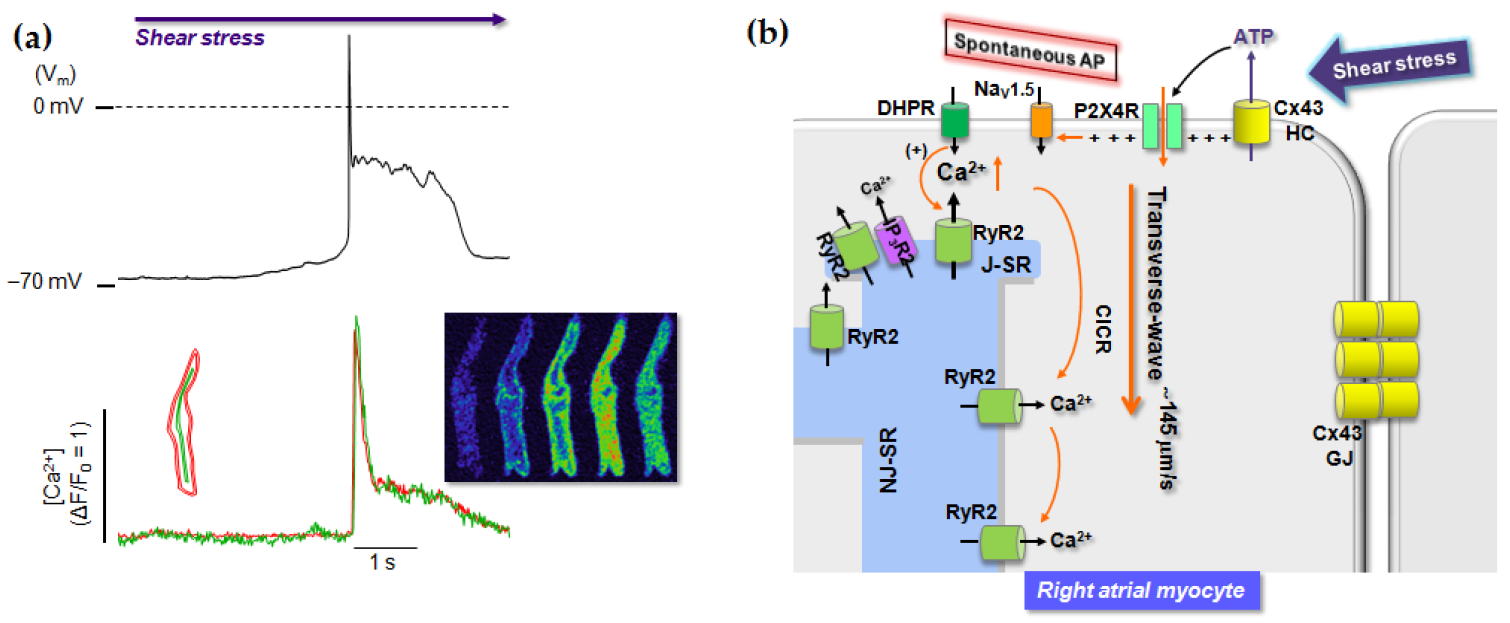
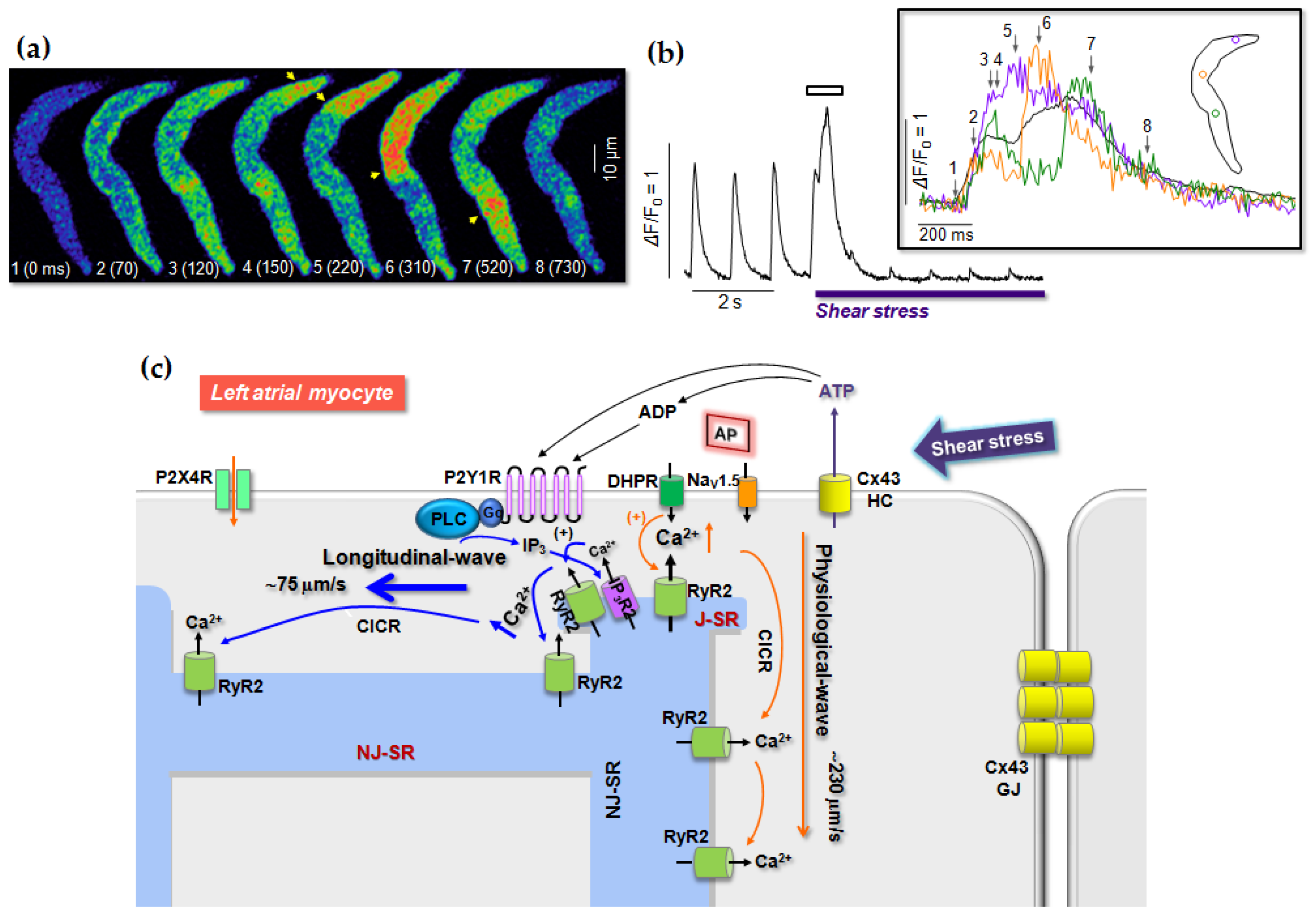
Table 1.
P2Y-receptor subtypes expressed in mammalian tissues and their agonist affinities.
| Type | Species | Principal Agonists | Tissue Distribution | Selected References |
|---|---|---|---|---|
| P2Y1 | Rat | 2-MeS-ADP = 2-MeS-ATP > ADP | Heart, platelet, skeletal muscle, neuron, intestine | [85,86] |
| Mouse | 2-MeS-ATP > 2Cl-ATP > ATP | [85] | ||
| Bovine | 2-MeS-ATP > ADP > ATP | [87] | ||
| Human | (N)-mc-2-MeS-ADP > 2-MeS-ADP > ADP = ADPβS ATP | [66,88,89,90,91,92,93] | ||
| P2Y2 | Rat | UTP = ATP > CTP > GTP | Heart, lung, skeletal muscle, spleen, kidney | [94,95,96] |
| Mouse | UTP = ATP> Ap4A | [82] | ||
| Canine | UTP ≥ ATP > ADP > 2-MeS-ATP | [97] | ||
| Porcine | UTP > ITP > ATP > UDP | [98] | ||
| Human | UTP = ATP > INS37217 > Ap4A > ATPγS | [99,100,101,102] | ||
| P2Y4 | Rat | UTP = ATP = ITP = Ap4A | Placenta, lung, vascular smooth muscle, brain, liver | [96,103,104,105] |
| Mouse | UTP = ATP | [106] | ||
| Human | UTP > UTPγS | [101,107,108,109,110] | ||
| P2Y6 | Rat | UDP > UTP > ADP > 2-MeS-ATP | Heart, lung, spleen, placenta, thymus, intestine, brain | [101,111] |
| Mouse | UDP > UTP > ADP > 2-MeS-ATP | [112] | ||
| Human | UDP = 5-Br-UDP >> UTP > 2-MeS-ADP | [113,114,115] | ||
| P2Y11 | Canine | ADPβS = 2-MeS-ADP ≥ 2-MeS-ATP > ATP | Spleen, intestine, immune cells | [116,117] |
| Human | ARC67085 ≥ ATPγS = BzATP > ATP, (UTP) > 2-MeSAT | [116,117,118,119,120] | ||
| P2Y12 | Rat | 2-MeSADP >ADP > ATP | Neuron, platelet | [121,122] |
| Mouse | 2-MeSADP >ADP > ADPβS | [123,124,125] | ||
| Bovine | 2-MeS-ADP ≫ ADP, ATP | [126] | ||
| Human | 2-MeS-ADP >ADP >> (N)-mc-2-MeS-ADP | [121,127,128] | ||
| P2Y13 | Rat | ADP > 2-MeS-ADP >> HATP | Spleen, leucocytes, bone marrow, liver, brain | [129] |
| Mouse | ADP = 2-MeS-ADP = ADPβS > ATP | [130] | ||
| Human | 2-MeS-ADP > (=) ADP > ADPβS | [130,131,132] | ||
| P2Y14 | Rat | UDP-glucose | Placenta, adipose tissue, intestine, brain, spleen | [133] |
| Mouse | UDP-glucose | [134] | ||
| Human | UDP-glucose > UDP-galactose | [135] |
ARC67085, 2-propylthio-β,γ-dichloromethylene-D-ATP; Ap4A, diadenosine-tetraphosphate; ATPγS, adenosine-(O-3-thiotriphosphate); 5-Br-UDP, 5-bromo-UDP; BzATP, benzoyl–benzoyl–ATP; 2-Cl-ATP, 2-chloro-ATP; INS37217, P1-(uridine 5′)-P4-(2′-deoxycytidine-5′)tetraphosphate; 2-MeSADP, 2-methylthio-ADP; (N)-mc-2-MeSADP, (N)-methanocarba-2-methylthio-ADP (= MRS2365); 2-MeSATP, 2-methylthio-ATP; UTPγS, uridine-(O-3-thiotriphosphate).
Table 2.
Pathophysiological functions of P2 receptor subtypes in cardiac cells.
| Function | Cardiac Regions | Agonist | Effect | R | Species | References |
|---|---|---|---|---|---|---|
| Contraction | LA | ATP, ADP, UTP, Adenosine 2-MeS-ATP | Biphasic(Neg-pos) Neg Neg | A1 | Rat | [136] |
| A | ATP, ATPγS 2-MeS-ATP | Biphasic (neg-pos) Pos | P2X4(?) | Human | [137] | |
| A | UTP | Pos | P2Y | Human, rat, mouse | [136,137,138] | |
| A | ATP | Biphasic (pos-neg) | P2-A1 | Rat, guinea-pig | [136,139] | |
| A | 2-MeS-ATP | Neg | P2X | Rat | [136] | |
| A | 2-MeS-ATP | Pos | Mouse, chicken | [136] | ||
| V V | ATP 2-MeS-ATP, ivermectin | Pos Pos | P2X4 | Rat Mouse | [140] [72,141] | |
| Heart rate | A SAN RA | Adenosine ATP, 2-MeS-ADP ATP | Neg Biphasic (pos-neg) Neg | P2Y1-SR Ca2+ (↓) P2X4 | Frog Toad Rat | [61] [84] [72,142] |
| Pathology | V V V V A(HL-1) Fiboblast V V | ATP UTP ATP | Anti-HF IR-injury (↓) Inflammation (?) Hypertrophy Hypertrophy Fibrosis Fibrosis Antihypertensive | P2X4 P2X7 P2X7 P2Y P2Y(?) P2Y2 P2Y6 P2X4 | Mouse Rat Mouse Rat Mouse Human Mouse Mouse | [141] [143,144] [78] [145] [9] [146] [7] [147] |
| Mechano- transduction | RA | ATP | Shear stress, depolarization | P2X4 | Rat | [9,11] |
| LA | ATP | Shear stress, Ca2+ dysregulation | P2Y1 | Rat | [11,148] | |
| Endo | ATP | Shear stress | P2X4 | Mouse | [147] |
A, atrium; Endo, endothelium; HF, heart failure; HL-1, HL-1 cells; IR, ischemia-reperfusion; LA, left atrium; neg, negative (: decrease, ↓); pos, positive (: increase); R, receptor; RA, right atrium; SAN, sino atrial node; V, ventricle; (?), hypothesis.
Publisher’s Note: MDPI stays neutral with regard to jurisdictional claims in published maps and institutional affiliations. |
© 2020 by the authors. Licensee MDPI, Basel, Switzerland. This article is an open access article distributed under the terms and conditions of the Creative Commons Attribution (CC BY) license (http://creativecommons.org/licenses/by/4.0/).
Share and Cite
MDPI and ACS Style
Woo, S.-H.; Trinh, T.N. P2 Receptors in Cardiac Myocyte Pathophysiology and Mechanotransduction. Int. J. Mol. Sci. 2021, 22, 251. https://0-doi-org.brum.beds.ac.uk/10.3390/ijms22010251
AMA Style
Woo S-H, Trinh TN. P2 Receptors in Cardiac Myocyte Pathophysiology and Mechanotransduction. International Journal of Molecular Sciences. 2021; 22(1):251. https://0-doi-org.brum.beds.ac.uk/10.3390/ijms22010251
Chicago/Turabian StyleWoo, Sun-Hee, and Tran Nguyet Trinh. 2021. "P2 Receptors in Cardiac Myocyte Pathophysiology and Mechanotransduction" International Journal of Molecular Sciences 22, no. 1: 251. https://0-doi-org.brum.beds.ac.uk/10.3390/ijms22010251
Note that from the first issue of 2016, this journal uses article numbers instead of page numbers. See further details here.