
G-Protein Coupled Receptors (GPCRs): Signaling Pathways, Characterization, and Functions in Insect Physiology and Toxicology


Abstract
:1. Introduction
2. GPCRs and the Regulatory Pathways Governing the Biological and Physiological Processes of Organisms
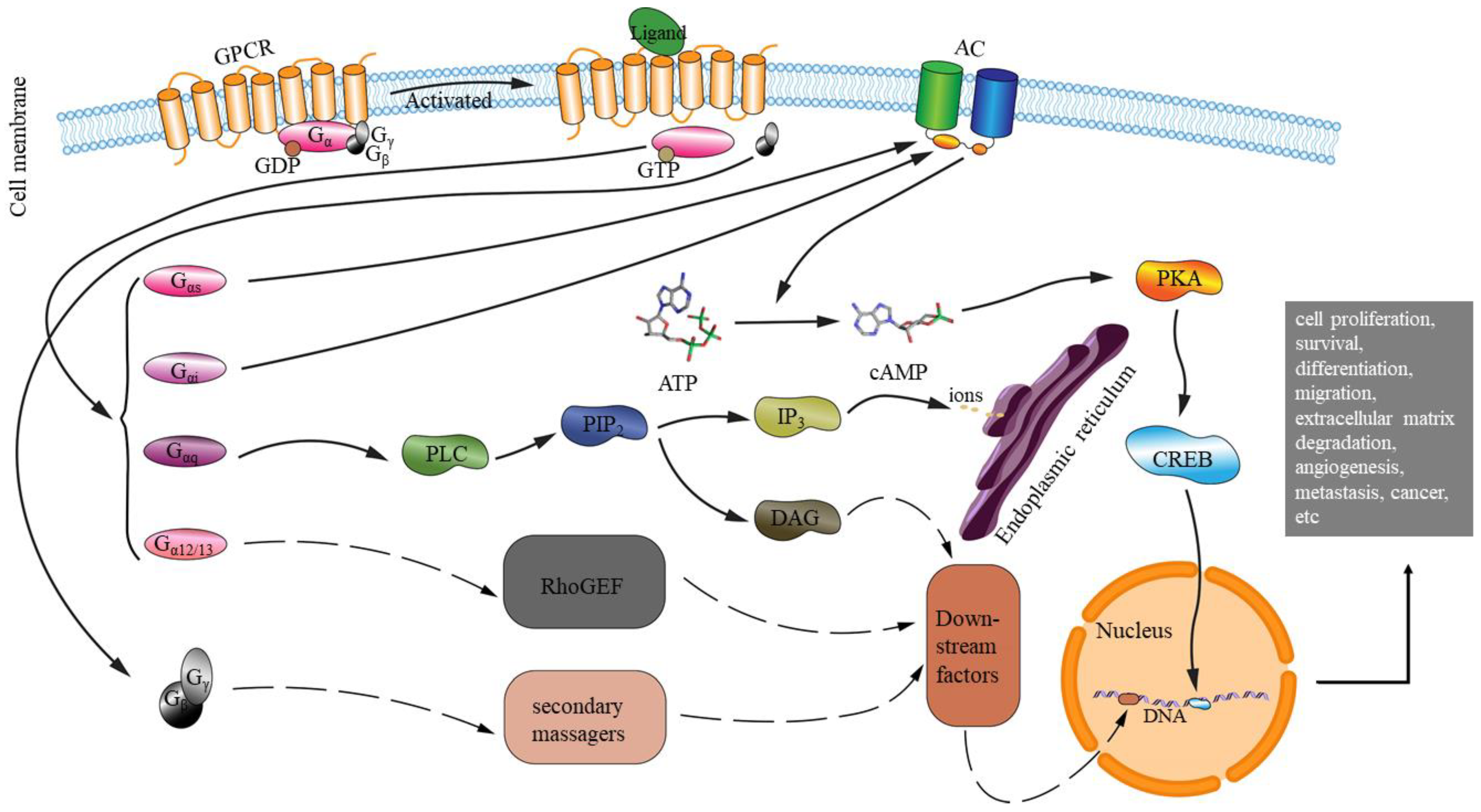
2.1. The GPCR Regulatory Pathways in General
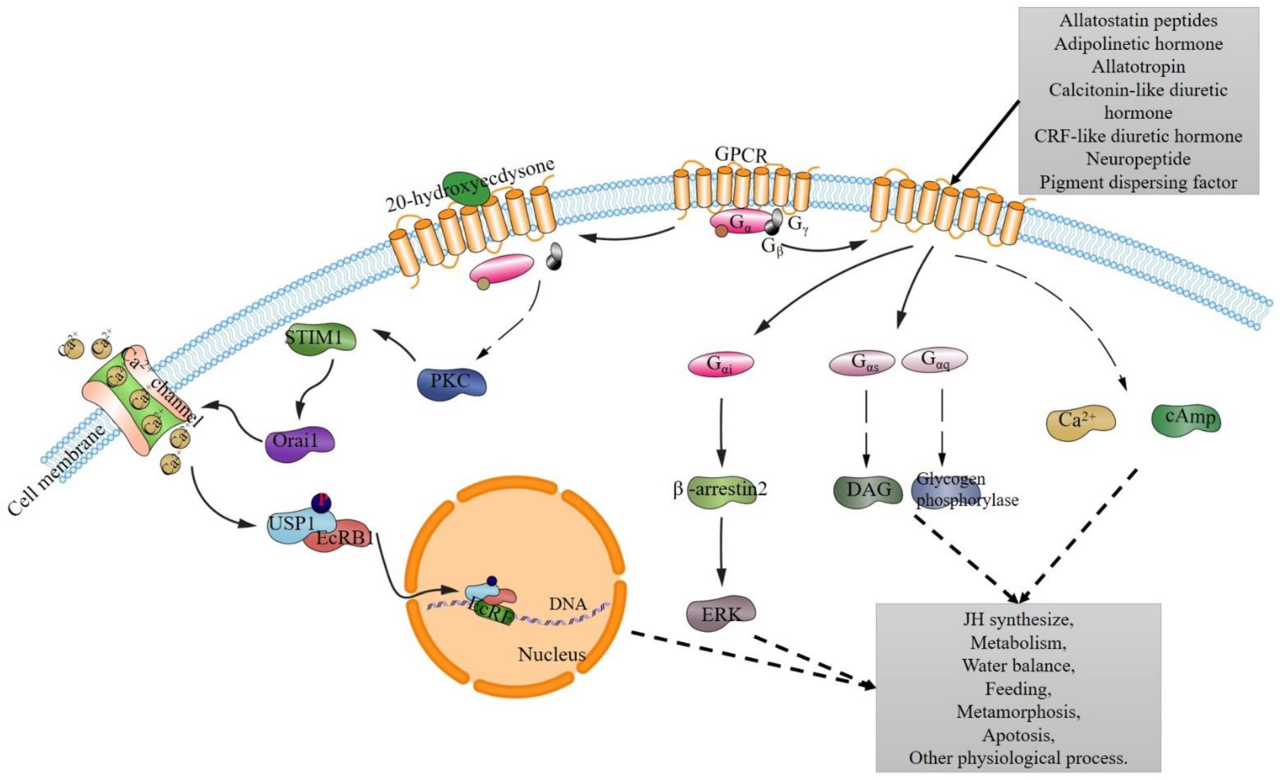
2.2. GPCR Pathways and GPCR Downstream Effectors in Insect Physiology
3. Gene Expression and Functional Characterization of the GPCRs in Insects
3.1. Whole Genome Sequence and Transcriptome Screening to Characterize the Genes and Efforts in the GPCR Regulatory Pathways
3.2. Quantitative Gene Expression and Single Nucleotide Polymorphism Analyses of GPCR Genes in Insects
3.3. Functional Studies of GPCR Genes in Insects
3.4. GPCR Characterization in Cell Line and Ligand-Binding Assays
3.5. Homology 3D Modeling Analyses of GPCRs
4. GPCRs, the Intracellular Pathway, and the Function into Insecticide Resistance
4.1. Overexpression of GPCR Genes in Insecticide Resistance
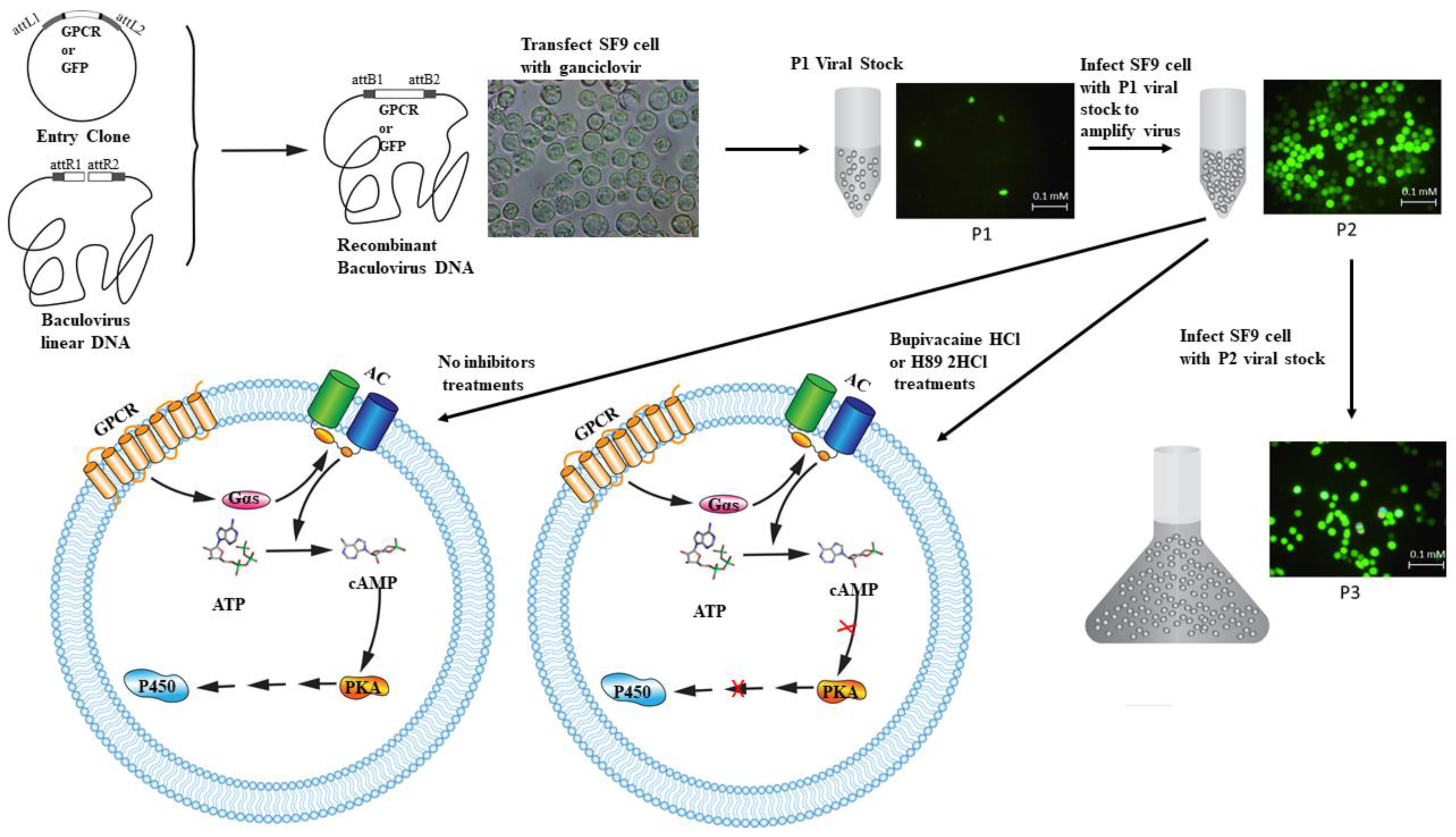
4.2. In Vitro Functional Studies of GPCR Regulation Pathways in Insecticide Resistance
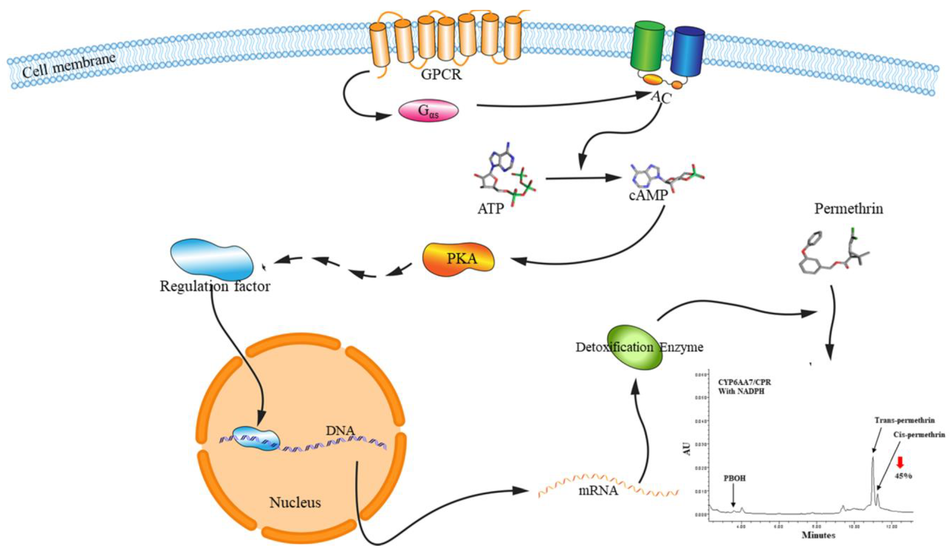
4.3. In Vivo Functional Studies of GPCR Regulation Pathways in Insecticide Resistance
5. Conclusions
Author Contributions
Funding
Conflicts of Interest
References
- Hopkins, A.L.; Groom, C.R. The druggable genome. Nat. Rev. Drug Discov. 2002, 1, 727–730. [Google Scholar] [CrossRef] [PubMed]
- Jacoby, E.; Bouhelal, R.; Gerspacher, M.; Seuwen, K. The 7TM G-protein-coupled receptor target family. ChemMedChem 2006, 1, 760–782. [Google Scholar] [CrossRef] [PubMed]
- Hauser, A.S.; Attwood, M.M.; Rask-Andersen, M.; Schiöth, H.B.; Gloriam, D.E. Trends in GPCR drug discovery: New agents, targets and indications. Nat. Rev. Drug Discov. 2017, 16, 829–842. [Google Scholar] [CrossRef] [PubMed]
- Congreve, M.; de Graaf, C.; Swain, N.A.; Tate, C.G. Impact of GPCR structures on drug discovery. Cell 2020, 181, 81–91. [Google Scholar] [CrossRef] [PubMed]
- Sharan, S.; Hill, C.A. Potential of GPCR-targeting insecticides for control of arthropod vectors. In Advances in Agrochemicals: Ion Channels and G Protein-Coupled Receptors (gpcrs) as Targets for Pest Control, Vol 2: Gpcrs and Ion Channels; Gross, A.D., Ozoe, Y., Coats, J.R., Eds.; American Chemical Society: Washington, DC, USA, 2017; Volume 1265, pp. 55–84. ISBN 978-0-8412-3260-0. [Google Scholar]
- Hapairai, L.K.; Mysore, K.; Sun, L.; Li, P.; Wang, C.-W.; Scheel, N.D.; Lesnik, A.; Scheel, M.P.; Igiede, J.; Wei, N.; et al. Characterization of an adulticidal and larvicidal interfering RNA pesticide that targets a conserved sequence in mosquito G protein-coupled dopamine 1 receptor genes. Insect Biochem. Mol. Biol. 2020, 120, 103359. [Google Scholar] [CrossRef]
- Xiong, C.; Kaczmarek, K.; Zabrocki, J.; Pietrantonio, P.V.; Nachman, R.J. Evaluation of Aib and PEG-polymer insect kinin analogs on mosquito and tick GPCRs identifies potent new pest management tools with potentially enhanced biostability and bioavailability. Gen. Comp. Endocrinol. 2019, 278, 58–67. [Google Scholar] [CrossRef]
- Bockaert, J.; Philippe Pin, J. Molecular tinkering of G protein-coupled receptors: An evolutionary success. EMBO J. 1999, 18, 1723–1729. [Google Scholar] [CrossRef] [PubMed] [Green Version]
- Maudsley, S.; Martin, B.; Luttrell, L.M. The origins of diversity and specificity in G protein-coupled receptor signaling. J. Pharmacol. Exp. Ther. 2005, 314, 485–494. [Google Scholar] [CrossRef] [Green Version]
- Lagerström, M.C.; Schiöth, H.B. Structural diversity of G protein-coupled receptors and significance for drug discovery. Nat. Rev. Drug Discov. 2008, 7, 339–357. [Google Scholar] [CrossRef]
- Spehr, M.; Munger, S.D. Olfactory receptors: G protein-coupled receptors and beyond. J. Neurochem. 2009, 109, 1570–1583. [Google Scholar] [CrossRef] [Green Version]
- Millar, R.P.; Newton, C.L. The year in G protein-coupled receptor research. Mol. Endocrinol. 2010, 24, 261–274. [Google Scholar] [CrossRef]
- Eglen, R.M.; Reisine, T. GPCRs revisited: New insights lead to novel drugs. Pharmaceuticals 2011, 4, 244–272. [Google Scholar] [CrossRef] [Green Version]
- Gether, U. Uncovering molecular mechanisms involved in activation of G protein-coupled receptors. Endocr. Rev. 2000, 21, 90–113. [Google Scholar] [CrossRef]
- Wise, A.; Gearing, K.; Rees, S. Target validation of G-protein coupled receptors. Drug Discov. Today 2002, 7, 235–246. [Google Scholar] [CrossRef]
- Oldham, W.M.; Hamm, H.E. Heterotrimeric G protein activation by G-protein-coupled receptors. Nat. Rev. Mol. Cell Biol. 2008, 9, 60–71. [Google Scholar] [CrossRef] [PubMed]
- Verrier, F.; An, S.; Ferrie, A.M.; Sun, H.; Kyoung, M.; Deng, H.; Fang, Y.; Benkovic, S.J. GPCRs Regulate the assembly of a multienzyme complex for purine biosynthesis. Nat. Chem. Biol. 2011, 7, 909–915. [Google Scholar] [CrossRef]
- Gardner, J.; Wu, S.; Ling, L.; Danao, J.; Li, Y.; Yeh, W.C.; Tian, H.; Baribault, H. G-protein-coupled receptor GPR21 knockout mice display improved glucose tolerance and increased insulin response. Biochem. Biophys. Res. Commun. 2012, 418, 1–5. [Google Scholar] [CrossRef] [PubMed]
- Flower, D.R. Modelling G-protein-coupled receptors for drug design. Biochim. Biophys. Acta Biomembr. 1999, 1422, 207–234. [Google Scholar] [CrossRef]
- Robas, N.; O’Reilly, M.; Katugampola, S.; Fidock, M. Maximizing serendipity: Strategies for identifying ligands for orphan G-protein-coupled receptors. Curr. Opin. Pharmacol. 2003, 3, 121–126. [Google Scholar] [CrossRef]
- Hauser, F.; Cazzamali, G.; Williamson, M.; Blenau, W.; Grimmelikhuijzen, C.J. A review of neurohormone GPCRs present in the fruitfly Drosophila melanogaster and the honey bee Apis mellifera. Prog. Neurobiol. 2006, 80, 1–19. [Google Scholar] [CrossRef] [Green Version]
- Hauser, F.; Cazzamali, G.; Williamson, M.; Park, Y.; Li, B.; Tanaka, Y.; Predel, R.; Neupert, S.; Schachtner, J.; Verleyen, P. A genome-wide inventory of neurohormone GPCRs in the red flour beetle Tribolium castaneum. Front. Neuroendocr. 2008, 29, 142–165. [Google Scholar] [CrossRef] [PubMed]
- Jiang, H.; Lkhagva, A.; Daubnerova, I.; Chae, H.-S.; Simo, L.; Jung, S.-H.; Yoon, Y.-K.; Lee, N.-R.; Seong, J.Y.; Zitnan, D.; et al. Natalisin, a tachykinin-like signaling system, regulates sexual activity and fecundity in insects. Proc. Natl. Acad. Sci. USA 2013, 110, E3526–E3534. [Google Scholar] [CrossRef] [Green Version]
- Bai, H.; Palli, S.R. Identification of G protein-coupled receptors required for vitellogenin uptake into the oocytes of the red flour beetle, Tribolium Castaneum. Sci. Rep. 2016, 6, 27648. [Google Scholar] [CrossRef] [PubMed] [Green Version]
- Jing, Y.-P.; An, H.; Zhang, S.; Wang, N.; Zhou, S. Protein kinase C mediates juvenile hormone-dependent phosphorylation of Na+/K+-ATPase to induce ovarian follicular patency for yolk protein uptake. J. Biol. Chem. 2018, 293, 20112–20122. [Google Scholar] [CrossRef] [PubMed] [Green Version]
- Marciniak, P.; Urbanski, A.; Lubawy, J.; Szymczak, M.; Pacholska-Bogalska, J.; Chowanski, S.; Kuczer, M.; Rosinski, G. Short neuropeptide F signaling regulates functioning of male reproductive system in Tenebrio Molitor beetle. J. Comp. Physiol. B, Biochem. Syst. Environ. Physiol. 2020, 190, 521–534. [Google Scholar] [CrossRef]
- Nagai, C.; Mabashi-Asazuma, H.; Nagasawa, H.; Nagata, S. Identification and characterization of receptors for ion transport peptide (ITP) and ITP-like (ITPL) in the silkworm Bombyx Mori. J. Biol. Chem. 2014, 289, 32166–32177. [Google Scholar] [CrossRef] [PubMed] [Green Version]
- Poels, J.; Van Loy, T.; Vandersmissen, H.P.; Van Hiel, B.; Van Soest, S.; Nachman, R.J.; Vanden Broeck, J. Myoinhibiting peptides are the ancestral ligands of the promiscuous Drosophila sex peptide receptor. Cell. Mol. Life Sci. 2010, 67, 3511–3522. [Google Scholar] [CrossRef] [PubMed]
- Bai, H.; Zhu, F.; Shah, K.; Palli, S.R. Large-scale RNAi screen of G protein-coupled receptors involved in larval growth, molting and metamorphosis in the red flour beetle. BMC Genom. 2011, 12, 388. [Google Scholar] [CrossRef] [Green Version]
- Regna, K.; Kurshan, P.T.; Harwood, B.N.; Jenkins, A.M.; Lai, C.-Q.; Muskavitch, M.A.T.; Kopin, A.S.; Draper, I. A critical role for the drosophila dopamine D-1-like receptor Dop1R2 at the onset of metamorphosis. BMC Dev. Biol. 2016, 16, 15. [Google Scholar] [CrossRef] [Green Version]
- Mendive, F.M.; Van Loy, T.; Claeysen, S.; Poels, J.; Williamson, M.; Hauser, F.; Grimmelikhuijzen, C.J.P.; Vassart, G.; Broeck, J.V. Drosophila molting neurohormone bursicon is a heterodimer and the natural agonist of the orphan receptor DLGR2. FEBS Lett. 2005, 579, 2171–2176. [Google Scholar] [CrossRef] [Green Version]
- Kang, X.-L.; Zhang, J.-Y.; Wang, D.; Zhao, Y.-M.; Han, X.-L.; Wang, J.-X.; Zhao, X.-F. The steroid hormone 20-hydroxyecdysone binds to dopamine receptor to repress Lepidopteran insect feeding and promote pupation. PLoS Genet. 2019, 15, e1008331. [Google Scholar] [CrossRef] [Green Version]
- Zitnan, D.; Kim, Y.-J.; Zitnanova, I.; Roller, L.; Adams, M.E. Complex steroid-peptide-receptor cascade controls insect ecdysis. Gen. Comp. Endocrinol. 2007, 153, 88–96. [Google Scholar] [CrossRef] [PubMed] [Green Version]
- Iga, M.; Nakaoka, T.; Suzuki, Y.; Kataoka, H. Pigment dispersing factor regulates ecdysone biosynthesis via Bombyx neuropeptide G protein coupled receptor-B2 in the prothoracic glands of Bombyx Mori. PLoS ONE 2014, 9, e103239. [Google Scholar] [CrossRef]
- Yamanaka, N.; Hua, Y.-J.; Roller, L.; Spalovska-Valachova, I.; Mizoguchi, A.; Kataoka, H.; Tanaka, Y. Bombyx prothoracicostatic peptides activate the sex peptide receptor to regulate ecdysteroid biosynthesis. Proc. Natl. Acad. Sci. USA 2010, 107, 2060–2065. [Google Scholar] [CrossRef] [Green Version]
- Li, B.; Beeman, R.W.; Park, Y. Functions of duplicated genes encoding CCAP receptors in the red flour beetle, Tribolium castaneum. J. Insect Physiol. 2011, 57, 1190–1197. [Google Scholar] [CrossRef] [PubMed]
- Li, K.; Jia, Q.-Q.; Li, S. Juvenile hormone signaling—A mini review. Insect Sci. 2019, 26, 600–606. [Google Scholar] [CrossRef] [PubMed]
- Nouzova, M.; Brockhoff, A.; Mayoral, J.G.; Goodwin, M.; Meyerhof, W.; Noriega, F.G. Functional characterization of an allatotropin receptor expressed in the corpora allata of mosquitoes. Peptides 2012, 34, 201–208. [Google Scholar] [CrossRef] [PubMed] [Green Version]
- Kim, Y.-J.; Zitnan, D.; Cho, K.-H.; Schooley, D.A.; Mizoguchi, A.; Adams, M.E. Central peptidergic ensembles associated with organization of an innate behavior. Proc. Natl. Acad. Sci. USA 2006, 103, 14211–14216. [Google Scholar] [CrossRef] [Green Version]
- Terhzaz, S.; Cabrero, P.; Robben, J.H.; Radford, J.C.; Hudson, B.D.; Milligan, G.; Dow, J.A.; Davies, S.-A. Mechanism and function of Drosophila capa GPCR: A desiccation stress-responsive receptor with functional homology to human neuromedinU receptor. PLoS ONE 2012, 7, e29897. [Google Scholar] [CrossRef] [Green Version]
- Petruccelli, E.; Lark, A.; Mrkvicka, J.A.; Kitamoto, T. Significance of DopEcR, a G-protein coupled dopamine/ecdysteroid receptor, in physiological and behavioral response to dtressors. J. Neurogenet. 2020, 34, 55–68. [Google Scholar] [CrossRef]
- Bryon, A.; Wybouw, N.; Dermauw, W.; Tirry, L.; Van Leeuwen, T. Genome wide gene-expression analysis of facultative reproductive diapause in the two-spotted spider mite Tetranychus urticae. BMC Genom. 2013, 14, 815. [Google Scholar] [CrossRef] [Green Version]
- Homma, T.; Watanabe, K.; Tsurumaru, S.; Kataoka, H.; Imai, K.; Kamba, M.; Niimi, T.; Yamashita, O.; Yaginuma, T. G protein-coupled receptor for diapause hormone, an inducer of Bombyx embryonic diapause. Biochem. Biophys. Res. Commun. 2006, 344, 386–393. [Google Scholar] [CrossRef] [PubMed]
- Jiang, H.; Wei, Z.; Nachman, R.J.; Park, Y. Molecular cloning and functional characterization of the diapause hormone receptor in the corn earworm Helicoverpa zea. Peptides 2014, 53, 243–249. [Google Scholar] [CrossRef] [PubMed] [Green Version]
- Shen, Z.; Jiang, X.; Yan, L.; Chen, Y.; Wang, W.; Shi, Y.; Shi, L.; Liu, D.; Zhou, N. Structural basis for the interaction of diapause hormone with its receptor in the dilkworm, Bombyx mori. FASEB J. 2018, 32, 1338–1353. [Google Scholar] [CrossRef] [Green Version]
- Choi, M.-Y.; Estep, A.; Sanscrainte, N.; Becnel, J.; Meer, R.K.V. Identification and expression of PBAN/diapause hormone and GPCRs from Aedes aegypti. Mol. Cell. Endocrinol. 2013, 375, 113–120. [Google Scholar] [CrossRef]
- Chen, C.-H.; Di, Y.-Q.; Shen, Q.-Y.; Wang, J.-X.; Zhao, X.-F. The steroid hormone 20-hydroxyecdysone induces phosphorylation and aggregation of stromal interacting molecule 1 for store-operated calcium entry. J. Biol. Chem. 2019, 294, 14922–14936. [Google Scholar] [CrossRef]
- Devambez, I.; Agha, M.A.; Mitri, C.; Bockaert, J.; Parmentier, M.-L.; Marion-Poll, F.; Grau, Y.; Soustelle, L. G alpha o is required for L-canavanine detection in drosophila. PLoS ONE 2013, 8, e63484. [Google Scholar] [CrossRef] [Green Version]
- Li, T.; Liu, L.; Zhang, L.; Liu, N. Role of G-protein-coupled receptor-related genes in insecticide resistance of the mosquito, Culex quinquefasciatus. Sci. Rep. 2014, 4, 6474. [Google Scholar] [CrossRef] [Green Version]
- Li, T.; Cao, C.; Yang, T.; Zhang, L.; He, L.; Xi, Z.; Bian, G.; Liu, N. A G-protein-coupled receptor regulation pathway in cytochrome P450-mediated permethrin-resistance in mosquitoes, Culex quinquefasciatus. Sci. Rep. 2015, 5, 17772. [Google Scholar] [CrossRef]
- Li, T.; Liu, N. Regulation of P450-mediated permethrin resistance in Culex quinquefasciatus by the GPCR/Gαs/AC/CAMP/PKA signaling cascade. Biochem. Biophys. Rep. 2017, 12, 12–19. [Google Scholar] [CrossRef] [PubMed]
- Kersch, C.N.; Pietrantonio, P.V. Mosquito Aedes aegypti (L.) leucokinin receptor is critical for in vivo fluid excretion post blood feeding. FEBS Lett. 2011, 585, 3507–3512. [Google Scholar] [CrossRef] [PubMed] [Green Version]
- Chen, M.E.; Pietrantonio, P.V. The short neuropeptide F-like receptor from the red imported fire ant, Solenopsis invicta Buren (Hymenoptera: Formicidae). Arch. Insect Biochem. Physiol. 2006, 61, 195–208. [Google Scholar] [CrossRef] [PubMed]
- Dillen, S.; Zels, S.; Verlinden, H.; Spit, J.; Van Wielendaele, P.; Vanden Broeck, J. Functional characterization of the short neuropeptide F receptor in the desert locust, Schistocerca gregaria. PLoS ONE 2013, 8, e53604. [Google Scholar] [CrossRef] [Green Version]
- Sangha, V.; Lange, A.B.; Orchard, I. Identification and cloning of the kinin receptor in the chagas disease vector, Rhodnius prolixus. Gen. Comp. Endocrinol. 2020, 289, 113380. [Google Scholar] [CrossRef]
- Marchal, E.; Schellens, S.; Monjon, E.; Bruyninckx, E.; Marco, H.G.; Gade, G.; Vanden Broeck, J.; Verlinden, H. Analysis of peptide ligand specificity of different insect adipokinetic hormone receptors. Int. J. Mol. Sci. 2018, 19, 542. [Google Scholar] [CrossRef] [Green Version]
- Yang, H.; Huang, J.; Liu, Y.; Li, J.; Luo, S.; Wu, J. Prediction of the post-translational modifications of adipokinetic hormone receptors from solitary to eusocial bees. Sociobiology 2018, 65, 264–272. [Google Scholar] [CrossRef]
- Lin, F.; Hossain, M.A.; Post, S.; Karashchuk, G.; Tatar, M.; De Meyts, P.; Wade, J.D. Total solid-phase synthesis of biologically active drosophila insulin-like peptide 2 (DILP2). Aust. J. Chem. 2017, 70, 208–212. [Google Scholar] [CrossRef] [Green Version]
- Baumbach, J.; Xu, Y.; Hehlert, P.; Kuehnlein, R.P. G alpha q, G gamma 1 and Plc21C control drosophila body fat storage. J. Genet. Genom. 2014, 41, 283–292. [Google Scholar] [CrossRef] [Green Version]
- Lu, K.; Zhang, X.; Chen, X.; Li, Y.; Li, W.; Cheng, Y.; Zhou, J.; You, K.; Zhou, Q. Adipokinetic hormone receptor mediates lipid mobilization to regulate starvation resistance in the brown planthopper, Nilaparvata lugens. Front. Physiol. 2018, 9, 1730. [Google Scholar] [CrossRef] [Green Version]
- Hyun, S.; Lee, Y.; Hong, S.T.; Bang, S.; Paik, D.; Kang, J.K.; Shin, J.; Lee, J.; Jeon, K.; Hwang, S.; et al. Drosophila GPCR han is a receptor for the circadian clock neuropeptide PDF. Neuron 2005, 48, 267–278. [Google Scholar] [CrossRef] [Green Version]
- Hou, Q.-L.; Chen, E.-H.; Jiang, H.-B.; Wei, D.-D.; Gui, S.-H.; Wang, J.-J.; Smagghe, G. Adipokinetic hormone receptor gene identification and its role in triacylglycerol mobilization and sexual behavior in the oriental fruit fly (Bactrocera dorsalis). Insect Biochem. Mol. Biol. 2017, 90, 1–13. [Google Scholar] [CrossRef]
- Abrieux, A.; Debernard, S.; Maria, A.; Gaertner, C.; Anton, S.; Gadenne, C.; Duportets, L. Involvement of the G-protein-coupled dopamine/ecdysteroid receptor DopEcR in the behavioral response to sex pheromone in an insect. PLoS ONE 2013, 8, e72785. [Google Scholar] [CrossRef] [Green Version]
- Bainton, R.J.; Tsai, L.T.Y.; Schwabe, T.; DeSalvo, M.; Gaul, U.; Heberlein, U. Moody encodes two GPCRs that regulate cocaine behaviors and blood-brain barrier permeability in Drosophila. Cell 2005, 123, 145–156. [Google Scholar] [CrossRef] [Green Version]
- Kwon, H.; Agha, M.A.; Smith, R.C.; Nachman, R.J.; Marion-Poll, F.; Pietrantonio, P.V. Leucokinin mimetic elicits aversive behavior in mosquito Aedes aegypti (L.) and inhibits the sugar taste neuron. Proc. Natl. Acad. Sci. USA 2016, 113, 6880–6885. [Google Scholar] [CrossRef] [Green Version]
- Thamm, M.; Balfanz, S.; Scheiner, R.; Baumann, A.; Blenau, W. Characterization of the 5-HT1A receptor of the honeybee (Apis mellifera) and involvement of serotonin in phototactic behavior. Cell. Mol. Life Sci. 2010, 67, 2467–2479. [Google Scholar] [CrossRef]
- Johnson, E.C.; Bohn, L.M.; Taghert, P.H. Drosophila CG8422 encodes a functional diuretic hormone receptor. J. Exp. Biol. 2004, 207, 743–748. [Google Scholar] [CrossRef] [Green Version]
- Pietrantonio, P.V.; Xiong, C.; Nachman, R.J.; Shen, Y. G protein-coupled receptors in arthropod vectors: Omics and pharmacological approaches to elucidate ligand-receptor interactions and novel organismal functions. Curr. Opin. Insect Sci. 2018, 29, 12–20. [Google Scholar] [CrossRef]
- Uchiyama, H.; Maehara, S.; Ohta, H.; Seki, T.; Tanaka, Y. Elevenin regulates the body color through a G protein-coupled receptor NlA42 in the brown planthopper Nilaparvata lugens. Gen. Comp. Endocrinol. 2018, 258, 33–38. [Google Scholar] [CrossRef]
- Kwon, H.; Pietrantonio, P.V. Calcitonin receptor 1 (AedaeGPCRCAL1) hindgut expression and direct role in myotropic action in females of the mosquito Aedes aegypti (L.). Insect Biochem. Mol. Biol. 2013, 43, 588–593. [Google Scholar] [CrossRef]
- Kwon, H.; Lu, H.-L.; Longnecker, M.T.; Pietrantonio, P.V. Role in diuresis of a calcitonin receptor (GPRCAL1) expressed in a distal-proximal gradient in renal organs of the mosquito Aedes aegypti (L.). PLoS ONE 2012, 7, e50374. [Google Scholar] [CrossRef] [Green Version]
- Petruccelli, E.; Li, Q.; Rao, Y.; Kitamoto, T. The unique dopamine/ecdysteroid receptor modulates ethanol-induced sedation in drosophila. J. Neurosci. 2016, 36, 4647–4657. [Google Scholar] [CrossRef] [PubMed]
- Grohmann, L.; Blenau, W.; Erber, J.; Ebert, P.R.; Strunker, T.; Baumann, A. Molecular and functional characterization of an octopamine receptor from honeybee (Apis mellifera) brain. J. Neurosci. 2003, 86, 725–735. [Google Scholar] [CrossRef] [Green Version]
- Lee, D.; Vanden Broeck, J.; Lange, A.B. Identification and expression of the CCAP receptor in the chagas’ disease vector, Rhodnius prolixus, and its involvement in cardiac control. PLoS ONE 2013, 8, e68897. [Google Scholar] [CrossRef] [Green Version]
- Li, M.; Reid, W.R.; Zhang, L.; Scott, J.G.; Gao, X.; Kristensen, M.; Liu, N. A whole transcriptomal linkage analysis of gene co-regulation in insecticide resistant house flies, Musca domestica. BMC Genom. 2013, 14, 803. [Google Scholar] [CrossRef] [PubMed] [Green Version]
- Arensburger, P.; Megy, K.; Waterhouse, R.M.; Abrudan, J.; Amedeo, P.; Antelo, B.; Bartholomay, L.; Bidwell, S.; Caler, E.; Camara, F.; et al. Sequencing of Culex quinquefasciatus establishes a platform for mosquito comparative genomics. Science 2010, 330, 86–88. [Google Scholar] [CrossRef] [PubMed] [Green Version]
- Scott, J.G.; Warren, W.C.; Beukeboom, L.W.; Bopp, D.; Clark, A.G.; Giers, S.D.; Hediger, M.; Jones, A.K.; Kasai, S.; Leichter, C.A.; et al. Genome of the house fly, Musca domestica l., a global vector of diseases with adaptations to a septic environment. Genome Biol. 2014, 15, 466. [Google Scholar] [CrossRef] [Green Version]
- Hopf, T.A.; Morinaga, S.; Ihara, S.; Touhara, K.; Marks, D.S.; Benton, R. Amino acid coevolution reveals three-dimensional structure and functional domains of insect odorant receptors. Nat. Commun. 2015, 6, 1–7. [Google Scholar] [CrossRef] [Green Version]
- Baron, S.; van der Merwe, N.A.; Madder, M.; Maritz-Olivier, C. SNP analysis infers that recombination is involved in the evolution of amitraz resistance in Rhipicephalus microplus. PLoS ONE 2015, 10, e0131341. [Google Scholar] [CrossRef] [Green Version]
- Choi, M.-Y.; Jurenka, R.A. Site-directed mutagenesis and PBAN activation of the Helicoverpa zea pban-receptor. FEBS Lett. 2010, 584, 1212–1216. [Google Scholar] [CrossRef] [Green Version]
- Oryan, A.; Wahedi, A.; Paluzzi, J.-P.V. Functional characterization and quantitative expression analysis of two GNRH-related peptide receptors in the mosquito, Aedes aegypti. Biochem. Biophys. Res. Commun. 2018, 497, 550–557. [Google Scholar] [CrossRef] [PubMed]
- Li, T.; Liu, N. Role of the g-protein-coupled receptor signaling pathway in insecticide resistance. Int. J. Mol. Sci. 2019, 20, 4300. [Google Scholar] [CrossRef] [PubMed] [Green Version]
- Caers, J.; Verlinden, H.; Zels, S.; Vandersmissen, H.P.; Vuerinckx, K.; Schoofs, L. More than two decades of research on insect neuropeptide GPCRs: An overview. Front. Endocrinol. 2012, 3, 151. [Google Scholar] [CrossRef] [PubMed] [Green Version]
- Hanlon, C.D.; Andrew, D.J. Outside-in signaling–a brief review of GPCR signaling with a focus on the Drosophila GPCR family. J. Cell. Sci. 2015, 128, 3533–3542. [Google Scholar] [CrossRef] [Green Version]
- Stevens, R.C.; Cherezov, V.; Katritch, V.; Abagyan, R.; Kuhn, P.; Rosen, H.; Wüthrich, K. The GPCR network: A large-scale collaboration to determine human GPCR structure and function. Nat. Rev. Drug Discov. 2013, 12, 25–34. [Google Scholar] [CrossRef] [Green Version]
- Burnell, A.M.; O’Halloran, D.M. Chemoreceptor genes: What can we learn from Caenorhabditis elegans and how can we apply this information to studies on other nematodes? In Proceeding of the Fourth International Congress of Nematology; Cook, R.C., Hunt, D.J., Eds.; E. J. Brill: Leiden, The Netherlands, 2004; Volume 2, pp. 707–714. ISBN 90-04-13921-4. [Google Scholar]
- Bjarnadottir, T.K.; Gloriam, D.E.; Hellstrand, S.H.; Kristiansson, H.; Fredriksson, R.; Schioth, H.B. Comprehensive repertoire and phylogenetic analysis of the G protein-coupled receptors in human and mouse. Genomics 2006, 88, 263–273. [Google Scholar] [CrossRef] [PubMed]
- Meethal, S.V.; Gallego, M.J.; Haasl, R.J.; Petras, S.J.; Sgro, J.-Y.; Atwood, C.S. Identification of a gonadotropin-releasing hormone receptor orthologue in Caenorhabditis elegans. BMC Evol. Biol. 2006, 6, 103. [Google Scholar] [CrossRef] [Green Version]
- Radeff-Huang, J.; Seasholtz, T.M.; Matteo, R.G.; Brown, J.H. G protein mediated signaling pathways in lysophospholipid induced cell proliferation and survival. J. Cell. Biochem. 2004, 92, 949–966. [Google Scholar] [CrossRef]
- Hsiao, E.C.; Millard, S.M.; Nissenson, R.A. Gs/Gi Regulation of Bone Cell differentiation: Review and insights from engineered receptors. Horm. Metab. Res. 2016, 48, 689–699. [Google Scholar] [CrossRef]
- Cotton, M.; Claing, A. G protein-coupled receptors stimulation and the control of cell migration. Cell. Signal. 2009, 21, 1045–1053. [Google Scholar] [CrossRef]
- Chellini, L.; Caprara, V.; Spadaro, F.; Sestito, R.; Bagnato, A.; Rosanò, L. Regulation of extracellular matrix degradation and metastatic spread by IQGAP1 through endothelin-1 receptor signalling in ovarian cancer. Matric Biol. 2019, 81, 17–33. [Google Scholar] [CrossRef]
- Richard, D.E.; Vouret-Craviari, V.; Pouysségur, J. Angiogenesis and G-protein-coupled receptors: Signals that bridge the gap. Oncogene 2001, 20, 1556–1562. [Google Scholar] [CrossRef] [PubMed]
- Bagnato, A.; Rosanò, L. New routes in GPCR/β-arrestin-driven signaling in cancer progression and metastasis. Front. Pharmacol. 2019, 10. [Google Scholar] [CrossRef] [Green Version]
- Dorsam, R.T.; Gutkind, J.S. G-protein-coupled receptors and cancer. Nat. Rev. Cancer 2007, 7, 79–94. [Google Scholar] [CrossRef]
- Li, T.; Liu, N. G-protein-coupled receptors: Their expression, function and regulation in insecticide resistance. In Advances in the Biorational Control of Medical and Veterinary Pests; Norris, E.J., Coats, J.R., Gross, A.D., Clark, J.M., Eds.; American Chemical Society: Washington, DC, USA, 2018; Volume 1289, pp. 153–172. ISBN 978-0-8412-3358-4. [Google Scholar]
- Liu, L.; Parent, C.A. TOR kinase complexes and cell migration. J. Cell Biol. 2011, 194, 815–824. [Google Scholar] [CrossRef] [PubMed] [Green Version]
- Harvey, K.F.; Zhang, X.; Thomas, D.M. The hippo pathway and human cancer. Nat. Rev. Cancer 2013, 13, 246–257. [Google Scholar] [CrossRef]
- Marinissen, M.J.; Gutkind, J.S. G-protein-coupled receptors and signaling networks: Emerging paradigms. Trends Pharmacol. Sci. 2001, 22, 368–376. [Google Scholar] [CrossRef]
- Neves, S.R.; Ram, P.T.; Iyengar, R. G protein pathways. Science 2002, 296, 1636–1639. [Google Scholar] [CrossRef]
- Chen-Goodspeed, M.; Lukan, A.N.; Dessauer, C.W. Modeling of Gαs and Gαi regulation of human type V and VI adenylyl cyclase. J. Biol. Chem. 2005, 280, 1808–1816. [Google Scholar] [CrossRef] [PubMed] [Green Version]
- Wettschureck, N.; Offermanns, S. Mammalian G proteins and their cell type specific functions. Physiol. Rev. 2005, 85, 1159–1204. [Google Scholar] [CrossRef] [Green Version]
- Sassone-Corsi, P. The cyclic AMP pathway. Cold Spring Harb. Perspect. Biol. 2012, 4. [Google Scholar] [CrossRef] [PubMed]
- Syrovatkina, V.; Alegre, K.O.; Dey, R.; Huang, X.-Y. Regulation, signaling, and physiological functions of g-proteins. J. Mol. Biol. 2016, 428, 3850–3868. [Google Scholar] [CrossRef] [Green Version]
- Kim, C.; Cheng, C.Y.; Saldanha, S.A.; Taylor, S.S. PKA-I holoenzyme structure reveals a mechanism for CAMP-dependent activation. Cell 2007, 130, 1032–1043. [Google Scholar] [CrossRef] [PubMed] [Green Version]
- Bradley, J.; Li, J.; Davidson, N.; Lester, H.A.; Zinn, K. Heteromeric olfactory cyclic nucleotide-gated channels: A subunit that confers increased sensitivity to CAMP. Proc. Natl. Acad. Sci. USA 1994, 91, 8890–8894. [Google Scholar] [CrossRef] [Green Version]
- Zhang, X.; Candas, M.; Griko, N.B.; Taussig, R.; Bulla, L.A. A mechanism of cell death involving an adenylyl cyclase/PKA signaling pathway is induced by the Cry1Ab toxin of Bacillus thuringiensis. Proc. Natl. Acad. Sci. USA 2006, 103, 9897–9902. [Google Scholar] [CrossRef] [PubMed] [Green Version]
- Rhee, S.G. Regulation of phosphoinositide-specific phospholipase C. Annu. Rev. Biochem. 2001, 70, 281–312. [Google Scholar] [CrossRef]
- Philip, F.; Kadamur, G.; Silos, R.G.; Woodson, J.; Ross, E.M. Synergistic activation of phospholipase C-Β3 by Gαq and Gβγ describes a simple two-state coincidence detector. Curr. Biol. 2010, 20, 1327–1335. [Google Scholar] [CrossRef] [Green Version]
- Falkenburger, B.H.; Dickson, E.J.; Hille, B. Quantitative properties and receptor reserve of the DAG and PKC branch of Gq-coupled receptor signaling. J. Gen. Physiol. 2013, 141, 537–555. [Google Scholar] [CrossRef] [PubMed] [Green Version]
- Jafri, M.S.; Keizer, J. On the roles of Ca2+ diffusion, Ca2+ buffers, and the endoplasmic reticulum in IP3-induced Ca2+ waves. Biophys. J. 1995, 69, 2139–2153. [Google Scholar] [CrossRef] [Green Version]
- Worzfeld, T.; Wettschureck, N.; Offermanns, S. G12/G13-mediated signalling in mammalian physiology and disease. Trends Pharmacol. Sci. 2008, 29, 582–589. [Google Scholar] [CrossRef]
- Dhanasekaran, N.; Dermott, J.M. Signaling by the G12 class of G proteins. Cell. Signal. 1996, 8, 235–245. [Google Scholar] [CrossRef]
- Jiang, Y.; Ma, W.; Wan, Y.; Kozasa, T.; Hattori, S.; Huang, X.-Y. The G protein Gα12 stimulates Bruton’s tyrosine kinase and a RasGAP through a conserved PH/BM Domain. Nature 1998, 395, 808–813. [Google Scholar] [CrossRef] [PubMed]
- Meigs, T.E.; Fields, T.A.; McKee, D.D.; Casey, P.J. Interaction of Gα12 and Gα13 with the cytoplasmic domain of cadherin provides a mechanism for β-catenin release. Proc. Natl. Acad. Sci. USA 2001, 98, 519–524. [Google Scholar] [CrossRef] [Green Version]
- Suzuki, N.; Nakamura, S.; Mano, H.; Kozasa, T. Gα12 activates Rho GTPase through tyrosine-phosphorylated leukemia-associated RhoGEF. Proc. Natl. Acad. Sci. USA 2003, 100, 733–738. [Google Scholar] [CrossRef] [PubMed] [Green Version]
- Rosenbaum, D.M.; Rasmussen, S.G.; Kobilka, B.K. The structure and function of G-protein-coupled receptors. Nature 2009, 459, 356–363. [Google Scholar] [CrossRef] [PubMed] [Green Version]
- Kimura, K.I.; Kodama, A.; Hayasaka, Y.; Ohta, T. Activation of the CAMP/PKA signaling pathway is required for post-ecdysial cell death in wing epidermal cells of Drosophila melanogaster. Development 2004, 131, 1597–1606. [Google Scholar] [CrossRef] [PubMed] [Green Version]
- Davies, S.-A.; Cabrero, P.; Povsic, M.; Johnston, N.R.; Terhzaz, S.; Dow, J.A.T. Signaling by Drosophila CAPA neuropeptides. Gen. Comp. Endocrinol. 2013, 188, 60–66. [Google Scholar] [CrossRef]
- Yang, H.; He, X.; Yang, J.; Deng, X.; Liao, Y.; Zhang, Z.; Zhu, C.; Shi, Y.; Zhou, N. Activation of CAMP-response element-binding protein is positively regulated by PKA and calcium-sensitive calcineurin and negatively by PKC in insect. Insect Biochem. Mol. Biol. 2013, 43, 1028–1036. [Google Scholar] [CrossRef]
- Lin, P.; Cheng, T.; Ma, S.; Gao, J.; Jin, S.; Jiang, L.; Xia, Q. Bacillus bombysepticus α-toxin binding to G protein-coupled receptor kinase 2 regulates CAMP/PKA signaling pathway to induce host death. PLOS Pathog. 2016, 12, e1005527. [Google Scholar] [CrossRef] [PubMed] [Green Version]
- Li, Y.-B.; Pei, X.-Y.; Wang, D.; Chen, C.-H.; Cai, M.-J.; Wang, J.-X.; Zhao, X.-F. The steroid hormone 20-hydroxyecdysone upregulates calcium release-activated calcium channel modulator 1 expression to induce apoptosis in the midgut of Helicoverpa armigera. Cell Calcium 2017, 68, 24–33. [Google Scholar] [CrossRef]
- Wang, D.; Zhao, W.-L.; Cai, M.-J.; Wang, J.-X.; Zhao, X.-F. G-protein-coupled receptor controls steroid hormone signaling in cell membrane. Sci. Rep. 2015, 5, 8675. [Google Scholar] [CrossRef] [Green Version]
- Zhang, X.-Q.; Li, X.-R.; Ren, J.; Li, Y.-B.; Cai, M.-J.; Wang, J.-X.; Zhao, X.-F. Beta-arrestin1 interacts with G protein-coupled receptor to desensitize signaling of the steroid hormone 20-hydroxyecdysone in the lepidopteran insect Helicoverpa armigera. Cell. Signal. 2015, 27, 878–886. [Google Scholar] [CrossRef] [PubMed]
- Yamanaka, N.; Yamamoto, S.; Zitnan, D.; Watanabe, K.; Kawada, T.; Satake, H.; Kaneko, Y.; Hiruma, K.; Tanaka, Y.; Shinoda, T.; et al. Neuropeptide receptor transcriptome reveals unidentified neuroendocrine pathways. PLoS ONE 2008, 3, e3048. [Google Scholar] [CrossRef] [PubMed] [Green Version]
- Coast, G.M.; Webster, S.G.; Schegg, K.M.; Tobe, S.S.; Schooley, D.A. The Drosophila melanogaster homologue of an insect calcitonin-like diuretic peptide stimulates V-ATPase activity in fruit fly malpighian tubules. J. Exp. Biol. 2001, 204, 1795–1804. [Google Scholar] [CrossRef] [PubMed]
- Zandawala, M. Calcitonin-like diuretic hormones in insects. Insect Biochem. Mol. Biol. 2012, 42, 816–825. [Google Scholar] [CrossRef] [PubMed]
- Esquivel, C.J.; Cassone, B.J.; Piermarini, P.M. A de novo transcriptome of the malpighian tubules in non-blood-fed and blood-fed asian tiger mosquitoes Aedes albopictus: Insights into diuresis, detoxification, and blood meal processing. PeerJ 2016, 4, e1784. [Google Scholar] [CrossRef] [Green Version]
- Işbilir, A.; Duan Sahbaz, B.; Tuncgenc, G.; Bünemann, M.; Lohse, M.J.; Birgül-Iyison, N. Pharmacological characterization of the stick insect Carausius morosus allatostatin-C receptor with its endogenous agonist. ACS Omega 2020, 5, 32183–32194. [Google Scholar] [CrossRef]
- Gäde, G.; Auerswald, L. Mode of action of neuropeptides from the adipokinetic hormone family. Gen. Comp. Endocrinol. 2003, 132, 10–20. [Google Scholar] [CrossRef]
- Horodyski, F.M.; Verlinden, H.; Filkin, N.; Vandersmissen, H.P.; Fleury, C.; Reynolds, S.E.; Kai, Z.; Broeck, J.V. Isolation and functional characterization of an allatotropin receptor from Manduca sexta. Insect Biochem. Mol. Biol. 2011, 41, 804–814. [Google Scholar] [CrossRef]
- Vuerinckx, K.; Verlinden, H.; Lindemans, M.; Broeck, J.V.; Huybrechts, R. Characterization of an allatotropin-like peptide receptor in the red flour beetle, Tribolium castaneum. Insect Biochem. Mol. Biol. 2011, 41, 815–822. [Google Scholar] [CrossRef]
- Brugge, V.T.; Paluzzi, J.-P.; Schooley, D.A.; Orchard, I. Identification of the elusive peptidergic diuretic hormone in the blood-feeding bug Rhodnius prolixus: A CRF-related peptide. J. Exp. Biol. 2011, 214, 371–381. [Google Scholar] [CrossRef] [PubMed] [Green Version]
- Garczynski, S.F.; Brown, M.R.; Shen, P.; Murray, T.F.; Crim, J.W. Characterization of a functional neuropeptide F receptor from Drosophila melanogaster. Peptides 2002, 23, 773–780. [Google Scholar] [CrossRef]
- Mertens, I.; Vandingenen, A.; Johnson, E.C.; Shafer, O.T.; Li, W.; Trigg, J.S.; De Loof, A.; Schoofs, L.; Taghert, P.H. PDF receptor signaling in Drosophila contributes to both circadian and geotactic behaviors. Neuron 2005, 48, 213–219. [Google Scholar] [CrossRef] [Green Version]
- Hill, C.A.; Fox, A.N.; Pitts, R.J.; Kent, L.B.; Tan, P.L.; Chrystal, M.A.; Cravchik, A.; Collins, F.H.; Robertson, H.M.; Zwiebel, L.J. G protein coupled receptors in Anopheles gambiae. Science 2002, 298, 176–178. [Google Scholar] [CrossRef] [PubMed]
- Anstead, C.A.; Korhonen, P.K.; Young, N.D.; Hall, R.S.; Jex, A.R.; Murali, S.C.; Hughes, D.S.T.; Lee, S.F.; Perry, T.; Stroehlein, A.J.; et al. Lucilia cuprina genome unlocks parasitic fly biology to underpin future interventions. Nat. Commun. 2015, 6, 7344. [Google Scholar] [CrossRef] [PubMed] [Green Version]
- Calkins, T.L.; Tamborindeguy, C.; Pietrantonio, P. GPCR annotation, G proteins, and transcriptomics of fire ant (Solenopsis invicta) queen and worker brain: An improved view of signaling in an invasive superorganism. Gen. Comp. Endocrinol. 2019, 278, 89–103. [Google Scholar] [CrossRef] [PubMed]
- Duan Sahbaz, B.; Birgul Iyison, N. Prediction and expression analysis of G protein-coupled receptors in the laboratory stick insect, Carausius morosus. Turk. J. Biol. 2019, 43, 77–88. [Google Scholar] [CrossRef]
- Ma, Z.; Zhang, Y.; You, C.; Zeng, X.; Gao, X. The Role of G protein-coupled receptor-related genes in cytochrome P450-mediated resistance of the house fly, Musca domestica (Diptera: Muscidae), to imidacloprid. Insect Mol. Biol. 2020, 29, 92–103. [Google Scholar] [CrossRef] [PubMed]
- Liu, N.; Liu, H.; Zhu, F.; Zhang, L. Differential expression of genes in pyrethroid resistant and susceptible mosquitoes, Culex quinquefasciatus (S.). Gene 2007, 394, 61–68. [Google Scholar] [CrossRef] [PubMed]
- Bai, H.; Palli, S.R. Functional characterization of bursicon receptor and genome-wide analysis for identification of genes affected by bursicon receptor RNAi. Dev. Biol. 2010, 344, 248–258. [Google Scholar] [CrossRef]
- Deng, X.; Yang, H.; He, X.; Liao, Y.; Zheng, C.; Zhou, Q.; Zhu, C.; Zhang, G.; Gao, J.; Zhou, N. Activation of bombyx neuropeptide g protein-coupled receptor A4 via a G alpha i-dependent signaling pathway by direct interaction with neuropeptide f from silkworm, Bombyx mori. Insect Biochem. Mol. Biol. 2014, 45, 77–88. [Google Scholar] [CrossRef] [PubMed]
- Hamoudi, Z.; Lange, A.B.; Orchard, I. Identification and characterization of the corazonin receptor and possible physiological roles of the corazonin-signaling pathway in Rhodnius prolixus. Front. Neurosci. 2016, 10, 357. [Google Scholar] [CrossRef]
- Cao, C.; Sun, L.; Du, H.; Moural, T.W.; Bai, H.; Liu, P.; Zhu, F. Physiological functions of a methuselah-like G protein coupled receptor in Lymantria dispar Linnaeus. Pestic. Biochem. Physiol. 2019, 160, 1–10. [Google Scholar] [CrossRef] [PubMed]
- Bayliss, A.; Roselli, G.; Evans, P.D. A comparison of the signalling properties of two tyramine receptors from drosophila. J. Neurochem. 2013, 125, 37–48. [Google Scholar] [CrossRef] [PubMed]
- Belmont, M.; Cazzamali, G.; Williamson, M.; Hauser, F.; Grimmelikhuijzen, C.J.P. Identification of four evolutionarily related G protein-coupled receptors from the malaria mosquito Anopheles gambiae. Biochem. Biophys. Res. Commun. 2006, 344, 160–165. [Google Scholar] [CrossRef]
- Cazzamali, G.; Torp, M.; Hauser, F.; Williamson, M.; Grimmelikhuijzen, C.J.P. The Drosophila gene CG9918 codes for a pyrokinin-1 receptor. Biochem. Biophys. Res. Commun. 2005, 335, 14–19. [Google Scholar] [CrossRef] [PubMed]
- Dolezelova, E.; Nothacker, H.-P.; Civelli, O.; Bryant, P.J.; Zurovec, M. A Drosophila adenosine receptor activates CAMP and calcium signaling. Insect Biochem. Mol. Biol. 2007, 37, 318–329. [Google Scholar] [CrossRef]
- Blenau, W.; Wilms, J.A.; Balfanz, S.; Baumann, A. AmOct Alpha 2R: Functional characterization of a honeybee octopamine receptor inhibiting adenylyl cyclase activity. Int. J. Mol. Sci. 2020, 21, 9334. [Google Scholar] [CrossRef]
- Guo, Z.; Kang, S.; Sun, D.; Gong, L.; Zhou, J.; Qin, J.; Guo, L.; Zhu, L.; Bai, Y.; Ye, F.; et al. MAPK-dependent hormonal signaling plasticity contributes to overcoming Bacillus thuringiensis toxin action in an insect host. Nat. Commun. 2020, 11, 3003. [Google Scholar] [CrossRef]
- Gondalia, K.; Qudrat, A.; Bruno, B.; Medina, J.F.; Paluzzi, J.-P.V. Identification and functional characterization of a pyrokinin neuropeptide receptor in the lyme disease vector, Ixodes scapularis. Peptides 2016, 86, 42–54. [Google Scholar] [CrossRef]
- Hansen, K.K.; Hauser, F.; Cazzamali, G.; Williamson, M.; Grimmelikhuijzen, C.J.P. Cloning and characterization of the adipokinetic hormone receptor from the cockroach Periplaneta americana. Biochem. Biophys. Res. Commun. 2006, 343, 638–643. [Google Scholar] [CrossRef]
- Holmes, S.P.; Barhoumi, R.; Nachman, R.J.; Pietrantonio, P.V. Functional analysis of a G protein-coupled receptor from the southern cattle tick Boophilus microplus (Acari: Ixodidae) identifies it as the first arthropod myokinin receptor. Insect Mol. Biol. 2003, 12, 27–38. [Google Scholar] [CrossRef]
- Cui, L.; Li, J.; Xie, X. Rediocide A, an Insecticide, Induces G-protein-coupled receptor desensitization via activation of conventional protein kinase C. J. Nat. Prod. 2012, 75, 1058–1062. [Google Scholar] [CrossRef]
- Gross, A.D.; Temeyer, K.B.; Day, T.A.; de Leon, A.A.P.; Kimber, M.J.; Coats, J.R. Pharmacological characterization of a tyramine receptor from the southern cattle tick, Rhipicephalus (Boophilus) microplus. Insect Biochem. Mol. Biol. 2015, 63, 47–53. [Google Scholar] [CrossRef] [PubMed] [Green Version]
- Hill, C.A.; Meyer, J.M.; Ejendal, K.F.K.; Echeverry, D.F.; Lang, E.G.; Avramova, L.V.; Conley, J.M.; Watts, V.J. Re-invigorating the insecticide discovery pipeline for vector control: GPCRs as targets for the identification of next gen insecticides. Pestic. Biochem. Physiol. 2013, 106, 141–148. [Google Scholar] [CrossRef]
- Akermoun, M.; Koglin, M.; Zvalova-Iooss, D.; Folschweiller, N.; Dowell, S.J.; Gearing, K.L. Characterization of 16 human G protein-coupled receptors expressed in baculovirus-infected insect cells. Protein Expr. Purif. 2005, 44, 65–74. [Google Scholar] [CrossRef] [PubMed]
- Palczewski, K.; Kumasaka, T.; Hori, T.; Behnke, C.A.; Motoshima, H.; Fox, B.A.; Le Trong, I.; Teller, D.C.; Okada, T.; Stenkamp, R.E. Crystal structure of rhodopsin: A G protein-coupled receptor. Science 2000, 289, 739–745. [Google Scholar] [CrossRef] [PubMed] [Green Version]
- Yang, D.; Zhou, Q.; Labroska, V.; Qin, S.; Darbalaei, S.; Wu, Y.; Yuliantie, E.; Xie, L.; Tao, H.; Cheng, J.; et al. G protein-coupled receptors: Structure- and function-based drug discovery. Signal Transduct. Target. Ther. 2021, 6, 1–27. [Google Scholar] [CrossRef] [PubMed]
- Braza, M.K.E.; Gazmen, J.D.N.; Yu, E.T.; Nellas, R.B. Ligand-induced conformational dynamics of a tyramine receptor from Sitophilus oryzae. Sci. Rep. 2019, 9, 16275. [Google Scholar] [CrossRef] [Green Version]
- Di Giglio, M.G.; Muttenthaler, M.; Harpsoe, K.; Liutkeviciute, Z.; Keov, P.; Eder, T.; Rattei, T.; Arrowsmith, S.; Wray, S.; Marek, A.; et al. Development of a human vasopressin V-1A-receptor antagonist from an evolutionary-related insect neuropeptide. Sci. Rep. 2017, 7, 41002. [Google Scholar] [CrossRef] [Green Version]
- Hawkins, N.J.; Bass, C.; Dixon, A.; Neve, P. The evolutionary origins of pesticide resistance. Biol. Rev. 2019, 94, 135–155. [Google Scholar] [CrossRef]
- Liu, N. Insecticide resistance in mosquitoes: Impact, mechanisms, and research directions. Annu. Rev. Entomol. 2015, 60, 537–559. [Google Scholar] [CrossRef]
- Feyereisen, R. Insect cytochrome P450. Biochem. Soc. Trans. 2005. [Google Scholar] [CrossRef] [Green Version]
- Scott, J.G. Cytochromes P450 and insecticide resistance. Insect Biochem. Mol. Biol. 1999, 29, 757–777. [Google Scholar] [CrossRef]
- Marcombe, S.; Fustec, B.; Cattel, J.; Chonephetsarath, S.; Thammavong, P.; Phommavanh, N.; David, J.-P.; Corbel, V.; Sutherland, I.W.; Hertz, J.C.; et al. Distribution of insecticide resistance and mechanisms involved in the arbovirus vector Aedes aegypti in Laos and implication for vector control. PLOS Negl. Trop. Dis. 2019, 13, e0007852. [Google Scholar] [CrossRef] [Green Version]
- Liu, N.; Li, M.; Gong, Y.; Liu, F.; Li, T. Cytochrome P450s-their expression, regulation, and role in insecticide resistance. Pestic. Biochem. Physiol. 2015, 120, 77–81. [Google Scholar] [CrossRef]
- Yang, T.; Liu, N. Genome analysis of cytochrome p450s and their expression profiles in insecticide resistant mosquitoes, Culex quinquefasciatus. PLoS ONE 2011, 6, e29418. [Google Scholar] [CrossRef] [Green Version]
- Yang, T.; Li, T.; Feng, X.; Li, M.; Liu, S.; Liu, N. Multiple cytochrome P450 genes conferring high levels of permethrin resistance in mosquitoes, Culex quinquefasciatus. Sci. Rep. 2021, in press. [Google Scholar] [CrossRef]
- Gong, Y.; Li, T.; Feng, Y.; Liu, N. The function of two P450s, CYP9M10 and CYP6AA7, in the permethrin resistance of Culex quinquefasciatus. Sci. Rep. 2017, 7, 587. [Google Scholar] [CrossRef]
- Sun, Y.; Chang, X.-L.; Chen, C.; Hong, S.-C.; Ma, L.; Shen, B.; Shi, L.-N.; Wang, W.-J.; Yu, J.; Yu, X.-Y.; et al. Functional characterization of an arrestin gene on insecticide resistance of culex pipiens pallens. Parasit Vectors 2012, 5. [Google Scholar] [CrossRef] [PubMed] [Green Version]
- Li, T.; Liu, N. The function of G-protein-coupled receptor-regulatory cascade in southern house mosquitoes (Diptera: Culicidae). J. Med. Entomol. 2018, 55, 862–870. [Google Scholar] [CrossRef]
- Feng, X.; Liu, N. Functional characterization of carboxylesterases in insecticide resistant house flies, Musca domestica. J. Vis. Exp. 2018, e58106. [Google Scholar] [CrossRef] [PubMed]


Publisher’s Note: MDPI stays neutral with regard to jurisdictional claims in published maps and institutional affiliations. |
© 2021 by the authors. Licensee MDPI, Basel, Switzerland. This article is an open access article distributed under the terms and conditions of the Creative Commons Attribution (CC BY) license (https://creativecommons.org/licenses/by/4.0/).
Share and Cite
Liu, N.; Wang, Y.; Li, T.; Feng, X. G-Protein Coupled Receptors (GPCRs): Signaling Pathways, Characterization, and Functions in Insect Physiology and Toxicology. Int. J. Mol. Sci. 2021, 22, 5260. https://0-doi-org.brum.beds.ac.uk/10.3390/ijms22105260
Liu N, Wang Y, Li T, Feng X. G-Protein Coupled Receptors (GPCRs): Signaling Pathways, Characterization, and Functions in Insect Physiology and Toxicology. International Journal of Molecular Sciences. 2021; 22(10):5260. https://0-doi-org.brum.beds.ac.uk/10.3390/ijms22105260
Chicago/Turabian StyleLiu, Nannan, Yifan Wang, Ting Li, and Xuechun Feng. 2021. "G-Protein Coupled Receptors (GPCRs): Signaling Pathways, Characterization, and Functions in Insect Physiology and Toxicology" International Journal of Molecular Sciences 22, no. 10: 5260. https://0-doi-org.brum.beds.ac.uk/10.3390/ijms22105260