The Study on Molecular Profile Changes of Pathogens via Zinc Nanocomposites Immobilization Approach

 , , ,
, , , 
Abstract
:1. Introduction
2. Results and Discussion
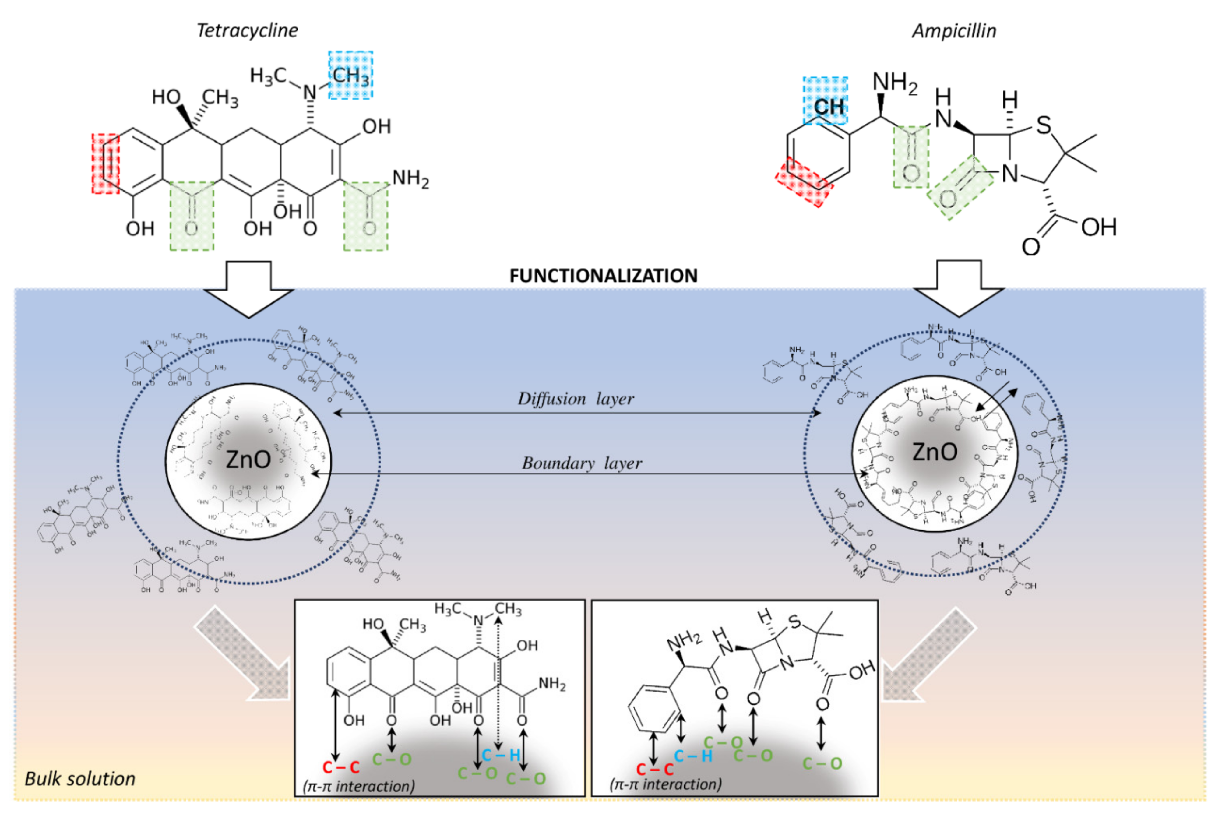
2.1. Investigation of the Mechanism of Zinc Oxide Nanoparticles’ Immobilization with Antibiotics
2.2. Physico-Chemical Characteristics of Immobilized Zinc Oxide Nanoparticles
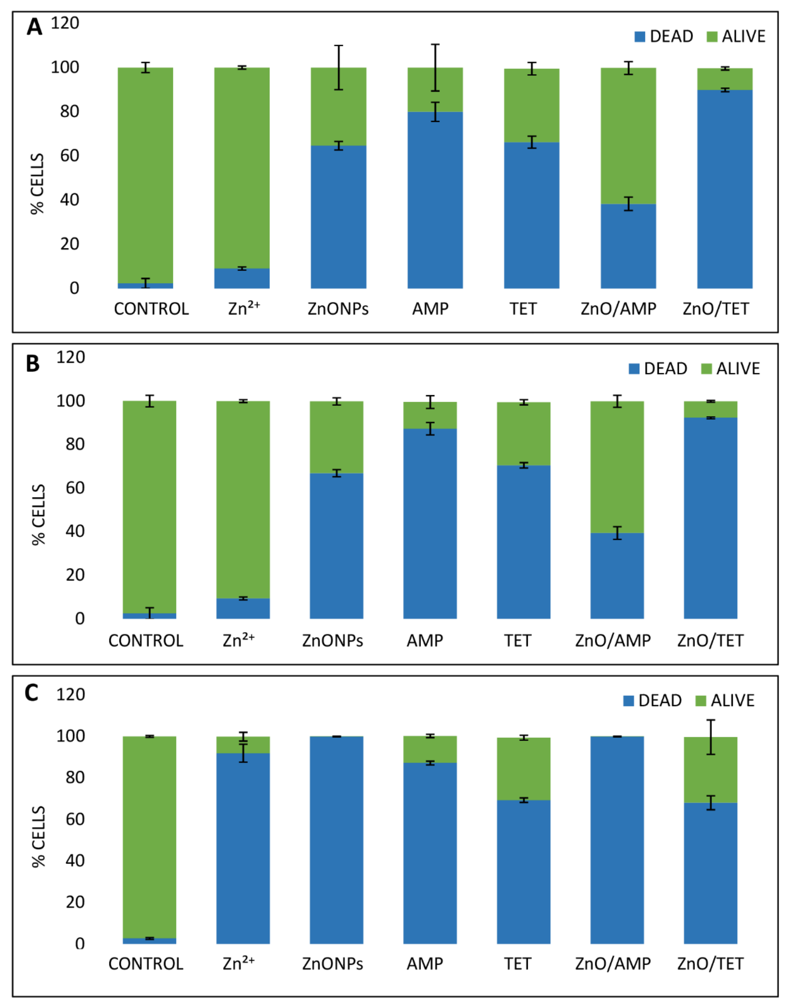
2.3. Investigation of Antimicrobial Properties of Immobilized Zinc Oxide Nanoparticles
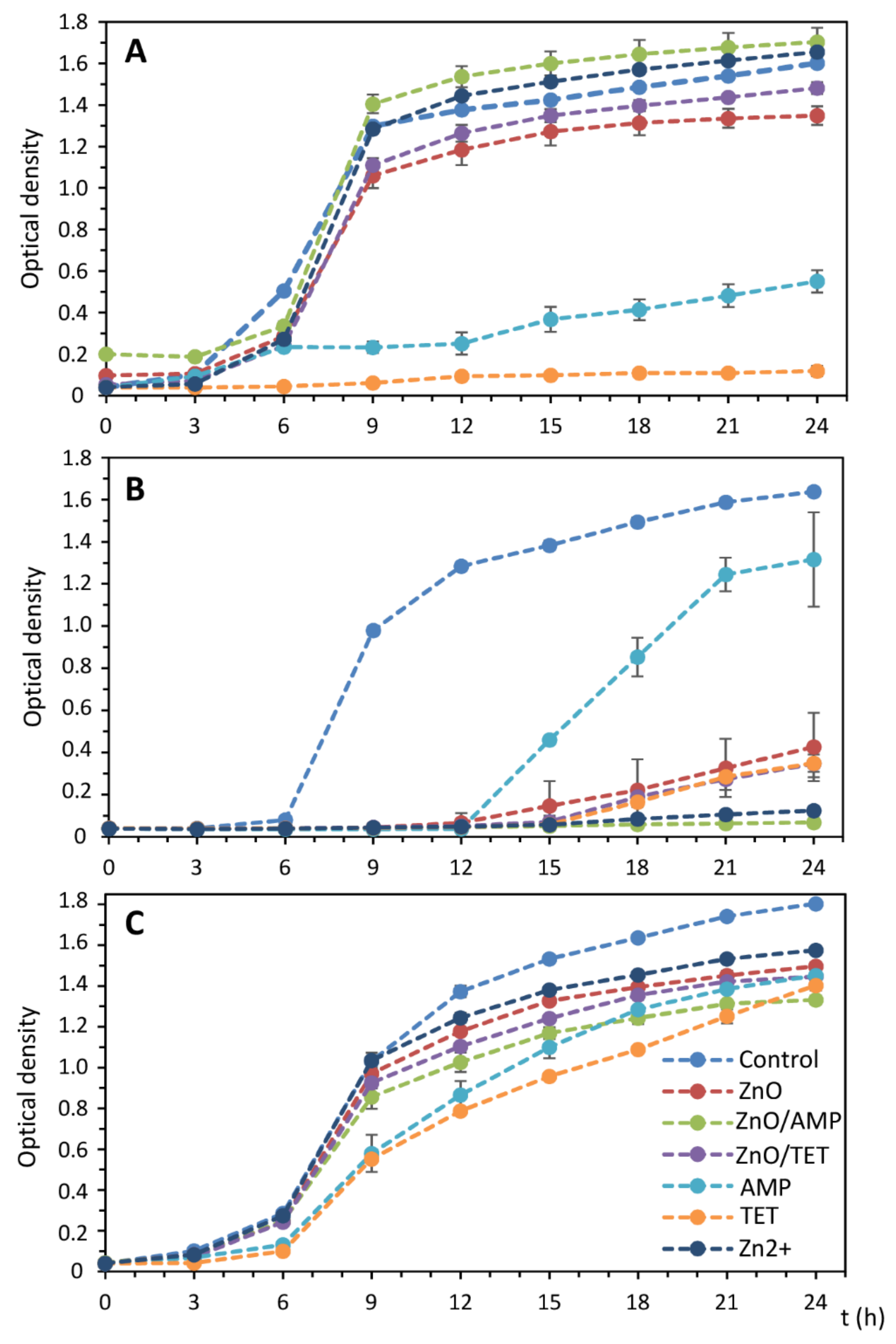
2.4. Growth Kinetics of the Selected Bacteria Strains in the Presence of Tested Antibacterial Agents
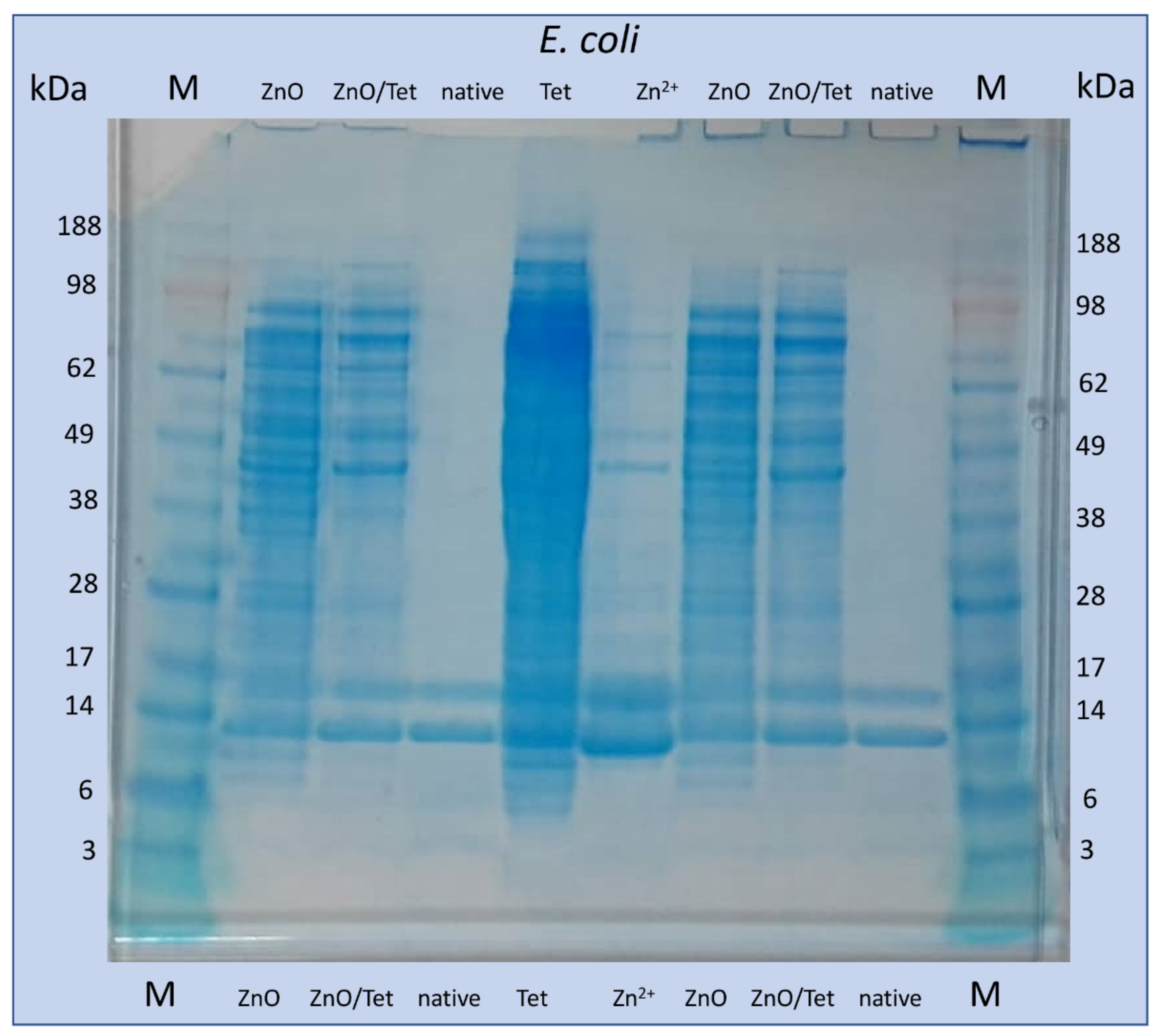
2.5. Electrophoretic Analysis of Isolated Proteins from the Bacterial Cells after Treatment with Selected Antimicrobial Agents
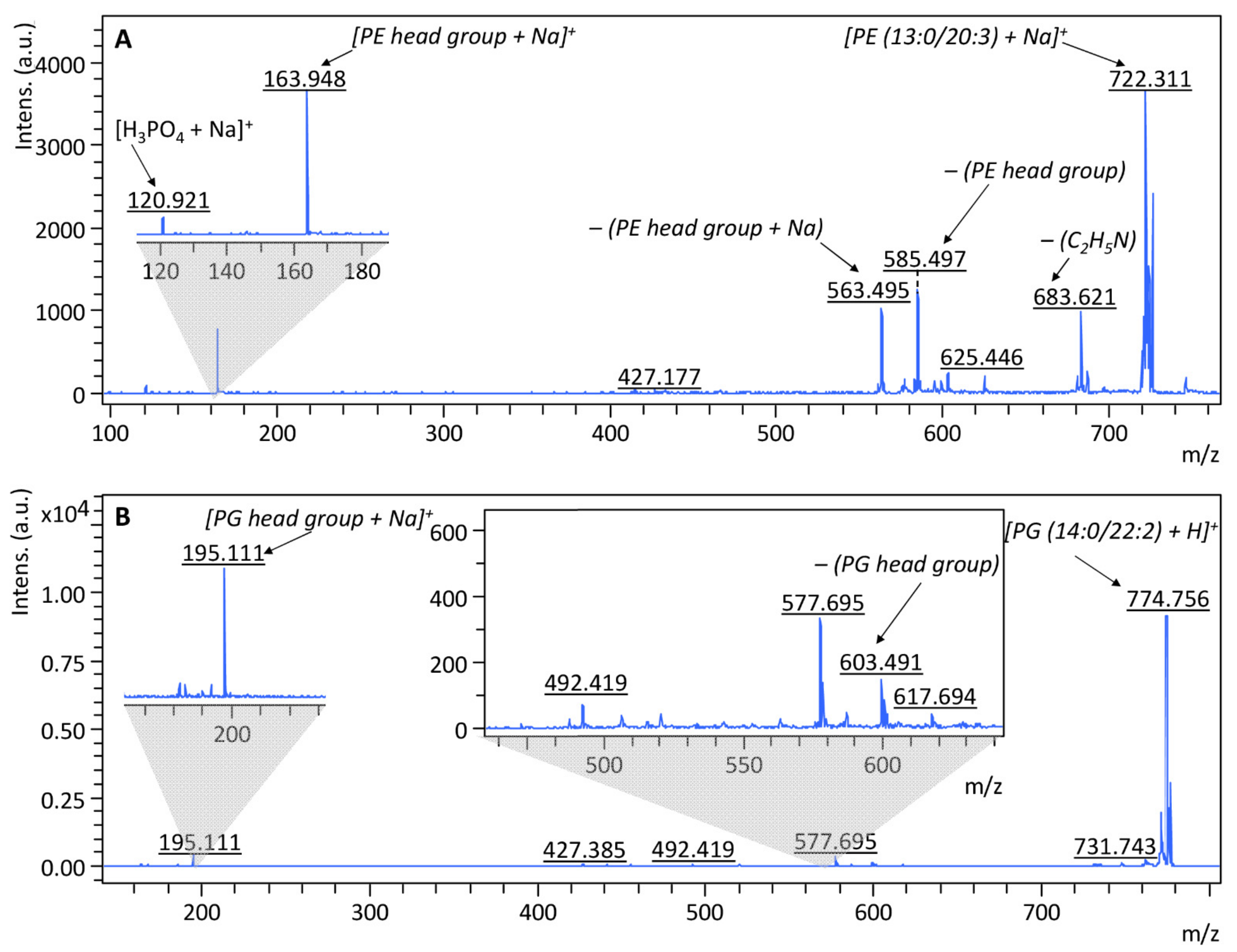
2.6. MALDI-TOF MS Analysis of Proteins and Lipids Extracted from the Bacterial Cells Treated with Selected Antimicrobial Agents
3. Materials and Methods
3.1. Chemicals and Reagents
3.2. Kinetic Study of the Antibiotics Sorption onto Zinc Oxide Nanoparticles
3.3. Immobilization Procedure
3.4. Physico-Chemical Characteristics of Immobilized Zinc Oxide Nanoparticles
3.5. Minimum Inhibitory Concentration (MIC) Assay
3.6. Flow Cytometry Analysis
3.7. Growth Kinetics of the Selected Bacteria Strains in the Presence of Tested Antibacterial Agents
3.8. Electrophoretic Analysis of Isolated Proteins from the Bacterial Cells after Treatment with Selected Antimicrobial Agents
3.9. Samples Preparation for MALDI-TOF MS Analysis
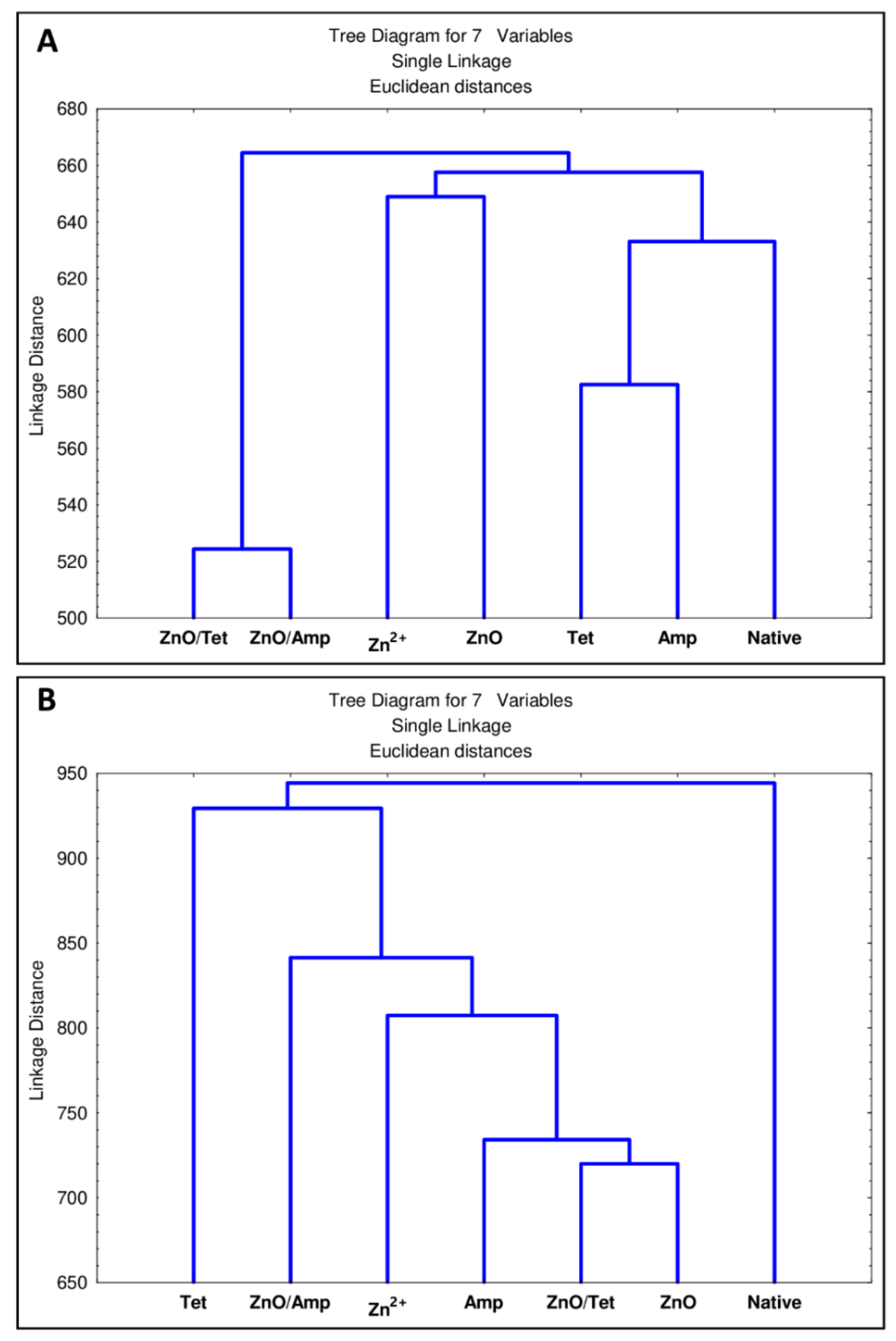
3.10. Statistical Analysis
4. Conclusions
Author Contributions
Funding
Conflicts of Interest
References
- De Souza, R.C.; Haberbeck, L.U.; Riella, H.G.; Ribeiro, D.H.B.; Carciofi, B.A.M. Antibacterial activity of zinc oxide nanoparticles synthesized by solochemical process. Brazilian J. Chem. Eng. 2019, 36, 885–893. [Google Scholar] [CrossRef] [Green Version]
- Sirelkhatim, A.; Mahmud, S.; Seeni, A.; Kaus, N.H.M.; Ann, L.C.; Bakhori, S.K.M.; Hasan, H.; Mohamad, D. Review on zinc oxide nanoparticles: Antibacterial activity and toxicity mechanism. Nano-Micro Lett. 2015, 7, 219–242. [Google Scholar] [CrossRef] [PubMed] [Green Version]
- WHO Publishes List of Bacteria for Which New Antibiotics Are Urgently Needed. Available online: https://www.who.int/news/item/27-02-2017-who-publishes-list-of-bacteria-for-which-new-antibiotics-are-urgently-needed (accessed on 5 February 2021).
- EFSA. The European Union Summary Report on Antimicrobial Resistance in zoonotic and indicator bacteria from humans, animals and food in 2018/2019. EFSA J. 2021, 19. [Google Scholar] [CrossRef]
- Kaushik, D.; Mohan, M.; Borade, D.M.; Swami, O.C. Ampicillin: Rise fall & resurgence. J. Clin. Diagn. Res. 2014, 8, ME01. [Google Scholar] [CrossRef] [PubMed]
- Hansson, J.; Körner, U.; Ludwigs, K.; Johnsson, E.; Jönsson, C.; Lundholm, K. Antibiotics as first-line therapy for acute appendicitis: Evidence for a change in clinical practice. World J. Surg. 2012, 36, 2028–2036. [Google Scholar] [CrossRef] [PubMed] [Green Version]
- Rubab, F.; Chaudhary, M.F.; Butt, N.M. Comparative and synergistic studies of antibacterial effect of ZnO nanoparticles and antibiotics for Pathogens in Drinking Water. TechConnect Briefs 2016, 3, 17–22. [Google Scholar]
- Pasquet, J.; Chevalier, Y.; Pelletier, J.; Couval, E.; Bouvier, D.; Bolzinger, M.A. The contribution of zinc ions to the antimicrobial activity of zinc oxide. Colloids Surf. A Physicochem. Eng. Asp. 2014, 457, 263–274. [Google Scholar] [CrossRef]
- Siddiqi, K.S.; ur Rahman, A.; Tajuddin; Husen, A. Properties of Zinc Oxide Nanoparticles and Their Activity against Microbes. Nanoscale Res. Lett. 2018, 13, 1–3. [Google Scholar] [CrossRef]
- Padmavathy, N.; Vijayaraghavan, R. Enhanced bioactivity of ZnO nanoparticles—An antimicrobial study. Sci. Technol. Adv. Mater. 2008, 9. [Google Scholar] [CrossRef] [PubMed]
- Zhang, L.; Jiang, Y.; Ding, Y.; Daskalakis, N.; Jeuken, L.; Povey, M.; O’Neill, A.J.; York, D.W. Mechanistic investigation into antibacterial behaviour of suspensions of ZnO nanoparticles against E. coli. J. Nanopart. Res. 2010, 12, 1625–1636. [Google Scholar] [CrossRef]
- Atmaca, S.; Gul, K.; Clcek, R. The effect of zinc on microbial growth. Turkish J. Med. Sci. 1998, 28, 595–597. [Google Scholar] [CrossRef] [Green Version]
- Ann, L.C.; Mahmud, S.; Bakhori, S.K.M.; Sirelkhatim, A.; Mohamad, D.; Hasan, H.; Seeni, A.; Rahman, R.A. Antibacterial responses of zinc oxide structures against Staphylococcus aureus, Pseudomonas aeruginosa and Streptococcus pyogenes. Ceram. Int. 2014, 40, 2993–3001. [Google Scholar] [CrossRef]
- Duffy, L.L.; Osmond-McLeod, M.J.; Judy, J.; King, T. Investigation into the antibacterial activity of silver, zinc oxide and copper oxide nanoparticles against poultry-relevant isolates of Salmonella and Campylobacter. Food Control 2018, 92, 293–300. [Google Scholar] [CrossRef]
- Vargas-Reus, M.A.; Memarzadeh, K.; Huang, J.; Ren, G.G.; Allaker, R.P. Antimicrobial activity of nanoparticulate metal oxides against peri-implantitis pathogens. Int. J. Antimicrob. Agents 2012, 40, 135–139. [Google Scholar] [CrossRef]
- Buszewski, B.; Rafińska, K.; Pomastowski, P.; Walczak, J.; Rogowska, A. Novel aspects of silver nanoparticles functionalization. Colloids Surf. A Physicochem. Eng. Asp. 2016, 506, 170–178. [Google Scholar] [CrossRef]
- Rogowska, A.; Rafińska, K.; Pomastowski, P.; Walczak, J.; Railean-Plugaru, V.; Buszewska-Forajta, M.; Buszewski, B. Silver nanoparticles functionalized with ampicillin. Electrophoresis 2017, 38, 2757–2764. [Google Scholar] [CrossRef] [PubMed]
- Buszewski, B.; Rogowska, A.; Railean-Plugaru, V.; Złoch, M.; Walczak-Skierska, J.; Pomastowski, P. The Influence of Different Forms of Silver on Selected Pathogenic Bacteria. Materials 2020, 13, 2403. [Google Scholar] [CrossRef] [PubMed]
- Sharma, N.; Jandaik, S.; Kumar, S. Synergistic activity of doped zinc oxide nanoparticles with antibiotics: Ciprofloxacin, ampicillin, fluconazole and amphotericin B against pathogenic microorganisms. An. Acad. Bras. Cienc. 2016, 88, 1689–1698. [Google Scholar] [CrossRef] [PubMed]
- Shopsin, B.; Gomez, M.; Montgomery, S.O.; Smith, D.H.; Waddington, M.; Dodge, D.E.; Bost, D.A.; Riehman, M.; Naidich, S.; Kreiswirth, B.N. Evaluation of protein A gene polymorphic region DNA sequencing for typing of Staphylococcus aureus strains. J. Clin. Microbiol. 1999, 37, 3556–3563. [Google Scholar] [CrossRef] [Green Version]
- Thati, V.; Roy, A.S.; Prasad, M.V.N.A.; Shivannavar, C.T.; Gaddad, S.M. Nanostructured zinc oxide enhances the activity of antibiotics against Staphylococcus aureus. J. Biosci Tech. 2010, 1, 64–69. [Google Scholar]
- Solomon, S.D.; Bahadory, M.; Jeyarajasingam, A.V.; Rutkowsky, S.A.; Boritz, C.; Mulfinger, L. Synthesis and study of silver nanoparticles. J. Chem. Educ. 2007, 84, 322–325. [Google Scholar] [CrossRef]
- Namasivayam, S.K.R.; Prasanna, M.; Subathra, S. Synergistic antibacterial activity of zinc oxide nanoparticles with antibiotic against the human pathogenic bacteria. J. Chem. Pharm. Res. 2015, 7, 729–735. [Google Scholar]
- Gupta, M.; Tomar, R.S.; Kaushik, S.; Mishra, R.K.; Sharma, D. Effective antimicrobial activity of green ZnO nano particles of Catharanthus roseus. Front. Microbiol. 2018, 9. [Google Scholar] [CrossRef]
- Banoee, M.; Seif, S.; Nazari, Z.E.; Jafari-Fesharaki, P.; Shahverdi, H.R.; Moballegh, A.; Moghaddam, K.M.; Shahverdi, A.R. ZnO nanoparticles enhanced antibacterial activity of ciprofloxacin against Staphylococcus aureus and Escherichia coli. J. Biomed. Mater. Res. Part B Appl. Biomater. 2010, 93, 557–561. [Google Scholar] [CrossRef] [Green Version]
- Ramani, M.; Ponnusamy, S.; Muthamizhchelvan, C. From zinc oxide nanoparticles to microflowers: A study of growth kinetics and biocidal activity. Mater. Sci. Eng. C 2012, 32, 2381–2389. [Google Scholar] [CrossRef]
- Ramani, M.; Ponnusamy, S.; Muthamizhchelvan, C.; Cullen, J.; Krishnamurthy, S.; Marsili, E. Morphology-directed synthesis of ZnO nanostructures and their antibacterial activity. Colloids Surf. B Biointerfaces 2013, 105, 24–30. [Google Scholar] [CrossRef]
- Allahverdiyev, A.M.; Kon, K.V.; Abamor, E.S.; Bagirova, M.; Rafailovich, M. Coping with antibiotic resistance: Combining nanoparticles with antibiotics and other antimicrobial agents. Expert Rev. Anti. Infect. Ther. 2011, 9, 1035–1052. [Google Scholar] [CrossRef] [PubMed]
- Railean-Plugaru, V.; Pomastowski, P.; Kowalkowski, T.; Sprynskyy, M.; Buszewski, B. Physicochemical study of natural fractionated biocolloid by asymmetric flow field-flow fractionation in tandem with various complementary techniques using biologically synthesized silver nanocomposites. Anal. Bioanal. Chem. 2018, 410, 2837–2847. [Google Scholar] [CrossRef] [Green Version]
- Gołębiowski, A.; Pomastowski, P.; Rodzik, A.; Król-Górniak, A.; Kowalkowski, T.; Górecki, M.; Buszewski, B. Isolation and self-association studies of beta-lactoglobulin. Int. J. Mol. Sci. 2020, 21, 9711. [Google Scholar] [CrossRef] [PubMed]
- Gołębiowski, A.; Kowalkowski, T.; Buszewski, B. Molecular parameters of low methoxylated pectin affected by gelation with copper and cadmium cations. Bioact. Carbohydr. Diet. Fibre 2020, 21, 100211. [Google Scholar] [CrossRef]
- Amde, M.; Tan, Z.Q.; Liu, J. Separation and size characterization of zinc oxide nanoparticles in environmental waters using asymmetrical flow field-flow fractionation. Talanta 2019, 200, 357–365. [Google Scholar] [CrossRef]
- Medina, J.; Bolaños, H.; Mosquera-Sanchez, L.P.; Rodriguez-Paez, J.E. Controlled synthesis of ZnO nanoparticles and evaluation of their toxicity in Mus musculus mice. Int. Nano Lett. 2018, 8, 165–179. [Google Scholar] [CrossRef] [Green Version]
- Barman, A.; De, A.; Das, M. Stabilization and Dispersion of ZnO Nanoparticles in PVA Matrix. J. Inorg. Organomet. Polym. Mater. 2020, 30, 2248–2257. [Google Scholar] [CrossRef]
- Gunasekaran, S.; Varadhan, S.R.; Karunanidhi, N. Qualitative analysis on the infrared bands of Tetracycline and Ampicillin. Proc. Indian Natl. Sci. Acad. 1996, 62, 309–316. [Google Scholar]
- Baraldi, C.; Tinti, A.; Ottani, S.; Gamberini, M.C. Characterization of polymorphic ampicillin forms. J. Pharm. Biomed. Anal. 2014, 100, 329–340. [Google Scholar] [CrossRef]
- Kumar Trivedi, M. Spectroscopic Characterization of Chloramphenicol and Tetracycline: An Impact of Biofield Treatment. Pharm. Anal. Acta 2015, 6. [Google Scholar] [CrossRef]
- Long, D.A. Infrared and Raman characteristic group frequencies. J. Raman Spectrosc. 2004, 35, 905. [Google Scholar] [CrossRef]
- Król, A.; Pomastowski, P.; Rafińska, K.; Railean-Plugaru, V.; Walczak, J.; Buszewski, B. Microbiology neutralization of zearalenone using Lactococcus lactis and Bifidobacterium sp. Anal. Bioanal. Chem. 2018, 410, 943–952. [Google Scholar] [CrossRef] [PubMed]
- Ennaceri, H.; Wang, L.; Erfurt, D.; Riedel, W.; Mangalgiri, G.; Khaldoun, A.; El Kenz, A.; Benyoussef, A.; Ennaoui, A. Water-resistant surfaces using zinc oxide structured nanorod arrays with switchable wetting property. Surf. Coat. Technol. 2016, 299, 169–176. [Google Scholar] [CrossRef] [Green Version]
- Buszewski, B.; Žuvela, P.; Król-Górniak, A.; Railean-Plugaru, V.; Rogowska, A.; Wong, M.W.; Yi, M.; Rodzik, A.; Sprynskyy, M.; Pomastowski, P. Interactions of zinc aqua complexes with ovalbumin at the forefront of the Zn2+/ZnO-OVO hybrid complex formation mechanism. Appl. Surf. Sci. 2021, 542, 148641. [Google Scholar] [CrossRef]
- MIC EUCAST. Available online: https://mic.eucast.org/search/show-registration/41519?back=https://mic.eucast.org/search/?search%255Bmethod%255D%3Dmic%26search%255Bantibiotic%255D%3D-1%26search%255Bspecies%255D%3D461%26search%255Bdisk_content%255D%3D1%26search%255Blimit%255D%3D50 (accessed on 6 May 2021).
- Silva, M.S.; Rabadzhiev, Y.; Eller, M.R.; Iliev, I.; Ivanova, I.; Santana, W.C. Microorganisms in Honey. In Honey Analysis; InTech: Vienna, Austria, 2017. [Google Scholar] [CrossRef] [Green Version]
- McMahon, M.A.S.; Xu, J.; Moore, J.E.; Blair, I.S.; McDowell, D.A. Environmental stress and antibiotic resistance in food-related pathogens. Appl. Environ. Microbiol. 2007, 73, 211–217. [Google Scholar] [CrossRef] [Green Version]
- Boor, K.J. Bacterial Stress Responses: What Doesn’t Kill Them Can Make Them Stronger. PLoS Biol. 2006, 4, e23. [Google Scholar] [CrossRef] [PubMed]
- Brayner, R.; Ferrari-Iliou, R.; Brivois, N.; Djediat, S.; Benedetti, M.F.; Fiévet, F. Toxicological impact studies based on Escherichia coli bacteria in ultrafine ZnO nanoparticles colloidal medium. Nano Lett. 2006, 6, 866–870. [Google Scholar] [CrossRef]
- Stoimenov, P.K.; Klinger, R.L.; Marchin, G.L.; Klabunde, K.J. Metal oxide nanoparticles as bactericidal agents. Langmuir 2002, 18, 6679–6686. [Google Scholar] [CrossRef]
- El Fattah, M.A.K.; Gamal, N.; Ibrahim, F.; Elm, G.M.; Saleh, P. Investigation of the Efficacy of Synthesized Silver and Zinc Oxide Nanoparticles against Multi-Drug Resistant Gram Negative Bacterial Clinical Isolates. iMedPub J. 2017, 8, 1–12. [Google Scholar]
- Abo-Shama, U.H.; El-Gendy, H.; Mousa, W.S.; Hamouda, R.A.; Yousuf, W.E.; Hetta, H.F.; Abdeen, E.E. Synergistic and antagonistic effects of metal nanoparticles in combination with antibiotics against some reference strains of pathogenic microorganisms. Infect. Drug Resist. 2020, 13, 351–362. [Google Scholar] [CrossRef] [Green Version]
- Egelman, E.H. Bacterial helicases. J. Struct. Biol. 1998, 124, 123–128. [Google Scholar] [CrossRef] [PubMed]
- Frank, E.G.; Ennis, D.G.; Gonzalez, M.; Levine, A.S.; Woodgate, R. Regulation of SOS mutagenesis by proteolysis. Proc. Natl. Acad. Sci. USA 1996, 93, 10291–10296. [Google Scholar] [CrossRef] [PubMed] [Green Version]
- Walshaw, D.L.; Poole, P.S. The general L-amino acid permease of Rhizobium leguminosarum is an ABC uptake system that also influences efflux of solutes. Mol. Microbiol. 1996, 21, 1239–1252. [Google Scholar] [CrossRef]
- Sun, X.; Xiao, C.; Ge, R.; Yin, X.; Li, H.; Li, N.; Yang, X.; Zhu, Y.; He, X.; He, Q.-Y. Putative copper- and zinc-binding motifs in Streptococcus pneumoniae identified by immobilized metal affinity chromatography and mass spectrometry. Proteomics 2011, 11, 3288–3298. [Google Scholar] [CrossRef] [PubMed]
- Rice, K.C.; Bayles, K.W. Molecular Control of Bacterial Death and Lysis. Microbiol. Mol. Biol. Rev. 2008, 72, 85–109. [Google Scholar] [CrossRef] [Green Version]
- Masuda, H.; Awano, N.; Inouye, M. ydfD encodes a novel lytic protein in Escherichia coli. FEMS Microbiol. Lett. 2016, 363. [Google Scholar] [CrossRef] [Green Version]
- Nguyen Le Minh, P.; de Cima, S.; Bervoets, I.; Maes, D.; Rubio, V.; Charlier, D. Ligand binding specificity of RutR, a member of the TetR family of transcription regulators in Escherichia coli. FEBS Open Bio 2015, 5, 76–84. [Google Scholar] [CrossRef] [PubMed] [Green Version]
- AlMasoud, N.; Xu, Y.; Trivedi, D.K.; Salivo, S.; Abban, T.; Rattray, N.J.W.; Szula, E.; AlRabiah, H.; Sayqal, A.; Goodacre, R. Classification of Bacillus and Brevibacillus species using rapid analysis of lipids by mass spectrometry. Anal. Bioanal. Chem. 2016, 408, 7865–7878. [Google Scholar] [CrossRef] [PubMed] [Green Version]
- Gidden, J.; Denson, J.; Liyanage, R.; Ivey, D.M.; Lay, J.O. Lipid compositions in Escherichia coli and Bacillus subtilis during growth as determined by MALDI-TOF and TOF/TOF mass spectrometry. Int. J. Mass Spectrom. 2009, 283, 178–184. [Google Scholar] [CrossRef] [Green Version]
- Kuyukina, M.S.; Ivshina, I.B.; Rychkova, M.I.; Chumakov, O.B. Effect of cell lipid composition on the formation of nonspecific antibiotic resistance in alkanotrophic rhodococci. Microbiology 2000, 69, 51–57. [Google Scholar] [CrossRef]
- Pomastowski, P.; Złoch, M.; Rodzik, A.; Ligor, M.; Kostrzewa, M.; Buszewski, B. Analysis of bacteria associated with honeys of different geographical and botanical origin using two different identification approaches: MALDI-TOF MS and 16S rDNA PCR technique. PLoS ONE 2019, 14. [Google Scholar] [CrossRef] [Green Version]
- Elshikh, M.; Ahmed, S.; Funston, S.; Dunlop, P.; McGaw, M.; Marchant, R.; Banat, I.M. Resazurin-based 96-well plate microdilution method for the determination of minimum inhibitory concentration of biosurfactants. Biotechnol. Lett. 2016, 38, 1015–1019. [Google Scholar] [CrossRef] [Green Version]
- Sarker, S.D.; Nahar, L.; Kumarasamy, Y. Microtitre plate-based antibacterial assay incorporating resazurin as an indicator of cell growth, and its application in the in vitro antibacterial screening of phytochemicals. Methods 2007, 42, 321–324. [Google Scholar] [CrossRef]
- Ratiu, I.A.; Railean Plugaru, V.; Pomastowski, P.; Milanowski, M.; Mametov, R.; Bocos-Bintintan, V.; Buszewski, B. Temporal influence of different antibiotics onto the inhibition of Escherichia coli bacterium grown in different media. Anal. Biochem. 2019, 585. [Google Scholar] [CrossRef]


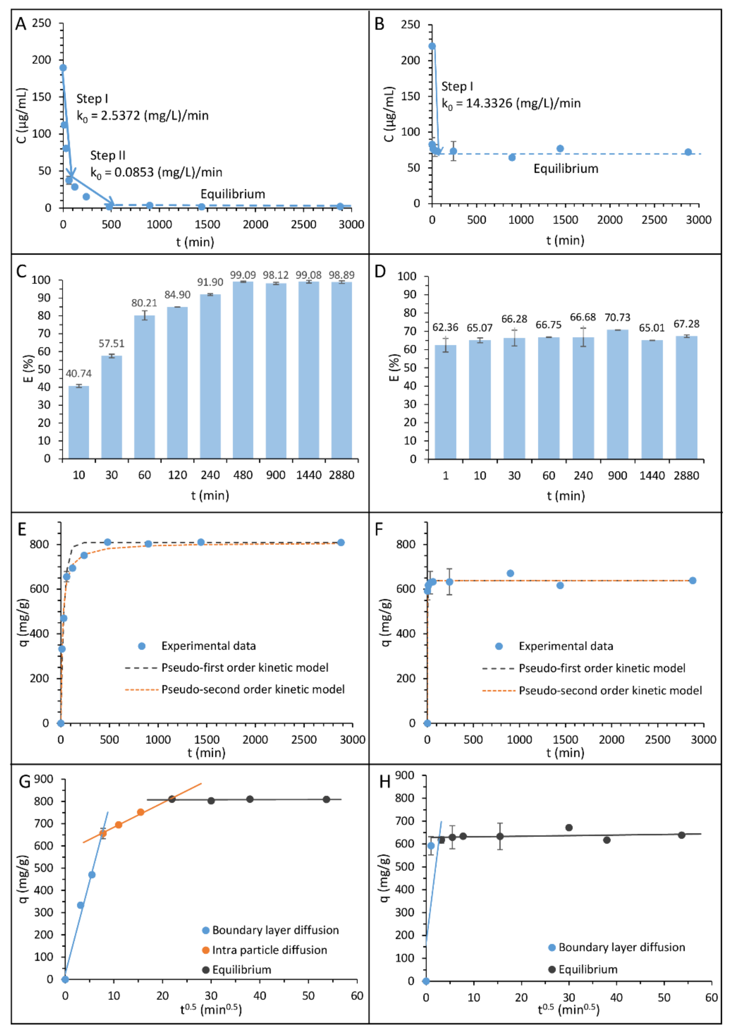
| Zero Order Kinetics Model | Pseudo-First Order Kinetics Model | Pseudo-Second Order Kinetics Model | Intra-Particle Diffusion Model | ||||
|---|---|---|---|---|---|---|---|
| Ampicillin | |||||||
| First step | |||||||
| k0 (mg L−1 min−1) | 2.537 | qe (mg g−1) | 808.999 | qe (mg g−1) | 804.298 | A (mg g−1) | 574.577 |
| Second step | |||||||
| k0 (mg L−1 min−1) | 0.085 | k1 (min−1) | 0.031 | k2 (min−1) | 7.343 × 10⁻⁵ | Kip (mg g−1min−0.5) | 10.959 |
| Aapprox. % | 3.405 | Aapprox. % | 1.030 | ||||
| Tetracycline | |||||||
| First step | |||||||
| k0 (mg L−1 min−1) | 14.333 | qe (mg g−1) | 638.784 | qe (mg g−1) | 638.766 | A (mg g−1) | 628.753 |
| k1 (min−1) | 0.976 | k2 (min−1) | 0.019 | Kip (mg g−1min−0.5) | 0.263 | ||
| Aapprox. % | 5.512 | Aapprox. % | 0.380 | ||||
| qe (mg/kg) | Ce (mg/L) | Kd | T (K) | ΔG0 (kJ mol−1) |
|---|---|---|---|---|
| Ampicillin | ||||
| 8.1 × 10⁵ | 2.102 | 3.8 × 10⁵ | 295 | −31.542 |
| Tetracycline | ||||
| 6.4 × 10⁵ | 72.077 | 8.9 × 10³ | 295 | −22.293 |
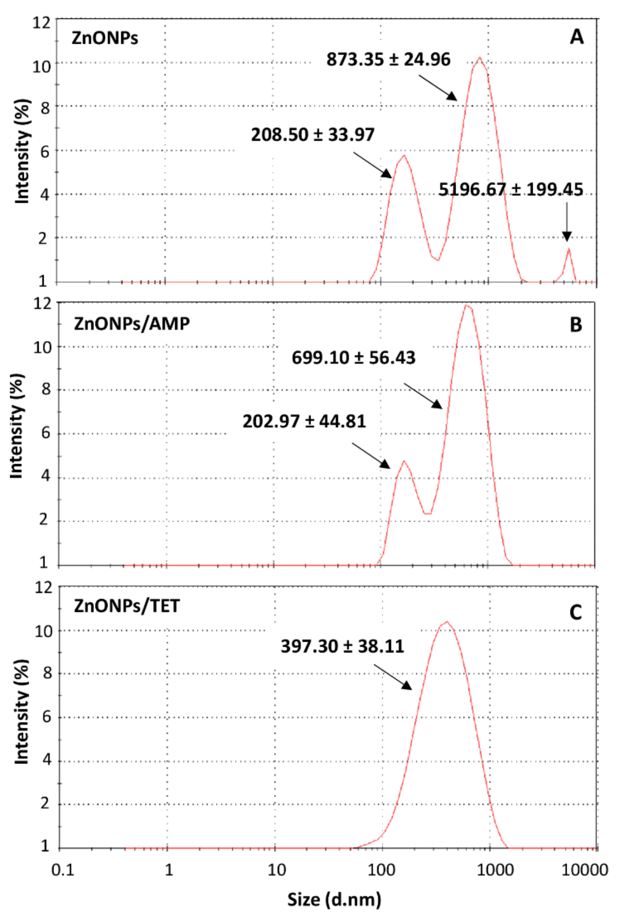
| ZnONPs | ZnONPs/AMP | ZnONPs/TET | |||
|---|---|---|---|---|---|
| pH | |||||
| 7.04 | 7.01 | 7.11 | |||
| Zeta potential (mV) | |||||
| −25.23 ± 0.97 | −24.57 ± 0.80 | −25.17 ± 0.59 | |||
| Hydrodynamic size (nm) | |||||
| Pk 1 | Pk 2 | Pk 3 | Pk 1 | Pk 2 | Pk3 |
| 873.35 ± 24.96 | 208.50 ± 33.97 | 5196.67 ± 199.45 | 699.10 ± 56.43 | 202.97 ± 44.81 | 397.30 ± 38.11 |
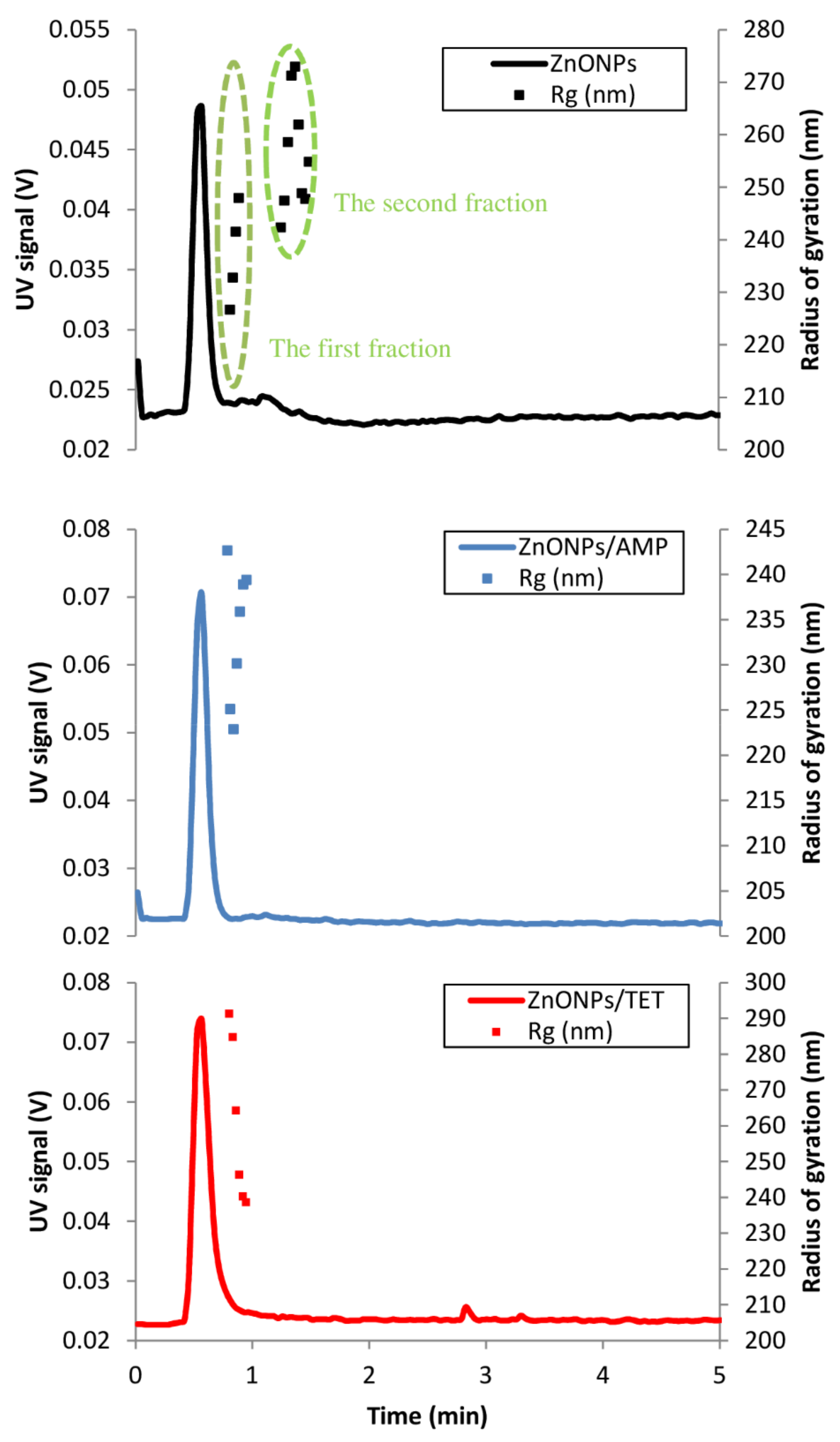
| Radius of gyration (nm) | |||||
| First fraction | Second fraction | Fraction | Fraction | ||
| 237.7 | 259.0 | 231.6 | 263.1 | ||
| Polydispersity index Pdi | |||||
| First fraction | Second fraction | Fraction | Fraction | ||
| 0.99 | 1.01 | 0.99 | 1.01 | ||
| Minimum Inhibitory Concentration [µg/mL] | ||||||
|---|---|---|---|---|---|---|
| AMP | TET | Zn2+ | ZnONPs | ZnONPs/AMP | ZnONPs/TET | |
| E. coli | 6.25 | 3.125 | 300 | 6.25 | 6.25 | 3.125 |
| S. epidermidis | 50 | 6.25 | 150 | 0.78 | 1.56 | 0.78 |
| K. pneumoniae | 50 | 0.78 | 150 | 1.56 | 1.56 | 1.56 |
Publisher’s Note: MDPI stays neutral with regard to jurisdictional claims in published maps and institutional affiliations. |
© 2021 by the authors. Licensee MDPI, Basel, Switzerland. This article is an open access article distributed under the terms and conditions of the Creative Commons Attribution (CC BY) license (https://creativecommons.org/licenses/by/4.0/).
Share and Cite
Rogowska, A.; Railean-Plugaru, V.; Pomastowski, P.; Walczak-Skierska, J.; Król-Górniak, A.; Gołębiowski, A.; Buszewski, B. The Study on Molecular Profile Changes of Pathogens via Zinc Nanocomposites Immobilization Approach. Int. J. Mol. Sci. 2021, 22, 5395. https://0-doi-org.brum.beds.ac.uk/10.3390/ijms22105395
Rogowska A, Railean-Plugaru V, Pomastowski P, Walczak-Skierska J, Król-Górniak A, Gołębiowski A, Buszewski B. The Study on Molecular Profile Changes of Pathogens via Zinc Nanocomposites Immobilization Approach. International Journal of Molecular Sciences. 2021; 22(10):5395. https://0-doi-org.brum.beds.ac.uk/10.3390/ijms22105395
Chicago/Turabian StyleRogowska, Agnieszka, Viorica Railean-Plugaru, Paweł Pomastowski, Justyna Walczak-Skierska, Anna Król-Górniak, Adrian Gołębiowski, and Bogusław Buszewski. 2021. "The Study on Molecular Profile Changes of Pathogens via Zinc Nanocomposites Immobilization Approach" International Journal of Molecular Sciences 22, no. 10: 5395. https://0-doi-org.brum.beds.ac.uk/10.3390/ijms22105395