The Unusual Role of Pro in Cu(II) Binding by His2-Cyclopentapeptide


Department of Inorganic Chemistry, Wroclaw Medical University, Borowska 211A, 50-552 Wrocław, Poland
*
Authors to whom correspondence should be addressed.
Int. J. Mol. Sci. 2021, 22(12), 6628; https://0-doi-org.brum.beds.ac.uk/10.3390/ijms22126628
Submission received: 12 May 2021
/
Revised: 10 June 2021
/
Accepted: 15 June 2021
/
Published: 21 June 2021
(This article belongs to the Section Biochemistry)
Abstract
:In this paper, we present findings from studying the interaction of copper(II) ions with the His2-cyclopentapeptide and the role of proline used for the purpose of potentiometric titration and UV-Vis, CD and EPR spectroscopic measurements. Experiments of two homodetic peptides differing by one amino acid residue were conducted for a ligand to metal ratio of 1:1 in the pH range 2.5–11.0. The presented studies reveal that peptides form only mononuclear complexes, and the CuH2L complex appears in the system first (for both L1 and L2). Study results show that the presence of Pro influences the structure of formed complexes and their stabilities and has a strong impact on the efficiency of copper(II) coordination.
1. Introduction
Proline is among the naturally occurring amino acid residues with a cyclic structure and has a significant impact on the abilities of peptides. Proline influences the peptide/protein secondary structure. The cyclic structure of the side chain causes conformational stiffness. Accordingly, the most common proline residues occur at the ends of secondary structures, such as the β-pleated sheet or α-helix [1,2]. Moreover, the Pro moiety has exceptional conformational rigidity, which reduces the flexibility of the entire molecule [3]. The double Pro-Gly motif forces antiparallel β-sheet conformation [4,5].
Of all the amino acids, proline plays a special role in the coordination of copper (II) ions by peptides. This amino acid residue can act as a “break point”, with the exception of proline present at the N-terminus. This amino acid, which is usually not directly involved in the binding of metal ions in proteins, modulates the coordination process. The presence of proline in the middle of a peptide sequence is considered an amino acid that does not directly participate in the binding of divalent metal ions. Due to the pyrrolidine ring, the nitrogen atom, which creates the peptide bond and is not able to participate in the bounding metal ions, is considered to be the “break point” of the bond cascade along the peptide chain [1].
The binding of metal ions by protein and small peptides is an important issue in the area of biochemistry and has been widely explored in recent years. Previous studies enabled the understanding of copper ion homeostasis and its role in living organisms. This trace element occurs in the prosthetic group of metalloenzymes, e.g., cytochrome c oxidase, superoxide dismutase, and tyrosinase [6,7,8]. A frequentative occurring amino acid responsible for binding divalent metal ions in protein is a side chain of His. This amino acid is often found in the active center of metalloenzymes [3].
Cyclic peptides (CP) have a huge potential as therapeutic agents. The specific peptide ring scaffold is resistant to enzymatic degradation, which makes them more orally bioavailable. Moreover, they are also more thermally stable than their linear analogs. Head-to-tail cyclization implies increased lipophilicity and membrane permeability [9,10]. The cyclic analogs of conotoxin (potential treatment of pain) and chlorotoxin (imaging of brain tumors) are more stable than their linear counterparts [11,12]. Cyclopeptides exhibit antimicrobial activity (AMPs) against Gram-positive and Gram-negative bacteria, yeasts, fungi, and viruses [13]. Some cyclopseudopeptides, such as cyclosporine A, sirolimus, and tacrolimus, are able to modulate the immune system and are used in skin disorder treatment and the prevention of organ transplant rejection [14]. The family of cyclic peptides with RGD fragments is widely studied as a radiotracer for imaging tumors [15,16,17]. This specific amino acid motif is responsible for binding to αvβ3 transmembrane glycoproteins, which play a key role in tumor growth, progression, and angiogenesis [15]. This proves that the cyclization of small tetra-, penta-, and hexa- RGD peptides exhibits an increased receptor binding affinity and selectivity to integrin [18,19].
Previously, studies have shown that cyclic peptides are attractive chelators for divalent metal ions, such Hg2+, Pb2+, and Cd2+, and can be used as fluorescence sensors for toxic ions [20,21]. Furthermore, copper(II) complexes with CP could mimic antioxidant enzyme activity as a superoxide dismutase [7,22,23]. The coordination properties of CP could be modulated by various chemical modifications of ligands. The head-to-tail cyclization of peptide leads to the deprivation of terminal groups (NH2− amino and –COO− carboxyl), which promotes the side chains of amino acid residues for metal coordination. CP analogs have been shown to interact more effectively with copper(II) ions than their linear counterparts with blocked N- and C-termini [24]. Additionally, the peptide ring size of CP impacts the binding of Cu(II) ions. The flexibility of the peptide chain depends on the number of amino acids in the sequence, which is related to the available donor atoms for metal ions.
Accordingly, cyclopeptides in general constitute a group of compounds with an interesting topology and biology and a broad spectrum of utility. Understanding the interaction of these ligands with metal ions has high potential for application in medical and biological chemistry and underlies a detailed explanation of more complex interactions. This paper concerns basic research on the interaction of copper ions with model cyclopentapeptides and the atypical influence of the Pro residue on the interaction with metal ions.
In this work, we focused on describing the binding abilities toward Cu(II) ions of two homodetic, cyclic pentapeptides differing by one amino acid residue. The first peptide has a c(GlyHisProHisLys) sequence, whilst the second one has c(GlyHisGlyHisLys), hereinafter referred to as L1 and L2 (Scheme 1).
Owing to these facts, we decided to analyze the effect of Pro residue on the binding abilities of simple model His2-cyclopentapeptides. Studies were performed using an equimolar system with copper(II) ions. The application of the potentiometric titration allowed us to determine the acid/base equilibrium and stoichiometry of complexes in the studied systems, and UV-Vis, CD, and EPR spectroscopy were used to analyze atom donors directly bound to metal ions.
2. Results and Discussion
The acid-based abilities of the free ligands and the stability constants of formed complexes are presented in Table 1. Both ligands are characterized by three protonation constants related to the presence of two His residues and the ε-amino group on the lysine side chain and are comparable to constants found in the literature [25,26,27].
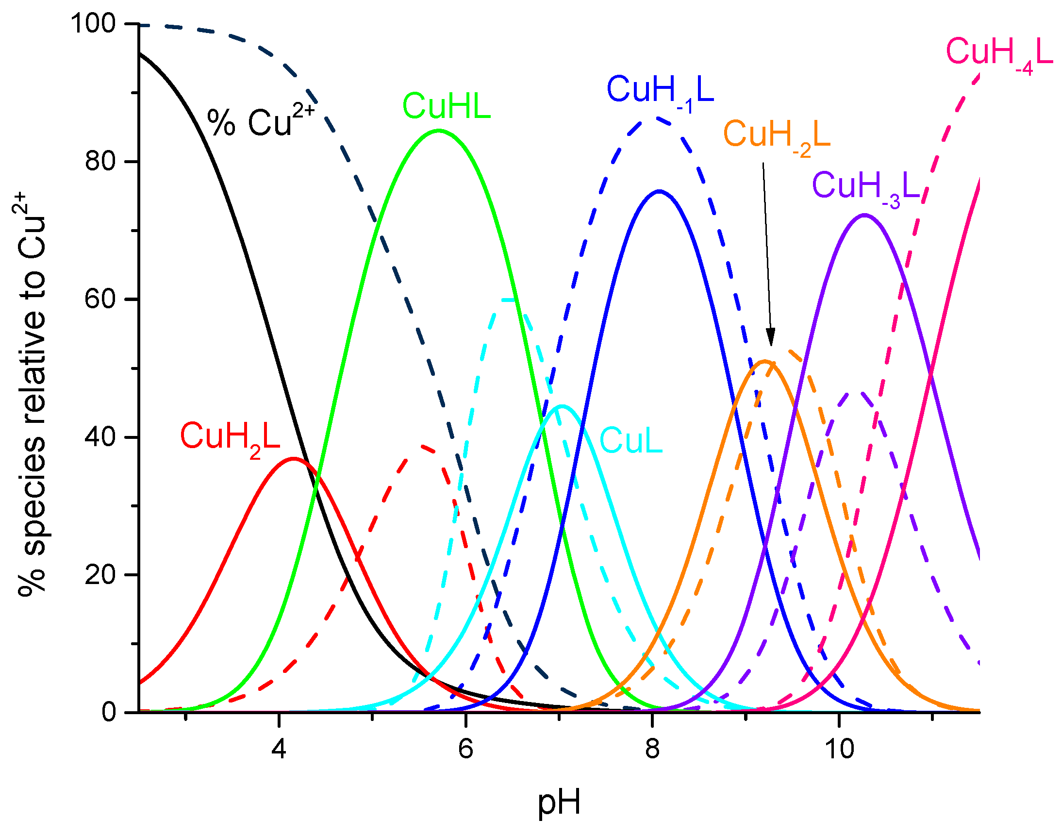
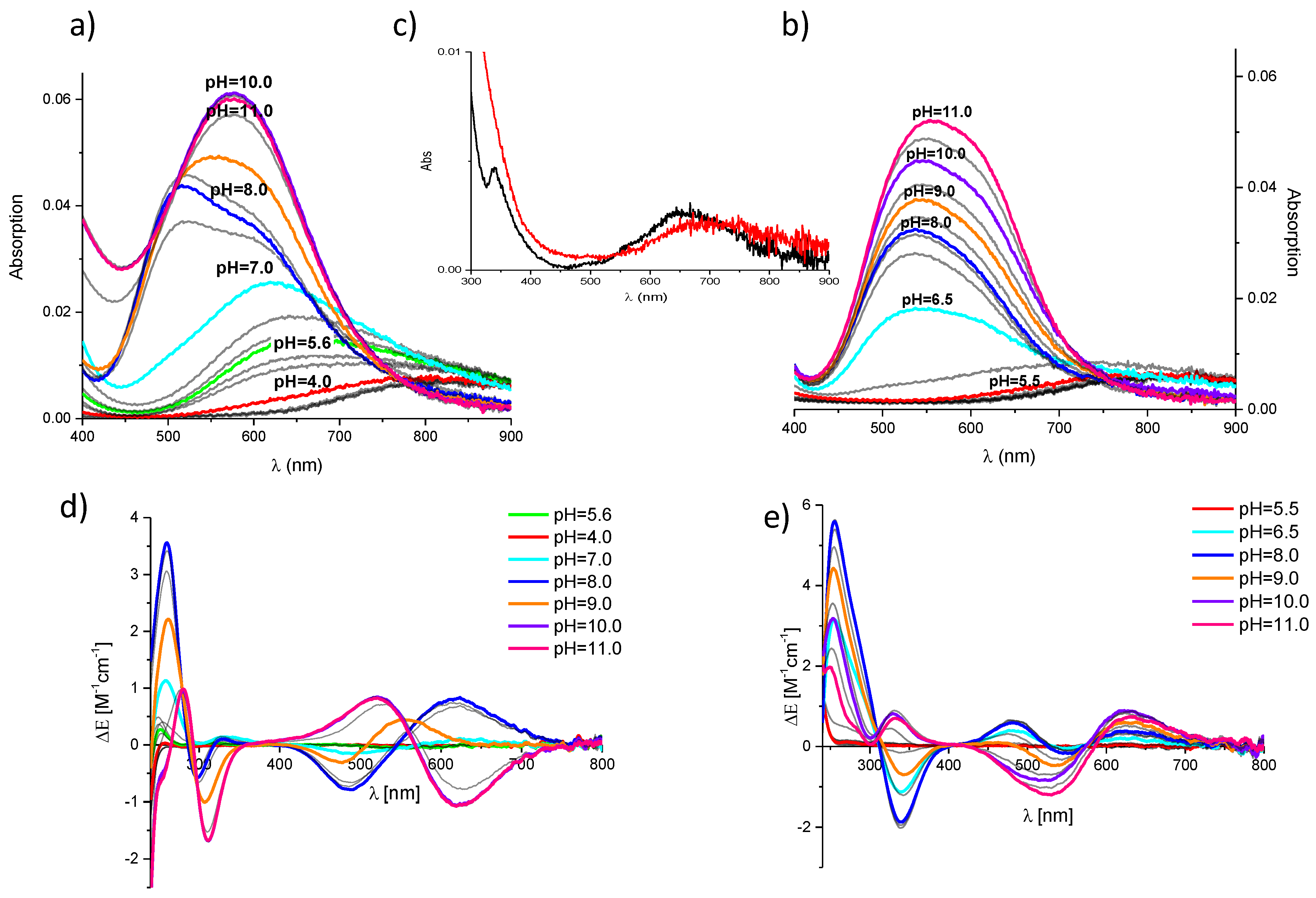
Generally, both peptides, L1 and L2, form complexes with the same stoichiometry (Table 1). The CuH2L complex appears first. Its formation is related to the dissociation of one proton from the peptide molecule, and the binding of the metal ion (Figure 1) takes place on imidazole nitrogen [28,29]. This assumption is strongly supported by the value of the corrected stability constant, logβ*L1 = logβCuH2L − logβH2L. Its value for L2, logβ*L2 = 3.60, is characteristic of the complex with the {Nim} binding mode [22,28,30,31], whilst the logβ*L1 for L1 is equal to 4.53 and significantly higher than that calculated for L2. The characteristic feature of L1 is the substitution of Gly by Pro residue in the HisXaaHis sequentional motif (Scheme 1). The increase in the logβ*L1 may be related to the extraordinary axial interaction of the metal ion with the free electron pair of amide Pro residues (Scheme 2a). This assumption is also supported by the comparison of the simulated absorption spectra, obtained by subtracting the spectrum of the copper(II) aqua ion from the spectra obtained for the discussed systems: at pH 4.0 (for L1) and 5.5 (for L2), where the CuH2L achieves the highest concentrations (Figure 2c). The d-d band for L1 is redshifted (λmaxL1 ≅ 654 nm vs. λmaxL2 ≅ 694 nm), although the absorption is minor due to the low concentration of the complexes, which supports additional donors in the coordination sphere of Cu(II).
In contrast to L2, with the increase in pH, L1 forms the CuHL complex. The formation of this species does not significantly influence the absorption or the CD spectra, and only an increase in the intensity of the bands was observed. This shows the same binding mode in both CuH2L and CuHL complexes. The value of the logKCuH2L→CuHL = 6.82 is almost the same as logKIm2 = 6.76 in the free ligand, which suggests proton dissociation from the second imidazole ring without changes in the coordination sphere of the Cu(II) ion.
With the increase in pH, L1 and L2 form the CuL species, which is dominant around pH 7 (L1) and 6 (L2). Consequently, in the system with L1, at pH 7, there are two additional complexes CuHL and CuH−1L and it was not possible to obtain the spectral parameters only for the CuL species of L2. The creation of this complex by L2 is the consequence of two protons dissociating from the CuH2L species and the involvement of two additional N-donors in the copper(II) binding, which causes the redshift of the d-d band in the absorption spectrum (Figure 2b). Moreover, the presence of three CT bands in the CD spectrum at 253, 292, and 342 nm supports the involvement of the imidazole as well as the amide donors. In the system of L1, the creation of this species is related to the deprotonation of the amide donor and its involvement in the Cu(II) binding. Nevertheless, the comparison of the corrected stability constants for these complexes, logβ*corr = logβCuL − logKLys (logβ*L1 = 0.03 vs. logβ*L2 = −1.12), shows the higher stability of the species created by L1, which may be explained by the presence of the NPro instead of the second NIm in the coordination sphere of the Cu(II) ion (Scheme 1b).
Next, the CuH-1L form achieves the highest concentration at pH 8.0. Based on the fact that the Lys side chain is still protonated, it can be assumed that H+ is dissociated from the next amide nitrogen, and its coordination takes place. The UV-Vis spectrum for the L1/Cu(II) system shows a wide band with double λmax = 515 and 613 nm, whilst for the L2/Cu(II) system, d-d bands at 538 nm and shband ≈ 608 nm were observed. The involvement of the second amide donor in the Cu(II) ion binding significantly influences the CD spectra recorded at pH 8.0 (Figure 2a,b). Based on the analysis presented above, the CuH-1L species may be described by the {(NIm, 2Nam)NPro} and {2NIm, 2Nam} binding modes for the L1 and L2 ligands, respectively. The proposed structure of the complex when the Pro residue is located between both His moieties is presented in Scheme 2c.
Next, CuH-2L is only a minor species in the L1 system, but it dominates at pH 9.20 in the Cu(II)/L2 solution (Figure 1). The logK values = 8.88 (L1) and = 9.13 (L2) (Table 1) strongly support the dissociation of the proton from the subsequent peptide bond and the involvement of the next amide in the Cu(II) binding. Owing to this fact, the discussed complex may be described by the {NIm, 3Nam} binding manner for L1 and L2. The spectral parameters confirm the same coordination model. Finally, the formation of the last two forms, CuH-3L and CuH-4L, has no impact on the spectral abilities of both systems, and the logK values (Table 1) support proton dissociation from the Lys side chain and water or imidazole bound to the Cu(II) ion.
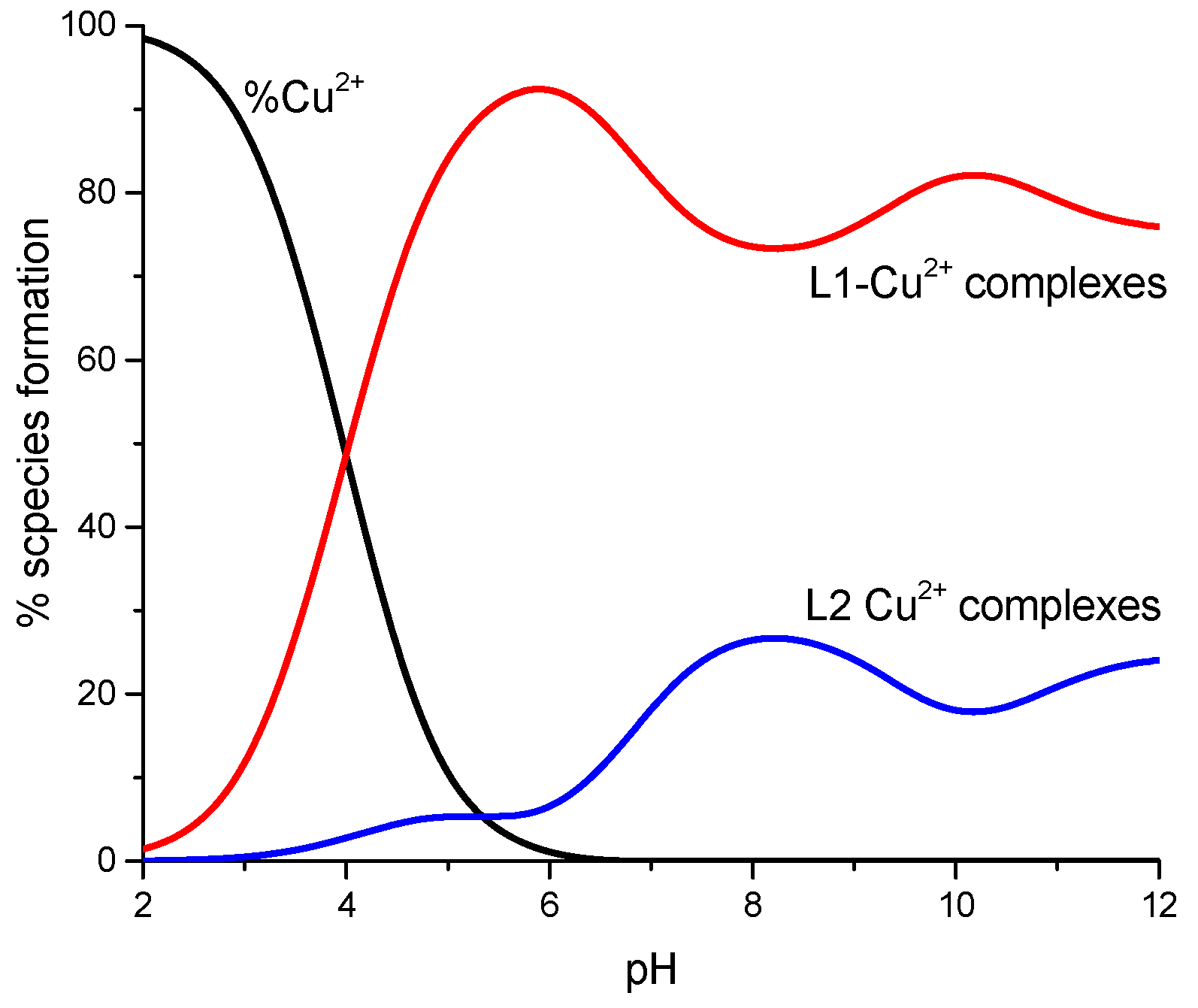
The presence of Pro in the L1 structure influences not only the structure of the formed complexes and their stabilities, as presented above, but it also has a strong impact on the efficiency of the copper(II) coordination. Figure 3 presents the competition diagram simulated for the system L1/Cu(II)/L2 with a nL1:nCu(II):nL2 = 1:1:1 molar ratio, and it can be clearly observed that the L1 peptide binds to metal ions significantly more efficiently.
The characteristic structural feature of the investigated peptide is the presence of the HXXH motif. Due to this fact, its binding abilities were compared with the properties of the simple linear and cyclic His2 analogs (Scheme 3).
Table 2 shows the comparison of the stability constants of the complexes with only NIm in the coordination sphere of Cu(II) and with one, two, or three amide donors. It is clear that independent of the structure (linear or cyclic) of the peptide, the 1NIm of the 2NIm complexes of L1 are significantly more stable, which supports the postulated interaction of NPro with Cu(II) ions.
According to the durability of the complexes with amide, the presence of Pro residue in the peptide sequence promotes the coordination of the second and third amides. However, the role of Pro seems to be able to stiffen the peptide cycle, which promotes the favorable steric position of Namide.
3. Materials and Methods
3.1. Materials
Both peptides, L1 and L2, were purchased commercially from STI (Poland, Poznań). The purity of the researched ligands was confirmed by the MS method. Cu(II) solution was prepared by the dilution of solid metal salts CuCl2 × 2H2O (POCH) in distilled water. For potentiometric and spectroscopic measurements, the acid solution was prepared using concentrated HCl (37%) and solid KCl to obtain the final concentration of 3.16 × 10⁻³ M and the ionic strength equal to 0.1 M. The base (KOH) was used as a titrant at 0.1 M concentration and was purchased from Sigma-Aldrich. CO2 was removed from the solution.
3.2. Potentiometric Measurements
Potentiometric studies were carried out using a Metrohm pH-metr system (Switzerland, Herisau) with a semimicro combined electrode at 25 °C calibrated in a hydrogen ion concentration using HCl [34]. The peptide concentration was 8 × 10⁻⁴ M, and pH-metric titration was performed at a constant ionic strength of 0.1 M KCl using sample volumes of 1.3 mL. Systems with 1:1 ligand to Cu(II) metal ion molar ratios were analyzed. Alkali (KOH) was added using a 0.250 mL micrometer syringe. Protonation constants of the ligands and stability constants (logβ) of the complexes and stoichiometry were calculated from titration curves using SUPERQUAD and HYPERQUAD programs [35,36].
3.3. Spectroscopic Measurements
The UV-Vis spectra of the copper(II) complexes were recorded on a Varian Cary 50 Bio spectrophotometer (Varian Inc., USA, Palo Alto, California) in 1 cm path length quartz cells. All UV-Vis spectra were collected in the 350–900 nm and 2.5–11 pH range. Circular dichroism (CD) spectra were recorded on a Jasco J-1500 spectrophotometer (Jasco, Japan, Tokyo) in the 240–800 nm range using 1 cm cuvettes. The same concentration, ionic strength, and molar ratio were used for both spectroscopic and potentiometric studies.
4. Conclusions
The coordination abilities and impact of proline on the two cyclopentapeptides c(GlyHisProHisLys) and c(GlyHisGlyHisLys) are presented in this paper. Studies were conducted on Cu(II) ions with a ligand-to-metal ratio of 1:1. Both peptides form complexes with the same stoichiometry. The coordination started with the CuH2L complex. At pH 6–7, CuL is the dominant species. The final solution of both ligands contains species with the {NIm, 3Nam} binding mode.
The competition between L1 and L2 in the presence of Cu(II) ions under equimolar conditions shows that the L1 peptide with proline in the sequence binds to metal ions significantly more efficiently. Additionally, complexes of L1 are significantly more stable, which strongly supports the extraordinary axial interaction of NPro with Cu(II) ions.
Author Contributions
Conceptualization, J.B.; Methodology, J.B., A.K. and A.P.; Formal Analysis, A.K. and J.B.; Investigation, A.P.; Data Curation, J.B., A.K. and A.P.; Writing—Original Draft Preparation, A.K. and A.P.; Writing—Review and Editing, J.B.; Visualization, J.B. and A.K.; Supervision, J.B.; Project Administration, J.B.; Funding Acquisition, J.B. and A.P. All authors have read and agreed to the published version of the manuscript.
Funding
The presented studies were financially supported by Wroclaw Medical University (STM.D080.20.123).
Institutional Review Board Statement
Not applicable.
Informed Consent Statement
Not applicable.
Data Availability Statement
Not applicable.
Conflicts of Interest
The author Aleksandra Pieniężna received research grants from Wroclaw Medical University (STM.D080.20.123). The other coauthors declare that they have no known competing financial interests or personal relationships that could have appeared to influence the work reported in this paper.
References
- Bataille, M.; Formicka-Kozlowska, G.; Kozlowski, H.; Pettit, L.D.; Steel, I. The L-proline residue as a ‘break-point’ in the co-ordination of metal–peptide systems. J. Chem. Soc. Chem. Commun. 1984, 231–232. [Google Scholar] [CrossRef]
- Pettit, L.D.; Steel, I.; Formicka-Kozlowska, G.; Tatarowski, T.; Bataille, M. The L-proline residue as a ?break-point? in metal?peptide systems. J. Chem. Soc. Dalton Trans. 1985, 535–539. [Google Scholar] [CrossRef]
- Kozłowski, H.; Bal, W.; Dyba, M.; Kowalik-Jankowska, T. Specific structure–stability relations in metallopeptides. Co-ord. Chem. Rev. 1999, 184, 319–346. [Google Scholar] [CrossRef]
- Peluso, S.; Rückle, T.; Lehmann, C.; Mutter, M.; Peggion, C.; Crisma, M. Crystal structure of a synthetic cyclodecapeptide for template-assembled synthetic protein design. ChemBioChem 2001, 2, 432–437. [Google Scholar] [CrossRef]
- Dumy, P.; Eggleston, I.M.; Esposito, G.; Nicula, S.; Mutter, M. Solution structure of Regioselectively Addressable Functional-ized Templates: An NMR and restrained molecular dynamics investigation. Biopolymers 1996, 39, 297–308. [Google Scholar] [CrossRef]
- Fontanesi, F.; Soto, I.C.; Horn, D.; Barrientos, A. Assembly of mitochondrial cytochromec-oxidase, a complicated and highly regulated cellular process. Am. J. Physiol. Physiol. 2006, 291, C1129–C1147. [Google Scholar] [CrossRef] [Green Version]
- Kotynia, A.; Janek, T.; Czyżnikowska, Ż.; Bielińska, S.; Kamysz, W.; Brasuń, J. The Analysis of Cu(II)/Zn(II) Cyclopeptide System as Potential Cu, ZnSOD Mimic Center. Int. J. Pept. Res. Ther. 2017, 23, 431–439. [Google Scholar] [CrossRef] [PubMed] [Green Version]
- Decker, H.; Schweikardt, T.; Tuczek, F. The First Crystal Structure of Tyrosinase: All Questions Answered? Angew. Chem. Int. Ed. 2006, 45, 4546–4550. [Google Scholar] [CrossRef] [PubMed]
- Vorherr, T.; Lewis, I.; Berghausen, J.; Desrayaud, S.; Schaefer, M. Modulation of Oral Bioavailability and Metabolism for Closely Related Cyclic Hexapeptides. Int. J. Pept. Res. Ther. 2018, 24, 35–48. [Google Scholar] [CrossRef] [Green Version]
- Bockus, A.T.; McEwen, C.M.; Lokey, R.S. Form and Function in Cyclic Peptide Natural Products: A Pharmacokinetic Perspective. Curr. Top. Med. Chem. 2013, 999, 29–35. [Google Scholar] [CrossRef]
- Clark, R.; Fischer, H.; Dempster, L.; Daly, N.L.; Rosengren, K.J.; Nevin, S.T.; Meunier, F.A.; Adams, D.; Craik, D.J. Engineering stable peptide toxins by means of backbone cyclization: Stabilization of the -conotoxin MII. Proc. Natl. Acad. Sci. USA 2005, 102, 13767–13772. [Google Scholar] [CrossRef] [Green Version]
- Akcan, M.; Stroud, M.R.; Hansen, S.J.; Clark, R.; Daly, N.L.; Craik, D.J.; Olson, J.M. Chemical Re-engineering of Chlorotoxin Improves Bioconjugation Properties for Tumor Imaging and Targeted Therapy. J. Med. Chem. 2011, 54, 782–787. [Google Scholar] [CrossRef] [Green Version]
- Namjoshi, S.; Benson, H.A.E. Cyclic peptides as potential therapeutic agents for skin disorders. Biopolymer 2010, 94, 673–680. [Google Scholar] [CrossRef] [PubMed]
- Velkov, T.; Roberts, K.D.; Nation, R.L.; Thompson, P.E.; Li, J. Pharmacology of polymyxins: New insights into an ‘old’ class of antibiotics. Future Microbiol. 2013, 8, 711–724. [Google Scholar] [CrossRef] [Green Version]
- Zhou, Y.; Chakraborty, S.; Liu, S. Radiolabeled Cyclic RGD Peptides as Radiotracers for Imaging Tumors and Thrombosis by SPECT. Theranostics 2011, 1, 58–82. [Google Scholar] [CrossRef] [PubMed] [Green Version]
- Liu, S. Radiolabeled Cyclic RGD Peptide Bioconjugates as Radiotracers Targeting Multiple Integrins. Bioconjugate Chem. 2015, 26, 1413–1438. [Google Scholar] [CrossRef] [Green Version]
- Shi, J.; Wang, F.; Liu, S. Radiolabeled cyclic RGD peptides as radiotracers for tumor imaging. Biophys. Rep. 2016, 2, 1–20. [Google Scholar] [CrossRef] [PubMed] [Green Version]
- Müller, G.; Gurrath, M.; Kessler, H.; Timpl, R. Dynamic Forcing, a Method for Evaluating Activity and Selectivity Profiles of RGD(Arg-Gly-Asp) Peptides. Angew. Chem. Int. Ed. 1992, 31, 326–328. [Google Scholar] [CrossRef]
- Haubner, R.; Gratias, R.; Diefenbach, B.; Goodman, S.L.; Jonczyk, A.; Kessler, H. Structural and Functional Aspects of RGD-Containing Cyclic Pentapeptides as Highly Potent and Selective Integrin αVβ3Antagonists. J. Am. Chem. Soc. 1996, 118, 7461–7472. [Google Scholar] [CrossRef]
- Ngu-Schwemlein, M.; Gilbert, W.; Askew, K.; Schwemlein, S. Thermodynamics and fluorescence studies of the interactions of cyclooctapeptides with Hg2+, Pb2+, and Cd2+. Bioorganic Med. Chem. 2008, 16, 5778–5787. [Google Scholar] [CrossRef]
- Ngu-Schwemlein, M.; Butko, P.; Cook, B.; Whigham, T. Interactions of an acidic cyclooctapeptide with metal ions: Microcalorimetric and fluorescence analyses. J. Pept. Res. 2008, 66, 72–81. [Google Scholar] [CrossRef]
- Bonomo, R.P.; Impellizzeri, G.; Pappalardo, G.; Purrello, R.; Rizzarelli, E.; Tabbì, G. Co-ordinating properties of cyclopeptides. Thermodynamic and spectroscopic study on the formation of copper(II) complexes with cyclo(Gly-His)4 and cyclo(Gly-His-Gly)2 and their superoxide dismutase-like activity. J. Chem. Soc. Dalton Trans. 1998, 3851–3858. [Google Scholar] [CrossRef]
- Bonomo, R.P.; Conte, E.; Impellizzeri, G.; Pappalardo, G.; Purrello, R.; Rizzarelli, E. Copper(II) complexes with cyclo(L-aspartyl-L-aspartyl) and cyclo(L-glutamyl-L-glutamyl) derivatives and their antioxidant properties. J. Chem. Soc. Dalton Trans. 1996, 3093–3099. [Google Scholar] [CrossRef]
- Kotynia, A.; Pap, J.S.; Brasun, J. The binding abilities of homodetic cyclic His-peptides toward copper ions. Inorg. Chim. Acta 2018, 472, 3–11. [Google Scholar] [CrossRef]
- Kotynia, A.; Bielinska, S.; Kamysz, W.; Brasun, J. The coordination abilities of the multiHis-cyclopeptide with two metal-binding centers—Potentiometric and spectroscopic investigation. Dalton Trans. 2012, 41, 12114. [Google Scholar] [CrossRef]
- Kotynia, A.; Czyżnikowska, Ż.; Bielinska, S.; Szyrwiel, Ł.; Kamysz, W.; Malinka, W.; Brasun, J. The impact of two–GlyProGly–motifs on formation of di-copper complexes by His 4-cyclopeptides. New J. Chem. 2014, 38, 5198–5206. [Google Scholar] [CrossRef]
- Kállay, C.; Várnagy, K.; Malandrinos, G.; Hadjiliadis, N.; Sanna, D.; Sóvágó, I. Copper(ii) complexes of terminally protected pentapeptides containing three histidyl residues in alternating positions, Ac-His-Xaa-His-Yaa-His-NH2. Dalton Trans. 2006, 4545–4552. [Google Scholar] [CrossRef] [PubMed]
- Kállay, C.; Várnagy, K.; Malandrinos, G.; Hadjiliadis, N.; Sanna, D.; Sóvágó, I. Thermodynamic and structural characterization of the macrochelates formed in the reactions of copper(II) and zinc(II) ions with peptides of histidine. Inorg. Chim. Acta 2009, 362, 935–945. [Google Scholar] [CrossRef]
- Brasun, J.; Gabbiani, C.; Ginanneschi, M.; Messori, L.; Orfei, M.; Swiatek-Kozlowska, J. The copper(II) binding properties of the cyclic peptide c(HGHK). J. Inorg. Biochem. 2004, 98, 2016–2021. [Google Scholar] [CrossRef]
- Brasuń, J.; Matera-Witkiewicz, A.; Ołdziej, S.; Pratesi, A.; Ginanneschi, M.; Messori, L. Impact of ring size on the copper(II) coordination abilities of cyclic tetrapeptides. J. Inorg. Biochem. 2009, 103, 813–817. [Google Scholar] [CrossRef]
- Kállay, C.; Nagy, Z.; Várnagy, K.; Malandrinos, G.; Hadjiliadis, N.; Sóvágó, I. Thermodynamic and Structural Characterization of the Copper(II) Complexes of Peptides Containing Both Histidyl and Aspartyl Residues. Bioinorg. Chem. Appl. 2007, 2007, 030394. [Google Scholar] [CrossRef] [Green Version]
- Csire, G.; Timári, S.; Asztalos, J.; Király, J.M.; Kiss, M.; Várnagy, K. Coordination, redox properties and SOD activity of Cu(II) complexes of multihistidine peptides. J. Inorg. Biochem. 2017, 177, 198–210. [Google Scholar] [CrossRef]
- Rajković, S.; Kállay, C.; Serényi, R.; Malandrinos, G.; Hadjiliadis, N.; Sanna, D.; Sóvágó, I. Complex formation processes of terminally protected peptides containing two or three histidyl residues. Characterization of the mixed metal complexes of peptides. Dalton Trans. 2008, 5059–5071. [Google Scholar] [CrossRef] [PubMed]
- Irving, H.; Miles, M.; Pettit, L. A study of some problems in determining the stoicheiometric proton dissociation constants of complexes by potentiometric titrations using a glass electrode. Anal. Chim. Acta 1967, 38, 475–488. [Google Scholar] [CrossRef]
- Gans, P.; Sabatini, A.; Vacca, A. SUPERQUAD: An improved general program for computation of formation constants from potentiometric data. J. Chem. Soc. Dalton Trans. 1985, 1195–1200. [Google Scholar] [CrossRef]
- Gans, P.; Sabatini, A.; Vacca, A. Investigation of equilibria in solution. Determination of equilibrium constants with the HYPERQUAD suite of programs. Talanta 1996, 43, 1739–1753. [Google Scholar] [CrossRef]
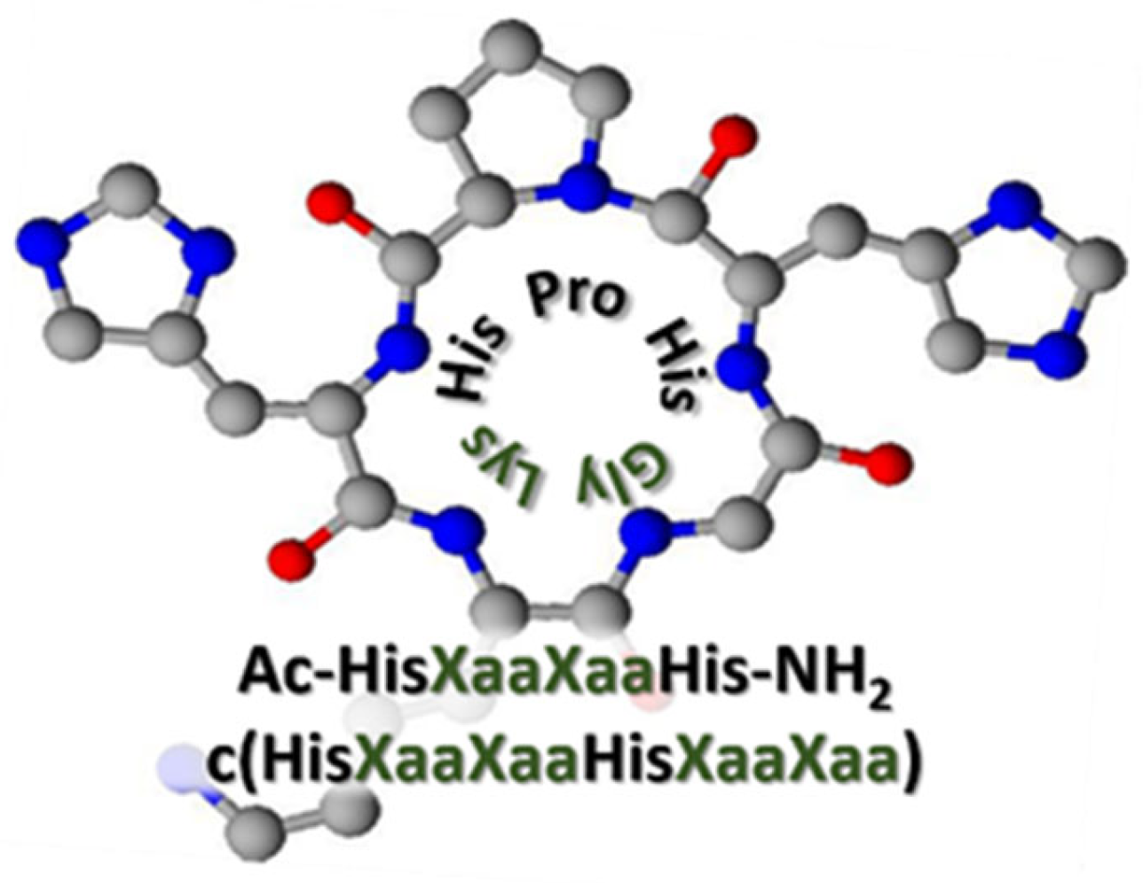
Scheme 1.
Schematic structures of investigated pentacyclopeptides: (a) L1—c(GlyHisProHisLys) and (b) L2—c(GlyHisGlyHisLys). The hydrogen atoms were omitted for simplicity; carbon atoms are grey; nitrogen atoms are blue; oxygen atoms are red.
Scheme 1.
Schematic structures of investigated pentacyclopeptides: (a) L1—c(GlyHisProHisLys) and (b) L2—c(GlyHisGlyHisLys). The hydrogen atoms were omitted for simplicity; carbon atoms are grey; nitrogen atoms are blue; oxygen atoms are red.

Figure 1.
The species distribution curves for the system of L1−c(GlyHisProHisLys) (solid line) and L2−c(GlyHisGlyHisLys) (dashed line).
Figure 1.
The species distribution curves for the system of L1−c(GlyHisProHisLys) (solid line) and L2−c(GlyHisGlyHisLys) (dashed line).

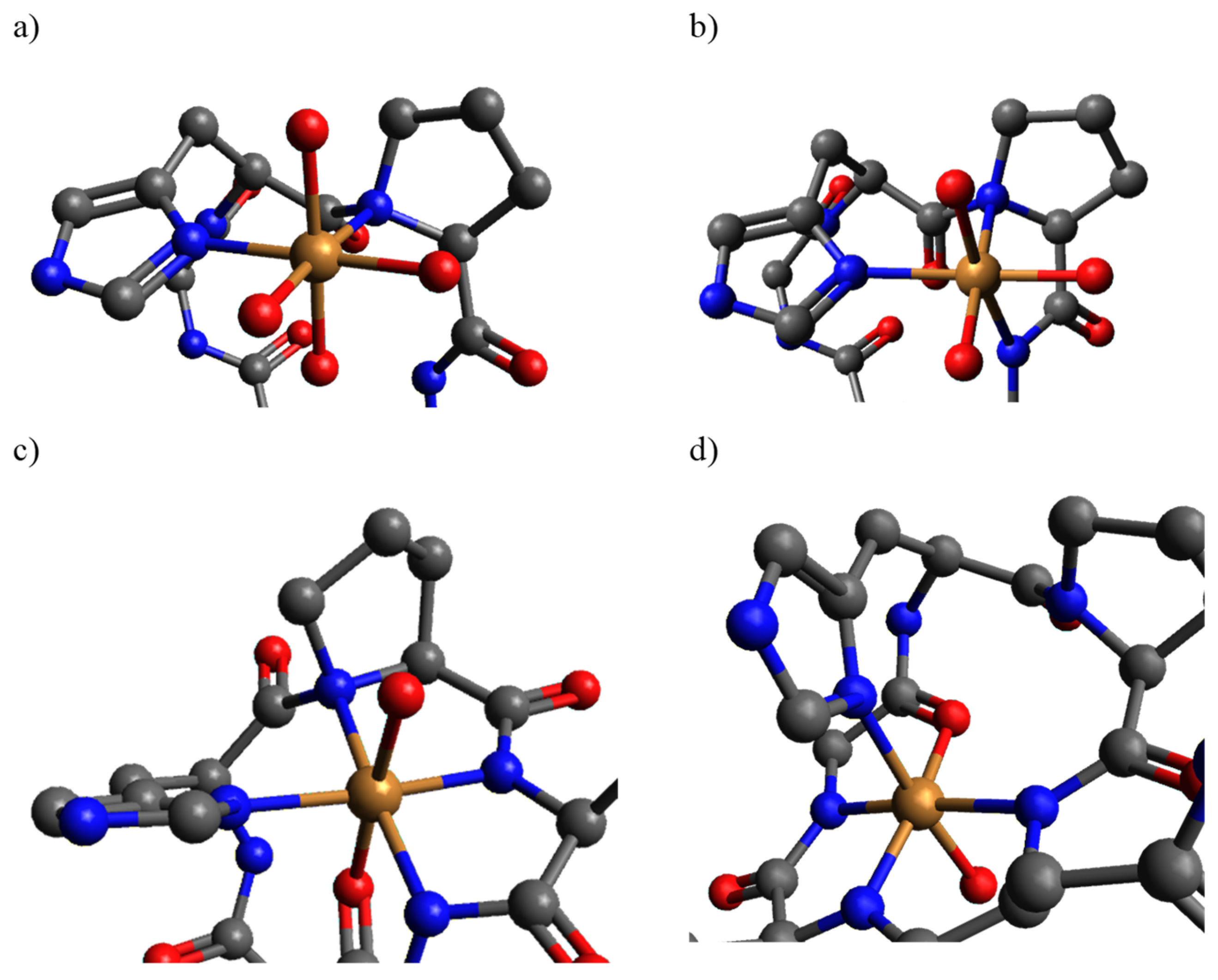
Scheme 2.
The proposed binding modes of (a) CuH2L, (b) CuL, (c) CuH-1L, and (d) CuH-2L visualized for L1−c(GlyHisProHisLys) peptide prepared by Avogadro Version 1.2.0.
Scheme 2.
The proposed binding modes of (a) CuH2L, (b) CuL, (c) CuH-1L, and (d) CuH-2L visualized for L1−c(GlyHisProHisLys) peptide prepared by Avogadro Version 1.2.0.

Figure 2.
The UV-Vis spectra for the solution with an equimolar quantity of (a) L1 and Cu(II) ions and (b) L2 and Cu(II) ions, at pH between 2.5 and 11.0. (c) The comparison of the experimental spectra for L1 at pH 4.0 (black) and L2 at pH 5.5 (red) obtained by the subtraction of the Cu(H2O)62+ spectrum. The CD spectra for the solution with an equimolar quantity of (d) L1−c(GlyHisProHisLys) and (e) L2−c(GlyHisGlyHisLys) with Cu(II) ions at pH between 2.5 and 11.0.

Figure 3.
The competition plot between L1−c(GlyHisProHisLys) and L2−c(GlyHisGlyHisLys) in the presence of Cu(II) ions under the equimolar conditions.
Figure 3.
The competition plot between L1−c(GlyHisProHisLys) and L2−c(GlyHisGlyHisLys) in the presence of Cu(II) ions under the equimolar conditions.

Scheme 3.
The characteristic HXXH motif presence in the investigated peptide.


Table 1.
The protonation constants and stability constants for ligands L1—c(GlyHisProHisLys) and L2—c(GlyHisGlyHisLys) calculated by fitting potentiometric curves by HYPERQUAD.
Table 1.
The protonation constants and stability constants for ligands L1—c(GlyHisProHisLys) and L2—c(GlyHisGlyHisLys) calculated by fitting potentiometric curves by HYPERQUAD.
| L1 | L2 | |||
|---|---|---|---|---|
| logβ | logK | logβ | logK | |
| HL | 9.79 ± 0.04 | 9.79 | 9.63 ± 0.05 | 9.63 |
| H2L | 16.53 ± 0.06 | 6.74 | 16.71 ± 0.08 | 7.08 |
| H3L | 21.89 ± 0.06 | 5.36 | 22.53 ± 0.08 | 5.82 |
| CuH2L | 21.06 ± 0.05 | 4.42 | 20.31 ± 0.07 | − |
| CuHL | 16.64 ± 0.03 | 6.82 | − | − |
| CuL | 9.82 ± 0.05 | 7.25 | 8.51 ± 0.06 | 6.90 |
| CuH-1L | 2.57 ± 0.04 | 8.88 | 1.61 ± 0.05 | 9.13 |
| CuH-2L | −6.31 ± 0.04 | 9.53 | −7.52 ± 0.07 | 9.87 |
| CuH-3L | −15.84 ± 0.05 | 10.98 | −17.39 ± 0.07 | 10.40 |
| CuH-4L | −26.82 ± 0.05 | − | −27.79 ± 0.07 | − |
Table 2.
Calculated value of logβ* independent of the protonation state of the selected ligands.
| logβ Independent of the Protonation State of the Ligand | |||||
|---|---|---|---|---|---|
| Ligand | 1NIm | 2NIm | 1Nam | 2Nam | 3Nam |
| Ac-HGGH-NH2 [32] | − | 5.97 | − | −7.88 | −16.10 |
| Ac-HAAH-NH2 [32] | − | 6.08 | −0.93 | −8.05 | −16.39 |
| Ac-HVVH-NH2 [33] | − | 5.79 | −1.05 | −9.29 | −17.60 |
| c(HGKHP)—L1 | 4.54 | 6.85 | 0.03 | −7.22 | −15.84 |
| c(HGGHGG) [22] | 3.63 | 5.45 | −1.40 | −8.00 | − |
| logβ* = logβCuHxL − logβHxL | |||||
Publisher’s Note: MDPI stays neutral with regard to jurisdictional claims in published maps and institutional affiliations. |
© 2021 by the authors. Licensee MDPI, Basel, Switzerland. This article is an open access article distributed under the terms and conditions of the Creative Commons Attribution (CC BY) license (https://creativecommons.org/licenses/by/4.0/).
Share and Cite
MDPI and ACS Style
Pieniężna, A.; Kotynia, A.; Brasuń, J. The Unusual Role of Pro in Cu(II) Binding by His2-Cyclopentapeptide. Int. J. Mol. Sci. 2021, 22, 6628. https://0-doi-org.brum.beds.ac.uk/10.3390/ijms22126628
AMA Style
Pieniężna A, Kotynia A, Brasuń J. The Unusual Role of Pro in Cu(II) Binding by His2-Cyclopentapeptide. International Journal of Molecular Sciences. 2021; 22(12):6628. https://0-doi-org.brum.beds.ac.uk/10.3390/ijms22126628
Chicago/Turabian StylePieniężna, Aleksandra, Aleksandra Kotynia, and Justyna Brasuń. 2021. "The Unusual Role of Pro in Cu(II) Binding by His2-Cyclopentapeptide" International Journal of Molecular Sciences 22, no. 12: 6628. https://0-doi-org.brum.beds.ac.uk/10.3390/ijms22126628
Note that from the first issue of 2016, this journal uses article numbers instead of page numbers. See further details here.