
The Potential Role of REG Family Proteins in Inflammatory and Inflammation-Associated Diseases of the Gastrointestinal Tract

 ,
,
Abstract
:1. Introduction
2. Background of the Reg Family
3. Regulation of Reg Family Genes
4. Reg and Inflammatory Diseases (GI Tract and Systemic Inflammation)
5. Involvement of Reg Family Proteins in Inflammation-Associated Carcinogenesis
6. Reg Family Proteins as Biological Markers of Inflammatory/Neoplastic Diseases

| Reg Family | Diseases | Findings | References |
|---|---|---|---|
| REG Iα | RA | The efficacy of infliximab treatment | [73] |
| REG Iα | Celiac disease | For diagnosis and monitor | [74] |
| REG Iα | Posttraumatic sepsis | Related to the severity of inflammation | [75] |
| REG Iα | Septic shock | Increased and associated with in-hospital mortality | [76] |
| REG Iα | SIRS | Increased at the onset of organ dysfunction | [77] |
| REG Iα | GC | Relevant to infiltrating pattern and function as prognostic factor | [80] |
| REG Iα | GC | Independent predictor of poor outcomes | [81] |
| REG Iα | Stage IV GC | Potent indicator for resistance to chemotherapy | [82] |
| REG I/REG IIIα | Early CRC | Indicator of adverse effect on survival | [86] |
| REG Iα/REG IIIα | Abdominal/cardiac surgery | Identification of septic complications | [78,79] |
| REG Iβ/ REG IIIα | CRC | Predictor of favorable prognosis | [85] |
| REG IIIα | Gastrointestinal GVHD | Prediction of response to therapy and 1-year survival following HCT | [22] |
| REG IIIα | Mucosal enteropathies | Useful marker to distinguish mucosal enteropathies from IBS | [90] |
| REG IIIα | CD | Increased in active conditions | [91] |
| REG IV | GC | Predictor of resistance to 5-FU-based chemotherapy | [83] |
| REG IV | GC | Prognostic indicator and early diagnosis of GC | [84] |
| REG IV | Advanced CRC | Prognosticator of poor survival with liver metastasis | [87] |
| REG IV | CRC | Crucial modulator of sensitivity and specificity to radiotherapy | [88,89] |
| REG IV | UC | Upregulation in remitting mucosa | [92] |
7. Conclusions
Author Contributions
Funding
Institutional Review Board Statement
Informed Consent Statement
Data Availability Statement
Conflicts of Interest
Abbreviations
| REG | regenerating gene in human |
| Reg | regenerating gene in mouse/rat |
| GI | gastrointestinal |
| IBD | inflammatory bowel disease |
| RA | rheumatoid arthritis |
| ECL | enterochromaffin-like |
| NF-κB | nuclear factor kappa B |
| TNF-α | tumor necrosis factor-α |
| UC | ulcerative colitis |
| EXTL3 | exostoses-like gene 3 |
| ATF-2 | activating transcription factor-2 |
| CRC | colorectal cancer |
| GPR37 | G protein-coupled receptor 37 |
| STAT3 | signal transducer and activator of transcription 3 |
| CDX2 | caudal type homeobox 2 |
| CD | Crohn’s disease |
| Th2 | T-helper type 2 |
| DSS | dextran sulfate sodium |
| GATA6 | GATA DNA-binding protein 6 |
| ERK | extracellular signal-regulated kinase |
| MAPK | mitogen-activated protein kinase |
| MZF1 | myeloid zinc finger 1 |
| RTEF1 | related transcriptional enhancer factor-1 |
| HLTF | helicase-like transcription factor |
| bFGF | basic fibroblast growth factor |
| HGF | hepatocyte growth factor |
| AP1 | activator protein-1 |
| EGFR | epidermal growth factor receptor |
| GSK-3β | glycogen synthase kinase 3β |
| TCF4 | T-cell factor 4 |
| SIRS | systemic inflammatory response syndrome |
| GVHD | graft-versus-host disease |
| HCT | hematopoietic cell transplantation |
| GC | gastric cancer |
| IBS | irritable bowel syndrome |
References
- Edwards, J.A.; Tan, N.; Toussaint, N.; Ou, P.; Mueller, C.; Stanek, A.; Zinsou, V.; Roudnitsky, S.; Sagal, M.; Dresner, L.; et al. Role of regenerating islet-derived proteins in inflammatory bowel disease. World J. Gastroenterol. 2020, 26, 2702–2714. [Google Scholar] [CrossRef]
- Dieckgraefe, B.K.; Crimmins, D.L.; Landt, V.; Houchen, C.; Anant, S.; Porche-Sorbet, R.; Ladenson, J.H. Expression of the regenerating gene family in inflammatory bowel disease mucosa: Reg Iα upregulation, processing, and antiapoptotic activity. J. Investig. Med. 2002, 50, 421–434. [Google Scholar] [CrossRef] [PubMed]
- Ho, M.R.; Lou, Y.C.; Wei, S.Y.; Luo, S.C.; Lin, W.C.; Lyu, P.C.; Chen, C. Human RegIV protein adopts a typical C-type lectin fold but binds mannan with two calcium-independent sites. J. Mol. Biol. 2010, 402, 682–695. [Google Scholar] [CrossRef]
- Lehotzky, R.E.; Partch, C.L.; Mukherjee, S.; Cash, H.L.; Goldman, W.E.; Gardner, K.H.; Hooper, L.V. Molecular basis for peptidoglycan recognition by a bactericidal lectin. Proc. Natl. Acad. Sci. USA 2010, 107, 7722–7727. [Google Scholar] [CrossRef] [Green Version]
- De Reggi, M.; Gharib, B. Protein-X, pancreatic stone-, pancreatic thread-, reg-protein, P19, lithostathine, and now what? Characterization, structural analysis and putative function(s) of the major non-enzymatic protein of pancreatic secretions. Curr. Protein Pept. Sci. 2001, 2, 19–42. [Google Scholar] [CrossRef] [PubMed]
- Graf, R.; Schiesser, M.; Scheele, G.A.; Marquardt, K.; Frick, T.W.; Ammann, R.W.; Bimmler, D. A family of 16-kDa pancreatic secretory stress proteins form highly organized fibrillar structures upon tryptic activation. J. Biol. Chem. 2001, 276, 21028–21038. [Google Scholar] [CrossRef] [Green Version]
- Sekikawa, A.; Fukui, H.; Suzuki, K.; Karibe, T.; Fujii, S.; Ichikawa, K.; Tomita, S.; Imura, J.; Shiratori, K.; Chiba, T.; et al. Involvement of the IL-22/REG Iα axis in ulcerative colitis. Lab. Invest. 2010, 90, 496–505. [Google Scholar] [CrossRef] [Green Version]
- Sun, C.; Fukui, H.; Hara, K.; Kitayama, Y.; Eda, H.; Yang, M.; Yamagishi, H.; Tomita, T.; Oshima, T.; Watari, J.; et al. Expression of Reg family genes in the gastrointestinal tract of mice treated with indomethacin. Am. J. Physiol. Gastrointest. Liver Physiol. 2015, 308, G736–G744. [Google Scholar] [CrossRef] [Green Version]
- Hakata, Y.; Fukui, H.; Sekikawa, A.; Yamagishi, H.; Ichikawa, K.; Tomita, S.; Imura, J.; Kawamata, H.; Imai, Y.; Fujimori, T. Expression of β-catenin and REG Iα in relation to cell proliferative ability in salivary gland tumors. Exp. Ther. Med. 2010, 1, 437–443. [Google Scholar] [CrossRef] [PubMed] [Green Version]
- Takasawa, S. Regenerating gene (REG) product and its potential clinical usage. Expert Opin. Ther. Targets 2016, 20, 541–550. [Google Scholar] [CrossRef]
- Terazono, K.; Yamamoto, H.; Takasawa, S.; Shiga, K.; Yonemura, Y.; Tochino, Y.; Okamoto, H. A novel gene activated in regenerating islets. J. Biol. Chem. 1988, 263, 2111–2114. [Google Scholar] [CrossRef]
- Watanabe, T.; Yonemura, Y.; Yonekura, H.; Suzuki, Y.; Miyashita, H.; Sugiyama, K.; Moriizumi, S.; Unno, M.; Tanaka, O.; Kondo, H.; et al. Pancreatic beta-cell replication and amelioration of surgical diabetes by Reg protein. Proc. Natl. Acad. Sci. USA 1994, 91, 3589–3592. [Google Scholar] [CrossRef] [Green Version]
- Parikh, A.; Stephan, A.F.; Tzanakakis, E.S. Regenerating proteins and their expression, regulation and signaling. Biomol. Concepts 2012, 3, 57–70. [Google Scholar] [CrossRef] [PubMed] [Green Version]
- Tezel, E.; Nagasaka, T.; Tezel, G.; Kaneko, T.; Takasawa, S.; Okamoto, H.; Nakao, A. REG I as a marker for human pancreatic acinoductular cells. Hepato-Gastroenterol. 2004, 51, 91–96. [Google Scholar]
- Zenilman, M.E.; Magnuson, T.H.; Swinson, K.; Egan, J.; Perfetti, R.; Shuldiner, A.R. Pancreatic thread protein is mitogenic to pancreatic-derived cells in culture. Gastroenterology 1996, 110, 1208–1214. [Google Scholar] [CrossRef]
- Asahara, M.; Mushiake, S.; Shimada, S.; Fukui, H.; Kinoshita, Y.; Kawanami, C.; Watanabe, T.; Tanaka, S.; Ichikawa, A.; Uchiyama, Y.; et al. Reg gene expression is increased in rat gastric enterochromaffin-like cells following water immersion stress. Gastroenterology 1996, 111, 45–55. [Google Scholar] [CrossRef] [PubMed]
- Fukui, H.; Kinoshita, Y.; Maekawa, T.; Okada, A.; Waki, S.; Hassan, S.; Okamoto, H.; Chiba, T. Regenerating gene protein may mediate gastric mucosal proliferation induced by hypergastrinemia in rats. Gastroenterology 1998, 115, 1483–1493. [Google Scholar] [CrossRef] [Green Version]
- Unno, M.; Yonekura, H.; Nakagawara, K.; Watanabe, T.; Miyashita, H.; Moriizumi, S.; Okamoto, H.; Itoh, T.; Teraoka, H. Structure, chromosomal localization, and expression of mouse reg genes, reg I and reg II. A novel type of reg gene, reg II, exists in the mouse genome. J. Biol. Chem. 1993, 268, 15974–15982. [Google Scholar] [CrossRef]
- Ortiz, E.M.; Dusetti, N.J.; Vasseur, S.; Malka, D.; Bödeker, H.; Dagorn, J.C.; Iovanna, J.L. The pancreatitis-associated protein is induced by free radicals in AR4-2J cells and confers cell resistance to apoptosis. Gastroenterology 1998, 114, 808–816. [Google Scholar] [CrossRef]
- Malka, D.; Vasseur, S.; Bödeker, H.; Ortiz, E.M.; Dusetti, N.J.; Verrando, P.; Dagorn, J.C.; Iovanna, J.L. Tumor necrosis factor alpha triggers antiapoptotic mechanisms in rat pancreatic cells through pancreatitis-associated protein I activation. Gastroenterology 2000, 119, 816–828. [Google Scholar] [CrossRef]
- Vasseur, S.; Folch-Puy, E.; Hlouschek, V.; Garcia, S.; Fiedler, F.; Lerch, M.M.; Dagorn, J.C.; Closa, D.; Iovanna, J.L. p8 improves pancreatic response to acute pancreatitis by enhancing the expression of the anti-inflammatory protein pancreatitis-associated protein I. J. Biol. Chem. 2004, 279, 7199–7207. [Google Scholar] [CrossRef] [Green Version]
- Ferrara, J.L.; Harris, A.C.; Greenson, J.K.; Braun, T.M.; Holler, E.; Teshima, T.; Levine, J.E.; Choi, S.W.; Huber, E.; Landfried, K.; et al. Regenerating islet-derived 3-alpha is a biomarker of gastrointestinal graft-versus-host disease. Blood 2011, 118, 6702–6708. [Google Scholar] [CrossRef] [PubMed] [Green Version]
- Zhao, D.; Kim, Y.H.; Jeong, S.; Greenson, J.K.; Chaudhry, M.S.; Hoepting, M.; Anderson, E.R.; van den Brink, M.R.M.; Peled, J.U.; Gomes, A.L.C.; et al. Survival signal REG3α prevents crypt apoptosis to control acute gastrointestinal graft-versus-host disease. J. Clin. Invest. 2018, 128, 4970–4979. [Google Scholar] [CrossRef] [Green Version]
- Hartupee, J.C.; Zhang, H.; Bonaldo, M.F.; Soares, M.B.; Dieckgraefe, B.K. Isolation and characterization of a cDNA encoding a novel member of the human regenerating protein family: Reg IV. Biochim. Biophys. Acta. 2001, 1518, 287–293. [Google Scholar] [CrossRef]
- Li, F.Y.; Ren, X.B.; Xu, E.P.; Huang, Q.; Sheng, H.Q.; Lv, B.J.; Lai, M.D. RegIV expression showing specificity to gastrointestinal tract and its potential role in diagnosing digestive tract neuroendocrine tumor. J. Zhejiang Univ. Sci. B. 2010, 11, 258–266. [Google Scholar] [CrossRef] [PubMed] [Green Version]
- Numata, M.; Oshima, T. Significance of regenerating islet-derived type IV gene expression in gastroenterological cancers. World J. Gastroenterol. 2012, 18, 3502–3510. [Google Scholar] [CrossRef]
- Kobayashi, S.; Akiyama, T.; Nata, K.; Abe, M.; Tajima, M.; Shervani, N.J.; Unno, M.; Matsuno, S.; Sasaki, H.; Takasawa, S.; et al. Identification of a receptor for reg (regenerating gene) protein, a pancreatic beta-cell regeneration factor. J. Biol. Chem. 2000, 275, 10723–10726. [Google Scholar] [CrossRef] [PubMed] [Green Version]
- Takasawa, S.; Ikeda, T.; Akiyama, T.; Nata, K.; Nakagawa, K.; Shervani, N.J.; Noguchi, N.; Murakami-Kawaguchi, S.; Yamauchi, A.; Takahashi, I.; et al. Cyclin D1 activation through ATF-2 in Reg-induced pancreatic beta-cell regeneration. FEBS. Lett. 2006, 580, 585–591. [Google Scholar] [CrossRef] [Green Version]
- Acquatella-Tran Van Ba, I.; Marchal, S.; Francois, F.; Silhol, M.; Lleres, C.; Michel, B.; Benyamin, Y.; Verdie, J.M.; Trousse, F.; Marcilhac, A. Regenerating islet-derived 1α (Reg-1α) protein is new neuronal secreted factor that stimulates neurite outgrowth via exostosin Tumor-like 3 (EXTL3) receptor. J. Biol. Chem. 2012, 287, 4726–4739. [Google Scholar] [CrossRef] [Green Version]
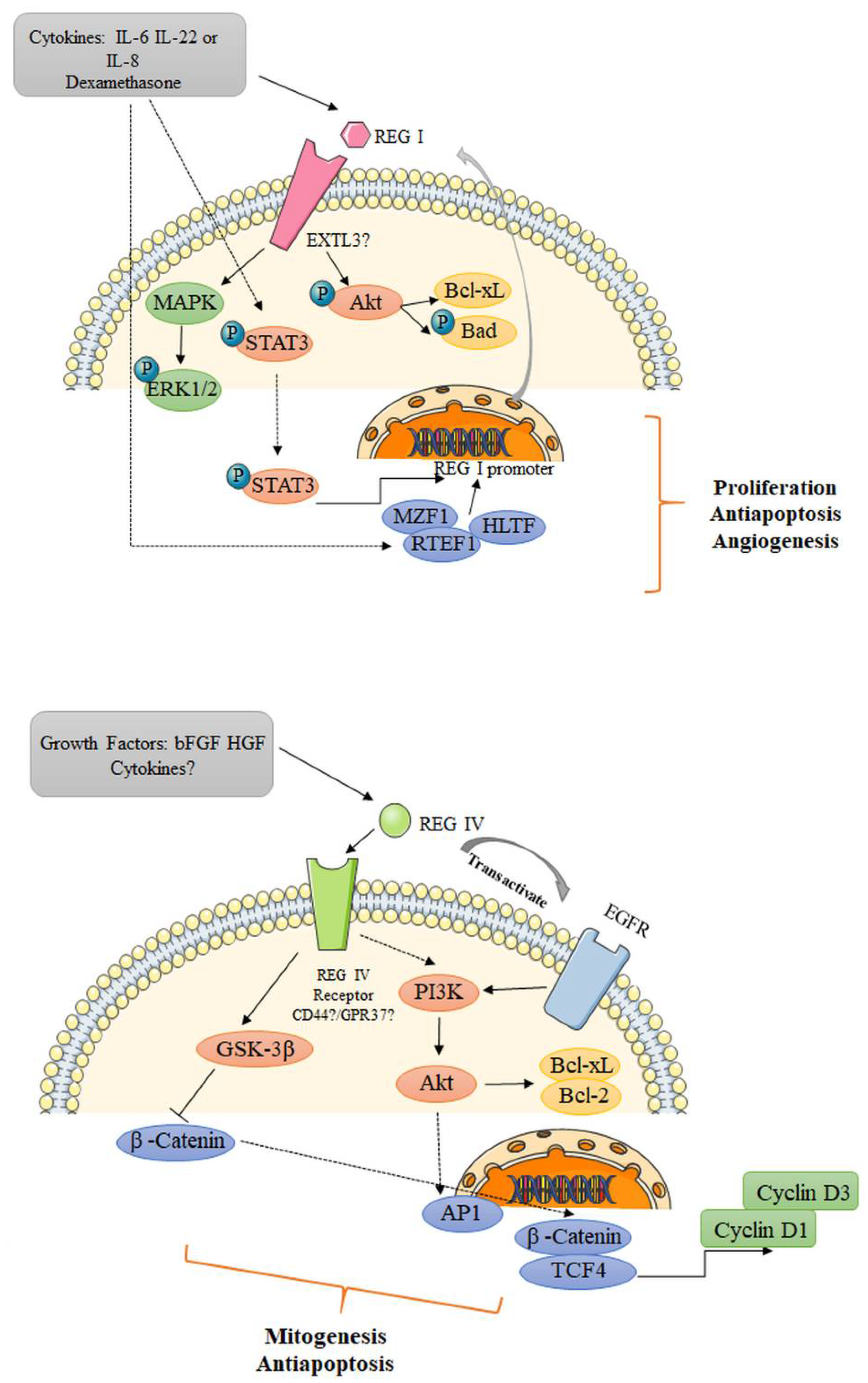
- Bishnupuri, K.S.; Luo, Q.; Murmu, N.; Houchen, C.W.; Anant, S.; Dieckgraefe, B.K. Reg IV activates the epidermal growth factor receptor/Akt/AP-1 signaling pathway in colon adenocarcinomas. Gastroenterology 2006, 130, 137–149. [Google Scholar] [CrossRef] [PubMed]
- Wang, H.; Hu, L.; Zang, M.; Zhang, B.; Duan, Y.; Fan, Z.; Li, J.; Su, L.; Yan, M.; Zhu, Z.; et al. REG4 promotes peritoneal metastasis of gastric cancer through GPR37. Oncotarget 2016, 7, 27874–27888. [Google Scholar] [CrossRef] [Green Version]
- Akiyama, T.; Takasawa, S.; Nata, K.; Kobayashi, S.; Abe, M.; Shervani, N.J.; Ikeda, T.; Nakagawa, K.; Unno, M.; Matsuno, S.; et al. Activation of Reg gene, a gene for insulin-producing beta -cell regeneration: Poly(ADP-ribose) polymerase binds Reg promoter and regulates the transcription by autopoly(ADP-ribosyl)ation. Proc. Natl. Acad. Sci. USA 2001, 98, 48–53. [Google Scholar] [CrossRef] [PubMed]
- Dusetti, N.J.; Mallo, G.V.; Ortiz, E.M.; Keim, V.; Dagorn, J.C.; Iovanna, J.L. Induction of lithostathine/reg mRNA expression by serum from rats with acute pancreatitis and cytokines in pancreatic acinar AR-42J cells. Arch. Biochem. Biophys. 1996, 330, 129–132. [Google Scholar] [CrossRef]
- Kazumori, H.; Ishihara, S.; Hoshino, E.; Kawashima, K.; Moriyama, N.; Suetsugu, H.; Sato, H.; Adachi, K.; Fukuda, R.; Watanabe, S.; et al. Neutrophil chemoattractant 2 beta regulates expression of the Reg gene in injured gastric mucosa in rats. Gastroenterology 2000, 119, 1610–1622. [Google Scholar] [CrossRef] [PubMed]
- Yoshino, N.; Ishihara, S.; Rumi, M.A.; Ortega-Cava, C.F.; Yuki, T.; Kazumori, H.; Takazawa, S.; Okamoto, H.; Kadowaki, Y.; Kinoshita, Y. Interleukin-8 regulates expression of Reg protein in Helicobacter pylori-infected gastric mucosa. Am. J. Gastroenterol. 2005, 100, 2157–2166. [Google Scholar] [CrossRef] [PubMed]
- Aggarwal, S.; Xie, M.H.; Maruoka, M.; Foster, J.; Gurney, A.L. Acinar cells of the pancreas are a target of interleukin-22. J. Interferon Cytokine Res. 2001, 21, 1047–1053. [Google Scholar] [CrossRef] [PubMed]
- Folch-Puy, E.; Granell, S.; Dagorn, J.C.; Iovanna, J.L.; Closa, D. Pancreatitis-associated protein I suppresses NF-kappa B activation through a JAK/STAT-mediated mechanism in epithelial cells. J. Immunol. 2006, 176, 3774–3779. [Google Scholar] [CrossRef] [PubMed] [Green Version]
- Sekikawa, A.; Fukui, H.; Fujii, S.; Ichikawa, K.; Tomita, S.; Imura, J.; Chiba, T.; Fujimori, T. REG Iα protein mediates an anti-apoptotic effect of STAT3 signaling in gastric cancer cells. Carcinogenesis 2008, 29, 76–83. [Google Scholar] [CrossRef] [Green Version]
- Liu, X.; Wang, J.; Wang, H.; Yin, G.; Liu, Y.; Lei, X.; Xiang, M. REG3A accelerates pancreatic cancer cell growth under IL-6-associated inflammatory condition: Involvement of a REG3A-JAK2/STAT3 positive feedback loop. Cancer Lett. 2015, 362, 45–60. [Google Scholar] [CrossRef]
- Naito, Y.; Oue, N.; Hinoi, T.; Sakamoto, N.; Sentani, K.; Ohdan, H.; Yanagihara, K.; Sasaki, H.; Yasui, W. Reg IV is a direct target of intestinal transcriptional factor CDX2 in gastric cancer. PLoS ONE 2012, 7, e47545. [Google Scholar] [CrossRef] [Green Version]
- Strober, W.; Fuss, I.J. Proinflammatory cytokines in the pathogenesis of inflammatory bowel diseases. Gastroenterology 2011, 140, 1756–1767. [Google Scholar] [CrossRef] [PubMed] [Green Version]
- Ogawa, H.; Fukushima, K.; Naito, H.; Funayama, Y.; Unno, M.; Takahashi, K.; Kitayama, T.; Matsuno, S.; Ohtani, H.; Takasawa, S.; et al. Increased expression of HIP/PAP and regenerating gene III in human inflammatory bowel disease and a murine bacterial reconstitution model. Inflamm. Bowel Dis. 2003, 9, 162–170. [Google Scholar] [CrossRef]
- Xu, X.; Fukui, H.; Ran, Y.; Wang, X.; Inoue, Y.; Ebisudani, N.; Nishimura, H.; Tomita, T.; Oshima, T.; Watari, J.; et al. The link between type III Reg and STAT3-associated cytokines in inflamed colonic tissues. Mediat. Inflamm. 2019, 2019, 7859460. [Google Scholar] [CrossRef] [PubMed] [Green Version]
- Darnaud, M.; Dos Santos, A.; Gonzalez, P.; Augui, S.; Lacoste, C.; Desterke, C.; De Hertogh, G.; Valentino, E.; Braun, E.; Zheng, J.; et al. Enteric delivery of regenerating family member 3 alpha alters the intestinal microbiota and controls inflammation in mice with colitis. Gastroenterology 2018, 154, 1009–1023. [Google Scholar] [CrossRef] [PubMed] [Green Version]
- Shindo, R.; Katagiri, T.; Komazawa-Sakon, S.; Ohmuraya, M.; Takeda, W.; Nakagawa, Y.; Nakagata, N.; Sakuma, T.; Yamamoto, T.; Nishiyama, C.; et al. Regenerating islet-derived protein (Reg)3β plays a crucial role in attenuation of ileitis and colitis in mice. Biochem. Biophys. Rep. 2020, 21, 100738. [Google Scholar] [CrossRef]
- Bishnupuri, K.S.; Sainathan, S.K.; Bishnupuri, K.; Leahy, D.R.; Luo, Q.; Anant, S.; Houchen, C.W.; Dieckgraefe, B.K. Reg4-induced mitogenesis involves Akt-GSK3β-β-Catenin-TCF-4 signaling in human colorectal cancer. Mol. Carcinog. 2014, 53 (Suppl. 1), E169–E180. [Google Scholar] [CrossRef] [Green Version]
- Nanakin, A.; Fukui, H.; Fujii, S.; Sekikawa, A.; Kanda, N.; Hisatsune, H.; Seno, H.; Konda, Y.; Fujimori, T.; Chiba, T. Expression of the REG IV gene in ulcerative colitis. Lab. Invest. 2007, 87, 304–314. [Google Scholar] [CrossRef] [PubMed]
- Tsuchida, C.; Sakuramoto-Tsuchida, S.; Takeda, M.; Itaya-Hironaka, A.; Yamauchi, A.; Misu, M.; Shobatake, R.; Uchiyama, T.; Makino, M.; Pujol-Autonell, I.; et al. Expression of REG family genes in human inflammatory bowel diseases and its regulation. Biochem. Biophys. Rep. 2017, 12, 198–205. [Google Scholar] [CrossRef]
- Takasawa, S.; Tsuchida, C.; Sakuramoto-Tsuchida, S.; Takeda, M.; Itaya-Hironaka, A.; Yamauchi, A.; Misu, M.; Shobatake, R.; Uchiyama, T.; Makino, M.; et al. Expression of human REG family genes in inflammatory bowel disease and their molecular mechanism. Immunol. Res. 2018, 66, 800–805. [Google Scholar] [CrossRef]
- Fukui, H.; Franceschi, F.; Penland, R.L.; Sakai, T.; Sepulveda, A.R.; Fujimori, T.; Terano, A.; Chiba, T.; Genta, R.M. Effects of Helicobacter pylori infection on the link between regenerating gene expression and serum gastrin levels in Mongolian gerbils. Lab. Invest. 2003, 83, 1777–1786. [Google Scholar] [CrossRef] [Green Version]
- Kitayama, Y.; Fukui, H.; Hara, K.; Eda, H.; Kodani, M.; Yang, M.; Sun, C.; Yamagishi, H.; Tomita, T.; Oshima, T.; et al. Role of regenerating gene I in claudin expression and barrier function in the small intestine. Transl. Res. 2016, 173, 92–100. [Google Scholar] [CrossRef]
- Peterson, K.M.; Guo, X.; Elkahloun, A.G.; Mondal, D.; Bardhan, P.K.; Sugawara, A.; Duggal, P.; Haque, R.; Petri, W.A. The expression of REG 1A and REG 1B is increased during acute amebic colitis. Parasitol. Int. 2011, 60, 296–300. [Google Scholar] [CrossRef] [Green Version]
- van Ampting, M.T.; Loonen, L.M.; Schonewille, A.J.; Konings, I.; Vink, C.; Iovanna, J.; Chamaillard, M.; Dekker, J.; van der Meer, R.; Wells, J.M.; et al. Intestinally secreted C-type lectin Reg3b attenuates salmonellosis but not listeriosis in mice. Infect. Immun. 2012, 80, 1115–1120. [Google Scholar] [CrossRef] [Green Version]
- Zheng, Y.; Valdez, P.A.; Danilenko, D.M.; Hu, Y.; Sa, S.M.; Gong, Q.; Abbas, A.R.; Modrusan, Z.; Ghilardi, N.; de Sauvage, F.J.; et al. Interleukin-22 mediates early host defense against attaching and effacing bacterial pathogens. Nat. Med. 2008, 14, 282–289. [Google Scholar] [CrossRef]
- Sasaki, N.; Sachs, N.; Wiebrands, K.; Ellenbroek, S.I.; Fumagalli, A.; Lyubimova, A.; Begthel, H.; van den Born, M.; van Es, J.H.; Karthaus, W.R.; et al. Reg4+ deep crypt secretory cells function as epithelial niche for Lgr5+ stem cells in colon. Proc. Natl. Acad. Sci. USA 2016, 113, E5399–E5407. [Google Scholar] [CrossRef] [Green Version]
- Qi, H.; Wei, J.; Gao, Y.; Yang, Y.; Li, Y.; Zhu, H.; Su, L.; Su, X.; Zhang, Y.; Yang, R. Reg4 and complement factor D prevent the overgrowth of E. coli in the mouse gut. Commun. Biol. 2020, 3, 483. [Google Scholar] [CrossRef] [PubMed]
- Fujishiro, M.; Nozawa, K.; Kawasaki, M.; Yamaguchi, A.; Iwabuchi, K.; Yanagida, M.; Suzuki, F.; Miyazawa, K.; Fukui, H.; Kaneko, K.; et al. Regenerating gene (REG) 1 alpha promotes pannus progression in patients with rheumatoid arthritis. Mod. Rheumatol. 2012, 22, 228–237. [Google Scholar] [CrossRef] [PubMed]
- Kimura, T.; Fukui, H.; Sekikawa, A.; Yamagishi, H.; Ichikawa, K.; Tomita, S.; Fujii, S.; Imura, J.; Kawamata, H.; Chiba, T.; et al. Involvement of REG Iα protein in the regeneration of ductal epithelial cells in the minor salivary glands of patients with Sjögren’s syndrome. Clin. Exp. Immunol. 2009, 155, 16–20. [Google Scholar] [CrossRef] [PubMed]
- Yoshimoto, K.; Fujimoto, T.; Itaya-Hironaka, A.; Miyaoka, T.; Sakuramoto-Tsuchida, S.; Yamauchi, A.; Takeda, M.; Kasai, T.; Nakagawara, K.; Nonomura, A.; et al. Involvement of autoimmunity to REG, a regeneration factor, in patients with primary Sjögren’s syndrome. Clin. Exp. Immunol. 2013, 174, 1–9. [Google Scholar] [CrossRef]
- Fujimura, T.; Fujimoto, T.; Itaya-Hironaka, A.; Miyaoka, T.; Yoshimoto, K.; Sakuramoto-Tsuchida, S.; Yamauchi, A.; Takeda, M.; Tsujinaka, H.; Tanaka, Y.; et al. Significance of interleukin-6/STAT pathway for the gene expression of REG Iα, a new autoantigen in Sjögren’s syndrome patients, in salivary duct epithelial cells. Clin. Rev. Allergy Immunol. 2017, 52, 351–363. [Google Scholar] [CrossRef] [PubMed]
- Wijnands, A.M.; de Jong, M.E.; Lutgens, M.; Hoentjen, F.; Elias, S.G.; Oldenburg, B. Dutch Initiative on Crohn and Colitis (ICC). Prognostic factors for advanced colorectal neoplasia in inflammatory bowel disease: Systematic review and meta-analysis. Gastroenterology 2021, 160, 1584–1598. [Google Scholar] [CrossRef]
- Nebbia, M.; Yassin, N.A.; Spinelli, A. Colorectal cancer in inflammatory bowel disease. Clin. Colon Rectal Surg. 2020, 33, 305–317. [Google Scholar] [CrossRef]
- Rabbenou, W.; Ullman, T.A. Risk of colon cancer and recommended surveillance strategies in patients with ulcerative colitis. Gastroenterol. Clin. North. Am. 2020, 49, 791–807. [Google Scholar] [CrossRef]
- Tanaka, H.; Fukui, H.; Fujii, S.; Sekikawa, A.; Yamagishi, H.; Ichikawa, K.; Tomita, S.; Imura, J.; Yasuda, Y.; Chiba, T.; et al. Immunohistochemical analysis of REG Iα expression in ulcerative colitis-associated neoplastic lesions. Digestion 2011, 83, 204–209. [Google Scholar] [CrossRef] [PubMed]
- Sekikawa, A.; Fukui, H.; Fujii, S.; Nanakin, A.; Kanda, N.; Uenoyama, Y.; Sawabu, T.; Hisatsune, H.; Kusaka, T.; Ueno, S.; et al. Possible role of REG Iα protein in ulcerative colitis and colitic cancer. Gut 2005, 54, 1437–1444. [Google Scholar] [CrossRef] [PubMed]
- Rafa, L.; Dessein, A.F.; Devisme, L.; Buob, D.; Truant, S.; Porchet, N.; Huet, G.; Buisine, M.; Lesuffleur, T. REG4 acts as a mitogenic, motility and pro-invasive factor for colon cancer cells. Int. J. Oncol. 2010, 36, 689–698. [Google Scholar]
- Zhang, Y.; Lai, M.; Lv, B.; Gu, X.; Wang, H.; Zhu, Y.; Zhu, Y.; Shao, L.; Wang, G. Overexpression of Reg IV in colorectal adenoma. Cancer Lett. 2003, 200, 69–76. [Google Scholar] [CrossRef]
- Kinoshita, Y.; Ishihara, S.; Kadowaki, Y.; Fukui, H.; Chiba, T. Reg protein is a unique growth factor of gastric mucosal cells. J. Gastroenterol. 2004, 39, 507–513. [Google Scholar] [CrossRef] [PubMed]
- Kadowaki, Y.; Ishihara, S.; Miyaoka, Y.; Rumi, M.A.; Sato, H.; Kazumori, H.; Adachi, K.; Takasawa, S.; Okamoto, H.; Chiba, T.; et al. Reg protein is overexpressed in gastric cancer cells, where it activates a signal transduction pathway that converges on ERK1/2 to stimulate growth. FEBS. Lett. 2002, 530, 59–64. [Google Scholar] [CrossRef]
- Fukui, H.; Fujii, S.; Takeda, J.; Kayahara, T.; Sekikawa, A.; Nanakin, A.; Suzuki, K.; Hisatsune, H.; Seno, H.; Sawada, M.; et al. Expression of REG Iα protein in human gastric cancers. Digestion 2004, 69, 177–184. [Google Scholar] [CrossRef]
- Sekikawa, A.; Fukui, H.; Fujii, S.; Takeda, J.; Nanakin, A.; Hisatsune, H.; Seno, H.; Takasawa, S.; Okamoto, H.; Fujimori, T.; et al. REG Iα protein may function as a trophic and/or anti-apoptotic factor in the development of gastric cancer. Gastroenterology 2005, 128, 642–653. [Google Scholar] [CrossRef]
- Hara, K.; Fukui, H.; Sun, C.; Kitayama, Y.; Eda, H.; Yamasaki, T.; Kondo, T.; Tomita, T.; Oshima, T.; Watari, J.; et al. Effect of REG Iα protein on angiogenesis in gastric cancer tissues. Oncol. Rep. 2015, 33, 2183–2189. [Google Scholar] [CrossRef] [PubMed] [Green Version]
- Sekigawa, I.; Yanagida, M.; Iwabuchi, K.; Kaneda, K.; Kaneko, H.; Takasaki, Y.; Jung, G.; Sone, S.; Tanaka, Y.; Ogawa, H.; et al. Protein biomarker analysis by mass spectrometry in patients with rheumatoid arthritis receiving anti-tumor necrosis factor-alpha antibody therapy. Clin. Exp. Rheumatol. 2008, 26, 261–267. [Google Scholar]
- Vives-Pi, M.; Takasawa, S.; Pujol-Autonell, I.; Planas, R.; Cabre, E.; Ojanguren, I.; Montraveta, M.; Santos, A.L.; Ruiz-Ortiz, E. Biomarkers for diagnosis and monitoring of celiac disease. J. Clin. Gastroenterol. 2013, 47, 308–313. [Google Scholar] [CrossRef]
- Keel, M.; Harter, L.; Reding, T.; Sun, L.K.; Hersberger, M.; Seifert, B.; Bimmler, D.; Graf, R. Pancreatic stone protein is highly increased during posttraumatic sepsis and activates neutrophil granulocytes. Crit. Care Med. 2009, 37, 1642–1648. [Google Scholar] [CrossRef] [PubMed]
- Que, Y.A.; Delodder, F.; Guessous, I.; Graf, R.; Bain, M.; Calandra, T.; Liaudet, L.; Eggimann, P. Pancreatic stone protein as an early biomarker predicting mortality in a prospective cohort of patients with sepsis requiring ICU management. Crit. Care 2012, 16, R114. [Google Scholar] [CrossRef] [Green Version]
- Jiri, Z.; Kyr, M.; Vavrina, M.; Fedora, M. Pancreatic stone protein—a possible biomarker of multiorgan failure and mortality in children sepsis. Cytokine 2014, 66, 106–111. [Google Scholar] [CrossRef]
- Fisher, O.M.; Oberkofler, C.E.; Raptis, D.A.; Soll, C.; Béchir, M.; Schiesser, M.; Graf, R. Pancreatic stone protein (PSP) and pancreatitis-associated protein (PAP): A protocol of a cohort study on the diagnostic efficacy and prognostic value of PSP and PAP as postoperative markers of septic complications in patients undergoing abdominal surgery (PSP study). BMJ Open 2014, 4, e004914. [Google Scholar] [PubMed]
- Klein, H.J.; Csordas, A.; Falk, V.; Slankamenac, K.; Rudiger, A.; Schönrath, F.; Rodriguez Cetina Biefer, H.; Starck, C.T.; Graf, R. Pancreatic stone protein predicts postoperative infection in cardiac surgery patients irrespective of cardiopulmonary bypass or surgical technique. PLoS ONE 2015, 10, e0120276. [Google Scholar] [CrossRef] [Green Version]
- Yonemura, Y.; Sakurai, S.; Yamamoto, H.; Endou, Y.; Kawamura, T.; Bandou, E.; Elnemr, A.; Sugiyama, K.; Sasaki, T.; Akiyama, T.; et al. REG gene expression is associated with the infiltrating growth of gastric carcinoma. Cancer 2003, 98, 1394–1400. [Google Scholar] [CrossRef]
- Yamagishi, H.; Fukui, H.; Sekikawa, A.; Kono, T.; Fujii, S.; Ichikawa, K.; Tomita, S.; Imura, J.; Hiraishi, H.; Chiba, T.; et al. Expression profile of REG family proteins REG Iα and REG IV in advanced gastric cancer: Comparison with mucin phenotype and prognostic markers. Mod. Pathol. 2009, 22, 906–913. [Google Scholar] [CrossRef] [Green Version]
- Sekikawa, A.; Fukui, H.; Zhang, X.; Maruo, T.; Tsumura, T.; Okabe, Y.; Wakasa, T.; Osaki, Y.; Chiba, T.; Tomita, T.; et al. REG Iα is a biomarker for predicting response to chemotherapy with S-1 plus cisplatin in patients with unresectable stage IV gastric cancer. Br. J. Cancer 2013, 108, 395–401. [Google Scholar] [CrossRef] [Green Version]
- Mitani, Y.; Oue, N.; Matsumura, S.; Yoshida, K.; Noguchi, T.; Ito, M.; Tanaka, S.; Kuniyasu, H.; Kamata, N.; Yasui, W. Reg IV is a serum biomarker for gastric cancer patients and predicts response to 5-fluorouracil-based chemotherapy. Oncogene 2007, 26, 4383–4393. [Google Scholar] [CrossRef] [Green Version]
- Tao, H.Q.; He, X.J.; Ma, Y.Y.; Wang, H.J.; Xia, Y.J.; Ye, Z.Y.; Zhao, Z.S. Evaluation of REG4 for early diagnosis and prognosis of gastric cancer. Hum. Pathol. 2011, 42, 1401–1409. [Google Scholar] [CrossRef]
- Zheng, H.C.; Sugawara, A.; Okamoto, H.; Takasawa, S.; Takahashi, H.; Masuda, S.; Takano, Y. Expression profile of the REG gene family in colorectal carcinoma. J. Histochem. Cytochem. 2011, 59, 106–115. [Google Scholar] [CrossRef] [Green Version]
- Macadam, R.C.; Sarela, A.I.; Farmery, S.M.; Robinson, P.A.; Markham, A.F.; Guillou, P.J. Death from early colorectal cancer is predicted by the presence of transcripts of the REG gene family. Br. J. Cancer 2000, 83, 188–195. [Google Scholar] [CrossRef] [PubMed] [Green Version]
- Oue, N.; Kuniyasu, H.; Noguchi, T.; Sentani, K.; Ito, M.; Tanaka, S.; Setoyama, T.; Sakakura, C.; Natsugoe, S.; Yasui, W. Serum concentration of Reg IV in patients with colorectal cancer: Overexpression and high serum levels of Reg IV are associated with liver metastasis. Oncology 2007, 72, 371–380. [Google Scholar] [CrossRef] [PubMed]
- Kobunai, T.; Watanabe, T.; Fukusato, T. REG4, NEIL2, and BIRC5 gene expression correlates with gamma-radiation sensitivity in patients with rectal cancer receiving radiotherapy. Anticancer Res. 2011, 31, 4147–4153. [Google Scholar] [PubMed]
- Bishnupuri, K.S.; Luo, Q.; Sainathan, S.K.; Kikuchi, K.; Sureban, S.M.; Sabarinathan, M.; Gross, J.H.; Aden, K.; May, R.; Houchen, C.W.; et al. Reg IV regulates normal intestinal and colorectal cancer cell susceptibility to radiation-induced apoptosis. Gastroenterology 2010, 138, 616–626, 626.e1–2. [Google Scholar] [CrossRef] [PubMed] [Green Version]
- Marafini, I.; Di Sabatino, A.; Zorzi, F.; Monteleone, I.; Sedda, S.; Cupi, M.L.; Antenucci, C.; Biancheri, P.; Giuffrida, P.; Di Stefano, M.; et al. Serum regenerating islet-derived 3-alpha is a biomarker of mucosal enteropathies. Aliment. Pharmacol. Ther. 2014, 40, 974–981. [Google Scholar] [CrossRef] [Green Version]
- Nunes, T.; Etchevers, M.J.; Sandi, M.J.; Pino Donnay, S.; Grandjean, T.; Pellisé, M.; Panés, J.; Ricart, E.; Iovanna, J.L.; Dagorn, J.C.; et al. Pancreatitis-associated protein does not predict disease relapse in inflammatory bowel disease patients. PLoS ONE 2014, 9, e84957. [Google Scholar] [CrossRef]
- Planell, N.; Lozano, J.J.; Mora-Buch, R.; Masamunt, M.C.; Jimeno, M.; Ordás, I.; Esteller, M.; Ricart, E.; Piqué, J.M.; Panés, J.; et al. Transcriptional analysis of the intestinal mucosa of patients with ulcerative colitis in remission reveals lasting epithelial cell alterations. Gut 2013, 62, 967–976. [Google Scholar] [CrossRef] [Green Version]


Publisher’s Note: MDPI stays neutral with regard to jurisdictional claims in published maps and institutional affiliations. |
© 2021 by the authors. Licensee MDPI, Basel, Switzerland. This article is an open access article distributed under the terms and conditions of the Creative Commons Attribution (CC BY) license (https://creativecommons.org/licenses/by/4.0/).
Share and Cite
Sun, C.; Wang, X.; Hui, Y.; Fukui, H.; Wang, B.; Miwa, H. The Potential Role of REG Family Proteins in Inflammatory and Inflammation-Associated Diseases of the Gastrointestinal Tract. Int. J. Mol. Sci. 2021, 22, 7196. https://0-doi-org.brum.beds.ac.uk/10.3390/ijms22137196
Sun C, Wang X, Hui Y, Fukui H, Wang B, Miwa H. The Potential Role of REG Family Proteins in Inflammatory and Inflammation-Associated Diseases of the Gastrointestinal Tract. International Journal of Molecular Sciences. 2021; 22(13):7196. https://0-doi-org.brum.beds.ac.uk/10.3390/ijms22137196
Chicago/Turabian StyleSun, Chao, Xiaoyu Wang, Yangyang Hui, Hirokazu Fukui, Bangmao Wang, and Hiroto Miwa. 2021. "The Potential Role of REG Family Proteins in Inflammatory and Inflammation-Associated Diseases of the Gastrointestinal Tract" International Journal of Molecular Sciences 22, no. 13: 7196. https://0-doi-org.brum.beds.ac.uk/10.3390/ijms22137196