
Protein Corona Hinders N-CQDs Oxidative Potential and Favors Their Application as Nanobiocatalytic System


Abstract
:1. Introduction
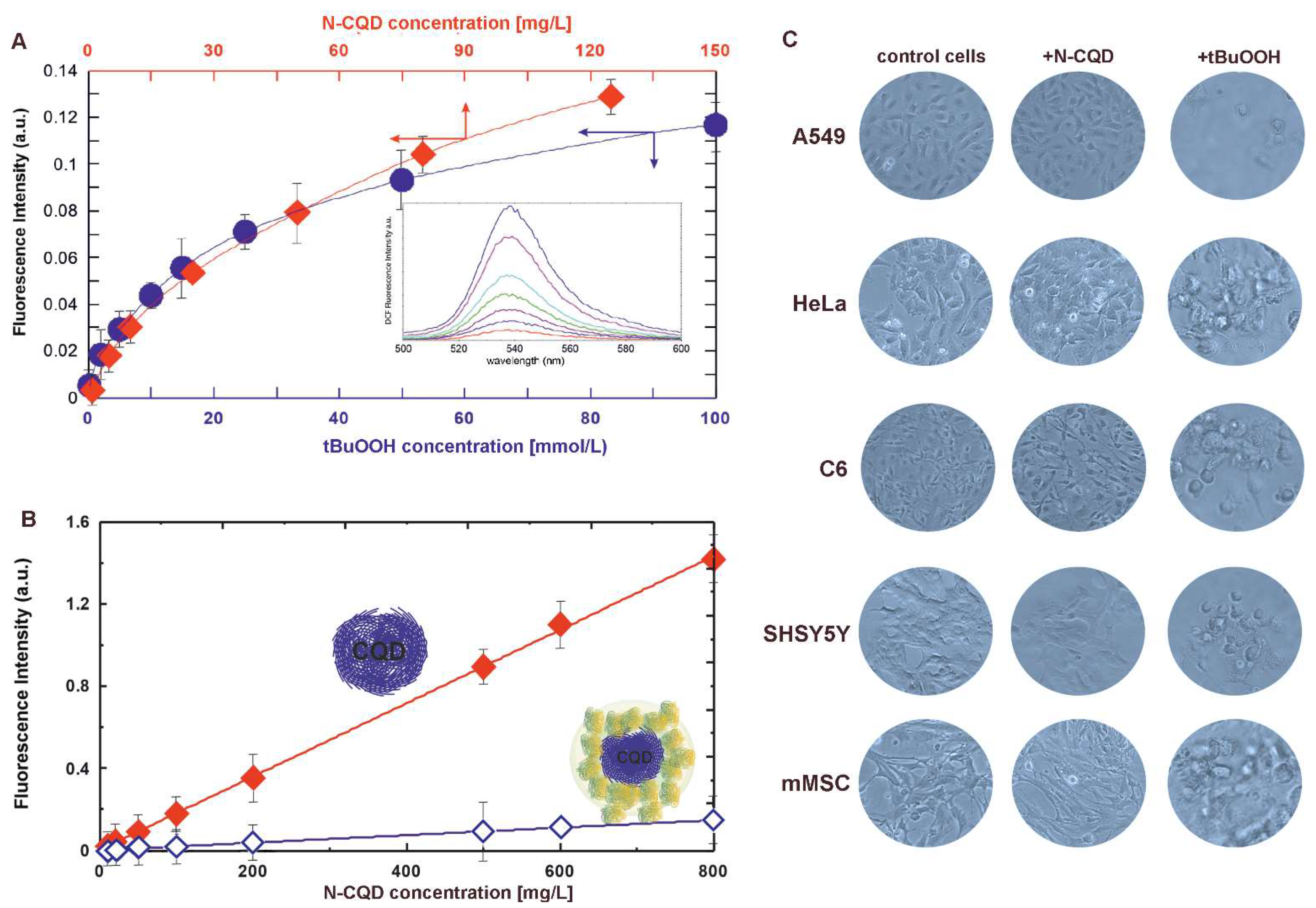
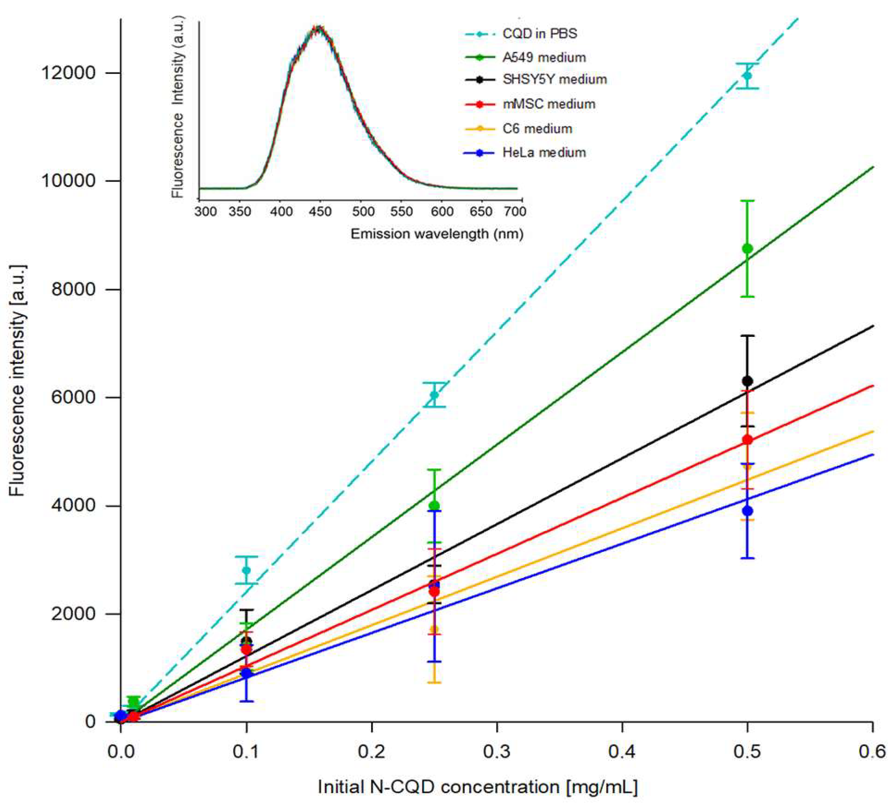
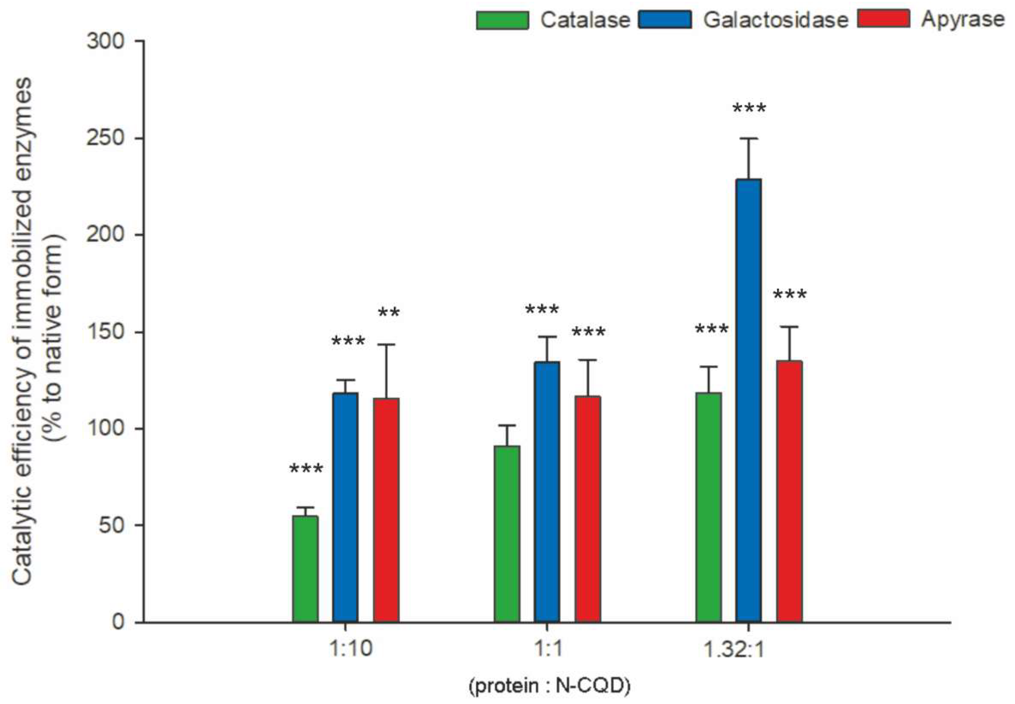
2. Results
N-CQD Influence on Oxidative Stress and Cell Viability
3. Discussion
4. Materials and Methods
4.1. N-CQD Preparation
4.2. In Vitro Cell Culture
4.3. Viability Assays
4.4. Quantification of N-CQDs Internalization by Cells
4.5. Oxidative Properties of N-CQDs
4.6. Determination of ROS Removal Capability
4.7. Enzymes Immobilization on Carbon Quantum Dots
4.8. Determination of Catalase Activity
4.9. Determination of β-D-Galactosidase Activity
4.10. Determination of Apyrase Activity
4.11. Statistical Analyses
5. Conclusions
Author Contributions
Funding
Institutional Review Board Statement
Informed Consent Statement
Conflicts of Interest
References
- Havrdova, M.; Hola, K.; Skopalik, J.; Tomankova, K.; Petr, M.; Cepe, K.; Polakova, K.; Tucek, J.; Bourlinos, A.B.; Zboril, R. Toxicity of carbon dots—Effect of surface functionalization on the cell viability, reactive oxygen species generation and cell cycle. Carbon 2016, 99, 238–248. [Google Scholar] [CrossRef]
- Ronzani, C.; Van Belle, C.; Didier, P.; Spiegelhalter, C.; Pierrat, P.; Lebeau, L.; Pons, F. Lysosome mediates toxicological effects of polyethyleneimine-based cationic carbon dots. J. Nanopart. Res. 2019, 21, 4. [Google Scholar] [CrossRef]
- Zhang, T.; Qu, J.; Yao, Y.; Zhang, Y.; Ma, Y.; Wu, D.; Cao, Y.; Yang, M.; Zhang, Y.; Tang, M. N-doped carbon dots triggered the induction of ROS-mediated cytoprotective autophagy in Hepa1-6 cells. Chemosphere 2020, 251, 126440. [Google Scholar] [CrossRef]
- Janus, Ł.; Radwan-Pragłowska, J.; Piątkowski, M.; Bogdał, D. Facile Synthesis of Surface-Modified Carbon Quantum Dots (CQDs) for Biosensing and Bioimaging. Materials 2020, 13, 3313. [Google Scholar] [CrossRef] [PubMed]
- Huang, H.; Cui, Y.; Liu, M.; Chen, J.; Wan, Q.; Wen, Y.; Deng, F.; Zhou, N.; Zhang, X.; Wei, Y. A one-step ultrasonic irradiation assisted strategy for the preparation of polymer-functionalized carbon quantum dots and their biological imaging. J. Colloid. Interface Sci. 2018, 532, 767–773. [Google Scholar] [CrossRef] [PubMed]
- Wang, X.; Yang, P.; Feng, Q.; Meng, T.; Wei, J.; Xu, C.; Han, J. Green Preparation of Fluorescent Carbon Quantum Dots from Cyanobacteria for Biological Imaging. Polymers 2019, 11, 616. [Google Scholar] [CrossRef] [PubMed] [Green Version]
- Du, J.; Xu, N.; Fan, J.; Sun, W.; Peng, X. Carbon dots for in vivo bioimaging and theranostics. Small 2019, 15, 1805087. [Google Scholar] [CrossRef]
- Falahati, M.; Attar, F.; Sharifi, M.; Haertlé, T.; Berret, J.F.; Khan, R.H.; Saboury, A.A. A health concern regarding the protein corona, aggregation and disaggregation. Biochim. Biophys. Acta Gen. Subj. 2019, 1863, 971–991. [Google Scholar] [CrossRef] [PubMed]
- Kopac, T. Protein corona, understanding the nanoparticle-protein interactions and future perspectives: A critical review. Int. J. Biol. Macromol. 2021, 169, 290–301. [Google Scholar] [CrossRef]
- Kunamneni, A.; Ogaugwu, C.; Goli, D. Enzymes as therapeutic agents. In Enzymes in Human and Animal Nutrition; Simões Nunes, C., Kumar, V., Eds.; Academic Press: Cambridge, MA, USA, 2018; pp. 301–312. [Google Scholar]
- Robinson, P.K. Enzymes: Principles and biotechnological applications. Essays Biochem. 2015, 59, 1–41. [Google Scholar] [CrossRef]
- Mateo, C.; Palomo, J.M.; Fernandez-Lorente, G.; Guisan, J.M.; Fernandez-Lafuente, R. Improvement of enzyme activity, stability and selectivity via immobilization techniques. Enzyme Microbial. Technol. 2007, 40, 1451–1463. [Google Scholar] [CrossRef]
- DiCosimo, R.; McAuliffe, J.; Poulose, A.J.; Bohlmann, G. Industrial use of immobilized enzymes. Chem. Soc. Rev. 2013, 42, 6437–6474. [Google Scholar] [CrossRef]
- Bolibok, P.; Wiśniewski, M.; Roszek, K.; Terzyk, A. Controlling enzymatic activity by immobilization on graphene oxide. Sci. Nat. 2017, 104, 36. [Google Scholar] [CrossRef] [PubMed] [Green Version]
- Barbosa, O.; Ortiz, C.; Berenguer-Murcia, A.; Torres, R.; Rodrigues, R.C.; Fernandez-Lafuente, R. Strategies for the one-step immobilization–purification of enzymes as industrial biocatalysts. Biotechnol. Adv. 2015, 33, 435–456. [Google Scholar] [CrossRef] [PubMed] [Green Version]
- Hermanová, S.; Zarevúcká, M.; Bouša, D.; Pumerad, M.; Sofer, Z. Graphene oxide immobilized enzymes show high thermal and solvent stability. Nanoscale 2015, 7, 5852. [Google Scholar] [CrossRef] [PubMed] [Green Version]
- Zdarta, J.; Meyer, A.S.; Jesionowski, T.; Pinelo, M. A General Overview of Support Materials for Enzyme Immobilization: Characteristics, Properties, Practical Utility. Catalysts 2018, 8, 92. [Google Scholar] [CrossRef] [Green Version]
- Mohamad, N.R.; Marzukia, N.H.C.; Buanga, N.A.; Huyopb, F.; Wahab, R.A. An overview of technologies for immobilization of enzymes and surface analysis techniques for immobilized enzymes. Biotechnol. Biotechnol. Equip. 2015, 29, 205–220. [Google Scholar] [CrossRef]
- Tischer, W.; Wedenkind, F. Immobilized enzymes: Methods and application. In Biocatalysis—From Discovery to Application; Fessner, W.D., Ed.; Springer: Berlin/Heidelberg, Germany, 1999; pp. 100–108. [Google Scholar]
- End, N.; Schoning, K.U. Immobilisation of biocatalyst in industrial research and production. Top. Curr. Chem. 2004, 242, 273–317. [Google Scholar]
- Mohajeri, M.; Behnam, B.; Sahebkar, A. Biomedical applications of carbon nanomaterials: Drug and gene delivery potentials. J. Cell Physiol. 2018, 234, 298–319. [Google Scholar] [CrossRef] [Green Version]
- Liu, D.M.; Dong, C. Recent advances in nano-carrier immobilized enzymes and their applications. Process. Biochem. 2020, 92, 464–475. [Google Scholar] [CrossRef]
- Yang, S.T.; Wang, X.; Wang, H. Carbon Dots as Nontoxic and High-Performance Fluorescence Imaging Agents. J. Phys. Chem. C 2009, 113, 18110–18114. [Google Scholar] [CrossRef] [Green Version]
- Luo, P.; Yang, F.; Yang, S.-T.; Sonkar, S.; Yang, L.; Jenkins, B.J.; Liu, Y.; Yaping, S. Carbon-based quantum dots for fluorescence imaging of cells and tissues. RSC Adv. 2014, 4, 10791–10807. [Google Scholar] [CrossRef]
- Lim, S.Y.; Shen, W.; Gao, Z. Carbon quantum dots and their applications. Chem. Soc. Rev. 2015, 44, 362. [Google Scholar] [CrossRef]
- Huang, C.; Dong, H.; Su, Y.; Wu, Y.; Narron, R.; Yong, Q. Synthesis of Carbon Quantum Dot Nanoparticles Derived from Byproducts in Bio-Refinery Process for Cell Imaging and In Vivo Bioimaging. Nanomaterials 2019, 9, 387. [Google Scholar] [CrossRef] [PubMed] [Green Version]
- Qin, X.; Qiang, T.; Chen, L.; Wang, S. Construction of 3D N-CQD/MOF-5 photocatalyst to improve the photocatalytic performance of MOF-5 by changing the electron transfer path. Microp. Mesoporous Mater. 2021, 315, 110889. [Google Scholar] [CrossRef]
- Lee, J.J.; Yazan, L.S.; Che Abdullah, C.A. A review on current nanomaterials and their drug conjugate for targeted breast cancer treatment. Int. J. Nanomed. 2017, 12, 2373–2384. [Google Scholar] [CrossRef] [PubMed] [Green Version]
- Karakoçak, B.B.; Laradji, A.; Primeau, T.; Berezin, M.Y.; Li, S.; Ravi, N. Hyaluronan-Conjugated Carbon Quantum Dots for Bioimaging Use. ACS Appl. Mater. Interfaces 2021, 13, 277–286. [Google Scholar] [CrossRef] [PubMed]
- Wiśniewski, M.; Czarnecka, J.; Bolibok, P.; Świdziński, M.; Roszek, K. Quenching of carbon quantum dots fluorescence—Practical implications for drug delivery nanocarriers. Materials 2021, 14, 2454. [Google Scholar] [CrossRef]
- Zu, F.; Yan, F.; Bai, Z.; Xu, J.; Wang, Y.; Huang, Y.; Zhou, X. The quenching of the fluorescence of carbon dots: A review on mechanisms and applications. Microchim. Acta 2017, 184, 1–16. [Google Scholar] [CrossRef]
- Hetmann, A.; Wujak, M.; Bolibok, P.; Zięba, W.; Wiśniewski, M.; Roszek, K. Novel biocatalytic systems for maintaining the nucleotide balance based on adenylate kinase immobilized on carbon nanostructures. Mater. Sci. Eng. C Mater. Biol. Appl. 2018, 88, 130–139. [Google Scholar] [CrossRef]
- Feng, H.; Qian, Z. Functional Carbon Quantum Dots: A Versatile Platform for Chemosensing and Biosensing. Chem. Rec. 2017, 18, 491–505. [Google Scholar] [CrossRef] [PubMed]
- Wang, Y.; Hu, A. Carbon quantum dots: Synthesis, properties and applications. J. Mater. Chem. C 2014, 2, 6921–6939. [Google Scholar] [CrossRef] [Green Version]
- Kiss, E.; Gyulai, G.; Pénzes, C.B.; Idei, M.; Horváti, K.; Bacsa, B.; Bősze, S. Tuneable surface modification of PLGA nanoparticles carrying new antitubercular drug candidate. Colloids Surf. A 2014, 458, 178–186. [Google Scholar] [CrossRef] [Green Version]
- Zhao, Y.; Sun, X.; Zhang, G.; Trewyn, B.G.; Slowing, I.I.; Lin, V.S.-Y. Interaction of Mesoporous Silica Nanoparticles with Human Red Blood Cell Membranes: Size and Surface Effects. ACS Nano 2011, 5, 1366–1375. [Google Scholar] [CrossRef] [PubMed] [Green Version]
- Song, Y.; Wang, H.; Zhang, L.; Lai, B.; Liu, K.; Tan, M. Protein corona formation of human serum albumin with carbon quantum dots from roast salmon. Food Funct. 2020, 11, 2358–2367. [Google Scholar] [CrossRef]
- Zhang, Z.; Duan, Y.; Yu, Y.; Yan, Z.; Chen, J. Carbon quantum dots: Synthesis, characterization, and assessment of cytocompatibility. J. Mater. Sci. Mater. Med. 2015, 26, 213. [Google Scholar] [CrossRef]
- Wei, Y.; Jin, X.; Kong, T.; Zhang, W.; Zhu, B. The endocytic pathways of carbon dots in human adenoid cystic carcinoma cells. Cell Prolif. 2019, 52, e12586. [Google Scholar] [CrossRef] [PubMed] [Green Version]
- Jung, Y.K.; Shin, E.; Kim, B.S. Cell nucleus-targeting zwitterionic carbon dots. Sci. Rep. 2015, 5, 18807. [Google Scholar] [CrossRef] [PubMed] [Green Version]
- Gyulai, G.; Ouanzi, F.; Bertóti, I.; Mohai, M.; Kolonits, T.; Horváti, K.; Bősze, S. Chemical structure and in vitro cellular uptake of luminescent carbon quantum dots prepared by solvothermal and microwave assisted techniques. J. Colloid Interface Sci. 2019, 549, 150–161. [Google Scholar] [CrossRef] [PubMed] [Green Version]
- Aigrain, L.; Sustarsic, M.; Crawford, R.; Plochowietz, A.; Kapanidis, A.N. Internalization and Observation of Fluorescent Biomolecules in Living Microorganisms via Electroporation. J. Vis. Exp. 2015, 96, 52208. [Google Scholar] [CrossRef] [PubMed] [Green Version]
- Kang, Y.F.; Li, Y.H.; Fang, Y.W.; Xu, Y.; Wei, X.M.; Yin, X.B. Carbon Quantum Dots for Zebrafish Fluorescence Imaging. Sci. Rep. 2015, 5, 11835. [Google Scholar] [CrossRef] [PubMed]
- Zhou, N.; Zhu, S.; Maharjan, S.; Hao, Z.; Song, Y.; Zhao, X.; Jiang, Y.; Yang, B.; Lu, L. Elucidating the endocytosis, intracellular trafficking, and exocytosis of carbon dots in neural cells. RSC Adv. 2014, 4, 62086–62095. [Google Scholar] [CrossRef]
- Zhao, J.; Stenzel, M.H. Entry of nanoparticles into cells: The importance of nanoparticle properties. Polym. Chem. 2018, 9, 259–272. [Google Scholar] [CrossRef]
- Wu, D.; Yotnda, P. Production and Detection of Reactive Oxygen Species (ROS) in Cancers. J. Vis. Exp. 2011, 57, 3357. [Google Scholar] [CrossRef] [PubMed]
- Ścibior-Bentkowska, D.; Czeczot, H. Cancer cells and oxidative stress. Adv. Hyg. Exp. Med. 2009, 63, 58–72. [Google Scholar]
- Orive, G.; Hernandez, R.M.; Gascon, A.; Pedraz, J.L. Biomedical application of immobilized cells. Methods Biotech. 2006, 22, 427–437. [Google Scholar]
- Verma, M.L.; Naebe, M.; Barrow, C.J.; Puri, M. Enzyme Immobilisation on Amino-Functionalised Multi-Walled Carbon Nanotubes: Structural and Biocatalytic Characterisation. PLoS ONE 2013, 8, e73642. [Google Scholar] [CrossRef]
- Razmi, H.; Mohammad-Rezaei, R. Graphene quantum dots as a new substrate for immobilization and direct electrochemistry of glucose oxidase: Application to sensitive glucose determination. Biosens. Bioelectron. 2013, 41, 498–504. [Google Scholar] [CrossRef]
- Bilal, M.; Ashraf, S.; Ferreira, L.F.R.; Cui, J.; Lou, W.; Franco, M.; Iqbal, H.M.N. Nanostructured materials as a host matrix to develop robust peroxidases-based nanobiocatalytic systems. Int. J. Biol. Macromol. 2020, 162, 1906–1923. [Google Scholar] [CrossRef]
- Chen, M.; Zeng, G.; Xu, P.; Lai, C.; Tang, L. How Do Enzymes ‘Meet’ Nanoparticles and Nanomaterials? Trends Biochem. Sci. 2017, 42, 914–930. [Google Scholar] [CrossRef] [PubMed]
- Yu, S.; Ding, L.; Lin, H.; Wu, W.; Huang, J. A novel optical fiber glucose biosensor based on carbon quantum dots-glucose oxidase/cellulose acetate complex sensitive film. Biosens. Bioelectron. 2019, 146, 111760. [Google Scholar] [CrossRef] [PubMed]
- Pan, Y.; Neupane, S.; Farmakes, J.; Bridges, M.; Froberg, J.; Rao, J.; Qian, S.Y.; Liu, G.; Choi, Y.; Yang, Z. Probing the structural basis and adsorption mechanism of an enzyme on nano-sized protein carriers. Nanoscale 2017, 9, 3512–3523. [Google Scholar] [CrossRef] [PubMed]
- Berti, I.R.; Islan, G.A.; Castro, G.R. Enzymes and biopolymers. The opportunity for the smart design of molecular delivery systems. Bioresour. Technol. 2021, 322, 124546. [Google Scholar] [CrossRef] [PubMed]


| Cell Line | Depletion of N-CQD from Initial 500 µg/mL Concentration [%] | Concentration of Internalized N-CQD [ng per Cell] |
|---|---|---|
| A549 | 23.89 ± 0.5 | 1.91 ± 0.05 |
| HeLa | 59.38 ± 2.5 | 4.99 ± 0.4 |
| C6 | 58.9 ± 1.2 | 3.34 ± 0.1 |
| SH-SY5Y | 45.2 ± 1.1 | 3.25 ± 0.1 |
| mMSC | 54.62 ± 3.1 | 3.79 ± 0.2 |
| Protein to N-CQD Ratio | Vmax [µmol/mL/min] ± SD | Km [mM] ± SD | Vmax/Km [1/min] |
|---|---|---|---|
| Native enzyme | 204.0 ± 13.1 | 1.78 ± 0.23 | 112.36 |
| 1:10 CQD | 264.1 ± 25.4 ** | 4.22 ± 0. 65 *** | 61.61 |
| 1:1 CQD | 230.4 ± 20.8 | 2.35 ± 0.47 * | 97.46 |
| 1.32:1 CQD | 201.3 ± 11.8 ** | 1.52 ± 0.09 *** | 131.58 |
| Protein to N-CQD Ratio | Vmax [µmol/mL/min] ± SD | Km [mM] ± SD | Vmax/Km [1/min] |
| Native enzyme | 17.27 ± 1.2 | 1.015 ± 0.09 | 17.01 |
| 1:10 CQD | 17.30 ± 2.2 *** | 0.861 ± 0.04 *** | 20.08 |
| 1:1 CQD | 20.16 ± 3.7 *** | 0.881 ± 0.02 *** | 22.88 |
| 1.32:1 CQD | 17.57 ± 2.1 *** | 0.451 ± 0.02 *** | 38.91 |
| Protein to N-CQD Ratio | Vmax [µmol/mL/min] ± SD | Km [mM] ± SD | Vmax/Km [1/min] |
|---|---|---|---|
| Native enzyme | 4.207 ± 0.44 | 1.210 ± 0.04 | 3.478 |
| 1:10 CQD | 3.827 ± 0.41 *** | 0.952 ± 0.10 ** | 4.019 |
| 1:1 CQD | 5.666 ± 0.38 *** | 1.396 ± 0.09 *** | 4.058 |
| 1.32:1 CQD | 4.342 ± 0.43 | 0.925 ± 0.06 *** | 4.693 |
Publisher’s Note: MDPI stays neutral with regard to jurisdictional claims in published maps and institutional affiliations. |
© 2021 by the authors. Licensee MDPI, Basel, Switzerland. This article is an open access article distributed under the terms and conditions of the Creative Commons Attribution (CC BY) license (https://creativecommons.org/licenses/by/4.0/).
Share and Cite
Czarnecka, J.; Kwiatkowski, M.; Wiśniewski, M.; Roszek, K. Protein Corona Hinders N-CQDs Oxidative Potential and Favors Their Application as Nanobiocatalytic System. Int. J. Mol. Sci. 2021, 22, 8136. https://0-doi-org.brum.beds.ac.uk/10.3390/ijms22158136
Czarnecka J, Kwiatkowski M, Wiśniewski M, Roszek K. Protein Corona Hinders N-CQDs Oxidative Potential and Favors Their Application as Nanobiocatalytic System. International Journal of Molecular Sciences. 2021; 22(15):8136. https://0-doi-org.brum.beds.ac.uk/10.3390/ijms22158136
Chicago/Turabian StyleCzarnecka, Joanna, Mateusz Kwiatkowski, Marek Wiśniewski, and Katarzyna Roszek. 2021. "Protein Corona Hinders N-CQDs Oxidative Potential and Favors Their Application as Nanobiocatalytic System" International Journal of Molecular Sciences 22, no. 15: 8136. https://0-doi-org.brum.beds.ac.uk/10.3390/ijms22158136