Review

CDK4, CDK6/cyclin-D1 Complex Inhibition and Radiotherapy for Cancer Control: A Role for Autophagy

1
Department of Precision Medicine, University of Campania “L. Vanvitelli”, 80138 Naples, Italy
2
Department of Medical Biotechnologies, University of Siena, 53100 Siena, Italy
3
Medical Oncology Unit, Grand Metropolitan Hospital “Bianchi-Melacrino-Morelli”, 89124 Reggio Calabria, Italy
4
Sbarro Institute for Cancer Research and Molecular Medicine, Center for Biotechnology, College of Science and Technology, Temple University, Philadelphia, PA 19104, USA
5
Section of Radiation Oncology, Medical School, University of Siena, 53100 Siena, Italy
6
Pathological Anatomy Unit, Department of Medical, Surgical and Neurological Science, University of Siena, 53100 Siena, Italy
*
Author to whom correspondence should be addressed.
Int. J. Mol. Sci. 2021, 22(16), 8391; https://0-doi-org.brum.beds.ac.uk/10.3390/ijms22168391
Submission received: 21 July 2021 / Revised: 1 August 2021 / Accepted: 2 August 2021 / Published: 4 August 2021
(This article belongs to the Section Molecular Oncology)

Abstract

:
The expanding clinical application of CDK4- and CDK6-inhibiting drugs in the managements of breast cancer has raised a great interest in testing these drugs in other neoplasms. The potential of combining these drugs with other therapeutic approaches seems to be an interesting work-ground to explore. Even though a potential integration of CDK4 and CDK6 inhibitors with radiotherapy (RT) has been hypothesized, this kind of approach has not been sufficiently pursued, neither in preclinical nor in clinical studies. Similarly, the most recent discoveries focusing on autophagy, as a possible target pathway able to enhance the antitumor efficacy of CDK4 and CDK6 inhibitors is promising but needs more investigations. The aim of this review is to discuss the recent literature on the field in order to infer a rational combination strategy including cyclin-D1/CDK4-CDK6 inhibitors, RT, and/or other anticancer agents targeting G1-S phase cell cycle transition.

1. Introduction

The expanding clinical application of CDK4- and CDK6-inhibiting drugs in the management of hormone-sensitive breast cancer [1,2,3,4] has raised a great interest in also testing these G1/S cell-cycle blocking agents in other neoplasms [5,6,7], for which the potential clinical benefit is still largely unknown.
The innovative use of CDK4 and CDK6 inhibitors in combination with hormone therapy (HT) as a treatment for estrogen receptor positive/human epidermal growth factor receptor-2 negative (ESR+/HER2−) metastatic breast cancer (BC) patientsis safe and successful in term of response rate (RR), progression-free survival (PFS), and overall survival (OS). Nevertheless, 10% of patients are refractory to this treatment whereas half of the patients show progression within 24 months of therapy [8]. Therefore, to fully unleash their impact, combining these drugs with other therapeutic approaches seems a rational work-ground to explore.
Even though a potential integration of CDK4 and CDK6 inhibitors with radiotherapy (RT) has been hypothesized, this kind of approach has not been sufficiently pursued, neither in preclinical nor in clinical studies. Similarly, the most recent discoveries focusing on autophagy as a possible target pathway able to enhance the antitumor efficacy of CDK4 and CDK6 inhibitors are promising but need more investigation.
Therefore, our aim is to review and discuss the recent literature in the field in order to infer a rational combination strategy including Cyclin-D1/CDK4 and CDK6 inhibitors, RT, and/or other anticancer agents targeting G1-S phase cell cycle transition.

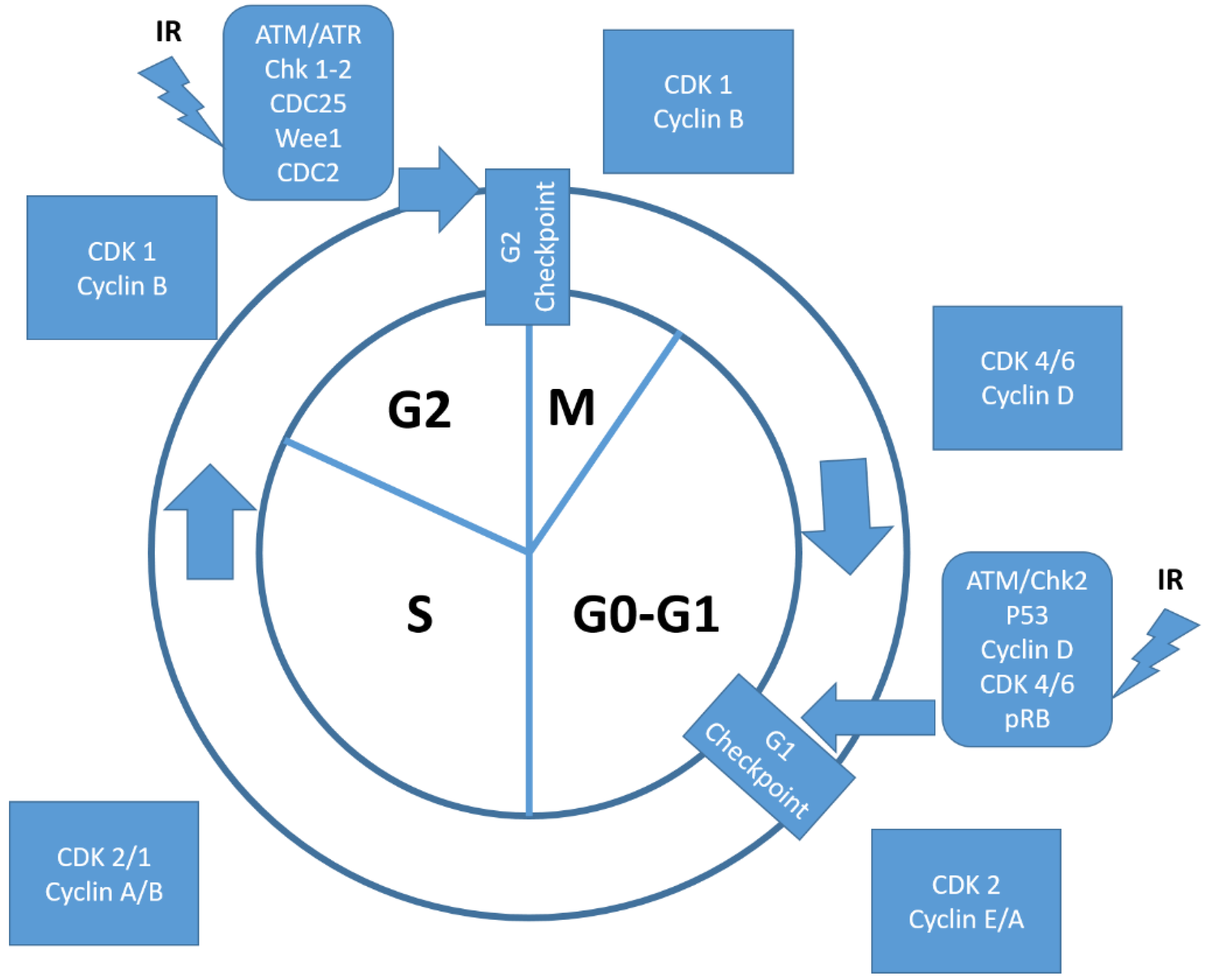
2. Molecular Background and Involved Pathways

The cell cycle has been defined as a set of subsequent events involved in the process of cell replication aiming to pass down the genetic information. In particular, cells are engaged into DNA replication (phase G1) followed by genome replication (DNA synthesis or S-phase), eventually leading to their segregation into two daughter cells (mitosis or M-phase). In a normal cell, this process is finely regulated and cell cycle checkpoints take place entering both S- and M-phase, respectively designated as G1 (between M and S) followed by G2 phase (between S and M). Cell cycle checkpoints represent a pivotal mechanism to preserve cell genomic integrity, prevent uncontrolled proliferation, and allow repair of damaged DNA [9].
Growth factors may induce the activation of positive cell checkpoints that in turn activate and promote cell cycle progression finely tuned by two groups of proteins, designated as cyclins and cyclin-dependent kinases (CDK). At present, four phase-specific cyclins (A, B, D, E) have been described. Cyclin D expression rises in G1 and falls in M phase while cyclin E, cyclin A, and cyclin B expression peaks within G1/S, G2, and G2/M phase, respectively. Experimental evidence suggests that the level of cyclins depends on their degradation by the endoplasmic ubiquitin/proteasome system (UPS).
In order to exert their regulatory functions, cyclins bind specific effector CDKs forming a cyclin/CDK hetero-dimeric complex (CCDK) whose phosphorylation in turn promotes cell cycle progression. Phosphorylation appears to be critical for CCDK functions and is under strict regulation by cyclin-dependent kinase inhibitors (CDKI). At present, two classes of CDKI have been identified: the INK4 family (p15, p16, p18, and p19) and the Cip/Kip protein family (p21, p27, p57) that disrupt the cyclin D1/CDK4 and CDK6 complex and both cyclin A,B/CDK 1 cyclin A,E/CDK 2 complexes, respectively.
CCDK regulation is pivotal in the event of DNA damage or misaligned chromosome during mitosis when negative checkpoint regulators, such as the retinoblastoma family of proteins (Rb), p53, or specific CDKIs, halt the cell cycle and activate DNA repair. In the event of irreparable damage, p53 triggers a pro-apoptotic cascade with consequent cell death [10,11,12,13,14,15]. In transformed cells, either negative or positive checkpoints are deregulated and affect the normal progression of cell cyle. In cancer cells, the additional amplification of the CCND1 gene, encoding for cyclin D1 (CCND1), is one of the most frequent alterations recorded in human solid neoplasms. This genomic alteration results in (1) CCND1 overexpression, (2) hyperactivation of cyclin D/CDK4 and CDK6 complex [16], (3) induced expression of E2F target genes, and 4) cell cycle engagement and progression from G1 to S phase.

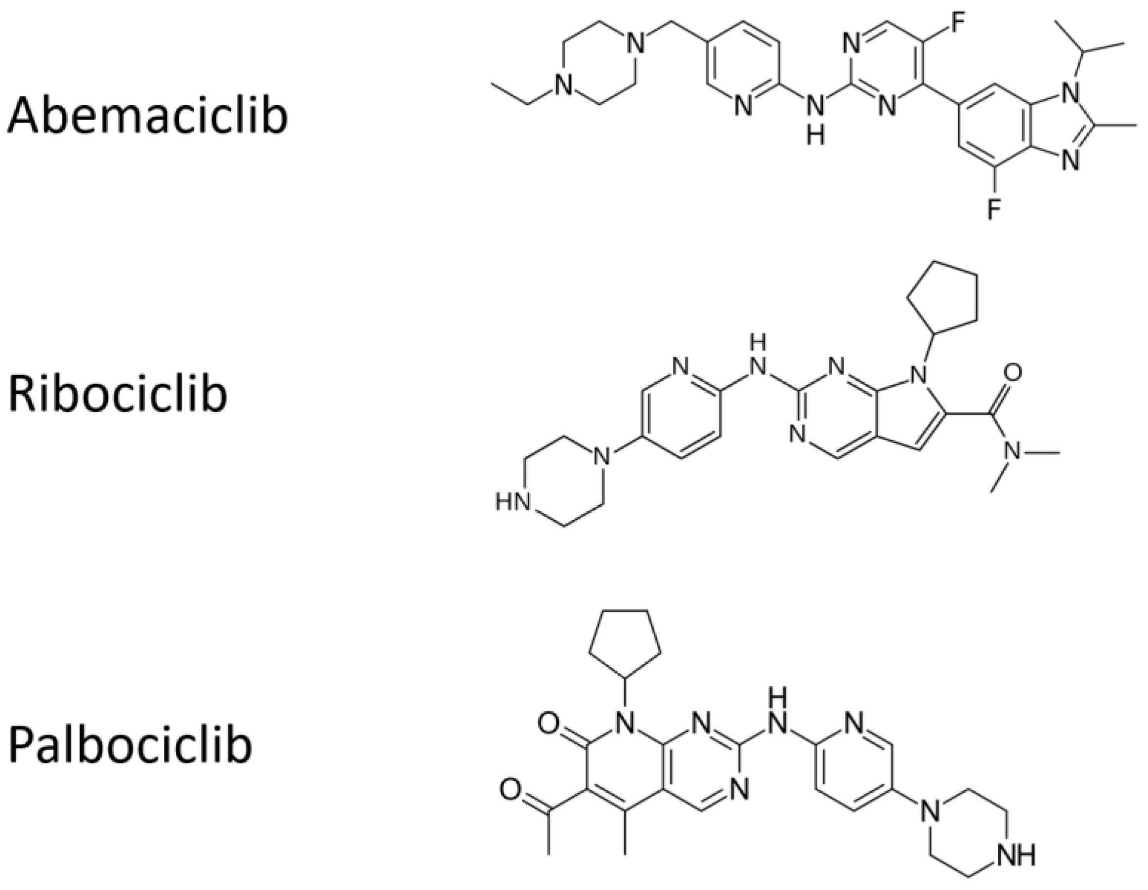
2.1. Antitumor Activity of Cyclin-D1/CDK4 and CDK6 Inhibition

A normal process of cell differentiation is associated with permanent irreversible proliferative quiescence referred to as G0 [17]. In contrast, stem cells involved in tissue renewal and repair (usually in a reversible quiescence) may restart cell cycle progression when stimulated by specific mitogenic factors. Cell cycle engagement and progression (G1/S phase) are regulated by D-type cyclins (D1, D2, and D3) in the early G1, with an outcome depending on the dynamic equilibrium among synthesis, accumulation, localization, and degradation of cyclin D. It has also been described that CCND1/CDK4 and CDK6 complex hetero-dymerization promotes cell cycle progression by sequestering the p21CIP1 and p27KIP1 inhibitors of the cyclin E/CDK2 complex in late G1 and inhibiting negative checkpoints regulators such as Rb through phosphorylation, (see Figure 1).
In light of the critical role of the pathway cyclin D/CDK4 and CDK6/Rb in cell cycle progression, one can easily argue the relevance of its deregulation in cancer cells [18]. The presence of defective cyclin D, CDK4 and CDK 6 proteins, or mutations in their genes can promote both tumor initiation and progression [5,11,19,20,21].
CDK4 gene amplification has been detected in 50% of glioblastomas (GB) [22] while CDK4 activating point mutation in R24C has been commonly reported in melanoma patients with primary resistance to the INK4 family of CDKIs [23]. Similarly, CCND1 over-expression has been reported in the carcinoma of the liver and CCND1 autophagy-dependent degradation leads to RB-driven inhibition of cell proliferation and tumor progression and subsequent suppression of the E2F transcriptional activity necessary for entering S phase [19]. Moreover in a xenograft model of nasopharynx carcinoma, other authors have shown that CDK4-6 inhibition with palpociclib exerts significant antitumor activity halting the cell cycle in G1 [24].
Altogether, these findings support the hypothesis that tumor cells may become functionally “addicted” to specific CDK alterations, thus making CDK inhibition a promising target for a new generation of anti-neoplastic agents.
Back in the early 1990s, the first CCDK2 inhibitor flavone L86-8275 blazed the trail toward the use of cell cycle inhibitors as a new ammunition against cancer [25]. More recently, a new generation of CCND1/CDK4 and CDK6 inhibitors, including alvocidib, palbociclib, ribociclib, and abemaciclib, demonstrated a relevant therapeutic activity in ESR+/HER2- advanced and metastatic breast cancer and is now a standard treatment in combination with aromatase inhibitors (AI) (anastrozole, letrozole, examestane), selective estrogen receptor (ESR) modulators (SERM: tamoxifen), and selective ESR down-regulators (SERD: fulvestrant).
Although several mechanisms have been evoked to explain the occurrence of primary and secondary resistance to this class of drugs, most of them involve CCND1/CDK 4 and CDK6/INK4/RB pathway [12] and highlight the critical role of Rb [26].

2.2. Ionizing Radiation and the Cyclin-D1/CDK4 and CDK6 Pharmacological Inhibition: Preclinical Evidence

From more than half a century [27], there has been a large consensus on the better response to ionizing radiation (IR) when cells are in G2/M rather than in G1/S [28,29]. Likewise, it is broadly agreed that IR determines a redistribution throughout the cell cycle; the repair of sub-lethal and potentially lethal DNA damage occur after IR; an indirect effect of IR is due to water radiolysis and reactive oxygen species (ROS); and IR induces endothelial activation and dysfunction in tissues.
Other evidence shows that cells treated with IR can repair damaged DNA or, on the contrary, undergo either programmed cell death pathways (PCD, e.g., apoptosis, autophagy) or necrosis [27]. An efficient cross-talk between the PCD pathways and necrosis has also been shown, which is not necessarily related to specific cell cycle phases [30]. Clear evidence does exist that tumor cells exposed to CCND1/CDK4 and CDK6 complex inhibitor increase their sensitivity to IR thanks to the induction of sustained DNA double-strand breaks (DSB) [31].
Although the precise mechanisms of these events have not been completely elucidated, several pre-clinical models provide the scientific rationale to investigate the combination pharmacological CDKIs [2,24,32,33,34,35,36] with IR [37] and suggest that it could be a paramount new avenue to achieve a better clinical performance of this class of drugs.
Consistently, other authors have provided evidence that in an orthotopic mouse glioblastoma model, combined use of palbociclib and IR achieved longer survival than palbociclib alone [2] and that in a xenograft cervical cancer model, two CDKIs, AT7519 and SNS-032, synergized with IR to induce apoptosis, senescence, and cytostasis [32].
These results were partially confirmed in clinical trials for brain cancer, when the use of CDKIs in combination with RT or temozolomyde or other anticancer agents showed promising results in terms of PFS and OS [38]. Similarly, the efficacy of CDKIs in combination with IR was also preliminarily tested in patients with lung [36], prostate, cervical, and breast cancer, suggesting a broad activity of this combination across these tumor types [38,39]. Notably, multiple pre-clinical studies have demonstrated the ability of CDKIs to promote apoptosis in tumor tissues exposed to IR, presumably by engaging p53 independent mechanisms
Interestingly, these studies also revealed the ability of CDKI to protect normal tissues by IR as well as chemotherapy-mediated damage by reducing the frequency of treatment associated intestinal injuries and hematological toxicity [40,41]. This phenomenon seems to be related to CDKIs’ ability to induce selective pharmacological quiescence of mucosal as well as hematopoietic cell precursors [42]. Several other similar points including the role of CDKIs in mitigating IR-induced neo-vasculogenesis, are certainly worthy of more future investigations [39].
Nevertheless, the optimal combined regimens with CDK4 and CDK6 inhibitors and RT, i.e., timing, dosage, treatment technique, and fractionation schedule, are still far from being identified.

2.3. Autophagy and the Cell Cycle

Autophagy is an evolutionary conserved process, aimed to maintain cellular homeostasis in response to stressful conditions (e.g., starvation, growth factors deprivation, excess of unfolded proteins, infection, gene mutation, etc., and altered signal pathways of cancer initiation and progression), and it can be also involved in type 2-PCD or autophagy-related cell death (APCD) in particular stressful situations [43].
Chaperone-mediated autophagy (CMA), micro-autophagy (or endosomal micro-autophagy (eMI), and macro-autophagy [44] are the three main process types. Among these, macro-autophagy, or simply “autophagy”, can be “basal” to degrade and recycle cytosolic components, as damaged macromolecules and organelles, to reconstitute the normal intracellular environment, or “induced” by external stress (e.g., starvation) to refill the cell aminoacidic pool for new proteins synthesis and/or energetic needs. Moreover, autophagy exerts pro-survival and anti-aging roles and is involved in tumor suppression and antigen presentation to immune effectors [45] and can contribute to apoptosis and other APCD processes. The autophagic process is divided into four different phases that are characterized by specific autophagy-related proteins (Atg) [46]: (1) initiation, (2) nucleation, (3) elongation and maturation, and (4) transport and fusion with lysosomes [47]. Briefly, the initial induction of the phagophore requires the formation of the Ulk1,2/Atg13/Atg101/FIP200 complex, whose activity is negatively or positively regulated by mTORC, depending on nutrients’ availability. The first phase depends on the formation of an autophagy-regulating PI3K complex (PI3K complex I) (Vps34/Beclin-1/Atg6/Atg14/p150/Vps15), that in turn is negatively regulated (nucleation) by the antiapoptotic proteins Bcl-2 or Bcl-xL that sequesters Beclin-1, thus inhibiting autophagosome’s formation. Subsequently, the third phase of the process requires the formation of the PI3K complex II (Atg12/Atg5/Atg16L1 complex) for the elongation of the pre-autophagosome membrane and the formation of the active form of the microtubule-associated protein 1/light chain 3 (LC3), designated as LC3-II, which is presently considered as the most reliable marker of autophagy. Finally, in the fourth phase of the process, mature autophagosomes fuse with endosomes, thus generating the amphysomes where the ultimate protein degradation occurs [48].
So far, no clear elucidation of the mechanisms underlying the cross-talk between autophagy and cell cycle has been achieved. CDKs-dependent cell progression can be related to autophagy inhibition, hence DNA damage or other inhibitory signals may be involved in inducing autophagy via CDKIs inhibition [49].
Many authors show a pivotal role of INK-4 and the Cip/Kip CDKIs families and, consequently, for Rb/E2F pathway in inducing autophagy [50].
p16, p21, and p27 Cip/Kip CDKIs have been strictly correlated to starvation-induced autophagy and cell cycle arrest. In particular, the cyclin-binding region of p27 appears to be essential in inducing autophagy and the depletion of CDK4 and CDK6 can reproduce, at least in part, this process. p16 overexpression has also been related to autophagy induction by promoting the Rb pathway. Moreover, some autophagy-promoting genes, including ULK and LC3, are the ultimate targets of the E2F transcription activity, and in turn, E2F factors were shown to bind the promoter site of Beclin-1. Therefore, the relationship between the Rb/E2F pathway and autophagy, and the possible correlation between autophagy and cell-cycle arrest, are characterized by a great complexity that explains some contrasting evidence due to multiple redundant pathways including, eight different known members within the E2F family with opposite regulative gene expression [51]. Polager et al. have provided evidence that the intensity of Rb/E2F pathway activation influences the autophagic response because a declined E2F1 expression hampers the Rb-driven DNA damage-induced autophagy. In the human bone osteosarcoma cell line U2OS [52], the presence of functional E2F1 transcription factors Rb pathway activation promotes autophagy inducing the expression of damage-regulated autophagy modulators genes encoding for LC3, Atg1, Atg5, and Dram. An excessive Rb activity or a defective E2F1 causes autophagy. In contrast, an excess in E2F1 activity antagonizes Rb-induced autophagy and promotes apoptosis [53]. This is consistent with the hypothesis that Bcl-2 might be a target for suppression of autophagy by binding Beclin-1.
It is noteworthy that Zheng et al. [54] reported several experimental conditions where both induction and suppression of autophagy by cell-cycle proteins (CDKs and CDKIs) were attributable to different stimuli and conditions. Similarly, recent evidence showed that chemical- or siRNA-based CDK4 and CDK6 inhibition can induce autophagy in multiple cancer cell lines [54,55]. In particular, treatment of myeloma cell lines with CDK4 and CDK6 inhibitor, abemaciclib, leads cells to accumulate in G0/G1, and exerts cytocidal activity accompanied by a dose-dependent autophagic vacuolization [56].
In addition, it has been demonstrated that autophagy itself can regulate the cell cycle. This two-way activity depends on the cell and TME conditions and may either lead cancer cells to cell death or may promote their survival [50,57]. The regulation of these events has been attributed to an autophagy-induced degradation of the cell cycle proteins, partially depending on the ubiquitin-proteasomal system (UPS). In particular, G1 arrest in breast cancer [58] and hepatocarcinoma cells [6] seems to be related to CCND1 autophagy-induced degradation. The role of autophagy on CCND1 was also supported by our group’s results obtained from surgical samples of resected GB relapsing after postoperative RT with concomitant and adjuvant temozolomide [59] (the gold standard for GB patients). We showed the co-localization of CCND1 with the key autophagy protein Beclin-1 and a possible role for autophagy in the control of cell cycle. One more promising strategy to overcome GB resistance is the use of PI3K/AKT/mTOR pathway inhibitors, which are demonstrated to increase IR-sensitivity and autophagy-related cell death in GB cell lines [60]. Altogether, these results prompted us to hypothesize that autophagy-related CCND1 degradation, mTOR inhibition, and autophagy activation could be a novel valuable therapeutic strategy to treat human tumors

2.4. The PI3K/AKT/mTOR Pathway in Endocrine Resistant ESR1+/HER2− Breast Cancer, and in other Neoplasms

The PI3K/AKT/mTOR signaling cascade is involved in escape pathways from hormonal therapy (HT) in breast cancer and may be independent of ER activation [61]. Because this signaling impacts on the cell cycle, cyclin-D1/CDK4 and CDK6 function, autophagy and IR are involved in many human neoplasms. It may be due to mutations or amplifications of genes, such as the encoding PI3KCA, or PI3K downstream components (PTEN, acting as a suppressor; AKT; mTOR). The relevance of PI3K signaling in particular has been shown in pre-clinical models of prostate and lung cancer, melanoma, leukemia, hematological malignancies, etc.
ER stimulation receptor tyrosine kinase (RTK) activation by growth factors and extracellular matrix components can activate PI3K, whereas RTK activation is cross-linked with the MAPK pathway [62]. Activation by PI3K is necessary for AKT through the phosphorylation of Thr-308 and Ser-473 by the phosphoinositide-dependent kinases (PDK)-1 and -2. Activated AKT, in turn, regulates downstream effectors including mammalian target of rapamycin (mTOR), often deregulated in human cancer.
It is noteworthy that AKT can be activated independently from the PI3K signaling, by IR-induced DNA double-strand break [63], and that PI3K/AKT/mTOR pathway is crucial for the functional interaction between CCND1 and CDK4 and CDK6 [64].

3. The State of the Art in the Clinical Domain

3.1. Clinical Trials of Cyclin-D1/CDK4 and CDK6 Inhibition in Breast Cancer

Despite the wide experimental preclinical work on cyclin-D1/CDK4 and CDK6 inhibitors in several neoplasms, these agents have been mostly clinically tested only for advanced and metastatic malignancies and just recently have been approved as a standard first-line strategy in HR-positive, HER2 negative breast cancer, in various combinations with AIs, SERMs, or SERDs.
In a phase II clinical trial (PALOMA-1), the addition of palbociclib to letrozole significantly increased the median progression-free survival (PFS) from 10.2 months to 20.2 months [15] vs. letrozole alone for post-menopausal women with previously untreated ESR1+/HER2− advanced breast cancer. Palbociclib received accelerated approval by the US Food and Drug Administration (FDA) in February 2015 [65]. The placebo-randomized, double-blind, controlled phase III trial (PALOMA-3) compared treatment with palbociclib and fulvestrant to fulvestrant plus placebo in women with ESR1+/HER2− metastatic breast cancer that had relapsed or progressed on prior hormone therapy, including a substantial portion of patients (33%) with prior chemotherapy for metastatic disease. The interim analysis of this study demonstrated a significantly improved median PFS (9.5 months versus 4.6 months, respectively) [12,66]. Palbociclib is presently under further investigation in over 50 trials in related clinical settings.
Ribociclib is a selective strong inhibitor of CDK4 and CDK6 [13], blocks Rb phosphorylation, and causes cell cycle arrest of Rb-positive tumor cells, similarly to palbociclib [14]. The MONALEESA-2 trial demonstrated that ribociclib plus letrozole significantly improved progression-free survival, compared with placebo plus letrozole as a first-line therapy in postmenopausal patients with HR-positive, HER2-negative advanced breast cancer [15].
Ongoing trials are demonstrating significant progression-free survival improvements with ribociclib in different settings [67,68]. However, both palpociclib and ribociclib were characterized by a not-negligible systemic toxicity [31].
Abemaciclib was tested in a phase I trial, showing efficacy against non-small cell lung cancer, melanoma, and mantle cell lymphoma, in addition to breast cancer [69]. In fact, other than its high affinity for CCND1/CDK4, it also inhibits cyclin-B/CDK1 (related to G2 arrest) and cyclin E-A/CDK2 (related to palpociclib resistance). Differently from the cytostatic effect alpociclib and ribociclib, abemaciclib achieved a durable cytotoxic effect in Rb-negative cells [70]. Moreover, in a first line randomized phase III double-blind study (MONARCH 3) for ESR1+/HER2− advanced breast cancer, abemaciclib presented a better hematological tolerability compared to palpociclib and ribociclib, and achieved impressive results in terms of PFS and objective response rate (ORR) [71]. A synthesis of these clinical trials is reported in Table 1.
A metanalysis demonstrated that three CDK4 and CDK6 inhibitors have similar efficacy when associated with AIs, and are superior to either fulvestrant or AI monotherapy independently of any stratification criteria, thus supporting AI plus CDK4 and CDK6 inhibition as the best approach in ESR1+/HER2− metastatic breast cancer [65,66]. Li et al., in another meta-analysis, included the most prominent nine previous prospective trials and 5043 patients [72]. Adding CDK4 and CDK6 inhibitors to endocrine therapy achieved a highly significant benefit for OS, compared to ET alone. Moreover, significant gain in OS also resulted in second-line treatment in patients affected by visceral metastases. However, the combined CDK4 and CDK6 inhibitor/ET treatment was characterized by a heavier G3-4 toxicity that was less pronounced for abemaciclib. Nevertheless, it should be remembered that 10% of the overall ESR1+/HER2− breast cancer patients show primary or secondary resistance to this combined therapeutic approach [6].
All the chemical structures of the molecules are presented in Figure 2.

3.2. Real-Life Clinical Reports on Cyclin-D1/CDK4 and CDK6 Inhibitors, Including Association with Radiotherapy

Delivering unplanned RT (mainly with palliative/symptomatic aims) in advanced breast cancer patients on Cyclin-D1/CDK4 and CDK6 inhibitors in prospective trials has produced empirically based treatment not supported by an appropriate rationale and consolidated approach, e g., in terms of RT intent, dose, and scheduling. However, several observational studies provided clearly suggest the lack of additive adverse events when palbociclib or ribociclib are combined with letrozole and concomitant palliative RT [73,74,75]. Additionally, a retrospective single-institution analysis suggested that 15 out a cohort of 42 pts with ESR1+ breast cancer with brain metastases showed promising results using CDK4 and CDK6 inhibitors palbociclib or abemaciclib for 6 months combined with stereotactic radiation [71] and only 5% of the patients developed radiation necrosis. However, only a slight improvement could be shown in survival. However, early radiation toxicities, including esophagitis and severe dermatitis have been reported in a breast cancer patient on palpociclib and palliative RT on cervical lymph nodes [72]. Similar studies evaluating CDK inhibitors and RT are presently ongoing in patients with head and neck, brain, breast, and pancreatic cancer as reported in Table 2.

4. Molecular Grounds for Future Developments

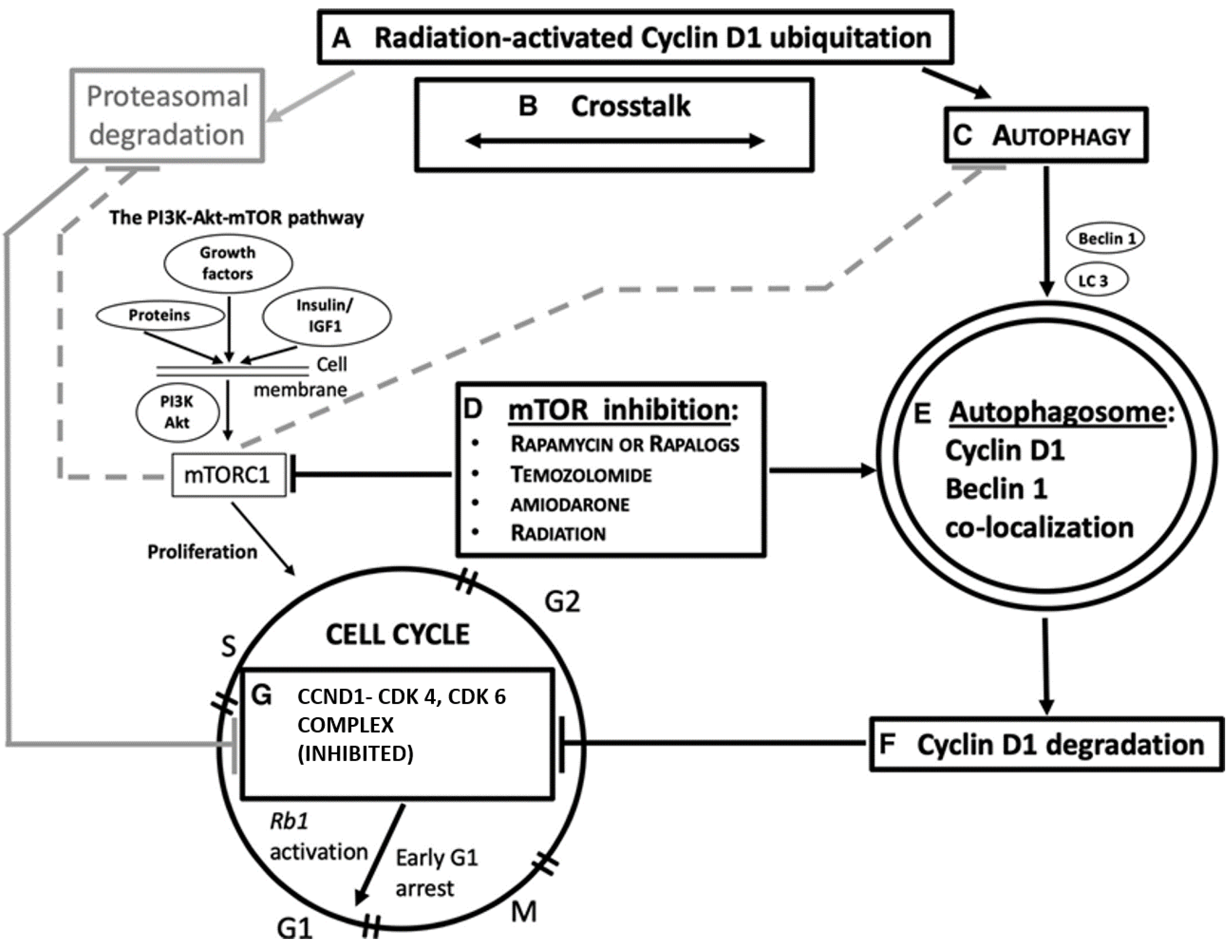
4.1. CCND1 as a Target of Ionizing Radiation in the CCND1-CDK4, and CDK6 Complex

Only the CCND1-CDK4 and CDK6 complex has raised interest as a possible actionable target, whereas, in contrast, despite its prognostic relevance in cancer [76], the regulatory member CCND1 per se has received less attention. As previously quoted, palpociclib induces autophagy in cancer, targeting CDK4 and CDK6 [54,55], via the LKB1 and Ser325 phosphorylation resulting in a CCND1-mediated, reduced activation of AMPK [77]. The CCND1 gene encoding CCND1 is activated in B-cell lymphomas and overexpressed in many other human cancers (esophageal, lung, breast, bladder carcinomas, etc.) where it acts as a proto-oncogene. Therefore, the CCND1 is a potential pivotal actionable target for cell cycle arrest. In normal cells, CCND1 has a very short life due to UPS degradation whereas in cancer cells, inhibition of turnover and increased levels of CCND1 were shown. Conversely, the knockdown of the activity of a specific deubiquitinase for CCND1 was shown, inducing growth suppression in cancer cells addicted to its expression [78]. Ubiquitination of CCND1 is achievable in response to IR [79] and, in cancer cells, IR or other DNA-damaging agents may activate selective autophagy [54]. Autophagy shares common molecular mechanisms with UPS, with which it is intertwined in a close and complex relationship [80]. As described by these authors, ubiquitin-specific recognition can have a pivotal role for CCND1 autophagy degradation. Selective autophagy requires some receptor proteins necessary for the process, such as p62 (also known as sequestosome 1 or SQSTM1), NBR1 (neighbor of the BRCA 1 gene), CALCOCO2 (calcium binding and coiled-coil domain 2), and optineurin. p62 (the most relevant receptor protein for the subject dealt with here) is a component of several transduction signal cascades, such as Keap-Nrf2, and essential for UPS degradation.
Possible future strategies aimed at enhancing the IR-autophagic degradation of CCND1 in combination with drugs targeting the CCND1-CDK4 and CDK6 complex for G1/S arrest could overcome the IR-resistance of G1 and S phases of cell cycle

4.2. Ionizing Radiation, Autophagy Enhancement, and CCND1

Ionizing radiation can directly induce autophagy; although the mechanism underlying this effect has not been fully elucidated so far, two main mechanisms have been evoked: (1) similarly to rapamycin, IR reverts auto-phosphorylation of mTOR (e.g., in MCF-7 breast cancer cell culture) leading to the formation of autophagic vacuoles, segregation of cytoplasmic materials, and appearance of LC3 positivity and eventually mitochondrial hyperpolarization and decreased cell survival [81].
(2) Radiation causes a protective pathway via endoplasmic reticulum (ER) stress, accumulation of ER misfolded proteins (unfolded protein response—UPR), and autophagy. Therefore, UPR pathway and autophagy are upregulated in some cancer types as an escape strategy leading to IR-activated autophagy responsible for resistance to RT [82,83,84]. Nonetheless, autophagy is always depictable as a “double-edged sword”. As previously reviewed [80], most of the literature dedicated to this subject is focused on drug manipulation of autophagy (i.e., either induction or inhibition) for improving IR-sensitivity in cancer cells, more than to IR in conditioning, per se, autophagy towards cell death.
Emphasis should be placed in autophagy during the cell cycle, as different studies have investigated the autophagy activation in the different phases of the cell cycle [81,82]. In contrast, other evidence suggests a protective suppression of autophagy during mitosis aimed at preventing the inappropriate inactivation of molecular species and organelles, functional for cellular replication, or even aggregated chromosomes [50]. IR-based autophagy induced during the G1 and S counteracts the accelerated cell cycle in cancer and lays the groundwork for combined therapies combined with IR. It has been shown that CCND1 suppresses autophagy in mammary epithelium [85] whereas CCND1 inhibits autophagy by activating AMPK in breast cancer cells [77]. Further, in 2016, our group published a study in recurrent GB patients that prompted us to hypothesize a role for the IR-temozolomide treatment in inducing CCND1 autophagic degradation in Beclin-1-positive GBs [59]. Consistently, another group demonstrated the inverse correlation between low autophagic activity and high CCND1 expression that were also related to poorer prognosis [4]. In human and murine models of hepatocellular carcinomas (HCC), induction of autophagy led to CCND1 ubiquitination and its recruitment into autophagosomes via SQSTM1, formation of autophago-lysosomes, and degradation with suppression of tumor growth. Further, Chen et al. in 2019 [58] showed that autophagy itself causes CCND1 autophagic degradation in breast cancer cell lines with subsequent G1 cell cycle arrest underpinning the hypothesis of cell cycle modulation by CCND1 via mTORC1 signaling inhibition. Finally, we proposed in 2020 an overview of the role of autophagy, and namely its pivotal Beclin-1 protein, in CCND1 degradation, as a possibly actionable mechanism for G1/S cell cycle arrest in cancer (Figure 3) [86].
It is still unclear whether autophagy could represent a true per se pro-death process, or can simply enhance the IR and chemotherapy sensitivity of cancer cells. The impact of autophagy in a cell death progress is cautiously named “autophagy-related cell death”. This definition is underpinned by the clinical observation that a high tissue expression of cytoplasmic Beclin-1 was related to better prognosis in patients with malignant glioma on temozolomide chemotherapy and radiotherapy [87]. Notwithstanding, one should also take account of the tumor suppression of the autophagy-independent functions of Beclin-1 [88].

5. Concluding Remarks, with Special Regard to Immunotherapy Developments

Although the efficacy of combining HT and CDK4 and CDK6 inhibitor in patients with ESR1+/HER2− breast cancer, the promising benefit of CDK4 and CDK6 drug inhibitors in other neoplasms has not been extensively tested.
Attempts to enhance the G1/S arrest action are currently under clinical investigation, e.g., based on a concurrent PI3K-pathway inhibition for breast cancer (triple therapy), or a sequential use of these agents in cases bearing PIK3CA mutations [8]. Of note, PALOMA trials showed that CCND1 amplification and overexpression did not correlate with reduced therapeutic results. We believe this latter observation does not rule out CCND1 being an actionable target. Notably, there is evidence that autophagy induction by IR and/or drug manipulation can induce CCND1 degradation not only in breast cancer [58], but also in hepatocellular carcinoma (HCC) [6] and GB cells [59,86]. Other than with IR in vitro, autophagy can be induced by alkylating drugs (e.g., temozolomide) and mTOR inhibitors (e.g., rapamycin and rapalogs), although their combination with CDK4 and CDK6 inhibitors raises safety concerns. On the other hand, in vitro experiments have shown that two pro-autophagy drugs, amiodarone (in HCC; [6]) and trehalose (in melanoma), are safer or even show no toxicity at all, respectively. Moreover, more recently, selective and less toxic mTOR inhibitors showed reduced impact rapamycin or rapalogs on glucose homeostasis, lipid metabolism, and immune response [89].
A high autophagy rate degrades cell cycle regulators, including cyclin-induced senescence [90]. Cancer cells secrete inflammatory cytokines, chemokines, and growth factors (collectively designated as senescence-associated secretory phenotype, or SASP) [31] and CDK inhibitors and IR may contribute to this process. SASP may also recruit immunity against the senescent cells, whereas other combined actions by CDK inhibitors and IR, i.e., enhancement of the MHC class I expression, activation of dendritic cells, increased antigen cross-presentation, and reduced the proliferation of Tregs. These effects do contribute not only to local but also systemic cancer-suppressive response (abscopal effect by IR) [91,92].
It is worth mentioning that, according to nutrient availability, microenvironment stress, pathogenic conditions, and a responsive immune system, autophagy plays neutral, tumor-suppressive, or tumor-promoting roles [93]. Different treatment responses may, therefore, be in part due to factors that influence the immune system (thus, autophagy) other than IR, such as genetic background, dietary habits, and microbiome. Accounting for these factors and their impact on the treatment response, the research framework of molecular pathological epidemiology (MPE) has already been demonstrated to offer a valid interdisciplinary integration when it was adopted to study breast, lung, prostate, and colorectal cancers. The MPE approach can be applied, for instance, to assess the influence of exogenous and endogenous factors on carcinogenic processes by gauging the immune response to a tumor, filling the research gap between tumor immunology and epidemiology (immune-MPE) [94].
MPE research must work with both in vivo and in vitro experimental research to improve our understanding of disease pathogenesis, given the complexities of human diseases, to fulfill an integrative scientific approach [95].
Moreover, platform database analysis addressed CCND1 amplification as a potential surrogate biomarker of efficacy and even target for immunotherapy [96]. CCND1 amplification was associated with the exclusion of cytotoxic cells, T CD8+ and B+ T cells and dendritic cells (DCs) in the tumor microenvironment (TME). These studies also suggest a possible correlation between CCND1 amplification and immunosuppressive features as the epithelial–mesenchymal transition (EMT), transforming growth factor (TGF)-b, KRAS and PI3K/AKT/mTOR signaling, p53 pathway, and hypoxia signaling.
In light of the data available, CCND1 is an attractive, multifaceted target for cancer therapy and is worth extensive study to explore its impact as a novel actionable target and when combined with IR.
Setting up proper patient stratification and study design will allow us to understand how to unleash this new ammunition against cancer.

Author Contributions

Conceptualization, V.N., P.C., and L.P.; methodology, V.N., L.P., C.M., A.R., and S.C.; formal analysis, L.M., P.P., and P.C.; writing—original draft preparation, V.N., M.B., L.P., P.C., and L.M.; writing—review and editing, V.N., M.B., A.A., P.C., P.P., S.C., A.R., L.M., C.M., R.G., A.G., and L.P.; visualization, V.N., M.B., A.A., P.C., P.P., S.C., A.R., L.M., C.M., R.G., A.G., and L.P.; supervision, L.M. All authors have read and agreed to the published version of the manuscript.

Funding

This research received no external funding.

Institutional Review Board Statement

Not applicable.

Informed Consent Statement

Not applicable.

Data Availability Statement

Not applicable.

Conflicts of Interest

The authors declare no conflict of interest.

References

  1. Pajonk, F.; Vlashi, E.; McBride, W.H. Radiation resistance of cancer stem cells: The 4 R’s of radiobiology revisited. Stem Cells 2010, 28, 639–648. [Google Scholar] [CrossRef] [Green Version]
  2. Whittaker, S.; Madani, D.; Joshi, S.; Chung, S.A.; Johns, T.; Day, B.; Khasraw, M.; McDonald, K.L. Combination of palbociclib and radiotherapy for glioblastoma. Cell Death Discov. 2017, 3, 17033. [Google Scholar] [CrossRef] [PubMed]
  3. Nardone, V.; Falivene, S.; Giugliano, F.M.; Gaetano, M.; Giordano, P.; Muto, M.; Daniele, B.; Guida, C. The role of radiation therapy and systemic therapies in elderly with breast cancer. Transl. Cancer Res. 2020, 9, S97–S109. [Google Scholar] [CrossRef]
  4. Renzulli, M.; Zanotti, S.; Clemente, A.; Mineo, G.; Tovoli, F.; Reginelli, A.; Barile, A.; Cappabianca, S.; Taffurelli, M.; Golfieri, R. Hereditary breast cancer: Screening and risk reducing surgery. Gland. Surg. 2019, 8, S142–S149. [Google Scholar] [CrossRef] [Green Version]
  5. Sicinska, E.; Aifantis, I.; Le Cam, L.; Swat, W.; Borowski, C.; Yu, Q.; Ferrando, A.A.; Levin, S.D.; Geng, Y.; Von Boehmer, H.; et al. Requirement for cyclin D3 in lymphocyte development and T cell leukemias. Cancer Cell 2003, 4, 451–461. [Google Scholar] [CrossRef] [Green Version]
  6. Wu, S.Y.; Lan, S.H.; Wu, S.R.; Chiu, Y.C.; Lin, X.Z.; Su, I.J.; Tsai, T.F.; Yen, C.J.; Lu, T.H.; Liang, F.W.; et al. Hepatocellular carcinoma-related cyclin D1 is selectively regulated by autophagy degradation system. Haepatology 2018, 68, 141–154. [Google Scholar] [CrossRef] [PubMed] [Green Version]
  7. Hamilton, E.; Infante, J.R. Targeting CDK4/6 in patients with cancer. Cancer Treat. Rev. 2016, 45, 129–138. [Google Scholar] [CrossRef] [Green Version]
  8. McCartney, A.; Migliaccio, I.; Bonechi, M.; Biagioni, C.; Romagnoli, D.; De Luca, F.; Galardi, F.; Risi, E.; De Santo, I.; Benelli, M.; et al. Mechanisms of Resistance to CDK4/6 Inhibitors: Potential Implications and Biomarkers for Clinical Practice. Front. Oncol. 2019, 9, 666. [Google Scholar] [CrossRef] [PubMed]
  9. MacLachlan, T.K.; Sang, N.; Giordano, A. Cyclins, cyclin-dependent kinases and cdk inhibitors: Implications in cell cycle control and cancer. Crit. Rev. Eukaryot. Gene Expr. 1995, 5, 127–156. [Google Scholar] [CrossRef] [PubMed]
  10. Prokhorova, E.A.; Egorshina, A.Y.; Zhivotovsky, B.; Kopeina, G.S. The DNA-damage response and nuclear events as regulators of nonapoptotic forms of cell death. Oncogene 2019, 39, 1–16. [Google Scholar] [CrossRef] [PubMed]
  11. Puyol, M.; Martín, A.; Dubus, P.; Mulero, F.; Pizcueta, P.; Khan, G.; Guerra, C.; Santamaría, D.; Barbacid, M. A synthetic lethal interaction between K-Ras oncogenes and Cdk4 unveils a therapeutic strategy for non-small cell lung carcinoma. Cancer Cell. 2010, 18, 63–73. [Google Scholar] [CrossRef]
  12. Cristofanilli, M.; Turner, N.C.; Bondarenko, I.; Ro, J.; Im, S.-A.; Masuda, N.; Colleoni, M.; DeMichele, A.; Loi, S.; Verma, S.; et al. Fulvestrant plus palbociclib versus fulvestrant plus placebo for treatment of hormone-receptor-positive, HER2-negative metastatic breast cancer that progressed on previous endocrine therapy (PALOMA-3): Final analysis of the multicentre, double-blind, phase 3 randomised controlled trial. Lancet Oncol. 2016, 17, 425–439. [Google Scholar] [CrossRef] [PubMed] [Green Version]
  13. Asghar, U.; Witkiewicz, A.K.; Turner, N.C.; Knudsen, E.S. The history and future of targeting cyclin-dependent kinases in cancer therapy. Nat. Rev. Drug Discov. 2015, 14, 130–146. [Google Scholar] [CrossRef] [PubMed] [Green Version]
  14. Rader, J.; Russell, M.R.; Hart, L.S.; Nakazawa, M.S.; Belcastro, L.T.; Martinez, D.; Li, Y.; Carpenter, E.L.; Attiyeh, E.F.; Diskin, S.J.; et al. Dual CDK4/CDK6 inhibition induces cell-cycle arrest and senescence in neuroblastoma. Clin. Cancer Res. 2013, 19, 6173–6182. [Google Scholar] [CrossRef] [Green Version]
  15. Hortobagyi, G.; Stemmer, S.; Burris, H.; Yap, Y.-S.; Sonke, G.; Paluch-Shimon, S.; Campone, M.; Petrakova, K.; Blackwell, K.; Winer, E.; et al. Updated results from MONALEESA-2, a phase III trial of first-line ribociclib plus letrozole versus placebo plus letrozole in hormone receptor-positive, HER2-negative advanced breast cancer. Ann. Oncol. 2019, 30, 1842. [Google Scholar] [CrossRef]
  16. Qie, S.; Diehl, J.A. Cyclin D1, cancer progression, and opportunities in cancer treatment. J. Mol. Med. 2016, 94, 1313–1326. [Google Scholar] [CrossRef] [Green Version]
  17. Kaufmann, W.K.; Paules, R.S. DNA damage and cell cycle checkpoints. FASEB J. 1996, 10, 238–247. [Google Scholar] [CrossRef] [PubMed]
  18. Jardim, D.L.; Millis, S.Z.; Ross, J.S.; Woo, M.S.; Ali, S.M.; Kurzrock, R. Cyclin Pathway Genomic Alterations Across 190,247 Solid Tumors: Leveraging Large-Scale Data to Inform Therapeutic Directions. Oncologist 2020, 26, e78–e89. [Google Scholar] [CrossRef] [PubMed]
  19. Choi, Y.J.; Li, X.; Hydbring, P.; Sanda, T.; Stefano, J.; Christie, A.L.; Signoretti, S.; Look, A.T.; Kung, A.; von Boehmer, H.; et al. The Requirement for Cyclin D Function in Tumor Maintenance. Cancer Cell 2012, 22, 438–451. [Google Scholar] [CrossRef] [PubMed] [Green Version]
  20. Sawai, C.M.; Freund, J.; Oh, P.; Ndiaye-Lobry, D.; Bretz, J.C.; Strikoudis, A.; Genesca, L.; Trimarchi, T.; Kelliher, M.A.; Clark, M.; et al. Therapeutic targeting of the cyclin D3:CDK4/6 complex in T cell leukemia. Cancer Cell. 2012, 22, 452–465. [Google Scholar] [CrossRef] [Green Version]
  21. Hu, M.G.; Deshpande, A.; Enos, M.; Mao, D.; Hinds, E.A.; Hu, G.F.; Chang, R.; Guo, Z.; Dose, M.; Mao, C.; et al. A requirement for cyclin-dependent kinase 6 in thymocyte development and tumorigenesis. Cancer Res. 2009, 69, 810–818. [Google Scholar] [CrossRef] [Green Version]
  22. Schmidt, E.E.; Ichimura, K.; Reifenberger, G.; Collins, V.P. CDKN2 (p16/MTS1) gene deletion or CDK4 amplification occurs in the majority of glioblastomas. Cancer Res. 1994, 54, 6321–6324. [Google Scholar]
  23. Wölfel, T.; Hauer, M.; Schneider, J.; Serrano, M.; Wolfel, C.; Klehmann-Hieb, E.; De Plaen, E.; Hankeln, T.; Zum Buschenfelde, K.M.; Beach, D. A p16INK4a-insensitive CDK4 mutant targeted by cytolytic T lymphocytes in a human melanoma. Science 1995, 269, 1281–1284. [Google Scholar] [CrossRef] [PubMed]
  24. Hsu, C.L.; Lui, K.W.; Chi, L.M.; Kuo, Y.C.; Chao, Y.K.; Yeh, C.N.; Lee, L.Y.; Huang, Y.; Lin, T.L.; Huang, M.Y.; et al. Integrated genomic analyses in PDX model reveal a cyclin-dependent kinase inhibitor Palbociclib as a novel candidate drug for nasopharyngeal carcinoma. J. Exp. Clin. Cancer Res. 2018, 37, 233. [Google Scholar] [CrossRef]
  25. Kaur, G.; Stetler-Stevenson, M.; Sebers, S.; Worland, P.; Sedlacek, H.; Myers, C.; Czech, J.; Naik, R.; Sausville, E. Growth inhibition with reversible cell cycle arrest of carcinoma cells by flavone L86-8275. J. Natl. Cancer Inst. 1992, 84, 1736–1740. [Google Scholar] [CrossRef]
  26. Sammons, S.L.; Topping, D.L.; Blackwell, K.L. HR+, HER2- Advanced Breast Cancer and CDK4/6 Inhibitors: Mode of Action, Clinical Activity, and Safety Profiles. Curr. Cancer Drug Targets 2017, 17, 637–649. [Google Scholar] [CrossRef] [PubMed]
  27. Sinclair, W.K.; Morton, R.A. X-ray sensitivity during the cell generation cycle of cultured Chinese hamster cells. Radiat. Res. 1966, 29, 450–474. [Google Scholar] [CrossRef] [PubMed]
  28. Gurley, L.R.; Walters, R.A.; Tobey, R.A. Cell cycle-specific changes in histone phosphorylation associated with cell proliferation and chromosome condensation. J. Cell. Biol. 1974, 60, 356–364. [Google Scholar] [CrossRef] [Green Version]
  29. Terzoudi, G.I.; Pantelias, G.E. Conversion of DNA damage into chromosome damage in response to cell cycle regulation of chromatin condensation after irradiation. Mutagenesis 1997, 12, 271–276. [Google Scholar] [CrossRef] [Green Version]
  30. Chen, Q.; Kang, J.; Fu, C. The independence of and associations among apoptosis, autophagy, and necrosis. Signal Transduct. Target. Ther. 2018, 3, 1–11. [Google Scholar] [CrossRef] [PubMed] [Green Version]
  31. Yang, Y.; Luo, J.; Chen, X.; Yang, Z.; Mei, X.; Ma, J.; Zhang, Z.; Guo, X.; Yu, X. CDK4/6 inhibitors: A novel strategy for tumor radiosensitization. J. Exp. Clin. Cancer Res. 2020, 39, 188. [Google Scholar] [CrossRef] [PubMed]
  32. Kang, M.A.; Kim, W.; Jo, H.-R.; Shin, Y.-J.; Kim, M.-H.; Jeong, J.-H. Anticancer and radiosensitizing effects of the cyclin-dependent kinase inhibitors, AT7519 and SNS-032, on cervical cancer. Int. J. Oncol. 2018, 53, 703–712. [Google Scholar] [CrossRef] [Green Version]
  33. Lin, Z.P.; Zhu, Y.L.; Ratner, E.S. Targeting Cyclin-Dependent Kinases for Treatment of Gynecologic Cancers. Front. Oncol. 2018, 8, 303. [Google Scholar] [CrossRef] [PubMed]
  34. Lee, C.L.; Oh, P.; Xu, E.S.; Ma, Y.; Kim, Y.; Daniel, A.R.; Kirsch, D.G. Blocking Cyclin-Dependent Kinase 4/6 During Single Dose Versus Fractionated Radiation Therapy Leads to Opposite Effects on Acute Gastrointestinal Toxicity in Mice. Int. J. Radiat. Oncol. Biol. Phys. 2018, 102, 1569–1576. [Google Scholar] [CrossRef]
  35. Naz, S.; Sowers, A.; Choudhuri, R.; Wissler, M.; Gamson, J.; Mathias, A.; Cook, J.A.; Mitchell, J.B. Abemaciclib, a Selective CDK4/6 Inhibitor, Enhances the Radiosensitivity of Non-Small Cell Lung Cancer In Vitro and In Vivo. Clin. Cancer Res. 2018, 24, 3994–4005. [Google Scholar] [CrossRef] [Green Version]
  36. Jung, S.-Y.; Nam, K.-Y.; Park, J.-I.; Song, K.-H.; Ahn, J.; Park, J.K.; Um, H.-D.; Hwang, S.-G.; Choi, S.U.; Song, J.-Y. Radiosensitizing Effect of Novel Phenylpyrimidine Derivatives on Human Lung Cancer Cells via Cell Cycle Perturbation. J. Pharmacol. Exp. Ther. 2019, 370, 514–527. [Google Scholar] [CrossRef] [Green Version]
  37. Wang, J.Y.; Naderi, S.; Chen, T.T. Role of retinoblastoma tumor suppressor protein in DNA damage response. Acta. Oncol. 2001, 40, 689–695. [Google Scholar] [PubMed]
  38. Juric, V.; Murphy, B. Cyclin-dependent kinase inhibitors in brain cancer: Current state and future directions. Cancer Drug Resist. 2020, 3, 48–65. [Google Scholar] [CrossRef] [Green Version]
  39. Naz, S.; Cook, J.A.; Mitchell, J.B. Abemaciclib: A multi-functional radiation modifier. Oncotarget 2019, 10, 1230–1232. [Google Scholar] [CrossRef]
  40. Johnson, S.M.; Torrice, C.D.; Bell, J.F.; Monahan, K.B.; Jiang, Q.; Wang, Y.; Ramsey, M.R.; Jin, J.; Wong, K.K.; Su, L.; et al. Mitigation of hematologic radiation toxicity in mice through pharmacological quiescence induced by CDK4/6 inhibition. J. Clin. Investig. 2010, 120, 2528–2536. [Google Scholar] [CrossRef]
  41. Wei, L.; Leibowitz, B.J.; Wang, X.; Epperly, M.; Greenberger, J.; Zhang, L.; Yu, J. Inhibition of CDK4/6 protects against radiation-induced intestinal injury in mice. J. Clin. Investig. 2016, 126, 4076–4087. [Google Scholar] [CrossRef] [PubMed]
  42. Martin, K.L.; Hill, G.A.; Klein, R.R.; Arnett, D.G.; Burd, R.; Limesand, K.H. Prevention of radiation-induced salivary gland dysfunction utilizing a CDK inhibitor in a mouse model. PLoS ONE 2012, 7, e51363. [Google Scholar] [CrossRef] [Green Version]
  43. Janji, B.; Viry, E.; Baginska, J.; Van Moer, K.; Berchem, G. Role of Autophagy in Cancer and Tumor Progression. In Autophagy—A Double-Edged Sword—Cell Survival or Death? Bailly, Y., Ed.; IntechOpen: London, UK, 2013. [Google Scholar] [CrossRef] [Green Version]
  44. Galluzzi, L.; Baehrecke, E.H.; Ballabio, A.; Boya, P.; Bravo-San Pedro, J.M.; Cecconi, F.; Choi, A.M.; Chu, C.T.; Codogno, P.; Colombo, M.I.; et al. Molecular definitions of autophagy and related processes. EMBO J. 2017, 36, 1811–1836. [Google Scholar] [CrossRef] [PubMed]
  45. Mizushima, N. Autophagy: Process and function. Genes Dev. 2007, 21, 2861–2873. [Google Scholar] [CrossRef] [Green Version]
  46. Klionsky, D.J.; Abdalla, F.C.; Abeliovich, H.; Abraham, R.T.; Acevedo-Arozena, A.; Adeli, K.; Agholme, L.; Agnello, M.; Agostinis, P.; Aguirre-Ghiso, J.; et al. Guidelines for the use and interpretation of assays for monitoring autophagy. Autophagy 2012, 8, 445–544. [Google Scholar] [CrossRef]
  47. Toscano, M.; Palumbo, S.; Tini, P.; Miracco, C.; Gravina, G.; Comincini, S. Cell Death Pathways, with Special Regard to Ionizing Radiation and Temozolomide. In Radiobiology of Glioblastoma-Recent Advances and Related Pathobiology; Pirtoli, L., Gravina, G.L., Giordano, A., Eds.; Springer: Berlin/Heidelberg, Germany, 2016; pp. 209–224. [Google Scholar] [CrossRef]
  48. Rogov, V.; Dötsch, V.; Johansen, T.; Kirkin, V. Interactions between autophagy receptors and ubiquitin-like proteins form the molecular basis for selective autophagy. Mol. Cell. 2014, 53, 167–178. [Google Scholar] [CrossRef] [PubMed] [Green Version]
  49. Capparelli, C.; Chiavarina, B.; Whitaker-Menezes, D.; Pestell, T.G.; Pestell, R.G.; Hulit, J.; Andò, S.; Howell, A.; Martinez-Outschoorn, U.E.; Sotgia, F.; et al. CDK inhibitors (p16/p19/p21) induce senescence and autophagy in cancer-associated fibroblasts, “fueling” tumor growth via paracrine interactions, without an increase in neo-angiogenesis. Cell Cycle 2012, 11, 3599–3610. [Google Scholar] [CrossRef] [Green Version]
  50. Mathiassen, S.G.; De Zio, D.; Cecconi, F. Autophagy and the Cell Cycle: A Complex Landscape. Front. Oncol. 2017, 7, 51. [Google Scholar] [CrossRef] [Green Version]
  51. Polager, S.; Ginsberg, D. p53 and E2f: Partners in life and death. Nat. Rev. Cancer 2009, 9, 738–748. [Google Scholar] [CrossRef]
  52. Polager, S.; Ofir, M.; Ginsberg, D. E2F1 regulates autophagy and the transcription of autophagy genes. Oncogene 2008, 27, 4860–4864. [Google Scholar] [CrossRef] [PubMed] [Green Version]
  53. Jiang, H.; Martin, V.; Gomez-Manzano, C.; Johnson, D.G.; Alonso, M.; White, E.; Xu, J.; McDonnell, T.J.; Shinojima, N.; Fueyo, J. The RB-E2F1 pathway regulates autophagy. Cancer Res. 2010, 70, 7882–7893. [Google Scholar] [CrossRef] [Green Version]
  54. Zheng, K.; He, Z.; Kitazato, K.; Wang, Y. Selective Autophagy Regulates Cell Cycle in Cancer Therapy. Theranostics 2019, 9, 104–125. [Google Scholar] [CrossRef]
  55. Knudsen, E.S.; Witkiewicz, A.K. The Strange Case of CDK4/6 Inhibitors: Mechanisms, Resistance, and Combination Strategies. Trends Cancer 2017, 3, 39–55. [Google Scholar] [CrossRef] [PubMed] [Green Version]
  56. Iriyama, N.; Hino, H.; Moriya, S.; Hiramoto, M.; Hatta, Y.; Takei, M.; Miyazawa, K. The cyclin-dependent kinase 4/6 inhibitor, abemaciclib, exerts dose-dependent cytostatic and cytocidal effects and induces autophagy in multiple myeloma cells. Leuk. Lymphoma 2018, 59, 1439–1450. [Google Scholar] [CrossRef]
  57. White, E. The role for autophagy in cancer. J. Clin. Investig. 2015, 125, 42–46. [Google Scholar] [CrossRef] [Green Version]
  58. Chen, G.; Ding, X.F.; Bouamar, H.; Pressley, K.; Sun, L.Z. Everolimus induces G(1) cell cycle arrest through autophagy-mediated protein degradation of cyclin D1 in breast cancer cells. Am. J. Physiol. Cell. Physiol. 2019, 317, C244–C252. [Google Scholar] [CrossRef] [PubMed]
  59. Pirtoli, L.; Belmonte, G.; Toscano, M.; Tini, P.; Miracco, C. Cyclin D1 Co-localizes with Beclin-1 in Glioblastoma Recurrences: A Clue to a Therapy-induced, Autophagy-mediated Degradative Mechanism? Anticancer Res. 2016, 36, 4057–4062. [Google Scholar] [PubMed]
  60. Palumbo, S.; Pirtoli, L.; Tini, P.; Cevenini, G.; Calderaro, F.; Toscano, M.; Miracco, C.; Comincini, S. Different involvement of autophagy in human malignant glioma cell lines undergoing irradiation and temozolomide combined treatments. J. Cell. Biochem. 2012, 113, 2308–2318. [Google Scholar] [CrossRef]
  61. Bostner, J.; Skoog, L.; Fornander, T.; Nordenskjöld, B.; Stål, O. Estrogen receptor-alpha phosphorylation at serine 305, nuclear p21-activated kinase 1 expression, and response to tamoxifen in postmenopausal breast cancer. Clin. Cancer Res. 2010, 16, 1624–1633. [Google Scholar] [CrossRef] [Green Version]
  62. Presti, D.; Quaquarini, E. The PI3K/AKT/mTOR and CDK4/6 Pathways in Endocrine Resistant HR+/HER2- Metastatic Breast Cancer: Biological Mechanisms and New Treatments. Cancers 2019, 11, 1242. [Google Scholar] [CrossRef] [PubMed] [Green Version]
  63. Toulany, M.; Rodemann, H.P. Potential of Akt mediated DNA repair in radioresistance of solid tumors overexpressing erbB-PI3K-Akt pathway. Transl. Cancer Res. 2013, 2, 190–202. [Google Scholar]
  64. Miller, T.W.; Rexer, B.N.; Garrett, J.T.; Arteaga, C.L. Mutations in the phosphatidylinositol 3-kinase pathway: Role in tumor progression and therapeutic implications in breast cancer. Breast Cancer Res. 2011, 13, 224. [Google Scholar] [CrossRef] [Green Version]
  65. Beaver, J.A.; Amiri-Kordestani, L.; Charlab, R.; Chen, W.; Palmby, T.; Tilley, A.; Zirkelbach, J.F.; Yu, J.; Liu, Q.; Zhao, L.; et al. FDA Approval: Palbociclib for the Treatment of Postmenopausal Patients with Estrogen Receptor-Positive, HER2-Negative Metastatic Breast Cancer. Clin. Cancer Res. 2015, 21, 4760–4766. [Google Scholar] [CrossRef] [PubMed] [Green Version]
  66. Turner, N.C.; Ro, J.; André, F.; Loi, S.; Verma, S.; Iwata, H.; Harbeck, N.; Loibl, S.; Huang Bartlett, C.; Zhang, K. Palbociclib in Hormone-Receptor-Positive Advanced Breast Cancer. N. Engl. J. Med. 2015, 373, 209–219. [Google Scholar] [CrossRef] [Green Version]
  67. Slamon, D.J.; Neven, P.; Chia, S.; Fasching, P.A.; De Laurentiis, M.; Im, S.A.; Petrakova, K.; Bianchi, G.V.; Esteva, F.J.; Martín, M.; et al. Overall Survival with Ribociclib plus Fulvestrant in Advanced Breast Cancer. N. Engl. J. Med. 2020, 382, 514–524. [Google Scholar] [CrossRef] [PubMed]
  68. Tripathy, D.; Im, S.A.; Colleoni, M.; Franke, F.; Bardia, A.; Harbeck, N.; Hurvitz, S.A.; Chow, L.; Sohn, J.; Lee, K.S.; et al. Ribociclib plus endocrine therapy for premenopausal women with hormone-receptor-positive, advanced breast cancer (MONALEESA-7): A randomised phase 3 trial. Lancet Oncol. 2018, 19, 904–915. [Google Scholar] [CrossRef]
  69. Patnaik, A.; Rosen, L.S.; Tolaney, S.M.; Tolcher, A.W.; Goldman, J.W.; Gandhi, L.; Papadopoulos, K.P.; Beeram, M.; Rasco, D.W.; Hilton, J.F.; et al. Efficacy and Safety of Abemaciclib, an Inhibitor of CDK4 and CDK6, for Patients with Breast Cancer, Non-Small Cell Lung Cancer, and Other Solid Tumors. Cancer Discov. 2016, 6, 740–753. [Google Scholar] [CrossRef] [Green Version]
  70. Rossi, V.; Berchialla, P. Should All Patients with HR-Positive HER2-Negative Metastatic Breast Cancer Receive CDK 4/6 Inhibitor as First-Line Based Therapy? A Network Meta-Analysis of Data from the PALOMA 2, MONALEESA 2, MONALEESA 7, MONARCH 3, FALCON, SWOG and FACT trials. Cancers 2019, 11, 1661. [Google Scholar] [CrossRef] [Green Version]
  71. Goetz, M.P.; Toi, M.; Campone, M.; Sohn, J.; Paluch-Shimon, S.; Huober, J.; Park, I.H.; Trédan, O.; Chen, S.C.; Manso, L.; et al. MONARCH 3: Abemaciclib As Initial Therapy for Advanced Breast Cancer. J. Clin. Oncol. 2017, 35, 3638–3646. [Google Scholar] [CrossRef]
  72. Li, J.; Huo, X.; Zhao, F.; Ren, D.; Ahmad, R.; Yuan, X.; Du, F.; Zhao, J. Association of Cyclin-Dependent Kinases 4 and 6 Inhibitors with Survival in Patients with Hormone Receptor-Positive Metastatic Breast Cancer: A Systematic Review and Meta-analysis. JAMA Netw. Open 2020, 3, e2020312. [Google Scholar] [CrossRef]
  73. Meattini, I.; Desideri, I.; Scotti, V.; Simontacchi, G.; Livi, L. Ribociclib plus letrozole and concomitant palliative radiotherapy for metastatic breast cancer. Breast 2018, 42, 1–2. [Google Scholar] [CrossRef]
  74. Ratosa, I.; Orazem, M.; Scoccimarro, E.; Steinacher, M.; Dominici, L.; Aquilano, M.; Cerbai, C.; Desideri, I.; Ribnikar, D.; Marinko, T.; et al. Cyclin-Dependent Kinase 4/6 Inhibitors Combined with Radiotherapy for Patients with Metastatic Breast Cancer. Clin. Breast Cancer 2020, 20, 495–502. [Google Scholar] [CrossRef]
  75. Ippolito, E.; Greco, C.; Silipigni, S.; Dell’Aquila, E.; Petrianni, G.M.; Tonini, G.; Fiore, M.; D’Angelillo, R.M.; Ramella, S. Concurrent radiotherapy with palbociclib or ribociclib for metastatic breast cancer patients: Preliminary assessment of toxicity. Breast 2019, 46, 70–74. [Google Scholar] [CrossRef]
  76. VanArsdale, T.; Boshoff, C.; Arndt, K.T.; Abraham, R.T. Molecular Pathways: Targeting the Cyclin D-CDK4/6 Axis for Cancer Treatment. Clin. Cancer Res. 2015, 21, 2905–2910. [Google Scholar] [CrossRef] [PubMed] [Green Version]
  77. Casimiro, M.C.; Di Sante, G.; Di Rocco, A.; Loro, E.; Pupo, C.; Pestell, T.G.; Bisetto, S.; Velasco-Velázquez, M.A.; Jiao, X.; Li, Z.; et al. Cyclin D1 Restrains Oncogene-Induced Autophagy by Regulating the AMPK-LKB1 Signaling Axis. Cancer Res. 2017, 77, 3391–3405. [Google Scholar] [CrossRef] [PubMed] [Green Version]
  78. Shan, J.; Zhao, W.; Gu, W. Suppression of cancer cell growth by promoting cyclin D1 degradation. Mol. Cell 2009, 36, 469–476. [Google Scholar] [CrossRef] [PubMed] [Green Version]
  79. Choo, D.W.; Baek, H.J.; Motoyama, N.; Cho, K.H.; Kim, H.S.; Kim, S.S. ATM is required for rapid degradation of cyclin D1 in response to gamma-irradiation. Biochem. Biophys. Res. Commun. 2009, 378, 847–850. [Google Scholar] [CrossRef] [PubMed]
  80. Liu, W.J.; Ye, L.; Huang, W.F.; Guo, L.J.; Xu, Z.G.; Wu, H.L.; Yang, C.; Liu, H.F. p62 links the autophagy pathway and the ubiqutin-proteasome system upon ubiquitinated protein degradation. Cell Mol. Biol. Lett. 2016, 21, 29. [Google Scholar] [CrossRef] [Green Version]
  81. Paglin, S.; Lee, N.Y.; Nakar, C.; Fitzgerald, M.; Plotkin, J.; Deuel, B.; Hackett, N.; McMahill, M.; Sphicas, E.; Lampen, N.; et al. Rapamycin-sensitive pathway regulates mitochondrial membrane potential, autophagy, and survival in irradiated MCF-7 cells. Cancer Res. 2005, 65, 11061–11070. [Google Scholar] [CrossRef] [Green Version]
  82. Kim, K.W.; Moretti, L.; Mitchell, L.R.; Jung, D.K.; Lu, B. Endoplasmic reticulum stress mediates radiation-induced autophagy by perk-eIF2alpha in caspase-3/7-deficient cells. Oncogene 2010, 29, 3241–3251. [Google Scholar] [CrossRef] [Green Version]
  83. De Cataldo, C.; Bruno, F.; Palumbo, P.; Di Sibio, A.; Arrigoni, F.; Clemente, A.; Bafile, A.; Gravina, G.L.; Cappabianca, S.; Barile, A.; et al. Apparent diffusion coefficient magnetic resonance imaging (ADC-MRI) in the axillary breast cancer lymph node metastasis detection: A narrative review. Gland Surg. 2020, 9, 2225–2234. [Google Scholar] [CrossRef] [PubMed]
  84. Reginelli, A.; Silvestro, G.; Fontanella, G.; Sangiovanni, A.; Conte, M.; Nuzzo, I.; Calvanese, M.; Traettino, M.; Ferraioli, P.; Grassi, R.; et al. Validation of DWI in assessment of radiotreated bone metastases in elderly patients. Int. J. Surg. 2016, 33, S148–S153. [Google Scholar] [CrossRef]
  85. Brown, N.E.; Jeselsohn, R.; Bihani, T.; Hu, M.G.; Foltopoulou, P.; Kuperwasser, C.; Hinds, P.W. Cyclin D1 activity regulates autophagy and senescence in the mammary epithelium. Cancer Res. 2012, 72, 6477–6489. [Google Scholar] [CrossRef] [PubMed] [Green Version]
  86. Pirtoli, L.; Belmonte, G.; Toscano, M.; Tini, P.; Miracco, C. Comment on “Everolimus induces G1 cell cycle arrest through autophagy-mediated protein degradation of cyclin D1 in breast cancer cells”. Am. J. Physiol. Physiol. 2020, 318, C448–C449. [Google Scholar] [CrossRef] [Green Version]
  87. Pirtoli, L.; Cevenini, G.; Tini, P.; Vannini, M.; Oliveri, G.; Marsili, S.; Mourmouras, V.; Rubino, G.; Miracco, C. The prognostic role of Beclin 1 protein expression in high-grade gliomas. Autophagy 2009, 5, 930–936. [Google Scholar] [CrossRef] [Green Version]
  88. Matthew-Onabanjo, A.N.; Janusis, J.; Mercado-Matos, J.; Carlisle, A.; Kim, D.; Levine, F.; Cruz-Gordillo, P.; Richards, R.; Lee, M.J.; Shaw, L.M. Beclin 1 Promotes Endosome Recruitment of Hepatocyte Growth Factor Tyrosine Kinase Substrate to Suppress Tumor Proliferation. Cancer Res. 2019, 80, 249–262. [Google Scholar] [CrossRef]
  89. Schreiber, K.H.; Apelo, S.I.A.; Yu, D.; Brinkman, J.A.; Velarde, M.C.; Syed, F.A.; Liao, C.-Y.; Baar, E.L.; Carbajal, K.A.; Sherman, D.S.; et al. A novel rapamycin analog is highly selective for mTORC1 in vivo. Nat. Commun. 2019, 10, 1–12. [Google Scholar] [CrossRef]
  90. Maes, H.; Rubio, N.; Garg, A.D.; Agostinis, P. Autophagy: Shaping the tumor microenvironment and therapeutic response. Trends Mol. Med. 2013, 19, 428–446. [Google Scholar] [CrossRef] [PubMed]
  91. Tini, P.; Nardone, V.; Pastina, P.; Pirtoli, L.; Correale, P.; Giordano, A. The effects of radiotherapy on the survival of patients with unresectable non-small cell lung cancer. Expert Rev. Anticancer Ther. 2018, 18, 593–602. [Google Scholar] [CrossRef]
  92. Nardone, V.; Pastina, P.; Giannicola, R.; Agostino, R.; Croci, S.; Tini, P.; Pirtoli, L.; Giordano, A.; Tagliaferri, P.; Correale, P. How to Increase the Efficacy of Immunotherapy in NSCLC and HNSCC: Role of Radiation Therapy, Chemotherapy, and Other Strategies. Front. Immunol. 2018, 9. [Google Scholar] [CrossRef] [PubMed]
  93. Li, X.; He, S.; Ma, B. Autophagy and autophagy-related proteins in cancer. Mol. Cancer 2020, 19, 1–16. [Google Scholar] [CrossRef] [PubMed]
  94. Ogino, S.; Nowak, J.A.; Hamada, T.; Phipps, A.I.; Peters, U.; Milner, D.A., Jr.; Giovannucci, E.L.; Nishihara, R.; Giannakis, M.; Garrett, W.S.; et al. Integrative analysis of exogenous, endogenous, tumour and immune factors for precision medicine. Gut 2018, 67, 1168–1180. [Google Scholar] [CrossRef]
  95. Ogino, S.; Nowak, J.A.; Hamada, T.; Milner, D.A., Jr.; Nishihara, R. Insights into Pathogenic Interactions Among Environment, Host, and Tumor at the Crossroads of Molecular Pathology and Epidemiology. Annu. Rev. Pathol. 2019, 14, 83–103. [Google Scholar] [CrossRef] [PubMed]
  96. Chen, Y.; Huang, Y.; Gao, X.; Li, Y.; Lin, J.; Chen, L.; Chang, L.; Chen, G.; Guan, Y.; Pan, L.K.; et al. CCND1 Amplification Contributes to Immunosuppression and Is Associated with a Poor Prognosis to Immune Checkpoint Inhibitors in Solid Tumors. Front. Immunol. 2020, 11, 1620. [Google Scholar] [CrossRef] [PubMed]
Figure 1. Schematic overview of cell cycle regulation, with an emphasis on radiotherapy-induced pathways and CDK/cyclin regulation. In M phase and in G2 resting phase, cancer cells are respectively very sensitive and moderately sensitive to radiation injury, whereas in G1 phase and in S phase, cancer cells are moderately resistant to radiation injury. Irradiation induces G1 and G2 cell cycle checkpoint activation and DNA repair. Most cancer cells are defective in G1 checkpoint, commonly due to the mutations/alterations of the key regulators of the G1 checkpoint, but contain a functional G2 checkpoint.
Ijms 22 08391 g001
Figure 2. Chemical structures of cyclin inhibitors that are currently approved.
Ijms 22 08391 g002
Figure 3. A simplified overview of the role of autophagy and Beclin-1 in CCND1 degradation, as a possible mechanism actionable for G 1/S cell cycle arrest in cancer. Radiation and chemotherapy are also considered in this domain. Some steps of the pathway are indicated, as shown in different tumors. A: Ubiquitation in CCND1 levels is achievable by gamma irradiation [3]. B: Ubiquitin–proteasome system and autophagy functionally cooperate with one another for protein homeostasis [1]. C: Autophagy is induced in response to environmental stress resulting in abnormal or degraded proteins [5]. Beclin-1 (encoded by the BECN1 gene) is an autophagy protein, which has a critical role in the autophagy machinery. LC3 is a protein of the late autophagy pathway, necessary for autophagosome, biogenesis, and is used as an autophagy marker. D: Mammalian target of rapamycin (mTOR) inhibitors (everolimus [2], rapamycin [6,11], amiodarone [11]) activate autophagy, also concurrently with radiation or temozolomide chemotherapy [6]. E: The autophagosome engulfs degraded proteins [5], subsequently digested via its fusion with lysosomes. CCND1 colocalizes with Beclin-1 in the cytoplasm [7] after cancer therapy and this suggests its autophagy degradation. F and G: CCND1 degradation deactivates the formation of the CCND1/CDK4 and CDK6 complex, thus activating the Rb1-dependent onco-suppressive pathway [10] and hampering cell cycle progression [2,6]. In gray: an alternative pathway of CCND1 degradation is through the ubiquitin–proteasome mechanism (UPD). Active mTOR complex 1 (mTORC1) inhibits both UPD and autophagy (dashed lines). PI3K, phosphatidylinositol 3-kinase. Image copyright © 2021, The American Physiological Society [86].
Ijms 22 08391 g003
Table 1. Synthesis of the registrative clinical trials of cyclin inhibitors in metastatic breast cancer.
Scheme.INTERVENTIONPATIENT CHARACTERISTICSMEDIAN PFS(Months)Clinical Gain/Approval
PALOMA-1Palbociclib + letrozole
vs.
letrozole alone
Post-menopausal women with untreated ER+/HER2−advanced breast cancer20.2
vs.
10.2
Significant gain in terms of median PFS (10 Months).
FDA approval in 2015
PALOMA-2Palbociclib + letrozole
vs.
letrozole alone
ESR1+/HER2−advanced breast cancer27.6
vs.
14.5
Delayed ChT: 40.4
vs.
29.9 months
PALOMA-3Palbociclib + fulvestrant
vs.
placebo + fulvestrant
ESR1+/HER2− metastatic breast cancer after hormone therapy (30% received prior ChT)9.5 (11.2)
vs.
4.6
Significant gain in terms of median PFS
MONALEESA-2Ribociclib + letrozole
vs.
letrozole alone
Postmenopausal women with ESR1+/HER2 advanced breast cancerNot reached
vs.
14.7
FDA approval in 2017
MONARCH-3Abemaciclib + aromatase inhib.
vs.
placebo + aromatase inhib.
Postmenopausal women with ESR1+/HER2 locoregionally recurrent or metastatic breast cancer with no prior systemic therapy28.1
vs.
14.7
Significantly prolonged PFSFDA approval in 2018
Table 2. Ongoing clinical trials evaluating combination of CDK inhibitors and RT in different diseases.
NCT NumberStudy TypeCancer TypeTrial TitleSponsors and Collaborators
NCT02290145Interventional, Phase IISquamous cell
carcinoma of mouth
CCND1 Based TPF Induction Chemotherapy for Oral Squamous Cell Carcinoma Patients at Clinical N2 Stage•Ninth People’s Hospital, Shanghai Jiao Tong University School of Medicine, Shanghai, Shanghai, China
NCT04585724Interventional, Phase IMetastatic breast
carcinoma
Stereotactic Radiosurgery with Abemaciclib, Ribociclib, or Palbociclib in Treating Patients with Hormone Receptor Positive Breast Cancer with Brain Metastases•Grady Health System, Atlanta, GA, United States
NCT03024489Interventional, Phase IIHead and neck cancerPalbociclib with Cetuximab and IMRT for Locally Advanced Squamous Cell Carcinoma•Faculty of Medicine, Ramathibodi Hospital, Bangkok, Thailand
NCT02607124Interventional, Phase IIHigh grade gliomaA Phase I/II Study of Ribociclib, a CDK4 and CDK6 Inhibitor, Following Radiation Therapy•Cincinnati Children’s Hospital Medical Center, Cincinnati, OH, United States
NCT04298983Interventional, Phase IIProstate cancerAbemaciclib in Combination with Androgen Deprivation Therapy for Locally Advanced Prostate CancerUniversity of Alabama at Birmingham
NCT04923542Interventional, Phase IIBrain metastases from breast cancerStereotactic Radiation and Abemaciclib in the Management of ESR1+/HER2- Breast Cancer Brain Metastases•H. Lee Moffitt Cancer Centerand Research Institute
NCT04220892Interventional, Phase IHigh grade gliomaPilot Study of Pembrolizumab Combined with Pemetrexed or Abemaciclib for High Grade Glioma•Jose Carrillo
•Eli Lilly and Company
•John Wayne Cancer Institute
NCT03355794Interventional, Phase ICerebral gliomaA Study of Ribociclib and Everolimus Following Radiation Therapy in Children with Newly Diagnosed Non-biopsied Diffuse Pontine Gliomas (DIPG) and RB+ Biopsied DIPG and High Grade Gliomas (HGG)Children’s Hospital Medical Center, Cincinnati, OH, United States
•Novartis
NCT03691493Interventional, Phase IIBone metastases from breast cancerRadiation Therapy, Palbociclib, and Hormone Therapy in Treating Breast Cancer Patients with Bone Metastasis•Emory University
•Pfizer
NCT03870919Interventional, Phase IBreast cancer stage IVLocoregional Treatment and Palbociclib in de Novo, Treatment Naive, Stage IV ESR1+, HER2- Breast Cancer Patients•UNICANCER
•Pfizer
NCT03024489Interventional, Phase IIHead and neck cancerPalbociclib with Cetuximab and IMRT for Locally Advanced Squamous Cell Carcinoma•Mahidol University
NCT04563507Interventional, Phase IIBreast cancer stage IVCombined Immunotherapies in Metastatic ESR1+ Breast Cancer•Weill Medical College of Cornell University
NCT03389477Interventional, Phase IIHead and neck cancerLos Tres Paso: Neoadjuvant Palbociclib Monotherapy, Concurrent Chemoradiation Therapy, Adjuvant Palbociclib Monotherapy in Patients with p16INK4a Negative, HPV- Unrelated Head and Neck Squamous Cell Carcinoma•Washington University School of Medicine
•Pfizer
NCT04605562Interventional, Phase IINasopharyngeal carcinomaUmbrella Biomarker-Guided Therapy in NPC•Sun Yat-sen University
NCT02624973Interventional, Phase IIBreast cancerPErsonalized TREatment of High-risk MAmmary Cancer—the PETREMAC Trial•Haukeland University Hospital
•Helse Vest
•Pfizer
•AstraZeneca
Publisher’s Note: MDPI stays neutral with regard to jurisdictional claims in published maps and institutional affiliations.

