
Quantitative Structure-Activity Relationship (QSAR) Studies on the Toxic Effects of Nitroaromatic Compounds (NACs): A Systematic Review


Abstract
:1. Introduction
2. QSAR Studies on Toxic Effects of NACs
2.1. Aquatic Toxicity
2.1.1. QSAR Studies on Aquatic Crustaceans
2.1.2. QSAR Studies on Algaes
2.1.3. QSAR Studies on Aquatic Interspecies Toxicity
2.2. Acute Toxicity
2.2.1. QSAR Studies on Fish and Algae
2.2.2. QSAR Studies on Rodents
2.3. QSAR Studies on Mutagenicity and Carcinogenicity
2.3.1. QSAR Studies on Bacteria
2.3.2. QSAR Studies on Mammals
3. Conclusions and Future Scope
Author Contributions
Funding
Institutional Review Board Statement
Informed Consent Statement
Data Availability Statement
Conflicts of Interest
References
- Ju, K.S.; Parales, R.E. Nitroaromatic Compounds, from Synthesis to Biodegradation. Microbiol. Mol. Biol. Rev. 2010, 74, 250–272. [Google Scholar] [CrossRef] [Green Version]
- Akhavan, J. The Chemistry of Explosives; The Royal Society of Chemistry: London, UK, 2011. [Google Scholar]
- Lee, P.R. Explosives development and fundamentals of explosives technology. In Explosive Effects and Applications; Springer: New York, NY, USA, 2002; pp. 23–45. [Google Scholar]
- Mulla, S.I.; Bharagava, R.N.; Belhaj, D.; Saratale, G.D.; Bagewadi, Z.K.; Saxena, G.; Kumar, A.; Mohan, H.; Yu, C.-P.; Ninnekar, H.Z. An overview of nitro group-containing compounds and herbicides degradation in microorganisms. In Microbial Metabolism of Xenobiotic Compounds; Springer: Singapore, 2019; pp. 319–335. [Google Scholar]
- Tiwari, J.; Tarale, P.; Sivanesan, S.; Bafana, A. Environmental persistence, hazard, and mitigation challenges of nitroaromatic compounds. Environ. Sci. Pollut. Res. Int. 2019, 26, 28650–28667. [Google Scholar] [CrossRef]
- Parry, R.; Nishino, S.; Spain, J. Naturally-occurring nitro compounds. Nat. Prod. Rep. 2011, 28, 152–167. [Google Scholar] [CrossRef] [PubMed]
- Schuhmann, I.; Yao, C.B.F.-F.; Al-Zereini, W.; Anke, H.; Helmke, E.; Laatsch, H. Nitro derivatives from the Arctic ice bacterium Salegentibacter sp. isolate T436. J. Antibiot. 2009, 62, 453–460. [Google Scholar] [CrossRef] [PubMed] [Green Version]
- Zhang, C.-L.; Yu, Y.-Y.; Fang, Z.; Naraginti, S.; Zhang, Y.; Yong, Y.-C. Recent advances in nitroaromatic pollutants bioreduction by electroactive bacteria. Process Biochem. 2018, 70, 129–135. [Google Scholar] [CrossRef]
- Berthe-Corti, L.; Jacobi, H.; Kleihauer, S.; Witte, I. Cytotoxicity and mutagenicity of a 2,4,6-trinitrotoluene (TNT) and hexogen contaminated soil in S. typhimurium and mammalian cells. Chemosphere 1998, 37, 209–218. [Google Scholar] [CrossRef]
- Bilal, M.; Bagheri, A.R.; Bhatt, P.; Chen, S. Environmental occurrence, toxicity concerns, and remediation of recalcitrant nitroaromatic compounds. J. Environ. Manag. 2021, 291, 112685. [Google Scholar] [CrossRef]
- Rickert, D.E.; Butterworth, B.E.; Popp, J.A.; Krahn, D.F. Dinitrotoluene: Acute toxicity, oncogenicity, genotoxicity, and metabolism. Crit. Rev. Toxicol. 1984, 13, 217–234. [Google Scholar] [CrossRef]
- Grundlingh, J.; Dargan, P.I.; El-Zanfaly, M.; Wood, D.M. 2,4-Dinitrophenol (DNP): A Weight Loss Agent with Significant Acute Toxicity and Risk of Death. J. Med. Toxicol. 2011, 7, 205. [Google Scholar] [CrossRef] [Green Version]
- Vass, M.; Hruska, K.; Franek, M. Nitrofuran antibiotics: A review on the application, prohibition and residual analysis. Vet. Med. 2008, 53, 469. [Google Scholar] [CrossRef] [Green Version]
- Karim, K.; Gupta, S. Biotransformation of nitrophenols in upflow anaerobic sludge blanket reactors. Bioresour. Technol. 2001, 80, 179–186. [Google Scholar] [CrossRef]
- Marshall, T.C.; Royer, R.E.; Li, A.P.; Kusewitt, D.F.; Brooks, A.L. Acute and genetic toxicity of 1-nitropyrene and its fate after single oral doses to rats. J. Toxicol. Environ. Health Part A 1982, 10, 373–384. [Google Scholar] [CrossRef]
- Wang, W.; Jariyasopit, N.; Schrlau, J.; Jia, Y.; Tao, S.; Yu, T.-W.; Dashwood, R.H.; Zhang, W.; Wang, X.; Simonich, S.L.M. Concentration and photochemistry of PAHs, NPAHs, and OPAHs and toxicity of PM2. 5 during the Beijing Olympic Games. Environ. Sci. Technol. 2011, 45, 6887–6895. [Google Scholar] [CrossRef] [Green Version]
- Deng, K.; Wong, T.-Y.; Wang, Y.; Leung, E.M.K.; Chan, W. Combination of Precolumn Nitro-reduction and Ultraperformance Liquid Chromatography with Fluorescence Detection for the Sensitive Quantification of 1-Nitronaphthalene, 2-Nitrofluorene, and 1-Nitropyrene in Meat Products. J. Agric. Food Chem. 2015, 63, 3161–3167. [Google Scholar] [CrossRef]
- Kurian, J.R.; Chin, N.A.; Longlais, B.J.; Hayes, K.L.; Trepanier, L.A. Reductive Detoxification of Arylhydroxylamine Carcinogens by Human NADH Cytochrome b5 Reductase and Cytochrome b5. Chem. Res. Toxicol 2006, 19, 1366–1373. [Google Scholar] [CrossRef] [PubMed] [Green Version]
- Benbrahim-Tallaa, L.; Baan, R.A.; Grosse, Y.; Lauby-Secretan, B.; El Ghissassi, F.; Bouvard, V.; Guha, N.; Loomis, D.; Straif, K. Carcinogenicity of diesel-engine and gasoline-engine exhausts and some nitroarenes. Lancet Oncol. 2012, 13, 663–664. [Google Scholar] [CrossRef] [Green Version]
- Keith, L.; Telliard, W. ES&T Special Report: Priority pollutants: I-a perspective view. Environ. Sci. Technol. 1979, 13, 416–423. [Google Scholar]
- Preiss, A.; Elend, M.; Gerling, S.; Berger-Preiss, E.; Steinbach, K. Identification of highly polar nitroaromatic compounds in leachate and ground water samples from a TNT-contaminated waste site by LC-MS, LC-NMR, and off-line NMR and MS investigations. Anal. Bioanal. Chem. 2007, 389, 1979–1988. [Google Scholar] [CrossRef] [PubMed]
- Birceanu, O.; Sorensen, L.A.; Henry, M.; McClelland, G.B.; Wang, Y.S.; Wilkie, M.P. The effects of the lampricide 3-trifluoromethyl-4-nitrophenol (TFM) on fuel stores and ion balance in a non-target fish, the rainbow trout (Oncorhynchus mykiss). Comp. Biochem. Physiol. Part C Toxicol. Pharmacol. 2014, 160, 30–41. [Google Scholar] [CrossRef] [Green Version]
- Brüning, T.; Thier, R.; Bolt, H.Μ. Nephrotoxicity and Nephrocarcinogenicity of Dinitrotoluene: New Aspects to be Considered. Rev. Environ. Health 2002, 17, 163–172. [Google Scholar] [CrossRef] [PubMed]
- Karnjanapiboonwong, A.; Zhang, B.; Freitag, C.M.; Dobrovolny, M.; Salice, C.J.; Smith, P.N.; Kendall, R.J.; Anderson, T.A. Reproductive toxicity of nitroaromatics to the cricket, Acheta domesticus. Sci. Total Environ. 2009, 407, 5046–5049. [Google Scholar] [CrossRef]
- Deng, Y.; Zhao, R. Advanced Oxidation Processes (AOPs) in Wastewater Treatment. Curr. Pollut. Rep. 2015, 1, 167–176. [Google Scholar] [CrossRef] [Green Version]
- Min, J.; Wang, J.; Chen, W.; Hu, X. Biodegradation of 2-chloro-4-nitrophenol via a hydroxyquinol pathway by a Gram-negative bacterium, Cupriavidus sp. strain CNP-8. AMB Express 2018, 8, 43. [Google Scholar] [CrossRef] [Green Version]
- Liedtke, C.; Luedde, T.; Sauerbruch, T.; Scholten, D.; Streetz, K.; Tacke, F.; Tolba, R.; Trautwein, C.; Trebicka, J.; Weiskirchen, R. Experimental liver fibrosis research: Update on animal models, legal issues and translational aspects. Fibrog. Tissue Repair 2013, 6, 1–25. [Google Scholar] [CrossRef] [Green Version]
- Verma, J.; Khedkar, V.M.; Coutinho, E.C. 3D-QSAR in drug design-a review. Curr. Top. Med. Chem. 2010, 10, 95–115. [Google Scholar] [CrossRef]
- Dudek, A.Z.; Arodz, T.; Gálvez, J. Computational methods in developing quantitative structure-activity relationships (QSAR): A review. Comb. Chem. High Throughput Screen. 2006, 9, 213–228. [Google Scholar] [CrossRef]
- Golbraikh, A.; Tropsha, A. Beware of q2! J. Mol. Graph. Modell. 2002, 20, 269–276. [Google Scholar] [CrossRef]
- Vehtari, A.; Gelman, A.; Gabry, J. Practical Bayesian model evaluation using leave-one-out cross-validation and WAIC. Stat. Comput. 2017, 27, 1413–1432. [Google Scholar] [CrossRef] [Green Version]
- Gramatica, P.; Sangion, A. A Historical Excursus on the Statistical Validation Parameters for QSAR Models: A Clarification Concerning Metrics and Terminology. J. Chem. Inf. Model. 2016, 56, 1127–1131. [Google Scholar] [CrossRef]
- Gramatica, P. Principles of QSAR models validation: Internal and external. QSAR Comb. Sci. 2007, 26, 694–701. [Google Scholar] [CrossRef]
- Gramatica, P.; Chirico, N.; Papa, E.; Cassani, S.; Kovarich, S. QSARINS: A new software for the development, analysis, and validation of QSAR MLR models. J. Comput. Chem. 2013, 34, 2121–2132. [Google Scholar] [CrossRef]
- Worth, A.P.; Bassan, A.; De Bruijn, J.; Gallegos Saliner, A.; Netzeva, T.; Pavan, M.; Patlewicz, G.; Tsakovska, I.; Eisenreich, S. The role of the European Chemicals Bureau in promoting the regulatory use of (Q)SAR methods. SAR QSAR Environ. Res. 2007, 18, 111–125. [Google Scholar] [CrossRef]
- OECD. Environment Health and Safety Publications Series on Testing and Assessment No. 69, Guidance Document on the Validation of (Quantitative) Structure-Activity Relationships [(Q) SAR] Models; OECD: Paris, France, 2007. [Google Scholar]
- Combes, R.; Grindon, C.; Cronin, M.T.D.; Roberts, D.W.; Garrod, J.F. Integrated Decision-tree Testing Strategies for Acute Systemic Toxicity and Toxicokinetics with Respect to the Requirements of the EU REACH Legislation. Altern. Lab. Anim. 2008, 36, 45–63. [Google Scholar] [CrossRef]
- Schultz, T.W.; Cronin, M.T.D.; Walker, J.D.; Aptula, A.O. Quantitative structure–activity relationships (QSARs) in toxicology: A historical perspective. J. Mol. Struct. THEOCHEM 2003, 622, 1–22. [Google Scholar] [CrossRef]
- Verhaar, H.J.M.; van Leeuwen, C.J.; Hermens, J.L.M. Classifying environmental pollutants. Chemosphere 1992, 25, 471–491. [Google Scholar] [CrossRef]
- Wen, Y.; Su, L.; Qin, W.; Zhao, Y.; Madden, J.C.; Steinmetz, F.P.; Cronin, M.T.D. Investigation of Critical Body Residues and Modes of Toxic Action Based on Injection and Aquatic Exposure in Fish. Water Air Soil Pollut. 2015, 226, 1–11. [Google Scholar] [CrossRef]
- Wang, J.; Yang, Y.; Huang, Y.; Zhang, X.; Huang, Y.; Qin, W.C.; Wen, Y.; Zhao, Y.H. Evaluation of modes of action of pesticides to Daphnia magna based on QSAR, excess toxicity and critical body residues. Ecotoxicol. Environ. Saf. 2020, 203, 111046. [Google Scholar] [CrossRef]
- Zhao, Y.H.; Yuan, X.; Ji, G.D.; Sheng, L.X.; Wang, L.S. Quantitative structure-activity relationships of nitroaromatic compounds to four aquatic organisms. Chemosphere 1997, 34, 1837–1844. [Google Scholar] [CrossRef]
- Schmitt, H.; Altenburger, R.; Jastorff, B.; Schüürmann, G. Quantitative Structure−Activity Analysis of the Algae Toxicity of Nitroaromatic Compounds. Chem. Res. Toxicol. 2000, 13, 441–450. [Google Scholar] [CrossRef]
- Tugcu, G.; Ertürk, M.D.; Saçan, M.T. On the aquatic toxicity of substituted phenols to Chlorella vulgaris: QSTR with an extended novel data set and interspecies models. J. Hazard. Mater. 2017, 339, 122–130. [Google Scholar] [CrossRef]
- Roy, K.; Das, R.N.; Ambure, P.; Aher, R.B. Be aware of error measures. Further studies on validation of predictive QSAR models. Chemom. Intell. Lab. Syst. 2016, 152, 18–33. [Google Scholar] [CrossRef]
- Šarlauskas, J.; Polmickaitė-Smirnova, E.; Čėnas, N.; Krikštopaitis, K.; Anusevičius, Ž. The QSAR study for antibacterial activity of structurally diverse nitroaromatics. Chemija 2019, 30. [Google Scholar] [CrossRef] [Green Version]
- Hao, Y.X.; Sun, G.H.; Fan, T.J.; Sun, X.D.; Liu, Y.D.; Zhang, N.; Zhao, L.J.; Zhong, R.G.; Peng, Y.Z. Prediction on the mutagenicity of nitroaromatic compounds using quantum chemistry descriptors based QSAR and machine learning derived classification methods. Ecotoxicol. Environ. Saf. 2019, 186, 109822. [Google Scholar] [CrossRef]
- Fan, T.; Sun, G.; Zhao, L.; Cui, X.; Zhong, R. QSAR and Classification Study on Prediction of Acute Oral Toxicity of N-Nitroso Compounds. Int. J. Mol. Sci. 2018, 19, 3015. [Google Scholar] [CrossRef] [Green Version]
- Ertürk, M.D.; Saçan, M.T. Assessment and modeling of the novel toxicity data set of phenols to Chlorella vulgaris. Ecotoxicol. Environ. Saf. 2013, 90, 61–68. [Google Scholar] [CrossRef]
- Artemenko, A.G.; Muratov, E.N.; Kuz’min, V.E.; Muratov, N.N.; Varlamova, E.V.; Kuz’mina, A.V.; Gorb, L.G.; Golius, A.; Hill, F.C.; Leszczynski, J.; et al. QSAR analysis of the toxicity of nitroaromatics in Tetrahymena pyriformis: Structural factors and possible modes of action. SAR QSAR Environ. Res. 2011, 22, 575–601. [Google Scholar] [CrossRef] [Green Version]
- Cassani, S.; Kovarich, S.; Papa, E.; Roy, P.P.; van der Wal, L.; Gramatica, P. Daphnia and fish toxicity of (benzo)triazoles: Validated QSAR models, and interspecies quantitative activity–activity modelling. J. Hazard. Mater. 2013, 258–259, 50–60. [Google Scholar] [CrossRef]
- Aruoja, V.; Sihtmäe, M.; Dubourguier, H.C.; Kahru, A. Toxicity of 58 substituted anilines and phenols to algae Pseudokirchneriella subcapitata and bacteria Vibrio fischeri: Comparison with published data and QSARs. Chemosphere 2011, 84, 1310–1320. [Google Scholar] [CrossRef] [PubMed]
- Lang, P.Z.; Ma, X.F.; Lu, G.H.; Wang, Y.; Bian, Y. QSAR for the acute toxicity of nitroaromatics to the carp (Cyprinus carpio). Chemosphere 1996, 32, 1547–1552. [Google Scholar] [CrossRef]
- Yan, X.F.; Xiao, H.M.; Gong, X.D.; Ju, X.H. Quantitative structure–activity relationships of nitroaromatics toxicity to the algae (Scenedesmus obliguus). Chemosphere 2005, 59, 467–471. [Google Scholar] [CrossRef]
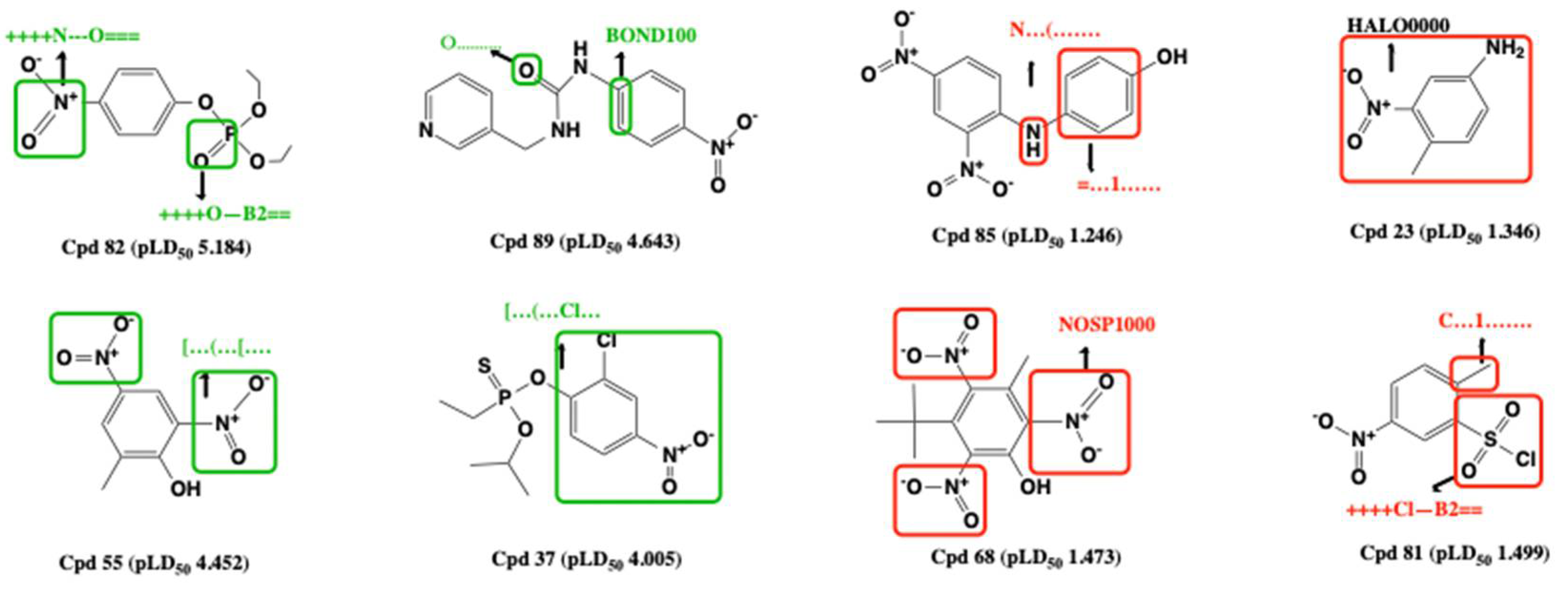
- Hao, Y.X.; Sun, G.H.; Fan, T.J.; Tang, X.Y.; Zhang, J.; Liu, Y.D.; Zhang, N.; Zhao, L.J.; Zhong, R.G.; Peng, Y.Z. In vivo toxicity of nitroaromatic compounds to rats: QSTR modelling and interspecies toxicity relationship with mouse. J. Hazard. Mater. 2020, 399, 122981. [Google Scholar] [CrossRef]
- Wang, L.L.; Ding, J.J.; Pan, L.; Fu, L.; Tian, J.H.; Cao, D.S.; Jiang, H.; Ding, X.Q. Quantitative structure-toxicity relationship model for acute toxicity of organophosphates via multiple administration routes in rats and mice. J. Hazard. Mater. 2021, 401, 123724. [Google Scholar] [CrossRef] [PubMed]
- Sun, Y.Z.; Li, Z.J.; Yan, X.L.; Wang, L.; Meng, F.H. Study on the quantitative structure–toxicity relationships of benzoic acid derivatives in rats via oral LD 50. Med. Chem. Res. 2009, 18, 712–724. [Google Scholar] [CrossRef]
- Mondal, D.; Ghosh, K.; Baidya, A.T.; Gantait, A.M.; Gayen, S. Identification of structural fingerprints for in vivo toxicity by using Monte Carlo based QSTR modeling of nitroaromatics. Toxicol. Mech. Methods 2020, 30, 257–265. [Google Scholar] [CrossRef] [PubMed]
- Kuz’min, V.E.; Muratov, E.N.; Artemenko, A.G.; Gorb, L.; Qasim, M.; Leszczynski, J. The effects of characteristics of substituents on toxicity of the nitroaromatics: HiT QSAR study. J. Comput.-Aided Mol. Des. 2008, 22, 747–759. [Google Scholar] [CrossRef]
- Keshavarz, M.H.; Akbarzadeh, A.R. A simple approach for assessment of toxicity of nitroaromatic compounds without using complex descriptors and computer codes. SAR QSAR Environ. Res. 2019, 30, 347–361. [Google Scholar] [CrossRef] [PubMed]
- Harvey, R.G. Polycyclic Aromatic Hydrocarbons: Chemistry and Carcinogenicity; CUP Archive: Cambridge, UK, 1991. [Google Scholar]
- Cogliano, V.; Baan, R.; Straif, K.; Grosse, Y.; Secretan, B.; El Ghissassi, F. Carcinogenicity of human papillomaviruses. Lancet Oncol. 2005, 6, 204. [Google Scholar] [CrossRef]
- Hornberg, J.J.; Laursen, M.; Brenden, N.; Persson, M.; Thougaard, A.V.; Toft, D.B.; Mow, T. Exploratory toxicology as an integrated part of drug discovery. Part II: Screening strategies. Drug Discov. Today 2014, 19, 1137–1144. [Google Scholar] [CrossRef]
- Wang, X.; Lin, Z.; Yin, D.; Liu, S.; Wang, L. 2D/3D-QSAR comparative study on mutagenicity of nitroaromatics. Sci. China Ser. B Chem. 2005, 48, 246–252. [Google Scholar] [CrossRef]
- Zhang, Z.; Niu, J.; Zhi, X. A QSAR model for predicting mutagenicity of nitronaphthalenes and methylnitronaphthalenes. Bull. Environ. Contam. Toxicol. 2008, 81, 498–502. [Google Scholar] [CrossRef] [PubMed]
- Ding, Y.L.; Lyu, Y.C.; Leong, M.K. In silico prediction of the mutagenicity of nitroaromatic compounds using a novel two-QSAR approach. Toxicol. In Vitro 2017, 40, 102–114. [Google Scholar] [CrossRef] [PubMed]
- Papa, E.; Pilutti, P.; Gramatica, P. Prediction of PAH mutagenicity in human cells by QSAR classification. SAR QSAR Environ. Res. 2008, 19, 115–127. [Google Scholar] [CrossRef] [PubMed]
- Morales, A.H.; Pérez, M.Á.C.; Combes, R.D.; González, M.P. Quantitative structure activity relationship for the computational prediction of nitrocompounds carcinogenicity. Toxicology 2006, 220, 51–62. [Google Scholar] [CrossRef] [PubMed]


| Name | Source or Application | Toxicity | Organism | References |
|---|---|---|---|---|
| TNT | explosive | inhibition of cell growth and cell viability, mutagenic, liver damage, cataract | mammal | [9,10] |
| DNT | dye, medicine, rubber | central nervous system and respiratory system depression, ataxia, reproductive toxicity | rat | [11] |
| DNP | dye, pesticide, herbicide | weight loss, lethal, cataract, tumorigenicity | human | [12] |
| Nitrofuran | antibiotics | tumorigenicity, mutagenicity, carcinogenicity | human | [13] |
| Mononitrophenol | explosive, dye, pharmaceutical, rubber, | carcinogenicity | human | [14] |
| 1-Nitropyrene | diesel | mutagenicity, carcinogenicity | human, bacteria | [15] |
| 2-Nitronaphthalene | vehicle fuel | DNA damage | human, bacteria | [16] |
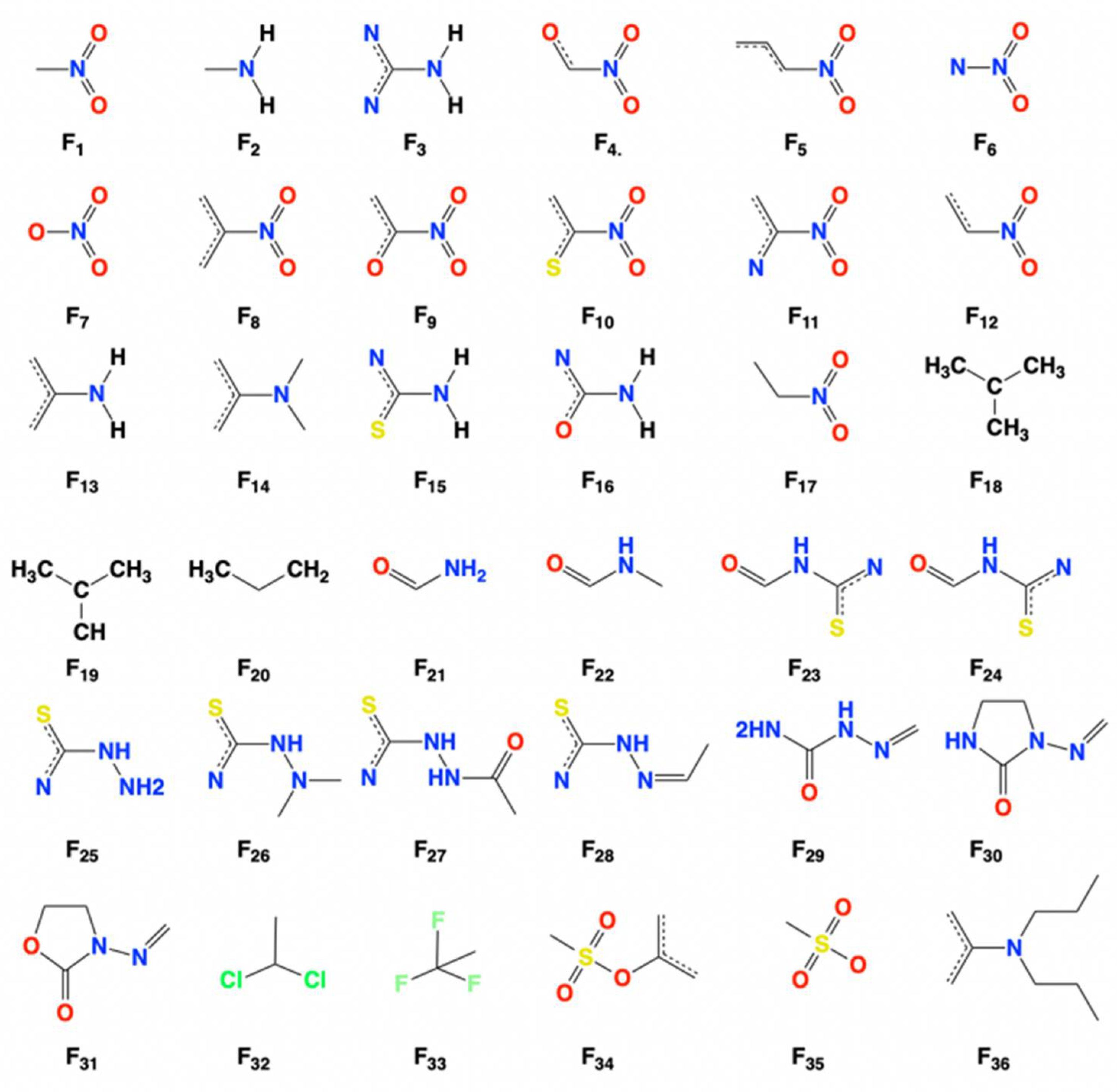
| Studied Fragments | Fragment Contributions | Studied Fragments | Fragment Contributions |
|---|---|---|---|
| F1 | −0.036 | F19 | +0.188 |
| F2 | −0.420 | F20 | +0.649 |
| F3 | −0.718 | F21 | −0.235 |
| F4 | −0.134 | F22 | +0.581 |
| F5 | −0.260 | F23 | −0.466 |
| F6 | −0.058 | F24 | +0.791 |
| F7 | −0.040 | F25 | −1.390 |
| F8 | −0.243 | F26 | −2.329 |
| F9 | −0.233 | F27 | −0.799 |
| F10 | −0.321 | F28 | −0.779 |
| F11 | −0.186 | F29 | −0.661 |
| F12 | −0.175 | F30 | −0.202 |
| F13 | −1.112 | F31 | −0.092 |
| F14 | −0.638 | F32 | −2.770 |
| F15 | −0.912 | F33 | +0.010 |
| F16 | −0.666 | F34 | −0.871 |
| F17 | +0.180 | F35 | −0.605 |
| F18 | −1.206 | F36 | +0.890 |
| Toxicity Endpoints | Chemicals | Molecular Descriptors | Organisms | References |
|---|---|---|---|---|
| Aquatic Toxicity | Pesticides | PSA, , , S, B, V, CV | aquatic crustaceans | [41] |
| Nitrophenols | Hydrophobicity, harness, electrophilicity | Chlorella vulgaris | [44,49] | |
| 26 NACs | E12, BCF, EHOMO, ELUMO, KOW | Scenedesmus obliguus | [42] | |
| 19 NACs | EHOMO, ELUMO, KOW, distribution coefficient | Scenedesmus vacuolatus | [43] | |
| Acute Toxicity | 19 NACs | Xv, Kα, Σσ−, ELUMO, logP and I | carp | [53] |
| 25 NACs | KOW, ELUMO and | Scenedesmus obliguus | [54] | |
| 28 NACs | Hydrophobicity, electrostatic, Van der Waals interactions, -F and -OH | rat | [59] | |
| 128 NACs | The van der Waals surface area, the presence of C-F at topological distance 6, and high frequency of C-N at topological distance 9 | rat | [55] | |
| 90 NACs | Presence of specific substructures generated from Monte Carlo | rat | [58] | |
| Mutagenicity & Carcinogenicity | 219 NACs | Hydrophobicity and ELUMO | TA98/100 | [64] |
| 48 NACs | EHOMO, Hypnotic-80, Infective-50, TIC2, and CATS2D_04_LL | TA100 | [47] | |
| 16 NNs and MNNs | Hf, CCR (core-core repulsion energy), EHOMO-1, ELUMO + EHOMO | TA98 | [65] | |
| 282 NACs | ω, hydrophobicity, ELUMO, qc2, MRo, I(diNO2) and dipole moment | TA98 | [66] | |
| 11 NPAHs | S1K and nArNO2 | human h1A1v2 cells | [67] | |
| 48 NACs | Hydrophobicity, bond dipole moment, Gasteiger–Marsili charge, molar refractivity and specific molecular fragments | female rat | [68] |
Publisher’s Note: MDPI stays neutral with regard to jurisdictional claims in published maps and institutional affiliations. |
© 2021 by the authors. Licensee MDPI, Basel, Switzerland. This article is an open access article distributed under the terms and conditions of the Creative Commons Attribution (CC BY) license (https://creativecommons.org/licenses/by/4.0/).
Share and Cite
Huang, T.; Sun, G.; Zhao, L.; Zhang, N.; Zhong, R.; Peng, Y. Quantitative Structure-Activity Relationship (QSAR) Studies on the Toxic Effects of Nitroaromatic Compounds (NACs): A Systematic Review. Int. J. Mol. Sci. 2021, 22, 8557. https://0-doi-org.brum.beds.ac.uk/10.3390/ijms22168557
Huang T, Sun G, Zhao L, Zhang N, Zhong R, Peng Y. Quantitative Structure-Activity Relationship (QSAR) Studies on the Toxic Effects of Nitroaromatic Compounds (NACs): A Systematic Review. International Journal of Molecular Sciences. 2021; 22(16):8557. https://0-doi-org.brum.beds.ac.uk/10.3390/ijms22168557
Chicago/Turabian StyleHuang, Tao, Guohui Sun, Lijiao Zhao, Na Zhang, Rugang Zhong, and Yongzhen Peng. 2021. "Quantitative Structure-Activity Relationship (QSAR) Studies on the Toxic Effects of Nitroaromatic Compounds (NACs): A Systematic Review" International Journal of Molecular Sciences 22, no. 16: 8557. https://0-doi-org.brum.beds.ac.uk/10.3390/ijms22168557