Article

Vitamin D Reverses Disruption of Gut Epithelial Barrier Function Caused by Campylobacter jejuni

by
Fábia D. Lobo de Sá
1,
Steffen Backert
2,
Praveen K. Nattramilarasu
1,
Soraya Mousavi
3,
Geoffrey I. Sandle
4,
Stefan Bereswill
3,
Markus M. Heimesaat
3,
Jörg-Dieter Schulzke
1 and
Roland Bücker
1,*
1
Nutritional Medicine/Clinical Physiology, Medical Department, Division of Gastroenterology, Infectious Diseases, Rheumatology, Charité—Universitätsmedizin Berlin, 12203 Berlin, Germany
2
Division of Microbiology, Department of Biology, University of Erlangen-Nürnberg, 91058 Erlangen, Germany
3
Institute of Microbiology, Infectious Diseases and Immunology, Charité—Universitätsmedizin Berlin, 12203 Berlin, Germany
4
Institute of Medical Research at St James’s, St James’s University Hospital, Leeds LS9 7TF, UK
*
Author to whom correspondence should be addressed.
Int. J. Mol. Sci. 2021, 22(16), 8872; https://0-doi-org.brum.beds.ac.uk/10.3390/ijms22168872
Submission received: 29 July 2021 / Revised: 9 August 2021 / Accepted: 16 August 2021 / Published: 18 August 2021
(This article belongs to the Special Issue The Mechanism of Action of Food Components in Disease Prevention 2020)

Abstract

:
Infections by the zoonotic foodborne bacterium Campylobacter jejuni (C. jejuni) are among the most frequent causes of bacterial gastroenteritis worldwide. The aim was to evaluate the relationship between epithelial barrier disruption, mucosal immune activation, and vitamin D (VD) treatment during C. jejuni infection, using intestinal epithelial cells and mouse models focused on the interaction of C. jejuni with the VD signaling pathway and VD treatment to improve C. jejuni-induced barrier dysfunction. Our RNA-Seq data from campylobacteriosis patients demonstrate inhibition of VD receptor (VDR) downstream targets, consistent with suppression of immune function. Barrier-preserving effects of VD addition were identified in C. jejuni-infected epithelial cells and IL-10−/− mice. Furthermore, interference of C. jejuni with the VDR pathway was shown via VDR/retinoid X receptor (RXR) interaction. Paracellular leakiness of infected epithelia correlated with tight junction (TJ) protein redistribution off the TJ domain and apoptosis induction. Supplementation with VD reversed barrier impairment and prevented inhibition of the VDR pathway, as shown by restoration of transepithelial electrical resistance and fluorescein (332 Da) permeability. We conclude that VD treatment restores gut epithelial barrier functionality and decreases bacterial transmigration and might, therefore, be a promising compound for C. jejuni treatment in humans and animals.

1. Introduction

The most common cause of bacterial gastroenteritis worldwide is the foodborne zoonotic bacterium Campylobacter jejuni (C. jejuni). C. jejuni is an intestinal commensal in a variety of animals (particularly in poultry), but after fecal–oral transmission is pathogenic in the human intestine, causing an acute infective colitis. Campylobacteriosis is characterized by watery, bloody diarrhea, abdominal cramps, fever, and intestinal inflammation [1]. The infection is usually self-limiting and only symptomatic treatment is required, but chronic progression or infections in immunocompromised patients require antibiotic treatment. A proportion of campylobacteriosis patients develop sequelae such as Guillain–Barré syndrome, reactive arthritis, or irritable bowel syndrome (IBS). Studies in C. jejuni-infected volunteers indicated that an acute inflammatory response is a hallmark of the infection [1]. Experimental studies revealed a strong interrelation between the mucosal immune response and epithelial barrier dysfunction in C. jejuni infection: the ‘leaky gut’ phenomenon [2,3,4].
The pre-hormone vitamin D (VD) regulates innate and adaptive immune responses. Its precursor cholecalciferol (calciol; (3β,5Z,7E)-9,10-secocholesta-5,7,10(19)-trien-3-ol) is metabolized to the intermediate calcidiol (25-hydroxycholecalciferol) and then to biologically active vitamin D3 (calcitriol; 1,25-dihydroxycholecalciferol). Low serum VD levels increase susceptibility to acute and chronic illnesses such as infectious diseases, IBS, inflammatory bowel disease (IBD), and colorectal cancer [5,6,7,8]. VD is well known for its role in bone metabolism, but is also involved in regulating the absorptive and immune functions of the gastrointestinal tract [9]. In the intestine, vitamin D is required for cell differentiation, proliferation, and for the maintenance of epithelial barrier function, in addition to its effects on calcium and phosphate transport [10]. The biological active form of VD is hydroxylated to calcitriol by 1-α-hydroxylase (also known as cytochrome P450 enzyme CYP27B1) and regulates further target genes in a complex with the VD receptor (VDR) and the retinoid X receptor (RXR). Evidence for the barrier-strengthening potential of VD has come from studies in IBD [11]. VD also exhibited barrier-protective and anti-inflammatory properties in in vitro and in vivo studies, but without a clear explanation for its molecular mechanism of action [12,13,14,15,16,17]. Nevertheless, clinical studies have shown a beneficial effect of VD treatment on diarrhea in IBD patients [18], which raises the possibility that it might also improve bacteria-induced diarrhea.
In this regard, VD supplementation increased the viability of intestinal epithelial cells, and partially restored the reduced transepithelial electrical resistance (TER) that occurred during C. jejuni infection [2]. Furthermore, in a preclinical intervention mouse study, VD decreased the inflammatory response that occurred during campylobacteriosis [14]. In the present study, we built on our previous work using bioinformatics to identify the relevance of the VD pathway in campylobacteriosis [2] by investigating (i) whether C. jejuni-induced barrier dysfunction can be treated or prevented by VD supplementation, and (ii) how C. jejuni interferes with the VD pathway.

2. Results

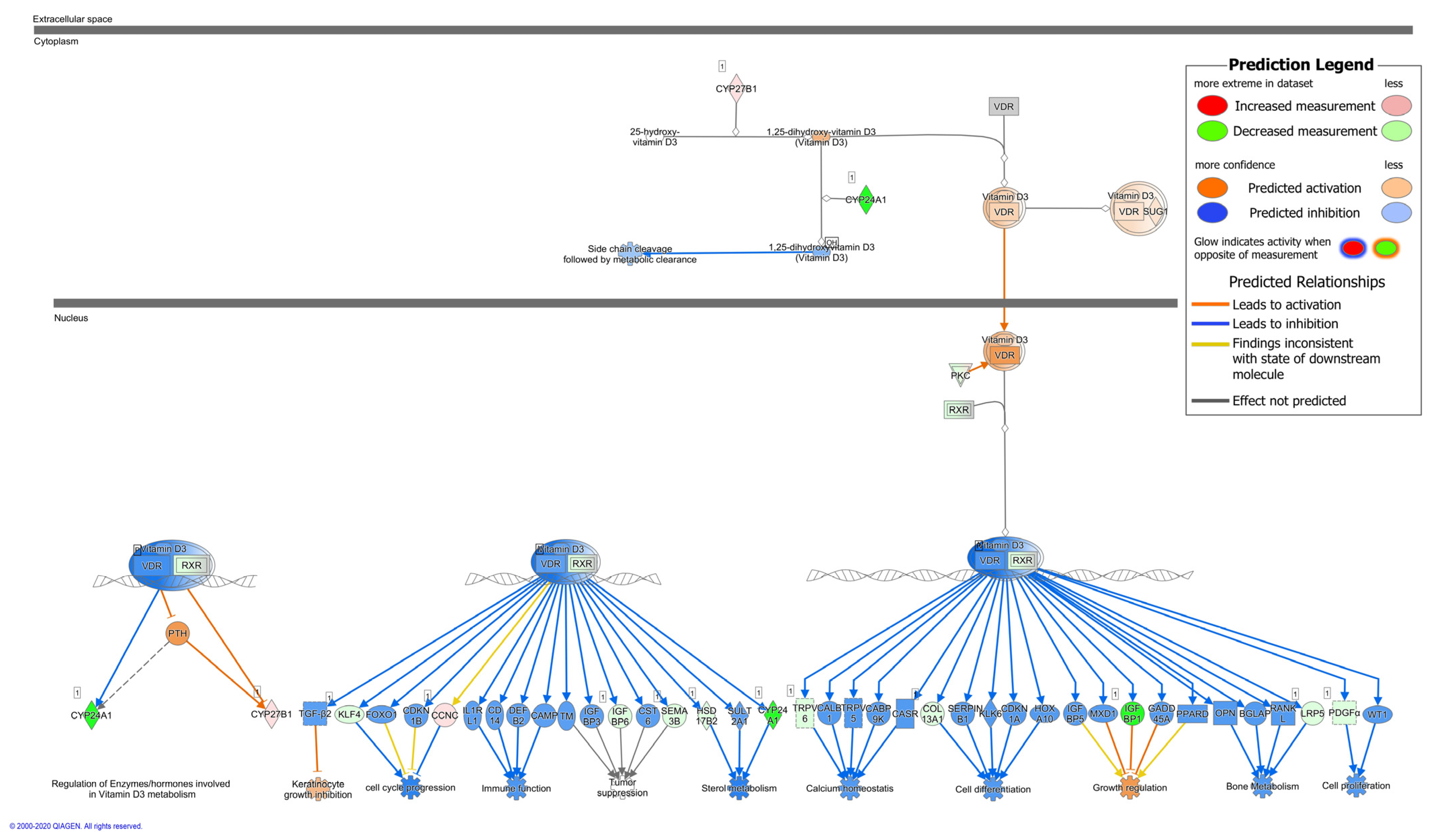
2.1. Upstream Regulator Analysis of C. jejuni—Infected Human Colonic Mucosa and VDR Downstream Targets

Our RNA-Seq analysis of colonic mucosa from C. jejuni-infected patients predicted inhibition of the ability of vitamin D (VD) to act as an upstream regulator [2]. Examination of this analysis in detail revealed that active VD (calcitriol) had a negative activation Z-score of −6.3 with an overlap p-value of 8.97 × 10⁻⁵. This highly significant negative Z-score with inhibited VDR pathways suggested that administration of VD might counter-regulate inhibited VD downstream targets and protect the host intestine from C. jejuni infection. Based on the RNA-Seq data from colonic mucosa of C. jejuni-infected patients, activity patterns of vitamin D receptor (VDR)/retinoid X receptor (RXR) pathways were analyzed using Ingenuity Pathway Analysis (IPA) software, which demonstrated that downstream targets related to immune regulation, cell proliferation, and cell differentiation in the patients’ colon mucosae were largely inhibited during campylobacteriosis (Figure 1).
For example, in the case of downstream VDR/RXR pathways regulating immune function, target genes for beta-defensin 2 (DEFB2, DEFB4), cathelicidin (CAMP), CD14 (CD14) and IL-1 receptor (IL1RL1, activated by IL-1β), were inhibited during C. jejuni infection as indicated (Figure 1, blue arrows). During infection, most downstream targets should show activation of their respective pathways (Figure 1, orange arrows). This has led to the hypothesis that C. jejuni directly interferes with the VDR pathway, which is targeted for activation in other infections [19].

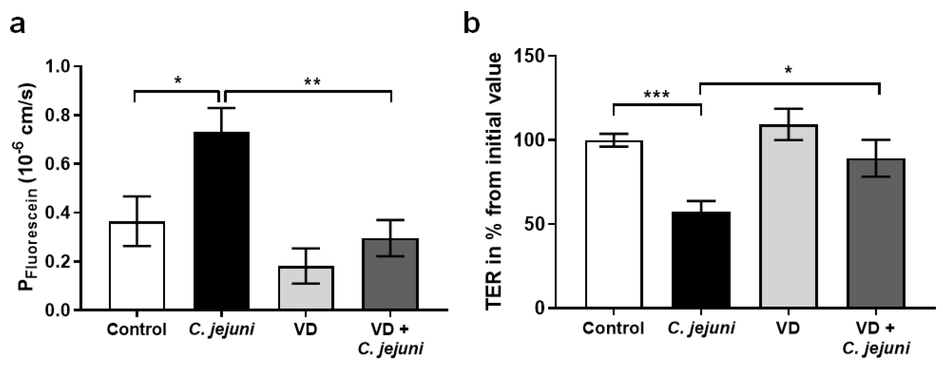
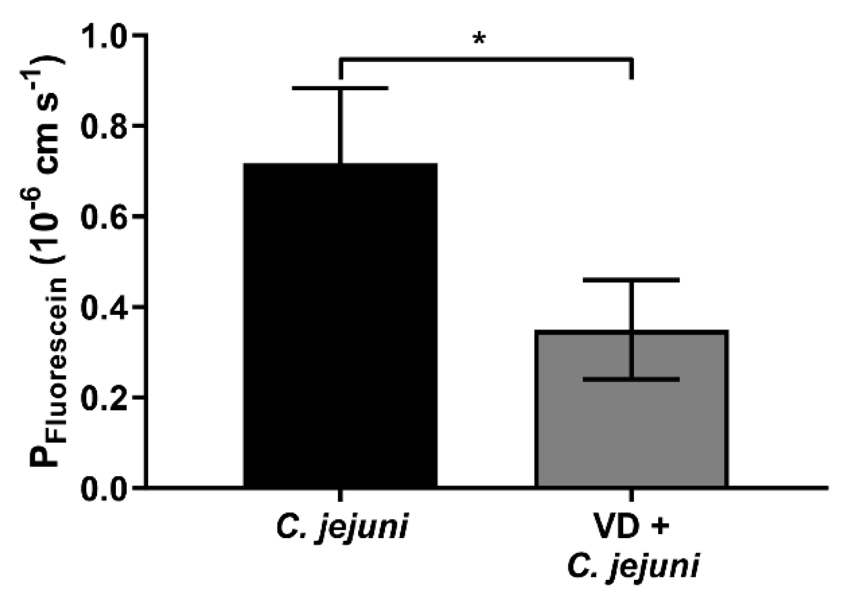
2.2. Epithelial Barrier Dysfunction in C. jejuni Infection Is Reversed by Vitamin D

Based on RNA-Seq data from campylobacteriosis patients, the predicted potential effectiveness of active VD against the pathophysiological changes induced by C. jejuni was investigated in more detail using different in vitro and in vivo approaches. In in vitro studies, C. jejuni increased the paracellular permeability of HT-29/B6-GR/MR cell monolayers to fluorescein (332 Da) (Figure 2a). In the presence of VD, permeability to fluorescein was even lower than in controls. Moreover, C. jejuni decreased transepithelial electrical resistance (TER), reflecting an increase in ionic permeability (Figure 2b). These changes point to disruption of barrier function in the intestinal epithelial cell monolayers. However, the presence of VD in C. jejuni-infected monolayers prevented the barrier-disrupting effect of the bacterium, and TER and epithelial permeability were restored to control levels (Figure 2).
In order to obtain confirmatory evidence in an in vivo ‘leaky gut’ mouse model, pre-conditioned IL-10−/− mice were infected with C. jejuni and treated with cholecalciferol added to their drinking water. Animals receiving cholecalciferol showed a lower permeability to 332 Da fluorescein compared to placebo-treated C. jejuni-infected mice (Figure 3). In accordance with 3R principles guidelines, the number of animals studied was minimized by using animals from a collaborative study [14].

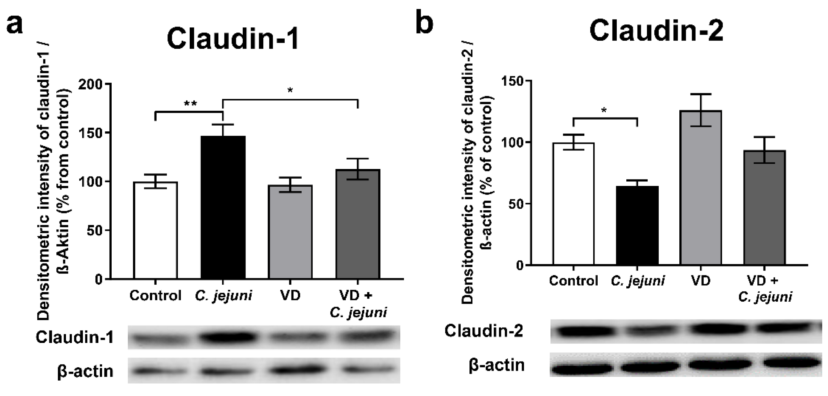
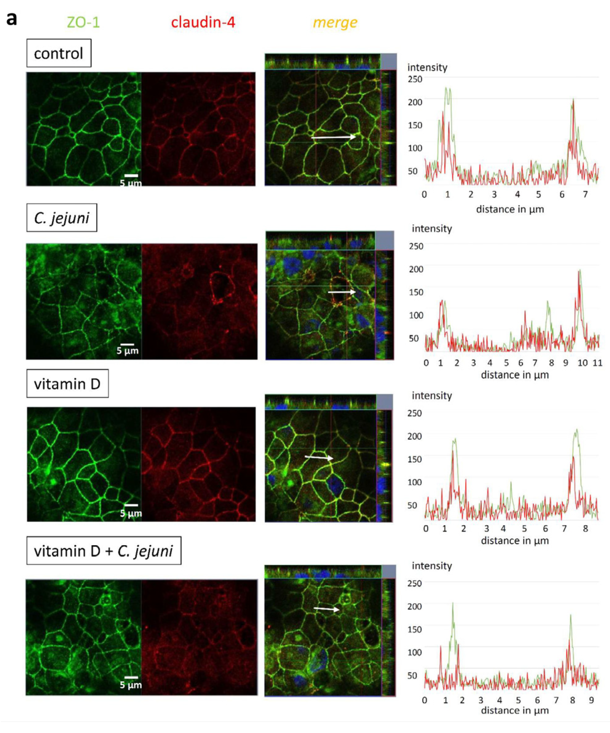
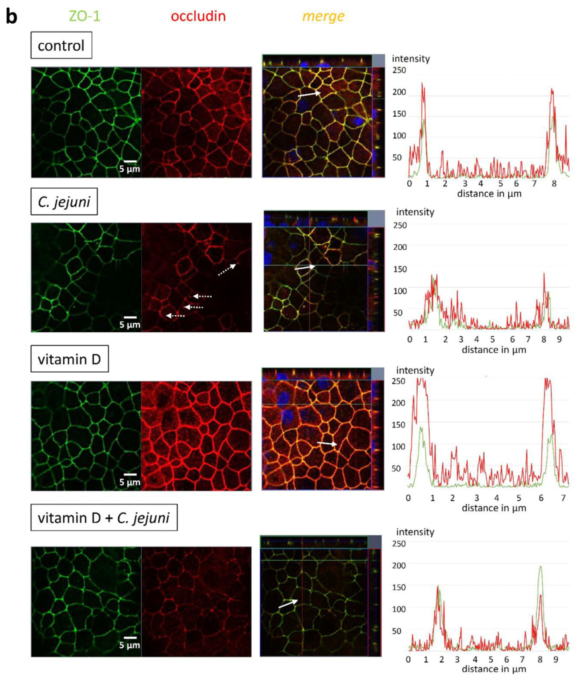
2.3. Influence of C. jejuni and Vitamin D on the Protein Expression and Distribution of Tight Junction Proteins

Intestinal epithelial barrier dysfunction correlates with changes in the expression or subcellular distribution of tight junction (TJ) proteins. Protein expression of different barrier-relevant TJ proteins in epithelial monolayers was quantified by densitometric analysis of Western blots, and revealed that C. jejuni infection caused no decreases in the expression of the sealing TJ proteins claudin-4, -5, -8, and the TJ proteins claudin-7, or occludin (Table 1).
C. jejuni infection also increased the expression of the barrier-forming TJ protein claudin-1 (Figure 4a) and decreased that of channel-forming claudin-2 (Figure 4b) but neither of these changes could account for C. jejuni-induced barrier impairment. VD inhibited the C. jejuni-induced changes in claudin-1 expression, which also does not explain the changes in the barrier function induced by infection.
C. jejuni-infected epithelial monolayers, either treated with VD or not, were immunostained for different barrier-relevant TJ proteins and visualized with confocal laser-scanning microscopy (CLSM). Microscopic signals from TJ proteins claudin-4 (Figure 5a) and occludin (Figure 5b) showed that the TJ domain was redistributed into intracellular compartments after C. jejuni infection, and both were less co-localized with zonula occludens protein-1 (ZO-1), changes consistent with impaired epithelial barrier function. This C. jejuni infection-induced redistribution of TJ proteins was inhibited by VD treatment (Figure 5) in keeping with the ability of VD to restore barrier function normal levels.

2.4. Vitamin D Prevents C. jejuni—Induced Epithelial Apoptosis

As the observed changes in TJ protein expression and distribution may not have been the only cause of barrier dysfunction in the epithelial cell monolayers, the role of apoptosis was analyzed by fluorescence microscopy and TdT-mediated dUTP-biotin nick end labeling (TUNEL) assay. C. jejuni infection increased the apoptosis ratio, which was prevented by VD treatment (Figure 6).

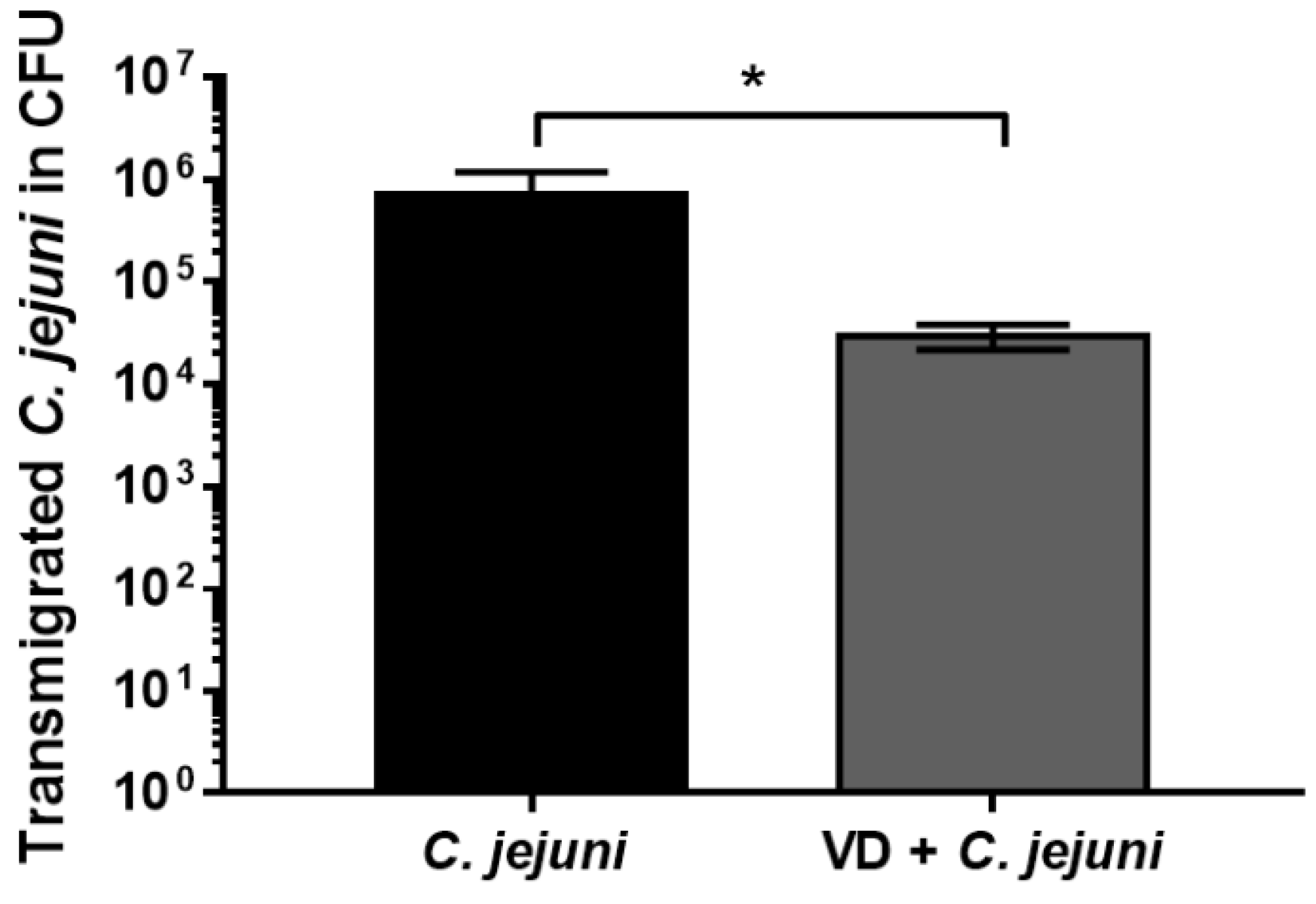
2.5. Vitamin D Supplementation Reduces the Number of Transmigrated C. jejuni

C. jejuni invades the intestinal epithelium, transmigrating from the lumen to the underlying immune cells, resulting in activation of the innate immune response. In our HT-29/B6 cell culture system, VD inhibited bacterial transmigration 8 h after infection with C. jejuni (Figure 7).
In addition, the presence of 200 nM VD inhibited bacterial transmigration between MKN-28 cells after infection from 3.4 ± 0.5 × 10⁷ CFU to 1 ± 0.8 × 10⁵ CFU (n = 3, p-value = 0.000645). These observations indicate that VD inhibited the development of barrier dysfunction, thereby maintaining barrier functionality, and thus minimizing bacterial translocation.

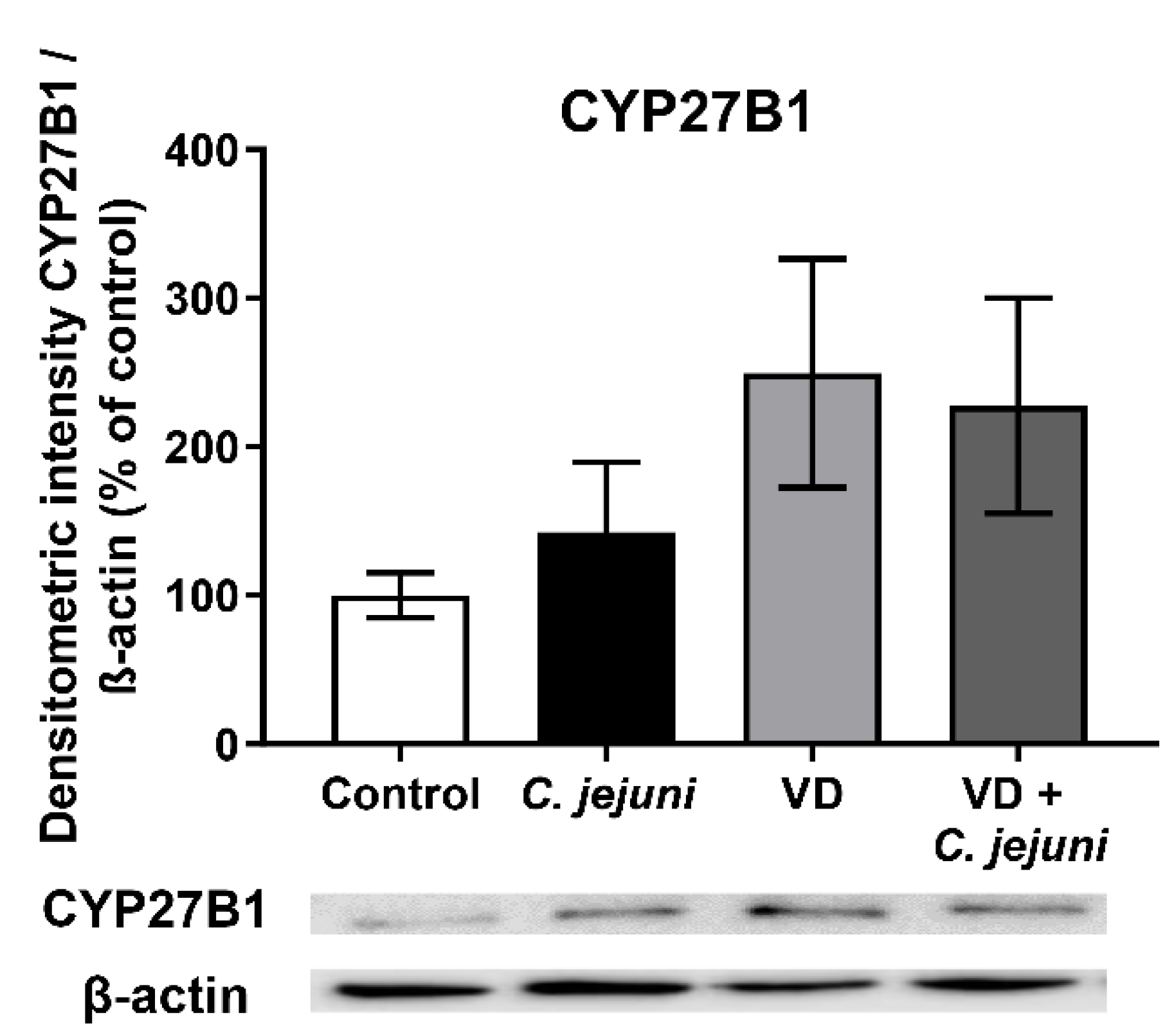
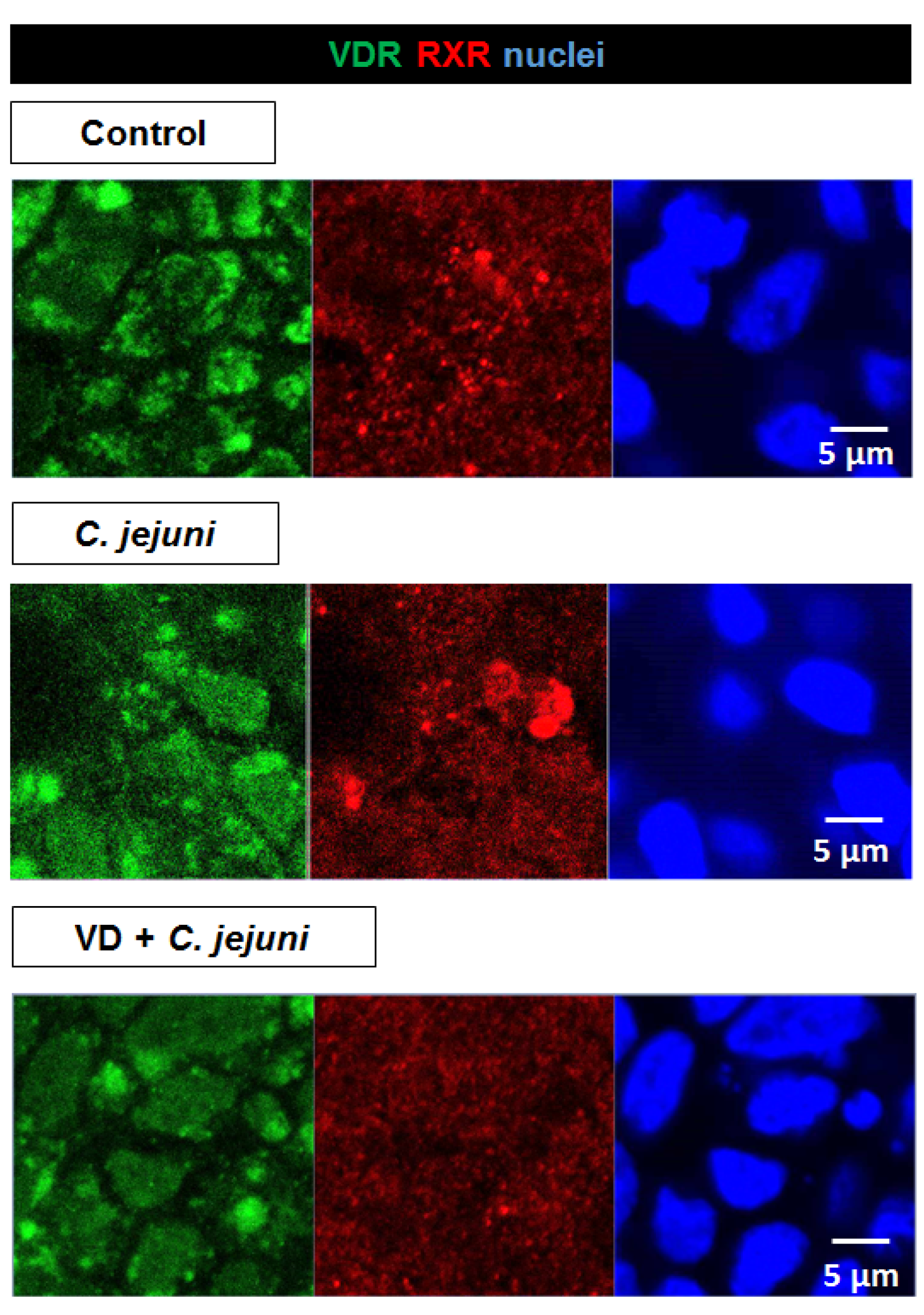
2.6. Influence of C. jejuni on the Vitamin D Pathway and Its Functional Importance

Using IPA, upstream regulator analysis of colon mucosae from C. jejuni-infected patients revealed inhibited activation states of the VDR (negative Z-score −3.2, p = 1 × 10⁻⁴) and CYP27B1 (negative Z-score −2.4, p = 5.5 × 10⁻³). However, RNA-Seq data indicated that VDR gene expression was unchanged, while that of CYP27B1 was increased (Figure 1). Similar results for CYP27B1 were obtained in Western blot analyses of C. jejuni-infected human HT-29/B6-GR/MR intestinal epithelial cells (Figure 8). CYP27B1 protein expression was not significantly different between C. jejuni-infected epithelial cells and controls, although it tended to increase during infection, particularly in the presence of VD.
VDRs are normally located within the nuclei. However, in C. jejuni-infected cells, VDR signals moved from the nucleoplasm to cytosolic compartments and were re-localized to the nuclei after VD treatment (Figure 9).
In addition, we used CLSM to analyze the VDR/retinoid X receptor (RXR) complex, VDR/RXR heterodimers being generally present in both cytoplasm and nucleus, with a greater amount in the nucleus [20]. Interestingly, fluorescence signals from RXR were more condensed (i.e., accumulated) in the C. jejuni-infected group than in the controls (Figure 9). VD treatment of C. jejuni-infected cells inhibited this condensation of the VDR/RXR complexes, so that the distribution of VDR/RXR complexes was similar to that seen in uninfected untreated controls (Figure 9). Condensation of VDR/RXR in the C. jejuni-infected group suggests strongly that C. jejuni infection interferes with downstream VDR signaling, leading to inhibition of downstream target genes and functional inhibition of their DNA transcripts, as predicted by IPA in the RNA-Seq data from the colon mucosae of C. jejuni-infected patients (Figure 1).

3. Discussion

Vitamin D status has an impact on the pathogenesis of IBD, colorectal cancer [5,6,7], and acute enteric infections such as that produced by Escherichia coli [8,21,22]. This suggests that a VD therapy in the form of cholecalciferol or calcitriol may have a place in the management of some common intestinal infections, and patients have been treated previously with doses ranging from 1800 to 10,000 international units (IU) per day [18,23]. VD status has also been shown to influence the disease outcome of corona virus (COVID-19) infection in risk groups [24]. As C. jejuni infections are a major clinical problem, we questioned whether VD might also influence the pathogenesis of C. jejuni enteritis and, if so, whether VD might have a positive impact on the prevention and/or treatment of this type of intestinal infection. To date, our previous studies have established that VD has a role in C. jejuni-induced barrier dysfunction [2,14] without identifying the mechanisms underlying its protective properties.

3.1. Effect of C. jejuni and Vitamin D on Tight Junction Protein Expression, Localization, and Apoptosis Induction

Previous studies have demonstrated epithelial barrier disturbance, with decreased TER and increased permeabilities for high molecular weight paracellular markers in C. jejuni-infected in vitro cell lines, in vivo mouse models, and colonic mucosae from C. jejuni-infected patients. In all of these models, barrier dysfunction reflected a combination of changes in TJ protein expression and localization and the induction of apoptosis [2,3,4,14]. Similarly, in the present study in C. jejuni-infected HT-29/B6 cells, we observed a decrease in TER and an increased permeability to fluorescein (332 Da), and both changes were reversed by VD. VD has also been shown to restore impaired barrier function induced by ethanol [12], TNF-α [13,25], and dextran sulfate sodium (DSS) [26,27] in other in vitro models.
Using C. jejuni-infected IL-10−/− mice as an in vivo ‘leaky gut’ model, high-dosage cholecalciferol added to their drinking water reversed the infection-induced increase in colonic permeability to fluorescein, confirming the results of our in vitro experiments. The restorative effects of cholecalciferol on the permeability of larger molecules such as FITC-dextran (4 kDa) have also been reported in DSS and 2,4,6-trinitrobenzenesulfonic acid (TNBS)-induced mouse model of colitis [25,27]. In colon mucosae of C. jejuni-infected IL-10−/− mice treated with cholecalciferol, TER was similar to that in control mice 6 days post-infection, and this effect of cholecalciferol was associated with reduced intestinal and extra-intestinal inflammatory responses, and a decrease in innate immune cells as well as diminished diarrheal frequency during the development of campylobacteriosis [14]. We speculate that calcitriol administration may have an even greater beneficial effect.
Disruption of the epithelial barrier function reflects changes in the expression and/or localization of TJ proteins. As in other models, expression of the sealing TJ protein claudin-1 increased during C. jejuni infection, despite the decrease in TER. This is called the ‘claudin-1 paradox’, and is due to the disassembly of claudin-1 away from the TJ domain [2]. Further mechanisms involve trafficking of TJ components or disruption of the perijunctional actomyosin cytoskeleton such as F-actin reorganization as shown, e.g., for Aeromonas hydrophila toxin, where TJ proteins were retraced from the TJ domain by actomyosin constriction or caveolin-dependent endocytosis of intrajunctional occludin [28,29,30]. In addition, C. jejuni downregulates the water- and cation-channel TJ protein claudin-2, which shows some increase after VD treatment and, in our model, may represent counter-regulation. Claudin-2 gene expression is a direct target of the VDR and is upregulated via the VDR promotor binding site VDRE [15]. Interestingly, recent studies demonstrated a link between downregulation of VDR and overexpression of CLDN2 in Salmonella infection [16,31]. Redistribution of occludin and of the sealing TJ protein claudin-4 induced by C. jejuni contributes to barrier dysfunction by opening the paracellular leak pathway to ions, water, and macromolecules, so that antigen influx activates immune cells and triggers cytokine release, which further compromises the epithelial barrier and results in a ‘leaky gut’. In our in vitro model, VD restored, at least in part, the expression and normal distribution of the TJ proteins, as reported in TNF-α-stimulated Caco-2 cells [25] and in T84 cells or in colonic biopsies from patients with ulcerative colitis [32].
Apoptosis also contributes to epithelial leakiness and the ‘leaky gut’. The C. jejuni-induced apoptosis contributes to epithelial barrier dysfunction as shown in a previous study, with the pan-caspase inhibitor Q-VD-OPh, which restores the decreased TER and the increased permeability to fluorescein (332 Da) and FITC-dextran (4 kDa) [4]. Due to its anti-tumorigenic and either pro- or anti-apoptotic effects in different cell types, VD is commonly used as a supplement in cancer therapy [33]. We found that VD treatment inhibited apoptosis in C. jejuni-infected HT-29/B6 cells. This anti-apoptotic effect has also been reported in cholecalciferol-treated C. jejuni-infected IL-10−/− mice [14], resulting in a decrease in antigen influx via the paracellular route.
C. jejuni first adhere to intestinal epithelial cells, then invade the epithelial layer and transmigrate via paracellular and transcellular pathways [34]. C. jejuni secrets a virulence factor—serine protease high-temperature requirement A (HtrA)—which has a critical role in transmigration, in that it cleaves epithelial junction proteins such as occludin and E-cadherin in Caco-2 cells, leading to enhanced bacterial invasion and paracellular transmigration [35,36,37]. We found that VD treatment decreased bacterial transmigration across C. jejuni-infected cell monolayers, which suggests that VD had an effect on the regulation of host cell signaling in addition to the VD-dependent cellular anti-bacterial mediators. The ability of VD to decrease C. jejuni transmigration has important functional implications, as it will tend to minimize both immune activation and the secondary deleterious effect of pro-inflammatory cytokines on barrier function (sustaining the ‘leaky gut’). In practical terms, this should result in a milder disease outcome.

3.2. C. jejuni Interference with the Vitamin D Pathway

Based on bioinformatic predictions from IPA of colon mucosae from C. jejuni-infected patients, the VD pathway showed overall inhibition of its activation state. This led us to hypothesize that C. jejuni infection might interfere with the VD pathway, and that VD supplementation might ameliorate C. jejuni-induced epithelial barrier defects. Evidence that VD has a protective effect on the intestinal epithelium in campylobacteriosis came from our preclinical intervention study in IL-10−/− mice, which demonstrated that peroral administration of cholecalciferol reduced the severity of inflammation during acute C. jejuni infection [14]. Furthermore, C. jejuni infection inhibited the immune function mediators IL-1β, sCD14, β-defensin, and cathelicidin, which suggests a wider effect on the regulation of intestinal immunity. As defensins and cathelicidin are important components of the innate defense mechanisms at the surface of host gastrointestinal epithelia, we predicted from our data that C. jejuni would suppress innate immune responses for bacterial survival within the mucosa. However, contrary to our prediction, increased defensin and cathelicidin was reported in C. jejuni-infected cultured monolayers of Caco-2 or HT-29 cells [38]. In addition, a study with Mycobacterium tuberculosis demonstrated that treatment with VD increased the level of mRNA encoding protective cathelicidin [39], and the ability of VD to enhance the expression of antimicrobial cathelicidin has been reviewed in detail elsewhere [40].
CYP27B1 (25-hydroxyvitamin D-1-α-hydroxylase) is an important regulator of VD mechanism, which not only converts cholecalciferol to biologically active vitamin D3 (calcitriol) but also indirectly regulates antimicrobial responses in the gastrointestinal tract by inducing the expression of, for example, cathelicidin [41,42]. In our study, CYP27B1 protein expression tended to increase in C. jejuni-infected cells compared to uninfected control cells. Data generated in cytokine-stimulated PBMCs support this finding, with enhanced CYP27B1 expression in response to stimulation by IFN-γ and LPS [43] and in human monocytes when exposed to pathogens [44].
Immunostaining of VDR within its complex with RXR indicated that C. jejuni interfered with the VDR pathway. After C. jejuni infection, VDR signals appeared no longer restricted to the nuclei and were present increasingly in the cytosol. A change in VDR localization in colonic epithelial cells has also been reported during Salmonella infection [19]. As C. jejuni lacks enterotoxins, its intracellular appearance in the intestinal epithelial cells might directly influence the VDR/RXR complex, supposable via cytoskeleton interactions or via pro-inflammatory pathways such as NF-κB, as shown for Salmonella [13,19]. We found that VD treatment reduced the level of heterodimers in the cytoplasm, as described in previous studies [20]. In addition, the VDR/RXR complex accumulates in the cytosol after C. jejuni infection, probably as a result of the effect of RXR on the distribution of VDR [20], which supports the view that downstream gene expression is negatively affected. Thus, beneficial antimicrobial and immune strengthening downstream target genes, and therefore genes involved in the protective functionality of the VD pathway are inhibited during Campylobacter infection, but can be rescued by therapeutic doses of VD.
In conclusion, C. jejuni infection suppresses VD signaling pathways, which may be part of the explanation for the rogue host response seen in campylobacteriosis. Nevertheless, administration of the pre-hormone VD protects against C. jejuni-induced epithelial barrier dysfunction in vitro and in vivo. VD should therefore be considered as a potentially useful therapeutic supplement, when combined with other barrier-protective, immunomodulatory, and anti-inflammatory compounds, such as curcumin or resveratrol [3,45,46], for prevention and treatment of acute and chronic gastrointestinal disorders.

4. Materials and Methods

4.1. Cell Culture and Treatment of the Cells

The human colon epithelial cell line HT-29/B6-GR/MR [47] was cultured as described previously [3]. Briefly, cells were grown in RPMI 1640 medium (Sigma Aldrich, St. Louis, MO, USA) supplemented with 10% fetal calf serum (FCS; Gibco, Carlsbad, CA, USA), 1% penicillin/streptomycin (Corning, Wiesbaden, Germany), G418 (300 µg/mL; Invitrogen, Carlsbad, CA, USA), and hygromycin B (200 µg/mL; Biochrom GmbH, Berlin, Germany) at 37 °C in a humidified 5% CO2 atmosphere. Cells were passaged once a week and seeded on Millicell PCF filter membranes with a pore size of 3 µm and an effective growth area of 0.6 cm2 (Merck Millipore, Billerica, MA, USA). Seven days after seeding, confluent cell monolayers were ready for use (TER 600–900 Ω·cm2). One day before infection, cells were washed twice with antibiotic-free cell culture medium supplemented with heat-inactivated FCS. Cells were pre-treated with 200 nM of the biological active vitamin D calcitriol (VD; Cayman Chemicals, Ann Arbor, MA, USA) for 2 h from the apical and basal sides.

4.2. C. jejuni Cultivation and Experimental Infection In Vitro

Campylobacter jejuni wildtype (wt) 81–176 reference strain was cultured for two days on blood agar plates (Oxoid, Thermo Scientific, Waltham, MA, USA) under microaerobic conditions (5–10% O2, 10% CO2, 85% N2) generated with CampyGen gaspacks (Oxoid, Thermo Scientific, Waltham, MA, USA) in plastic jars at 37 °C. Bacteria were then cultured microaerobically in Mueller-–Hinton broth (200 rpm) for 2 h at 37 °C. Bacteria were centrifuged (2 min, 5000× g, 10 °C) and the optical density at OD600 adjusted to OD 1 with RPMI 1640 cell culture medium. Cultured cells were infected from the apical side with a multiplicity of infection (MOI) of 100. After infection, cells were incubated for 48 h at 37 °C under microaerobic conditions to encourage bacterial growth. Polarized MKN-28 cells were infected with C. jejuni wt strain NCTC11168 for up to 24 h with (50 nM to 200 nM) or without the addition of VD.

4.3. Generation of Secondary Abiotic IL-10−/− Mice, Treatment and Experimental Infection In Vivo

IL-10−/− mice with a C57BL/6j background were bred and housed under specific pathogen-free conditions in the animal facility of the Forschungseinrichtung für Experimentelle Medizin of Charité—Universitätsmedizin Berlin. Animal experiments were performed in the animal facility of Charité—Universitätsmedizin Berlin according to the European animal welfare guidelines (2010/63/EU), the ARRIVE guidelines and the German animal protection law (ethic committee: LAGeSo Berlin with the approval number G0172/16, 13 October 2016). The standard housing conditions, generation of secondary abiotic mice, treatment, and the infection of mice has been described previously [14]. To remove commensal gut microbiota, mice were transferred to sterile cages and treated for 8 weeks with an antibiotic cocktail containing ampicillin/sulbactam (1 g/L; Ratiopharm GmbH, Ulm, Germany), ciprofloxacin (200 mg/L; Bayer Vital, Leverkusen, Germany), imipenem (250 mg/L; MSD, Kenilworth, NJ, USA), metronidazole (1 g/L; Fresenius, Bad Homburg, Germany), and vancomycin (500 mg/L; Cell Pharm, Bad Vilbel, Germany) added to drinking water provided ad libitum. The antibiotic cocktail was replaced by cholecalciferol (25-OH-cholecalciferol; Sigma Aldrich, St. Louis, MO, USA) 4 days before oral infection. Cholecalciferol was dissolved in Tween 80 (0.2% v/v) and administered to mice via the drinking water. Mice received a daily cholecalciferol treatment dose of 500 µg per kg body weight (equivalent to 20,000 IU per kg) [14]. At days 0 and 1, mice were then perorally infected with 108 colony forming units (CFU) of C. jejuni 81–176 in a volume of 0.3 mL PBS by gavage. Six days after infection, mice were sacrificed by isoflurane inhalation. Colon samples were removed and mounted in Ussing chambers in order to measure fluorescein fluxes (332 Da; Sigma Aldrich, St. Louis, MO, USA), as described previously [3].

4.4. Electrophysiological Methods

Transepithelial electrical resistance (TER) of cell monolayers was measured in vitro using chopstick electrodes under sterile conditions. TER was corrected for the resistance of an empty filter and calculated assuming a growth area of 0.6 cm2.

4.5. Epithelial Permeability Measurements

Measurements of fluorescein (332 Da) fluxes from apical to basolateral compartment were performed either in Ussing chambers for ex vivo samples or in 12-well plates for in vitro samples. Samples were taken every 15 min for one hour from the basolateral side, fluorescence measured spectrophotometrically (Tecan GmbH, Maennendorf, Switzerland), and permeability was calculated as flux/concentration differences.

4.6. Transmigration Assay

Polarized epithelial cell monolayers grown on 3 µm filter inserts were placed in 12-well plates and infected from the apical side with C. jejuni 81-176 or NCTC11168. A total of 25 µL of basolateral supernatant was removed in a time course after 2, 4, 8, and 24 h. The supernatant was diluted in cell culture medium and plated on to blood agar plates, which were incubated for 48 h under microaerobic conditions. The number of CFU was counted and multiplied with the factor of dilution to determine the number of transmigrated C. jejuni.

4.7. Western Blot

Changes in protein expression were quantified by Western blot analysis. Cells were washed twice with ice-cold PBS and lysed with whole cell lysis buffer containing 150 nM NaCl, 10 mM Tris buffer (pH 7.5), 0.5% Triton X-100, 1% SDS, and one tablet of Complete Protease Inhibitor (Roche AG, Mannheim, Germany) in a total volume of 10 mL. Cell lysis, protein quantification, and Western blot procedure were as described previously [3]. In brief, after cell lysis, cells were scraped carefully from the filters, transferred to reaction tubes, incubated 30–60 min on ice with regular vortexing, and centrifuged for 30 min (15,000× g at 4 °C). Proteins were detected on 12.5% polyacrylamide gels. Primary antibodies occludin (1:1000; Sigma Aldrich, St. Louis, MO, USA), claudin-1, -2, -4, -5, -7, -8 (1:1000; Invitrogen, Carlsbad, CA, USA), CYP27B1 (1:1000; Merck, Darmstadt, Germany), and β-actin (1:10,000; Sigma Aldrich, St. Louis, MO, USA) as loading control, were incubated slewing over night at 4 °C. Peroxidase-conjugated secondary antibodies to goat anti-rabbit IgG or goat anti-mouse IgG (Jackson ImmunoResearch, Ely, UK) were incubated slewing at room temperature for 2 h. Proteins were detected by incubating membranes in SuperSignal West Pico PLUS Stable Peroxide Solution (Thermo Scientific, Waltham, MA, USA), which were then visualized using a Fusion FX7 imaging system (Vilber Lourmat Deutschland GmbH, Eberhardzell, Germany), and analyzed by densitometry using Image Studio Lite version 5.2.

4.8. Immunostaining, Apoptosis Assay, and Confocal Microscopy

Epithelial cell monolayers were washed twice with PBS and fixed with 2% paraformaldehyde (PFA; Electron Microscopy Science, Hatfield, PA, USA) before staining to detect apoptosis, TJ proteins, nuclear factors, and effector proteins of the VD pathway as described previously [3]. The antibodies used were primary antibodies to occludin (1:100; Sigma Aldrich, St. Louis, MO, USA), claudin-4 (1:100; Invitrogen, Carlsbad, CA, USA), ZO-1 (1:100; BD Bioscience, Franklin Lakes, NJ, USA), VDR (1:100; Abcam, Cambridge, UK), RXR (1:100; Novus Biologicals, Littleton, CO, USA) as well as secondary antibodies conjugated to Alexa-Fluor 488 or 594 (1:500; Invitrogen, Carlsbad, CA, USA) with incubation for 1 h at 37 °C. Apoptotic events were detected using the TUNEL assay (In situ Cell Death Detection Kit, Roche, Mannheim, Germany) according to the manufacturer’s instructions. Nuclei were stained with 4′,6-diamidino-2-phenylindole (DAPI; 1:1000; Roche, Basel, Switzerland). Tight junction structure, subcellular protein distribution, and apoptotic cells were visualized by confocal laser-scanning microscopy (CLSM; Zeiss LSM780, Jena, Germany).

4.9. RNA Expression Analysis

RNA isolation from colonic samples from six control patients and four C. jejuni-infected patients, next-generation sequencing, and RNA-Seq expression analysis was performed and described previously [2]. Data were deposited in NCBI’s Gene Expression Omnibus (GEO ID GSE88710). Ingenuity Pathway Analysis software (IPA; Qiagen, Redwood, CA, USA) was used to analyze C. jejuni-dependent pathway activation.

4.10. Statistical Analysis

All data are presented as mean values ± standard error of the mean (SEM). Statistical analyses were performed with GraphPad Prism version 8.0 using Student´s t-test for comparison of two or one-way ANOVA comparing more than two groups with Bonferroni adjustment for multiple comparison. Data which were not normally distributed were analyzed using the nonparametric Mann–Whitney U-test. p < 0.05 was considered to be statistically significant.

Author Contributions

Conceptualization, F.D.L.d.S. and R.B.; Methodology, R.B., S.B. (Steffen Backert), J.-D.S., S.B. (Stefan Bereswill) and G.I.S.; Formal analysis, J.-D.S., F.D.L.d.S., S.B. (Steffen Backert), S.B. (Stefan Bereswill) and G.I.S.; Investigation, F.D.L.d.S., P.K.N., M.M.H. and S.M.; Data curation, M.M.H., F.D.L.d.S., P.K.N., and S.M.; Writing—original draft preparation, F.D.L.d.S. and R.B.; Writing—review and editing, J.-D.S., M.M.H., S.B. (Steffen Backert), S.B. (Stefan Bereswill) and G.I.S.; Supervision, R.B. and J.-D.S.; Project administration, R.B.; Funding acquisition, R.B., J.-D.S., S.B. (Steffen Backert), M.M.H. and S.B. (Stefan Bereswill). All authors have read and agreed to the published version of the manuscript.

Funding

This research was funded by the German Federal Ministry of Education and Research (BMBF) in frame of the zoonoses research consortium PAC-Campylobacter to RB and JDS (project IP8/01KI2007D), to SBa (IP9/01KI1725E), and to MMH and SB (IP7/01KI2007D). This work was supported by the German Research Foundation (DFG Schu559/11-3 and -5 and GRK 2318) to JDS.

Institutional Review Board Statement

For the use of human material, this study adhered to the Declaration of Helsinki. The study was approved by the local institutional review board “The Ethics Committee of the Charité” under the approval number EA4/123/09 (16 November 2009) and written informed consent was obtained from all patients 24 h prior to the intervention. Animal experiments were performed in the animal facility of Charité—Universitätsmedizin Berlin according to the European animal welfare guidelines (2010/63/EU), the ARRIVE guidelines and the German animal protection law (approved by LAGeSo Berlin with the approval number G0172/16, 13 October 2016).

Informed Consent Statement

Informed consent was obtained from all subjects involved in the study. Identification of patients and participants in the study is not possible from the data shown here.

Data Availability Statement

All generated data supporting the key findings are available in the manuscript. Raw data are available upon request. RNA-seq raw data are deposited at NCBI’s Gene Expression Omnibus under the GEO accession ID GSE88710.

Acknowledgments

We gratefully thank Anja Fromm and In-Fah Maria Lee for their excellent technical support. Manja Böhm is gratefully acknowledged for her contribution during transmigration experiments. We thank the CCG and Michal R. Schweiger from University of Cologne for the support in RNA-Seq.

Conflicts of Interest

The authors declare no conflict of interest.

Abbreviations

CFUcolony forming unit
CLSMconfocal laser-scanning microscopy
DAPI4′,6-diamidino-2-phenylindole
DSSdextran sulfate sodium
FCSfetal calf serum
HtrAhigh-temperature requirement A
IBDinflammatory bowel disease
IBSirritable bowel syndrome
IFNinterferon
IPAIngenuity Pathway Analysis
LPSlipopolysaccharide
MOImultiplicity of infection
ODoptical density
RXRretinoid X receptor
TERtransepithelial electrical resistance
TJtight junction
TNBS2,4,6-trinitrobenzenesulfonic acid
TNFtumor necrosis factor
TUNELTdT-mediated dUTP-biotin nick end labeling
VDvitamin D
VDRvitamin D receptor
ZO-1zonula occludens-1

References

  1. Black, R.E.; Levine, M.M.; Clements, M.L.; Hughes, T.P.; Blaser, M.J. Experimental Campylobacter jejuni Infection in Humans. J. Infect. Dis. 1988, 157, 472–479. [Google Scholar] [CrossRef]
  2. Bücker, R.; Krug, S.M.; Moos, V.; Bojarski, C.; Schweiger, M.R.; Kerick, M.; Fromm, A.; Janßen, S.; Fromm, M.; Hering, N.A.; et al. Erratum: Campylobacter jejuni impairs sodium transport and epithelial barrier function via cytokine release in human colon. Mucosal Immunol. 2017, 11, 575–577. [Google Scholar] [CrossRef] [Green Version]
  3. Lobo de Sá, F.D.; Butkevych, E.; Nattramilarasu, P.K.; Fromm, A.; Mousavi, S.; Moos, V.; Golz, J.C.; Stingl, K.; Kittler, S.; Seinige, D.; et al. Curcumin Mitigates Immune-Induced Epithelial Barrier Dysfunction by Campylobacter jejuni. Int. J. Mol. Sci. 2019, 20, 4830. [Google Scholar] [CrossRef] [Green Version]
  4. Butkevych, E.; Lobo de Sá, F.D.; Nattramilarasu, P.K.; Bücker, R. Contribution of Epithelial Apoptosis and Subepithelial Immune Responses in Campylobacter jejuni-Induced Barrier Disruption. Front. Microbiol. 2020, 11, 344. [Google Scholar] [CrossRef]
  5. Meeker, S.; Seamons, A.; Maggio-Price, L.; Paik, J. Protective links between vitamin D, inflammatory bowel disease and colon cancer. World J. Gastroenterol. 2016, 22, 933–948. [Google Scholar] [CrossRef] [PubMed]
  6. Assa, A.; Vong, L.; Pinnell, L.J.; Avitzur, N.; Johnson-Henry, K.C.; Sherman, P.M. Vitamin D Deficiency Promotes Epithelial Barrier Dysfunction and Intestinal Inflammation. J. Infect. Dis. 2014, 210, 1296–1305. [Google Scholar] [CrossRef] [PubMed] [Green Version]
  7. Abbasnezhad, A.; Amani, R.; Hasanvand, A.; Rad, E.Y.; Alipour, M.; Saboori, S.; Choghakhori, R. Association of Serum Vitamin D Concentration With Clinical Symptoms and Quality of Life in Patients With Irritable Bowel Syndrome. J. Am. Coll. Nutr. 2018, 38, 327–333. [Google Scholar] [CrossRef] [PubMed]
  8. Mahyar, A.; Ayazi, P.; Saffari Rad, M.; Dalirani, R.; Javadi, A.; Esmaeily, S. The Correlation Between Vitamin D and Bacterial Diarrhea in Children. Arch. Pediatr. Infect. Dis. 2019, 7, e84382. [Google Scholar] [CrossRef] [Green Version]
  9. Ooi, J.H.; Li, Y.; Rogers, C.J.; Cantorna, M.T. Vitamin D Regulates the Gut Microbiome and Protects Mice from Dextran Sodium Sulfate–Induced Colitis. J. Nutr. 2013, 143, 1679–1686. [Google Scholar] [CrossRef]
  10. Barbáchano, A.; Fernández-Barral, A.; Ferrer-Mayorga, G.; Costales, A.; Larriba, M.J.; Muñoz, A. The endocrine vitamin D system in the gut. Mol. Cell. Endocrinol. 2017, 453, 79–87. [Google Scholar] [CrossRef] [Green Version]
  11. Stio, M.; Retico, L.; Annese, V.; Bonanomi, A.G. Vitamin D regulates the tight-junction protein expression in active ulcerative colitis. Scand. J. Gastroenterol. 2016, 51, 1193–1199. [Google Scholar] [CrossRef] [PubMed]
  12. Chen, S.-W.; Ma, Y.-Y.; Zhu, J.; Zuo, S.; Zhang, J.-L.; Chen, Z.-Y.; Chen, G.-W.; Wang, X.; Pan, Y.-S.; Liu, Y.-C.; et al. Protective effect of 1,25-dihydroxyvitamin D3 on ethanol-induced intestinal barrier injury both in vitro and in vivo. Toxicol. Lett. 2015, 237, 79–88. [Google Scholar] [CrossRef] [PubMed]
  13. Chen, S.; Zhu, J.; Chen, G.; Zuo, S.; Zhang, J.; Chen, Z.; Wang, X.; Li, J.; Liu, Y.; Wang, P. 1,25-Dihydroxyvitamin D3 preserves intestinal epithelial barrier function from TNF-α induced injury via suppression of NF-kB p65 mediated MLCK-P-MLC signaling pathway. Biochem. Biophys. Res. Commun. 2015, 460, 873–878. [Google Scholar] [CrossRef] [PubMed]
  14. Mousavi, S.; Lobo de Sá, F.D.; Schulzke, J.-D.; Bücker, R.; Bereswill, S.; Heimesaat, M.M. Vitamin D in Acute Campylobacteriosis–Results From an Intervention Study Applying a Clinical Campylobacter jejuni Induced Enterocolitis Model. Front. Immunol. 2019, 10, 2094. [Google Scholar] [CrossRef] [Green Version]
  15. Zhang, Y.-G.; Wu, S.; Lu, R.; Zhou, D.; Zhou, J.; Carmeliet, G.; Petrof, E.; Claud, E.C.; Sun, J. Tight junction CLDN2 gene is a direct target of the vitamin D receptor. Sci. Rep. 2015, 5, 1–12. [Google Scholar] [CrossRef] [PubMed] [Green Version]
  16. Zhang, Y.-G.; Lu, R.; Xia, Y.; Zhou, D.; Petrof, E.; Claud, E.C.; Sun, J. Lack of Vitamin D Receptor Leads to Hyperfunction of Claudin-2 in Intestinal Inflammatory Responses. Inflamm. Bowel Dis. 2018, 25, 97–110. [Google Scholar] [CrossRef]
  17. Meckel, K.; Li, Y.C.; Lim, J.; Kocherginsky, M.; Weber, C.; Almoghrabi, A.; Chen, X.; Kaboff, A.; Sadiq, F.; Hanauer, S.B.; et al. Serum 25-hydroxyvitamin D concentration is inversely associated with mucosal inflammation in patients with ulcerative colitis. Am. J. Clin. Nutr. 2016, 104, 113–120. [Google Scholar] [CrossRef] [Green Version]
  18. Sikaroudi, M.K.; Mokhtare, M.; Shidfar, F.; Janani, L.; Kashani, A.F.; Masoodi, M.; Agah, S.; Dehnad, A.; Shidfar, S. Effects of vitamin D3 supplementation on clinical symptoms, quality of life, serum serotonin (5-hydroxytryptamine), 5-hydroxy-indole acetic acid, and ratio of 5-HIAA/5-HT in patients with diarrhea-predominant irritable bowel syndrome: A randomized clinical trial. EXCLI J. 2020, 19, 652–667. [Google Scholar]
  19. Wu, S.; Liao, A.P.; Xia, Y.; Li, Y.C.; Li, J.-D.; Sartor, R.B.; Sun, J. Vitamin D Receptor Negatively Regulates Bacterial-Stimulated NF-κB Activity in Intestine. Am. J. Pathol. 2010, 177, 686–697. [Google Scholar] [CrossRef]
  20. Prüfer, K.; Racz, A.; Lin, G.C.; Barsony, J. Dimerization with Retinoid X Receptors Promotes Nuclear Localization and Subnuclear Targeting of Vitamin D Receptors. J. Biol. Chem. 2000, 275, 41114–41123. [Google Scholar] [CrossRef] [Green Version]
  21. Assa, A.; Vong, L.; Pinnell, L.J.; Rautava, J.; Avitzur, N.; Johnson-Henry, K.; Sherman, P. Vitamin D Deficiency Predisposes to Adherent-invasive Escherichia coli-induced Barrier Dysfunction and Experimental Colonic Injury. Inflamm. Bowel Dis. 2015, 21, 297–306. [Google Scholar] [CrossRef] [PubMed]
  22. Ahmed, A.M.S.; Magalhaes, R.S.; Long, K.Z.; Ahmed, T.; Alam, A.; Hossain, I.; Islam, M.; Mahfuz, M.; Mondal, D.; Haque, R.; et al. Association of vitamin D status with incidence of enterotoxigenic, enteropathogenic and enteroaggregative Escherichia coli diarrhoea in children of urban Bangladesh. Trop. Med. Int. Health 2016, 21, 973–984. [Google Scholar] [CrossRef] [PubMed] [Green Version]
  23. Hlavaty, T.; Krajcovicova, A.; Payer, J. Vitamin D Therapy in Inflammatory Bowel Diseases: Who, in What Form, and How Much? J. Crohns Colitis 2014, 9, 198–209. [Google Scholar] [CrossRef] [PubMed] [Green Version]
  24. Alexander, J.; Tinkov, A.; Strand, T.A.; Alehagen, U.; Skalny, A.; Aaseth, J. Early Nutritional Interventions with Zinc, Selenium and Vitamin D for Raising Anti-Viral Resistance Against Progressive COVID-19. Nutrients 2020, 12, 2358. [Google Scholar] [CrossRef] [PubMed]
  25. Du, J.; Chen, Y.; Shi, Y.; Liu, T.; Cao, Y.; Tang, Y.; Ge, X.; Nie, H.; Zheng, C.; Li, Y.C. 1,25-Dihydroxyvitamin D Protects Intestinal Epithelial Barrier by Regulating the Myosin Light Chain Kinase Signaling Pathway. Inflamm. Bowel Dis. 2015, 21, 2495–2506. [Google Scholar] [CrossRef] [PubMed]
  26. Kong, J.; Zhang, Z.; Musch, M.W.; Ning, G.; Sun, J.; Hart, J.; Bissonnette, M.; Li, Y.C. Novel role of the vitamin D receptor in maintaining the integrity of the intestinal mucosal barrier. Am. J. Physiol. Gastrointest. Liver Physiol. 2008, 294, G208–G216. [Google Scholar] [CrossRef] [PubMed] [Green Version]
  27. Zhao, H.; Zhang, H.; Wu, H.; Li, H.; Liu, L.; Guo, J.; Li, C.; Shih, D.Q.; Zhang, X. Protective role of 1,25(OH)2vitamin D3 in the mucosal injury and epithelial barrier disruption in DSS-induced acute colitis in mice. BMC Gastroenterol. 2012, 12, 57. [Google Scholar] [CrossRef] [PubMed] [Green Version]
  28. Ivanov, A.I.; McCall, I.C.; Parkos, C.A.; Nusrat, A. Role for Actin Filament Turnover and a Myosin II Motor in Cytoskeleton-driven Disassembly of the Epithelial Apical Junctional Complex. Mol. Biol. Cell 2004, 15, 2639–2651. [Google Scholar] [CrossRef] [PubMed] [Green Version]
  29. Bücker, R.; Krug, S.; Rosenthal, R.; Günzel, D.; Fromm, A.; Zeitz, M.; Chakraborty, T.; Fromm, M.; Epple, H.-J.; Schulzke, J.-D. Aerolysin From Aeromonas hydrophila Perturbs Tight Junction Integrity and Cell Lesion Repair in Intestinal Epithelial HT-29/B6 Cells. J. Infect. Dis. 2011, 204, 1283–1292. [Google Scholar] [CrossRef] [Green Version]
  30. Marchiando, A.M.; Shen, L.; Graham, W.; Weber, C.; Schwarz, B.T.; Austin, J.R.; Raleigh, D.R.; Guan, Y.; Watson, A.J.; Montrose, M.H.; et al. Caveolin-1–dependent occludin endocytosis is required for TNF-induced tight junction regulation in vivo. J. Cell Biol. 2010, 189, 111–126. [Google Scholar] [CrossRef] [Green Version]
  31. Zhang, Y.-G.; Wu, S.; Xia, Y.; Sun, J. Salmonella Infection Upregulates the Leaky Protein Claudin-2 in Intestinal Epithelial Cells. PLoS ONE 2013, 8, e58606. [Google Scholar] [CrossRef] [PubMed] [Green Version]
  32. Domazetovic, V.; Iantomasi, T.; Bonanomi, A.G.; Stio, M. Vitamin D regulates claudin-2 and claudin-4 expression in active ulcerative colitis by p-Stat-6 and Smad-7 signaling. Int. J. Colorectal Dis. 2020, 35, 1231–1242. [Google Scholar] [CrossRef]
  33. Johnson, C.S.; Muindi, J.R.; Hershberger, P.A.; Trump, D.L. The antitumor efficacy of calcitriol: Preclinical studies. Anticancer Res. 2006, 26, 2543–2549. [Google Scholar] [PubMed]
  34. Backert, S.; Boehm, M.; Wessler, S.; Tegtmeyer, N. Transmigration route of Campylobacter jejuni across polarized intestinal epithelial cells: Paracellular, transcellular or both? Cell Commun. Signal. 2013, 11, 72. [Google Scholar] [CrossRef] [Green Version]
  35. Boehm, M.; Lind, J.; Backert, S.; Tegtmeyer, N. Campylobacter jejuni serine protease HtrA plays an important role in heat tolerance, oxygen resistance, host cell adhesion, invasion, and transmigration. Eur. J. Microbiol. Immunol. 2015, 5, 68–80. [Google Scholar] [CrossRef] [PubMed] [Green Version]
  36. Boehm, M.; Hoy, B.; Rohde, M.; Tegtmeyer, N.; Bæk, K.T.; Oyarzabal, O.A.; Brøndsted, L.; Wessler, S.; Backert, S. Rapid paracellular transmigration of Campylobacter jejuni across polarized epithelial cells without affecting TER: Role of proteolytic-active HtrA cleaving E-cadherin but not fibronectin. Gut Pathog. 2012, 4, 3. [Google Scholar] [CrossRef] [Green Version]
  37. Harrer, A.; Bücker, R.; Boehm, M.; Zarzecka, U.; Tegtmeyer, N.; Sticht, H.; Schulzke, J.-D.; Backert, S. Campylobacter jejuni enters gut epithelial cells and impairs intestinal barrier function through cleavage of occludin by serine protease HtrA. Gut Pathog. 2019, 11, 1–16. [Google Scholar] [CrossRef]
  38. Zilbauer, M.; Dorrell, N.; Boughan, P.K.; Harris, A.; Wren, B.W.; Klein, N.J.; Bajaj-Elliott, M. Intestinal Innate Immunity to Campylobacter jejuni Results in Induction of Bactericidal Human Beta-Defensins 2 and 3. Infect. Immun. 2005, 73, 7281–7289. [Google Scholar] [CrossRef] [Green Version]
  39. Liu, P.T.; Stenger, S.; Li, H.; Wenzel, L.; Tan, B.H.; Krutzik, S.R.; Ochoa, M.T.; Schauber, J.; Wu, K.; Meinken, C.; et al. Toll-Like Receptor Triggering of a Vitamin D-Mediated Human Antimicrobial Response. Science 2006, 311, 1770–1773. [Google Scholar] [CrossRef]
  40. Guo, C.; Gombart, A.F. The antibiotic effects of vitamin D. Endocr. Metab. Immune Disord. Drug Targets 2014, 14, 255–266. [Google Scholar] [CrossRef] [PubMed]
  41. Lagishetty, V.; Chun, R.; Liu, N.Q.; Lisse, T.S.; Adams, J.; Hewison, M. 1α-Hydroxylase and innate immune responses to 25-hydroxyvitamin D in colonic cell lines. J. Steroid Biochem. Mol. Biol. 2010, 121, 228–233. [Google Scholar] [CrossRef] [Green Version]
  42. Adams, J.; Hewison, M. Extrarenal expression of the 25-hydroxyvitamin D-1-hydroxylase. Arch. Biochem. Biophys. 2012, 523, 95–102. [Google Scholar] [CrossRef] [Green Version]
  43. Stoffels, K.; Overbergh, L.; Giulietti, A.; Verlinden, L.; Bouillon, R.; Mathieu, C. Immune Regulation of 25-Hydroxyvitamin-D3-1α-Hydroxylase in Human Monocytes. J. Bone Miner. Res. 2005, 21, 37–47. [Google Scholar] [CrossRef] [PubMed]
  44. Krutzik, S.R.; Hewison, M.; Liu, P.T.; Robles, J.A.; Stenger, S.; Adams, J.S.; Modlin, R.L. IL-15 Links TLR2/1-Induced Macrophage Differentiation to the Vitamin D-Dependent Antimicrobial Pathway. J. Immunol. 2008, 181, 7115–7120. [Google Scholar] [CrossRef]
  45. Lobo de Sá, F.D.; Heimesaat, M.M.; Bereswill, S.; Nattramilarasu, P.K.; Schulzke, J.-D.; Bücker, R. Resveratrol Prevents Campylobacter jejuni-Induced Leaky gut by Restoring Occludin and Claudin-5 in the Paracellular Leak Pathway. Front. Pharmacol. 2021, 12. [Google Scholar] [CrossRef] [PubMed]
  46. Lobo de Sá, F.D.; Schulzke, J.-D.; Bücker, R. Diarrheal Mechanisms and the Role of Intestinal Barrier Dysfunction in Campylobacter Infections. Curr. Top. Microbiol. Immunol. 2021, 431, 203–231. [Google Scholar] [CrossRef]
  47. Bergann, T.; Plöger, S.; Fromm, A.; Zeissig, S.; Borden, S.A.; Fromm, M.; Schulzke, J.-D. A colonic mineralocorticoid receptor cell model expressing epithelial Na+ channels. Biochem. Biophys. Res. Commun. 2009, 382, 280–285. [Google Scholar] [CrossRef]
Figure 1. Ingenuity Pathway Analysis (IPA) of RNA-Seq data showing VDR/RXR activation in colonic mucosae from C. jejuni-infected patients. CYP27B1 activates 25-hydroxy vitamin D3 (calcidiol) to biologically active 1,25-dihydroxy-vitamin D3 (calcitriol). CYP24A1 inactivates vitamin D and therefore opposes the action of CYP27B1. Active vitamin D binds to the VD receptor (VDR), enters the nucleus, forms a complex with the retinoid X receptor (RXR), and influences the expression of different target genes. Blue arrows indicate predicted inhibition and orange arrows predict activation of the network components. A colored legend is shown in the upper right corner of the figure.
Ijms 22 08872 g001
Figure 2. Vitamin D inhibits C. jejuni-induced barrier dysfunction in vitro. HT-29/B6-GR/MR cells grown on filter supports were pre-treated with or without 200 nM vitamin D (VD) from the apical and basolateral sides and infected with C. jejuni from the apical side with a multiplicity of infection (MOI) of 100. After 48 h microaerobic incubation (a) 332 Da fluorescein permeability between the apical and basolateral compartments was determined (* p < 0.05, ** p < 0.01, one-way ANOVA with Bonferroni correction, n = 6–7) and (b) transepithelial electrical resistance (TER) was measured using chopstick electrodes (* p < 0.05, *** p < 0.001, one-way ANOVA with Bonferroni correction, n = 13–14).
Ijms 22 08872 g002
Figure 3. Vitamin D treatment prevents C. jejuni-induced barrier dysfunction in vivo. Secondary abiotic IL-10−/− mice were orally infected with 108 colony forming units (CFU) C. jejuni. Cholecalciferol was administered via the drinking water ad libitum four days prior the infection with C. jejuni. Six days after infection with C. jejuni, mice were scarified, colonic biopsies were mounted immediately in miniaturized Ussing chambers, and fluorescein (332 Da) permeability was measured from the apical to basolateral compartment (* p < 0.05, Mann–Whitney U-test, n = 7–14).
Ijms 22 08872 g003
Figure 4. Tight junction protein expression changes in vitamin D-treated C. jejuni-infected epithelial cells. C. jejuni-infected HT-29/B6-GR/MR cells were treated with 200 nM vitamin D. Cells were lysed 48 h after infection and changes in (a) claudin-1 and (b) claudin-2 determined by Western blot analysis and normalized to β-actin (* p < 0.05, ** p < 0.01, one-way ANOVA with Bonferroni correction, n = 9–10).
Ijms 22 08872 g004
Figure 5. C. jejuni-induced redistribution of tight junction proteins is prevented by vitamin D treatment in vitro. C. jejuni-infected HT-29/B6-GR/MR cells were treated with 200 nM vitamin D. 48 h after infection with C. jejuni, cells were stained with antibodies to ZO-1 (green) and (a) claudin-4 (red) or (b) occludin (red), and visualized by confocal laser-scanning microscopy. Nuclei were stained blue with 4′,6-diamidino-2-phenylindole (DAPI) (bar 5 µm). Co-localization of the TJ proteins is depicted by a yellow merge signal. White arrows in the z-stacks indicate the analyzed area of the displayed intensity distance plot. Intensity distance plots indicate changes in the signal intensity on the abscissa, and redistribution to intracellular regions on the ordinate.
Ijms 22 08872 g005aIjms 22 08872 g005b
Figure 6. Vitamin D prevents epithelial cells from C. jejuni-induced apoptosis. C. jejuni-infected HT-29/B6-GR/MR cells were treated with 200 nM vitamin D (VD). After 48 h of infection, cells were stained for apoptotic cells with TUNEL reagent, and visualized by confocal laser-scanning microscopy. Nuclei were stained blue with 4´,6-diamidino-2-phenylindole (DAPI) and counted in relation to TUNEL-positive signals. The number of apoptotic cells was estimated in six low-power fields per sample (** p < 0.01, *** p < 0.001, one-way ANOVA with Bonferroni correction, n = 4).
Ijms 22 08872 g006
Figure 7. Transmigration of C. jejuni across intestinal epithelial cell monolayers. HT-29/B6-GR/MR cells were treated with 200 nM vitamin D and infected with C. jejuni from the apical side. Eight hours after infection, bacteria that had transmigrated into the basolateral compartment were collected from the bathing medium and quantified by counting CFU on blood agar plates (* p < 0.05, Mann–Whitney U-test, n = 11–16).
Ijms 22 08872 g007
Figure 8. C. jejuni influences CYP27B1 protein expression. HT-29/B6-GR/MR cells were infected with C. jejuni from the apical side with an MOI 100 and treated with active vitamin D (VD). Cells were lysed 48 h after infection with C. jejuni, and changes in CYP27B1 protein expression analyzed by Western blot and normalized to β-actin. There were no significant differences between the groups (one-way ANOVA with Bonferroni correction, n = 9–10).
Ijms 22 08872 g008
Figure 9. Changes in the localization of the VDR/RXR complex in epithelial cells after C. jejuni infection and vitamin D (VD) treatment. After 48 h of infection with C. jejuni and treatment with VD, HT-29/B6-GR/MR cells were immunostained with antibodies to the vitamin D receptor (VDR; green), and the retinoid-X receptor (RXR; red), and nuclei were stained blue with 4′,6-diamidino-2-phenylindole (DAPI). Changes in localization and intensity of VDR-RXR staining were determined by confocal laser-scanning microscopy (bar 5 µm).
Ijms 22 08872 g009
Table 1. Tight junction (TJ) protein expression change from control level (100%) in HT-29/B6-GR/MR cells after C. jejuni infection with or without addition of 200 nM vitamin D.
TJ ProteinC. jejuniVitamin DVitamin D + C. jejuni
claudin-498 ± 9%95 ± 12%90 ± 10%
claudin-585 ± 13%93 ± 27%86 ± 12%
claudin-789 ± 15%101 ± 11%100 ± 14%
claudin-892 ± 15%93 ± 17%110 ± 17%
occludin103 ± 18%109 ± 16%99 ± 16%
TJ: tight junction; one-way ANOVA with Bonferroni correction, all comparisons not significant.
Publisher’s Note: MDPI stays neutral with regard to jurisdictional claims in published maps and institutional affiliations.

