Review

Extracellular DNA (eDNA). A Major Ubiquitous Element of the Bacterial Biofilm Architecture

by
Davide Campoccia
1,*,
Lucio Montanaro
1,2 and
Carla Renata Arciola
1,2,*
1
Laboratorio di Patologia delle Infezioni Associate all’Impianto, IRCCS Istituto Ortopedico Rizzoli, via di Barbiano 1/10, 40136 Bologna, Italy
2
Department of Experimental, Diagnostic and Specialty Medicine, University of Bologna, via San Giacomo 14, 40126 Bologna, Italy
*
Authors to whom correspondence should be addressed.
Int. J. Mol. Sci. 2021, 22(16), 9100; https://0-doi-org.brum.beds.ac.uk/10.3390/ijms22169100
Submission received: 7 July 2021 / Revised: 19 August 2021 / Accepted: 20 August 2021 / Published: 23 August 2021
(This article belongs to the Section Molecular Microbiology)

Abstract

:
After the first ancient studies on microbial slime (the name by which the biofilm matrix was initially indicated), multitudes of studies on the morphology, composition and physiology of biofilms have arisen. The emergence of the role that biofilms play in the pathogenesis of recalcitrant and persistent clinical infections, such as periprosthetic orthopedic infections, has reinforced scientific interest. Extracellular DNA (eDNA) is a recently uncovered component that is proving to be almost omnipresent in the extracellular polymeric substance (EPS) of biofilm. This macromolecule is eliciting unprecedented consideration for the critical impact on the pathogenesis of chronic clinical infections. After a systematic review of the literature, an updated description of eDNA in biofilms is presented, with a special focus on the latest findings regarding its fundamental structural role and the contribution it makes to the complex architecture of bacterial biofilms through interactions with a variety of other molecular components of the biofilm matrix.

1. Introduction

Bacterial biofilm-associated infections are known to be particularly difficult to treat with conventional antibiotics and represent a major clinical challenge. This type of infection is primarily known to occur in the presence of implant biomaterials, mainly used in the orthopedic clinical field, and other indwelling devices, where solid material surfaces constitute a substratum for bacterial anchorage, colonization and subsequent establishment of bacterial communities encased within protective extracellular polymeric matrices [1,2,3]. However, similar biofilms, where bacteria form aggregates enveloped in polymeric substances, can be observed even in infections that develop in the absence of foreign bodies as in the case of osteomyelitis, cystic fibrosis, periodontitis, rhinosinusitis, and infective endocarditis [4,5]. This second circumstance also occurs in pathologic conditions such as septic arthritis, when biofilms form in joint synovial fluids by suspended bacteria [6]. In the absence of any supporting abiotic surface, biofilms are mobile and develop in form of flocs. Under both circumstances, i.e., in the presence as well as in the absence of a solid substratum, intercellular adhesion factors such as adhesins and the extracellular polymeric substance (EPS) play a fundamental role in mediating bacterial cells aggregation, taking part to the composition of the biofilm matrix architecture and contributing to its many protective functions. In clinical biofilms, EPS often comprises not only polymers produced by bacteria, but also components of the extracellular matrix (ECM) of host tissues or molecules released from host cells such as leukocytes. Bacteria have special ability in recycling these basic molecules and repurposing them for their own scopes. Once occupied a niche in human tissues, they take advantage of the many substances available in the host interstitial milieu to gain protection. These substances are largely represented by organic molecules that constitute the ECM architecture, remnant materials generated from tissue damage and from the death of host cells, substances actively secreted by bacteria or residual components derived from bacterial cell lysis processes.
Biofilm organic components include different categories of substances, among them: polysaccharides [7,8], proteins [9,10], nucleic acids [11,12,13], teichoic acids [14], and even lipids [15,16]. Biofilm formation is influenced by numerous factors, among them: the strain’s genetic background and the presence of genes that are mobile among lineages [17,18,19], a different constitutive expression of the genes [19], the presence or the absence of the insertion element IS256 within the intercellular adhesion (ica) locus [20], the presence of mobile genetic elements [21], the production of lytic enzymes [22], the modulation by quorum sensing systems [23,24,25,26,27], these often presenting many allelic variants, and cues from the external environment [28,29,30]. A remarkable diversity characterizes biofilm EPS in different bacterial species, but even strain types of the same species as well as the same bacterial strain under varying environmental conditions have been found capable to form different biofilms. These alternative forms of biofilm, indicated by some authors as “biofilm morphotypes” [18], may vary in structure and EPS composition and offer flexibility for environmental adaptation.
There is a high biodiversity in the EPS that takes part to the molecular structure of the biofilm matrix, with distinct exopolysaccharides and proteic factors being expressed in different Gram-positive and Gram-negative bacterial species. On the contrary to other known EPS components, extracellular DNA (eDNA) has emerged to be a nearly universal component of microbial biofilms across environmental and pathogenic species, these including not only bacteria but also fungi (e.g., Candida albicans [31]). Initially thought a remnant of lysed cells retained in biofilms, eDNA was more recently discovered to take part to supramolecular structures such as stable filamentous networks [32]. Surprisingly, the different taxa of microorganisms have independently redirected and repurposed the use of DNA, sometimes reaching convergent evolutive solutions/functions, but frequently completely reinventing the ways this molecule is released in the outer space and it interacts with the other EPS components. Due to a high degree of complexity, despite all the intense research work conducted over several decades, in many species of microorganisms the mechanisms of release and regulation of eDNA still remain unknown [33].
A profound comprehension of bacterial extracellular matrix architecture represents the first critical step to the development of new effective strategies for preventing and treating persistent chronic infections. In this regard, eDNA and its interactions with eDNA-binding molecules are becoming primary targets. In clinical infections, eDNA is not only a common trait of the extracellular matrix of biofilms established on biomaterials surfaces, but also a main component of most biofilms growing in suspension. Concentrations of up to 4 mg/mL of eDNA have been reported in the lungs of patients affected by cystic fibrosis, nearly an order of magnitude greater than in normal tissues [34]. This polyanionic molecule plays an important role in facilitating early adhesion to abiotic material surfaces, enabling the attachment of bacterial cells even to less adhesive hydrophobic surfaces and thus supporting the initial, but also fundamental, steps of surface colonization. It promotes intercellular aggregation and plays a fundamental structural function for the integrity of the biofilm architecture.
eDNA is an extremely versatile molecule that represents much more than a mere inert structural element of the biofilm architecture. As a real multitask player, eDNA has been found to mediate horizontal gene transfer [35,36], bind and sequester cations, including magnesium, from the surrounding environment [37], neutralize important cationic effector molecules of the innate immunity such as antimicrobial peptides (AMPs) [38,39], actively contribute to biofilm tolerance by restricting the diffusion of antimicrobials [40,41,42], acidify the local environment and promote antibiotic resistant phenotypes [43], interact with cells of the host immune system and condition both the innate and the cell-mediate immune response [44,45,46,47,48,49,50], function as a precursor of leukocidal molecules [51], guide biofilm spreading [52], and serve as a nutrient source during starvation [53], promote efficient extracellular electron transfer by phenazines [54]. The interest on eDNA is further enhanced by some recent findings that enlighten an existing link between its interaction with amyloid proteins and the pathogenesis of autoimmune disorders [55,56].
Over the years, the general concept of biofilm architecture has impressively evolved. In the early overly simplified view, biofilm was initially conceived as clumps of sessile microorganisms encased in an amorphous accumulation of viscous sticky or gummy substances also referred to as slime for the muddy consistency [57,58]. The “slime layer present around the organisms” was progressively associated to bacterial virulence [59,60], while parallel studies were running to investigate slime chemical composition in clinically relevant species [61]. The adoption of the term film to indicate a layer of sessile bacterial microorganisms attached onto a trickling filter substrate, comprising a self-secreted matrix, is generally attributed to Mack et al. and dates back to 1975 [62]. The role of biofilm formation in the pathogenesis of infections associated to implant related materials became new object of study [63,64] and so became the morphology and ultrastructure of biofilms formed on biomaterial surfaces in clinical infections [65]. With the contribution of emerging techniques such as confocal laser scanning microscopy (CLSM), the direct observation of the morphology of biofilms formed by different bacteria under static or dynamic conditions became feasible, unveiling the heterogeneous structure varying with time and space of bacterial sessile communities and their basic building block, the microcolony. With this important impulse, the development of bacterial biofilms and their physiology started to be better understood and new definitions and concepts were formulated [66,67,68]. In 2002, Whitchurch et al. [12] observed that eDNA is required for the initial establishment of Pseudomonas aeruginosa biofilm and suggested that also other biofilms could specifically release DNA. Nonetheless, the extracellular biofilm matrix was still seen as a mere accumulation of hydrated and differently distributed EPS and not as the assembly of a well-defined molecular architecture, where polymeric molecules develop specific interactions and bind to each other based on their affinity and specific physico-chemical characteristics, conferring to the biofilm stability and resistance to mechanical stress. Only recently, with a more detailed knowledge of the many components that take part to the composition of the biofilm matrix and their possibilities of reciprocal molecular interactions, a new type of exploration has started and eDNA has progressively emerged as a principal structural element of biofilm architecture. After ten years, this review article follows our previous paper [69] focused on extracellular DNA in biofilms and intends to provide a more updated and detailed overview on the mechanisms that bacteria adopt to release/secrete DNA in the outer space, repurposing this versatile molecule to architect their stable and highly protective biofilms.
In view of the ubiquity of this important EPS component, eDNA can be regarded as an ideal broad-spectrum molecular target for preventing and treating biofilm-based infections [70]. Indeed, eDNA represents a critical molecule for the structural integrity of biofilm and its shielding effects, which confer tolerance to physical and chemical stress as well as protection from host immune response and medical therapies.

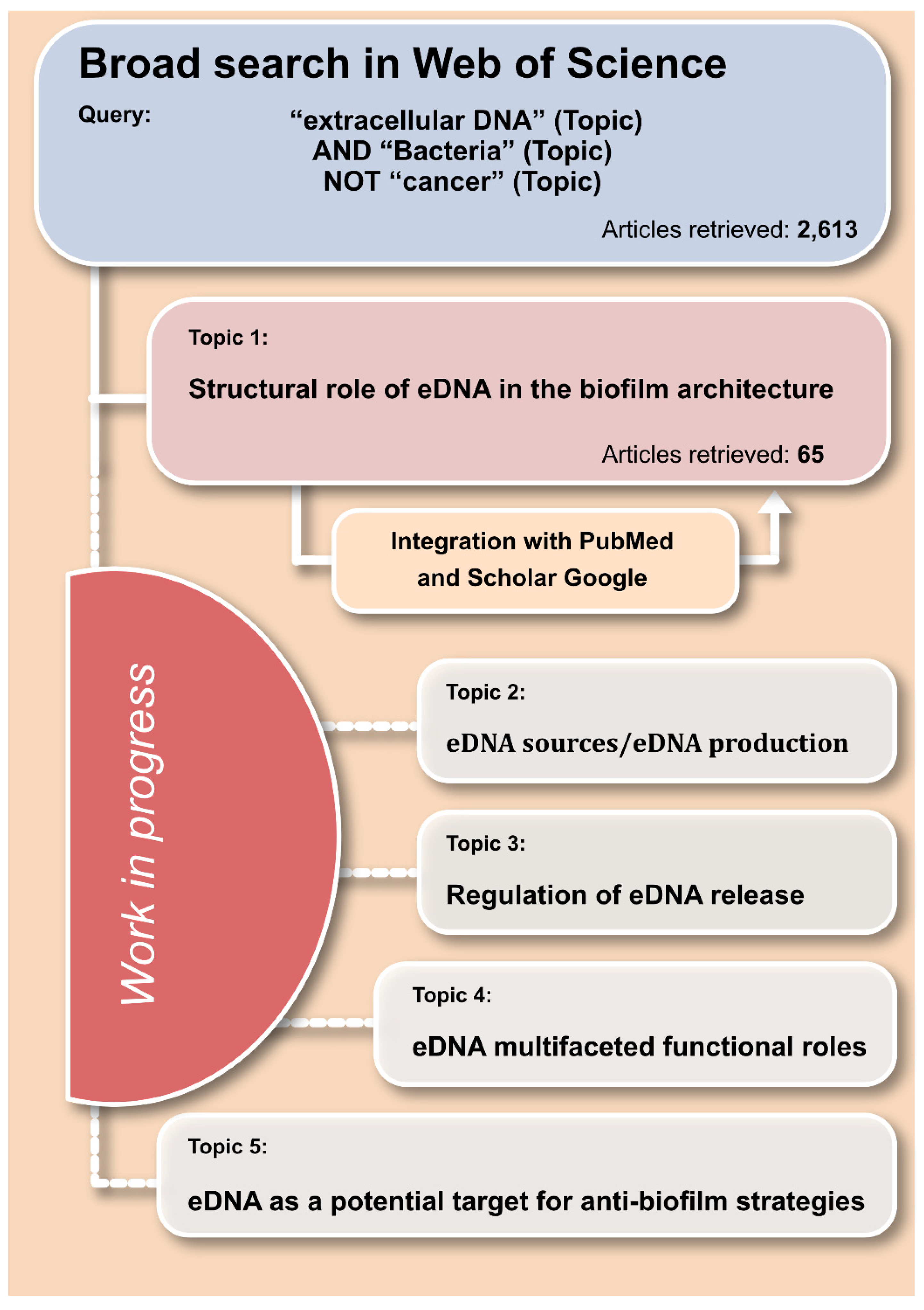
2. Criteria Adopted for the Review of the Literature

This review was generated in a context of a broader systematic survey aimed at gathering all the information currently available in the literature on extracellular DNA, with special reference to the physiology and functions in bacterial biofilms. The bibliographic research was primarily conducted by Web of Science and then extended by PubMed and Google Scholar. The initial broad query adopted in Web of Science was as follows: “extracellular DNA” (Topic) AND “Bacteria” (Topic) NOT “cancer” (Topic) (Figure 1). An alert was set, and the search results were regularly updated until the 5 May 2021. Overall, 2613 article entries were obtained from this broad research. Experimental articles were thoroughly screened based on the title and the abstract. The content of review articles was examined, assessing, and eventually including their relevant citations. All pertinent manuscripts were clustered together based on the relevant treated topics into the following categories: “eDNA sources/eDNA production”, “regulation of eDNA release”, “structural role of eDNA in the biofilm architecture”, “eDNA multifaceted functional roles”, and a further special category concerning “eDNA as a potential target for anti-biofilm strategies”. The present systematic review was based on the articles selected as relevant to the category “structural role of eDNA in the biofilm architecture”.

3. eDNA Structural Role in Biofilm Architecture

For diverse bacterial species, the main EPS components of biofilm extracellular matrix have progressively been identified, with eDNA appearing ubiquitous. Consequently, current research efforts are progressively being focused on establishing if and how each single polymeric substance structurally integrates with the others to originate the typical biofilm architectural frame. In this regard, eDNA has clearly emerged to play a pivotal role and develop different physico-chemical interactions not only with main EPS components of the biofilm extracellular matrix, but also with factors expressed on the bacterial surfaces and with abiotic surfaces. Thus, among other important roles, eDNA contributes to bacterial aggregation promoting intercellular adhesion [71], mediates attachment to abiotic material surfaces enabling bacterial anchorage [72] and stabilizes the biofilm architecture [73,74,75] by interlacing different polymeric components. Hu et al. [76] studied Myxococcus xanthus biofilms and reported as the direct interactions of eDNA with exopolysaccharides enhance the physical strength and the resistance of biofilms to biological stress. Further work by Peterson et al. [77] has highlighted a distinguishable role of different matrix constituents in stress relaxation. Under mechanical stress, eDNA would appear to modulate its interactions with other EPS components and, in this way, control its contribution to viscoelastic relaxation. Therefore, in view of recent advancements in the comprehension of the organization of the biofilm, eDNA would not simply accumulate in the extracellular space as an inert polymer with filtering and sheltering functions. Rather, it would form molecular complexes and interact with a multiplicity of chemical substances, generating a sort of crosslinked auto-setting gel with heterogeneous features, undergoing progressive modifications, and remodeling during different phases of biofilm maturation.
Gradients of pH generated by bacteria growing in hypoxic conditions would influence electrostatic charges of eDNA and determine its local propensity to interact with other cationic substances. The formation of gels consisting of polyelectrolyte complexes starting from charged biopolymers is well established and has been exploited for the fabrication of biocompatible hydrogels for biomedical applications [78,79]. A long molecule (if fully stretched out, the bacterial chromosome would measure nearly 1000 times the size of a bacterial cell [80]), eDNA may generate a sort of main grid and attract and help in retaining soluble and diffusible polycationic molecules otherwise lost to the biofilm matrix construction. The particular importance of a polyanionic molecule such as eDNA in the biofilm architecture is suggested by heparin, another polyanionic molecule, which was found to mimic eDNA in binding to cell-surface Staphylococcus aureus proteins and enhance biofilm formation [81]. However, in bacterial biofilm matrices the contribution to the architecture stability is provided by additional, more complex physico-chemical interactions involving different categories of compounds, which are described in the following dedicated paragraphs. The interactions of eDNA with biofilm exopolysaccharides and proteins will be treated in the following paragraphs.

4. Interaction of eDNA with Biofilm Exopolysaccharides

Given its polyanionic nature, some molecular interactions developed by eDNA with other biofilm components are mediated by its electrostatic charge. In staphylococcal species such as S. aureus and Staphylococcus epidermidis, the polysaccharide intercellular adhesin (PIA) represents a main exopolysaccharide biofilm component. PIA, whose expression is associated to the icaADBC locus [25], consists of poly-β-(1,6)-N-acetylglucosamine, a polysaccharide that, during its processing, is partially de-acetylated by the enzyme IcaB and acquires a net positive charge. In S. epidermidis biofilms, PIA is recognized to be a main EPS component of the extracellular matrix. Conversely, different past investigations supported the idea that S. aureus would produce biofilms through two main distinct pathways, alternatively leading to the assembly of a polysaccharide-based biofilm morphotype or an eDNA/protein biofilm morphotype [82,83]. The polysaccharide-based biofilm morphotype was reported as predominantly observed in methicillin-sensitive S. aureus (MSSA) strains, while the eDNA/protein biofilm morphotype would be more typical of methicillin-resistant S. aureus strains (MRSA). However, very recent data by Mlynek et al. [84] would suggest that these forms of biofilm extracellular matrices are not mutually exclusive. Rather, the relative proportions of matrix components and sensitivity to extracellular-matrix-degrading agents such as DNase I and proteinase are governed by the conditions in which biofilms develop and the differential expression of factors associated to biofilm formation or their respective degradation/catabolism (e.g., proteases). Interestingly, Mlynek and colleagues propose a model where, in reason of their respective electrostatic charge, PIA directly interacts with eDNA. In turn, eDNA binds one or more lipoproteins in the biofilm [85]. This electrostatic net model would finally support and combine some earlier observations suggesting the occurrence of both eDNA/protein [86] and eDNA/PIA complexation [17]. In 2018, the work of Sugimoto et al. [70] earlier highlighted a broad impact of eDNA on the biofilm of clinical MSSA and MRSA strains. They reported even in PIA-dependent biofilms less but sufficient levels of eDNA to promote biofilm formation and maintain biofilm structures. Levels were found to depend on the strain and on the culture condition and not always correlated with the extent of biofilm biomass. Conversely, in a past investigation assessing the composition of biofilms formed by 55 clinical S. epidermidis strains, levels of eDNA and PIA were found to some degree correlated [17]. This is somehow consistent with the findings in S. aureus of Mlynek and colleagues [84], suggesting a synergistic function in biofilm formation and bacterial aggregation by eDNA and PIA, under the control of the global regulator CodY. Very recently, Skovdal et al. [87] have questioned the role played by PIA and other S. epidermidis adhesins such as Embp under in vivo real clinical conditions with respect to in vitro laboratory conditions. The authors showed as, in an S. epidermidis 1585 WT strain, which was deficient in icaADBC, and in its derivative strain lacking the embp gene, host factors in human plasma could restore bacterial aggregation and vancomycin tolerance. Biofilms formed in human plasma were reported to be loosely attached and consisting of suspended aggregates. It should be said that, traditionally, loosely attached bacterial aggregates that deposit during sedimentation under static conditions are normally removed by gentle rinsing before the assessment of the firmly adhered biofilm on a material surface.
Thus, the appeal of Flemming et al. [88] to reconsider and revise the definition of biofilm in all its possible manifestations is overly due. Moreover, bacterial flexibility to adapt to different environmental conditions often relies on redundant mechanisms to aggregate, condition the external environment, and generate their protected niches. Opportunistic pathogens such as staphylococci, Escherichia coli and P. aeruginosa can inhabit different environmental niches, including drinking water distribution systems [89] and natural waters [90]. It is conceivable that depending on the local availability of polymeric substances, bacteria may rely on their own production of EPS or on available external sources. It is certainly true that there is need to gain a more complete information on the composition of bacterial biofilms in vivo in clinical samples and that in vitro models should more closely simulate the in vivo human physiological fluids found at the different anatomic sites [91].
Important molecular interactions between eDNA and biofilm exopolysaccharides have been described for the biofilms of other important pathogens. In P. aeruginosa biofilms, depending on the strain type, the architecture of the extracellular matrix is variously characterized by the presence of different exopolysaccharides, these including: alginate, a polyanionic polysaccharide consisting of guluronic and mannuronic acids and typically overproduced in mucoid strains; Psl polysaccharide (the product of the polysaccharide synthesis locus, Psl), a neutral mannose-rich polysaccharide; and pellicle (Pel), a polycationic, partially de-N-acetylated polysaccharide consisting of N-acetylglucosamine and N-acetylgalactosamine [92,93,94,95].
During the morphogenesis of biofilm matrix architecture, thick-structured biofilms are formed even by mutant strains producing just one or two of the above-mentioned polysaccharides. The sole exception is observed when the mutant is uniquely capable of producing alginate [96]. Psl exopolysaccharide typically accumulates at the periphery of the 3D-structured mushroom-like microcolonies that form during biofilm maturation [97], where it appears to accomplish a scaffolding structural function. Conversely, the microcolony center remains Pls-free and occupied by bacterial cells. Cell death and lysis occurring in this central area of the microcolony result in an abundant release of eDNA. Interestingly, Jennings et al. [93] found that the polycationic Pel exopolysaccharide develops ionic interactions with eDNA as earlier described for PIA in staphylococci, determining the formation of molecular cross-links. The authors observed that Pel and eDNA generally colocalize to the stalk of the microcolony structure. Pel distribution is largely influenced by the strain type and its respective ability of expressing Psl. In fact, a mutant strain defective for the psl gene expressed substantial amounts of Pel, which could be detected even at the periphery of the biofilm, this documenting as Pel can eventually compensate and replace Psl functions. Jennings and colleagues [93] could not ascertain differences in Pel transcriptional control that could explain its prevalent distribution in microcolonies stalk. They offered the alternative explanation that, at least in part, pH gradients with their influence on ionic polymer charge and presence of eDNA drive the Pel/eDNA interactions, generating the structural core of the stalk. In this regard, Reichhardt & Parsek [94] demonstrated as Pel becomes positively charged only with a pH at or below the isoelectric point (corresponding to 6.3), which can be reached at the core of the microcolony. Through the interaction with eDNA, Pel appears to contribute to the formation of meshwork-like structures and influence biofilm cell density and/or the compactness of the biofilm [96]. Wang et al. [98] reported that even Psl can interact with eDNA and, in in vitro grown P. aeruginosa biofilms, form fiber-like structures, where the two polymers colocalize. Psl-based fibers were further confirmed in vivo and found to emanate from the body of the biofilms [99]. However, the nature of the interaction between the Psl and eDNA remains unexplained. In contrast, Reichhardt and Parsek [94] found that exogenous DNA did not accumulate in negative control biofilms formed by a PAO1ΔwspF Δpel pBADpsl mutant strain, which lacks Pel and overexpresses Psl, this suggesting that only Pel could specifically interact with eDNA. Better established are the interactions of both Psl [94] and Pel [95] with the extracellular adhesin CdrA, implicated in promoting biofilm formation through biofilm matrix cross-linking of different exopolysaccharides. Dispersion of P. aeruginosa cells in the late biofilm maturation stage is finally achieved through the degradation of both Pel and Psl polysaccharides [100].
An important recent discovery was that Pel-DNA aggregates, which form at pH 6.3 by salmon sperm DNA in Pel-overproducing cultures, resist digestion with DNase I even after extended digestion [101]. eDNA returned DNase sensitive only after re-solubilizing the aggregates by sonication or increasing the pH to disrupt ionic interactions between the two polymers. This protection provided by the Pel-eDNA interaction from the disruption by nucleases has significant implications for future antibiofilm strategies. Moreover, the authors found that the Pel-eDNA interaction was also associated to an increased tolerance to cationic aminoglycoside antibiotics such as tobramycin but not to the neutral ciprofloxacin antibiotic, pinpointing a multifunctional role. Psl was not found to exhibit any of these two activities.
For many species such as Bacillus subtilis, the molecular structure of the exopolysaccharides found in the biofilm extracellular matrix are still unknown. Peng et al. [102] have recently reported that the expression of the main exopolysaccharide in B. subtilis, SBE1, may be associated to the expression of the epsG gene and eDNA was found to colocalize with such exopolysaccharide in B. subtilis SBE1 pellicles, suggesting a potential physical interaction between these two components.
Table 1 reports the polysaccharides that have been demonstrated to interact with eDNA in the biofilm architecture.

5. Interaction of eDNA with Biofilm Proteins

Investigations on S. aureus strains that produce ica-independent, proteinase K-sensitive biofilms have shown as treatments with proteinase K or DNase I (but not RNase) disrupt cells clumping [86]. Conversely, the addition of exogenous DNA (either autologous or heterologous) can restore cells clumping after DNase I treatment, but not in proteinase K pretreated biofilms. These observations highlight as proteins and eDNA are two interrelated main players in bacterial aggregation and biofilm stabilization. Nowadays, several proteins have emerged to interact with eDNA and contribute to the biofilm skeletal framework. Payne and Boles [103] have earlier reviewed some of the proteins that take part to the biofilm architecture and interact with eDNA. Among the different categories of proteins that have been found to express eDNA-binding activity there are: secreted proteins (e.g., the exotoxin β-toxin); proteins and lipoproteins expressed on the bacterial surface (e.g., S. aureus SaeP and the immunodominant surface antigen B, IsaB); nucleoid proteins and other cytoplasmic moonlighting proteins (e.g., DNABII family proteins); and amyloid/amyloidogenic proteins (e.g., phenol-soluble modulins, PSMs). Table 2 summarizes the main proteins that have been described to interact with eDNA playing a potential role in the organization of biofilm architecture.
As seen for the electrostatic net model involving polycationic exopolysaccharides and eDNA, Foulston et al. [104] suggested that eDNA might serve as an electrostatic net and link protein-coated cells in the biofilm. The authors reported as in S. aureus an important contribution to biofilm formation derives from the repurposing of moonlighting proteins derived from cytolysis processes. Once released in the outer space, cytoplasmic proteins would reversibly associate with the cell surface in a manner that depends on local pH. Under conditions of oxygen limitations, a drop in pH is associated to the release in the extracellular space of fermentation products, these including, among others: formate, lactate, and acetate [105]. At pH 4.5 to 5, surface-associated, cytoplasmic proteins are likely to carry a net positive charge and interact with both eDNA and with negatively charged cell surfaces characterized by the presence of polyanionic teichoic acids.
The identification of eDNA-binding proteins in the biofilms of the different bacterial species has only recently started to be unveiled. Increasing evidence has enlightened as different members of the DNABII protein family develop important interactions with eDNA and contribute to organize and stabilize the architecture of the biofilm matrix. Histone-like proteins of about 10 kDa (alternatively known as HLPs), DNABII form dimers that bind dsDNA. Initially known for their intracellular role, DNABII have progressively emerged to take part to the composition of extracellular matrix, where they critically contribute to the integrity of the biofilms containing eDNA [106,107]. Bacterial DNABII include the integration host factor (IHF) protein and the histone-like nucleoid-associated protein HU [108]. Different in vitro and in vivo studies clearly indicate a general involvement of the homologs of IHF (found in α- and γ-proteobacteria such as Burkholderia cenocepacia and H. influenzae) and of HU (broadly expressed by eubacteria), even in the biofilm architecture of relevant pathogens such as E. coli, S. aureus and P. aeruginosa [109,110,111]. Similarly, both IHF and HU bind dsDNA, exhibiting special affinity to pre-bent DNA. However, HU binding is not driven by specific DNA sequences, in this differing from IHF that has specific affinity for strands with the consensus WATCAANNNNTTR (W = A/T, R = A/G; N = any nucleotide; R = purine) [112]. Apart from binding of pre-bent dsDNA, HU exhibits a peculiarity with respect to many other DNA-binding proteins: an even greater affinity for cruciform branched nucleic acid structures known as Holliday junctions (HJs), up to 1000-fold higher than the affinity for double-stranded DNA or single-stranded oligonucleotides with no secondary structure [113,114]. Single-strand cross-over intermediates of homologous recombination, HJs are known to form in bacterial as well as in eukaryotic cells.
A series of interesting investigations conducted on in vivo monomicrobial and polymicrobial infections has documented that, in in vivo biofilms, eDNA is in a lattice structure [73,109,115,116,117]. Using immunohistochemical techniques, Devaraj et al. [118] were able to detect a diffuse presence of HJs in the complex lattice-like eDNA structure of biofilms formed in nontypeable Haemophilus influenzae (NTHI) infected middle ears of chinchillas. Similar observations were reported when analyzing the sputum of cystic fibrosis patients, where the biofilm was formed by multiple mixed bacterial species. DNABII proteins have been found to critically contribute to stabilization of the lattice structure of eDNA by binding to HJs. Indeed, HJ DNA-binding protein RuvA, normally implicated in the resolution of homologous recombination events in eubacteria, was found to functionally complement DNABII proteins within the extracellular matrix and stabilize the biofilms formed in vitro by E. coli, NTHI, and S. epidermidis. Conversely, sequestration of the DNABII proteins or competition for the HJs by HJ resolvases resulted in biofilm disruption.
Concerning the origin of eDNA-binding proteins in the extracellular space, DNABII proteins can be released in the outer space by bacterial cytolysis, but also through alternative routes as in the case of NTHI, where it occurs via inner membrane pore T4SS-like complex and ComE [117].
Apart from IHF and HU, there are several other nucleoid associated proteins (NAPs) that could potentially act as moonlighting proteins. Devaraj et al. [118] investigated if, in NTHI, NAPs other than DNABII such as H-NS, CbpA, HfQ and Dps could be found extracellularly and play an important role in the biofilm. Although NAPs other than DNABII were detected in the extracellular matrix, only DNABII were found to play a major role in maintaining the structural integrity of the biofilm. Interestingly, visualizing the biofilm with immunostaining techniques, IHF and HU were found distributed at distinct locations and did not show overlapping [118]. The critical importance of DNABII proteins had earlier been abundantly documented by a series of studies by the same group of authors, proving as the use of DNABII protein specific antibodies is capable to disrupt in vitro preformed biofilms of pathogens such as NTHI, B. cenocepacia, Streptococcus gordonii and Porphyromonas gingivalis and result in the dismantling and eradication of the biofilm in vivo [106,115,119,120].
Lipoproteins represent another important category of proteins capable to directly interact with eDNA and participate to the biofilm architecture as eDNA anchoring points on the cell surface. The recent work of Kavanaugh et al. [85] showed as specific membrane-attached lipoproteins can interact with the eDNA in the biofilm matrix and promote S. aureus biofilm formation. These authors adopted an innovative approach combining Southwestern blotting and mass spectrometry for screening the bacterial proteins capable of eDNA binding. While confirming the role of some proteins with known eDNA-binding activity (e.g., IsaB, Atl, Eap and PSMs), they also identified membrane-associated proteins and membrane-anchored lipoproteins previously unknown to bind DNA. The overexpression of identified eDNA-binding proteins was found to result in an increased retention of surface eDNA and, thus, an enhanced biofilm biomass. This finding is consistent with the hypothesis that the membrane-bound lipoproteins can function as anchor points and link bacterial cells together through noncovalent bonds with eDNA. Interestingly, one of the newly identified eDNA-binding lipoproteins by Kavanaugh and colleagues [85] was SaeP, which was previously known as an auxiliary membrane protein modulating, in concert with protein SaeQ, the activity of the SaeRS two-component system (SaeRS TCS). SaeRS TCS regulates the expression of several virulence factors (e.g., hemolysins, leukocidins, superantigens, surface proteins, and so on) and, among them, of the staphylococcal nuclease (Nuc), which has been implicated in the disruption of the biofilm during the exodus phase [121]. Therefore, SaeP could potentially express its biofilm-enhancing activity through two distinct mechanisms, respectively: (1) the inhibition of Nuc production through a modulating activity on SaeRS TCS and (2) its favorable interactions with the eDNA, strengthening the biofilm electrostatic net.
eDNA was also found to interact with and promote the polymerization of amyloidogenic peptides into self-assembled fibrillar structures. Amyloidogenic peptides represent a further category of proteins that are expressed in the biofilms of several bacterial species. For E. coli, a key amyloid component that binds eDNA promoting curli amyloids assembly is CsgA. Yan et al. (2020) [122] have reported some insights on the biogenesis of curli amyloids by the cooperations of CsgA with six distinct peptides. After recognition by CsgG, CsgA would be secreted through the CsgFG secretion channel into the extracellular space, where it would be eventually nucleated by CsgB and forms fibrils. In addition to E. coli, curli is known to be expressed also in Salmonella enterica serovar Typhimurium and in other species of the Enterobacteriaceae family, where it has been found to form curli-DNA complexes that contribute to the biofilm structure. Under in vivo conditions amyloid-DNA composites of bacterial biofilms have been observed and found capable to stimulate autoimmunity diseases through the generation of anti-dsDNA antibodies [55,123]. DNA was found to accelerate the polymerization of curli fibers [123]. Thus, eDNA would not only bind to curli forming complexes, but also promote the formation of stable amyloid fibrils acting on the polymerization rate. Both these actions contribute to an increased structural stability of the biofilms.
Phenol-soluble modulins (PSM) represent another group of bacterial amyloidogenic proteins capable to interact with eDNA in the extracellular space. Small surfactant-like amphipathic peptides, PSMs are multifunctional proteins that are known to act as cytotoxic virulence factors protecting bacteria from the host immune response, but also intervene in the process of staphylococcal biofilm maturation. In particular, they have been found to act as surfactant molecules and lead to biofilm disassembly when in monomeric form, but also contribute to the integrity of the biofilm architecture when self-assembled into amyloid-like fibers exhibiting amyloid-like properties [124]. Marinelli et al. [125] reported that, in S. aureus, α-PSM1 and α-PSM4 peptides are main amyloidogenic proteins involved in the α-PSMs fibrillogenesis. At a low concentration PSMs monomers alone do not readily polymerize. Nonetheless, positively charged PSMs such as α-PSM1 would interact with polyanionic eDNA, thus raising the local peptide concentration and triggering polymerization [126]. The formation of the DNA/PSM complex was also found to reduce the cytolytic activity of α-PSM10 [126], suggesting that the presence of eDNA would modulate at the same time biofilm stability and leukocidal action toward host cell-mediated response. It would be interesting to investigate if other eDNA-binding substances could play a kind of regulatory function by competing with the binding of PSMs.
S. aureus immunodominant surface antigen B (IsaB) is a further eDNA-binding protein, which is secreted in the extracellular space, where it remains retained on the bacterial surface [127]. Elevated IsaB has been reported to inhibit host autophagic flux and promote MRSA virulence. It remains a matter of speculation if its eDNA binding activity could play a further role in anchoring nucleic acids to the cell surface [128]. In addition to IsaB, DNA-binding activity is expressed also by the β-Toxin, a neutral sphingomyelinase hemolysin, which forms covalent cross-links to itself in the presence of DNA [129]. However, even in this case, its role in the biofilm structure is still to be completely clarified.
Table 2. Proteins interacting with eDNA in the biofilm architecture.
ComponentCategoryDescriptionMain Bacterial SpeciesReferences
IHFDNABII protein familyIntegration host factor proteinα- and γ-proteobacteria[106,107,108,109,110,111]
HUDNABII protein familyHistone-like nucleoid-associated protein HUBroadly expressed by eubacteria[106,107,108,109,110,111]
DNABII proteinsProteinBind at the vertices of crossed eDNA strands and act as lynchpin-like molecules to stabilize the structure of eDNANontypeable Haemophilus influenzae (NTHI)[117]
SaePLipoproteinSaeP inhibits Nuc production through a modulating activity on SaeRS TCS and interacts with the eDNA, strengthening the biofilm electrostatic net.S. aureus[85]
β-toxinExotoxinA neutral sphingomyelinase hemolysin, β-toxin has been speculated to form covalent cross-links to itself in the presence of DNAS. aureus[129]
IsaBExtra-cellular protein retained on bacterial surfaceS. aureus immunodominant surface antigen B (IsaB) has been speculated to anchor nucleic acids to the bacterial cell surfaceS. aureus[128]
LytCExtra-cellular proteinLysozyme LytC, an extra-cellular cell wall hydrolase, has been suggested to form DNA-LytC protein complexesS. pneumoniae[130]
PrgBSurface adhesinIts adhesin domain has been reporteed to bind and compact DNAEnterococcus faecalis[131,132]
CsgAAmyloid/amyloidogenic proteinsAmyloid component that binds eDNA promoting curli amyloids assemblyE. coli, Salmonella enterica serovar Typhimurium, Enterobacteriaceae family[122]
α-PSM1Amyloid/amyloidogenic proteinsA positively charged phenol-soluble modulin (PSM), α-PSM1 can interact with polyanionic eDNA and trigger α-PSMs fibrillogenesisS. aureus[124,125,126]
Other bacterial factors described to interact with eDNA and hypothesized to play a function in stabilizing the biofilm architecture include: lysozyme LytC, a S. pneumoniae extra-cellular cell wall hydrolase that forms intercellular DNA-LytC protein complexes in pneumococcal biofilms [130]; Enterococcus faecalis PrgB, whose adhesin domain binds and compacts DNA with a histone-like DNA condensation mechanism [131,132]; Streptococcus intermedius histone-like DNA-binding protein (Si-HLP) [133,134]; GAPDH and enolase S. aureus proteins, which are positively charged at pH 5 [81,86,104]; and Neisseria heparin-binding antigen (NhbA) and α-peptide of IgA protease both expressed by Neisseria meningitidis [135].

6. Bacterial eDNA Binding to Human Inflammatory Cells

All microbial structural motifs are recognized by the innate immune system via the TLR family of pattern recognition receptors (PRRs) [136]. TRL9 is a pattern recognition receptor, which is predominantly located intracellularly in immune cells and recognizes unmethylated CpG motifs characteristic of bacterial DNA [44,45]. TLR9-dependent activation can be triggered not only by phagocytosis of whole S. aureus cells but also by that of the extracellular DNA molecules, extensively contained in the biofilm matrix. The role of TLR9 in regulating host immunity to S. aureus biofilm growth has been examined by Thurlow et al. [137]. Although TLR9 is pivotal for host immune responses to planktonic S. aureus, immune response appears different in S. aureus biofilm infections. Using a mouse model of catheter-associated biofilm infection, these authors demonstrated a significant reduction in cytokine/chemokine production associated with biofilm in infected tissues compared with the wound healing response elicited by sterile catheters and concluded that S. aureus biofilms can circumvent traditional antimicrobial effector pathways and persist in an immuno-competent host. Human inflammatory cells rapidly detect CpG sequences in bacterial eDNA as a human evolutionary response to combat the ability of bacteria to rapidly form biofilm communities. In wild type mice, intraperitoneal application of small synthetic oligodeoxynucleotides containing unmethylated CpG dinucleotides has been found to lead to both a local and systemic inflammatory response. Conversely, in TLR9-deficient mice, the inflammatory response was abolished [138]. These findings would suggest that bacterial eDNA would potentially influence the inflammatory response even systemically [138].
In immune cells, the recognition of bacterial extracellular DNA by a surface receptor has evolved as a signal to alert the immune system to the presence of bacterial invasion, mainly in infections in which bacteria grow as biofilms. This effect might be relevant in biofilm infections in which extracellular DNA could function as a potent neutrophil agonist [139].
However, whether TLR9 plays a protective or deleterious role in host defense against P. aeruginosa remained to be determined. TLR9 plays an essential role in activating innate immunity by recognizing CpG specific motifs present in microbial DNA [10,140]. It is however reported that TLR9 down-regulates the innate immune response against P. aeruginosa and that the absence of TLR9 leads to an early increase in the inflammatory response [141]. This leads to the improvement of the clearance of P. aeruginosa in the lungs and increased mouse survival. The apparent enhancement in the ability of TLR9-negative mice to eliminate P. aeruginosa seems due to a TLR9 deletion that improves alveolar macrophages killing of this bacterium by increasing the production of IL-1b and NO, two inflammatory mediators necessary for P. aeruginosa killing by alveolar macrophages. According to the authors, this finding may open therapeutic strategies, based on TLR9 inhibition, to control P. aeruginosa-induced pneumonia.

7. Interaction of eDNA with Other Substances/Metabolites

Apart from the binding to other EPS components such as exopolysaccharides and proteins, eDNA has been shown to interact also with substances of different nature and functions. Redox-active metabolites like phenazines have been found to bind DNA through intercalation. In P. aeruginosa, pyocyanin binding to e-DNA has been reported to increase EPS stability and cellular aggregation [142]. In reason of its electrostatic charge, eDNA is known to complexate with polycationic ions and substances. For instance, the interaction between DNA and Ca2+ was found thermodynamically favorable and the binding process is spontaneous and exothermic. At biologically relevant concentrations, Ca2+ was found to enhance cell aggregation via cationic bridging of DNA [143]. Moreover, through its drug binding activity, eDNA confers to the biofilm a shielding function, protecting the encased bacteria from the bactericidal action of antibiotics as well as antimicrobial peptides [42,144]. Finally, eDNA has also been found to interact with supramolecular materials such as membrane vesicles [145], which have been observed in the biofilm to associate to the nucleic acid.

8. Attachment to Abiotic Surfaces and Bridging Processes

In the biofilm architecture eDNA has been seen to interact with different substances and form a diffuse web-like interconnected lattice structure. This diffuse net, we have seen, can be anchored to the bacterial surfaces through externally exposed lipoproteins and other surface associated eDNA-binding proteins. Eventually, polycationic substances of polysaccharidic or proteic nature may additionally interact with other competing anionic superficial bacterial molecules such as lipoteichoic acids and interlace these molecules with eDNA. The action of all these intermolecular interactions would explicate cell aggregating activity and letting emerge a clear role for eDNA as an intercellular adhesin. The heterogeneity of environmental conditions within a biofilm, in particular local pH, dictates the electrostatic charges of EPS components, so affecting the interactivity of the biofilm architectural elements. Physicochemical mechanistic explanations for eDNA-mediated adhesion and aggregation come also from the extended Derjaguin, Landau, Verwey, Overbeek (DLVO) theory. In this perspective, eDNA-mediated bacterial adhesion and aggregation would occur through attractive Lifshitz–Van der Waals and acid–base interactions despite existing electrostatic repulsion. In the extended DLVO theory, acid–base attraction results from interactions between electron-accepting and electron-donating moieties on cells surfaces and eDNA [146,147]. Attractive short-range acid-base interactions would contribute to eDNA binding with other EPS biopolymers and other intercalating metabolites such as pyocyanin, facilitating EPS anchoring on bacterial cell surfaces.
When bacteria are suspended in a fluid, in the absence of a substrate to adhere to and form a stratified biofilm, autoaggregation (also termed flocculation or agglutination) is believed to provide bacteria with the benefits of biofilm while maintaining mobility [148]. This is the case of many chronic, recalcitrant bacterial infections such as cystic fibrosis, where bacteria can persist and elude both host immune defenses and medical antibiotic therapies forming mono- or polymicrobial biofilms in the absence of a foreign body.
In the presence of materials with slippery, non-adhesive surfaces, eDNA has been found to play a central role in the formation of “biofilm bridges”. Biofilm bridging is a newly discovered phenomenon. Bacterial microcolonies do not remain confined to restricted adhesive surface microniches on the substrate but can successfully spread across bacteria-repulsive regions and develop contact with other distant biofilm clusters. All this is accomplished by forming thin biofilm bridges, which originate as a stress response to harsh environmental conditions (e.g., limited iron ion availability). The formation of biofilm bridges has been demonstrated to occur in P. aeruginosa, S. aureus and Stenotrophomonas maltophilia [149]. The significant reduction in the formation of biofilm bridges when treating bacterial cultures with DNase clearly pinpoints to the involvement of eDNA in the process. Indeed, eDNA was detected not only within the bridge but also in the adjacent areas, encountering the main biofilm clusters. However, apart from its structural role, eDNA has also been found to facilitate twitching motility of bacteria and, thus promote migration and biofilm spreading. eDNA would explicate the activity on cell motility by orderly directing the traffic flow of cells to the leading edges of biofilm through a sort of contact guidance [52].
Apart from intervening as an intercellular glue, eDNA has also been found to mediate and influence bacterial adhesion on material surfaces, which represents a fundamental step in the pathogenesis of biofilm-associate infections developing in the presence of foreign bodies and implant materials. Regina et al. [150] found that eDNA promotes bacterial adhesion to abiotic surfaces characterized by a wide range of surface chemistries. However, its effects on bacterial adhesion were found not only to depend on the material surface chemistry but also on the ionic strength (I) of the surrounding fluid [150,151]. eDNA would participate both in short-range acid-base interactions and long-range electrostatic and Lifshitz-van der Waals interactions. On hydrophilic surfaces, eDNA-stimulated Staphylococcus xylosus bacterial adhesion was only evident at low and intermediate I, while no effect was seen on glass surfaces and carboxyl-functionalized surfaces at high I of the surrounding liquid [150]. Conversely, eDNA was found to create favorable conditions for bacterial adhesion to neutrally charged hydrophobic surfaces, independently from I [150]. DNA is an amphipathic molecule consisting of a hydrophilic backbone and hydrophobic nitrogenous bases, which are potentially implicated in hydrophobic interactions with bacterial surfaces.
Figure 2 graphically summarizes some principal interactions emerged to occur in the biofilm of S. aureus, an exemplary, largely investigated staphylococcal species.

9. Conclusions

Biofilm formation is being universally recognized as a principal virulence mechanism enabling bacteria to elude the host immune response and circumvent conventional antibiotic treatments. Thus, bacterial growth in the biofilm state has progressively become the primary target of an increasing number of strategies aimed at preventing and eradicating difficult to treat clinical infections. However, as also pointed out by Flemming et al. [88], the concept of biofilm has been and is constantly evolving and human categories are often difficult to adapt for interpreting complex natural phenomena still incompletely understood. Surprisingly, for many bacterial species there is still very limited knowledge available on the biofilm matrix composition and the structural and functional role of each single molecule that may be expressed during different phases of biofilm maturation. A high degree of biodiversity is overall observed, with biofilms markedly varying in composition depending on bacterial species, strain types, stage of maturation and environmental conditions. Nonetheless, eDNA appears to be widespread and ubiquitously participate to the biofilm matrix composition of most bacterial and fungal species. This versatile molecule exhibits physico-chemical properties that are ideal to cover structural and functional roles required to microbial cells to strive in different environments, including the hostile interstices of the human body. Based on convergent recent findings, eDNA consistently emerges as a main structural element of a self-assembling architecture, which coordinates and bind a broad range of compounds secreted by bacteria, present in the environment, or provided by the host. Its chemistry enables a prompt interaction, often based on electrostatic charges, with molecules of various nature, promotes the formation of stabilizing eDNA networks and the spontaneous assembly of supramolecular structures that are fundamental for bacterial intercellular aggregation, bacterial anchorage to the substrate and formation of a functional and definite biofilm architecture. In view of its wide distribution across biofilms formed by main pathogenic species, eDNA probably represents the best target for broadly addressing anti-biofilm strategies. An important point is however the decreased vulnerability to DNase degradation acquired by biofilms during their maturation. It was initially thought that eDNA cornerstone function during the initial morphogenesis of biofilm matrix was finally supplanted/superseded by that of other EPS constituents. However, the protective complexation/interaction of other EPS components, in first place amyloidogenic and DNABII proteins, with eDNA has been found a critical point of vulnerability and an Achilles’ heel of biofilm structural integrity. These latest advancements in the knowledge of the biofilm matrix architecture are already inspiring new powerful therapeutic approaches to efficaciously treat and prevent biofilm-based infections, particularly the dreaded and multifaceted infections associated with orthopedic implants.

Author Contributions

Conceptualization, D.C., L.M. and C.R.A.; Research Design and Methodology, D.C.; Software, D.C.; Writing—original draft preparation, D.C.; Writing—Review and editing D.C., L.M. and C.R.A. All authors have read and agreed to the published version of the manuscript.

Funding

The financial contribution by the Theory-enhancing Project RF-2018-12368273: “Implant orthopaedic infections: the challenge of bacterial antibiotic resistance and new anti-infective materials” funded by the Ministry of Health for the IRCCS Rizzoli Orthopaedic Institute of Bologna is gratefully acknowledged.

Institutional Review Board Statement

Not applicable.

Data Availability Statement

Not applicable.

Conflicts of Interest

The authors declare no conflict of interest.

References

  1. Arciola, C.R.; Campoccia, D.; Montanaro, L. Implant infections: Adhesion, biofilm formation and immune evasion. Nat. Rev. Microbiol. 2018, 16, 397–409. [Google Scholar] [CrossRef] [PubMed]
  2. Campoccia, D.; Mirzaei, R.; Montanaro, L.; Arciola, C.R. Hijacking of immune defences by biofilms: A multifront strategy. Biofouling 2019, 35, 1055–1074. [Google Scholar] [CrossRef]
  3. Arciola, C.R.; Campoccia, D.; Ehrlich, G.D.; Montanaro, L. Biofilm-based implant infections in orthopaedics. Adv. Exp. Med. Biol. 2015, 830, 29–46. [Google Scholar] [CrossRef] [PubMed]
  4. Mirzaei, R.; Mohammadzadeh, R.; Alikhani, M.Y.; Moghadam, M.S.; Karampoor, S.; Kazemi, S.; Barfipoursalar, A.; Yousefimashouf, R. The biofilm-associated bacterial infections unrelated to indwelling devices. IUBMB Life 2020, 72, 1271–1285. [Google Scholar] [CrossRef] [PubMed]
  5. Gupta, P.; Sarkar, S.; Das, B.; Bhattacharjee, S.; Tribedi, P. Biofilm, pathogenesis and prevention—A journey to break the wall: A review. Arch. Microbiol. 2016, 198, 1–15. [Google Scholar] [CrossRef] [PubMed]
  6. Dastgheyb, S.; Parvizi, J.; Shapiro, I.M.; Hickok, N.J.; Otto, M. Effect of biofilms on recalcitrance of staphylococcal joint infection to antibiotic treatment. J. Infect. Dis. 2015, 211, 641–650. [Google Scholar] [CrossRef] [PubMed]
  7. Ying, Q.L.; Kemme, M.; Simon, S.R. Alginate, the slime exopolysaccharide of Pseudomonas aeruginosa, binds human leukocyte elastase, retards inhibition by alpha 1-proteinase inhibitor, and accelerates inhibition by secretory leukoprotease inhibitor. Am. J. Respir. Cell. Mol. Biol. 1996, 15, 283–291. [Google Scholar] [CrossRef] [PubMed]
  8. Mack, D.; Fischer, W.; Krokotsch, A.; Leopold, K.; Hartmann, R.; Egge, H.; Laufs, R. The intercellular adhesin involved in biofilm accumulation of Staphylococcus epidermidis is a linear beta-1,6-linked glucosaminoglycan: Purification and structural analysis. J. Bacteriol. 1996, 178, 175–183. [Google Scholar] [CrossRef] [Green Version]
  9. Martí, M.; Trotonda, M.P.; Tormo-Más, M.A.; Vergara-Irigaray, M.; Cheung, A.L.; Lasa, I.; Penadés, J.R. Extracellular proteases inhibit protein-dependent biofilm formation in Staphylococcus aureus. Microbes Infect. 2010, 12, 55–64. [Google Scholar] [CrossRef] [PubMed] [Green Version]
  10. Jahn, A.; Griebe, T.; Nielsen, P.H. Composition of Pseudomonas putida biofilms: Accumulation of protein in the biofilm matrix. Biofouling 1999, 14, 49–57. [Google Scholar] [CrossRef]
  11. Izano, E.A.; Amarante, M.A.; Kher, W.B.; Kaplan, J.B. Differential roles of poly-N-acetylglucosamine surface polysaccharide and extracellular DNA in Staphylococcus aureus and Staphylococcus epidermidis biofilms. Appl. Environ. Microbiol. 2008, 74, 470–476. [Google Scholar] [CrossRef] [Green Version]
  12. Whitchurch, C.B.; Tolker-Nielsen, T.; Ragas, P.C.; Mattick, J.S. Extracellular DNA required for bacterial biofilm formation. Science 2002, 295, 1487. [Google Scholar] [CrossRef] [PubMed]
  13. Palmgren, R.; Nielsen, P.H. Accumulation of DNA in the exopolymeric matrix of activated sludge and bacterial cultures. Water Sci. Technol. 1996, 34, 233–240. [Google Scholar] [CrossRef]
  14. Sadovskaya, I.; Vinogradov, E.; Li, J.; Jabbouri, S. Structural elucidation of the extracellular and cell-wall teichoic acids of Staphylococcus epidermidis RP62A, a reference biofilm-positive strain. Carbohydr. Res. 2004, 339, 1467–1473. [Google Scholar] [CrossRef] [PubMed]
  15. Flemming, H.C.; Wingender, J. The biofilm matrix. Nat. Rev. Microbiol. 2010, 8, 623–633. [Google Scholar] [CrossRef] [PubMed]
  16. Gehrke, T.; Telegdi, J.; Thierry, D.; Sand, W. Importance of Extracellular Polymeric Substances from Thiobacillus ferrooxidans for Bioleaching. Appl. Environ. Microbiol. 1998, 64, 2743–2747. [Google Scholar] [CrossRef] [PubMed] [Green Version]
  17. Campoccia, D.; Montanaro, L.; Ravaioli, S.; Pirini, V.; Cangini, I.; Arciola, C.R. Exopolysaccharide production by Staphylococcus epidermidis and its relationship with biofilm extracellular DNA. Int. J. Artif. Organs 2011, 34, 832–839. [Google Scholar] [CrossRef] [PubMed]
  18. Harris, L.G.; Murray, S.; Pascoe, B.; Bray, J.; Meric, G.; Mageiros, L.; Wilkinson, T.S.; Jeeves, R.; Rohde, H.; Schwarz, S.; et al. Biofilm Morphotypes and Population Structure among Staphylococcus epidermidis from Commensal and Clinical Samples. PLoS ONE 2016, 11, e0151240. [Google Scholar] [CrossRef]
  19. Ravaioli, S.; Campoccia, D.; Speziale, P.; Pietrocola, G.; Zatorska, B.; Maso, A.; Presterl, E.; Montanaro, L.; Arciola, C.R. Various biofilm matrices of the emerging pathogen Staphylococcus lugdunensis: Exopolysaccharides, proteins, eDNA and their correlation with biofilm mass. Biofouling 2020, 36, 86–100. [Google Scholar] [CrossRef]
  20. Arciola, C.R.; Campoccia, D.; Gamberini, S.; Rizzi, S.; Donati, M.E.; Baldassarri, L.; Montanaro, L. Search for the insertion element IS256 within the ica locus of Staphylococcus epidermidis clinical isolates collected from biomaterial-associated infections. Biomaterials 2004, 25, 4117–4125. [Google Scholar] [CrossRef] [PubMed]
  21. Pozzi, C.; Waters, E.M.; Rudkin, J.K.; Schaeffer, C.R.; Lohan, A.J.; Tong, P.; Loftus, B.J.; Pier, G.B.; Fey, P.D.; Massey, R.C.; et al. Methicillin resistance alters the biofilm phenotype and attenuates virulence in Staphylococcus aureus device-associated infections. PLoS Pathog. 2012, 8, e1002626. [Google Scholar] [CrossRef] [PubMed]
  22. Arciola, C.R.; Baldassarri, L.; Campoccia, D.; Creti, R.; Pirini, V.; Huebner, J.; Montanaro, L. Strong biofilm production, antibiotic multi-resistance and high gelE expression in epidemic clones of Enterococcus faecalis from orthopaedic implant infections. Biomaterials 2008, 29, 580–586. [Google Scholar] [CrossRef] [PubMed]
  23. Le, K.Y.; Villaruz, A.E.; Zheng, Y.; He, L.; Fisher, E.L.; Nguyen, T.H.; Ho, T.V.; Yeh, A.J.; Joo, H.S.; Cheung, G.Y.C.; et al. Role of Phenol-Soluble Modulins in Staphylococcus epidermidis Biofilm Formation and Infection of Indwelling Medical Devices. J. Mol. Biol. 2019, 431, 3015–3027. [Google Scholar] [CrossRef] [PubMed]
  24. Mukherjee, S.; Moustafa, D.; Smith, C.D.; Goldberg, J.B.; Bassler, B.L. The RhlR quorum-sensing receptor controls Pseudomonas aeruginosa pathogenesis and biofilm development independently of its canonical homoserine lactone autoinducer. PLoS Pathog. 2017, 13, e1006504. [Google Scholar] [CrossRef] [PubMed]
  25. Le, K.Y.; Otto, M. Quorum-sensing regulation in staphylococci-an overview. Front. Microbiol. 2015, 6, 1174. [Google Scholar] [CrossRef] [Green Version]
  26. Arciola, C.R.; Campoccia, D.; Ravaioli, S.; Montanaro, L. Polysaccharide intercellular adhesin in biofilm: Structural and regulatory aspects. Front. Cell. Infect. Microbiol. 2015, 5, 7. [Google Scholar] [CrossRef] [Green Version]
  27. Nadell, C.D.; Xavier, J.B.; Levin, S.A.; Foster, K.R. The evolution of quorum sensing in bacterial biofilms. PLoS Biol. 2008, 6, e14. [Google Scholar] [CrossRef] [PubMed] [Green Version]
  28. Arciola, C.R.; Campoccia, D.; Speziale, P.; Montanaro, L.; Costerton, J.W. Biofilm formation in Staphylococcus implant infections. A review of molecular mechanisms and implications for biofilm-resistant materials. Biomaterials 2012, 33, 5967–5982. [Google Scholar] [CrossRef] [PubMed]
  29. Knobloch, J.K.; Bartscht, K.; Sabottke, A.; Rohde, H.; Feucht, H.H.; Mack, D. Biofilm formation by Staphylococcus epidermidis depends on functional RsbU, an activator of the sigB operon: Differential activation mechanisms due to ethanol and salt stress. J. Bacteriol. 2001, 183, 2624–2633. [Google Scholar] [CrossRef] [PubMed] [Green Version]
  30. Cramton, S.E.; Ulrich, M.; Götz, F.; Döring, G. Anaerobic conditions induce expression of polysaccharide intercellular adhesin in Staphylococcus aureus and Staphylococcus epidermidis. Infect. Immun. 2001, 69, 4079–4085. [Google Scholar] [CrossRef] [Green Version]
  31. Martins, M.; Uppuluri, P.; Thomas, D.P.; Cleary, I.A.; Henriques, M.; Lopez-Ribot, J.L.; Oliveira, R. Presence of extracellular DNA in the Candida albicans biofilm matrix and its contribution to biofilms. Mycopathologia 2010, 169, 323–331. [Google Scholar] [CrossRef] [Green Version]
  32. Böckelmann, U.; Janke, A.; Kuhn, R.; Neu, T.R.; Wecke, J.; Lawrence, J.R.; Szewzyk, U. Bacterial extracellular DNA forming a defined network-like structure. FEMS Microbiol. Lett. 2006, 262, 31–38. [Google Scholar] [CrossRef] [PubMed]
  33. de Aldecoa, A.L.I.; Zafra, O.; González-Pastor, J.E. Mechanisms and Regulation of Extracellular DNA Release and Its Biological Roles in Microbial Communities. Front. Microbiol. 2017, 8, 1390. [Google Scholar] [CrossRef] [PubMed] [Green Version]
  34. Matthews, L.W.; Spector, S.; Lemm, J.; Potter, J.L. Studies on pulmonary secretions. I. The over-all chemical composition of pulmonary secretions from patients with cystic fibrosis, bronchiectasis, and laryngectomy. Am. Rev. Respir. Dis. 1963, 88, 199–204. [Google Scholar] [CrossRef] [PubMed]
  35. Molin, S.; Tolker-Nielsen, T. Gene transfer occurs with enhanced efficiency in biofilms and induces enhanced stabilisation of the biofilm structure. Curr. Opin. Biotechnol. 2003, 14, 255–261. [Google Scholar] [CrossRef]
  36. Hall, C.W.; Mah, T.F. Molecular mechanisms of biofilm-based antibiotic resistance and tolerance in pathogenic bacteria. FEMS Microbiol. Rev. 2017, 41, 276–301. [Google Scholar] [CrossRef] [PubMed]
  37. Mulcahy, H.; Charron-Mazenod, L.; Lewenza, S. Extracellular DNA chelates cations and induces antibiotic resistance in Pseudomonas aeruginosa biofilms. PLoS Pathog. 2008, 4, e1000213. [Google Scholar] [CrossRef] [PubMed] [Green Version]
  38. Jones, E.A.; McGillivary, G.; Bakaletz, L.O. Extracellular DNA within a nontypeable Haemophilus influenzae-induced biofilm binds human beta defensin-3 and reduces its antimicrobial activity. J. Innate Immun. 2013, 5, 24–38. [Google Scholar] [CrossRef] [PubMed]
  39. Batoni, G.; Maisetta, G.; Esin, S. Antimicrobial peptides and their interaction with biofilms of medically relevant bacteria. Biochim. Biophys. Acta 2016, 1858, 1044–1060. [Google Scholar] [CrossRef] [PubMed]
  40. Ramphal, R.; Lhermitte, M.; Filliat, M.; Roussel, P. The binding of anti-pseudomonal antibiotics to macromolecules from cystic fibrosis sputum. J. Antimicrob. Chemother. 1988, 22, 483–490. [Google Scholar] [CrossRef]
  41. Chiang, W.C.; Nilsson, M.; Jensen, P.Ø.; Høiby, N.; Nielsen, T.E.; Givskov, M.; Tolker-Nielsen, T. Extracellular DNA shields against aminoglycosides in Pseudomonas aeruginosa biofilms. Antimicrob. Agents Chemother. 2013, 57, 2352–2361. [Google Scholar] [CrossRef] [Green Version]
  42. Doroshenko, N.; Tseng, B.S.; Howlin, R.P.; Deacon, J.; Wharton, J.A.; Thurner, P.J.; Gilmore, B.F.; Parsek, M.R.; Stoodley, P. Extracellular DNA impedes the transport of vancomycin in Staphylococcus epidermidis biofilms preexposed to subinhibitory concentrations of vancomycin. Antimicrob. Agents Chemother. 2014, 58, 7273–7282. [Google Scholar] [CrossRef] [PubMed] [Green Version]
  43. Wilton, M.; Charron-Mazenod, L.; Moore, R.; Lewenza, S. Extracellular DNA Acidifies Biofilms and Induces Aminoglycoside Resistance in Pseudomonas aeruginosa. Antimicrob. Agents Chemother. 2015, 60, 544–553. [Google Scholar] [CrossRef] [PubMed] [Green Version]
  44. Hemmi, H.; Takeuchi, O.; Kawai, T.; Kaisho, T.; Sato, S.; Sanjo, H.; Matsumoto, M.; Hoshino, K.; Wagner, H.; Takeda, K.; et al. A Toll-like receptor recognizes bacterial DNA. Nature 2000, 408, 740–745. [Google Scholar] [CrossRef] [PubMed]
  45. Bauer, S.; Kirschning, C.J.; Häcker, H.; Redecke, V.; Hausmann, S.; Akira, S.; Wagner, H. Lipford GB. Human TLR9 confers responsiveness to bacterial DNA via species-specific CpG motif recognition. Proc. Natl. Acad. Sci. USA 2001, 98, 9237–9242. [Google Scholar] [CrossRef] [PubMed] [Green Version]
  46. Islam, D.; Bandholtz, L.; Nilsson, J.; Wigzell, H.; Christensson, B.; Agerberth, B.; Gudmundsson, G. Downregulation of bactericidal peptides in enteric infections: A novel immune escape mechanism with bacterial DNA as a potential regulator. Nat. Med. 2001, 7, 180–185. [Google Scholar] [CrossRef] [PubMed]
  47. Otto, M. Staphylococcal biofilms. Curr. Top. Microbiol. Immunol. 2008, 322, 207–228. [Google Scholar] [CrossRef] [PubMed]
  48. Bass, J.I.F.; Russo, D.M.; Gabelloni, M.L.; Geffner, J.R.; Giordano, M.; Catalano, M.; Zorreguieta, A.; Trevani, A.S. Extracellular DNA: A major proinflammatory component of Pseudomonas aeruginosa biofilms. J. Immunol. 2010, 184, 6386–6395. [Google Scholar] [CrossRef] [Green Version]
  49. Hornung, V.; Latz, E. Intracellular DNA recognition. Nat. Rev. Immunol. 2010, 10, 123–130. [Google Scholar] [CrossRef]
  50. Ciszek-Lenda, M.; Strus, M.; Walczewska, M.; Majka, G.; Machul-Żwirbla, A.; Mikołajczyk, D.; Górska, S.; Gamian, A.; Chain, B.; Marcinkiewicz, J. Pseudomonas aeruginosa biofilm is a potent inducer of phagocyte hyperinflammation. Inflamm. Res. 2019, 68, 397–413. [Google Scholar] [CrossRef] [Green Version]
  51. Thammavongsa, V.; Missiakas, D.M.; Schneewind, O. Staphylococcus aureus degrades neutrophil extracellular traps to promote immune cell death. Science 2013, 342, 863–866. [Google Scholar] [CrossRef] [Green Version]
  52. Gloag, E.S.; Turnbull, L.; Huang, A.; Vallotton, P.; Wang, H.; Nolan, L.M.; Mililli, L.; Hunt, C.; Lu, J.; Osvath, S.R.; et al. Self-organization of bacterial biofilms is facilitated by extracellular DNA. Proc. Natl. Acad. Sci. USA 2013, 110, 11541–11546. [Google Scholar] [CrossRef] [PubMed] [Green Version]
  53. Finkel, S.E.; Kolter, R. DNA as a nutrient: Novel role for bacterial competence gene homologs. J. Bacteriol. 2001, 183, 6288–6293. [Google Scholar] [CrossRef] [PubMed] [Green Version]
  54. Saunders, S.H.; Tse, E.C.M.; Yates, M.D.; Otero, F.J.; Trammell, S.A.; Stemp, E.D.A.; Barton, J.K.; Tender, L.M.; Newman, D.K. Extracellular DNA Promotes Efficient Extracellular Electron Transfer by Pyocyanin in Pseudomonas aeruginosa Biofilms. Cell 2020, 182, 919–932.e19. [Google Scholar] [CrossRef] [PubMed]
  55. Tursi, S.A.; Lee, E.Y.; Medeiros, N.J.; Lee, M.H.; Nicastro, L.K.; Buttaro, B.; Gallucci, S.; Wilson, R.P.; Wong, G.C.L.; Tükel, Ç. Bacterial amyloid curli acts as a carrier for DNA to elicit an autoimmune response via TLR2 and TLR9. PLoS Pathog. 2017, 13, e1006315. [Google Scholar] [CrossRef] [PubMed]
  56. Nicastro, L.; Tükel, Ç. Bacterial Amyloids: The Link between Bacterial Infections and Autoimmunity. Trends Microbiol. 2019, 27, 954–963. [Google Scholar] [CrossRef] [PubMed]
  57. Gorham, F.P. The Sixth Annual Meeting of the Society of American Bacteriologists. Science 1905, 21, 481–498. [Google Scholar] [CrossRef] [PubMed] [Green Version]
  58. Sanborn, J.R. The formation of semi-transparent membranes from cultures of slime-producing micro-organisms. Science 1933, 77, 290. [Google Scholar] [CrossRef] [PubMed] [Green Version]
  59. Yoshida, K.; Ekstedt, R.D. Relation of mucoid growth of Staphylococcus aureus to clumping factor reaction, morphology in serum-soft agar, and virulence. J. Bacteriol. 1968, 96, 902–908. [Google Scholar] [CrossRef] [PubMed] [Green Version]
  60. Schwarzmann, S.; Boring, J.R. Antiphagocytic Effect of Slime from a Mucoid Strain of Pseudomonas aeruginosa. Infect. Immun. 1971, 3, 762–767. [Google Scholar] [CrossRef] [Green Version]
  61. Bartell, P.F.; Orr, T.E.; Chudio, B. Purification and Chemical Composition of the Protective Slime Antigen of Pseudomonas aeruginosa. Infect. Immun. 1970, 2, 543–548. [Google Scholar] [CrossRef] [PubMed] [Green Version]
  62. Mack, W.N.; Mack, J.P.; Ackerson, A.O. Microbial film development in a trickling filter. Microb. Ecol. 1975, 2, 215–226. [Google Scholar] [CrossRef] [PubMed]
  63. Christensen, G.D.; Simpson, W.A.; Bisno, A.L.; Beachey, E.H. Adherence of slime-producing strains of Staphylococcus epidermidis to smooth surfaces. Infect. Immun. 1982, 37, 318–326. [Google Scholar] [CrossRef] [PubMed] [Green Version]
  64. Christensen, G.D.; Simpson, W.A.; Bisno, A.L.; Beachey, E.H. Experimental foreign body infections in mice challenged with slime-producing Staphylococcus epidermidis. Infect. Immun. 1983, 40, 407–410. [Google Scholar] [CrossRef] [PubMed] [Green Version]
  65. Marrie, T.J.; Costerton, J.W. Scanning and transmission electron microscopy of in situ bacterial colonization of intravenous and intraarterial catheters. J. Clin. Microbiol. 1984, 19, 687–693. [Google Scholar] [CrossRef] [PubMed] [Green Version]
  66. Costerton, J.W.; Stewart, P.S.; Greenberg, E.P. Bacterial biofilms: A common cause of persistent infections. Science 1999, 284, 1318–1322. [Google Scholar] [CrossRef] [PubMed] [Green Version]
  67. O’Toole, G.; Kaplan, H.B.; Kolter, R. Biofilm formation as microbial development. Annu. Rev. Microbiol. 2000, 54, 49–79. [Google Scholar] [CrossRef] [PubMed]
  68. Donlan, R.M.; Costerton, J.W. Biofilms: Survival mechanisms of clinically relevant microorganisms. Clin. Microbiol. Rev. 2002, 15, 167–193. [Google Scholar] [CrossRef] [PubMed] [Green Version]
  69. Montanaro, L.; Poggi, A.; Visai, L.; Ravaioli, S.; Campoccia, D.; Speziale, P.; Arciola, C.R. Extracellular DNA in biofilms. Int. J. Artif. Organs 2011, 34, 824–831. [Google Scholar] [CrossRef]
  70. Sugimoto, S.; Sato, F.; Miyakawa, R.; Chiba, A.; Onodera, S.; Hori, S.; Mizunoe, Y. Broad impact of extracellular DNA on biofilm formation by clinically isolated Methicillin-resistant and -sensitive strains of Staphylococcus aureus. Sci. Rep. 2018, 8, 2254. [Google Scholar] [CrossRef]
  71. Lister, J.L.; Horswill, A.R. Staphylococcus aureus biofilms: Recent developments in biofilm dispersal. Front. Cell. Infect. Microbiol. 2014, 4, 178. [Google Scholar] [CrossRef] [PubMed] [Green Version]
  72. Blakeman, J.T.; Morales-García, A.L.; Mukherjee, J.; Gori, K.; Hayward, A.S.; Lant, N.J.; Geoghegan, M. Extracellular DNA Provides Structural Integrity to a Micrococcus luteus Biofilm. Langmuir 2019, 35, 6468–6475. [Google Scholar] [CrossRef] [PubMed]
  73. Jurcisek, J.A.; Bakaletz, L.O. Biofilms formed by nontypeable Haemophilus influenzae in vivo contain both double-stranded DNA and type IV pilin protein. J. Bacteriol. 2007, 189, 3868–3875. [Google Scholar] [CrossRef] [PubMed] [Green Version]
  74. Mann, E.E.; Rice, K.C.; Boles, B.R.; Endres, J.L.; Ranjit, D.; Chandramohan, L.; Tsang, L.H.; Smeltzer, M.S.; Horswill, A.R.; Bayles, K.W. Modulation of eDNA release and degradation affects Staphylococcus aureus biofilm maturation. PLoS ONE 2009, 4, e5822. [Google Scholar] [CrossRef] [PubMed] [Green Version]
  75. Kiedrowski, M.R.; Kavanaugh, J.S.; Malone, C.L.; Mootz, J.M.; Voyich, J.M.; Smeltzer, M.S.; Bayles, K.W.; Horswill, A.R. Nuclease modulates bio- film formation in community-associated methicillin-resistant Staphylococcus aureus. PLoS ONE 2011, 6, e26714. [Google Scholar] [CrossRef] [Green Version]
  76. Hu, W.; Li, L.; Sharma, S.; Wang, J.; McHardy, I.; Lux, R.; Yang, Z.; He, X.; Gimzewski, J.K.; Li, Y.; et al. DNA builds and strengthens the extracellular matrix in Myxococcus xanthus biofilms by interacting with exopolysaccharides. PLoS ONE 2012, 7, e51905. [Google Scholar] [CrossRef]
  77. Peterson, B.W.; van der Mei, H.C.; Sjollema, J.; Busscher, H.J.; Sharma, P.K. A distinguishable role of eDNA in the viscoelastic relaxation of biofilms. mBio 2013, 4, e00497-13. [Google Scholar] [CrossRef] [Green Version]
  78. Lalevée, G.; Sudre, G.; Montembault, A.; Meadows, J.; Malaise, S.; Crépet, A.; David, L.; Delair, T. Polyelectrolyte complexes via desalting mixtures of hyaluronic acid and chitosan-Physicochemical study and structural analysis. Carbohydr. Polym. 2016, 154, 86–95. [Google Scholar] [CrossRef]
  79. Pellá, M.C.G.; Lima-Tenório, M.K.; Tenório-Neto, E.T.; Guilherme, M.R.; Muniz, E.C.; Rubira, A.F. Chitosan-based hydrogels: From preparation to biomedical applications. Carbohydr. Polym. 2018, 196, 233–245. [Google Scholar] [CrossRef]
  80. Badrinarayanan, A.; Le, T.B.; Laub, M.T. Bacterial chromosome organization and segregation. Annu. Rev. Cell. Dev. Biol. 2015, 31, 171–199. [Google Scholar] [CrossRef] [Green Version]
  81. Mishra, S.; Horswill, A.R. Heparin Mimics Extracellular DNA in Binding to Cell Surface-Localized Proteins and Promoting Staphylococcus aureus Biofilm Formation. mSphere 2017, 2, e00135-17. [Google Scholar] [CrossRef] [PubMed] [Green Version]
  82. Fitzpatrick, F.; Humphreys, H.; O’Gara, J.P. Evidence for icaADBC-independent biofilm development mechanism in methicillin-resistant Staphylococcus aureus clinical isolates. J. Clin. Microbiol. 2005, 43, 1973–1976. [Google Scholar] [CrossRef] [PubMed] [Green Version]
  83. O’Neill, E.; Pozzi, C.; Houston, P.; Humphreys, H.; Robinson, D.A.; Loughman, A.; Foster, T.J.; O’Gara, J.P. A novel Staphylococcus aureus biofilm phenotype mediated by the fibronectin-binding proteins, FnBPA and FnBPB. J. Bacteriol. 2008, 190, 3835–3850. [Google Scholar] [CrossRef] [PubMed] [Green Version]
  84. Mlynek, K.D.; Bulock, L.L.; Stone, C.J.; Curran, L.J.; Sadykov, M.R.; Bayles, K.W.; Brinsmade, S.R. Genetic and Biochemical Analysis of CodY-Mediated Cell Aggregation in Staphylococcus aureus Reveals an Interaction between Extracellular DNA and Polysaccharide in the Extracellular Matrix. J. Bacteriol. 2020, 202, e00593-19. [Google Scholar] [CrossRef] [PubMed]
  85. Kavanaugh, J.S.; Flack, C.E.; Lister, J.; Ricker, E.B.; Ibberson, C.B.; Jenul, C.; Moormeier, D.E.; Delmain, E.A.; Bayles, K.W.; Horswill, A.R. Identification of Extracellular DNA-Binding Proteins in the Biofilm Matrix. mBio 2019, 10, e01137-19. [Google Scholar] [CrossRef] [Green Version]
  86. Dengler, V.; Foulston, L.; DeFrancesco, A.S.; Losick, R. An Electrostatic Net Model for the Role of Extracellular DNA in Biofilm Formation by Staphylococcus aureus. J. Bacteriol. 2015, 197, 3779–3787. [Google Scholar] [CrossRef] [Green Version]
  87. Skovdal, S.M.; Hansen, L.K.; Ivarsen, D.M.; Zeng, G.; Büttner, H.; Rohde, H.; Jørgensen, N.P.; Meyer, R.L. Host factors abolish the need for polysaccharides and extracellular matrix-binding protein in Staphylococcus epidermidis biofilm formation. J. Med. Microbiol. 2021, 70, 001287. [Google Scholar] [CrossRef]
  88. Flemming, H.C.; Baveye, P.; Neu, T.R.; Stoodley, P.; Szewzyk, U.; Wingender, J.; Wuertz, S. Who put the film in biofilm? The migration of a term from wastewater engineering to medicine and beyond. NPJ Biofilms Microbiomes 2021, 7, 10. [Google Scholar] [CrossRef]
  89. Liu, J.; Ren, H.; Ye, X.; Wang, W.; Liu, Y.; Lou, L.; Cheng, D.; He, X.; Zhou, X.; Qiu, S.; et al. Bacterial community radial-spatial distribution in biofilms along pipe wall in chlorinated drinking water distribution system of East China. Appl. Microbiol. Biotechnol. 2017, 101, 749–759. [Google Scholar] [CrossRef]
  90. Silva, V.; Caniça, M.; Capelo, J.L.; Igrejas, G.; Poeta, P. Diversity and genetic lineages of environmental staphylococci: A surface water overview. FEMS Microbiol. Ecol. 2020, 96, fiaa191. [Google Scholar] [CrossRef]
  91. Campoccia, D.; Cangini, I.; Selan, L.; Vercellino, M.; Montanaro, L.; Visai, L.; Arciola, C.R. An overview of the methodological approach to the in vitro study of anti-infective biomaterials. Int. J. Artif. Organs 2012, 35, 800–816. [Google Scholar] [CrossRef] [PubMed]
  92. Colvin, K.M.; Irie, Y.; Tart, C.S.; Urbano, R.; Whitney, J.C.; Ryder, C.; Howell, P.L.; Wozniak, D.J.; Parsek, M.R. The Pel and Psl polysaccharides provide Pseudomonas aeruginosa structural redundancy within the biofilm matrix. Environ. Microbiol. 2012, 14, 1913–1928. [Google Scholar] [CrossRef] [PubMed] [Green Version]
  93. Jennings, L.K.; Storek, K.M.; Ledvina, H.E.; Coulon, C.; Marmont, L.S.; Sadovskaya, I.; Secor, P.R.; Tseng, B.S.; Scian, M.; Filloux, A.; et al. Pel is a cationic exopolysaccharide that cross-links extracellular DNA in the Pseudomonas aeruginosa biofilm matrix. Proc. Natl. Acad. Sci. USA 2015, 112, 11353–11358. [Google Scholar] [CrossRef] [PubMed] [Green Version]
  94. Reichhardt, C.; Parsek, M.R. Confocal Laser Scanning Microscopy for Analysis of Pseudomonas aeruginosa Biofilm Architecture and Matrix Localization. Front. Microbiol. 2019, 10, 677. [Google Scholar] [CrossRef] [PubMed]
  95. Reichhardt, C.; Jacobs, H.M.; Matwichuk, M.; Wong, C.; Wozniak, D.J.; Parsek, M.R. The Versatile Pseudomonas aeruginosa Biofilm Matrix Protein CdrA Promotes Aggregation through Different Extracellular Exopolysaccharide Interactions. J. Bacteriol. 2020, 202, e00216-20. [Google Scholar] [CrossRef] [PubMed]
  96. Ghafoor, A.; Hay, I.D.; Rehm, B.H. Role of exopolysaccharides in Pseudomonas aeruginosa biofilm formation and architecture. Appl. Environ. Microbiol. 2011, 77, 5238–5246. [Google Scholar] [CrossRef] [PubMed] [Green Version]
  97. Ma, L.; Conover, M.; Lu, H.; Parsek, M.R.; Bayles, K.; Wozniak, D.J. Assembly and development of the Pseudomonas aeruginosa biofilm matrix. PLoS Pathog. 2009, 5, e1000354. [Google Scholar] [CrossRef] [PubMed] [Green Version]
  98. Wang, S.; Liu, X.; Liu, H.; Zhang, L.; Guo, Y.; Yu, S.; Wozniak, D.J.; Ma, L.Z. The exopolysaccharide Psl-eDNA interaction enables the formation of a biofilm skeleton in Pseudomonas aeruginosa. Environ. Microbiol. Rep. 2015, 7, 330–340. [Google Scholar] [CrossRef] [PubMed] [Green Version]
  99. Geddes-McAlister, J.; Kugadas, A.; Gadjeva, M. Tasked with a Challenging Objective: Why Do Neutrophils Fail to Battle Pseudomonas aeruginosa Biofilms. Pathogens 2019, 8, 283. [Google Scholar] [CrossRef] [PubMed] [Green Version]
  100. Cherny, K.E.; Sauer, K. Untethering and Degradation of the Polysaccharide Matrix Are Essential Steps in the Dispersion Response of Pseudomonas aeruginosa Biofilms. J. Bacteriol. 2020, 202, e00575-19. [Google Scholar] [CrossRef]
  101. Jennings, L.K.; Dreifus, J.E.; Reichhardt, C.; Storek, K.M.; Secor, P.R.; Wozniak, D.J.; Hisert, K.B.; Parsek, M.R. Pseudomonas aeruginosa aggregates in cystic fibrosis sputum produce exopolysaccharides that likely impede current therapies. Cell Rep. 2021, 34, 108782. [Google Scholar] [CrossRef] [PubMed]
  102. Peng, N.; Cai, P.; Mortimer, M.; Wu, Y.; Gao, C.; Huang, Q. The exopolysaccharide-eDNA interaction modulates 3D architecture of Bacillus subtilis biofilm. BMC Microbiol. 2020, 20, 115. [Google Scholar] [CrossRef] [PubMed]
  103. Payne, D.E.; Boles, B.R. Emerging interactions between matrix components during biofilm development. Curr. Genet. 2016, 62, 137–141. [Google Scholar] [CrossRef] [PubMed] [Green Version]
  104. Foulston, L.; Elsholz, A.K.; DeFrancesco, A.S.; Losick, R. The extracellular matrix of Staphylococcus aureus biofilms comprises cytoplasmic proteins that associate with the cell surface in response to decreasing pH. mBio 2014, 5, e01667-14. [Google Scholar] [CrossRef] [PubMed] [Green Version]
  105. Graf, A.C.; Leonard, A.; Schäuble, M.; Rieckmann, L.M.; Hoyer, J.; Maass, S.; Lalk, M.; Becher, D.; Pané-Farré, J.; Riedel, K. Virulence Factors Produced by Staphylococcus aureus Biofilms Have a Moonlighting Function Contributing to Biofilm Integrity. Mol. Cell. Proteom. 2019, 18, 1036–1053. [Google Scholar] [CrossRef] [PubMed]
  106. Goodman, S.D.; Obergfell, K.P.; Jurcisek, J.A.; Novotny, L.A.; Downey, J.S.; Ayala, E.A.; Tjokro, N.; Li, B.; Justice, S.S.; Bakaletz, L.O. Biofilms can be dispersed by focusing the immune system on a common family of bacterial nucleoid-associated proteins. Mucosal Immunol. 2011, 4, 625–637. [Google Scholar] [CrossRef] [PubMed] [Green Version]
  107. Devaraj, A.; Buzzo, J.; Rocco, C.J.; Bakaletz, L.O.; Goodman, S.D. The DNABII family of proteins is comprised of the only nucleoid associated proteins required for nontypeable Haemophilus influenzae biofilm structure. Microbiologyopen 2018, 7, e00563. [Google Scholar] [CrossRef] [Green Version]
  108. Swinger, K.K.; Rice, P.A. IHF and HU: Flexible architects of bent DNA. Curr. Opin. Struct. Biol. 2004, 14, 28–35. [Google Scholar] [CrossRef] [PubMed]
  109. Idicula, W.K.; Jurcisek, J.A.; Cass, N.D.; Ali, S.; Goodman, S.D.; Elmaraghy, C.A.; Jatana, K.R.; Bakaletz, L.O. Identification of biofilms in post-tympanostomy tube otorrhea. Laryngoscope 2016, 126, 1946–1951. [Google Scholar] [CrossRef] [PubMed]
  110. Estellés, A.; Woischnig, A.K.; Liu, K.; Stephenson, R.; Lomongsod, E.; Nguyen, D.; Zhang, J.; Heidecker, M.; Yang, Y.; Simon, R.J.; et al. A High-Affinity Native Human Antibody Disrupts Biofilm from Staphylococcus aureus Bacteria and Potentiates Antibiotic Efficacy in a Mouse Implant Infection Model. Antimicrob. Agents Chemother. 2016, 60, 2292–2301. [Google Scholar] [CrossRef] [PubMed] [Green Version]
  111. Xiong, Y.Q.; Estellés, A.; Li, L.; Abdelhady, W.; Gonzales, R.; Bayer, A.S.; Tenorio, E.; Leighton, A.; Ryser, S.; Kauvar, L.M. A Human Biofilm-Disrupting Monoclonal Antibody Potentiates Antibiotic Efficacy in Rodent Models of both Staphylococcus aureus and Acinetobacter baumannii Infections. Antimicrob. Agents Chemother. 2017, 61, e00904-17. [Google Scholar] [CrossRef] [PubMed] [Green Version]
  112. Friedman, D.I. Integration host factor: A protein for all reasons. Cell 1988, 55, 545–554. [Google Scholar] [CrossRef] [Green Version]
  113. Pontiggia, A.; Negri, A.; Beltrame, M.; Bianchi, M.E. Protein HU binds specifically to kinked DNA. Mol. Microbiol. 1993, 7, 343–350. [Google Scholar] [CrossRef] [PubMed]
  114. Bianchi, M.E. Prokaryotic HU and eukaryotic HMG1: A kinked relationship. Mol. Microbiol. 1994, 14. [Google Scholar] [CrossRef] [PubMed]
  115. Novotny, L.A.; Amer, A.O.; Brockson, M.E.; Goodman, S.D.; Bakaletz, L.O. Structural stability of Burkholderia cenocepacia biofilms is reliant on eDNA structure and presence of a bacterial nucleic acid binding protein. PLoS ONE 2013, 8, e67629. [Google Scholar] [CrossRef] [PubMed]
  116. Gustave, J.E.; Jurcisek, J.A.; McCoy, K.S.; Goodman, S.D.; Bakaletz, L.O. Targeting bacterial integration host factor to disrupt biofilms associated with cystic fibrosis. J. Cyst. Fibros. 2013, 12, 384–389. [Google Scholar] [CrossRef] [PubMed] [Green Version]
  117. Jurcisek, J.A.; Brockman, K.L.; Novotny, L.A.; Goodman, S.D.; Bakaletz, L.O. Nontypeable Haemophilus influenzae releases DNA and DNABII proteins via a T4SS-like complex and ComE of the type IV pilus machinery. Proc. Natl. Acad. Sci. USA 2017, 114, E6632–E6641. [Google Scholar] [CrossRef] [Green Version]
  118. Devaraj, A.; Buzzo, J.R.; Mashburn-Warren, L.; Gloag, E.S.; Novotny, L.A.; Stoodley, P.; Bakaletz, L.O.; Goodman, S.D. The extracellular DNA lattice of bacterial biofilms is structurally related to Holliday junction recombination intermediates. Proc. Natl. Acad. Sci. USA 2019, 116, 25068–25077. [Google Scholar] [CrossRef] [Green Version]
  119. Rocco, C.J.; Davey, M.E.; Bakaletz, L.O.; Goodman, S.D. Natural antigenic differences in the functionally equivalent extracellular DNABII proteins of bacterial biofilms provide a means for targeted biofilm therapeutics. Mol. Oral Microbiol. 2017, 32, 118–130. [Google Scholar] [CrossRef]
  120. Novotny, L.A.; Jurcisek, J.A.; Goodman, S.D.; Bakaletz, L.O. Monoclonal antibodies against DNA-binding tips of DNABII proteins disrupt biofilms in vitro and induce bacterial clearance in vivo. EBioMedicine 2016, 10, 33–44. [Google Scholar] [CrossRef] [Green Version]
  121. Moormeier, D.E.; Bose, J.L.; Horswill, A.R.; Bayles, K.W. Temporal and stochastic control of Staphylococcus aureus biofilm development. mBio 2014, 5, e01341-14. [Google Scholar] [CrossRef] [PubMed] [Green Version]
  122. Yan, Z.; Yin, M.; Chen, J.; Li, X. Assembly and substrate recognition of curli biogenesis system. Nat. Commun. 2020, 11, 241. [Google Scholar] [CrossRef] [PubMed] [Green Version]
  123. Gallo, P.M.; Rapsinski, G.J.; Wilson, R.P.; Oppong, G.O.; Sriram, U.; Goulian, M.; Buttaro, B.; Caricchio, R.; Gallucci, S.; Tükel, Ç. Amyloid-DNA Composites of Bacterial Biofilms Stimulate Autoimmunity. Immunity 2015, 42, 1171–1184. [Google Scholar] [CrossRef] [PubMed] [Green Version]
  124. Schwartz, K.; Syed, A.K.; Stephenson, R.E.; Rickard, A.H.; Boles, B.R. Functional amyloids composed of phenol soluble modulins stabilize Staphylococcus aureus biofilms. PLoS Pathog. 2012, 8, e1002744. [Google Scholar] [CrossRef] [PubMed]
  125. Marinelli, P.; Pallares, I.; Navarro, S.; Ventura, S. Dissecting the contribution of Staphylococcus aureus α-phenol-soluble modulins to biofilm amyloid structure. Sci. Rep. 2016, 6, 34552. [Google Scholar] [CrossRef] [PubMed]
  126. Schwartz, K.; Ganesan, M.; Payne, D.E.; Solomon, M.J.; Boles, B.R. Extracellular DNA facilitates the formation of functional amyloids in Staphylococcus aureus biofilms. Mol. Microbiol. 2016, 99, 123–134. [Google Scholar] [CrossRef] [Green Version]
  127. Mackey-Lawrence, N.M.; Potter, D.E.; Cerca, N.; Jefferson, K.K. Staphylococcus aureus immunodominant surface antigen B is a cell-surface associated nucleic acid binding protein. BMC Microbiol. 2009, 9, 61. [Google Scholar] [CrossRef] [Green Version]
  128. Liu, P.F.; Cheng, J.S.; Sy, C.L.; Huang, W.C.; Yang, H.C.; Gallo, R.L.; Huang, C.M.; Shu, C.W. IsaB Inhibits Autophagic Flux to Promote Host Transmission of Methicillin-Resistant Staphylococcus aureus. J. Investig. Dermatol. 2015, 135, 2714–2722. [Google Scholar] [CrossRef] [Green Version]
  129. Huseby, M.J.; Kruse, A.C.; Digre, J.; Kohler, P.L.; Vocke, J.A.; Mann, E.E.; Bayles, K.W.; Bohach, G.A.; Schlievert, P.M.; Ohlendorf, D.H.; et al. Beta toxin catalyzes formation of nucleoprotein matrix in staphylococcal biofilms. Proc. Natl. Acad. Sci. USA 2010, 107, 14407–14412. [Google Scholar] [CrossRef] [Green Version]
  130. Domenech, M.; García, E.; Prieto, A.; Moscoso, M. Insight into the composition of the intercellular matrix of Streptococcus pneumoniae biofilms. Environ. Microbiol. 2013, 15, 502–516. [Google Scholar] [CrossRef]
  131. Schulte, T.; Mikaelsson, C.; Beaussart, A.; Kikhney, A.; Deshmukh, M.; Wolniak, S.; Pathak, A.; Ebel, C.; Löfling, J.; Fogolari, F.; et al. The BR domain of PsrP interacts with extracellular DNA to promote bacterial aggregation; structural insights into pneumococcal biofilm formation. Sci. Rep. 2016, 6, 32371. [Google Scholar] [CrossRef] [PubMed]
  132. Schmitt, A.; Jiang, K.; Camacho, M.I.; Jonna, V.R.; Hofer, A.; Westerlund, F.; Christie, P.J.; Berntsson, R.P. PrgB promotes aggregation, biofilm formation, and conjugation through DNA binding and compaction. Mol. Microbiol. 2018, 109, 291–305. [Google Scholar] [CrossRef] [PubMed] [Green Version]
  133. Liu, D.; Yumoto, H.; Murakami, K.; Hirota, K.; Ono, T.; Nagamune, H.; Kayama, S.; Matsuo, T.; Miyake, Y. The essentiality and involvement of Streptococcus intermedius histone-like DNA-binding protein in bacterial viability and normal growth. Mol. Microbiol. 2008, 68, 1268–1282. [Google Scholar] [CrossRef] [PubMed]
  134. Nur, A.; Hirota, K.; Yumoto, H.; Hirao, K.; Liu, D.; Takahashi, K.; Murakami, K.; Matsuo, T.; Shu, R.; Miyake, Y. Effects of extracellular DNA and DNA-binding protein on the development of a Streptococcus intermedius biofilm. J. Appl. Microbiol. 2013, 115, 260–270. [Google Scholar] [CrossRef] [PubMed] [Green Version]
  135. Arenas, J.; Nijland, R.; Rodriguez, F.J.; Bosma, T.N.; Tommassen, J. Involvement of three meningococcal surface-exposed proteins, the heparin-binding protein NhbA, the α-peptide of IgA protease and the autotransporter protease NalP, in initiation of biofilm formation. Mol. Microbiol. 2013, 87, 254–268. [Google Scholar] [CrossRef] [PubMed]
  136. Pietrocola, G.; Arciola, C.R.; Rindi, S.; Di Poto, A.; Missineo, A.; Montanaro, L.; Speziale, P. Toll-like receptors (TLRs) in innate immune defense against Staphylococcus aureus. Int. J. Artif. Organs 2011, 34, 799–810. [Google Scholar] [CrossRef] [PubMed]
  137. Thurlow, L.R.; Hanke, M.L.; Fritz, T.; Angle, A.; Aldrich, A.; Williams, S.H.; Engebretsen, I.L.; Bayles, K.W.; Horswill, A.R.; Kielian, T. Staphylococcus aureus biofilms prevent macrophage phagocytosis and attenuate inflammation in vivo. J. Immunol. 2011, 186, 6585–6596. [Google Scholar] [CrossRef] [PubMed] [Green Version]
  138. Knuefermann, P.; Baumgarten, G.; Koch, A.; Schwederski, M.; Velten, M.; Ehrentraut, H.; Mersmann, J.; Meyer, R.; Hoeft, A.; Zacharowski, K.; et al. CpG oligonucleotide activates Toll-like receptor 9 and causes lung inflammation in vivo. Respir. Res. 2007, 8, 72. [Google Scholar] [CrossRef] [Green Version]
  139. Bass, J.I.F.; Gabelloni, M.L.; Alvarez, M.E.; Vermeulen, M.E.; Russo, D.M.; Zorreguieta, A.; Geffner, J.R.; Trevani, A.S. Characterization of bacterial DNA binding to human neutrophil surface. Lab. Investig. 2008, 88, 926–937. [Google Scholar] [CrossRef] [PubMed] [Green Version]
  140. Xia, P.; Wu, Y.; Lian, S.; Yan, L.; Meng, X.; Duan, Q.; Zhu, G. Research progress on Toll-like receptor signal transduction and its roles in antimicrobial immune responses. Appl. Microbiol. Biotechnol. 2021, 105, 5341–5355. [Google Scholar] [CrossRef]
  141. Benmohamed, F.; Medina, M.; Wu, Y.Z.; Maschalidi, S.; Jouvion, G.; Guillemot, L.; Chignard, M.; Manoury, B.; Touqui, L. Toll-like receptor 9 deficiency protects mice against Pseudomonas aeruginosa lung infection. PLoS ONE 2014, 9, e90466. [Google Scholar] [CrossRef] [PubMed] [Green Version]
  142. Das, T.; Sehar, S.; Manefield, M. The roles of extracellular DNA in the structural integrity of extracellular polymeric substance and bacterial biofilm development. Environ. Microbiol. Rep. 2013, 5, 778–786. [Google Scholar] [CrossRef] [PubMed]
  143. Das, T.; Sehar, S.; Koop, L.; Wong, Y.K.; Ahmed, S.; Siddiqui, K.S.; Manefield, M. Influence of calcium in extracellular DNA mediated bacterial aggregation and biofilm formation. PLoS ONE 2014, 9, e91935. [Google Scholar] [CrossRef] [PubMed] [Green Version]
  144. Lewenza, S. Extracellular DNA-induced antimicrobial peptide resistance mechanisms in Pseudomonas aeruginosa. Front. Microbiol. 2013, 4, 21. [Google Scholar] [CrossRef] [PubMed] [Green Version]
  145. Schooling, S.R.; Hubley, A.; Beveridge, T.J. Interactions of DNA with biofilm-derived membrane vesicles. J. Bacteriol. 2009, 191, 4097–4102. [Google Scholar] [CrossRef] [Green Version]
  146. Das, T.; Sharma, P.K.; Busscher, H.J.; van der Mei, H.C.; Krom, B.P. Role of extracellular DNA in initial bacterial adhesion and surface aggregation. Appl. Environ. Microbiol. 2010, 76, 3405–3408. [Google Scholar] [CrossRef] [PubMed] [Green Version]
  147. Das, T.; Krom, B.P.; van der Mei, H.C.; Busscher, H.J.; Sharma, P.K. DNA-mediated bacterial aggregation is dictated by acid-base interactions. Soft. Matter 2011, 7, 2927–2935. [Google Scholar] [CrossRef]
  148. Trunk, T.; Khalil, H.S.; Leo, J.C. Bacterial autoaggregation. AIMS Microbiol. 2018, 4, 140–164. [Google Scholar] [CrossRef] [PubMed]
  149. Lei, W.; Bruchmann, J.; Rüping, J.L.; Levkin, P.A.; Schwartz, T. Biofilm Bridges Forming Structural Networks on Patterned Lubricant-Infused Surfaces. Adv. Sci. 2019, 6, 1900519. [Google Scholar] [CrossRef] [Green Version]
  150. Regina, V.R.; Lokanathan, A.R.; Modrzyński, J.J.; Sutherland, D.S.; Meyer, R.L. Surface physicochemistry and ionic strength affects eDNA’s role in bacterial adhesion to abiotic surfaces. PLoS ONE 2014, 9, e105033. [Google Scholar] [CrossRef] [Green Version]
  151. Das, T.; Sharma, P.K.; Krom, B.P.; van der Mei, H.C.; Busscher, H.J. Role of eDNA on the adhesion forces between Streptococcus mutans and substratum surfaces: Influence of ionic strength and substratum hydrophobicity. Langmuir 2011, 27, 10113–10118. [Google Scholar] [CrossRef] [PubMed]
Figure 1. Scheme of the systematic review.
Ijms 22 09100 g001
Figure 2. This scheme attempts to illustrate the various and complex molecular interactions taking place within the matrix of staphylococcal biofilms. Water-soluble molecules are retained by their mutual stabilizing interactions. The attention is focused on the variety of molecular species emerged to be involved and the real size ratio among different categories of molecules is not fully respected. During biofilm maturation, eDNA becomes progressively protected by the complexation with the other polymers and resistant to DNase digestion. Legend: VSC, viable staphylococcal cell; DSC, dead staphylococcal cell; HJs, eDNA Holliday junctions; PIA, polysaccharide intercellular adhesin; HU, histone-like nucleoid-associated protein HU; IHF, integration host factor; SaeP, S. aureus Saep lipoprotein; TA, (negatively charged) teichoic acids; β-Tox, S. aureus β-Toxin; α-PSMs, amyloidogenic α-Phenol-soluble modulins; DBMAP, DNA-binding membrane-associated proteins and membrane-anchored lipoproteins such as S. aureus SaeP and IsaB; HEPs, host extracellular-matrix proteins.
Ijms 22 09100 g002
Table 1. Polysaccharides interacting with eDNA in the biofilm architecture.
ComponentCategoryDescriptionMain Bacterial SpeciesReferences
PIA, PNAGExopolysaccharidepoly-β-(1,6)-N-acetylglucosamine.
In S. aureus and S. epidermidis PIA represents a main exopolysaccharide biofilm component
S. aureus, S. epidermidis[17,25,82,83,84]
Pel polysaccharideExopolysaccharidePolycationic, partially de-N-acetylated polysaccharide consisting of N-acetylglucosamine and N-acetylgalactosamine P. aeruginosa[92,93,94,95,96,97,100]
Psl polysaccharideExopolysaccharideNeutral mannose-rich polysaccharide, whose direct interaction with eDNA is still debatedP. aeruginosa[92,93,94,95,96,98]
Publisher’s Note: MDPI stays neutral with regard to jurisdictional claims in published maps and institutional affiliations.

