Review

Impact of Stilbenes as Epigenetic Modulators of Breast Cancer Risk and Associated Biomarkers

by
Sebanti Ganguly
1,
Itika Arora
1 and
Trygve O. Tollefsbol
1,2,3,4,5,6,*
1
Department of Biology, University of Alabama at Birmingham, Birmingham, AL 35294, USA
2
Integrative Center for Aging Research, University of Alabama at Birmingham, Birmingham, AL 35294, USA
3
O’Neal Comprehensive Cancer Center, University of Alabama at Birmingham, Birmingham, AL 35294, USA
4
Nutrition Obesity Research Center, University of Alabama at Birmingham, Birmingham, AL 35294, USA
5
Comprehensive Diabetes Center, University of Alabama at Birmingham, Birmingham, AL 35294, USA
6
Cell Senescence Culture Facility, University of Alabama at Birmingham, Birmingham, AL 35294, USA
*
Author to whom correspondence should be addressed.
Int. J. Mol. Sci. 2021, 22(18), 10033; https://0-doi-org.brum.beds.ac.uk/10.3390/ijms221810033
Submission received: 29 June 2021 / Revised: 12 September 2021 / Accepted: 13 September 2021 / Published: 17 September 2021
(This article belongs to the Special Issue Dietary Bioactive Compounds and Breast Cancer)

Abstract

:
With the recent advancement of genetic screening for testing susceptibility to mammary oncogenesis in women, the relevance of the gene−environment interaction has become progressively apparent in the context of aberrant gene expressions. Fetal exposure to external stressors, hormones, and nutrients, along with the inherited genome, impact its traits, including cancer susceptibility. Currently, there is increasing interest in the role of epigenetic biomarkers such as genomic methylation signatures, plasma microRNAs, and alterations in cell-signaling pathways in the diagnosis and primary prevention of breast cancer, as well as its prognosis. Polyphenols like natural stilbenes have been shown to be effective in chemoprevention by exerting cytotoxic effects that can stall cell proliferation. Besides possessing antioxidant properties against the DNA-damaging effects of reactive oxygen species, stilbenes have also been observed to modulate cell-signaling pathways. With the increasing trend of early-life screening for hereditary breast cancer risks, the potency of different phytochemicals in harnessing the epigenetic biomarkers of breast cancer risk demand more investigation. This review will explore means of exploiting the abilities of stilbenes in altering the underlying factors that influence breast cancer risk, as well as the appearance of associated biomarkers.

1. Introduction

According to the 2020 global cancer statistics performed by GLOBOCAN, an initiative of the World Health Organization, breast cancer is the most prevalent form of cancer in women, and contributed to 11.7% of the total global cancer burden that was estimated to be 19.3 million cases in the year 2020 [1,2]. North American countries, including the United States, saw a substantial increase in the total number of reported incidences in female breast cancer patients in the years around 2007 due to rising awareness with respect to annual screening. Simultaneously, a lower number of reports on mortality rates reflected asymptomatic women’s willingness to adopt primary preventive measures by addressing external factors such as lifestyle, diet, age of conceiving a full-term pregnancy, duration of breastfeeding, and nulliparity. There is little to no control of factors such as somatic age; race; age of menarche; menopause; density of breast tissue; acquired and germline mutations in the form of deletion or truncation of tumor suppressor genes (TSGs), including BRCA1/2, TP53, STK11, CD1, and PTEN; and upregulation of oncogenes such as ERBB2, c-MYC, and PIK3C [3,4,5,6]. However, there is still a degree of control that medical science possesses in reversing epimutations or epigenetic changes that silence or activate the aforementioned genes, which serve as biomarkers of breast cancer. The current research not only ensures maximum survival rates, but also focuses on preventing the poor quality of life that breast cancer malignancies entail through the prevention of the occurrence of disease pathologies in high-risk individuals through the employment of primary prevention.
Primary prevention includes lifestyle changes; risk-reducing mastectomy; and undergoing simultaneous treatment with selective estrogen receptor modulators (SERMs) such as tamoxifen, raloxifene, lasofoxifene, and aromatase inhibitors (AI). Nelson et al., in 2019, in a very extensive meta-analysis, addressed some of the important questions regarding the effectiveness of adopting such a precautionary intake of SERMs by healthy individuals belonging to breast cancer-risk groups in the successful prevention of disease-occurrence and mortality over the long term [7]. SERMs indeed reduce the risk of breast cancer susceptibility significantly in estrogen receptor-positive (ER+) breast cancers, but some limitations in the meta-analysis caused by variability in the duration of drug administration and disregard with respect to the ages of the subjects undergoing the clinical trial has left grounds for skepticism [7]. Their body of work acknowledges the fact that the intake of SERMs in asymptomatic women as a measure of primary prevention is a rarely practiced clinical procedure because of the health-related concerns of the subjects. These include the long-term and short-term side-effects of these drugs, including increased risk of endometrial hyperplasia and endometrial cancer, blood clotting, menstrual abnormalities, decreased bone and muscle density, hot-flashes, body pains, and sexual dysfunction, as well as other consequences. Additionally, tamoxifen and raloxifene are synthetic derivatives of stilbenes that may interfere with the normal functioning of off-target tissues, causing the death of healthy cells in vivo [3,8,9,10,11], leading to considerable interest in finding less harmful derivatives of plant polyphenols such as stilbenes, and optimizing their activity within the cell.
Epigenetics encompasses all the heritable, reversible changes in gene expression, without the genetic code becoming altered. A major epigenetic mechanism in mammals is the methylation of DNA at the CpG islands of the gene promoters, specifically at the fifth carbon of cytosine in the DNA backbone. Other important epigenetic mechanisms consist of methylation, phosphorylation, ubiquitination, SUMOylation, and acetylation of histone molecules and the influence of non-coding RNAs (ncRNAs), rendering a change in the conformation of the DNA. These covalent additions to the DNA structure modulate the accessibility of the DNA to different transcription factors, thereby upregulating or down-regulating a specific gene. The enzymes that are responsible for causing these epigenetic marks are called “writers”, which include DNA methyltransferase (DNMT), histone methyltransferase (HMT), and histone acetylase (HAT). The enzymes involved in removing epigenetic marks are called “erasers”, and include histone deacetylases (HDACs), DNA demethylases, and histone demethylases. The enzymes responsible for recognizing these marks and conducting downstream signaling are termed “readers” and are characterized by proteins with two types of domains—recognition and effector domains. Aberrant epigenetic marks can upregulate oncogenes or down-regulate tumor suppressor genes, and hence hold immense importance. The gene expression of the writer, reader, and eraser proteins, as well as their activity, can also be monitored to discover new biomarkers of breast cancer susceptibility.

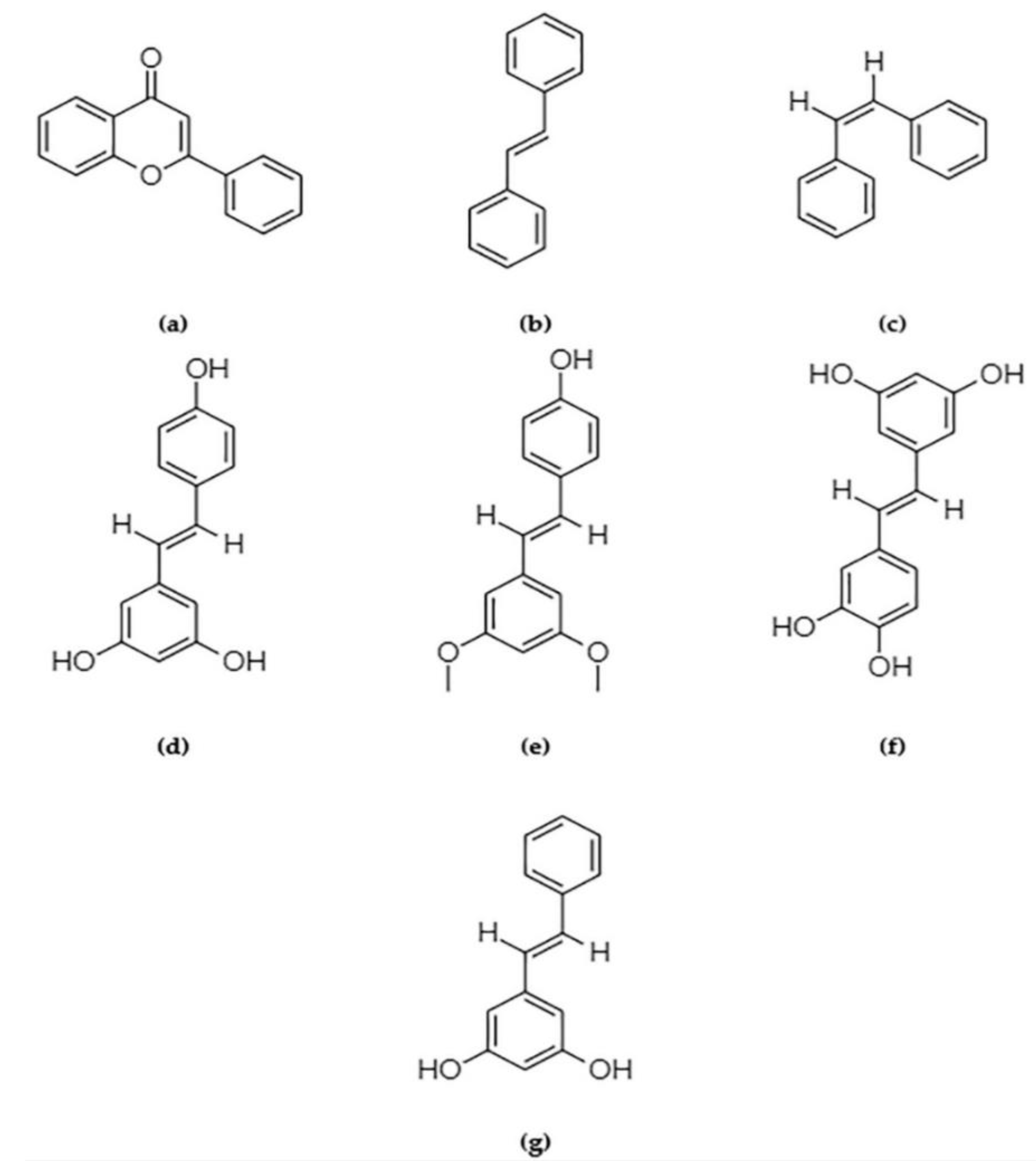
2. Scope of Primary Prevention of Breast Cancer Using Phytochemicals Including Stilbenes

Phytochemicals exert their anti-tumorigenic effect by modulating the gene expression of writer and reader proteins, by changing the pharmacokinetics of the same proteins or by potentiating the traditional chemopreventative methods [12,13,14].
Flavones and flavonoids (Figure 1a) represent the largest group of polyphenolic phytochemicals that have been abundantly studied. Currently, FDA-approved green tea catechins like epigallocatechin-3-gallate (EGCG), a naturally occurring flavone, is undergoing clinical trials (registered at clinicaltrials.gov as NCT00917735). The trial revealed no significant effect of EGCG in reducing breast cancer risk in postmenopausal women contrary to the dated observational studies [15]. Flavones such as apigenin [16] and luteolin [17], and isoflavones such as genistein and daidzein [18] have been evaluated in vitro and in vivo, and have shown promising results in the chemoprevention of breast cancer. Selvakumar et al. highlighted the mode of activity of flavonoids extensively [18]. EGCG increases the formation of S-adenosyl-L-homocysteine, which acts as a DNMT1 inhibitor and hence appears to be important for managing the methylated biomarkers discussed in the previous section. EGCG, when paired with an HDAC inhibitor like suberoylanilide hydroxamic acid (SAHA) or vorinostat, and administered to triple-negative breast cancer (TNBC) cell lines, has been shown to induce apoptosis and prevent metastatic tendencies by down-regulating the apoptosis inhibitor gene cIAP2 [13,18,19]. This combination has also been found to repress the expression of microRNAs such as miR-221/222, which is attributed to the cell-renewal capabilities in TNBC cell lines, thus maintaining the PTEN/AKT/mTOR/NF-κB expression necessary for normal stem-cell maintenance [19,20]. Genistein, found in soy products, is a controversial phytochemical that can reduce cancer risk by increasing nitric oxide bioavailability, thereby increasing oxidative stress and DNA damage and stalling cell cycle progression. Genistein promotes apoptosis by targeting proteins like BCL-2, BAX, and caspase3 functions and modulating NF-κB, PI3KC/AKT, ERK1/2, and MAPK pathway downstream signaling. However, it may promote cancer progression by upregulating estrogen receptor signaling [21,22]. Evidence has shown that genistein is capable of rendering epigenetic marks such as acetyl-H3 and H3K4me3, which are conducive to TSG (p21 and p16) expression [23].
Lignans are phytochemicals that are obtained from sources like flaxseeds, that also show promise in reducing breast cancer risk, with minimal side effects [24]. Cruciferous vegetables such as broccoli, watercress, and cabbage harbor two of the most potent phytochemicals that prevent breast cancer—phenethyl isothiocyanate (PEITC) and sulforaphane. Sulforaphane down-regulates HDAC6 expression, subsequently elevating global histone acetylation. This triggers PTEN-mediated tumor suppression in the form of autophagy in TNBC cell lines [25,26,27]. Sulforaphane also enriches H3ac, H3K9ac, and H4ac marks, which confer euchromatinization and block H3K9me3 and H3K27me3, and promote heterochromatinization in the hTERT gene, thus enabling repressors to bind. The product of hTERT gene prevents telomere shortening and therefore prevents cell death, which attributes stemness in cancer cells. Sulforaphane is also capable of demethylating selective CpG sites of hTERT, resulting in its repression [28].
One naturally occurring phytochemical found in grape skin, which has the potential to epigenetically decrease breast cancer risk, is resveratrol (3,5,4′-trihydroxy-trans-stilbene; Figure 1d). This compound belongs to the polyphenolic group of stilbenes (Figure 1b,c). Stilbenes are naturally occurring compounds derived from plants like grapevine, sorghum, pine, spruce, and mulberry, and the kinds that contain a 1,2-diphenylethylene nucleus and are used by the plants for protecting themselves from external attacks of pests, microbes, and UV exposures [29]. It can be consumed as a part of the regular diet, with no apparent toxicity other than against breast cancer cells and progenitors, although it has not yet been approved by the FDA as a dietary supplement [30]. Sinha et al. reported the mechanisms involving the chemistry by which resveratrol epigenetically suppress proliferative signals of breast tumors and the subsequent risk reduction. Resveratrol is anti-methylation and pro-acetylation, given its capability to inhibit DNMT1, DNMT3A, DNMT3B, HDAC1, and MeCP2 [31]. Computational predictions have shown that resveratrol might also have significant interactions with epigenetic readers such as BRD4 bromodomain 1, which reads acetylated histone lysine residues and plays significant roles in cell proliferation repression in cancer [32,33]. Recent studies have emphasized a resveratrol-derivative referred to as pterostilbene (trans-3,5-dimethoxy-4-hydroxystilbene; Figure 1e), which has a higher bioavailability and potency compared with resveratrol for inhibiting the growth of cancer cells and cancer stem cells in cervical cancer [34]. Studies have shown that contrary to single stilbene treatment, a combination of resveratrol and pterostilbene modulates global DNA methylation by targeting DNMT and histone acetylations by inhibiting m (an HDAC), causing an enrichment of acetyl-H3, acetyl-H3K9, and acetyl-H4 active chromatin marks, thereby inhibiting cancer cell proliferation. This same combination is successful for converting ERα- breast cancer cells into ERα+ cells, thereby sensitizing the cells to chemopreventive drugs [12]. Pterostilbene, which is safe to consume, is therefore likely an important player in the primary prevention of breast cancer. Lesser-known stilbenes include piceatannol (trans-3,3′,4′,5-tetrahydroxystilbene; Figure 1f) and pinosylvin (3,5-dihydroxy-trans-stilbene; Figure 1g), which act as resveratrol analogs and possess a similar HDAC inhibitory function [29,35]. Because of their lower levels of side effects, stilbenes can be easily integrated into the diet of individuals who are prone to breast cancer tumorigenesis.

3. Scope of Primary Prevention in Breast Cancer from a Detailed Epigenetic Perspective

The five established molecular subtypes of breast cancer are luminal A, luminal B, ERBB2/HER2-overexpressing, basal-like breast cancer (BLBC), and normal-like tumors, based on the expression levels of estrogen receptor (ER), progesterone receptor (PR), Human Epidermal Growth Factor receptor 2 (HER2), and Ki-67. Ki-67 is considered as one of the significant biomarkers in women with atypical hyperplasia, which changes depending on time. Tsang et al. (2020) extended our current understanding on the classification of molecular subtypes of breast cancer by subdividing the BLBC further into CNA-quiet (copy number alterations quiet), 1q/16q, chromosome 8 associated, CNA-high (copy number alterations-high), and mixed subtypes [36,37]. Breast cancer can be histologically classified as ductal and lobular, but a single tumor often consists of cells from diverse molecular subtypes, making it a heterogeneous disease by nature. Despite significant overlap between the histological and molecular subtyping of breast cancer, the latter, when applied to construct a spatial map of the entire breast tumor architecture, assists clinicians with a better prognosis of the disease and facilitates their ability to formulate the most effective therapeutic approach tailored specifically to a given patient [38,39,40,41].

3.1. Genetic and Epigenetic Biomarker Landscape: Breast Cancer Risk Factors and Susceptibility

3.1.1. DNA Methylation as Biomarkers

DNA methylation can be considered at two levels—focal or global. Focal DNA methylation deals with the methylation landscape on a single gene or locus basis, while global methylation accounts for the total 5-methylcytosine content in a biopsy sample methylome. There is a pattern in the focal counterpart; methylation at the upstream promoter of a gene is often associated with transcription repression, while methylation within the gene body is associated with an increased expression [42,43]. In HER2-enriched breast cancer patients or high-risk individuals, global methylation levels serve as successful biomarkers. Global methylation levels are lower in breast cancer patients and higher in normal individuals, especially at the repeat sequences like LINES or Alu, thereby keeping genomic instability in check [44,45,46]. However, there are some limitations to considering global methylation as a stable and strong biomarker for breast cancer risk determination. As pointed out by Ennour-Idrissi et al., the methylation signatures are subject to reversibility and variability based on tissue type [47]. To increase the precision of global methylation as a biomarker for breast cancer risk detection, collective methylation change in the global CpG landscape is being considered [48].
According to Knudson’s two hit hypothesis, for successful oncogenesis, both alleles of a tumor suppressor gene must be malfunctioning and silenced on one copy of a gene, and epigenetic alterations might provide a means for this process. From a gene-specific point of view, Nindrea et al. showed that hypermethylation at the BRCA1 promoter can act as “hit” by down-regulating transcription and initiating loss-of-function, thus disrupting the DNA damage repair response [49,50], which serves as an excellent biomarker for elevated risk of hereditary triple-negative breast cancer (TNBC) cases [51]. Gene body hypermethylation of ATM, a breast cancer susceptibility gene that codes for downstream signaling proteins for cell cycle arrest, can lead to the early onset of breast cancer in women, and thus is a very useful biomarker [52]. Promoter hypomethylation in PALB2, a breast cancer susceptibility gene that localizes the BRCA2 gene at the site of DNA damage, has been established as a biomarker for sporadic breast cancer [53]. Similarly, Masood et al. showed that hypermethylation in the 600 base pair region of the hTERT gene promoter can act as a potent biomarker for breast cancer diagnosis [54]. In the case of sporadic breast cancer, occasional rigorous screening of CpG landscapes of known breast cancer risk genes in healthy individuals may be the most logical way of assigning biomarkers. Ennour-Idrissi et al. provides information on seven significant genes that set breast cancer-prone individuals apart from resistant individuals [55]. There are also studies conducted on promoter methylation states of 100 genes, including BRCA1, CCND2, BCL2, MDR1, IL10, and TWIST, recorded to understand how alteration between hypermethylation and hypomethylation over time contributes to select breast cancer susceptibility biomarkers [56]. The list of breast cancer biomarkers involving DNA methylation continues to increase on a regular basis.

3.1.2. Histone Modifications as Biomarkers

Compared with DNA methylation, histone modifications are less constant epigenetic marks present on nucleosome structures that can change with every occasion of transcription. This helps to explain the scarcity of literature on anomalies in histone modifications as biomarkers for breast cancer risk assessment. Acetylation of histones is directly associated with euchromatic (open) and heterochromatic (tight) organization of DNA, which alters the accessibility to different transcription factors. Gene upregulation can often be found when acetyl groups are added to histone N terminal lysines (H3K9, H3K14, H3K18, H3K23, H4K5, H4K8, H4K12, and H4K16) [57]. Methylation at histone lysines (H3K9, H3K27, and H4K20, and histone arginine residues at H3R2me2a, H3R8me2a, H3R8me2s, and H4R3me2s) are associated with heterochromatin formation and gene-repression, while at H4R3me2a, H3R2me2s, H3R17me2a, and H3R26me2a, methylation confers an open chromatin structure [58,59].
There are other histone modifications such as ubiquitination, sumoylation, and phosphorylation. In patients who have already been diagnosed with breast cancer and are undergoing cancer surgery and treatment, global histone modifications have been shown to be an efficient tool for evaluating metastatic status, survival, and likelihood of relapse, as well as other outcomes [60,61].

3.1.3. Non-Coding RNA as Biomarkers

Non-coding RNAs (ncRNAs) are short fragments of RNA transcribed from the non-coding regions or regions of DNA that may or may not be originated from ultraconserved elements [62]. ncRNAs may possess certain functions, such as regulating the interactions between different proteins with genomic particles, enhancing or repressing transcription rates, influencing alternate splicing, the ability to change 3D conformation of genomic DNA, and maintaining genomic stability, to mention a few [63]. The ncRNAs that span only a few (19–25) nucleotides are called microRNAs (miRNAs), and those above 200 nucleotides are called long non-coding RNAs (lncRNAs) [64]. They serve as important biomarkers for breast cancer risk determination and are non-invasively detectable from circulating blood as they are transported around the periphery of the body within the exosomes. Farina et al. discovered a panel of 2500 miRNAs related to breast cancer and found six with an abnormal presence in the blood in apparently healthy individuals that could serve as a biomarker for high-risk individuals with tendencies for future breast tumorigenesis [65,66]. Short nucleotide polymorphisms (SNPs) in lncRNAs are often hotspots of methylation and, like DNA methylation, these epigenetic marks can upregulate or down-regulate the activity of the lncRNAs [62,67]. Probing ncRNA activities is achievable, which makes tagging them as biomarkers easier [68]. Table 1 enlists such non-coding RNAs associated with breast cancer risk.
In addition to RNAs, there are also cell-free DNAs and proteins with methylation marks circulating in the bloodstream that can act as epigenetic biomarkers of cancer risk assessment [43,69].
Table 1. A non-exhaustive table of relevant ncRNAs associated with breast cancer risk prediction serving as biomarkers.
ncRNATherapeutic SignificanceBreast Cancer SubtypeReference
miR-21-3p, miR-21-5p, and miR-99a-5p, miR-1246, miR-1307-3p, miR-4634, miR-6861-5p, and miR-6875-5p Risk prediction and early detection and overall survivalUnspecified[43]
Panel of 8 miRNAs (miR-139-5p, miR-10b-5p, miR-486-5p, miR-455-3p, miR-107, miR-146b-5p, miR-324-5p, and miR-20a-5p)Predicting risk of relapseTriple-negative breast cancer[70]
Panel of 6 miRNAs (miR-3124-5p, miR-1184, miR-4423-3p, miR-4529-3p, miR-7855-5p, and miR-4446-3p) Breast cancer risk predictionUnspecified[65]
miRNA-191, miRNA-484, miR-16, miR-25, miR-222, and miR-324-3p Breast cancer risk predictorUnspecified[71]
Panel of 4 miRNAs (hsa-miR-21, hsa-miR-494, hsa-miR-494, and hsa-miR-183) Metastatic risk predictionUnspecified[72]
Panel of 4 miRNAs (miR-1246, miR-206, miR-24, and miR-373) Early diagnosis of breast cancerUnspecified[66]
lncRNAs like PVT1, MAPT-AS1, LINC00667, and LINC00938Predicting breast cancer survivalUnspecified[73]
Panel of miR-127-3p, miR-148b, miR-376a, miR-376c, miR-409-3p, miR-652, and miR-801 Distinguishing healthy women from women carrying benign or malignant breast tumors with more accuracy in younger individualsUnspecified[74]
miR-200a,
miR-200b, miR-200c, miR-210, miR-215 and miR-486-5p
Metastasis onset predictorUnspecified[74]
mRNA-lncRNA conjugate (mRNA species for FCGR1A, RSAD2, CHRDL1, and the lncRNA species for HIF1A-AS2 and AK124454)Predicting risk of relapseTriple-negative breast cancer[32]

4. In-Practice Clinical Methods for Addressing Primary Prevention

As previously mentioned, tamoxifen and raloxifene are FDA-approved commonly prescribed synthetic stilbene derivatives successful in chemoprevention if administered for five years, in both pre- and post-menopausal women, but might have adverse effects on other body tissues. There is another major drawback with tamoxifen chemotherapy in patients. ER+ cancer cells can develop resistance against tamoxifen due to an array of reasons, such as acquired perpetuation of repressive methylation marks in CpG islands of the estrogen-sensitive gene promoter [75,76,77,78,79,80,81]. Tamoxifen is found to be the most effective in individuals with a high risk of ER+ breast cancer, because it is a strong antagonistic competitor of estrogen and prevents estrogen-related growth signaling when it binds to the estrogen receptor in breast epithelia. Recent findings have indicating the potential evolution of superior derivatives of tamoxifen, such as endoxifen, which has a better affinity towards estrogen receptors, with better pharmacokinetics, allowing for greater bioavailability in a time-dependent manner [82]. This is currently undergoing a clinical trial, as indicated on the NIH US National Library of Medicine website clinicaltrials.gov.
In 2018, the FDA approved a poly (ADP-ribose) polymerase (PARP) inhibitor called talazoparib (TALZENNA, Pfizer Inc.) for patients who are carriers of the germline BRCA mutation and have HER2- metastatic breast cancer. Pulliam et al., in 2018, showed that a combination of talazoparib and DNMT inhibitor guadecitabine rendered promising results in modulating DNMT1 action, thereby altering the DNA damage repair response, increasing cellular concentration of caspase 3 and finally asserting cytotoxic effects on cancer stem cells with minimal side-effects in contrast with what has been observed with the traditional chemotherapy [83,84].
The basic methods to prevent breast cancer epigenetically are not significantly different from genetic approaches. A schema has been reported by Hanahan et al. summarizing feasible methods of managing breast cancer [85]. There is a plethora of research regarding primary prevention of breast cancer through epigenetic means, although not as many phytochemicals and their derivatives have been identified that can be adopted as a part of preventing the appearances of the first signs of breast cancer. Figure 2 demonstrates some basic qualities that phytochemicals should possess in order to be considered as efficient epigenetic chemopreventive compounds. The following paragraphs summarize some novel epigenetic prospects of stilbenes that have relevance for its consideration as a potent chemical to reduce the risk of breast cancer.

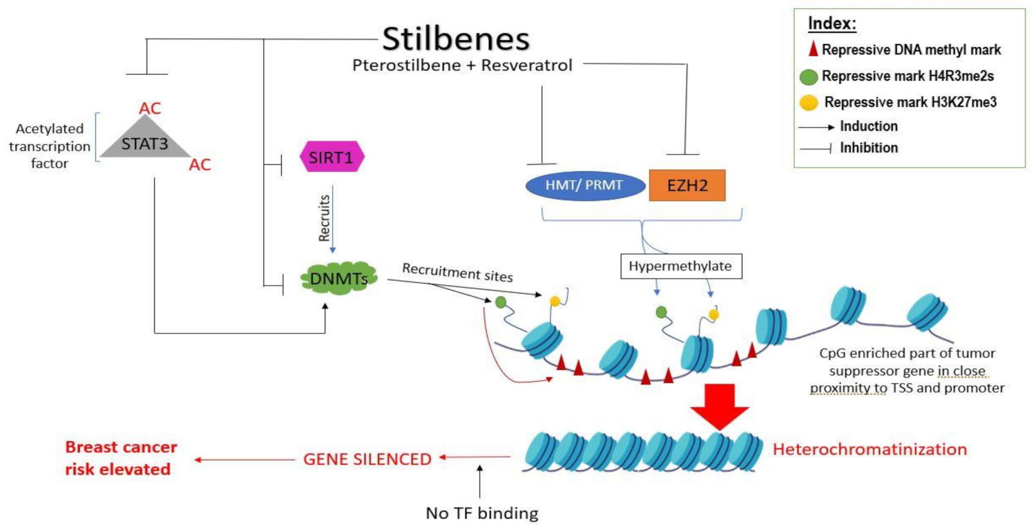
4.1. Role of Stilbenes in Differentially Modulating DNA Methylation of Genes and Gene Loci

Ongoing epigenetic research is mostly interested in discovering the mechanisms by which stilbenes differentially influence DNA methylation in both tumor suppressor genes and oncogenes or how they differentially methylate at two different CpG loci of the same gene. Harnessing these mechanisms can be the key to reversing the epigenetic marks that appear early at the onset of tumorigenesis; however, the biochemistry involved in this process is still elusive. In their experiment on both mild and aggressive breast cancer cell lines of MCF10A1a and MCF10A1h, Beetch et al. discovered 113 highly specific targets of resveratrol and pterostilbene. They also identified the SALL3 gene (sal-like 3), which is upregulated by these two stilbenes, which in turn down-regulates DNMT3A, binding to the promoters of tumor suppressor genes like SEMA3A [86]. As a result, a hypomethylation state is created on the silenced tumor suppressor gene, which mimics the wild-type methylation state, restoring its expression. Furthermore, these stilbenes influence Nuclear Factor 1C protein, a tumor suppressor that localizes heavily on the SEMA3A promoter. Similar patterns of reactivating silenced tumor suppressor genes such as p16, CCND2, APC, and RASSF1A by inducing demethylation with trans-resveratrol have been reported by Zhu et al., supporting the idea that resveratrol can play important roles in primary chemoprevention [87]. However, there are drawbacks to this idea, because resveratrol has multiple off-target effects that also require further investigation.
The selective nature of resveratrol and other similar stilbene compounds were discussed by Aldawsari et al. through in silico molecular docking simulations, where they showed the similarity in the chemical structure between resveratrol and some recently identified small molecules, including methylenedisalisylic acid, which binds to DNMT3As and DNMT3Bs at the catalytic site or at the cofactor binding site (S-adenosylhomocysteine) with a high degree of specificity, and inhibits their activity by preventing DNA binding. They also conducted in vitro work that showed that resveratrol is more potent when converted into a hydroxylated hybrid salicylate derivative [88].

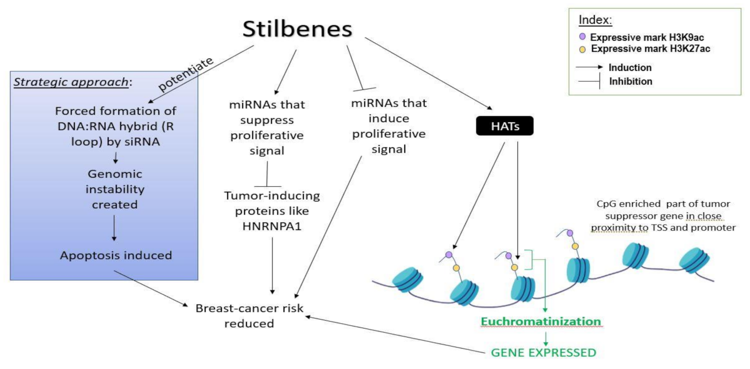
4.2. Role of Stilbenes in Differentially Modifying Histones

It is a well-known fact that histone acetylation regulates DNA conformation, thereby regulating the access of transcription factors and enzymes like DNMTs to the DNA. A combinatorial dosage of resveratrol and pterostilbene has been shown to down-regulate SIRT1, an HDAC III responsible for leading DNMTs to hypermethylate promoters and silence TSGs (Figure 3) [14]. Chatterjee et al. showed that resveratrol can successfully down-regulate the oncogenic activity of PRMT5 (protein arginine methyltransferases) and EZH2 (catalytic domain of Polycomb repressive protein 2) by reducing silencing histone methyl marks H4R3me2s and H3K27me3 from TSGs (Figure 3). In contrast with this, they also showed that HATs are positively influenced by resveratrol, leading to the increase of expressive histone marks like H3K9ac and H3K27ac on the histones in proximity of the TSG (BRCA1, p53, and p21) promoters, as shown in Figure 4. Removal of the H3K27me3 mark also prevents DNMT1 and DNMT3A binding to the promoters of TSGs like BRCA1, leading to its expression and causing p16-dependent cell senescence to occur [89]. Not just histones, but transcription factors can directly become acetylated, which subsequently hypermethylate the promoter of tumor suppressor genes, leading to the gene suppression. For instance, as shown by Lee et al., acetylated transcription factor STAT3 can silence tumor suppressor genes in basal-like breast cancer tissues, which can be reverted by stilbenes like resveratrol and pterostilbene. As a downstream effect of resveratrol on inhibiting STAT3 acetylation, the ERα gene is demethylated and its expression is upregulated, which provides an opportunity to TNBC patients to undergo hormonal therapy [90,91]. Besides STAT3 acetylation, STAT3 phosphorylation also plays an important role in tumorigenesis, and stilbenes can successfully prevent this process [91,92]. A hybrid derivative of pterostilbene and vorinostat (hydroxamate) has been shown to bind to the SH2 domain of STAT3 with stable interactions at arginine and serine residues, preventing STAT3 from interacting with DNA. This conjugate has also been proven to inhibit HDACs [91].

4.3. Role of Stilbenes in Differentially Modulating the Activity of Non-Coding RNAs and Preventing Breast Cancer Initiation

Hagiwara et al. showed that both pterostilbene and resveratrol, in both their natural form and demethylated condition, are capable of upregulating Argonaute2 protein and thereby increased the expression of tumor-suppressive microRNAs like miR-16, miR-141, miR-143, and miR-200c in the triple negative breast cancer cell line MDA-MB-231 [93]. Otsuka et al.’s work on breast cancer-associated tumor-suppressive miRNAs (miR-34a, miR-424, and miR-503) demonstrated that these microRNAs are upregulated by resveratrol, which in turn suppresses tumor-inducing protein HNRNPA1 (heterogeneous nuclear ribonucleoprotein A1) [94]. A summary is depicted in Figure 4.
There are indirect ways by which stilbenes can modulate non-coding RNAs, in favor of reducing the risk of breast cancer. Stilbenes can influence the three-dimensional structure of R-loops formed by DNA:RNA triple strand hybrids that are known to regulate normal cellular functions such as transcription, DNA replication, and telomere maintenance via epigenetic control. This involves subsequent prevention or allowing the reader or writer proteins of methylation to interact with the gene regulatory regions. Loss-of-tumor-suppression function of BRCA2, impaired ATP-dependent chromatin remodeler protein SWI/SNF complex, and stress of reactive oxygen species can give rise to sporadic R-loops, which can act as biomarkers for genomic instability and are suitable targets for the DNA damage repair response. Thus, R-loops or similar DNA:RNA complex structures are hotspots for targeting chemopreventive measures in cancer patients. From a strictly strategic point of view, one of the ways that primary chemoprevention works in cell cultures is through delivering synthetic small interfering RNA (siRNA) to the DNA duplex of cancer cells or progenitors via the transfection of the micelle-bound RNA interference (RNAi) protein complex, thereby forming a complex DNA:RNA hybrid that resembles R-loop. Stilbenes and stilbenoids, when amalgamated to the end of siRNA of a RNAi complex, have been shown to increase the uptake of these micelle-bound RNAi complexes by the cancer cells, thereby increasing the effectiveness of RNAi technology [95,96]. Stilbenes might therefore be used to potentiate RNAi-based therapeutics and topical or intratumoral siRNA vaccine (for precision delivery). Individuals at high risk of breast cancer might benefit from this kind of therapeutics. While there could be drawbacks to this approach, further investigations and trials are required.
Epithelial-to-mesenchymal transformation is a normal cellular process needed for embryogenesis and wound-healing, which is characterized by loss of cell−cell and cell−matrix adhesion and the gain of motile features in epithelial cells, which is also a hallmark for cancer initiation and progression. Huang et al. showed that pterostilbene can prevent the epithelial-to-mesenchymal transformation (EMT) and promote mesenchymal-to-epithelial transformation (MET) by increasing the expression of lncRNA H19 [97]. LncRNA H19 acts as a differential sponge of microRNAs miR-200b/c and let-7b. This leads to modulating their targets guanosine triphosphatase-activating protein gene Git2 and Cytohesin-3 protein coding gene Cyth3, which in turn regulate the RAS superfamily member adenosine 5’-diphosphate ribosylation factor (ARF) [98].

5. Epigenetic Effect of Stilbene on Genes and Proteins Related to Cell-Proliferation and Metastasis

Unabated cell proliferation is one of the major phenotypic hallmarks of breast cancer initiation. Constitutive activation of the PI3K catalytic domain p110α due to mutations in the PIK3CA gene causes cell migration and metastatic properties in breast cancer cells and is a strong biomarker for the early detection of breast cancer occurrence or recurrence, and is hence used for periodic molecular mammographic screening [99,100]. PI3K is also capable of exerting epigenetic effects by controlling the histone methyltransferases like EZH2, and thereby decreasing the global methylation and genome-wide upregulation of transcription. PI3K/AKT also modulates HATs like p300/CBP and has contributions towards euchromatin−heterochromatin modulation and DNA accessibility [101,102]. Hence, the targeted inhibition of PI3K has been proven to be useful for chemoprevention and therapy in luminal breast cancer [103,104]. However, one of the notable challenges that this mutation, combined with dysregulation of other cell cycle mediators like PTEN, pose is rendering insensitivity towards traditional chemotherapeutic reagents, like lapatinib and trastuzumab, and PI3K inhibitor therapy [105,106,107]. Stilbenes such as piceatannol have effects on the AKT/mTOR pathway, similar to the EGCG and SAHA combination. There is evidence of it successfully inhibiting the PI3K pathway and significantly reducing cell proliferation and migration in prostate cancer [108], and it might be promising for formulating a PI3Ki therapy. Part of this possibility lies in the fact that two stilbenes, resveratrol and piceatannol, inhibit the JAK/STAT pathway, thereby suppressing the cell survival signals [109] and potentially reversing the chemoresistance of malignant cells to cisplatin treatment, as shown in other types of cancer [110]. Further investigation is required in order to confirm these possibilities regarding targeted breast cancer therapy and chemoprevention. There are also limited data on the epigenetic effects of stilbenes on the genes that encode the proteins of these major pathways that regulate breast cancer risk.
Ki-67 is a protein that was traditionally used as a biomarker for cell proliferation, for which high levels provide information on distinguishing women with no tumors, benign tumors, and malignant breast tumors [111]. However, recent research posits that cell cycle progression depends substantially on the localization of this protein within a mitotic cell. Sun et al. made an account of all of the past research explaining the molecular relationship between Ki-67 deficiency in a dividing cell and cell cycle arrest [112]. Dearth of Ki-67 induces cyclin-dependent kinase inhibitor checkpoint protein p21 in human primary fibroblasts and can delay S phase initiation in hTERT-BJ skin cell lines. In addition, Ki-67 plays a major role in heterochromatin organization in nucleolar periphery, thereby controlling a cell’s entry to the G1/S phase [112]. Li et al. using in vitro gastric cancer cell lines, successfully suppressed the expression of Ki-67 by targeting methylated CpG binding protein 2 (MBD2) at the methylated promoter of the Ki-67 protein-coding gene, thereby preventing the transcription factor Sp1 from binding [113]. This concept seems to hold potential for formulating a therapy or prevention of breast cancer cells. However, Sun et al. challenged this concept as it might not be applicable for all breast cancer cells. MDA-MB-231 (ER-, PR-, and HER2-) cells express a higher Ki-67 expression contrary to MCF-F7 (ER+, PR+, and HER2-) cells and have been shown to have no p21 induction [112]. Employment of stilbenes like resveratrol to repress Ki-67 transcription might also be challenging because studies show that it prevents DNMT1 and MBD2 binding at the promoter of important oncogenes like BRCA1, thereby activating them [114]. The potential to make resveratrol work differentially, gene-to-gene, needs further research.

6. Bioavailability of Stilbenes in Target Tissue and Limitations

Unlike flavones, stilbenes are derived at low or variable concentrations from edible sources. In addition, stilbenes like resveratrol have a substantially short half-life (14.4 min in mice), low water-solubility, and faster metabolization and exclusion rate, not only in the cells of test subjects, but also their gut-microbiota, which reduces the functionality of stilbene as anti-cancer compounds [29,115,116]. Depending on the chemical conjugate that the stilbenes possess in food, their bioavailability varies greatly. For example, the pharmacokinetic profile of pterostilbene is better than that of resveratrol due to the presence of methoxy groups at the 3- and 5-carbon positions of the m-hydroquinone moiety and their higher lipophilicity [29,117]. This makes the formulation of an achievable stilbene-containing diet for daily consumption a difficult task. Apart from this, extensive preclinical and clinical studies conducted on cell lines and xenografted mouse and rat models have shown that the bioavailability of stilbenes, like resveratrol, in the plasma is as low as 42.8 ± 4.4 µM after 5 min, if administered independently and intravenously at a rate of 20 mg/kg body weight [118,119]. Similar results were recorded for other stilbenes like pterostilbene, pinostilbene, and gnetol, except for the stilbene piceatannol, which has a bioavailability 2.6 times that of resveratrol. The bioavailability of stilbenes at the target tissue also varies depending on the species, route of administration (some examples include oral capsule gavage, dietary intake, and intravenous injection), age, sex, and lifestyle of the subjects [119].
By combining the results generated on human subjects by Sergides et al. [120] and Ávila-Gálvez et al. [121], it can be concluded that a daily dose between 437.7 mg to 500 mg of pure resveratrol or plant-derived resveratrol combined with other staple nutrients has been observed to effectively be utilized by the adult patient body without any cytotoxicity. Metabolite profiling [121] has revealed that the major drawback with resveratrol is that it does not reach the healthy or malignant breast tissue in its original form. The resveratrol-metabolites generated in vivo in human patients were found to be highly bioavailable in both healthy and malignant breast tissue, but were significantly unsuccessful in exerting an antiestrogenic and antiproliferative activity on both of the tissue types. These observations reduce the possibility of resveratrol and similar stilbenes that are considered as potent risk-reducing dietary chemopreventive compounds.
To address all such problems, research is ongoing to find better mechanisms to deliver stilbenes to the target tissue. It has been hypothesized that stilbenes, being hydrophobic in nature, may be transported more efficiently through lipid-based cellular delivery mechanism (liposomes) or through emulsification, thereby facilitating intestinal absorption [122]. Other methods, like combining stilbenes with sulfobutylether-β-cyclodextrin, have been shown to have increased stability and a better bioavailability of the former at the target tissue [123,124].

7. Conclusions

Presently, there are not enough accurate methods to detect dormant risk factors in breast tissue before the occurrence of the disease. Zubor et al. [74] suggest a multiomics approach encompassing MRI, mammography, and liquid biopsy with added emphasis on epigenetic miRNA profiling of both blood and breast tissue as the best way to address primary prevention. This makes the primary prevention of breast cancer not as popular of a concept as secondary or tertiary prevention. Besides exposing oneself to invasive medical procedures, positive outcomes upon the administration of stilbenes in relevant doses are not ascertained for every individual. There could be adverse effects that also need evaluation, especially adopting stilbene-derived synthetic chemoprevention while gestating. There has been evidence of increased possibilities of transgenerational developmental deformities [125]. Epigenetic biomarkers are subject to constant change, given the continuous change of an individual’s external and internal environment, providing a scope for drug repurposing and combinatorial therapeutic and preventive measures. Presently, there is interest in the prospects of NSAIDs, like aspirin and ibuprofen, as risk-reducing medication for breast cancer as they can inhibit the overexpression of COX-2, a gene responsible for the initiation of tumorigenesis and inflammation in breast epithelia [126]. Regulski et al. recently identified 19 trans-stilbene and 6 trans-4-stilbazole derivatives that can serve as a replacement for NSAIDs, showing a similar interaction at the Tyr355 residue of the COX-2 protein N-terminal and the same docking energy in simulation [127], although the detailed epigenetic mechanisms of these chemicals remain to be evaluated.
With the discovery of new biomarkers and stilbene variants, more research on novel epigenetic reactions and targets is required. It is imperative to approach the first signs of breast cancer risk with combinatorial approaches instead of monotherapy for attaining the maximum efficacy of these chemicals.

Author Contributions

S.G. conceived the review paper with the guidance of T.O.T. The manuscript was drafted and edited by S.G., I.A. and T.O.T. All authors have read and agreed to the published version of the manuscript.

Funding

This work was supported in part by grants from the National Institute of Health (NCI R01CA178441 and NCI R01CA204346).

Institutional Review Board Statement

Not applicable.

Informed Consent Statement

Not applicable.

Acknowledgments

We thank Tollefsbol for this opportunity and want to extend our gratitude towards all members of the Tollefsbol lab for their supportive comments.

Conflicts of Interest

The authors declare no conflict of interest.

Abbreviations

AisAromatase inhibitors
Akt Protein kinase B
ARFAdenosine 5’-diphosphate ribosylation factor
ATMSerine/ Threonine kinase
BaxBcl-2-associated X protein
Bcl-2B-cell lymphoma 2 protein
BRCA1/2Breast cancer gene 1 and 2
CD1Cluster of differentiation 1
cIAP2Cellular Inhibitor Of Apoptosis 2 gene
c-MycMyelocytomatosis proto-oncogene
CNA-high Copy number alterations high
CNA-quiet Copy number alterations quiet
COX2Cyclooxygenase 2 gene
CpG Regions of DNA with repetitive occurrence of cytosine and guanosine nucleotides
Cyth3Cytohesin-3 gene
DNA Deoxyribonucleic acid
DNMT1 DNA methyltransferase 1
DNMT3A DNA methyltransferase 3 Alpha
DNMT3B DNA methyltransferase 3 Beta
DNMTs DNA methyl transferases
EGCG Epigallocatechin gallate
EMT Epithelial mesenchymal transformation
ER- Estrogen receptor negative
ER+Estrogen receptor positive
ERBB2Erythroblastic leukemia viral oncogene homologue 2
ERK1/2 Extracellular signal-regulated kinases
EZH2 Enhancer of zeste homolog 2 protein
Git2Guanosine triphosphatase-activating protein gene
HATs Histone acetyltransferases
HDACi Histone deacetylase inhibitor
HDACs Histone deacetylases
HMTs Histone methyltransferases
HNRNPA1 Heterogeneous nuclear ribonucleoprotein A1
hTERTHuman telomerase reverse transcriptase
H3R2me2a Asymmetrical dimethylation at arginine residue 2 of histone subunit 3
HER2 Human epidermal growth factor receptor 2
H4R3me2s Dimethylation at arginine residue 3 residue of histone subunit 4
H3ac Acetylation at histone subunit 3
H4ac Acetylation at histone subunit 4
HAC6Histone deacetylase 6
H3K4me3 Trimethylation at histone subunit 3 at lysine 4
H3R8me2a Asymmetrical dimethylation at arginine residue 8 residue of histone subunit 3
H3R8me2s Dimethylation at arginine residue 8 residue of histone subunit 3
H4K5 Histone subunit 4 at lysine residue 5
H3K9 Histone subunit 3 at lysine residue 9
H3K9me3 Trimethylation at histone subunit 3 at lysine 9
H4K8 Histone subunit 4 at lysine residue 8
H3K9ac Acetylation at histone subunit 3 at lysine 9
H4K12Histone subunit 4 at lysine residue 12
H3K14 Histone subunit 3 at lysine residue 14
H4K16 Histone subunit 4 at lysine residue 16
H3K18 Histone subunit 3 at lysine residue 18
H3K23 Histone subunit 3 at lysine residue 23
H3K27me3 Trimethylation at histone subunit 3 at lysine 27
JAK Janus kinase
Ki-67 Kiel-67 antibody
Let-7b MicroRNA Let-7b
LINEs Long interspersed nuclear elements
lncRNA Long non-coding RNA
MAPK Mitogen-activated protein kinase
MBD2Methyl-CpG-binding domain protein 2
MeCP2Methyl-CpG-binding protein 2
MET Mesenchymal-to-epithelial transformation
miRNA MicroRNA
mTOR Mammalian target of rapamycin
N terminal Nitrogen terminal
ncRNA Non-coding RNA
NF-κB Nuclear factor kappa light chain enhancer of activated B cells
NSAIDs Non-steroidal anti-inflammatory drugs
p300/CBP CREB-binding protein and its homolog p300
PALB2Partner and localizer of BRCA2
PARP Poly (ADP-ribose) polymerase
PEITCPhenethyl isothiocyanate
PI3K Phosphoinositide 3-Kinase
PI3Ki Phosphoinositide 3-Kinase inhibitor
PIK3CA Phosphatidylinositol-4,5-Bisphosphate 3-Kinase Catalytic Subunit Alpha
PR- Progesterone receptor negative
PR+ Progesterone receptor positive
PTENPhosphatase and Tensin homolog deleted on chromosome 10
RAS Rat sarcoma
RNA Ribonucleic acid
RNAi Interfering RNA
SAHA Suberanilohydroxamic acid
SALL3Spalt Like Transcription Factor 3 coding gene
SEMA3ASemaphorin 3A gene
SERMs Selective estrogen receptor modulators
siRNA Small interfering RNA
SIRT1Sirtuin 1
SNPs Single nucleotide polymorphism
STAT Signal transducer and activator of transcription proteins
STK11Serine/threonine kinase 11
TNBC Triple-negative breast cancer
TP53Tumor protein 53
TSGs Tumor suppressor genes
Tyr355 Tyrosine 355 residue

References

  1. Ahmad, A. Breast Cancer Statistics: Recent Trends. Adv. Exp. Med. Biol. 2019, 1152, 1–7. [Google Scholar] [CrossRef] [PubMed]
  2. Sung, H.; Ferlay, J.; Siegel, R.L.; Laversanne, M.; Soerjomataram, I.; Jemal, A.; Bray, F. Global Cancer Statistics 2020: GLOBOCAN Estimates of Incidence and Mortality Worldwide for 36 Cancers in 185 Countries. CA A Cancer J. Clin. 2021, 71, 209–249. [Google Scholar] [CrossRef] [PubMed]
  3. Thorat, M.A.; Balasubramanian, R. Breast cancer prevention in high-risk women. Best Pract. Res. Clin. Obstet. Gynaecol. 2020, 65, 18–31. [Google Scholar] [CrossRef]
  4. Lee, C.I.; Chen, L.E.; Elmore, J.G. Risk-based Breast Cancer Screening: Implications of Breast Density. Med. Clin. N. Am. 2017, 101, 725–741. [Google Scholar] [CrossRef]
  5. Sun, Y.S.; Zhao, Z.; Yang, Z.N.; Xu, F.; Lu, H.J.; Zhu, Z.Y.; Shi, W.; Jiang, J.; Yao, P.P.; Zhu, H.P. Risk Factors and Preventions of Breast Cancer. Int. J. Biol. Sci. 2017, 13, 1387–1397. [Google Scholar] [CrossRef] [Green Version]
  6. Coughlin, S.S. Epidemiology of Breast Cancer in Women. Adv. Exp. Med. Biol. 2019, 1152, 9–29. [Google Scholar] [CrossRef] [PubMed]
  7. Nelson, H.D.; Fu, R.; Zakher, B.; Pappas, M.; McDonagh, M. Medication Use for the Risk Reduction of Primary Breast Cancer in Women: Updated Evidence Report and Systematic Review for the US Preventive Services Task Force. JAMA 2019, 322, 868–886. [Google Scholar] [CrossRef] [PubMed] [Green Version]
  8. Shieh, Y.; Tice, J.A. Medications for Primary Prevention of Breast Cancer. JAMA 2020, 324, 291–292. [Google Scholar] [CrossRef]
  9. Graham, D.; DiNome, M.L.; Ganz, P.A. Breast Cancer Risk–Reducing Medications. JAMA 2020, 324, 310. [Google Scholar] [CrossRef]
  10. Li, F.; Dou, J.; Wei, L.; Li, S.; Liu, J. The selective estrogen receptor modulators in breast cancer prevention. Cancer Chemother. Pharmacol. 2016, 77, 895–903. [Google Scholar] [CrossRef]
  11. Kasiotis, K.M.; Lambrinidis, G.; Fokialakis, N.; Tzanetou, E.N.; Mikros, E.; Haroutounian, S.A. Novel Carbonyl Analogs of Tamoxifen: Design, Synthesis, and Biological Evaluation. Front. Chem. 2017, 5, 71. [Google Scholar] [CrossRef] [Green Version]
  12. Kala, R.; Tollefsbol, T.O. A Novel Combinatorial Epigenetic Therapy Using Resveratrol and Pterostilbene for Restoring Estrogen Receptor-α (ERα) Expression in ERα-Negative Breast Cancer Cells. PLoS ONE 2016, 11, e0155057. [Google Scholar] [CrossRef]
  13. Lee, W.J.; Shim, J.-Y.; Zhu, B.T. Mechanisms for the Inhibition of DNA Methyltransferases by Tea Catechins and Bioflavonoids. Mol. Pharmacol. 2005, 68, 1018. [Google Scholar] [CrossRef] [PubMed] [Green Version]
  14. Kala, R.; Shah, H.N.; Martin, S.L.; Tollefsbol, T.O. Epigenetic-based combinatorial resveratrol and pterostilbene alters DNA damage response by affecting SIRT1 and DNMT enzyme expression, including SIRT1-dependent γ-H2AX and telomerase regulation in triple-negative breast cancer. BMC Cancer 2015, 15, 672. [Google Scholar] [CrossRef] [Green Version]
  15. Samavat, H.; Wu, A.H.; Ursin, G.; Torkelson, C.J.; Wang, R.; Yu, M.C.; Yee, D.; Kurzer, M.S.; Yuan, J.M. Green Tea Catechin Extract Supplementation Does Not Influence Circulating Sex Hormones and Insulin-Like Growth Factor Axis Proteins in a Randomized Controlled Trial of Postmenopausal Women at High Risk of Breast Cancer. J. Nutr. 2019, 149, 619–627. [Google Scholar] [CrossRef] [PubMed] [Green Version]
  16. Yan, X.; Qi, M.; Li, P.; Zhan, Y.; Shao, H. Apigenin in cancer therapy: Anti-cancer effects and mechanisms of action. Cell Biosci. 2017, 7, 50. [Google Scholar] [CrossRef] [Green Version]
  17. Imran, M.; Rauf, A.; Abu-Izneid, T.; Nadeem, M.; Shariati, M.A.; Khan, I.A.; Imran, A.; Orhan, I.E.; Rizwan, M.; Atif, M.; et al. Luteolin, a flavonoid, as an anticancer agent: A review. Biomed. Pharmacother. 2019, 112, 108612. [Google Scholar] [CrossRef]
  18. Selvakumar, P.; Badgeley, A.; Murphy, P.; Anwar, H.; Sharma, U.; Lawrence, K.; Lakshmikuttyamma, A. Flavonoids and Other Polyphenols Act as Epigenetic Modifiers in Breast Cancer. Nutrients 2020, 12, 761. [Google Scholar] [CrossRef] [Green Version]
  19. Steed, K.L.; Jordan, H.R.; Tollefsbol, T.O. SAHA and EGCG Promote Apoptosis in Triple-negative Breast Cancer Cells, Possibly Through the Modulation of cIAP2. Anticancer Res. 2020, 40, 9–26. [Google Scholar] [CrossRef]
  20. Li, B.; Lu, Y.; Yu, L.; Han, X.; Wang, H.; Mao, J.; Shen, J.; Wang, B.; Tang, J.; Li, C.; et al. miR-221/222 promote cancer stem-like cell properties and tumor growth of breast cancer via targeting PTEN and sustained Akt/NF-κB/COX-2 activation. Chem.-Biol. Interact. 2017, 277, 33–42. [Google Scholar] [CrossRef]
  21. Tuli, H.S.; Tuorkey, M.J.; Thakral, F.; Sak, K.; Kumar, M.; Sharma, A.K.; Sharma, U.; Jain, A.; Aggarwal, V.; Bishayee, A. Molecular Mechanisms of Action of Genistein in Cancer: Recent Advances. Front Pharm. 2019, 10, 1336. [Google Scholar] [CrossRef] [Green Version]
  22. Ziaei, S.; Halaby, R. Dietary Isoflavones and Breast Cancer Risk. Medicines 2017, 4, 18. [Google Scholar] [CrossRef] [Green Version]
  23. Li, Y.; Chen, H.; Hardy, T.M.; Tollefsbol, T.O. Epigenetic Regulation of Multiple Tumor-Related Genes Leads to Suppression of Breast Tumorigenesis by Dietary Genistein. PLoS ONE 2013, 8, e54369. [Google Scholar] [CrossRef] [Green Version]
  24. Fabian, C.J.; Khan, S.A.; Garber, J.E.; Dooley, W.C.; Yee, L.D.; Klemp, J.R.; Nydegger, J.L.; Powers, K.R.; Kreutzjans, A.L.; Zalles, C.M.; et al. Randomized Phase IIB Trial of the Lignan Secoisolariciresinol Diglucoside in Premenopausal Women at Increased Risk for Development of Breast Cancer. Cancer Prev. Res. 2020, 13, 623. [Google Scholar] [CrossRef] [Green Version]
  25. Park, J.E.; Sun, Y.; Lim, S.K.; Tam, J.P.; Dekker, M.; Chen, H.; Sze, S.K. Dietary phytochemical PEITC restricts tumor development via modulation of epigenetic writers and erasers. Sci. Rep. 2017, 7, 40569. [Google Scholar] [CrossRef] [PubMed] [Green Version]
  26. Kuran, D.; Pogorzelska, A.; Wiktorska, K. Breast Cancer Prevention-Is there a Future for Sulforaphane and Its Analogs? Nutrients 2020, 12, 1559. [Google Scholar] [CrossRef]
  27. Yang, F.; Wang, F.; Liu, Y.; Wang, S.; Li, X.; Huang, Y.; Xia, Y.; Cao, C. Sulforaphane induces autophagy by inhibition of HDAC6-mediated PTEN activation in triple negative breast cancer cells. Life Sci. 2018, 213, 149–157. [Google Scholar] [CrossRef] [PubMed]
  28. Meeran, S.M.; Patel, S.N.; Tollefsbol, T.O. Sulforaphane causes epigenetic repression of hTERT expression in human breast cancer cell lines. PLoS ONE 2010, 5, e11457. [Google Scholar] [CrossRef] [PubMed]
  29. Sirerol, J.A.; Rodríguez, M.L.; Mena, S.; Asensi, M.A.; Estrela, J.M.; Ortega, A.L. Role of Natural Stilbenes in the Prevention of Cancer. Oxidative Med. Cell. Longev. 2016, 2016, 3128951. [Google Scholar] [CrossRef] [PubMed] [Green Version]
  30. Cottart, C.H.; Nivet-Antoine, V.; Laguillier-Morizot, C.; Beaudeux, J.L. Resveratrol bioavailability and toxicity in humans. Mol. Nutr. Food Res. 2010, 54, 7–16. [Google Scholar] [CrossRef]
  31. Sinha, D.; Sarkar, N.; Biswas, J.; Bishayee, A. Resveratrol for breast cancer prevention and therapy: Preclinical evidence and molecular mechanisms. Semin. Cancer Biol. 2016, 40–41, 209–232. [Google Scholar] [CrossRef]
  32. Jiang, Y.-Z.; Liu, Y.-R.; Xu, X.-E.; Jin, X.; Hu, X.; Yu, K.-D.; Shao, Z.-M. Transcriptome Analysis of Triple-Negative Breast Cancer Reveals an Integrated mRNA-lncRNA Signature with Predictive and Prognostic Value. Cancer Res. 2016, 76, 2105. [Google Scholar] [CrossRef] [Green Version]
  33. Dutra, L.A.; Heidenreich, D.; Silva, G.D.B.d.; Man Chin, C.; Knapp, S.; Santos, J.L.D. Dietary Compound Resveratrol Is a Pan-BET Bromodomain Inhibitor. Nutrients 2017, 9, 1172. [Google Scholar] [CrossRef] [Green Version]
  34. Shin, H.J.; Han, J.M.; Choi, Y.S.; Jung, H.J. Pterostilbene Suppresses both Cancer Cells and Cancer Stem-Like Cells in Cervical Cancer with Superior Bioavailability to Resveratrol. Molecules 2020, 25, 228. [Google Scholar] [CrossRef] [Green Version]
  35. Ko, H.S.; Lee, H.-J.; Kim, S.-H.; Lee, E.-O. Piceatannol Suppresses Breast Cancer Cell Invasion through the Inhibition of MMP-9: Involvement of PI3K/AKT and NF-κB Pathways. J. Agric. Food Chem. 2012, 60, 4083–4089. [Google Scholar] [CrossRef] [PubMed]
  36. Tsang, J.Y.S.; Tse, G.M. Molecular Classification of Breast Cancer. Adv. Anat. Pathol. 2020, 27, 27–35. [Google Scholar] [CrossRef]
  37. Santisteban, M.; Reynolds, C.; Barr Fritcher, E.G.; Frost, M.H.; Vierkant, R.A.; Anderson, S.S.; Degnim, A.C.; Visscher, D.W.; Pankratz, V.S.; Hartmann, L.C. Ki67: A time-varying biomarker of risk of breast cancer in atypical hyperplasia. Breast Cancer Res. Treat. 2010, 121, 431–437. [Google Scholar] [CrossRef] [PubMed]
  38. Provenzano, E.; Ulaner, G.A.; Chin, S.F. Molecular Classification of Breast Cancer. PET Clin. 2018, 13, 325–338. [Google Scholar] [CrossRef] [PubMed]
  39. Yeo, S.K.; Guan, J.L. Breast Cancer: Multiple Subtypes within a Tumor? Trends Cancer 2017, 3, 753–760. [Google Scholar] [CrossRef]
  40. Jackson, H.W.; Fischer, J.R.; Zanotelli, V.R.T.; Ali, H.R.; Mechera, R.; Soysal, S.D.; Moch, H.; Muenst, S.; Varga, Z.; Weber, W.P.; et al. The single-cell pathology landscape of breast cancer. Nature 2020, 578, 615–620. [Google Scholar] [CrossRef] [PubMed]
  41. Barnard, M.E.; Boeke, C.E.; Tamimi, R.M. Established breast cancer risk factors and risk of intrinsic tumor subtypes. Biochim. Biophys. Acta (BBA)-Rev. Cancer 2015, 1856, 73–85. [Google Scholar] [CrossRef]
  42. Yang, X.; Han, H.; De Carvalho, D.D.; Lay, F.D.; Jones, P.A.; Liang, G. Gene body methylation can alter gene expression and is a therapeutic target in cancer. Cancer Cell 2014, 26, 577–590. [Google Scholar] [CrossRef] [Green Version]
  43. Nassar, F.J.; Chamandi, G.; Tfaily, M.A.; Zgheib, N.K.; Nasr, R. Peripheral Blood-Based Biopsy for Breast Cancer Risk Prediction and Early Detection. Front. Med. 2020, 7, 28. [Google Scholar] [CrossRef]
  44. Pasha, H.A.; Rezk, N.A.; Riad, M.A. Circulating Cell Free Nuclear DNA, Mitochondrial DNA and Global DNA Methylation: Potential Noninvasive Biomarkers for Breast Cancer Diagnosis. Cancer Investig. 2019, 37, 432–439. [Google Scholar] [CrossRef] [PubMed]
  45. Park, S.Y.; Seo, A.N.; Jung, H.Y.; Gwak, J.M.; Jung, N.; Cho, N.Y.; Kang, G.H. Alu and LINE-1 hypomethylation is associated with HER2 enriched subtype of breast cancer. PLoS ONE 2014, 9, e100429. [Google Scholar] [CrossRef]
  46. Hansmann, T.; Pliushch, G.; Leubner, M.; Kroll, P.; Endt, D.; Gehrig, A.; Preisler-Adams, S.; Wieacker, P.; Haaf, T. Constitutive promoter methylation of BRCA1 and RAD51C in patients with familial ovarian cancer and early-onset sporadic breast cancer. Hum. Mol. Genet. 2012, 21, 4669–4679. [Google Scholar] [CrossRef] [PubMed]
  47. Ennour-Idrissi, K.; Dragic, D.; Durocher, F.; Diorio, C. Epigenome-wide DNA methylation and risk of breast cancer: A systematic review. BMC Cancer 2020, 20, 1048. [Google Scholar] [CrossRef] [PubMed]
  48. Yang, Y.; Wu, L.; Shu, X.-O.; Cai, Q.; Shu, X.; Li, B.; Guo, X.; Ye, F.; Michailidou, K.; Bolla, M.K.; et al. Genetically Predicted Levels of DNA Methylation Biomarkers and Breast Cancer Risk: Data From 228 951 Women of European Descent. J. Natl. Cancer Inst. 2020, 112, 295–304. [Google Scholar] [CrossRef]
  49. Shukla, S.; Penta, D.; Mondal, P.; Meeran, S.M. Epigenetics of Breast Cancer: Clinical Status of Epi-drugs and Phytochemicals. Adv. Exp. Med. Biol. 2019, 1152, 293–310. [Google Scholar] [CrossRef]
  50. Nindrea, R.D.; Harahap, W.A.; Aryandono, T.; Lazuardi, L. Association of BRCA1 Promoter Methylation with Breast Cancer in Asia: A Meta- Analysis. Asian Pac. J. Cancer Prev. 2018, 19, 885–889. [Google Scholar] [CrossRef]
  51. Iwamoto, T.; Yamamoto, N.; Taguchi, T.; Tamaki, Y.; Noguchi, S. BRCA1 promoter methylation in peripheral blood cells is associated with increased risk of breast cancer with BRCA1 promoter methylation. Breast Cancer Res. Treat. 2011, 129, 69–77. [Google Scholar] [CrossRef]
  52. Begam, N.; Jamil, K.; Raju, S.G. Promoter Hypermethylation of the ATM Gene as a Novel Biomarker for Breast Cancer. Asian Pac. J. Cancer Prev. 2017, 18, 3003–3009. [Google Scholar] [CrossRef] [PubMed]
  53. Potapova, A.; Hoffman, A.M.; Godwin, A.K.; Al-Saleem, T.; Cairns, P. Promoter hypermethylation of the PALB2 susceptibility gene in inherited and sporadic breast and ovarian cancer. Cancer Res. 2008, 68, 998–1002. [Google Scholar] [CrossRef] [PubMed] [Green Version]
  54. Masood, S.; El-Gabry, E.; Zhang, C.; Wang, Z. DNA Methylation of the hTERT Gene in Breast Cancer Revisited: Diagnostic and Clinical Implications. Lab. Med. 2016, 47, 293–299. [Google Scholar] [CrossRef] [PubMed] [Green Version]
  55. Ennour-Idrissi, K.; Dragic, D.; Issa, E.; Michaud, A.; Chang, S.-L.; Provencher, L.; Durocher, F.; Diorio, C. DNA Methylation and Breast Cancer Risk: An Epigenome-Wide Study of Normal Breast Tissue and Blood. Cancers 2020, 12, 3088. [Google Scholar] [CrossRef] [PubMed]
  56. Pasculli, B.; Barbano, R.; Parrella, P. Epigenetics of breast cancer: Biology and clinical implication in the era of precision medicine. Semin. Cancer Biol. 2018, 51, 22–35. [Google Scholar] [CrossRef]
  57. Marmorstein, R.; Zhou, M.M. Writers and readers of histone acetylation: Structure, mechanism, and inhibition. Cold Spring Harb. Perspect. Biol. 2014, 6, a018762. [Google Scholar] [CrossRef] [Green Version]
  58. Blanc, R.S.; Richard, S. Arginine Methylation: The Coming of Age. Mol. Cell 2017, 65, 8–24. [Google Scholar] [CrossRef] [Green Version]
  59. Daniel, M.; Tollefsbol, T.O. Epigenetic linkage of aging, cancer and nutrition. J. Exp. Biol. 2015, 218, 59–70. [Google Scholar] [CrossRef] [Green Version]
  60. Zhuang, J.; Huo, Q.; Yang, F.; Xie, N. Perspectives on the Role of Histone Modification in Breast Cancer Progression and the Advanced Technological Tools to Study Epigenetic Determinants of Metastasis. Front. Genet. 2020, 11, 603552. [Google Scholar] [CrossRef]
  61. Bhat, S.A.; Majid, S.; Wani, H.A.; Rashid, S. Diagnostic utility of epigenetics in breast cancer—A review. Cancer Treat. Res. Commun. 2019, 19, 100125. [Google Scholar] [CrossRef]
  62. Suvanto, M.; Beesley, J.; Blomqvist, C.; Chenevix-Trench, G.; Khan, S.; Nevanlinna, H. SNPs in lncRNA Regions and Breast Cancer Risk. Front. Genet. 2020, 11, 550. [Google Scholar] [CrossRef] [PubMed]
  63. Chandra Gupta, S.; Nandan Tripathi, Y. Potential of long non-coding RNAs in cancer patients: From biomarkers to therapeutic targets. Int. J. Cancer 2017, 140, 1955–1967. [Google Scholar] [CrossRef] [PubMed]
  64. Li, S.-Q.; Li, F.; Xiao, Y.; Wang, C.-M.; Tuo, L.; Hu, J.; Yang, X.-B.; Wang, J.-S.; Shi, W.-H.; Li, X.; et al. Comparison of long non-coding RNAs, microRNAs and messenger RNAs involved in initiation and progression of esophageal squamous cell carcinoma. Mol. Med. Rep. 2014, 10, 652–662. [Google Scholar] [CrossRef] [PubMed] [Green Version]
  65. Farina, N.H.; Ramsey, J.E.; Cuke, M.E.; Ahern, T.P.; Shirley, D.J.; Stein, J.L.; Stein, G.S.; Lian, J.B.; Wood, M.E. Development of a predictive miRNA signature for breast cancer risk among high-risk women. Oncotarget 2017, 8, 112170–112183. [Google Scholar] [CrossRef] [Green Version]
  66. Jang, J.Y.; Kim, Y.S.; Kang, K.N.; Kim, K.H.; Park, Y.J.; Kim, C.W. Multiple microRNAs as biomarkers for early breast cancer diagnosis. Mol. Clin. Oncol. 2021, 14, 31. [Google Scholar] [CrossRef]
  67. Zhang, C.; Wang, X.; Li, X.; Zhao, N.; Wang, Y.; Han, X.; Ci, C.; Zhang, J.; Li, M.; Zhang, Y. The landscape of DNA methylation-mediated regulation of long non-coding RNAs in breast cancer. Oncotarget 2017, 8, 51134–51150. [Google Scholar] [CrossRef] [Green Version]
  68. McFadden, E.J.; Hargrove, A.E. Biochemical Methods To Investigate lncRNA and the Influence of lncRNA:Protein Complexes on Chromatin. Biochemistry 2016, 55, 1615–1630. [Google Scholar] [CrossRef] [Green Version]
  69. Cheuk, I.W.Y.; Shin, V.Y.; Kwong, A. Detection of Methylated Circulating DNA as Noninvasive Biomarkers for Breast Cancer Diagnosis. J. Breast Cancer 2017, 20, 12–19. [Google Scholar] [CrossRef] [Green Version]
  70. Hong, H.C.; Chuang, C.H.; Huang, W.C.; Weng, S.L.; Chen, C.H.; Chang, K.H.; Liao, K.W.; Huang, H.D. A panel of eight microRNAs is a good predictive parameter for triple-negative breast cancer relapse. Theranostics 2020, 10, 8771–8789. [Google Scholar] [CrossRef]
  71. Hu, Z.; Dong, J.; Wang, L.-E.; Ma, H.; Liu, J.; Zhao, Y.; Tang, J.; Chen, X.; Dai, J.; Wei, Q.; et al. Serum microRNA profiling and breast cancer risk: The use of miR-484/191 as endogenous controls. Carcinogenesis 2012, 33, 828–834. [Google Scholar] [CrossRef] [Green Version]
  72. Marino, A.L.F.; Evangelista, A.F.; Vieira, R.A.C.; Macedo, T.; Kerr, L.M.; Abrahão-Machado, L.F.; Longatto-Filho, A.; Silveira, H.C.S.; Marques, M.M.C. MicroRNA expression as risk biomarker of breast cancer metastasis: A pilot retrospective case-cohort study. BMC Cancer 2014, 14, 739. [Google Scholar] [CrossRef] [Green Version]
  73. Zhu, M.; Lv, Q.; Huang, H.; Sun, C.; Pang, D.; Wu, J. Identification of a four-long non-coding RNA signature in predicting breast cancer survival. Oncol. Lett. 2020, 19, 221–228. [Google Scholar] [CrossRef] [Green Version]
  74. Zubor, P.; Kubatka, P.; Kajo, K.; Dankova, Z.; Polacek, H.; Bielik, T.; Kudela, E.; Samec, M.; Liskova, A.; Vlcakova, D.; et al. Why the Gold Standard Approach by Mammography Demands Extension by Multiomics? Application of Liquid Biopsy miRNA Profiles to Breast Cancer Disease Management. Int. J. Mol. Sci. 2019, 20, 2878. [Google Scholar] [CrossRef] [Green Version]
  75. Clarke, R.; Tyson, J.J.; Dixon, J.M. Endocrine resistance in breast cancer--An overview and update. Mol Cell Endocrinol 2015, 418 Pt 3, 220–234. [Google Scholar] [CrossRef] [Green Version]
  76. Ye, L.; Lin, C.; Wang, X.; Li, Q.; Li, Y.; Wang, M.; Zhao, Z.; Wu, X.; Shi, D.; Xiao, Y.; et al. Epigenetic silencing of SALL2 confers tamoxifen resistance in breast cancer. EMBO Mol. Med. 2019, 11, e10638. [Google Scholar] [CrossRef] [PubMed]
  77. Liu, R.; Wang, J.; Ukai, M.; Sewon, K.; Chen, P.; Suzuki, Y.; Wang, H.; Aihara, K.; Okada-Hatakeyama, M.; Chen, L. Hunt for the tipping point during endocrine resistance process in breast cancer by dynamic network biomarkers. J. Mol. Cell Biol. 2019, 11, 649–664. [Google Scholar] [CrossRef] [PubMed] [Green Version]
  78. Mills, J.N.; Rutkovsky, A.C.; Giordano, A. Mechanisms of resistance in estrogen receptor positive breast cancer: Overcoming resistance to tamoxifen/aromatase inhibitors. Curr. Opin. Pharmacol. 2018, 41, 59–65. [Google Scholar] [CrossRef] [PubMed]
  79. Shou, J.; Massarweh, S.; Osborne, C.K.; Wakeling, A.E.; Ali, S.; Weiss, H.; Schiff, R. Mechanisms of Tamoxifen Resistance: Increased Estrogen Receptor-HER2/neu Cross-Talk in ER/HER2–Positive Breast Cancer. J. Natl. Cancer Inst. 2004, 96, 926–935. [Google Scholar] [CrossRef] [PubMed] [Green Version]
  80. Merikhian, P.; Ghadirian, R.; Farahmand, L.; Mansouri, S.; Majidzadeh, A.K. MUC1 induces tamoxifen resistance in estrogen receptor-positive breast cancer. Expert Rev. Anticancer Ther. 2017, 17, 607–613. [Google Scholar] [CrossRef]
  81. Stone, A.; Valdés-Mora, F.; Gee, J.M.; Farrow, L.; McClelland, R.A.; Fiegl, H.; Dutkowski, C.; McCloy, R.A.; Sutherland, R.L.; Musgrove, E.A.; et al. Tamoxifen-induced epigenetic silencing of oestrogen-regulated genes in anti-hormone resistant breast cancer. PLoS ONE 2012, 7, e40466. [Google Scholar] [CrossRef] [Green Version]
  82. Sanchez-Spitman, A.B.; Swen, J.J.; Dezentje, V.O.; Moes, D.J.A.R.; Gelderblom, H.; Guchelaar, H.J. Clinical pharmacokinetics and pharmacogenetics of tamoxifen and endoxifen. Expert Rev. Clin. Pharmacol. 2019, 12, 523–536. [Google Scholar] [CrossRef] [PubMed]
  83. Pulliam, N.; Fang, F.; Ozes, A.R.; Tang, J.; Adewuyi, A.; Keer, H.; Lyons, J.; Baylin, S.B.; Matei, D.; Nakshatri, H.; et al. An Effective Epigenetic-PARP Inhibitor Combination Therapy for Breast and Ovarian Cancers Independent of BRCA Mutations. Clin. Cancer Res. 2018, 24, 3163–3175. [Google Scholar] [CrossRef] [PubMed] [Green Version]
  84. Boussios, S.; Abson, C.; Moschetta, M.; Rassy, E.; Karathanasi, A.; Bhat, T.; Ghumman, F.; Sheriff, M.; Pavlidis, N. Poly (ADP-Ribose) Polymerase Inhibitors: Talazoparib in Ovarian Cancer and Beyond. Drugs R&D 2020, 20, 55–73. [Google Scholar] [CrossRef] [Green Version]
  85. Hanahan, D.; Weinberg, R.A. Hallmarks of Cancer: The Next Generation. Cell 2011, 144, 646–674. [Google Scholar] [CrossRef] [Green Version]
  86. Beetch, M.; Lubecka, K.; Shen, K.; Flower, K.; Harandi-Zadeh, S.; Suderman, M.; Flanagan, J.M.; Stefanska, B. Stilbenoid-Mediated Epigenetic Activation of Semaphorin 3A in Breast Cancer Cells Involves Changes in Dynamic Interactions of DNA with DNMT3A and NF1C Transcription Factor. Mol. Nutr. Food Res. 2019, 63, 1801386. [Google Scholar] [CrossRef]
  87. Zhu, W.; Qin, W.; Zhang, K.; Rottinghaus, G.E.; Chen, Y.C.; Kliethermes, B.; Sauter, E.R. Trans-resveratrol alters mammary promoter hypermethylation in women at increased risk for breast cancer. Nutr. Cancer 2012, 64, 393–400. [Google Scholar] [CrossRef] [PubMed] [Green Version]
  88. Aldawsari, F.S.; Aguayo-Ortiz, R.; Kapilashrami, K.; Yoo, J.; Luo, M.; Medina-Franco, J.L.; Velázquez-Martínez, C.A. Resveratrol-salicylate derivatives as selective DNMT3 inhibitors and anticancer agents. J. Enzym. Inhib. Med. Chem. 2016, 31, 695–703. [Google Scholar] [CrossRef]
  89. Chatterjee, B.; Ghosh, K.; Kanade, S.R. Resveratrol modulates epigenetic regulators of promoter histone methylation and acetylation that restores BRCA1, p53, p21CIP1 in human breast cancer cell lines. BioFactors 2019, 45, 818–829. [Google Scholar] [CrossRef]
  90. Carlos-Reyes, Á.; Lopez-Gonzalez, J.S.; Meneses-Flores, M.; Gallardo-Rincón, D.; Ruíz-García, E.; Marchat, L.; La Vega, H.A.-D.; De La Cruz, O.N.H.; López-Camarillo, C. Dietary Compounds as Epigenetic Modulating Agents in Cancer. Front. Genet. 2019, 10, 79. [Google Scholar] [CrossRef] [Green Version]
  91. Ren, Y.; Li, S.; Zhu, R.; Wan, C.; Song, D.; Zhu, J.; Cai, G.; Long, S.; Kong, L.; Yu, W. Discovery of STAT3 and Histone Deacetylase (HDAC) Dual-Pathway Inhibitors for the Treatment of Solid Cancer. J. Med. Chem. 2021, 64, 7468–7482. [Google Scholar] [CrossRef]
  92. Lee, H.; Zhang, P.; Herrmann, A.; Yang, C.; Xin, H.; Wang, Z.; Hoon, D.S.; Forman, S.J.; Jove, R.; Riggs, A.D.; et al. Acetylated STAT3 is crucial for methylation of tumor-suppressor gene promoters and inhibition by resveratrol results in demethylation. Proc. Natl. Acad. Sci. USA 2012, 109, 7765–7769. [Google Scholar] [CrossRef] [Green Version]
  93. Hagiwara, K.; Kosaka, N.; Yoshioka, Y.; Takahashi, R.-u.; Takeshita, F.; Ochiya, T. Stilbene derivatives promote Ago2-dependent tumour-suppressive microRNA activity. Sci. Rep. 2012, 2, 314. [Google Scholar] [CrossRef] [Green Version]
  94. Otsuka, K.; Yamamoto, Y.; Ochiya, T. Regulatory role of resveratrol, a microRNA-controlling compound, in HNRNPA1 expression, which is associated with poor prognosis in breast cancer. Oncotarget 2018, 9, 24718–24730. [Google Scholar] [CrossRef] [Green Version]
  95. Wells, J.P.; White, J.; Stirling, P.C. R Loops and Their Composite Cancer Connections. Trends Cancer 2019, 5, 619–631. [Google Scholar] [CrossRef]
  96. Krawczyk, H. The stilbene derivatives, nucleosides, and nucleosides modified by stilbene derivatives. Bioorg. Chem. 2019, 90, 103073. [Google Scholar] [CrossRef]
  97. Huang, Y.; Du, J.; Mi, Y.; Li, T.; Gong, Y.; Ouyang, H.; Hou, Y. Long Non-coding RNAs Contribute to the Inhibition of Proliferation and EMT by Pterostilbene in Human Breast Cancer. Front. Oncol. 2018, 8, 8. [Google Scholar] [CrossRef] [PubMed]
  98. Zhou, W.; Ye, X.L.; Xu, J.; Cao, M.G.; Fang, Z.Y.; Li, L.Y.; Guan, G.H.; Liu, Q.; Qian, Y.H.; Xie, D. The lncRNA H19 mediates breast cancer cell plasticity during EMT and MET plasticity by differentially sponging miR-200b/c and let-7b. Sci. Signal. 2017, 10, eaak9557. [Google Scholar] [CrossRef] [PubMed] [Green Version]
  99. Mukohara, T. PI3K mutations in breast cancer: Prognostic and therapeutic implications. Breast Cancer 2015, 7, 111–123. [Google Scholar] [CrossRef] [PubMed] [Green Version]
  100. Litton, J.K.; Burstein, H.J.; Turner, N.C. Molecular Testing in Breast Cancer. Am. Soc. Clin. Oncol. Educ. Book 2019, 39, e1–e7. [Google Scholar] [CrossRef]
  101. Spangle, J.M.; Roberts, T.M.; Zhao, J.J. The emerging role of PI3K/AKT-mediated epigenetic regulation in cancer. Biochim. Biophys. Acta Rev. Cancer 2017, 1868, 123–131. [Google Scholar] [CrossRef] [PubMed]
  102. Toska, E.; Osmanbeyoglu, H.U.; Castel, P.; Chan, C.; Hendrickson, R.C.; Elkabets, M.; Dickler, M.N.; Scaltriti, M.; Leslie, C.S.; Armstrong, S.A.; et al. PI3K pathway regulates ER-dependent transcription in breast cancer through the epigenetic regulator KMT2D. Science 2017, 355, 1324–1330. [Google Scholar] [CrossRef] [Green Version]
  103. Fouqué, A.; Delalande, O.; Jean, M.; Castellano, R.; Josselin, E.; Malleter, M.; Shoji, K.F.; Hung, M.D.; Rampanarivo, H.; Collette, Y.; et al. A Novel Covalent mTOR Inhibitor, DHM25, Shows in Vivo Antitumor Activity against Triple-Negative Breast Cancer Cells. J. Med. Chem. 2015, 58, 6559–6573. [Google Scholar] [CrossRef] [PubMed]
  104. Mosele, F.; Stefanovska, B.; Lusque, A.; Tran Dien, A.; Garberis, I.; Droin, N.; Le Tourneau, C.; Sablin, M.P.; Lacroix, L.; Enrico, D.; et al. Outcome and molecular landscape of patients with PIK3CA-mutated metastatic breast cancer. Ann. Oncol. Off. J. Eur. Soc. Med. Oncol. 2020, 31, 377–386. [Google Scholar] [CrossRef] [Green Version]
  105. Yang, J.; Nie, J.; Ma, X.; Wei, Y.; Peng, Y.; Wei, X. Targeting PI3K in cancer: Mechanisms and advances in clinical trials. Mol. Cancer 2019, 18, 26. [Google Scholar] [CrossRef] [PubMed] [Green Version]
  106. Rimawi, M.F.; De Angelis, C.; Contreras, A.; Pareja, F.; Geyer, F.C.; Burke, K.A.; Herrera, S.; Wang, T.; Mayer, I.A.; Forero, A.; et al. Low PTEN levels and PIK3CA mutations predict resistance to neoadjuvant lapatinib and trastuzumab without chemotherapy in patients with HER2 over-expressing breast cancer. Breast Cancer Res. Treat. 2018, 167, 731–740. [Google Scholar] [CrossRef]
  107. Hu, H.; Zhu, J.; Zhong, Y.; Geng, R.; Ji, Y.; Guan, Q.; Hong, C.; Wei, Y.; Min, N.; Qi, A.; et al. PIK3CA mutation confers resistance to chemotherapy in triple-negative breast cancer by inhibiting apoptosis and activating the PI3K/AKT/mTOR signaling pathway. Ann. Transl. Med. 2021, 9, 410. [Google Scholar] [CrossRef]
  108. Banik, K.; Ranaware, A.M.; Harsha, C.; Nitesh, T.; Girisa, S.; Deshpande, V.; Fan, L.; Nalawade, S.P.; Sethi, G.; Kunnumakkara, A.B. Piceatannol: A natural stilbene for the prevention and treatment of cancer. Pharmacol. Res. 2020, 153, 104635. [Google Scholar] [CrossRef]
  109. Bose, S.; Banerjee, S.; Mondal, A.; Chakraborty, U.; Pumarol, J.; Croley, C.R.; Bishayee, A. Targeting the JAK/STAT Signaling Pathway Using Phytocompounds for Cancer Prevention and Therapy. Cells 2020, 9, 1451. [Google Scholar] [CrossRef]
  110. Farrand, L.; Byun, S.; Kim, J.Y.; Im-Aram, A.; Lee, J.; Lim, S.; Lee, K.W.; Suh, J.-Y.; Lee, H.J.; Tsang, B.K. Piceatannol Enhances Cisplatin Sensitivity in Ovarian Cancer via Modulation of p53, X-linked Inhibitor of Apoptosis Protein (XIAP), and Mitochondrial Fission*. J. Biol. Chem. 2013, 288, 23740–23750. [Google Scholar] [CrossRef] [Green Version]
  111. Ragab, H.M.; Samy, N.; Afify, M.; El Maksoud, N.A.; Shaaban, H.M. Assessment of Ki-67 as a potential biomarker in patients with breast cancer. J. Genet. Eng. Biotechnol. 2018, 16, 479–484. [Google Scholar] [CrossRef] [PubMed]
  112. Sun, X.; Kaufman, P.D. Ki-67: More than a proliferation marker. Chromosoma 2018, 127, 175–186. [Google Scholar] [CrossRef]
  113. Li, L.-T.; Wang, X.; Zhu, W.-T.; Qian, G.-W.; Pei, D.-S.; Zheng, J.-N. Reciprocal Role Of DNA Methylation And Sp1 Binding In Ki-67 Gene Transcription. Cancer Manag. Res. 2019, 11, 9749–9759. [Google Scholar] [CrossRef] [Green Version]
  114. Selmin, O.I.; Donovan, M.G.; Stillwater, B.J.; Neumayer, L.; Romagnolo, D.F. Epigenetic Regulation and Dietary Control of Triple Negative Breast Cancer. Front. Nutr. 2020, 7, 159. [Google Scholar] [CrossRef] [PubMed]
  115. Ávila-Gálvez, M.Á.; González-Sarrías, A.; Martínez-Díaz, F.; Abellán, B.; Martínez-Torrano, A.J.; Fernández-López, A.J.; Giménez-Bastida, J.A.; Espín, J.C. Disposition of Dietary Polyphenols in Breast Cancer Patients’ Tumors, and Their Associated Anticancer Activity: The Particular Case of Curcumin. Mol. Nutr. Food Res. 2021, 65, 2100163. [Google Scholar] [CrossRef]
  116. Ávila-Gálvez, M.Á.; Giménez-Bastida, J.A.; Espín, J.C.; González-Sarrías, A. Dietary Phenolics against Breast Cancer. A Critical Evidence-Based Review and Future Perspectives. Int. J. Mol. Sci. 2020, 21, 5718. [Google Scholar] [CrossRef] [PubMed]
  117. Chan, E.W.C.; Wong, C.W.; Tan, Y.H.; Foo, J.P.Y.; Wong, S.K.; Chan, H.T. Resveratrol and pterostilbene: A comparative overview of their chemistry, biosynthesis, plant sources and pharmacological properties. J. Appl. Pharm. Sci. 2019, 9, 124–129. [Google Scholar] [CrossRef] [Green Version]
  118. Ko, J.-H.; Sethi, G.; Um, J.-Y.; Shanmugam, M.K.; Arfuso, F.; Kumar, A.P.; Bishayee, A.; Ahn, K.S. The Role of Resveratrol in Cancer Therapy. Int. J. Mol. Sci. 2017, 18, 2589. [Google Scholar] [CrossRef] [PubMed] [Green Version]
  119. El Khawand, T.; Courtois, A.; Valls, J.; Richard, T.; Krisa, S. A review of dietary stilbenes: Sources and bioavailability. Phytochem. Rev. 2018, 17, 1007–1029. [Google Scholar] [CrossRef]
  120. Sergides, C.; Chirilă, M.; Silvestro, L.; Pitta, D.; Pittas, A. Bioavailability and safety study of resveratrol 500 mg tablets in healthy male and female volunteers. Exp. Med 2016, 11, 164–170. [Google Scholar] [CrossRef] [Green Version]
  121. Ávila-Gálvez, M.Á.; García-Villalba, R.; Martínez-Díaz, F.; Ocaña-Castillo, B.; Monedero-Saiz, T.; Torrecillas-Sánchez, A.; Abellán, B.; González-Sarrías, A.; Espín, J.C. Metabolic Profiling of Dietary Polyphenols and Methylxanthines in Normal and Malignant Mammary Tissues from Breast Cancer Patients. Mol. Nutr. Food Res. 2019, 63, 1801239. [Google Scholar] [CrossRef]
  122. Peng, R.-M.; Lin, G.-R.; Ting, Y.; Hu, J.-Y. Oral delivery system enhanced the bioavailability of stilbenes: Resveratrol and pterostilbene. BioFactors 2018, 44, 5–15. [Google Scholar] [CrossRef] [PubMed]
  123. Wang, X.; Parvathaneni, V.; Shukla, S.K.; Kanabar, D.D.; Muth, A.; Gupta, V. Cyclodextrin Complexation for Enhanced Stability and Non-invasive Pulmonary Delivery of Resveratrol-Applications in Non-small Cell Lung Cancer Treatment. AAPS PharmSciTech 2020, 21, 183. [Google Scholar] [CrossRef] [PubMed]
  124. Kershaw, J.; Kim, K.-H. The Therapeutic Potential of Piceatannol, a Natural Stilbene, in Metabolic Diseases: A Review. J. Med. Food 2017, 20, 427–438. [Google Scholar] [CrossRef] [PubMed] [Green Version]
  125. Papoutsis, A.J.; Selmin, O.I.; Borg, J.L.; Romagnolo, D.F. Gestational exposure to the AhR agonist 2,3,7,8-tetrachlorodibenzo-p-dioxin induces BRCA-1 promoter hypermethylation and reduces BRCA-1 expression in mammary tissue of rat offspring: Preventive effects of resveratrol. Mol. Carcinog. 2015, 54, 261–269. [Google Scholar] [CrossRef]
  126. Harris, R.E.; Casto, B.C.; Harris, Z.M. Cyclooxygenase-2 and the inflammogenesis of breast cancer. World J. Clin. Oncol. 2014, 5, 677–692. [Google Scholar] [CrossRef] [Green Version]
  127. Regulski, M.; Piotrowska, H.; Prukała, W.; Dutkiewicz, Z.; Regulska, K.; Stanisz, B.; Murias, M. Synthesis, in vitro and in silico evaluation of novel trans -stilbene analogues as potential COX-2 inhibitors. Bioorg. Med. Chem. 2018, 26, 141–151. [Google Scholar] [CrossRef] [PubMed]
Figure 1. Structural difference between the molecular skeleton of flavones and stilbenes (ac): (a) 15 carbon skeleton of flavonoids featuring a benzo-γ-pyrone structure; (b) trans-stilbene; (c) cis-stilbene; (d) resveratrol; (e) pterostilbene; (f) piceatannol; (g) pinosylvin. All of the chemical formulas are obtained from the PubChem database and are drawn with the help of ChemDraw.
Ijms 22 10033 g001
Figure 2. Factors that an effective phytochemical should control epigenetically for controlling cancer risk.
Ijms 22 10033 g002
Figure 3. A schematic representation of the mechanisms involved in the suppression of TSGs in breast cancer and its reversal by stilbenes causing a reduction in breast-cancer risk.
Ijms 22 10033 g003
Figure 4. Schematic representation of the mechanisms involved in the expression of TSGs and the role of the stilbenes involved in this process.
Ijms 22 10033 g004
Publisher’s Note: MDPI stays neutral with regard to jurisdictional claims in published maps and institutional affiliations.

