
Relationship between Antioxidant Activity and Ligand Basicity in the Dipicolinate Series of Oxovanadium(IV) and Dioxovanadium(V) Complexes

 , ,
, ,
Abstract
:1. Introduction
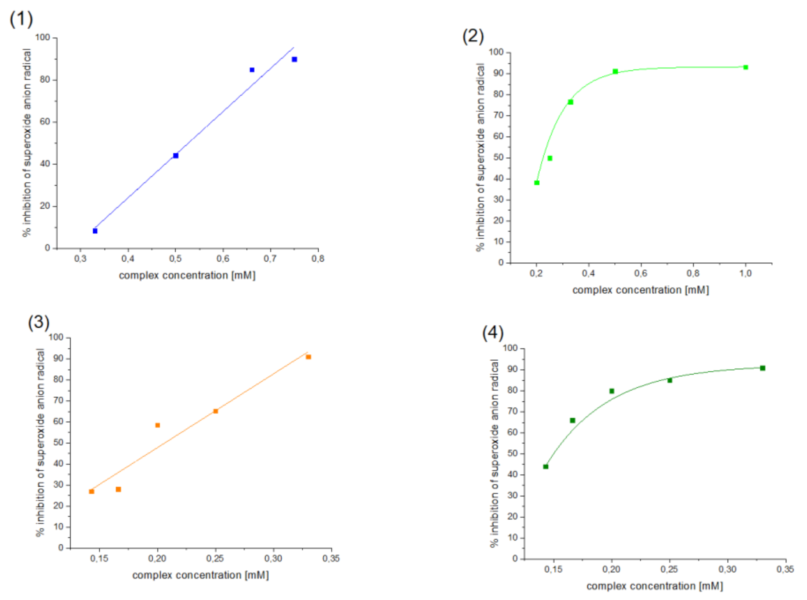
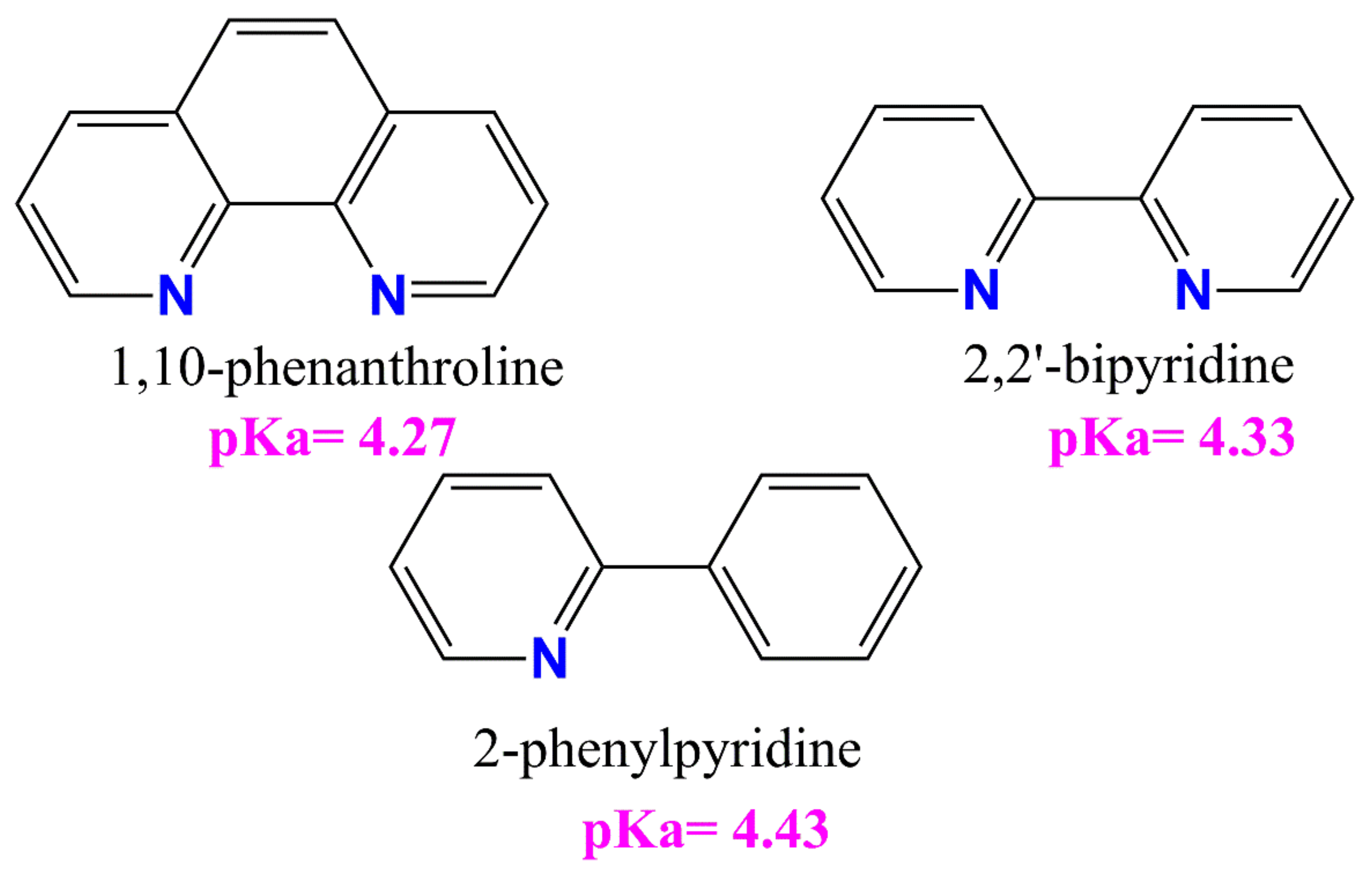
2. Results and Discussion
3. Materials and Methods
4. Conclusions
Supplementary Materials
Author Contributions
Funding
Institutional Review Board Statement
Informed Consent Statement
Acknowledgments
Conflicts of Interest
References
- Au Heluany, C.C.; Donate, P.B.; Schneider, A.H.; Fabris, A.L.; Gomes, R.A.; Villas-Boas, I.M.; Tambourgi, D.V.; da Silva, T.A.; Trossini, G.H.G.; Nalesso, G.; et al. Hydroquinone Exposure Worsens Rheumatoid Arthritis through the Activation of the Aryl Hydrocarbon Receptor and Interleukin-17 Pathways. Antioxidants 2021, 10, 929. [Google Scholar] [CrossRef] [PubMed]
- Fiedor, J.; Burda, K. Potential role of carotenoids as antioxidants in human health and disease. J. Nutr. 2014, 6, 466–488. [Google Scholar] [CrossRef] [PubMed] [Green Version]
- Yang, C.-C.; Wu, C.-J.; Chien, C.-Y.; Chien, C.-T. Green Tea Polyphenol Catechins Inhibit Coronavirus Replication and Potentiate the Adaptive Immunity and Autophagy-Dependent Protective Mechanism to Improve Acute Lung Injury in Mice. Antioxidants 2021, 10, 928. [Google Scholar] [CrossRef] [PubMed]
- Zhang, S.; Xu, M.; Zhang, W.; Liu, C.; Chen, S. Natural Polyphenols in Metabolic Syndrome: Protective Mechanisms and Clinical Applications. Int. J. Mol. Sci. 2021, 22, 6110. [Google Scholar] [CrossRef]
- Ulanowska, M.; Olas, B. Biological Properties and Prospects for the Application of Eugenol—A Review. Int. J. Mol. Sci. 2021, 22, 3671. [Google Scholar] [CrossRef]
- Konopko, A.; Kusio, J.; Litwinienko, G. Antioxidant Activity of Metal Nanoparticles Coated with Tocopherol-Like Residues—The Importance of Studies in Homo- and Heterogeneous Systems. Antioxidants 2020, 9, 5. [Google Scholar] [CrossRef] [Green Version]
- Romero, C.; Nardoia, M.; Arija, I.; Viveros, I.; Rey, A.I.; Prodanov, M.; Chamorro, S. Feeding Broiler Chickens with Grape Seed and Skin Meals to Enhance α- and γ-Tocopherol Content and Meat Oxidative Stability. Antioxidants 2021, 10, 699. [Google Scholar] [CrossRef] [PubMed]
- Kennedy, D.O. Polyphenols and the human brain: Plant “secondary metabolite” ecologic roles and endogenous signaling functions drive benefits. Adv. Nutr. 2014, 5, 515–533. [Google Scholar] [CrossRef] [Green Version]
- OusjiOrc, O.; Sleno, L. Identification of In Vitro Metabolites of Synthetic Phenolic Antioxidants BHT, BHA, and TBHQ by LC-HRMS/MS. Int. J. Mol. Sci. 2020, 21, 9525. [Google Scholar] [CrossRef]
- Adjimani, J.P.; Asare, P. Antioxidant and free radical scavenging activity of ironchelators. Toxicol. Rep. 2015, 2, 721–728. [Google Scholar] [CrossRef] [Green Version]
- Arts, M.J.; Haenen, G.R.; Voss, H.P.; Bast, A. Antioxidant capacity of reaction products limits the applicability of the Trolox Equivalent Antioxidant Capacity (TEAC) assay. Food Chem. Toxicol. 2004, 42, 45–49. [Google Scholar] [CrossRef]
- Liang, N.; Kitts, D.D. Antioxidant Property of Coffee Components: Assessment of Methods that Define Mechanisms of Action. Molecules 2014, 19, 19180–19208. [Google Scholar] [CrossRef] [PubMed] [Green Version]
- Apak, R.; Özyürek, M.; Güçlü, K.; Çapanoğlu, E. Antioxidant Activity/Capacity Measurement. 1. Classification, Physicochemical Principles, Mechanisms, and Electron Transfer (ET)-Based Assays. J. Agric. Food Chem. 2016, 64, 997–1027. [Google Scholar] [CrossRef]
- Apak, R.; Özyürek, M.; Güçlü, K.; Çapanoğlu, E. Antioxidant Activity/Capacity Measurement. 2. Hydrogen Atom Transfer (HAT)-Based, Mixed-Mode (Electron Transfer (ET)/HAT), and Lipid Peroxidation Assays. J. Agric. Food Chem. 2016, 64, 1028–1045. [Google Scholar] [CrossRef] [PubMed]
- Nicoli, M.C.; Anese, M.; Parpinel, M.T.; Franceschi, S.; Lerici, C.R. Loss and/or formation of antioxidants during food processing and storage. Cancer Lett. 1997, 114, 71. [Google Scholar] [CrossRef]
- Pranczk, J.; Wyrzykowski, D.; Jacewicz, D.; Sikorski, A.; Tesmar, A.; Chmurzyński, L. Structural, physico-chemical and antioxidant characteristics of 2,2′ bipyridyl(iminodiacetato) oxidovanadium(IV) dehydrate. Polyhedron 2015, 100, 74–81. [Google Scholar] [CrossRef]
- Crans, D.C.; Mahroof-Tahir, M.; Johnson, M.D.; Wilkins, P.C.; Yang, L.; Robbins, K.; Johnson, A.; Alfano, J.A.; GodzalaIII, M.E.; Austin, L.T.; et al. Vanadium (IV) and vanadium (V) complexes of dipicolinic acid and derivatives. Synthesis, X-ray structure, solution state properties: And effects in rats with STZ-induced diabetes. Inorg. Chim. Acta 2003, 356, 365–378. [Google Scholar] [CrossRef]
- Li, M.; Ding, W.; Smee, J.J.; Baruah, B.; Willsky, G.R.; Crans, D.C. Anti-diabetic effects of vanadium (III, IV, V)–chlorodipicolinate complexes in streptozotocin-induced diabetic rats. Biometals 2009, 22, 895–905. [Google Scholar] [CrossRef]
- Willsky, G.R.; Chi, L.H.; Godzala, M., III; Kostyniak, P.J.; Smee, J.J.; Trujillo, A.M.; Alfano, J.A.; Ding, W.; Huc, Z.; Crans, D.C. Anti-diabetic effects of a series of vanadium dipicolinate complexes in rats with streptozotocin-induced diabetes. Coord. Chem. Rev. 2011, 255, 2258–2269. [Google Scholar] [CrossRef] [Green Version]
- Crans, D.C.; Trujillo, A.M.; Pharazyn, P.S.; Cohen, M.D. How environment affects drug activity: Localization, compartmentalization and reactions of a vanadium insulin-enhancing compound, dipicolinatooxovanadium (V). Coord. Chem. Rev. 2011, 255, 2178–2192. [Google Scholar] [CrossRef]
- Rambaran, V.H.; Saumya, S.M.; Roy, S.; Sonu, K.P.; Eswaramoorthy, M.; Peter, S.C. The design, synthesis and in vivo biological evaluations of [V(IV)O(2, 6-pyridine diacetatato)(H2O)2](PDOV): Featuring its prolonged glucose lowering effect and non-toxic nature. Inorg. Chim. Acta 2020, 504, 119448. [Google Scholar] [CrossRef]
- Hoof, D.L.; Walton, R.A. Studies on metal carboxylates. VIII. Reactions of acetylacetonates of the first row transition elements with pyridine-2, 6-dicarboxylic acid. Inorg. Chim. Acta 1975, 12, 71–78. [Google Scholar] [CrossRef]
- Mahjoobizadeh, M.; Mirzaei, M.; Bauza, A.; Lippolis, V.; Carla Aragoni, M.; Shamsipur, M.; Ghanbari, M.; Frontera, A. Coordination Behavior of Chelidamic Acid With VV, NiII, FeIII, and CaII: Syntheses, X-ray Characterization and DFT Studies. Chem. Sel. 2016, 1, 1556–1566. [Google Scholar] [CrossRef]
- Kolesa-Dobravc, T.; Meden, A.; Perdih, F. Influence of noncovalent interactions on the structures of metal–organic hybrids based on a [VO2(2,6-pydc)]−tecton with cations of imidazole, pyridine and its derivatives. New J. Chem. 2015, 39, 4265–4277. [Google Scholar] [CrossRef] [Green Version]
- Available online: https://foodb.ca/compounds/FDB004404 (accessed on 21 July 2021).
- Available online: https://pubchem.ncbi.nlm.nih.gov/compound/1_10-Phenanthroline#section=LogP (accessed on 21 July 2021).
- Available online: https://pubchem.ncbi.nlm.nih.gov/compound/1474#section=Decomposition (accessed on 21 July 2021).
- CrysAlis CCD and CrysAlis RED. Version 1.171.36.24; Oxford Diffraction Ltd.: Yarnton, UK, 2012. [Google Scholar]
- Sheldrick, G.M. Crystal structure refinement with SHELXL. Acta Crystallogr. C 2015, 71, 3–8. [Google Scholar] [CrossRef]
- Spek, A.L. Structure validation in chemical crystallography. Acta Crystallogr. 2009, D65, 148–155. [Google Scholar] [CrossRef] [PubMed]
- Johnson, C.K. ORTEP II, Report ORNL-5138; Oak Ridge National Laboratory: Oak Ridge, TN, USA, 1976. [Google Scholar]
- Motherwell, S.; Clegg, S. PLUTO-78, Program for Drawing and Molecular Structure; University of Cambridge: Cambridge, UK, 1978. [Google Scholar]
- Macrae, C.F.; Bruno, I.J.; Chisholm, J.A.; Edgington, P.R.; McCabe, P.; Pidcock, E.; Rodriguez-Monge, L.; Taylor, R.; van de Streek, J.; Wood, P.A. Mercury CSD 2.0—New features for the visualization and investigation of crystal structures. J. Appl. Crystallogr. 2008, 41, 466–470. [Google Scholar] [CrossRef]


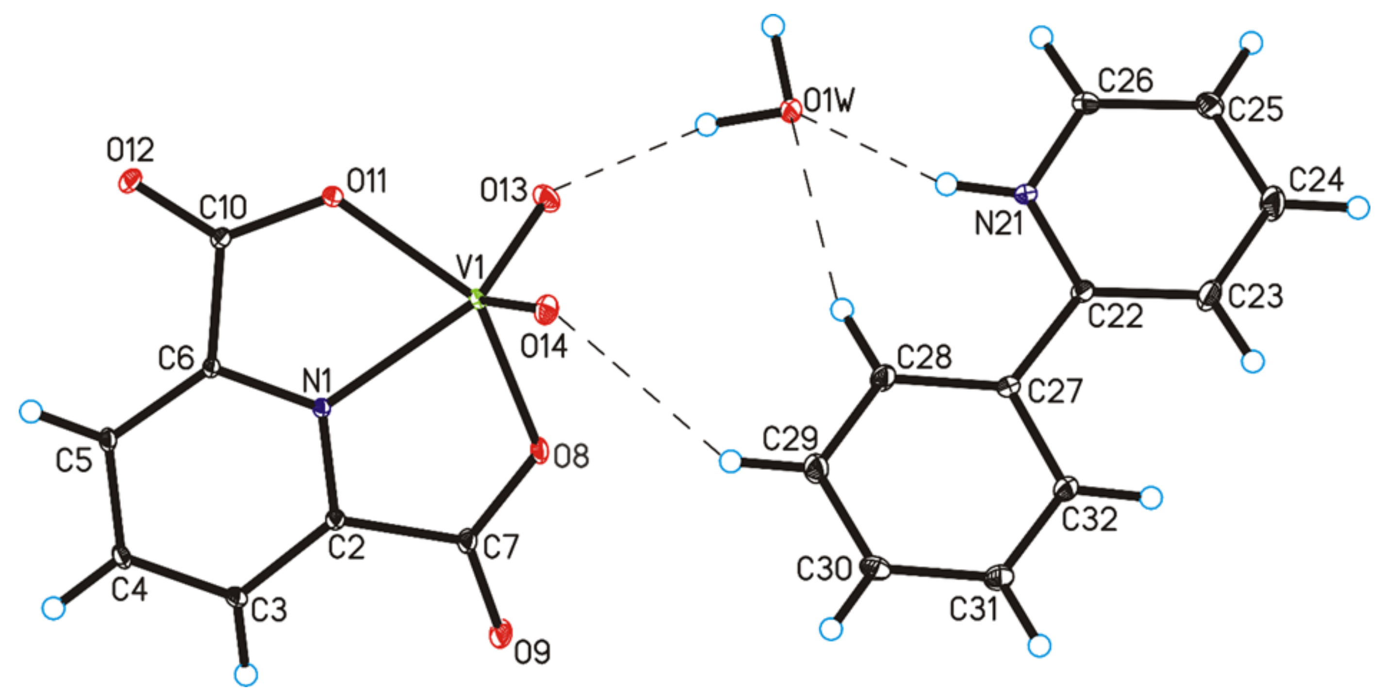
| Chemical Formula | C18H15N2O7V |
|---|---|
| Formula weight/g·mol−1 | 422.26 |
| Crystal system | monoclinic |
| Space group | P21/c |
| a/Å | 11.7061(9) |
| b/Å | 10.6882(5) |
| c/Å | 15.3271(11) |
| α/° | 90 |
| β/° | 111.967(8) |
| γ/° | 90 |
| V/Å3 | 1778.5(2) |
| Z | 2 |
| T/K | 295(2) |
| λMo/Å | 0.71073 |
| ρcalc/g·cm–3 | 1.577 |
| F(000) | 864 |
| µ/mm−1 | 0.603 |
| θ range/° | 3.35–25.00 |
| Completeness θ/% | 99.8 |
| Reflections collected | 11911 |
| Reflections unique | 3121 [Rint = 0.0525] |
| Data/restraints/parameters | 3121/3/262 |
| Goodness of fit on F2 | 1.058 |
| Final R1 value (I > 2σ(I)) | 0.0468 |
| Final wR2 value (I > 2σ(I)) | 0.0992 |
| Final R1 value (all data) | 0.0655 |
| Final wR2 value (all data) | 0.1069 |
| CCDC number | 2087422 |
| D–H···A | d(D–H) [Å] | d(H···A) [Å] | d(D⋯A) (Å) | ∠D–H⋯A (°) |
|---|---|---|---|---|
| O1W–H1WA···O12 i | 0.95(2) | 1.91(2) | 2.811(3) | 158(4) |
| O1W–H1WB···O13 | 0.95(2) | 1.91(2) | 2.826(3) | 161(4) |
| N21–H21···O1W | 0.87(3) | 1.86(3) | 2.699(4) | 161(4) |
| C3–H3···O9 ii | 0.93 | 2.40 | 3.330(4) | 177 |
| C5–H5···O9 iii | 0.93 | 2.38 | 3.250(4) | 157 |
| C26–H26···O11 i | 0.93 | 2.35 | 3.135(4) | 142 |
| C28–H28···O1W | 0.93 | 2.44 | 3.345(4) | 165 |
| C29–H29···O14 | 0.93 | 2.58 | 3.389(5) | 145 |
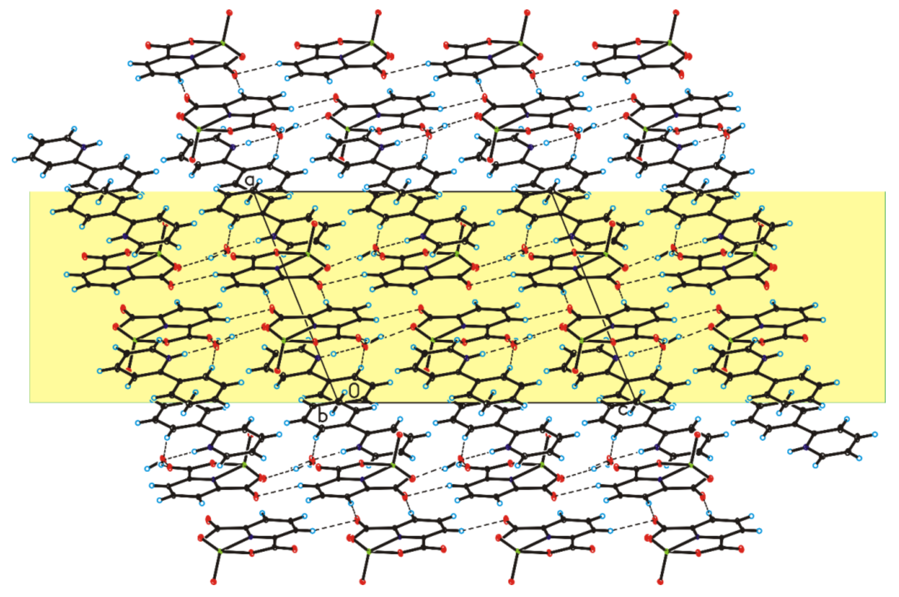
| [VO(dipic)(H2O)2]·2 H2O | [VO(dipic)(phen)]·3 H2O | [VO(dipic)(bipy)]·H2O | |
|---|---|---|---|
| V=O stretching frequency | 983 cm−1 | 980 cm−1 | 977 cm−1 |
| v(COO) of dipic [18] | 1352 cm−1 | 1319 cm−1 | 1335 cm−1 |
| 1665 cm−1 | 1666 cm−1 | 1649 cm−1 | |
| v(OH) [18] | 3571 cm−1 | 3427 cm−1 | 3536 cm−1 |
| stretching vibration of the V-N | 452 cm−1 | 416 cm−1 | 442 cm−1 |
Publisher’s Note: MDPI stays neutral with regard to jurisdictional claims in published maps and institutional affiliations. |
© 2021 by the authors. Licensee MDPI, Basel, Switzerland. This article is an open access article distributed under the terms and conditions of the Creative Commons Attribution (CC BY) license (https://creativecommons.org/licenses/by/4.0/).
Share and Cite
Drzeżdżon, J.; Pawlak, M.; Matyka, N.; Sikorski, A.; Gawdzik, B.; Jacewicz, D. Relationship between Antioxidant Activity and Ligand Basicity in the Dipicolinate Series of Oxovanadium(IV) and Dioxovanadium(V) Complexes. Int. J. Mol. Sci. 2021, 22, 9886. https://0-doi-org.brum.beds.ac.uk/10.3390/ijms22189886
Drzeżdżon J, Pawlak M, Matyka N, Sikorski A, Gawdzik B, Jacewicz D. Relationship between Antioxidant Activity and Ligand Basicity in the Dipicolinate Series of Oxovanadium(IV) and Dioxovanadium(V) Complexes. International Journal of Molecular Sciences. 2021; 22(18):9886. https://0-doi-org.brum.beds.ac.uk/10.3390/ijms22189886
Chicago/Turabian StyleDrzeżdżon, Joanna, Marta Pawlak, Natalia Matyka, Artur Sikorski, Barbara Gawdzik, and Dagmara Jacewicz. 2021. "Relationship between Antioxidant Activity and Ligand Basicity in the Dipicolinate Series of Oxovanadium(IV) and Dioxovanadium(V) Complexes" International Journal of Molecular Sciences 22, no. 18: 9886. https://0-doi-org.brum.beds.ac.uk/10.3390/ijms22189886