Ex Vivo Live Full-Thickness Porcine Skin Model as a Versatile In Vitro Testing Method for Skin Barrier Research

 and
and 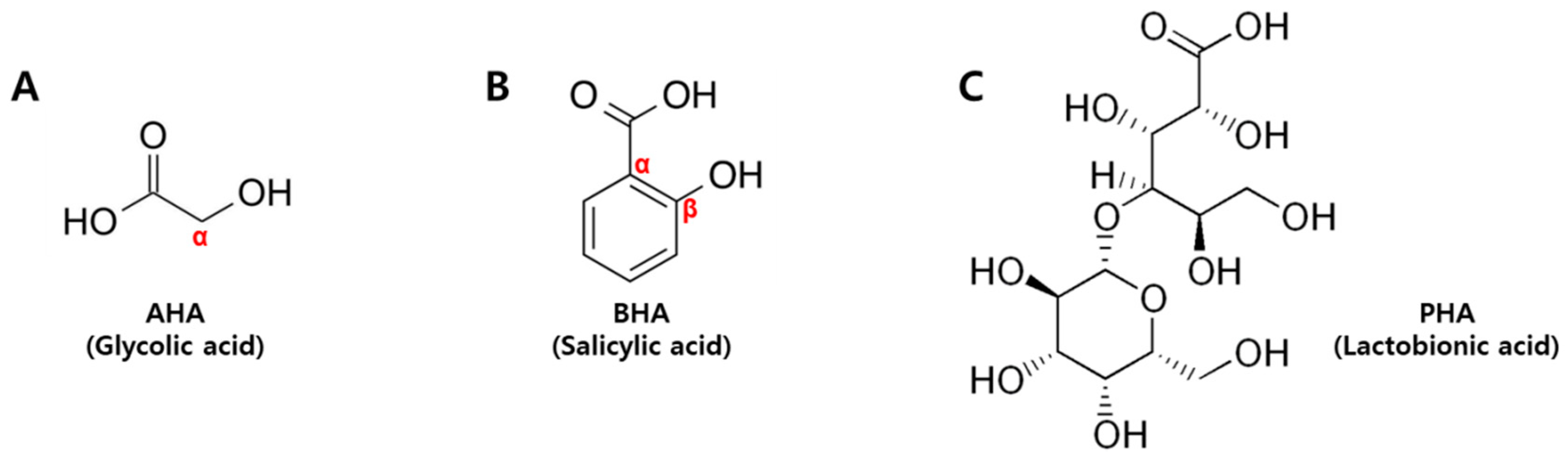
Abstract
:1. Introduction
2. Results
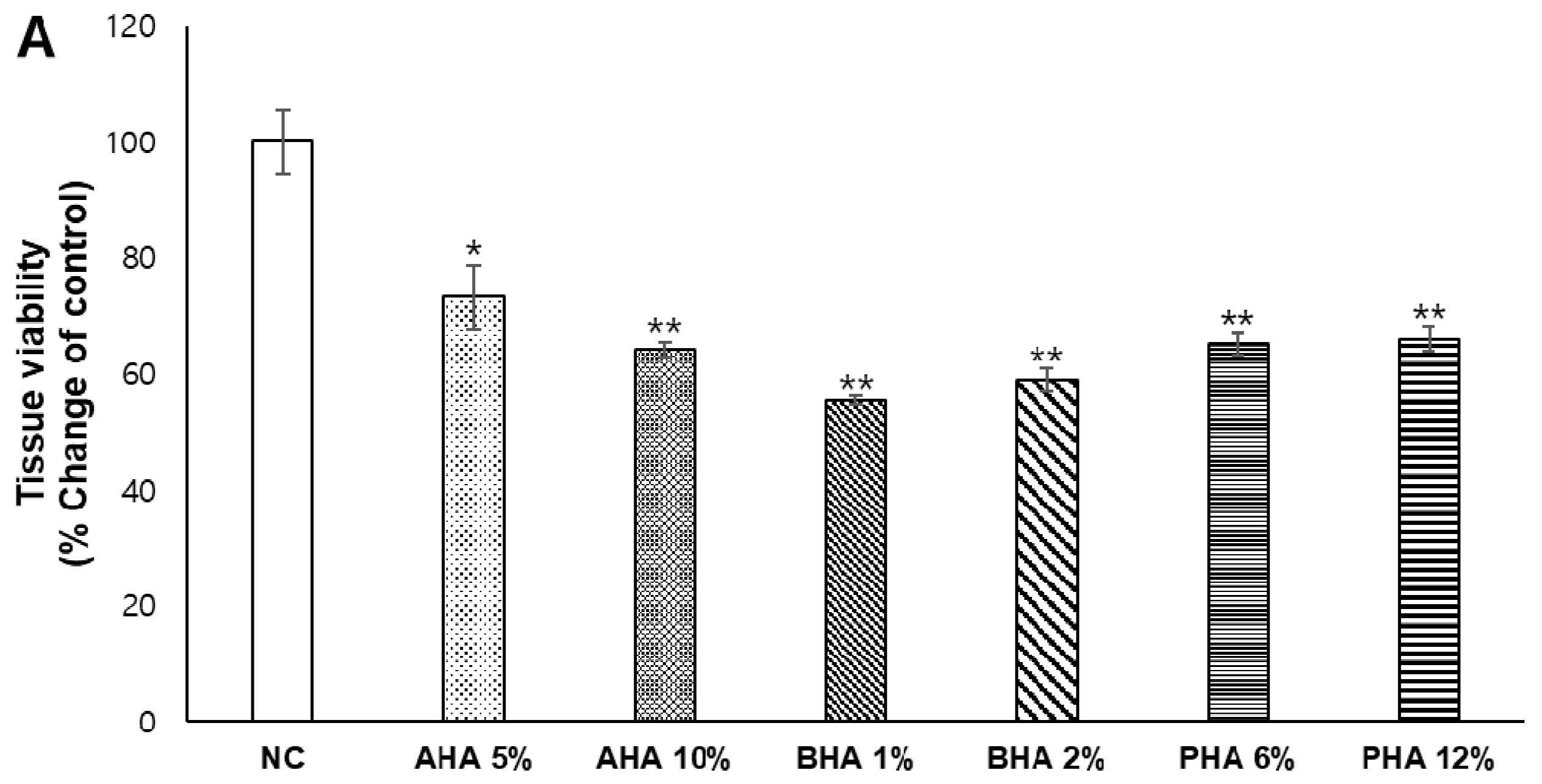
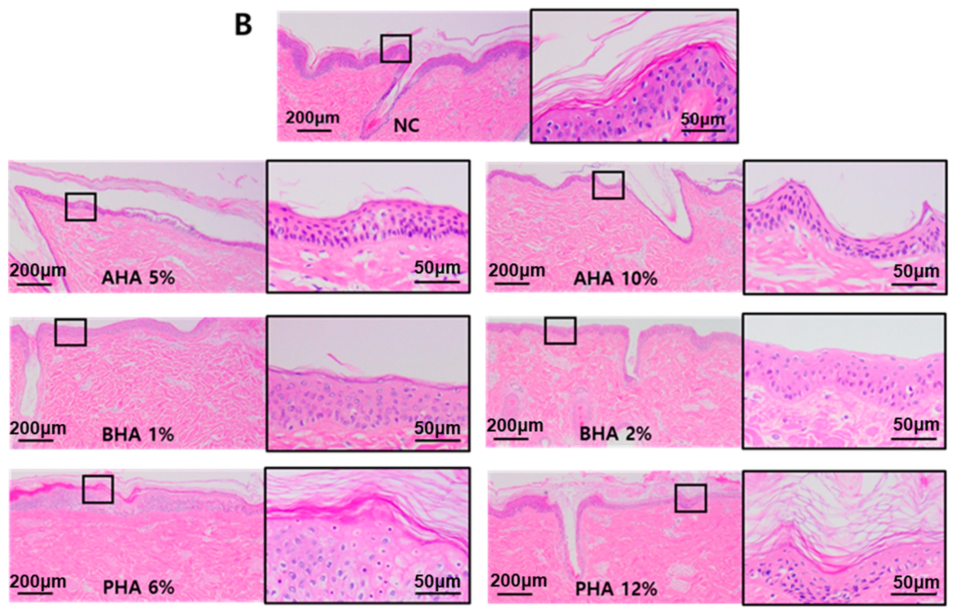
2.1. Assessment of Damage to Ex Vivo Porcine Skin Inflicted by Repeated Application of High-Concentration Hydroxyacids
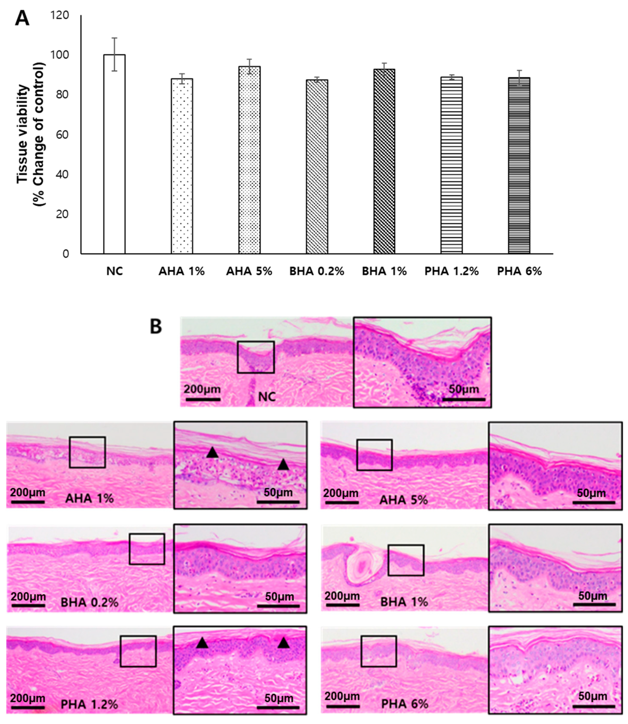
2.2. Effects of a Single Application of Hydroxyacids at Low Concentrations on the Tissue Integrity of Ex Vivo Porcine Skin
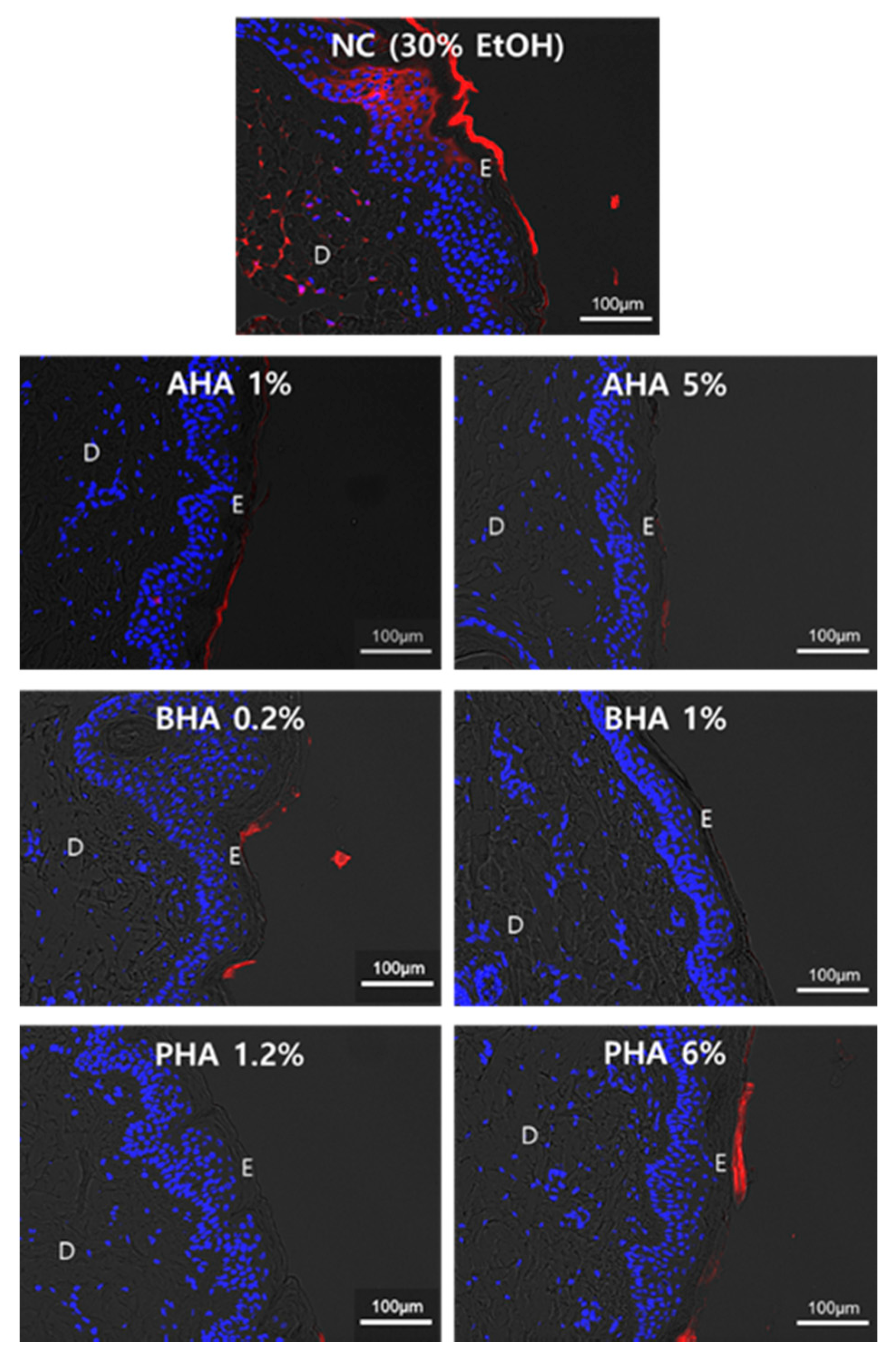
2.3. Effect of Hydroxyacids on the Skin Penetration of Rhodamine B
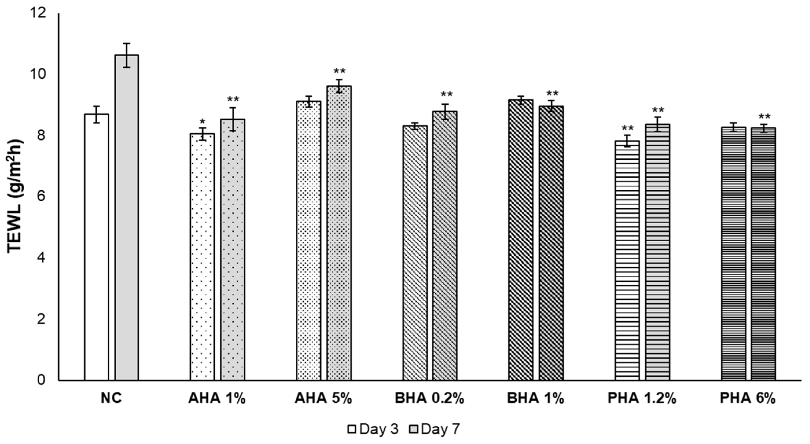
2.4. Effect of Hydroxyacids on the Skin Barrier Function as Measured by TEWL
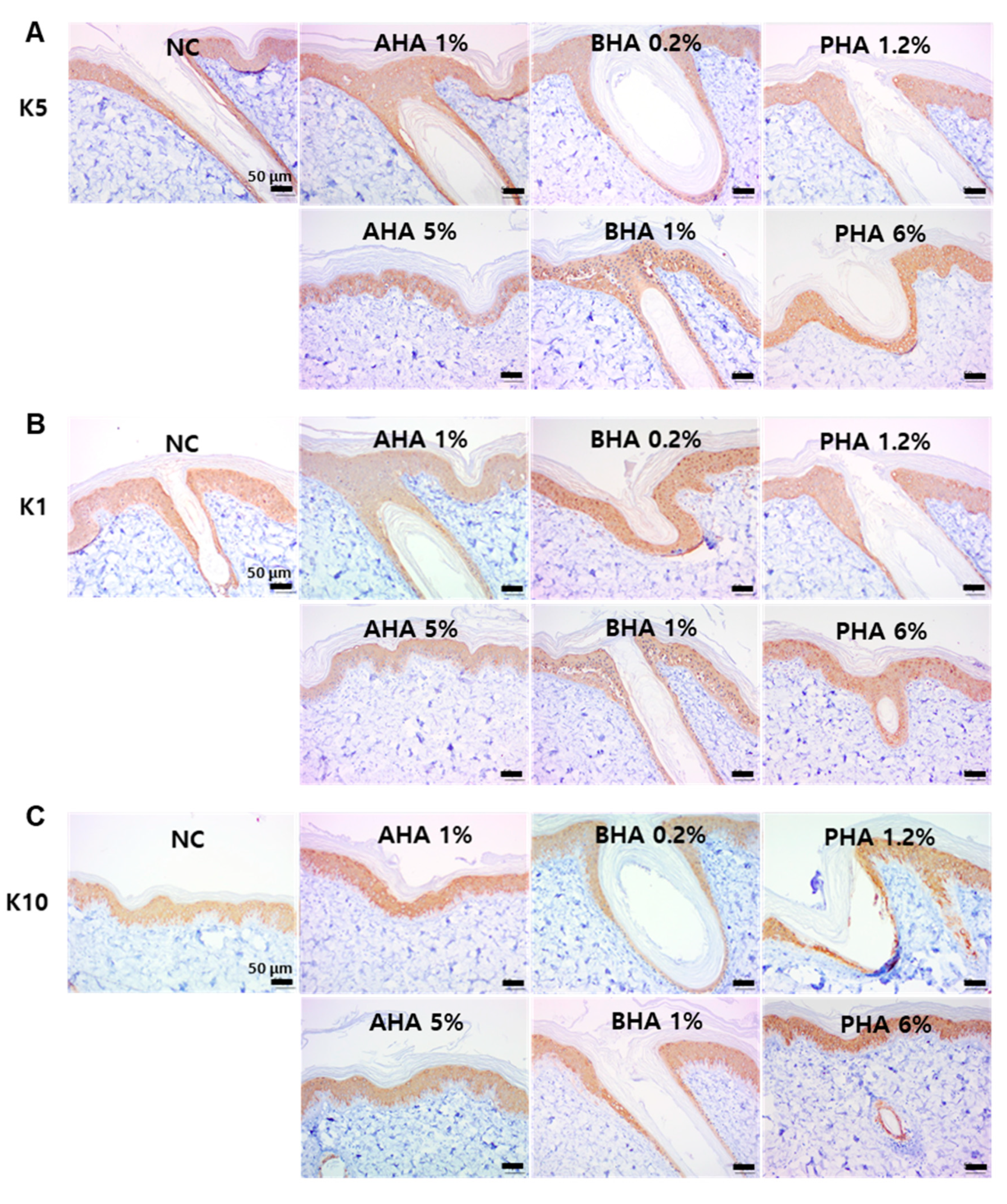
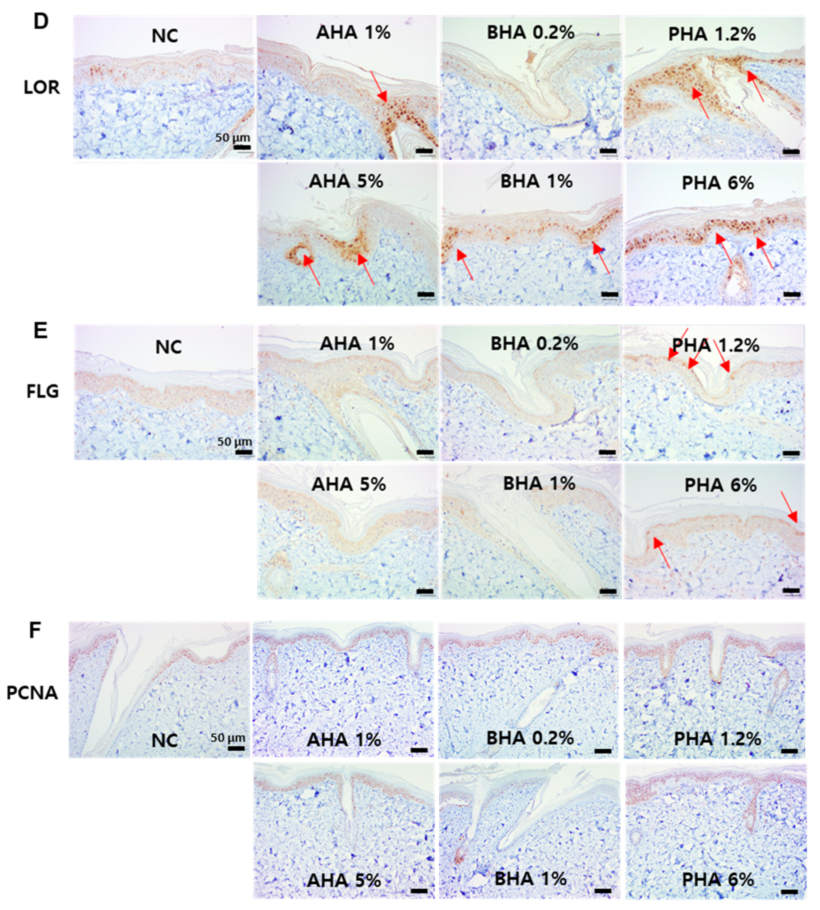
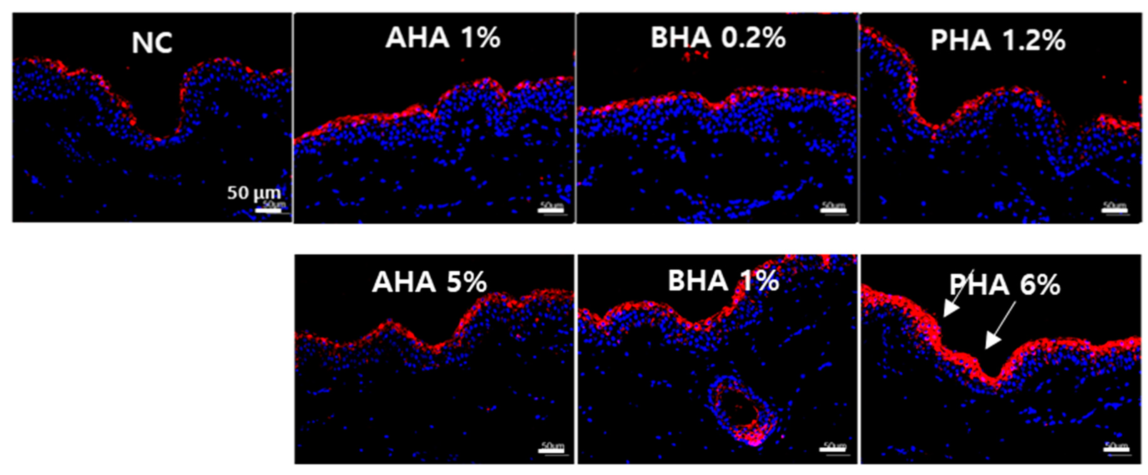
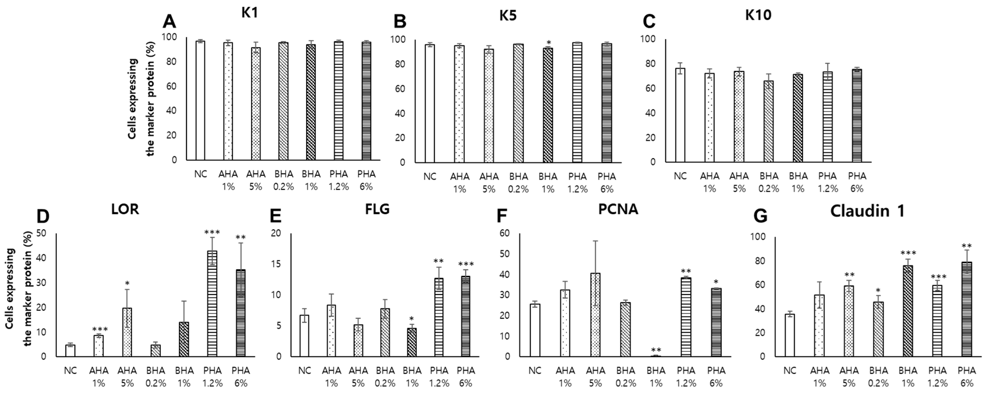
2.5. Immunohistochemical and Immunofluorescence Staining of Key Protein Components of the Skin Epidermis
3. Discussion
4. Materials and Methods
4.1. Materials and Reagents
4.2. Ex Vivo Live Full-Thickness Porcine Skin
4.3. WST-1 Assay
4.4. Histological Analysis
4.5. Skin Penetration Study
4.6. Immunohistochemistry (IHC) and Immunofluorescence (IF) Staining
4.7. Quantitation of Immunohistochemistry (IHC) and Immunofluorescence (IF) Staining
4.8. Trans-Epidermal Water Loss (TEWL)
4.9. Statistical Analysis
Author Contributions
Funding
Institutional Review Board Statement
Informed Consent Statement
Data Availability Statement
Conflicts of Interest
Abbreviations
| AHA | α-hydroxyacid |
| BHA | β-hydroxyacid |
| PHA | Polyhydroxyacid |
| TEWL | Trans-epidermal water loss |
| Rb | Rhodamine B |
| D | Dermis |
| E | Epidermis |
| IHC | Immunohistochemistry |
| IF | Immunofluorescence |
| K1 | Keratin 1 |
| K5 | Keratin 5 |
| K10 | Keratin 10 |
| LOR | Loricrin |
| FLG | Filaggrin |
| CLDN1 | Claudin 1 |
| WST-1 | (4-[3-(indophenyl)-2-(4-nitrophenyl)-2H-5-tetrazolio]-1,3-benzene disulfonate) |
| NC | Negative control |
| SC | Stratum corneum |
| SOC | Skin organ culture |
| PCNA | Proliferating cell nuclear antigen. |
References
- Forslind, B. A domain mosaic model of the skin barrier. Acta Dermato-Venereol. 1994, 74, 1–6. [Google Scholar] [PubMed]
- Choi, J.H.; Jin, S.W.; Lee, G.H.; Cho, S.M.; Jeong, H.G. Orostachys japonicus ethanol extract inhibits 2,4-dinitrochlorobenzene-induced atopic dermatitis-like skin lesions in NC/Nga mice and TNF-alpha/IFN-gamma-induced TARC expression in HaCaT cells. Toxicol. Res. 2020, 36, 99–108. [Google Scholar] [CrossRef] [PubMed]
- Reduan, F.H.; Shaari, R.M.; Sayuti, N.S.A.; Mustapha, N.M.; Abu Bakar, Z.; Sithambaram, S.; Hazilawati, H. Acute and subacute dermal toxicity of ethanolic extract of Melastoma malabathricum leaves in Sprague-Dawley rats. Toxicol. Res. 2020, 36, 203–210. [Google Scholar] [CrossRef] [PubMed]
- Barel, A.O.; Paye, M.; Maibach, H.I. Handbook of Cosmetic Science and Technology; CRC Press: Boca Raton, FL, USA, 2014. [Google Scholar]
- Duracher, L.; Visdal-Johnsen, L.; Mavon, A.; Oriflame Cosmetics, A.B. Novel Explant Model for Skin Delivery Assessment. Cosm. Toil 2015, 130, 30–40. [Google Scholar]
- Richert, S.; Schrader, A.; Schrader, K. Transdermal delivery of two antioxidants from different cosmetic formulations. Int. J. Cosmet. Sci. 2003, 25, 5–13. [Google Scholar] [CrossRef]
- Yu, R.J.; Scott, E.J.V. Hydroxycarboxylic Acids, N-Acetylamino Sugars, and N-Acetylamino Acids. SKINmed Dermatol. Clin. 2002, 1, 117–122. [Google Scholar] [CrossRef]
- Ruey, J.Y.; Van Scott, E.J. Organic Acids with Novel Functions: Hydroxy, Bionic, N-acetylamino Acids and N-acylpeptide Derivatives. In Textbook of Cosmetic Dermatology; CRC Press: Boca Raton, FL, USA, 2017; pp. 68–82. [Google Scholar]
- Yu, R.; Van Scott, E. Salicylic acid: Not a beta-hydroxy acid. J. Cosmet. Dermatol. 1997, 10, 27. [Google Scholar]
- Khalil, S.; Bardawil, T.; Saade, S.; Chedraoui, A.; Ramadan, N.; Hasbani, D.J.; Abbas, O.; Nemer, G.; Rubeiz, N.; Kurban, M. Use of Topical Glycolic Acid Plus a Lovastatin-Cholesterol Combination Cream for the Treatment of Autosomal Recessive Congenital Ichthyoses. JAMA Dermatol. 2018, 154, 1320–1323. [Google Scholar] [CrossRef]
- Geng, T.-M.; Wang, X.; Zhu, F.; Jiang, H.; Wang, Y. Sensing of polymeric sensor-based rhodamine B derivative for metal cations in complete aqueous solution. Bull. Mater. Sci. 2017, 40, 187–193. [Google Scholar] [CrossRef]
- Gomaa, Y.A.; El-Khordagui, L.K.; Garland, M.J.; Donnelly, R.F.; McInnes, F.; Meidan, V.M. Effect of microneedle treatment on the skin permeation of a nanoencapsulated dye. J. Pharm. Pharmacol. 2012, 64, 1592–1602. [Google Scholar] [CrossRef] [Green Version]
- Imhof, B.; McFeat, G. 11 Evaluation of the Barrier Function of Skin Using Transepidermal Water Loss (TEWL). In Handbook of Cosmetic Science Technology; CRC Press: Boca Raton, FL, USA, 2014; p. 131. [Google Scholar]
- Pöhler, E.; Cunningham, F.; Sandilands, A.; Cole, C.; Digby, S.; McMillan, J.; Aristodemou, S.; McGrath, J.; Smith, F.; McLean, W.; et al. Novel autosomal dominant mutation in loricrin presenting as prominent ichthyosis. Br. J. Dermatol. 2015, 173, 1291–1294. [Google Scholar] [CrossRef] [PubMed] [Green Version]
- Furuse, M.; Hirase, T.; Itoh, M.; Nagafuchi, A.; Yonemura, S. Tsukita Sa and Tsukita Sh. J. Cell Biol. 1994, 127, 1617–1626. [Google Scholar] [CrossRef] [Green Version]
- Bankhead, P.; Loughrey, M.B.; Fernández, J.A.; Dombrowski, Y.; McArt, D.G.; Dunne, P.D.; McQuaid, S.; Gray, R.T.; Murray, L.J.; Coleman, H.G.; et al. QuPath: Open source software for digital pathology image analysis. Sci. Rep. 2017, 7, 16878. [Google Scholar] [CrossRef] [Green Version]
- Ferguson-Smith, A.C. Genomic imprinting: The emergence of an epigenetic paradigm. Nat. Rev. Genet. 2011, 12, 565–575. [Google Scholar] [CrossRef]
- Roth, W.; Kumar, V.; Beer, H.-D.; Richter, M.; Wohlenberg, C.; Reuter, U.; Thiering, S.; Staratschek-Jox, A.; Hofmann, A.; Kreusch, F.; et al. Keratin 1 maintains skin integrity and participates in an inflammatory network in skin through interleukin-18. J. Cell Sci. 2012, 125, 5269–5279. [Google Scholar] [CrossRef] [PubMed] [Green Version]
- Skobowiat, C.; Brożyna, A.A.; Janjetovic, Z.; Jeayeng, S.; Oak, A.S.W.; Kim, T.-K.; Panich, U.; Reiter, R.J.; Slominski, A.T. Melatonin and its derivatives counteract the ultraviolet B radiation-induced damage in human and porcine skin ex vivo. J. Pineal Res. 2018, 65, e12501. [Google Scholar] [CrossRef] [PubMed]
- Murphy, S.V.; Skardal, A.; Nelson, R.A., Jr.; Sunnon, K.; Reid, T.; Clouse, C.; Kock, N.D.; Jackson, J.; Soker, S.; Atala, A. Amnion membrane hydrogel and amnion membrane powder accelerate wound healing in a full thickness porcine skin wound model. Stem Cells Transl. Med. 2020, 9, 80–92. [Google Scholar] [CrossRef] [Green Version]
- Yang, C.E.; Choi, S.; Lee, J.H.; Kang, E.H.; Ahn, H.M.; Roh, T.S.; Yun, C.-O.; Lee, W.J. Sustained Release of Decoy Wnt Receptor (sLRP6E1E2)-Expressing Adenovirus Using Gel-Encapsulation for Scar Remodeling in Pig Model. Int. J. Mol. Sci. 2020, 21, 2242. [Google Scholar] [CrossRef] [PubMed] [Green Version]
- Lombardi, F.; Palumbo, P.; Augello, F.R.; Cifone, M.G.; Cinque, B.; Giuliani, M. Secretome of Adipose Tissue-Derived Stem Cells (ASCs) as a Novel Trend in Chronic Non-Healing Wounds: An Overview of Experimental In Vitro and In Vivo Studies and Methodological Variables. Int. J. Mol. Sci. 2019, 20, 3721. [Google Scholar] [CrossRef] [Green Version]
- Şenyiğit, T.; Sonvico, F.; Rossi, A.; Tekmen, I.; Santi, P.; Colombo, P.; Nicoli, S.; Özer, Ö. In vivo assessment of clobetasol propionate-loaded lecithin-chitosan nanoparticles for skin delivery. Int. J. Mol. Sci. 2017, 18, 32. [Google Scholar] [CrossRef] [Green Version]
- Esposito, C.L.; Tardif, V.; Sarrazin, M.; Kirilov, P.; Roullin, V.G. Preparation and characterization of 12-HSA-based organogels as injectable implants for the controlled delivery of hydrophilic and lipophilic therapeutic agents. Mater. Sci. Eng. C 2020, 114, 110999. [Google Scholar] [CrossRef] [PubMed]
- Dick, I.P.; Scott, R.C. Pig Ear Skin as an In-vitro Model for Human Skin Permeability. J. Pharm. Pharmacol. 1992, 44, 640–645. [Google Scholar] [CrossRef] [PubMed]
- Gray, G.M.; Yardley, H.J. Lipid compositions of cells isolated from pig, human, and rat epidermis. J. Lipid Res. 1975, 16, 434–440. [Google Scholar] [CrossRef]
- Jacobi, U.; Kaiser, M.; Toll, R.; Mangelsdorf, S.; Audring, H.; Otberg, N.; Sterry, W.; Lademann, J. Porcine ear skin: An in vitro model for human skin. Ski. Res. Technol. 2007, 13, 19–24. [Google Scholar] [CrossRef] [PubMed]
- Muhammad, F.; Brooks, J.D.; Riviere, J.E. Comparative mixture effects of JP-8(100) additives on the dermal absorption and disposition of jet fuel hydrocarbons in different membrane model systems. Toxicol. Lett. 2004, 150, 351–365. [Google Scholar] [CrossRef]
- Wester, R.C.; Melendres, J.; Sedik, L.; Maibach, H.; Riviere, J.E. Percutaneous absorption of salicylic acid, theophylline, 2, 4-dimethylamine, diethyl hexyl phthalic acid, andp-aminobenzoic acid in the isolated perfused porcine skin flap compared to man in vivo. Toxicol. Appl. Pharmacol. 1998, 151, 159–165. [Google Scholar] [CrossRef]
- Choe, C.; Schleusener, J.; Lademann, J.; Darvin, M.E. Human skin in vivo has a higher skin barrier function than porcine skin ex vivo-comprehensive Raman microscopic study of the stratum corneum. J. Biophotonics 2018, 11, e201700355. [Google Scholar] [CrossRef]
- Franz, T.J. Percutaneous Absorption: Mechanisms—Methodology—Drug Delivery. Arch. Dermatol. 1990, 126, 1384. [Google Scholar] [CrossRef]
- Holbrook, K.A.; Odland, G.F. Regional Differences in the Thickness (Cell Layers) of the Human Stratum Corneum: An Ultrastructural Analysis. J. Investig. Dermatol. 1974, 62, 415–422. [Google Scholar] [CrossRef] [Green Version]
- Simon, G.A.; Maibach, H.I. The Pig as an Experimental Animal Model of Percutaneous Permeation in Man: Qualitative and Quantitative Observations—An Overview. Ski. Pharmacol. Physiol. 2000, 13, 229–234. [Google Scholar] [CrossRef]
- Msc, B.A.; Mucha, P.; Rotsztejn, H. Lactic and lactobionic acids as typically moisturizing compounds. Int. J. Dermatol. 2018, 58, 374–379. [Google Scholar] [CrossRef]
- Van Scott, E.; Yu, R. Hydroxyacids and their topical use in the elderly. Skin Dis. Elder. 2004, 27, 3–5. [Google Scholar]
- Jackson, E. AHA-type products proliferate in 1993. Cosmet. Dermatol. 1993, 6, 22–26. [Google Scholar]
- Bissonnette, R.; Bolduc, C.; Seité, S.; Nigen, S.; Provost, N.; Maari, C.; Rougier, A. Randomized study comparing the efficacy and tolerance of a lipophillic hydroxy acid derivative of salicylic acid and 5% benzoyl peroxide in the treatment of facial acne vulgaris. J. Cosmet. Dermatol. 2009, 8, 19–23. [Google Scholar] [CrossRef]
- Green, B. After 30 years… the future of hydroxyacids. J. Cosmet. Dermatol. 2005, 4, 44–45. [Google Scholar] [CrossRef] [PubMed]
- Dréno, B.; Fischer, T.; Perosino, E.; Poli, F.; Viera, M.; Rendon, M.; Berson, D.; Cohen, J.; Roberts, W.; Starker, I.; et al. Expert Opinion: Efficacy of superficial chemical peels in active acne management—What can we learn from the literature today? Evidence-based recommendations. J. Eur. Acad. Dermatol. Venereol. 2010, 25, 695–704. [Google Scholar] [CrossRef]
- Van, E.S.; Yu, R. Alpha hydroxy acids: Procedures for use in clinical practice. Cutis 1989, 43, 222–228. [Google Scholar]
- Jung, Y.-O.; Jeong, H.; Cho, Y.; Lee, E.-O.; Jang, H.-W.; Kim, J.; Nam, K.T.; Lim, K.-M. Lysates of a Probiotic, Lactobacillus rhamnosus, Can Improve Skin Barrier Function in a Reconstructed Human Epidermis Model. Int. J. Mol. Sci. 2019, 20, 4289. [Google Scholar] [CrossRef] [Green Version]


Publisher’s Note: MDPI stays neutral with regard to jurisdictional claims in published maps and institutional affiliations. |
© 2021 by the authors. Licensee MDPI, Basel, Switzerland. This article is an open access article distributed under the terms and conditions of the Creative Commons Attribution (CC BY) license (http://creativecommons.org/licenses/by/4.0/).
Share and Cite
Hwang, J.-h.; Jeong, H.; Lee, N.; Hur, S.; Lee, N.; Han, J.J.; Jang, H.W.; Choi, W.K.; Nam, K.T.; Lim, K.-M. Ex Vivo Live Full-Thickness Porcine Skin Model as a Versatile In Vitro Testing Method for Skin Barrier Research. Int. J. Mol. Sci. 2021, 22, 657. https://0-doi-org.brum.beds.ac.uk/10.3390/ijms22020657
Hwang J-h, Jeong H, Lee N, Hur S, Lee N, Han JJ, Jang HW, Choi WK, Nam KT, Lim K-M. Ex Vivo Live Full-Thickness Porcine Skin Model as a Versatile In Vitro Testing Method for Skin Barrier Research. International Journal of Molecular Sciences. 2021; 22(2):657. https://0-doi-org.brum.beds.ac.uk/10.3390/ijms22020657
Chicago/Turabian StyleHwang, Jee-hyun, Haengdueng Jeong, Nahyun Lee, Sumin Hur, Nakyum Lee, Jeong Jun Han, Hye Won Jang, Wang Keun Choi, Ki Taek Nam, and Kyung-Min Lim. 2021. "Ex Vivo Live Full-Thickness Porcine Skin Model as a Versatile In Vitro Testing Method for Skin Barrier Research" International Journal of Molecular Sciences 22, no. 2: 657. https://0-doi-org.brum.beds.ac.uk/10.3390/ijms22020657