Article

Long Non-Coding RNA ANRIL as a Potential Biomarker of Chemosensitivity and Clinical Outcomes in Osteosarcoma

1
Department of Experimental and Clinical Pharmacology, College of Pharmacy, University of Minnesota, Minneapolis, MN 55455, USA
2
Department of Pediatrics, Hematology & Oncology, University of Minnesota, Minneapolis, MN 55455, USA
3
Department of Natural Sciences, Zanvyl Krieger School of Arts and Sciences, The Johns Hopkins University, Baltimore, MD 21218, USA
4
Department of Medical Oncology and Therapeutics, City of Hope Comprehensive Cancer Center, Monrovia, CA 91007, USA
*
Author to whom correspondence should be addressed.
These authors contributed equally to this work.
Int. J. Mol. Sci. 2021, 22(20), 11168; https://0-doi-org.brum.beds.ac.uk/10.3390/ijms222011168
Submission received: 31 August 2021 / Revised: 4 October 2021 / Accepted: 12 October 2021 / Published: 16 October 2021

Abstract

:

Simple Summary

Osteosarcoma is a bone cancer with a poor prognosis. This is, in part, due to resistance to current standard-of-care chemotherapeutic treatment. As personalized treatment plans become more widely utilized, the role of patient-specific genome markers may serve to identify individuals with chemo-resistant disease at the outset of diagnosis. ANRIL, a long non-coding RNA, has promise as a predictive biomarker. Utilizing osteosarcoma cell lines, we observed that altering the expression of ANRIL significantly alters the sensitivity to cisplatin and doxorubicin, two agents that are a standard-of-care for treatment. Analysis of clinical data from the TARGET dataset confirmed higher ANRIL expression portending poorer prognosis, as evidenced by association with death and metastases at diagnosis.

Abstract

Osteosarcoma has a poor prognosis due to chemo-resistance and/or metastases. Increasing evidence shows that long non-coding RNAs (lncRNAs) can play an important role in drug sensitivity and cancer metastasis. Using osteosarcoma cell lines, we identified a positive correlation between the expression of a lncRNA and ANRIL, and resistance to two of the three standard-of-care agents for treating osteosarcoma—cisplatin and doxorubicin. To confirm the potential role of ANRIL in chemosensitivity, we independently inhibited and over-expressed ANRIL in osteosarcoma cell lines followed by treatment with either cisplatin or doxorubicin. Knocking-down ANRIL in SAOS2 resulted in a significant increase in cellular sensitivity to both cisplatin and doxorubicin, while the over-expression of ANRIL in both HOS and U2OS cells led to an increased resistance to both agents. To investigate the clinical significance of ANRIL in osteosarcoma, we assessed ANRIL expression in relation to clinical phenotypes using the osteosarcoma data from the Therapeutically Applicable Research to Generate Effective Treatments (TARGET) dataset. Higher ANRIL expression was significantly associated with increased rates of metastases at diagnosis and death and was a significant predictor of reduced overall survival rate. Collectively, our results suggest that the lncRNA ANRIL can be a chemosensitivity and prognosis biomarker in osteosarcoma. Furthermore, reducing ANRIL expression may be a therapeutic strategy to overcome current standard-of-care treatment resistance.

1. Introduction

Osteosarcoma is a skeletal neoplasm derived from primitive mesenchymal cells that affects children, adolescents, and young adults [1]. Although only representing 5% of pediatric malignancies, it is the most common primary solid malignancy of bone and has the highest incidence in adolescence [1,2]. A multimodal approach consisting of neoadjuvant chemotherapy and surgical resection is the mainstay of therapy [3,4]. Patients receive an initial two cycles of chemotherapy with the agents cisplatin, doxorubicin, and methotrexate [3]. Following this, patients undergo resection of the primary tumor, which is then assessed for extent of necrosis [4]. Patients then complete therapy with four more cycles of cisplatin, doxorubicin, and methotrexate [3].
Prior to the introduction, widespread use, and standardization of these chemotherapy agents, 5-year overall survival for osteosarcoma was poor at 20% [5]. Greater than 90% of patients died from pulmonary metastases [6]. The addition of neoadjuvant chemotherapy to surgical resection (or amputation) increased survival rates to 60–70% [7]. This initial increase in 5-year overall survival suggests that chemosensitivity, at least in part, can perhaps be viewed as a prognostic factor. However, this means that despite chemotherapy and surgical resection, 30–40% of patients do not survive beyond 5 years [8]. This is primarily due to the lack of initial response to the standard chemotherapy regimen of cisplatin, doxorubicin, and methotrexate, and demonstrates the urgent need to identify biomarkers that can be used to predict chemosensitivity [9].
Current known prognostic biomarkers for osteosarcoma, including Rb gene and/or TP53 mutations, are shown to be related to the incidence and outcomes of disease but are poor markers to predict chemosensitivity [10]. Therefore, the percent necrosis of tumor tissue after two cycles of chemotherapy has been used as a clinical biomarker of chemosensitivity [4,11,12]. However, a major drawback of this approach is that it requires patients to undergo toxic and potentially ineffective chemotherapy before determining resistance. Furthermore, this chemotherapy-only response predictor is no longer deemed sufficient to determine chemoresistance by many clinicians despite its initial promising use in The Children’s Oncology Group osteosarcoma trial [13].
Non-coding genes are a growing area of interest and many non-coding RNAs have been found to act as oncogenes or tumor suppressors [14]. LncRNAs (long non-coding RNA) make up 60% of the human genome and are characterized by their length of greater than 200 nucleotides [15]. They do not encode proteins but can manipulate local or global gene expression via transcription in addition to post-transcriptional and epigenetic regulation [16]. These manipulations can lead to either the inhibition or activation of protein-coding genes. To date, less than 1% of lncRNAs have been functionally characterized [17]. Given this, along with the fact that there are limited protein-coding gene mutations observed in pediatric cancers, lncRNAs present an exciting opportunity for clinical biomarker discovery as epigenetic regulators of cancer. Previous studies indicated that a growing number of lncRNAs play a key role in osteogenic differentiaton [18] and they were found to be associated with bone tumor onset and development [19,20]. Taken together, lncRNAs may serve as novel biomarkers for chemotherapeutic sensitivity in osteosarcoma.
The current knowledge of lncRNAs in terms of the initiation and development of human cancers has grown significantly, whereas investigations into the role of lncRNAs in regulating the sensitivity of cancer cells to antineoplastic treatments have just begun to make significant progress [21,22,23]. Recently, our research group performed a systematic analysis of the association between the somatic lncRNA transcriptome and genome of hundreds of cell lines with pharmacological profiles for hundreds of drugs, utilizing the Genomics of Drug Sensitivity in Cancer database (GDSC) [24,25,26]. LncRNAs were found to be just as potent as the protein-coding transcriptome at drug response prediction. In some instances, lncRNA outperformed known protein-coding gene biomarkers in predicting drug response. Among them, we observed significant associations between the lncRNA ANRIL (anti-sense non-coding RNA in the INK4 locus) and sensitivity to multiple drugs, including the mainstays of osteosarcoma treatment, cisplatin and doxorubicin (Supplementary Materials Figure S1).
The lncRNA ANRIL (also known as CDKN2B-AS1) was first identified from familial melanoma patients with a large germline deletion in the INK4B-ARF-INK4A gene cluster, and has been previously characterized as an oncogene in osteosarcoma and other cancer types [20,27,28,29,30]. In relation to chemotherapeutic response, previous studies have shown ANRIL to promote cell chemoresistance in multiple cancers; however, the impact of ANRIL in response to cisplatin and doxorubicin in the treatment of osteosarcoma is not fully elucidated [20,31,32,33,34,35,36,37].
Based on our previous analysis and the current existing evidence, we hypothesized that ANRIL may play a role in the variability of therapeutic response to cisplatin and doxorubicin and could serve as a potential clinical biomarker of therapeutic response in osteosarcoma. To investigate the relationship between ANRIL and chemotherapeutic sensitivity, this study utilized osteosarcoma cell lines to develop ANRIL knockdown and overexpression models to determine the effects on cell viability during cisplatin and doxorubicin treatment. To investigate the potential clinical utility of ANRIL, we also utilized expression data and clinical outcome measures in a publicly available osteosarcoma patient cohort.

2. Results

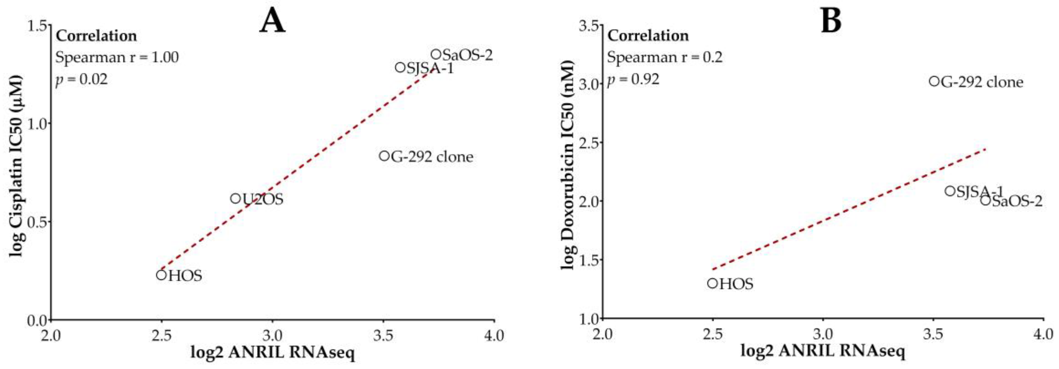
2.1. Higher ANRIL Expression Level Is Correlated with Cisplatin and Doxorubicin Resistance in Osteosarcoma Cell Lines

Using data from a publicly available high-throughput cancer cell line drug screening dataset, GDSC1 [38], we found significant correlations between the IC50 values of cisplatin and doxorubicin and ANRIL expression (Supplementary Figure S1) in a collection of cancer cell lines. When focusing these analyses only on available osteosarcoma cell lines from this GDSC1 screen, we again found positive correlations between the expression level of ANRIL and both drugs’ IC50 values (Figure 1). The correlation between ANRIL expression and IC50 was statistically significant for cisplatin but did not reach statistical significance for doxorubicin due to the small sample size.

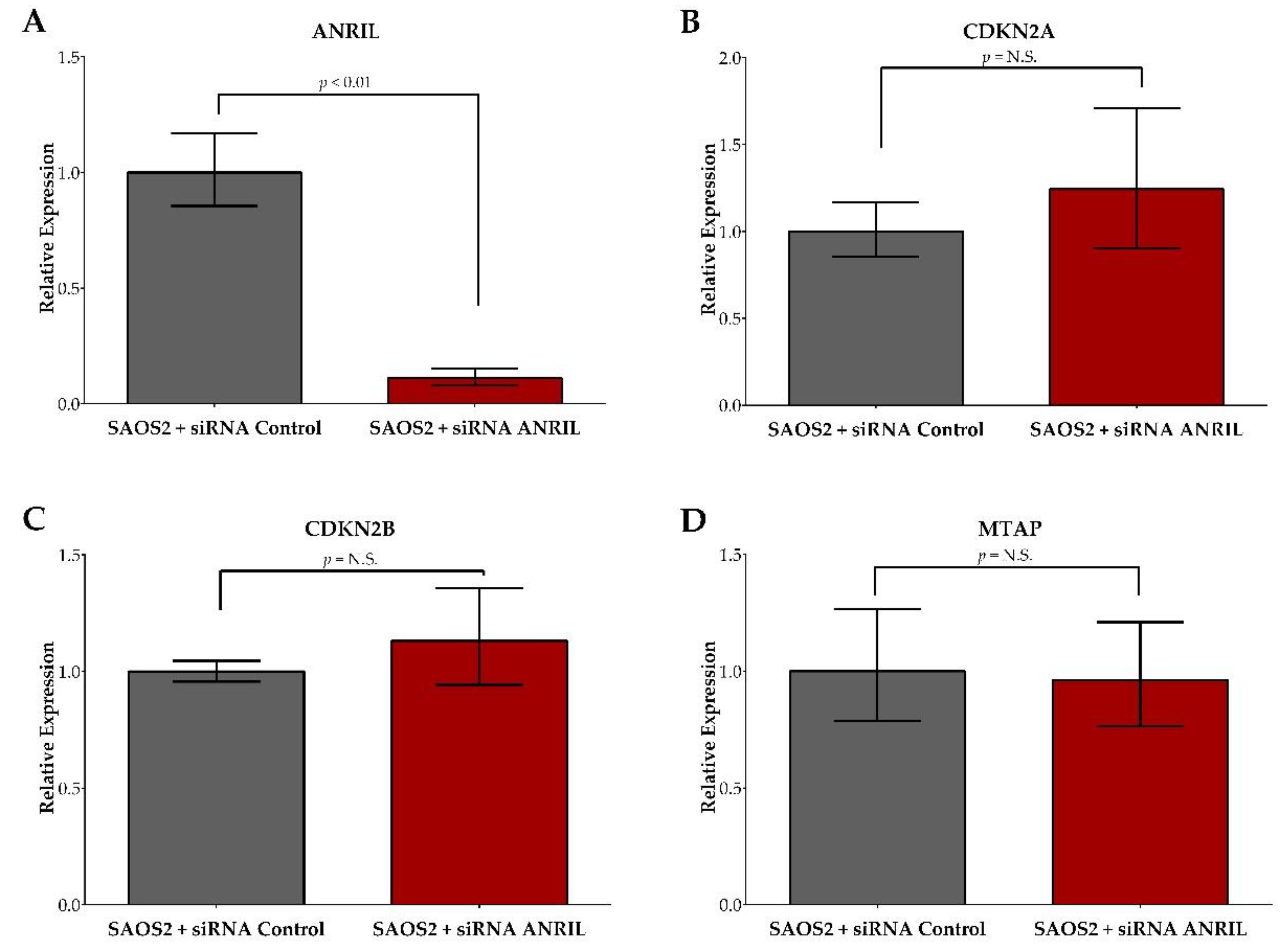
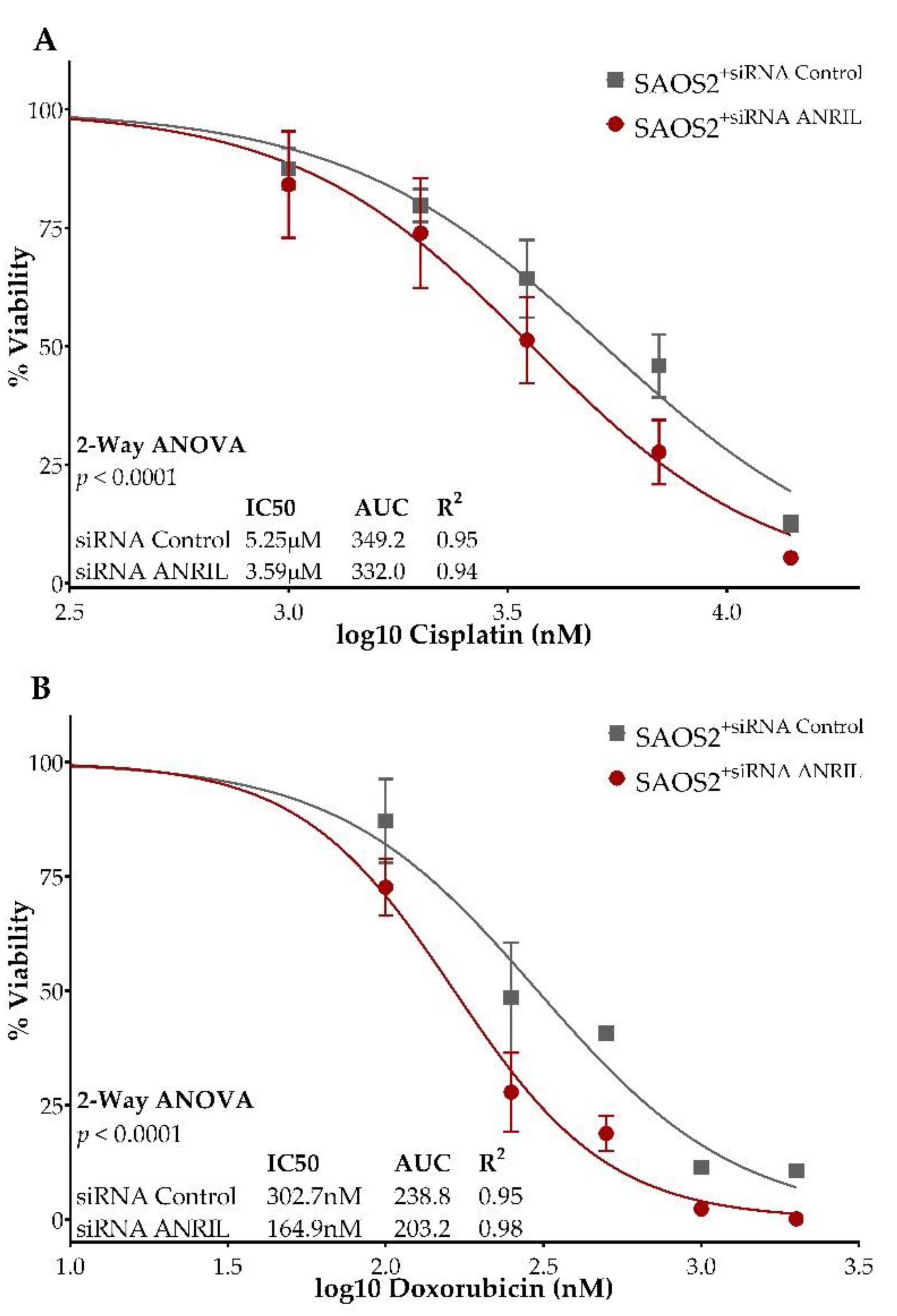
2.2. ANRIL Knockdown in SAOS2 and Impact on Cell Viability during Cisplatin and Doxorubicin Treatment

Knockdown of the lncRNA ANRIL in SAOS2 cells using siRNA displayed an 88.9% decrease in ANRIL expression compared to the SAOS2 cells transfected with the control siRNA at 24 h following transfection (Figure 2A).
The siRNA knockdown effect on ANRIL expression was maintained up to 72 h post-transfection at >80% knockdown efficiency. In order to assess the specificity of siRNA ANRIL knockdown, we additionally measured the expression of three neighboring protein-coding genes, CDKN2A, CDKN2B, and MTAP, and found no significant expression differences between SAOS2 cells transfected with ANRIL siRNA and SAOS2 cells transfected with control siRNA (Figure 2B,C). To determine the cellular response to decreased ANRIL expression, we measured cell viability at 24, 48, and 72 h post-transfection in SAOS2 cells transfected with either ANRIL or control siRNA using the WST-1 reagent. We observed a lower rate of cell proliferation (calculated by comparing cell viability at various post-treatment time points to those obtained at baseline) for SAOS2 ANRIL knockdown cells when compared to the control, but the difference was not statistically significant (Supplementary Figure S3).
To assess the impact of decreased ANRIL expression on cisplatin and doxorubicin sensitivity, we treated SAOS2 cells transfected with either ANRIL or control siRNA with serial dilutions of cisplatin and doxorubicin, and measured cell viability at 24, 48, and 72 h post-treatment using the cell viability reagent WST-1. We observed a significant difference in cell viability between ANRIL knockdown SAOS2 cells compared to the control assessed at 72 h post-treatment with either cisplatin or doxorubicin (two-way ANOVA, p < 0.0001 for both treatments). The siRNA ANRIL knockdown SAOS2 cells displayed a significantly decreased cisplatin IC50 of 3.6 uM compared to the siRNA control SAOS2 cells, which had a cisplatin IC50 of 5.3 uM (Figure 3A).
siRNA ANRIL knockdown SAOS2 cells displayed a significantly decreased doxorubicin IC50 of 165 nM compared to siRNA control SAOS2 cells with a doxorubicin IC50 of 302.7 nM (Figure 3B). Comparisons of cell viability in SAOS2 cells transfected with either ANRIL or control siRNA, at 24 and 48 h post-treatment with either cisplatin or doxorubicin, are provided in Supplementary Figure S4. Significant differences in drug sensitivity between ANRIL knockdown and control models were observed at both 24 h and 48 h post-treatment with cisplatin (Figure S4A,B) and doxorubicin (Figure S4C,D).

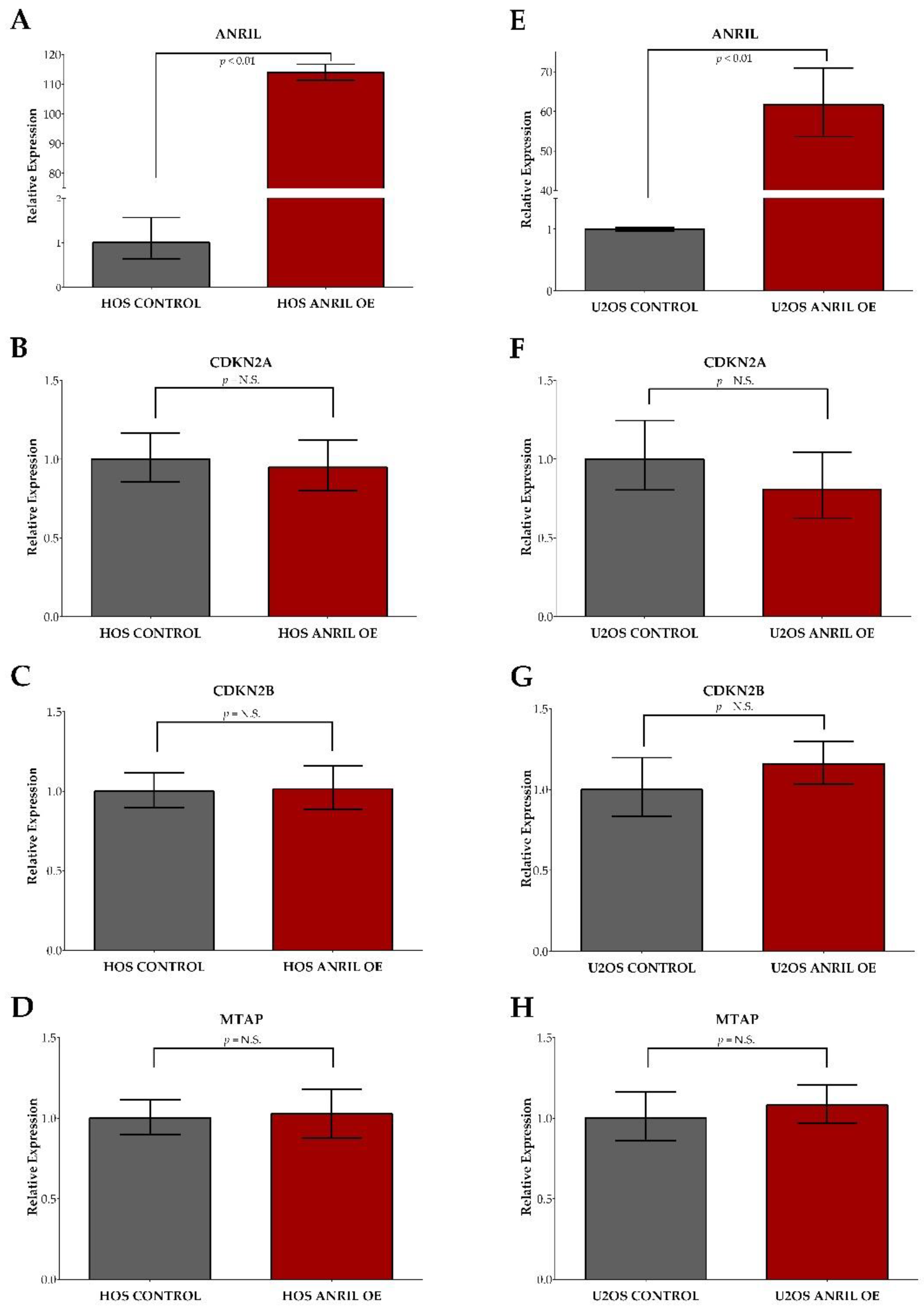
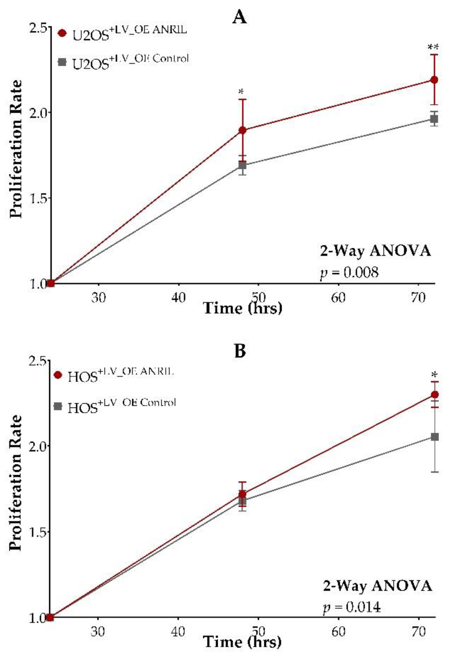
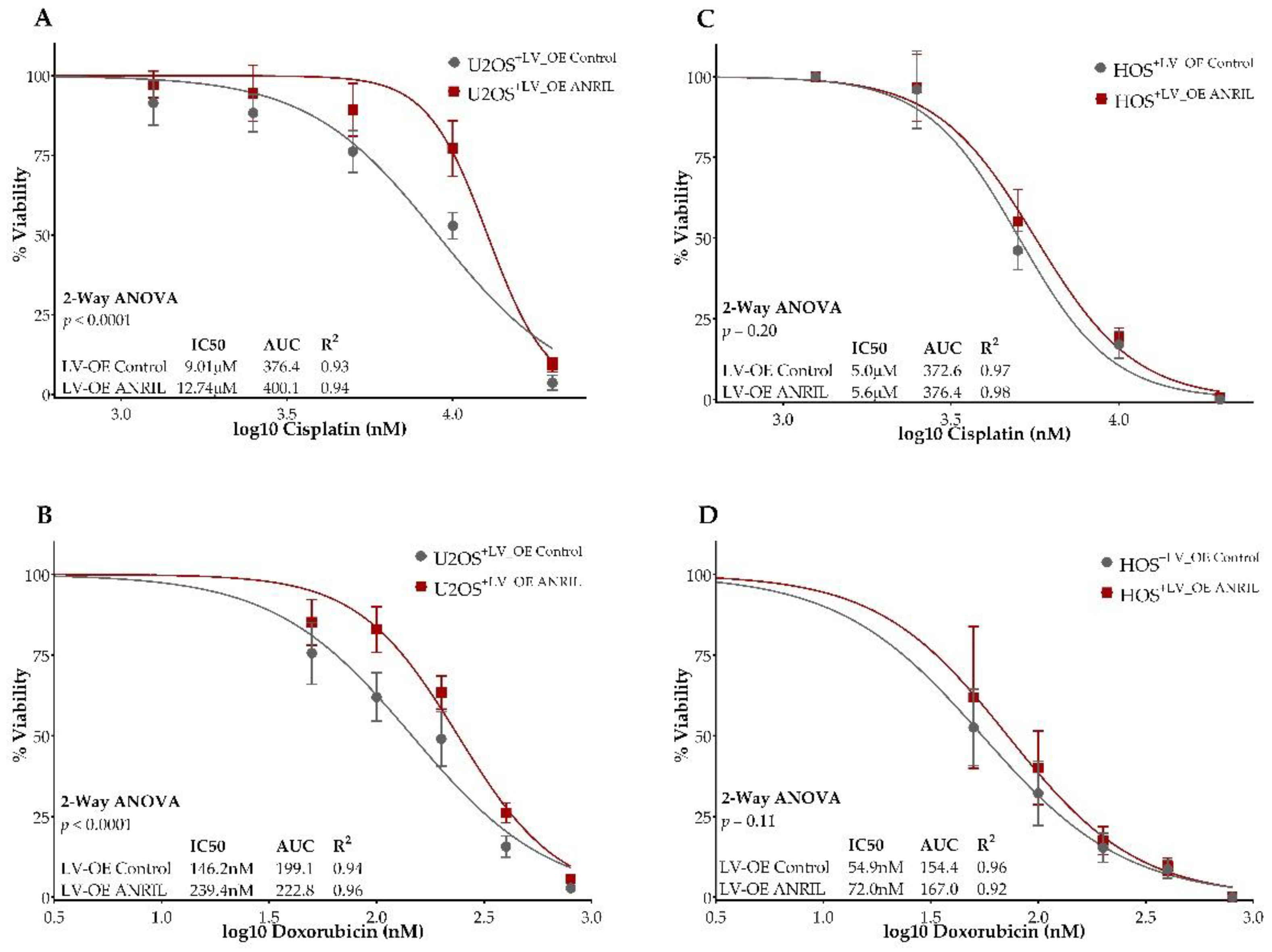
2.3. ANRIL Overexpression in HOS and U2OS and Impact on Cell Viability during Cisplatin and Doxorubicin Treatment

Stable ANRIL overexpression models using HOS and U2OS cell lines showed significant increases in ANRIL expression compared to the HOS and U2OS expression vector controls with a 114-fold increase observed in HOS and a 60-fold increase in U2OS (Figure 4A,D).
The assessment of CDKN2A, CDKN2B, and MTAP expression showed no significant differences between ANRIL overexpression HOS and U2OS models compared to their respective controls (Figure 4B,C,E,F). To determine if increased ANRIL expression promotes cell growth, we measured cell viability at 24, 48, and 72 h post-transfection in ANRIL overexpression and vector control models in both the HOS and U2OS cell lines using the cell viability reagent WST-1. We observed an increased rate of proliferation over time in both the HOS and U2OS ANRIL overexpression models compared to their respective controls in the absence of cisplatin and doxorubicin treatment (Figure 5A,B).
To assess the impact of increased ANRIL expression on cisplatin and doxorubicin sensitivity, we treated stable ANRIL overexpression and expression control models for HOS and U2OS cells with serial dilutions of cisplatin and doxorubicin, and measured cell viability at 24, 48, and 72 h post-treatment using the cell viability reagent WST-1. We observed a significant difference in cell viability between ANRIL overexpression U2OS cells compared to the control assessed at 72 h post-treatment with either cisplatin or doxorubicin (two-way ANOVA, p < 0.0001 for both treatments). Stable ANRIL overexpression in U2OS cells displayed a significantly increased cisplatin IC50 of 12.7 uM compared to the expression vector control U2OS cells with a cisplatin IC50 of 9.0 uM (Figure 6A).
Stable ANRIL overexpression in U2OS cells displayed a significantly increased doxorubicin IC50 of 239.4 nM compared to expression vector control U2OS cells with a doxorubicin IC50 of 146.2 nM (Figure 6B). In HOS cells, although we observed a similar trend of decreased sensitivity to cisplatin and doxorubicin at 72 h post-treatment in ANRIL overexpression HOS cells compared to the control (Figure 6C,D), the difference was not statistically significant. Comparisons of cell viability in both U2OS and HOS overexpression models at 24 and 48 h post-treatment with either cisplatin or doxorubicin are provided in Supplementary Figure S5. Significant differences in drug sensitivity between ANRIL overexpression U2OS cells and vector control were observed at both 24 h and 48 h post-treatment with cisplatin (Figure S5A,B) and doxorubicin (Figure S5C,D). HOS cells containing the ANRIL overexpression vector displayed no significant decreases in sensitivity to cisplatin at either 24 or 48 h post-treatment (Figure S5E,F), but did display a significant decrease in sensitivity to doxorubicin at 24 h post-treatment (Figure S5G). The observed change in doxorubicin sensitivity between ANRIL overexpression HOS cells and control were not maintained in the 48 h post-treatment measurement (Figure S5H).

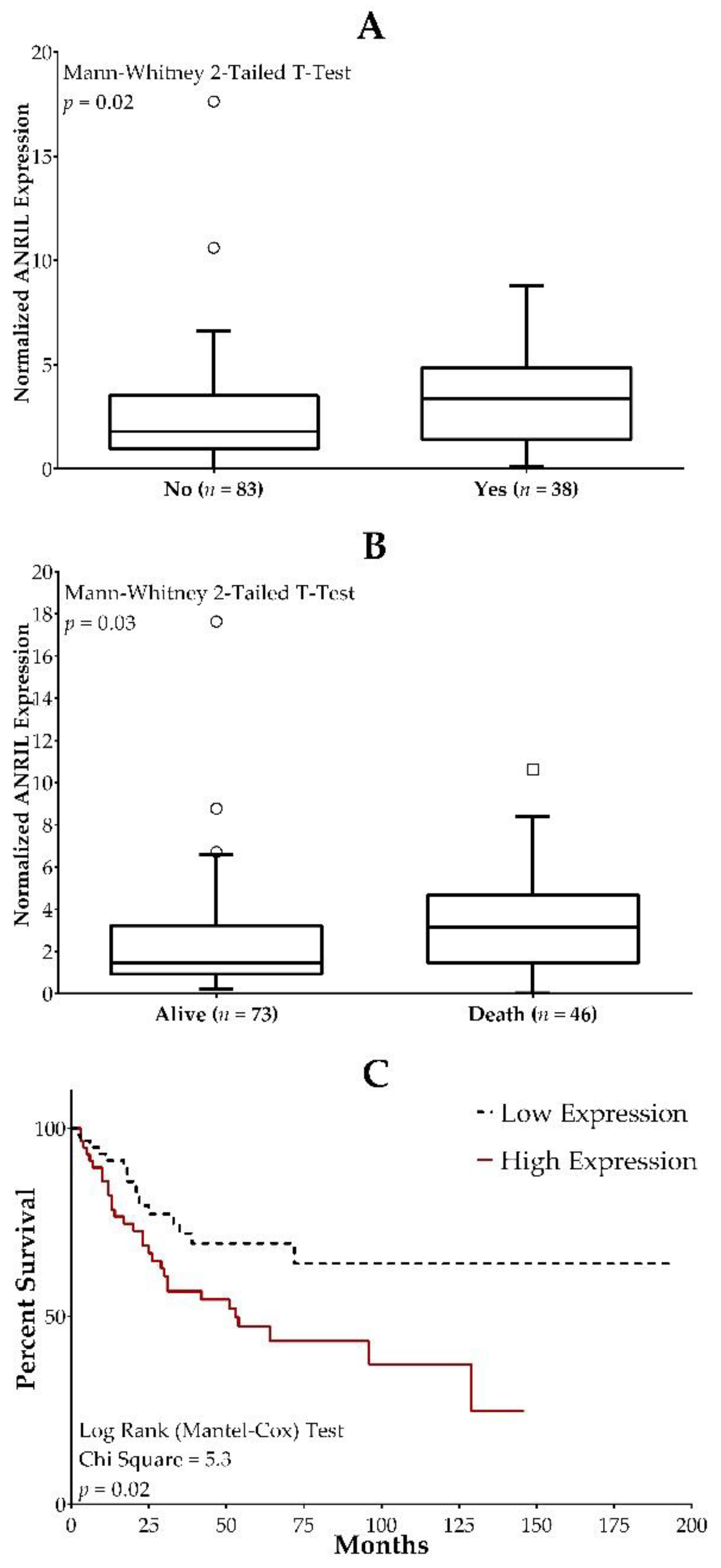
2.4. ANRIL Expression in Osteosarcoma Patients and Association with Clinical Outcome Measures

The role of lncRNA expression in osteosarcoma prognosis was examined utilizing the TARGET osteosarcoma database. Survival outcomes, metastases at diagnosis, and percent necrosis were evaluated in osteosarcoma patients (n = 86 and n = 43 for OS/metastases and percent necrosis, respectively) with various ANRIL expression levels. We found that higher ANRIL expression was significantly associated with increased incidences of metastases at diagnosis and death (Figure 7A,B). The survival curve showed that high ANRIL expression was a significant predictor of a reduced overall survival rate (Figure 7C). ANRIL expression did not predict % necrosis (p = 0.2).

3. Discussion

The large variability observed in response to the standard of care treatment in osteosarcoma presents a strong need for clinically applicable biomarkers to determine a patient’s likelihood of response in order to allow for individualized treatment regimens upfront. To date, patient clinical characteristics, along with many protein-coding gene expression and mutation statuses, have been assessed for clinical utility, but none have been incorporated into clinical practice to predict chemotherapeutic responses [10]. An investigation into the non-protein-coding genome may be fruitful and present a new opportunity to predict chemosensitivity prior to the administration of several cycles of toxic and potentially ineffective chemotherapy.
As a new frontier in cancer biomarker study, very little is known about the biological and physio-pathological role of lncRNAs [39]. To that end, our group previously evaluated lncRNAs in relation to chemosensitivity utilizing available data from the Genomics of Drug Sensitivity in Cancer database (GDSC) and identified significant correlations between decreased cisplatin and doxorubicin sensitivity and increased lncRNA ANRIL expression. The lncRNA ANRIL is transcribed in the opposite direction from the IN-K4b–ARF–INK4a gene cluster [40,41]. The protein-coding genes located within this gene cluster include S-methyl-5′-thioadenosine phosphorylase (MTAP), CDKN2A, which encodes splice variants p16INK4A and p14ARF, and CDKN2B, which encodes p15INK4B. This gene locus is transcriptionally silenced or homozygously deleted at a frequency of nearly 40% in various tumors [42]. Previous studies have shown that ANRIL expression is upregulated in osteosarcoma tissues by comparison to adjacent normal tissues and may play a role in the regulation of cell proliferation, growth, and metastasis of osteosarcoma and other cancer types [30,31,32,43,44,45,46,47,48,49,50,51]. In relation to chemotherapeutic response, ANRIL has also been reported to promote cell chemoresistance, in multiple cancer types, to various therapeutic agents including cisplatin [33,34,35,36,37,52,53,54].
Based on our previous findings and existing evidence, we hypothesized that increased ANRIL expression may play a role in the chemoresistance of osteosarcoma to cisplatin and doxorubicin and that ANRIL knockdown may lead to increased chemosensitivity to those agents. Our in vitro experiments confirmed this hypothesis, showing increased sensitivity to both cisplatin and doxorubicin in siRNA ANRIL-mediated SAOS2 cells. Using ANRIL overexpression models, we further demonstrated that increased ANRIL expression resulted in a significantly decreased sensitivity to cisplatin and doxorubicin in U2OS, but not in the HOS cell line. Given the complex and diverse genomic background of the osteosarcoma cell lines employed for this study, it is possible that genes in addition to and/or other than ANRIL may impact the chemosensitivity in HOS cells. It is also possible that there is a gene-dose effect, in which a certain level of ANRIL expression needs to be reached for conversion to chemo-resistance. All of these warrant subsequent studies. In addition, our results also showed that increased ANRIL expression resulted in significantly increased rates of cell proliferation under drug-naïve conditions in U2OS and HOS cells. Though not statistically significant, SAOS2 ANRIL knockdown cells displayed a predicted trend of decreased proliferation rates under drug-naïve conditions. The effect of ANRIL expression on cancer cell proliferation observed in our study is consistent with previous reports documented in retinoblastoma, liver cancer, gastric cancer and lung cancer [30,31,32,33,34,35,36]. Additionally, a previous study by Li G. et al. showed decreased cell proliferation under drug-naïve conditions and increased cisplatin sensitivity in U2OS cells after ANRIL knockdown [34].
It is important to note that because of the close physical proximity of ANRIL to several nearby protein-coding genes (CDKN2A, CDKN2B, MTAP), and the existence of multiple transcriptional isoforms of ANRIL, the role of ANRIL on the potential regulation of these genes in relation to drug sensitivity has not been fully elucidated. Previous functional studies have shown that ANRIL can mediate the epigenetic transcriptional repression of the neighboring genes CDKN2A and CDKN2B [55,56]. However, multiple studies have also reported positive correlations between ANRIL and nearby protein-coding genes, suggesting transcriptional co-regulation of these genes predominates in many tissues [57,58,59,60,61]. We carefully designed our ANRIL modification experiments to only target ANRIL and showed that there were no significant gene expression changes in three neighboring protein-coding genes. Previously, one of these nearby protein-coding genes (MTAP) was identified in osteosarcoma [62]. It is thought to play a role in cancer cell etiology given its behavior as a tumor suppressor [62,63,64,65]. However, correlation analyses between the expression of MTAP and cisplatin and doxorubicin sensitivity found no relationship between them.
Our analysis of TARGET data confirmed an association between osteosarcoma prognosis and ANRIL expression. As predicted, median ANRIL expression was associated with death and metastases at diagnosis. This is consistent with previous reports indicating that high ANRIL expression is a poor prognosis indicator in different cancer types including osteosarcoma [44,66,67,68,69].
In this study, we identified and validated the link between ANRIL expression and cisplatin and doxorubicin sensitivity in osteosarcoma. However, the underlying mechanisms of this relationship remain unclear, which may require a more comprehensive, longitudinal examination of protein-coding genes, both at the RNA and protein level, after ANRIL modification. Indeed, previous studies have provided evidence for significant relationships with select microRNAs and regulated genes, such as let-7a/HMGA2, miRNA-125a-5p/STAT3, microRNA-98, and microRNA-328/ABCG2, to further elucidate potential mechanisms involved in ANRIL-mediated chemoresistance to cisplatin across different cancer types [31,32,33,34,35,36,52]. A separate study in oral squamous cell carcinoma also identified significant impairment of drug transporters MRP1 and ABCC2 after ANRIL knockdown, which resulted in increased cisplatin cytotoxicity [53].
Our study focused on ANRIL’s impact on cancer cell growth inhibition induced by chemotherapy. Additional cellular phenotypes can be examined to assess whether changing the ANRIL expression level will impact other traits (e.g., migration and invasion). Since ANRIL is unlikely to be the only lncRNA that contributes to chemosensitivity, subsequent animal studies are warranted to assess the effect of ANRIL in the osteosarcoma response to chemotherapy in vivo. Furthermore, the microRNAs that are negatively regulated by ANRIL (such as miR-34a, miR-125a, and miR-186) should be investigated, as should their putative role in bone remodeling and osteosarcoma onset [70]. Further studies would include the investigation of these targets in osteosarcoma models that are particularly related to chemosensitivity.

4. Materials and Methods

4.1. Reagents

Cisplatin and doxorubicin were purchased from Sigma-Aldrich (St. Louis, MO, USA) and dissolved in either cell culture-grade water at a stock concentration of 3 mM for cisplatin, or in DMSO at a stock concentration of 10mM for doxorubicin. Reconstituted stocks were aliquoted into 1.5 mL black centrifuge tubes to protect from light and stored at −80 °C as single-use aliquots prior to experimentation.

4.2. Cell Lines

The osteosarcoma cancer cell lines SAOS2, HOS, and U2OS were obtained from the American Type Culture Collection (ATCC, Rockefeller, MD, USA) and selected based on ANRIL expression profiling and somatic mutation status (Supplementary Materials, Figure S2 and Table S1). All cell lines were grown using McCoy’s 5A modified medium (Thermo Fisher Scientific, Waltham, MA, USA), supplemented with 15% FBS (fetal bovine serum; Thermo Fisher Scientific) in a temperature-controlled incubator, and maintained at 37 °C with 5% CO2. Cells in the logarithmic phase of growth were used for all experiments. Cell lines were observed microscopically to confirm morphology, and population-doubling times were determined by viable cell counting using trypan blue on the TC20™ Automated Cell Counter (Bio-Rad Laboratories, Hercules, CA, USA). All cell lines were periodically monitored for mycoplasma using the Universal Mycoplasma Detection Kit following the manufacturer’s protocol (ATCC). Culture health and identity were monitored by microscopy and by comparing population doubling times to baseline values recorded at the time of receipt.

4.3. lncRNA ANRIL Knockdown in SAOS2 Cells

Knockdown of ANRIL was performed in the SAOS2 cell line, due to its high ANRIL expression, by utilizing Lincode SMARTpool small interfering RNA (siRNA) against ANRIL (CDKN2B-AS1) and the DharmaFECT transfection reagent 2 (Dharmacon, Horizon Discovery Biosciences, Boulder, CO, USA) following the manufacturer’s reverse transfection protocol. The Lincode Non-targeting Control Pool was used as a control in all siRNA knockdown experiments. Each siRNA pool consisted of four different siRNAs and was reconstituted to a 20 nM stock concentration. Knockdown efficiency was experimentally determined by reverse transfecting SAOS2 cells with 25 nM of either the ANRIL or control siRNA and 0.05% DharmaFECT transfection reagent 2 in a 6-well tissue culture plate, utilizing a seeding density of 5 × 10⁵ cells per well. Reverse transfection was performed in a total volume of 2 mL containing RPMI 1640 with 10% FBS and maintained at 37 °C in a humidified incubator with 5% CO2 atmosphere. Cells were harvested at 24, 48, and 72 h following reverse transfection to determine knockdown efficiency by assessing lncRNA ANRIL expression using quantitative real-time PCR.

4.4. lncRNA ANRIL Overexpression in HOS and U2OS Cells

Stable ANRIL overexpression models were created from the HOS and U2OS cell lines utilizing a custom designed Mammalian noncodingRNA Expression Lentiviral Vector (pLV(ncRNA)EGFP:T2A:Puro-CMV>hCDKN2B-AS1[NR_003529.3]; Vector ID:VB200211-1124awf) obtained from Vector Builder (VectorBuilder Inc., Chicago, IL, USA). A mCherry empty vector was utilized as a control (pLV[Exp]-EGFP:T2A:Puro-EF1A>mCherry; Vector ID: VB160109-10005). HOS and U2OS cells were plated at a seeding density of 1 × 10⁵ cells per well in a 6-well tissue culture plate and allowed to adhere for 24 h in a 37 °C humidified incubator with 5% CO2 atmosphere prior to transduction. Cells were transduced in 1 mL media containing the appropriate virus to obtain an MOI of 10 following the manufacturer’s protocol. Stable ANRIL expression lines and empty vector controls in both HOS and U2OS cells were generated following a 3-day antibiotic selection with puromycin. Positive GFP-expression cells were visualized and counted daily during selection to confirm transduction efficiency using the Cytation™ 1 Cell Imaging Multi-Mode Reader (BioTek Instruments, Winooski, VT, USA). Confirmation of target ANRIL expression was performed by assessing lncRNA ANRIL expression using quantitative real-time PCR.

4.5. lncRNA and Gene Expression Using Quantitative Real-Time PCR

Total RNA was extracted from cultured cells using the Quick-RNA MiniPrep Plus Kit (Zymo Research, Irvine, CA, USA) and quantified on the NanoDrop ND-8000 8-channel spectrophotometer (Thermo Fisher Scientific, Waltham, MA, USA). A total of 2 µg of RNA was used to synthesize cDNA, utilizing the High Capacity cDNA Reverse Transcription Kit (Thermo Fisher Scientific, Waltham, MA, USA). The Sso-Advanced Universal SYBR Green SuperMix (Bio-Rad, Hercules, CA, USA) was used to conduct real-time PCR analyses following the manufacturer’s protocol. In addition to the target lncRNA ANRIL, we assessed the expression of three protein-coding genes located near ANRIL to assess potential off-target effects following ANRIL knockdown or overexpression: CDKN2A, CDKN2B, and MTAP. GAPDH and ACTB (β-Actin) served as housekeeping genes. Pre-designed primers for all genes were selected from PrimeTime qPCR Primer Assays available from Integrated DNA Technologies, Inc. (IDT; Coralville, IA, USA). The PCR primer sequences utilized to assess gene expression are provided in Table 1.
Quantitative real-time PCR and data collection were performed using the 7500 Real-Time PCR System (Applied Biosystems, Foster City, CA, USA). All results were normalized with the expression of GAPDH and ACTB as a reference panel. All PCR reactions were performed in triplicate, and each knockdown and overexpression experiment was performed independently three times. Expression results were quantified using the ΔΔCt method relative to the appropriate control [71].

4.6. Cell Viability and Drug Response Curves in Knockdown and Overexpression Models following Cisplatin and Doxorubicin Exposure

To assess the impact of siRNA-mediated knockdown of the lncRNA ANRIL on proliferation and responses to cisplatin and doxorubicin, SAOS2 cells were reverse transfected in 96-well plates, with 25 nM of either the ANRIL or control siRNA and 0.05% DharmaFECT transfection reagent 2, using a seeding density of 1 × 10⁴ cells per well. Following a 24-h incubation in a 37 °C humidified incubator with 5% CO2 atmosphere, cells were treated with serial dilutions of either cisplatin at concentrations ranging from 1 µM to 14 µM, or of doxorubicin at concentrations ranging from 0.1 µM to 2 µM. Cell viability was measured at 24, 48, and 72 h after treatment by incubation with WST-1 (Roche Applied Science, Penzberg, Upper Bavaria, Germany) following the manufacturer’s protocol. Following a 2-h incubation at 37 °C after the addition of WST-1, absorbance at the 450 nm wavelength was assessed using the Synergy HTX Multi-Mode Plate Reader (BioTek, Winooski, VT, USA).
To assess the impact of ANRIL overexpression on proliferation and responses to cisplatin and doxorubicin, stable transduction models for the overexpression of ANRIL, or empty expression control generated in U2OS and HOS cell lines, were plated in 96-well plates using a seeding density of 1 × 10⁴ cells per well, and incubated for 24 h before treatment with serial dilutions of either cisplatin or doxorubicin. Cell viability was measured at 24, 48, and 72 h after treatment by incubation with WST-1 as described above.
Each experimental condition was performed in triplicate and measurements were reported as the mean ± standard deviation (S.D.) of three independent biological experiments.

4.7. ANRIL Expression and Clinical Outcomes Assessment

To investigate the impact of ANRIL expression on clinical outcome measures in osteosarcoma, we utilized available RNASeq data from the TARGET (Therapeutically Applicable Research to Generate Effective Treatments) osteosarcoma dataset (n = 86) (https://portal.gdc.cancer.gov/projects, dbGaP Study Accession: phs000468.v21.p8) accessed on 13 April 2021. This dataset is accessible via the National Cancer Institute Office of Cancer Genomics in the Genomic Data Commons (GDC). It includes tumors from children enrolled in Children’s Oncology Group (COG) protocols from which next-generation sequencing and transcriptome sequencing was performed to report long non-coding RNA expression. A summary of available clinical outcome measures and patient characteristics from TARGET osteosarcoma dataset are provided in Table 2.

4.8. Statistical Analysis

Statistical analyses of all in vitro cell culture experiments and patient data were performed in GraphPad Prism 6.07 (San Diego, CA, USA). For all cell culture experiments, significant differences between groups were tested using a two-tailed Student’s t-test or two-way analysis of variance (two-way ANOVA) [72]. For the patient cohort dataset, patients were categorized into high (>2) and low ANRIL expression groups based on the median ANRIL expression. Comparisons of ANRIL expression with clinical parameters were analyzed using the Student’s t-test or the Chi-square test [73]. Survival analysis was performed using the Kaplan–Meier method and the log-rank test [74,75]. A p-value of <0.05 was considered statistically significant.

5. Conclusions

The overexpression of ANRIL significantly increased cell proliferation and reduced both cisplatin and doxorubicin sensitivity, whereas the reduction in ANRIL expression led to increased sensitivity in our in vitro validation experiments. Analysis within the TARGET osteosarcoma patient cohort showed that higher ANRIL expression was significantly associated with death and metastases. These findings further support the hypothesis that ANRIL is a suitable chemo sensitivity and prognostic biomarker in osteosarcoma. Furthermore, reducing ANRIL expression may be a therapeutic strategy to overcome current standard-of-care treatment resistance in osteosarcoma.

Supplementary Materials

The following are available online at www.mdpi.com/article/10.3390/ijms222011168/s1, Figure S1: Increased ANRIL expression correlates with increased resistance to both cisplatin (A) and doxorubicin (B) in cancer cell lines; Table S1: Characteristics of osteosarcoma cell lines utilized for in vitro validation experiments; Figure S2: lncRNA ANRIL, CDKN2A, CDKN2B, and MTAP gene expression in osteosarcoma cell lines HOS (dark grey), U2OS (light grey), and SAOS2 (red); Figure S3: Comparison of proliferation rates between ANRIL knockdown SAOS2 cells and control; Figure S4: Increased sensitivity measured at 24 and 48 h post-treatment with cisplatin and doxorubicin following ANRIL knockdown in SAOS2 cells; Figure S5: Dose–response curves of U2OS and HOS ANRIL overexpression models measured at 24 and 48 h post-treatment with cisplatin and doxorubicin.

Author Contributions

Conceptualization, A.F., A.M.L., E.G. and R.S.H.; methodology, A.F., A.M.L. and R.S.H.; software, A.M.L., A.N. and D.C.K.; validation, A.M.L., E.M. and R.S.H.; formal analysis, A.F. and A.M.L.; investigation, A.F., A.M.L. and R.S.H.; resources, R.S.H.; data curation, A.F., A.M.L., A.N., D.C.K. and R.S.H.; writing—original draft preparation, A.F. and A.M.L.; writing—review and editing, A.F., A.M.L., A.N., D.C.K., E.G. and R.S.H.; supervision, R.S.H. All authors have read and agreed to the published version of the manuscript.

Funding

R.S.H received support from the NIH/NCI Grants R01CA229618 and R01CA204856.

Institutional Review Board Statement

Not applicable

Informed Consent Statement

Not applicable

Data Availability Statement

The results published here are, wholly or in part, based upon data generated by the Therapeutically Applicable Research to Generate Effective Treatments (https://ocg.cancer.gov/programs/target, dbGaP Study Accession: phs000468.v21.p8) accessed on 13 April 2021. The data used for this analysis are available at https://portal.gdc.cancer.gov/projects accessed on 13 April 2021.

Conflicts of Interest

The authors declare no conflict of interest.

References

  1. Friebele, J.C.; Peck, J.; Pan, X.; Abdel-Rasoul, M.; Mayerson, J.L. Osteosarcoma: A Meta-Analysis and Review of the Literature. Am. J. Orthop. 2015, 44, 547–553. [Google Scholar] [PubMed]
  2. Mirabello, L.; Troisi, R.J.; Savage, S.A. Osteosarcoma incidence and survival rates from 1973 to 2004: Data from the Surveillance, Epidemiology, and End Results Program. Cancer Interdisciplanary Int. J. Am. Cancer Soc. 2009, 115, 1531–1543. [Google Scholar] [CrossRef] [Green Version]
  3. Kager, L.; Tamamyan, G.; Bielack, S. Novel insights and therapeutic interventions for pediatric osteosarcoma. Future Oncol. 2017, 13, 357–368. [Google Scholar] [CrossRef]
  4. Sami, S.H.; Rafati, A.H.; Hodjat, P. Tissue necrosis after chemotherapy in osteosarcoma as the important prognostic factor. Saudi Med. J. 2008, 29, 1124–1129. [Google Scholar]
  5. Taran, S.J.; Taran, R.; Malipatil, N.B. Pediatric Osteosarcoma: An Updated Review. Indian J. Med. Paediatr. Oncol. 2017, 38, 33–43. [Google Scholar] [CrossRef] [Green Version]
  6. Durnali, A.G.; Turkoz, F.P.; Yukruk, F.A.; Tokluoglu, S.; Yazici, O.K.; Demirci, A.; Bal, O.; Buyukbas, S.G.; Esbah, O.; Oksuzoglu, B.; et al. Outcomes of Adolescent and Adult Patients with Lung Metastatic Osteosarcoma and Comparison of Synchronous and Metachronous Lung Metastatic Groups. PLoS ONE 2016, 11, e0152621. [Google Scholar] [CrossRef] [Green Version]
  7. Xu, J.; Xie, L.; Guo, W. Neoadjuvant Chemotherapy Followed by Delayed Surgery: Is it Necessary for All Patients with Nonmetastatic High-Grade Pelvic Osteosarcoma? Clin. Orthop. Relat. Res. 2018, 476, 2177–2186. [Google Scholar] [CrossRef]
  8. Smeland, S.; Bielack, S.S.; Whelan, J.; Bernstein, M.; Hogendoorn, P.; Krailo, M.D.; Gorlick, R.; Janeway, K.A.; Ingleby, F.C.; Anninga, J.; et al. Survival and prognosis with osteosarcoma: Outcomes in more than 2000 patients in the EURAMOS-1 (European and American Osteosarcoma Study) cohort. Eur. J. Cancer. 2019, 109, 36–50. [Google Scholar] [CrossRef] [Green Version]
  9. Misaghi, A.; Goldin, A.; Awad, M.; Kulidjian, A.A. Osteosarcoma: A comprehensive review. SICOT J. 2018, 4, 12. [Google Scholar] [CrossRef] [PubMed] [Green Version]
  10. Mirabello, L.; Zhu, B.; Koster, R.; Karlins, E.; Dean, M.; Yeager, M.; Gianferante, M.; Spector, L.; Morton, L.M.; Karyadi, D.; et al. Frequency of Pathogenic Germline Variants in Cancer-Susceptibility Genes in Patients with Osteosarcoma. JAMA Oncol. 2020, 6, 724–734. [Google Scholar] [CrossRef] [PubMed]
  11. Li, X.; Ashana, A.O.; Moretti, V.M.; Lackman, R.D. The relation of tumour necrosis and survival in patients with osteosarcoma. Int. Orthop. 2011, 35, 1847–1853. [Google Scholar] [CrossRef] [PubMed] [Green Version]
  12. Hanafy, E.; Al Jabri, A.; Gadelkarim, G.; Dasaq, A.; Nazim, F.; Al Pakrah, M. Tumor histopathological response to neoadjuvant chemotherapy in childhood solid malignancies: Is it still impressive? J. Investig. Med. 2018, 66, 289–297. [Google Scholar] [CrossRef] [PubMed] [Green Version]
  13. Whelan, J.S.; Bielack, S.S.; Marina, N.; Smeland, S.; Jovic, G.; Hook, J.M.; Krailo, M.; Anninga, J.; Butterfass-Bahloul, T.; Böhling, T.; et al. EURAMOS-1, an international randomised study for osteosarcoma: Results from pre-randomisation treatment. Ann. Oncol. 2015, 26, 407–414. [Google Scholar] [CrossRef]
  14. Huang, Y.; Ling, A.; Pareek, S.; Huang, R.S. Oncogene or tumor suppressor? Long noncoding RNAs role in patient’s prognosis varies depending on disease type. Transl. Res. 2021, 230, 98–110. [Google Scholar] [CrossRef]
  15. Jarroux, J.; Morillon, A.; Pinskaya, M. History, Discovery, and Classification of lncRNAs. Adv. Exp. Med. Biol. 2017, 1008, 1–46. [Google Scholar]
  16. Kopp, F.; Mendell, J.T. Functional Classification and Experimental Dissection of Long Noncoding RNAs. Cell 2018, 172, 393–407. [Google Scholar] [CrossRef] [Green Version]
  17. Wei, J.W.; Huang, K.; Yang, C.; Kang, C.S. Non-coding RNAs as regulators in epigenetics (Review). Oncol. Rep. 2017, 37, 3–9. [Google Scholar] [CrossRef] [Green Version]
  18. Lanzillotti, C.; De Mattei, M.; Mazziotta, C.; Taraballi, F.; Rotondo, J.C.; Tognon, M.; Martini, F. Long Non-coding RNAs and MicroRNAs Interplay in Osteogenic Differentiation of Mesenchymal Stem Cells. Front. Cell Dev. Biol. 2021, 9, 646032. [Google Scholar] [CrossRef]
  19. Moonmuang, S.; Chaiyawat, P.; Jantrapirom, S.; Pruksakorn, D.; Lo Piccolo, L. Circulating Long Non-Coding RNAs as Novel Potential Biomarkers for Osteogenic Sarcoma. Cancers 2021, 13, 4214. [Google Scholar] [CrossRef] [PubMed]
  20. Ghafouri-Fard, S.; Shirvani-Farsani, Z.; Hussen, B.M.; Taheri, M. The critical roles of lncRNAs in the development of osteosarcoma. Biomed. Pharmacother. 2021, 135, 111217. [Google Scholar] [CrossRef] [PubMed]
  21. Liu, K.; Gao, L.; Ma, X.; Huang, J.-J.; Chen, J.; Zeng, L.; Jr., C.R.A.; Zou, C.; Chen, Z.-S. Long non-coding RNAs regulate drug resistance in cancer. Mol. Cancer 2020, 19, 54. [Google Scholar] [CrossRef]
  22. Chen, Q.N.; Wei, C.C.; Wang, Z.X.; Sun, M. Long non-coding RNAs in anti-cancer drug resistance. Oncotarget 2017, 8, 1925–1936. [Google Scholar] [CrossRef] [Green Version]
  23. Li, Z.; Lü, M.; Zhou, Y.; Xu, L.; Jiang, Y.; Liu, Y.; Li, X.; Song, M. Role of Long Non-Coding RNAs in the Chemoresistance of Gastric Cancer: A Systematic Review. OncoTargets Ther. 2021, 14, 503–518. [Google Scholar] [CrossRef]
  24. Nath, A.; Geeleher, P.; Huang, R.S. Long non-coding RNA transcriptome of uncharacterized samples can be accurately imputed using protein-coding genes. Brief. Bioinform. 2020, 21, 637–648. [Google Scholar] [CrossRef]
  25. Nath, A.; Lau, E.Y.T.; Lee, A.M.; Geeleher, P.; Cho, W.C.S.; Huang, R.S. Discovering long noncoding RNA predictors of anticancer drug sensitivity beyond protein-coding genes. Proc. Natl. Acad. Sci. USA 2019, 116, 22020–22029. [Google Scholar] [CrossRef]
  26. Nath, A.; Huang, R.S. Emerging role of long non-coding RNAs in cancer precision medicine. Mol. Cell Oncol. 2020, 7, 1684130. [Google Scholar] [CrossRef]
  27. Pasmant, E.; Laurendeau, I.; Héron, D.; Vidaud, M.; Vidaud, D.; Bièche, I. Characterization of a germ-line deletion, including the entire INK4/ARF locus, in a melanoma-neural system tumor family: Identification of ANRIL, an antisense noncoding RNA whose expression coclusters with ARF. Cancer Res. 2007, 67, 3963–3969. [Google Scholar] [CrossRef] [PubMed] [Green Version]
  28. Liu, F.T.; Zhu, P.Q.; Luo, H.L.; Zhang, Y.; Hao, T.F.; Xia, G.F.; Zhu, Z.M.; Qiu, C. Long noncoding RNA ANRIL: A potential novel prognostic marker in cancer: A meta-analysis. Minerva Med. 2016, 107, 77–83. [Google Scholar] [PubMed]
  29. Li, Z.; Yu, X.; Shen, J. ANRIL: A pivotal tumor suppressor long non-coding RNA in human cancers. Tumour Biol. 2016, 37, 5657–5661. [Google Scholar] [CrossRef] [PubMed]
  30. Lou, N.; Liu, G.; Pan, Y. Long noncoding RNA ANRIL as a novel biomarker in human cancer. Future Oncol. 2020, 16, 2981–2995. [Google Scholar] [CrossRef] [PubMed]
  31. Huang, D.; Bi, C.; Zhao, Q.; Ding, X.; Bian, C.; Wang, H.; Wang, T.; Liu, H. Knockdown long non-coding RNA ANRIL inhibits proliferation, migration and invasion of HepG2 cells by down-regulation of miR-191. BMC Cancer 2018, 18, 919. [Google Scholar] [CrossRef] [PubMed]
  32. Lan, W.-G.; Xu, D.-H.; Xu, C.; Ding, C.-L.; Ning, F.-L.; Zhou, Y.-L.; Ma, L.-B.; Liu, C.-M.; Chang-Ling, D. Silencing of long non-coding RNA ANRIL inhibits the development of multidrug resistance in gastric cancer cells. Oncol. Rep. 2016, 36, 263–270. [Google Scholar] [CrossRef] [PubMed] [Green Version]
  33. Wang, Y.; Cheng, N.; Luo, J. Downregulation of lncRNA ANRIL represses tumorigenicity and enhances cisplatin-induced cytotoxicity via regulating microRNA let-7a in nasopharyngeal carcinoma. J. Biochem Mol. Toxicol. 2017, 31, e21904. [Google Scholar] [CrossRef] [PubMed]
  34. Li, G.; Zhu, Y. Effect of lncRNA ANRIL knockdown on proliferation and cisplatin chemoresistance of osteosarcoma cells in vitro. Pathol. Res. Pract. 2019, 215, 931–938. [Google Scholar] [CrossRef] [PubMed]
  35. Wang, X.; Zhang, G.; Cheng, Z.; Dai, L.; Jia, L.; Jing, X.; Wang, H.; Zhang, R.; Liu, M.; Jiang, T.; et al. Knockdown of lncRNA ANRIL inhibits the development of cisplatin resistance by upregulating miR-98 in lung cancer cells. Oncol. Rep. 2020, 44, 1025–1036. [Google Scholar] [CrossRef] [PubMed]
  36. Yin, X.; Liao, Y.; Xiong, W.; Zhang, Y.; Zhou, Y.; Yang, Y. Hypoxia-induced lncRNA ANRIL promotes cisplatin resistance in retinoblastoma cells through regulating ABCG2 expression. Clin. Exp. Pharmacol Physiol. 2020, 47, 1049–1057. [Google Scholar] [CrossRef]
  37. Ferretti, V.A.; León, I.E. Long Non-coding RNAs in Cisplatin Resistance in Osteosarcoma. Curr. Treat. Options Oncol. 2021, 22, 41. [Google Scholar] [CrossRef]
  38. Yang, W.; Soares, J.; Greninger, P.; Edelman, E.; Lightfoot, H.; Forbes, S.; Sridhar, R.; Futreal, P.A.; Haber, D.; Stratton, M.; et al. Genomics of Drug Sensitivity in Cancer (GDSC): A resource for therapeutic biomarker discovery in cancer cells. Nucleic Acids Res. 2013, 41, D955–D961. [Google Scholar] [CrossRef] [Green Version]
  39. Jathar, S.; Kumar, V.; Srivastava, J.; Tripathi, V. Technological Developments in lncRNA Biology. Adv. Exp. Med. Biol. 2017, 1008, 283–323. [Google Scholar]
  40. Aguilo, F.; Zhou, M.M.; Walsh, M.J. Long noncoding RNA, polycomb, and the ghosts haunting INK4b-ARF-INK4a expression. Cancer Res. 2011, 71, 5365–5369. [Google Scholar] [CrossRef] [Green Version]
  41. Kong, Y.; Hsieh, C.H.; Alonso, L.C. A lncRNA at the CDKN2A/B Locus with Roles in Cancer and Metabolic Disease. Front. Endocrinol 2018, 9, 405. [Google Scholar] [CrossRef] [PubMed] [Green Version]
  42. Wan, G.; Mathur, R.; Hu, X.; Liu, Y.; Zhang, X.; Peng, G.; Lu, X. Long non-coding RNA ANRIL (CDKN2B-AS) is induced by the ATM-E2F1 signaling pathway. Cell Signal. 2013, 25, 1086–1095. [Google Scholar] [CrossRef] [Green Version]
  43. Guan, H.; Mei, Y.; Mi, Y.; Li, C.; Sun, X.; Zhao, X.; Liu, J.; Cao, W.; Li, Y.; Wang, Y. Downregulation of lncRNA ANRIL suppresses growth and metastasis in human osteosarcoma cells. OncoTargets Ther. 2018, 11, 4893–4899. [Google Scholar] [CrossRef] [Green Version]
  44. Yu, G.; Liu, G.; Yuan, D.; Dai, J.; Cui, Y.; Tang, X. Long non-coding RNA ANRIL is associated with a poor prognosis of osteosarcoma and promotes tumorigenesis via PI3K/Akt pathway. J. Bone Oncol. 2018, 11, 51–55. [Google Scholar] [CrossRef]
  45. Qiu, J.J.; Lin, Y.Y.; Ding, J.X.; Feng, W.W.; Jin, H.Y.; Hua, K.Q. Long non-coding RNA ANRIL predicts poor prognosis and promotes invasion/metastasis in serous ovarian cancer. Int. J. Oncol. 2015, 46, 2497–2505. [Google Scholar] [CrossRef] [Green Version]
  46. Huang, M.-D.; Chen, W.-M.; Qi, F.-Z.; Xia, R.; Sun, M.; Xu, T.-P.; Yin, L.; Zhang, E.-B.; De, W.; Shu, Y.-Q. Long non-coding RNA ANRIL is upregulated in hepatocellular carcinoma and regulates cell proliferation by epigenetic silencing of KLF2. J. Hematol. Oncol. 2015, 8, 57. [Google Scholar] [CrossRef] [Green Version]
  47. Cheng, S.; Huang, T.; Li, P.; Zhang, W.; Wang, Z.; Chen, Y. Long non-coding RNA ANRIL promotes the proliferation, migration and invasion of human osteosarcoma cells. Exp. Ther. Med. 2017, 14, 5121–5125. [Google Scholar] [CrossRef]
  48. Naemura, M.; Tsunoda, T.; Inoue, Y.; Okamoto, H.; Shirasawa, S.; Kotake, Y. ANRIL regulates the proliferation of human colorectal cancer cells in both two- and three-dimensional culture. Mol. Cell. Biochem. 2016, 412, 141–146. [Google Scholar] [CrossRef]
  49. Zou, Z.W.; Ma, C.; Medoro, L.; Chen, L.; Wang, B.; Gupta, R.; Liu, T.; Yang, X.Z.; Chen, T.T.; Wang, R.Z.; et al. LncRNA ANRIL is up-regulated in nasopharyngeal carcinoma and promotes the cancer progression via increasing proliferation, reprograming cell glucose metabolism and inducing side-population stem-like cancer cells. Oncotarget 2016, 7, 61741–61754. [Google Scholar] [CrossRef] [PubMed] [Green Version]
  50. Yu, Y.; Chen, Q.; Zhang, X.; Yang, J.; Lin, K.; Ji, C.; Xu, A.; Yang, L.; Miao, L. Long noncoding RNA ANRIL promotes the malignant progression of cholangiocarcinoma by epigenetically repressing ERRFI1 expression. Cancer Sci. 2020, 111, 2297–2309. [Google Scholar] [CrossRef] [PubMed]
  51. Zhang, W.Y.; Liu, Y.J.; He, Y.; Chen, P. Down-regulation of long non-coding RNA ANRIL inhibits the proliferation, migration and invasion of cervical cancer cells. Cancer Biomark. 2018, 23, 243–253. [Google Scholar] [CrossRef]
  52. Miao, J.T.; Gao, J.H.; Chen, Y.Q.; Chen, H.; Meng, H.Y.; Lou, G. LncRNA ANRIL affects the sensitivity of ovarian cancer to cisplatin via regulation of let-7a/HMGA2 axis. Biosci. Rep. 2019, 39, BSR20182101. [Google Scholar] [CrossRef] [PubMed] [Green Version]
  53. Zhang, D.; Ding, L.; Li, Y.; Ren, J.; Shi, G.; Wang, Y.; Zhao, S.; Ni, Y.; Hou, Y. Midkine derived from cancer-associated fibroblasts promotes cisplatin-resistance via up-regulation of the expression of lncRNA ANRIL in tumour cells. Sci. Rep. 2017, 7, 16231. [Google Scholar] [CrossRef] [Green Version]
  54. Xu, R.; Mao, Y.; Chen, K.; He, W.; Shi, W.; Han, Y. The long noncoding RNA ANRIL acts as an oncogene and contributes to paclitaxel resistance of lung adenocarcinoma A549 cells. Oncotarget. 2017, 8, 39177–39184. [Google Scholar] [CrossRef] [PubMed] [Green Version]
  55. Kotake, Y.; Nakagawa, T.; Kitagawa, K.; Suzuki, S.; Liu, N.; Xiong, Y. Long non-coding RNA ANRIL is required for the PRC2 recruitment to and silencing of p15(INK4B) tumor suppressor gene. Oncogene 2011, 30, 1956–1962. [Google Scholar] [CrossRef] [PubMed] [Green Version]
  56. Yap, K.L.; Li, S.; Muñoz-Cabello, A.M.; Raguz, S.; Zeng, L.; Mujtaba, S.; Gil, J.; Walsh, M.J.; Zhou, M.-M. Molecular interplay of the noncoding RNA ANRIL and methylated histone H3 lysine 27 by polycomb CBX7 in transcriptional silencing of INK4a. Mol. Cell. 2010, 38, 662–674. [Google Scholar] [CrossRef] [Green Version]
  57. Burd, C.E.; Jeck, W.R.; Liu, Y.; Sanoff, H.K.; Wang, Z.; Sharpless, N.E. Expression of linear and novel circular forms of an INK4/ARF-associated non-coding RNA correlates with atherosclerosis risk. PLoS Genet. 2010, 6, e1001233. [Google Scholar] [CrossRef] [PubMed]
  58. Holdt, L.M.; Beutner, F.; Scholz, M.; Gielen, S.; Gábel, G.; Bergert, H.; Schuler, G.; Thiery, J.; Teupser, D. ANRIL expression is associated with atherosclerosis risk at chromosome 9p21. Arterioscler. Thromb. Vasc. Biol. 2010, 30, 620–627. [Google Scholar] [CrossRef] [Green Version]
  59. Cunnington, M.S.; Santibanez Koref, M.; Mayosi, B.M.; Burn, J.; Keavney, B. Chromosome 9p21 SNPs Associated with Multiple Disease Phenotypes Correlate with ANRIL Expression. PLoS Genet. 2010, 6, e1000899. [Google Scholar] [CrossRef] [Green Version]
  60. Jarinova, O.; Stewart, A.F.; Roberts, R.; Wells, G.; Lau, P.; Naing, T.; Buerki, C.; McLean, B.W.; Cook, R.C.; Parker, J.S.; et al. Functional analysis of the chromosome 9p21.3 coronary artery disease risk locus. Arterioscler. Thromb. Vasc. Biol. 2009, 29, 1671–1677. [Google Scholar] [CrossRef] [Green Version]
  61. Motterle, A.; Pu, X.; Wood, H.; Xiao, Q.; Gor, S.; Ng, F.L.; Chan, K.; Cross, F.; Shohreh, B.; Poston, R.N.; et al. Functional analyses of coronary artery disease associated variation on chromosome 9p21 in vascular smooth muscle cells. Hum. Mol. Genet. 2012, 21, 4021–4029. [Google Scholar] [CrossRef] [Green Version]
  62. Zhi, L.; Liu, D.; Wu, S.G.; Li, T.; Zhao, G.; Zhao, B.; Li, M. Association of common variants in MTAP with susceptibility and overall survival of osteosarcoma: A two-stage population-based study in HAN Chinese. J. Cancer 2016, 7, 2179–2186. [Google Scholar] [CrossRef] [Green Version]
  63. García-Castellano, J.M.; Villanueva, A.; Healey, J.H.; Sowers, R.; Cordon-Cardo, C.; Huvos, A.; Bertino, J.R.; Meyers, P.; Gorlick, R. Methylthioadenosine phosphorylase gene deletions are common in osteosarcoma. Clin. Cancer Res. 2002, 8, 782–787. [Google Scholar] [PubMed]
  64. Lubin, M.; Lubin, A. Selective killing of tumors deficient in methylthioadenosine phosphorylase: A novel strategy. PLoS ONE 2009, 4, e5735. [Google Scholar] [CrossRef] [PubMed] [Green Version]
  65. Gu, F.; Pfeiffer, R.M.; Bhattacharjee, S.; Han, S.S.; Taylor, P.R.; Berndt, S.; Yang, H.; Sigurdson, A.J.; Toro, J.; Mirabello, L.; et al. Common genetic variants in the 9p21 region and their associations with multiple tumours. Br. J. Cancer 2013, 108, 1378–1386. [Google Scholar] [CrossRef] [PubMed] [Green Version]
  66. Sun, Y.; Zheng, Z.P.; Li, H.; Zhang, H.Q.; Ma, F.Q. ANRIL is associated with the survival rate of patients with colorectal cancer, and affects cell migration and invasion In Vitro. Mol. Med. Rep. 2016, 14, 1714–1720. [Google Scholar] [CrossRef] [PubMed] [Green Version]
  67. Zhang, D.; Sun, G.; Zhang, H.; Tian, J.; Li, Y. Long non-coding RNA ANRIL indicates a poor prognosis of cervical cancer and promotes carcinogenesis via PI3K/Akt pathways. Biomed. Pharmacother. 2017, 85, 511–516. [Google Scholar] [CrossRef] [PubMed]
  68. Lin, L.; Gu, Z.T.; Chen, W.H.; Cao, K.J. Increased expression of the long non-coding RNA ANRIL promotes lung cancer cell metastasis and correlates with poor prognosis. Diagn. Pathol. 2015, 10, 14. [Google Scholar] [CrossRef] [Green Version]
  69. Liu, B.; Shen, E.-D.; Liao, M.-M.; Hu, Y.-B.; Wu, K.; Yang, P.; Zhou, L.; Chen, W.-D. Expression and mechanisms of long non-coding RNA genes MEG3 and ANRIL in gallbladder cancer. Tumour Biol. 2016, 37, 9875–9886. [Google Scholar] [CrossRef]
  70. Yin, Y.; Yang, W.; Zhang, L.; Liu, K.; Luo, Z. Long non-coding RNA ANRIL and its target microRNAs (microRNA-34a, microRNA-125a and microRNA-186) relate to risk stratification and prognosis in multiple myeloma. Hematology 2021, 26, 160–169. [Google Scholar] [CrossRef]
  71. Livak, K.J.; Schmittgen, T.D. Analysis of relative gene expression data using real-time quantitative PCR and the 2(-Delta Delta C(T)) Method. Methods 2001, 25, 402–408. [Google Scholar] [CrossRef]
  72. Kim, H.Y. Statistical notes for clinical researchers: Two-way analysis of variance (ANOVA)-exploring possible interaction between factors. Restor. Dent. Endod. 2014, 39, 143–147. [Google Scholar] [CrossRef] [PubMed] [Green Version]
  73. McHugh, M.L. The chi-square test of independence. Biochem Med. 2013, 23, 143–149. [Google Scholar] [CrossRef] [PubMed] [Green Version]
  74. Kaplan, E.L.; Meier, P. Nonparametric Estimation from Incomplete Observations. J. Am. Stat. Assoc. 1958, 53, 457–481. [Google Scholar] [CrossRef]
  75. Bland, J.M.; Altman, D.G. The logrank test. BMJ 2004, 328, 1073. [Google Scholar] [CrossRef] [Green Version]
Figure 1. Increased ANRIL expression correlates with increased resistance to both cisplatin (A) and doxorubicin (B) in osteosarcoma cell lines. In vitro drug response data were obtained from the Genomics of Drug Sensitivity in Cancer version 1 dataset (GDSC1; https://www.cancerrxgene.org/ release 8.3, June 2020) accessed on 4 June 2020. Measured ANRIL expression was obtained from the Cancer Cell Line Encyclopedia (CCLE, https://portals.broadinstitute.org/ccle/data, release date 1 February 2019) accessed on 4 June 2020. X-axis displays ANRIL expression after log2 transformation. Y-Axis displays the reported therapeutic IC50 (µM) value after log transformation.
Ijms 22 11168 g001
Figure 2. In vitro validation of lncRNA ANRIL knockdown in SAOS2 cells after siRNA transfection. SAOS2 cells transfected with ANRIL siRNA displayed an 88.9% decrease in ANRIL expression compared to control cells (A). No significant changes in gene expression of CDKN2A (B), CDKN2B (C), or MTAP (D) were observed between SAOS2 cells transfected with ANRIL siRNA and control. Red bars indicate relative expression generated from SAOS2 cells transfected with ANRIL siRNA. Grey bars indicate relative expression generated from SAOS2 transfected with control siRNA.
Ijms 22 11168 g002
Figure 3. ANRIL knockdown increases the sensitivity to cisplatin and doxorubicin in osteosarcoma. Dose–response curves of SAOS2 cells transfected with either ANRIL siRNA or control measured at 72 h post-treatment with either cisplatin (A) or doxorubicin (B). Y-axis displays percent viability. X-axis displays log-transformed treatment concentrations. ANRIL knockdown SAOS2 cells are represented by lines and circles shaded in red. Control siRNA SAOS2 cells are represented by lines and squares shaded in grey. Each experimental condition was performed in triplicate and measurements were reported as the mean ± standard deviation (S.D.) of three independent biological experiments. Dose points with minimal error (S.D. < 2) show no visible error bars.
Ijms 22 11168 g003
Figure 4. In vitro validation of lncRNA ANRIL overexpression in stable overexpression models. HOS cells containing stable ANRIL overexpression showed a 114-fold increase in ANRIL expression compared to the control (A). U2OS cells containing stable ANRIL overexpression showed a 60-fold increase in ANRIL expression compared to the control (E). No significant changes in gene expression of CDKN2A, CDKN2B, or MTAP were observed between the ANRIL overexpression and control models for HOS (BD) or U2OS (FH). Red bars indicate relative expression generated from ANRIL overexpression models. Grey bars indicate relative expression generated from empty vector controls.
Ijms 22 11168 g004
Figure 5. Overexpression of ANRIL increases cell proliferation. U2OS cell line models are shown in panel (A). HOS cell line models are shown in panel (B). Y-axis displays proliferation rate. X-axis displays time of measurement in hours. Calculated measurements of proliferation were normalized to measurements taken at 24 h after cell plating. ANRIL overexpression models are represented by lines and circles shaded in red. Empty vector expression control models are represented by lines and squares shaded in grey. * student t-test p-value < 0.05. ** student t-test p-value < 0.01.
Ijms 22 11168 g005
Figure 6. ANRIL overexpression decreases the sensitivity to cisplatin and doxorubicin in osteosarcoma. Dose–response curves of ANRIL overexpression U2OS lines treated with cisplatin or doxorubicin are displayed in panels (A,B). Dose–response curves of ANRIL overexpression HOS lines treated with cisplatin or doxorubicin are displayed in panels (C,D). Y-axis displays percent viability. X-axis displays log-transformed treatment concentrations. ANRIL overexpression models are represented by lines and circles shaded in red. Empty vector control models are represented by lines and squares shaded in grey. Each experimental condition was performed in triplicate and measurements were reported as the mean ± standard deviation (S.D.) of three independent biological experiments. Dose points with minimal error (S.D. < 2) show no visible error bars.
Ijms 22 11168 g006
Figure 7. Associations between ANRIL expression and clinical outcomes in osteosarcoma. Overexpression of ANRIL is associated with increased incidences of metastases at diagnosis (A) and death (B). Survival curve analysis showed that high ANRIL expression was a significant predictor of a reduced overall survival rate (C).
Ijms 22 11168 g007
Table 1. PCR primer sequences utilized to assess gene expression.
GenePCR Primer Sequence
ANRILF: 5′-CTCTCATCTGATCTCCGTCCT-3′
R: 5′-TCACATCCAAGACAGCAAGT-3′
CDKN2AF: 5′-GTGCCACATTCGCTAAGTG-3′
R: 5′-ACCCTGTCCCTCAAATCCT-3′
CDKN2BF: 5′-ATGCGTTCACTCCAATGTCT-3′
R: 5′-CTCCACTTTGTCCTCAGTCTTC-3′
MTAPF: 5′–GCAGCCATGCTACTTTAATGTC-3′
R: 5′–GCTTACTGCTCACTACCATACC–3′
GAPDHF: 5′-GAACATCATCCCTGCCTCTAC-3′
R: 5′-CCTGCTTCACCACCTTCTT-3′
ACTB (β-Actin)F: 5′-GTGGCCGAGGACTTTGATT-3′
R: 5′-TTTAGGATGGCAAGGGACTTC-3′
F: forward. R: Reverse.
Table 2. Clinical characteristics and outcomes from the TARGET Osteosarcoma patient cohort.
TARGET Osteosarcoma Patient Dataset
Clinical Characteristicn (%) or Mean (Range)
Sexn= 86
Males50 (58%)
Females36 (42%)
Age at Diagnosis, Mean (Range)15 (8–32)
<14 years of age35 (41%)
≥14 years of age51 (59%)
Metastases at Diagnosisn= 86
Yes21 (24%)
No 65 (76%)
Overall Survival in Months126 (0–487)
<36 months73 (86%)
≥36 months12 (14%)
% Necrosisn= 43
<91%19 (44%)
≥91%24 (56%)
Deathn= 86
Yes28 (33%)
No58 (67%)
Publisher’s Note: MDPI stays neutral with regard to jurisdictional claims in published maps and institutional affiliations.

