Article

Modulation of Prostanoids Profile and Counter-Regulation of SDF-1α/CXCR4 and VIP/VPAC2 Expression by Sitagliptin in Non-Diabetic Rat Model of Hepatic Ischemia-Reperfusion Injury

by
Małgorzata Krzystek-Korpacka
1,*,
Mariusz G. Fleszar
1,
Paulina Fortuna
1,
Kinga Gostomska-Pampuch
1,
Łukasz Lewandowski
1,
Tomasz Piasecki
2,
Bogna Kosyk
3,
Adam Szeląg
4 and
Małgorzata Trocha
4,*
1
Department of Biochemistry and Immunochemistry, Wroclaw Medical University, 50-368 Wrocław, Poland
2
Department of Epizootiology and Clinic of Bird and Exotic Animals, Wroclaw University of Environmental and Life Sciences, 50-366 Wrocław, Poland
3
Institute of Soil Science and Environmental Protection, Wroclaw University of Environmental and Life Sciences, 50-375 Wroclaw, Poland
4
Department of Pharmacology, Wroclaw Medical University, 50-345 Wrocław, Poland
*
Authors to whom correspondence should be addressed.
Int. J. Mol. Sci. 2021, 22(23), 13155; https://0-doi-org.brum.beds.ac.uk/10.3390/ijms222313155
Submission received: 1 November 2021 / Revised: 27 November 2021 / Accepted: 4 December 2021 / Published: 5 December 2021
(This article belongs to the Special Issue Liver Damage and Repair 2.0)

Abstract

:
Molecular mechanisms underlying the beneficial effect of sitagliptin repurposed for hepatic ischemia-reperfusion injury (IRI) are poorly understood. We aimed to evaluate the impact of IRI and sitagliptin on the hepatic profile of eicosanoids (LC-MS/MS) and expression/concentration (RTqPCR/ELISA) of GLP-1/GLP-1R, SDF-1α/CXCR4 and VIP/VPAC1, VPAC2, and PAC1 in 36 rats. Animals were divided into four groups and subjected to ischemia (60 min) and reperfusion (24 h) with or without pretreatment with sitagliptin (5 mg/kg) (IR and SIR) or sham-operated with or without sitagliptin pretreatment (controls and sitagliptin). PGI2, PGE2, and 13,14-dihydro-PGE1 were significantly upregulated in IR but not SIR, while sitagliptin upregulated PGD2 and 15-deoxy-12,14-PGJ2. IR and sitagliptin non-significantly upregulated GLP-1 while Glp1r expression was borderline detectable. VIP concentration and Vpac2 expression were downregulated in IR but not SIR, while Vpac1 was significantly downregulated solely in SIR. IRI upregulated both CXCR4 expression and concentration, and sitagliptin pretreatment abrogated receptor overexpression and downregulated Sdf1. In conclusion, hepatic IRI is accompanied by an elevation in proinflammatory prostanoids and overexpression of CXCR4, combined with downregulation of VIP/VPAC2. Beneficial effects of sitagliptin during hepatic IRI might be mediated by drug-induced normalization of proinflammatory prostanoids and upregulation of PGD2 and by concomitant downregulation of SDF-1α/CXCR4 and reinstating VIP/VCAP2 signaling.

1. Introduction

Liver transplantation is a life-saving procedure for patients with end-stage liver disease, the incidence of which is rising along with the increasing prevalence of its risk factors such as alcoholic and non-alcoholic steatohepatitis, viral infections, and cancer. While procedure frequency is constantly increasing as well, the number of patients requiring transplant exceeds the organ availability. Transplantation is associated with a significant risk of graft rejection, and an ischemia/reperfusion (IR) injury during transplantation is one of the key contributors. Moreover, the IR has a negative impact on the functioning of transplanted organs [1,2].
The IR injury is a complex phenomenon, and its pathology is not fully elucidated. A better understanding of its cellular and molecular mechanisms is needed to develop strategies protecting organs during and after transplantation [1,2]. Ischemia causes metabolic imbalance characterized by acidosis and ATP depletion, inducing apoptosis. Unfavorable changes in this phase intensify in the course of reperfusion. Activation of Kupffer cells and infiltration with T lymphocytes during the early phase of reperfusion and accumulation of macrophages and neutrophils during the late phase leads to the release of a plethora of inflammatory and immune mediators and generation of molecule-damaging reactive oxygen and nitrogen species [2].
IR event is accompanied by a release of vast amounts of arachidonic acid, an eicosanoid, from membrane phospholipids by phospholipase A2 (PLA2). Arachidonic acid is further converted to PGH2 by two isoforms of cyclooxygenase (COX), a constitutive COX1 and an inducible COX2. Subsequently, PGH2 is metabolized to prostacyclin PGI2, prostaglandins (PGs) D2, E2, and F2, and thromboxane (TX) A2, referred to as prostanoids. The 5-lipoxygenase, in turn, converts arachidonic acid into leukotriene (LT) A4. Less abundant dihomo-γ-linolenic acid, also metabolized by COX enzymes, becomes a precursor of series 1 of prostanoids, e.g., PGE1 [3]. Eicosanoids are frequently their own functional antagonists and display diverse biological activities, depending on their source, cell type, partner downstream receptors, and the context. Simplifying, COX1-derived prostanoids are mostly involved in housekeeping functions, while COX2-derived mediators are engaged in immune and inflammatory responses [4].
Hepatic IR is associated with the upregulated activity of PLA2 and COX2 and the accumulation of PGE2, produced mainly by hepatocytes and endothelial and Kupffer cells (reviewed in the work of [5]). However, the role of PGE2, a main proinflammatory prostanoid [3,4], in hepatic IR remains controversial. On the one hand, COX depletion [6] or inhibition by aspirin [7] protects the liver during IR. Consistently, inhibition of upstream PLA2 by dexamethasone [7,8] or downstream inducible PGE2 synthase (mPGES-1) [9] confers protection as well. On the other, mesenchymal stem cell-derived PGE2 [10] or PGE2 signaling via EP4 receptor [11] are claimed to exert hepatoprotective effects during liver injury.
Apart from steroidal and non-steroidal anti-inflammatory drugs, the efficacy of other well-established pharmaceuticals in alleviating organ IR injury is currently being investigated. Among these, gliptins (dipeptidyl peptidase-4 (DPP-4) inhibitors and primarily antidiabetics) arouse a growing interest. Gliptins, including their protagonist, sitagliptin, have been shown to exert cytoprotective effects in animal models of hepatic [12,13,14,15,16,17,18], cardiac [19,20], renal [21,22,23], cerebral [24], testicular [25], and intestinal [26,27] IR injury. Although anti-inflammatory, antioxidant, and antiapoptotic properties of gliptins seem to be implicated, the underlying molecular mechanisms are to be explained.
This work was designed to evaluate the effect of IR injury and sitagliptin on rat liver profile of eicosanoids: 6-ketoPGF, a stable metabolite of PGI2; PGE2; PGD2; PGF2; 15-deoxy-12,14-PGJ2, a PGD2 metabolite; 13,14-dihydro-PGE1, a PGE1 metabolite; TXB2, a stable metabolite of TXA2; and LTB4, a stable LTA4 metabolite, in the wide context of inflammatory mediators and oxidative, nitrosative, and halogenative stress markers. It was also aimed at analyzing the IR and sitagliptin impact on the expression and/or concentration of potentially relevant substrates of DPP-4 and their receptors, namely, SDF1/CXCR4, GLP1/GLP1R and VIP/VPAC1 and VPAC2, and PAC1.

2. Results

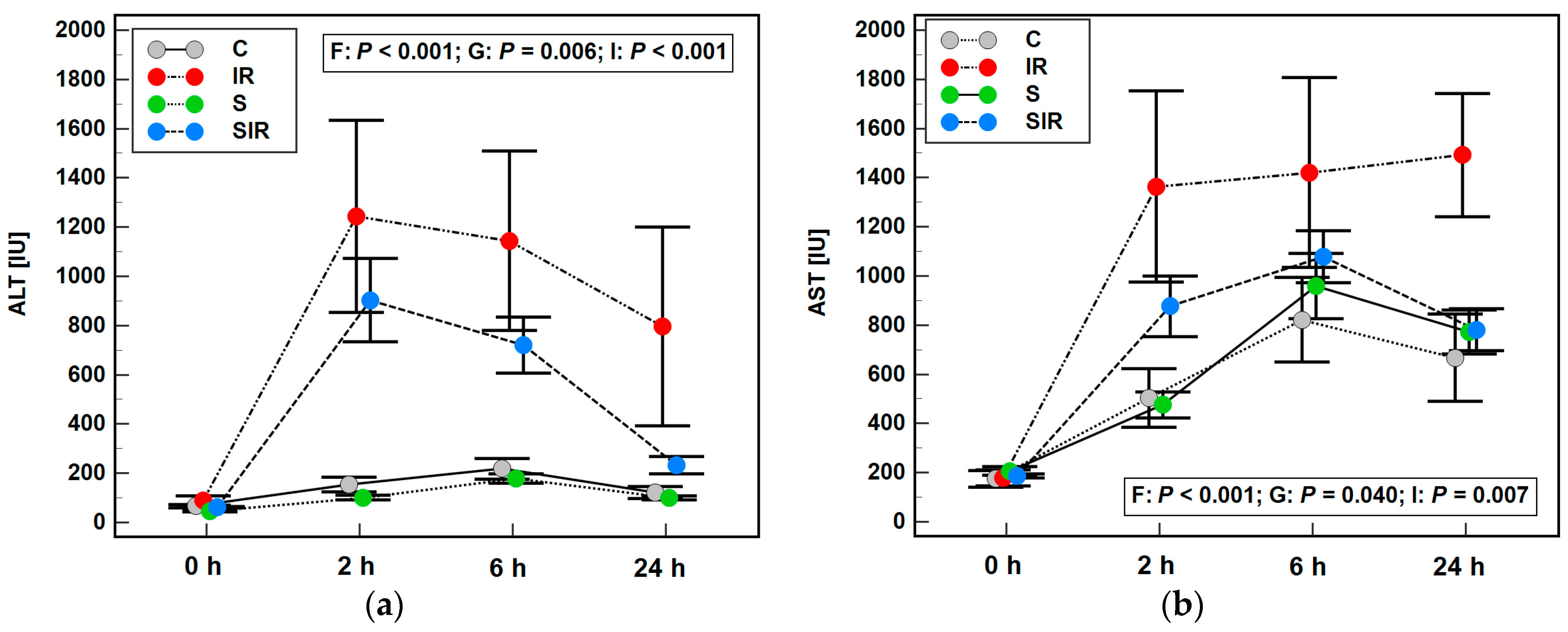
2.1. A Rat Model of IR and Its Validation

A rat IR model has previously been established [15,16], in which 36 animals were randomized into four groups: sham-operated (controls; n = 9), sham-operated following pretreatment with sitagliptin (5 mg/kg p.o.) (sitagliptin; n = 8), subjected to IR procedure (IR; n = 9), and subjected to IR procedure following pretreatment with sitagliptin (SIR; n = 10). It was validated by an elevation in the activity of alanine (ALT) and aspartate (AST) aminotransferases following injury, less pronounced in SIR than the IR group. The repeated-measures ANOVA indicated significant effect of factor (time of blood collection: 0, 2, 6, and 24 h), group (control, IR, sitagliptin, SIR) as well as factor × group interaction on ALT and AST dynamics (Figure 1). In addition, histopathological analysis indicated slight necrotic changes as well as neutrophil infiltration in IR and SIR animals and a significantly higher degree of steatosis in animals pretreated with sitagliptin [16] (Supplementary Figures S1–S4).

2.2. Effect of IR Injury and Sitagliptin on Liver Profile of Eicosanoids

The profile of eicosanoids was determined using liquid chromatography-tandem mass spectrometry (LC-MS/MS).
The concentrations of 6-ketoPGF (PGI2), PGE2, and 13,14-dihydro-PGE1 in the liver were significantly upregulated in the IR group as compared to controls and IR animals pretreated with sitagliptin (SIR group). In addition, 6-ketoPGF (PGI2), but not PGE2 and 13,14-dihydro-PGE1, were also upregulated in the sitagliptin group as compared to controls and SIR animals (Table 1).
Sitagliptin significantly upregulated PGD2, which was higher than in controls, IR, and SIR animals. It also upregulated 15-deoxy-12,14-PGJ2, which was elevated in sitagliptin and SIR groups as compared to controls and IR animals (Table 1).
Liver concentrations of PGF2, TBX2, and LTB4 were affected neither by IR nor sitagliptin. The PGI2-to-TXA4 ratio, calculated based on their stable metabolites (6-ketoPGF/TXB2), was higher in IR as compared to controls and SIR, but sitagliptin alone also elevated the ratio as compared to control animals (Table 1).

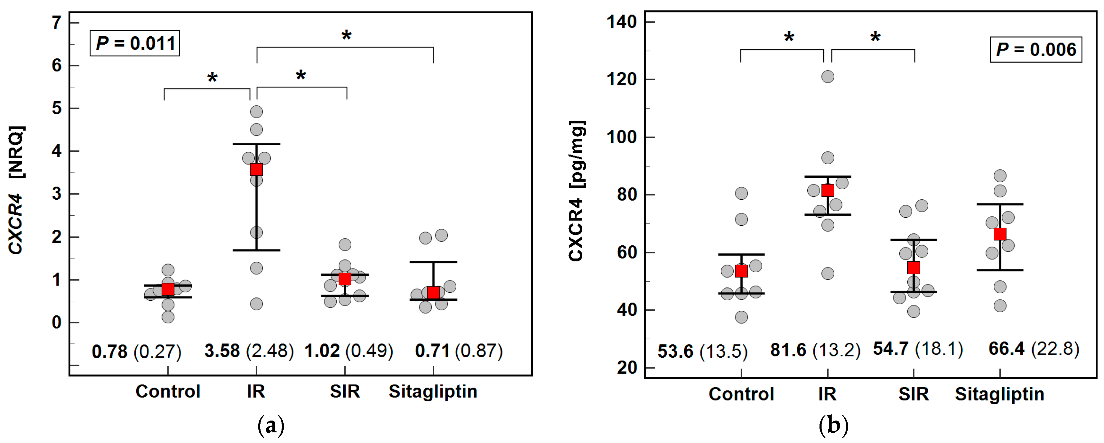
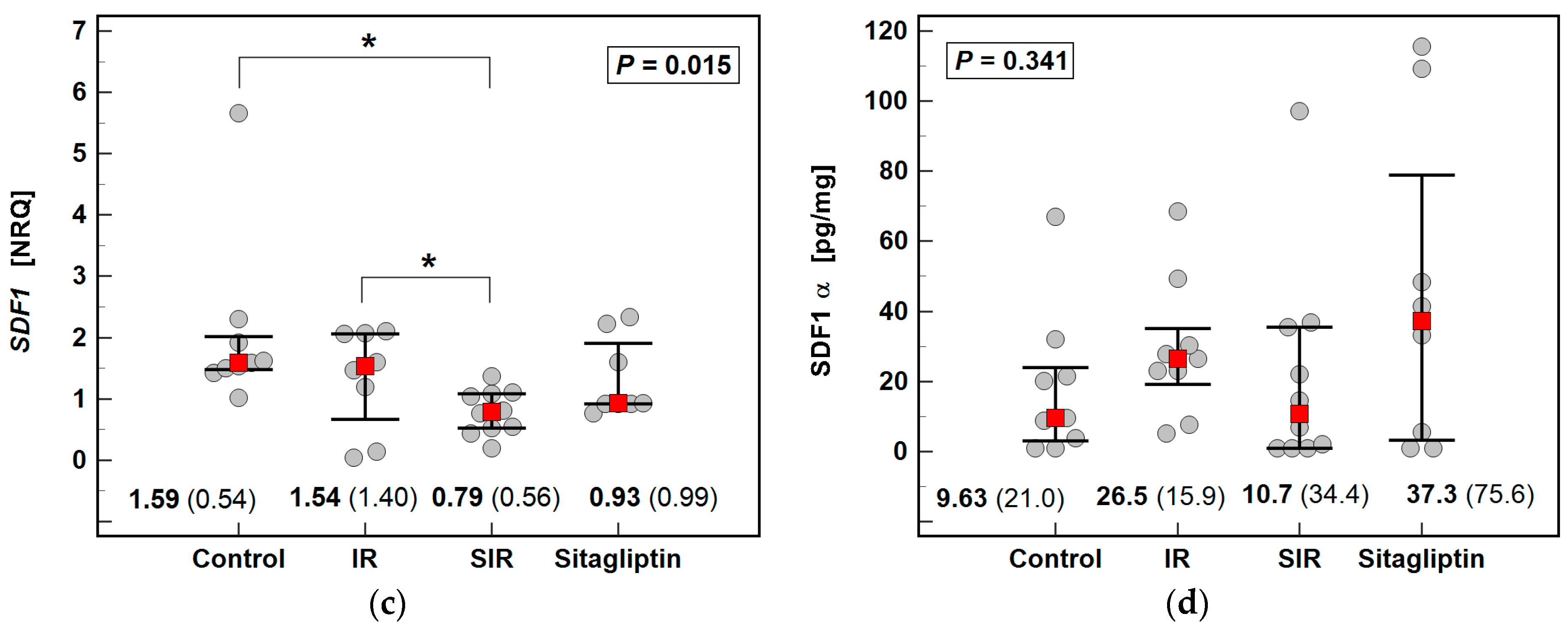
2.3. Effect of IR Injury and Sitagliptin on Liver Expression of CXCR4/SDF1

The expression and concentration of SDF1 cytokine and CXCR4, its receptor, were determined using reversely transcribed quantitative polymerase chain reaction (RTqPCR) and immunoenzymatic assays (ELISA).
The IR significantly upregulated CXCR4, both at mRNA and protein level, and pretreatment with sitagliptin downregulated its expression and concentration. Regarding the CXCR4 ligand SDF1, its expression (Sdf1) was significantly lower in the SIR group as compared to IR and controls. Neither IR nor sitagliptin had a significant impact on SDF1α (Figure 2).

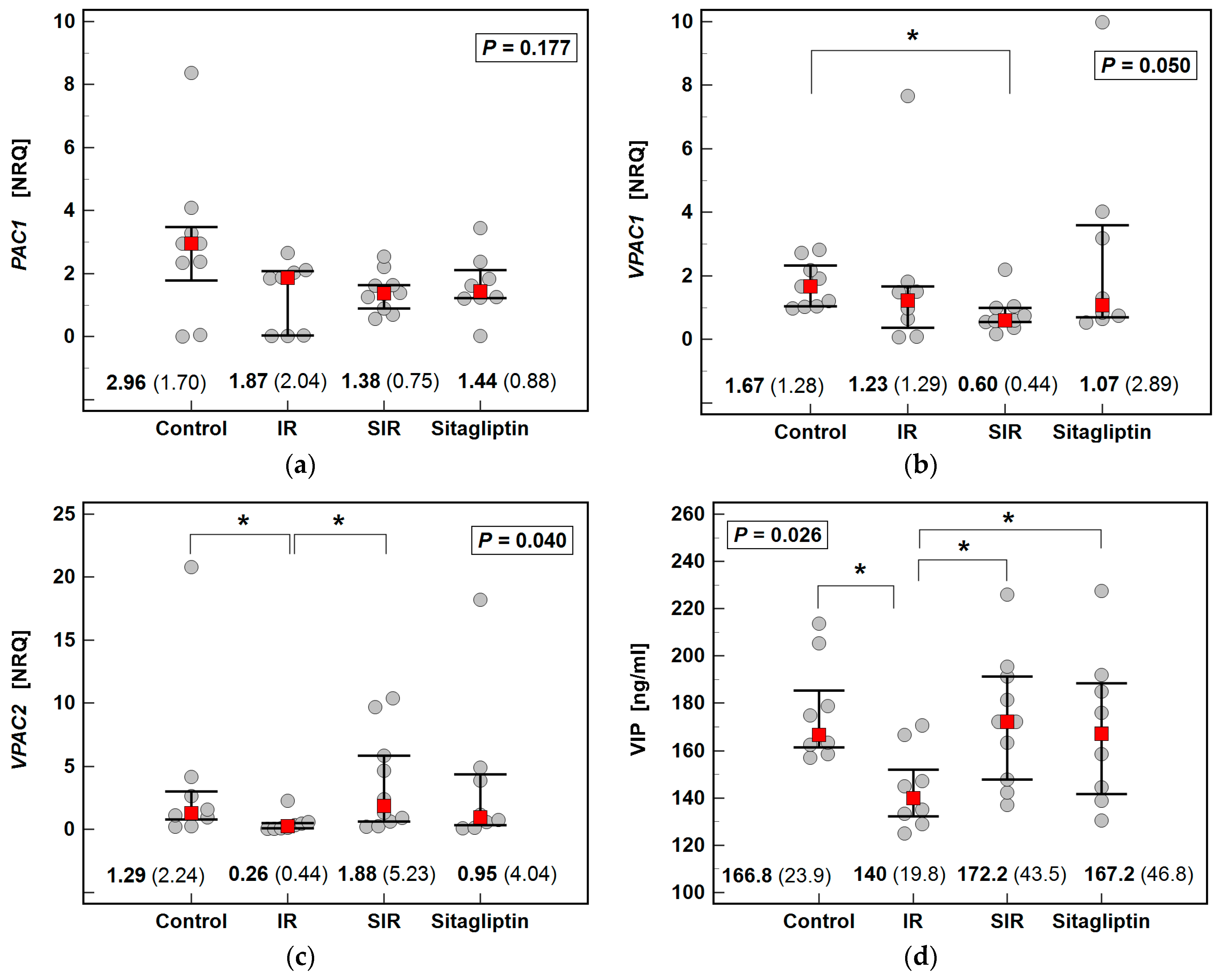
2.4. Effect of IR Injury and Sitagliptin on Liver Expression of PAC1, VPAC1, VPAC2/VIP

The IR significantly downregulated the expression of Vpac2 and concentration of VIP while pretreatment with sitagliptin prevented the downregulation of both the receptor expression and ligand concentration. The IR non-significantly downregulated Pac1 and Vpac1, the expression of which was more markedly downregulated in SIR animals, significantly so in the case of Vpac1 expression (Figure 3).

2.5. Effect of IR Injury and Sitagliptin on Liver Expression of GLP1R/GLP1

Neither IR nor sitagliptin had a significant impact on Glp1r expression and GLP1 concentration in the liver (Figure 4).

2.6. Relationship between Eicosanoids, DPP4 Ligands, and Their Receptors, and Mediators of Inflammation and Oxidative, Nitrosative, and Halogenative Stress (Univariate Analysis)

Liver eicosanoids as well as investigated DPP4 ligands and their receptors were related to the expression or concentration of mediators of inflammation and oxidative, nitrosative and halogenative stress: IL-1β, IL-10, IFNγ, VEGF-A, MIP-2, TNFα, Il6, Mmp1, Nampt, Tnfa, Nox1, Nox2, Nox4, Mdk, Ptn, 3-nitrotyrosine (NT), and 3-bromotyrosine (BT).

2.6.1. Eicosanoids

In univariate analysis, 6-ketoPGF (PGI2) correlated with the concentration of 13,14-dihydro-PGE1 (r = 0.36, p = 0.031), LBT4 (r = 0.35, p = 0.035), PGD2 (r = 0.45, p = 0.006), PGE2 (r = 0.61, p < 0.001), CXCR4 (r = 0.55, p < 0.001), 3-NT (r = 0.38, p = 0.021), SDF1α (r = 0.40, p = 0.016), VIP (ρ = −0.51, p = 0.002), and IL-1β (r = 0.33, p = 0.049) and the expression of Nox4 (r = −0.67, p < 0.001), Ptn (r = −0.38, p = 0.023), Sdf1 (r = −0.44, p = 0.009), Vpac2 (r = −0.38, p = 0.026), Vpac1 (r = −0.34, p = 0.045), and Cxcr4 (r = 0.48, p = 0.003).
Apart from 6-ketoPGF (PGI2), the concentration of PGE2 corelated with LBT4 (r = 0.64, p < 0.001), PGD2 (r = 0.55, p < 0.001), PGF2 (r = 0.69, p < 0.001), CXCR4 (r = 0.34, p = 0.043), SDF1α (r = 0.54, p < 0.001), and VIP (r = −0.42, p = 0.010) and the expression of Nox4 (r = −0.42, p = 0.012) and Cxcr4 (r = 0.43, p = 0.011).
Apart from PGE2, the concentration of PGF2 correlated with these of LTB4 (r = 0.42, p = 0.010), TXB2 (r = 0.59, p < 0.001), PGD2 (r = 0.36, p = 0.033), and SDF1α (r = 0.41, p = 0.012).
In addition to 6-ketoPGF (PGI2), PGE2, and PGF2, the concentration of PGD2 corelated with 15-deoxy-12,14-PGJ2 (r = 0.56, p < 0.001), LBT4 (r = 0.59, p < 0.001), and SDF1α (r = 0.59, p < 0.001).
Apart from PGD2, the concentration of 15-deoxy-12,14-PGJ2 correlated with that of LTB4 (r = 0.44, p = 0.009).
The 13,14-dihydro-PGE1 correlated solely with 6-ketoPGF (PGI2).
Apart from PGF2, the concentration of TXB2 correlated with the expression of Nox2 (r = −0.36, p = 0.035).
In addition to prostanoids: 15-deoxy-12,14-PGJ2, PGI2, PGD2, PGE2, and PGF2, the concentration of LBT4 correlated with that of SDF1α (r = 0.59, p < 0.001).

2.6.2. CXCR4/SDF1

The concentration of CXCR4 correlated with that of PGE2 and 6-ketoPGF (PGI2) and the expression of Cxcr4 (r = 0.48, p = 0.004) and Vpac2 (r = −0.40, p = 0.019). The receptor expression (Cxcr4) correlated with the expression of Nox4 (r = −0.34, p = 0.044) and Il6 (r = 0.40, p = 0.018) and GLP1 concentration (r = 0.34, p = 0.046), in addition to CXCR4 protein and the concentrations of 6-ketoPGF (PGI2) and PGE2.
The concentration of SDF1α correlated with that of 6-ketoPGF (PGI2), PGD2, PGE2, PGF2, and LTB4. Ligand expression (Sdf1), in turn, correlated with the concentration of 6-ketoPGF (PGI2) and IL-1β (r = −0.36, p = 0.034) and the expression of Mdk (r = 0.41, p = 0.014), Ptn (r = 0.70, p < 0.001), Nox1 (r = 0.38, p = 0.027), Nox4 (r = 0.53, p = 0.001), Vpac1 (r = 0.79, p < 0.001), Mmp1 (r = 0.51, p = 0.002), and Nampt (r = 0.68, p < 0.001).

2.6.3. PAC1, VPAC1, VPAC2/VIP

The expression of Vpac1 correlated with 6-ketoPGF (PGI2) concentration and the expression of Sdf1, Glp1r (r = 0.69, p < 0.001), Mdk (r = 0.79, p < 0.001), Ptn (r = 0.87, p < 0.001), Nox1 (r = 0.73, p < 0.001), Nox2 (r = −0.43, p = 0.009), Mmp1 (r = 0.80, p < 0.001), Nampt (r = 0.53, p = 0.001), and Tnfa (r = 0.37, p = 0.029).
The expression of Vpac2 correlated with liver concentrations of CXCR4, 6-ketoPGF (PGI2), and GLP1 (r = −0.37, p = 0.030), whereas the expression of Pac1 did not show any significant correlation.
The concentration of VIP correlated with 6-ketoPGF (PGI2), PGE2, and 3-BT (r = −0.33, p = 0.049) and the expression of Nox4 (r = 0.40, p = 0.017).

2.6.4. GLP1R/GLP1

The expression of Glp1r correlated with these of Vpac1, Mdk (r = 0.84, p < 0.001), Ptn (r = 0.68, p < 0.001), Nox1 (r = 0.90, p < 0.001), Nox2 (r = −0.43, p = 0.011), Il6 (r = 0.47, p = 0.004), Tnfa (r = 0.71, p < 0.001), and Mmp1 (r = 0.83, p < 0.001).
GLP1 correlated with IFNγ (r = −0.37, p = 0.025) in addition to Cxcr4 and Vpac2 expression.

2.7. Independent Predictors of Liver Eicosanoids and DPP4 Ligands and Their Receptors (Multivariate Analysis)

Multiple linear regression analysis (stepwise method) was conducted to discern independent predictors of variability in the concentration of individual eicosanoids as well as concentration/expression of investigated DPP4 ligands and their receptors.

2.7.1. Independent Predictors of Liver Eicosanoids

Of the variables found significantly associated in univariate analysis, 13,14-dihydro-PGE1, PGE2, 3-NT, VIP, IL-1β, Cxcr4, and Nox4 were independent predictors of 6-ketoPGF (PGI2), explaining 83% variability in its concentration.
6-ketoPGF (PGI2), PGF2, and LTB4 were independent predictors of PGE2, explaining 74% of variability, and PGE2 and TXB2 were independent predictors of PGF2, explaining 61% in its variability. PGF2 was independently associated with TXB2, explaining 28% in its variability, and 15-deoxy-12,14-PGJ2, PGE2, and SDF1α were independent predictors of LTB4, explaining 62% in its variability.
The IR was a sole predictor of 13,14-dihydro-PGE1, which explained 16% in concentration variability. Sitagliptin was an independent predictor of 15-deoxy-12,14-PGJ2, which, together with PGD2, explained 45% in concentration variability and of PGD2, for which it explained 73% of variability together with 15-deoxy-12,14-PGJ2, PGE2, and SDF1α (Table 2).

2.7.2. Independent Predictors of Liver Expression of DPP4 Ligands and Their Receptors

The IR was an independent predictor of the concentration of CXCR4, explaining 39% of its variability together with 6-ketoPGF (PGI2) and the concentration of VIP, explaining 39% of its variability together with PGE2 and 3-BT. Together with IFNγ, it also explained 32% of the variability in GLP1 concentration. The IR was a sole predictor of Cxcr4 and Vpac2 expression, explaining, respectively, 38% and 23% in their variability (Table 3).
The variability in SDF1α concentration was explained in 44% by LTB4 and PGD2, while that in Sdf1 expression in 84% by changes in expression of Vpac1, Mdk, Nox4, and Nampt (Table 3).
The variability in the expression of the Vpac1 receptor was explained in 91% by changes in Mdk, Nox2, Ptn, and Sdf1 expression and this in Glp1r in 83% by changes in Nox1 and Nox2 expression (Table 3).

3. Discussion

Deciphering molecular mechanisms of hepatic IR injury is a prerequisite to developing successful treatment strategies that would improve patients’ survival by reducing rates of graft rejection and improving the function of transplanted organs [2]. The ATP depletion in sinusoidal endothelial cells and hepatocytes caused by ischemia leads to the generation of reactive oxygen species (ROS), cell death, and release of alarmins, activating resident neutrophils and Kupfer cells. Released ROS and cytokines damage macromolecules and recruit circulating immune cells as well as hepatic stellate cells, engaging them in self-perpetuating inflammation and oxidative stress [2]. Therefore, targeting oxidative stress, inflammation, and preventing apoptosis are viewed as promising therapeutic options.
Prostanoids are potent mediators of immune and inflammatory responses. Under physiological conditions, prostanoids maintain homeostasis and are hepatoprotective. However, both their depletion and excess, occurring as a consequence of COX-2 activation in response to insult, might be detrimental [28]. In the IR settings accompanied by reduction in prostanoids’ concentration, a treatment with prostaglandins seems to be beneficial due to improved liver hemodynamics and survival of sinusoidal epithelial cells and reduced generation of ROS, leukocyte migration, and platelet aggregation (reviewed in the work of [29]). However, the efficacy of none of the tested prostaglandins, that is, PGE1, PGE2, or PGI2, has been confirmed in clinical trials [2]. Moreover, COX2 has been ascribed both a protective [30] and a critical enabling role in hepatic IR injury [6]. Adding to the confusion, COX2 inhibition [7] or inhibition of any other enzyme involved in the synthesis of eicosanoids [7,8,9] have proved beneficial.
In the IR injury model investigated here, hepatic concentrations of 13,14-dihydro-PGE1, 6-keto-PGF, and PGE2 were elevated in IR as compared to sham-operated animals while these of PGD2, its metabolite 15-deoxy-12,14-PGJ2, PGF2, TXB4, and LTB4 were not significantly altered. Similar IR-induced alterations in the profile of prostanoids were reported in the heart by Qiu et al. [31] and demonstrated to contribute to cardiac apoptosis. The authors have observed the upregulation of 6-keto-PGF, PGE2, PGD2 and no change in TXB2 and PGF (13,14-dihydro-PGE1, 15-deoxy-12,14-PGJ2, and LTB4 were not determined). The upregulation of PGI2 and an elevation of PGI2/TXA2 ratio have also characterized rat hippocamps in the course of cerebral IR injury but have been interpreted as a physiological mechanism aimed at neuroprotection [32].
The multivariate analysis conducted in the present study demonstrated that IR was an independent predictor of solely 13,14-dihydro-PGE1. However, 13,14-dihydro-PGE1 was, in turn, an independent predictor of 6-ketoPGF (PGI2), which explained variability in PGE2, a contributor to the variability in PGF2, PGI2, PGD2, and LTB4, stressing strong interrelationships between all studied eicosanoids. The stimulatory effect of inflammation and oxidative stress on their synthesis was indicated by the inclusion of IL-1β and 3-NT, an oxidative/nitrosative stress marker, in the regression models predicting prostacyclin 6-ketoPGF (PGI2) concentration as well as by including SDF-1α or CXCR4 in the regression models explaining the variability in 6-ketoPGF (PGI2), PGD2, and LTB4 concentration.
There is an increasing interest in repurposing the well-established drugs with already known toxicity, pharmacokinetics, and pharmacodynamics [33], including repositioning gliptins for IR injury. The contribution of antioxidant, anti-inflammatory, and antiapoptotic properties of sitagliptin to liver protection during IR injury has been well documented [13,14,15,16,17,18]. However, the drug effect on the liver profile of eicosanoids during IR has not been investigated, although sitagliptin has been demonstrated to reduce both PLA2 and COX2 concentration in various clinical and experimental settings [34,35]. Consistently, sitagliptin pretreatment of IR animals prevented significant elevation of 6-ketoPGF (PGI2), PGE2, and 13,14-dihydro-PGE1 (PGE1) and reduced 6-ketoPGF (PGI2)/TXB2 (TXA2) ratio, elevated in IR. Interestingly, sitagliptin in control sham-operated animals had an opposite effect; it elevated the expression of 6-ketoPGF (PGI2) and 6-ketoPGF (PGI2)/TXB2 (TXA2) ratio, indicating that the biological effect of the drug is context-dependent.
Sitagliptin, but not IR, was responsible for an elevation of PGD2 and 15-deoxy-12,14-PGJ2 (PGD2), significant solely in sitagliptin-pretreated control animals (PGD2) or in both control and IR animals in (PGD2 metabolite). The unfavorable proinflammatory character of PGD2 is manifested mostly in the lung and in allergic and autoimmune diseases, while in the liver, PGD2 has only recently been shown to alleviate the injury during IR [36]. Specifically, PGD2 metabolite 15-deoxy-Δ12,14-prostaglandin J2 exerted hepatoprotective effects by alleviating oxidative stress via activation of Nrf2, reducing systemic inflammation and liver infiltration with macrophages, decreasing apoptotic rates, and by downregulating an expression of beclin-1 and LC3, autophagy markers. Activation of Nrf2 signaling in IR liver in response to sitagliptin without exploring the mechanism has previously been noted by Abdel-Gaber et al. [14]. Sitagliptin has also been shown to alleviate hepatic fibrosis by mechanisms involving the enhancement of Nrf2 expression accompanied by the downregulation of NFκB [37]. Therefore, our observation on sitagliptin-mediated upregulation of PGD2 and its metabolite in the context of its ability to induce Nrf2 signaling might shed some light on the molecular mechanism involved. PGD2 might also mediate, recently reported [38], sitagliptin-induced downregulation of LC3 and beclin-1 in endothelial cells.
The number of studies on gliptins as attenuators of IR injury, which would explore possible mechanisms in addition to documenting drug anti-inflammatory and antioxidant properties, is limited. Sitagliptin, a competitive inhibitor of DPP4 also found to downregulate the enzyme expression [18], exerts its antidiabetic activity by preserving GLP-1, a peptide hormone synthesized by gut L cells [39]. GLP-1 and its analogs display antiapoptotic, anti-inflammatory, and antioxidant activities conferring protection during myocardial [40] and cerebral [41] IR. Consistently, sitagliptin attenuated intestinal IR injury by inducing GLP-1/GLP-1R signaling [26] and renal IR injury by upregulating GLP-1 secretion and local GLP-1R expression [23,42]. In the liver, GLP-1/GLP-1R has been speculated to mediate the upregulation of Nrf2 expression and subsequent elevation in heme oxygenase-1 [14]. Though, neither GLP-1R nor GLP-1 status in IR liver pretreated with sitagliptin has been investigated by the authors [14]. The speculation was based on, reported elsewhere [43], hepatoprotection conferred by activation of hepatic GLP-1R by receptor agonists. Cytoprotective effects of GLP-1 agonists have also been demonstrated in ischemic stroke [44] and myocardial infarct [45]. However, contrary to other organs, the presence of GLP-1R in the liver is controversial [46,47]. In the present study, GLP-1R mRNA expression was undetectable or borderline detectable in most of the examined livers, with six out of nine measurable cases (Cq < 40) present in sitagliptin-treated animals. The GLP-1 concentration in the liver did not differ significantly between analyzed groups, although a non-significant peptide upregulation in IR animals could be noted. Still, IR was an independent predictor of GLP-1, regardless of sitagliptin pretreatment, exerting a positive effect on hormone concentration. The local concentration of IFNγ, in turn, was a negative predictor of hepatic GLP-1, corroborating previous observations on an inverse relationship between the hormone and IFNγ signaling (reviewed in the work of [34]) and IFNγ involvement in hepatic IR injury [48].
To elucidate potential mechanisms of sitagliptin action in hepatic IR injury, we examined the drug effect on other potential DPP4 substrates, namely VIP and SDFα, and on their receptors. The VIP is a ubiquitous peptide hormone with anti-inflammatory and antioxidant properties, employing mainly VCAP1 and VCAP2 receptors. It can also interact with PACAP receptor (PAC1), although with lower affinity [49]. The VIP has been demonstrated to attenuate IR injury in mice liver [50] and rat lungs [51] and to protect rat kidneys from hemorrhagic ischemia and retransfusion [52]. Specifically, VIP has reduced liver infiltration with macrophages and neutrophils, increased concentration of anti-inflammatory IL-10, and decreased hepatocyte apoptotic rates while downregulating the expression of macrophage-derived TNFα, IL-6, and IL-12 [50]. However, whereas Ji et al. [50] observed an elevation in hepatic VIP mRNA expression in response to IR, our results indicate the downregulation of peptide concentration, which was avoided by sitagliptin preconditioning. Our finding is consistent with accelerated peptide degradation owing to upregulated DPP4 expression in response to hypoxia (reviewed in the work of [53]). Moreover, it also agrees well with an observation made in lung IR injury on the DPP4 inhibition upregulating both VIP concentration and expression [51]. Supporting an inverse relation between VIP and IR injury, peptide concentration in the present study was inversely related to both inflammation and oxidative stress. PGE2 and 3-BT concentrations were negative independent predictors of VIP concentration in addition to IR. In turn, VIP was an independent negative predictor of 6-ketoPGF1α, a stable metabolite of PGI2. The IR downregulated, and sitagliptin preconditioning also upregulated the expression of Vcap2, while Vcap1 expression was lower in all groups as compared to controls, significantly so in SIR. The IR was a sole, negative predictor of Vcap2 expression while that of Vcap1 was independently associated with oxidative stress marker Nox2 as a negative predictor and with Ptn, Mdk, and Sdf1 as positive predictors. Pleiotrophin (Ptn) and midkine (Mdk) are closely related to heparin-binding growth factors found to support liver regeneration [54] and protect against metal-induced liver toxicity [55] but being oppositely regulated in hepatic IR [17]. Moreover, pleiotrophin has been shown to play a cytoprotective role in brain and heart IR [56,57]. Midkine role during IR injury seems to depend on the affected organ as the cytokine exerted antiapoptotic, thus protective activity in the heart [58] but contributed to organ damage in the renal and hindlimb models of ischemia by promoting macrophage and neutrophil infiltration [59,60].
Instead of activation of GLP-1/GLP-1R, the upregulated Nrf2 expression in the liver subjected to IR injury observed by Abdel-Gaber et al. [14] might be attributed to increased stability of SDF-1α. In lung IR injury models, SDF-1α activates Nrf2 by triggering MAPK and PI3K/Akt pathways, the latter being also involved in SDF1α-mediated interference with NFκB signaling (reviewed in the work of [61]). However, as demonstrated here, hepatic IR had no effect on Sdf1 expression, and it elevated SDF-1α concentration only non-significantly. This observation opposes the notion of increased expression of SDF-1α upon acute and chronic liver injury, where the cytokine can contribute to both liver regeneration and fibrosis. Those contradictory roles of SDF-1α are believed to be executed by signaling via different receptors: pro-regenerative CXCR7 or profibrotic CXCR4 (reviewed in the work of [62]). Nonetheless, a known stimulatory effect of hypoxia and reactive oxygen species on SDF1α and CXCR4 [63] was, in the case of Sdf1, reflected by Nampt and Nox4 being independent positive predictors of cytokine expression. In the case of CXCR4, receptor expression and concentration were significantly upregulated in IR animals, and the IR was an independent predictor of both Cxcr4 and CXCR4. Receptor overexpression has been detrimental in animal models of renal [64] and cardiac IR [65], in which it induces senescence in progenitor epithelial cells [64] or recruited inflammatory cells and increased production of inflammatory cytokines while inducing apoptosis in cardiomyocytes [65]. Antioxidants [66] and CXCR4 inhibitors [67] have been shown to prevent stress-induced receptor upregulation. In the liver, plerixafor, a specific receptor inhibitor, improves recovery of the liver following IR injury by restoring the proliferative potential of hepatocytes, hampered by CXCR4 signaling [67]. Inhibiting CXCR4 has proven effective also in attenuating oxidative stress-induced podocyte injury and renal fibrosis associated with activation of the SDF-1α/CXCR4 axis [68]. In the present study, the IR animals that underwent sitagliptin preconditioning had significantly lower expression of Sdf1 and Cxcr4 as well as lower receptor concentration than animals subjected to IR procedure without preconditioning. This observation is in line with an unfavorable role of SDF-1α/CXCR4 signaling in the liver [67], kidney [64,68], heart [65], or PC12 cells [66]. However, it opposes the response of endothelial [69] and progenitor endothelial cells to sitagliptin [70], in which the drug induced healing by employing SDF-1α/CXCR4 signaling to increase the proliferative, migratory, and angiogenic potential of cells. In fact, the role of the SDF-1α/CXCR4 axis in inflammatory diseases is rather ambiguous, as indicated by both beneficial and detrimental consequences of pathway inhibition [71].

4. Materials and Methods

4.1. Experimental Setting

The current study uses biobanked livers (stored at −80 °C), collected during the original experiment [15,16], which was conducted as briefly described below. The experiment was carried out on healthy animals to eliminate the harmful impact of diabetes on IR injury.

4.1.1. Animals

Male Wistar rats, 2–3 months old, were housed under standard conditions (12:12 h day/night cycle, stable temperature of 19–21 °C, humidity of 45–60%, and continuous ventilation) with free access to standard food and water.

4.1.2. Chemicals

Sitagliptin (Januvia—tablets 100 mg) was purchased from MSD (Warsaw, Poland), heparin (Heparinum WZF—ampoules 25,000 U/5 mL) from Polfa Warszawa (Warsaw, Poland), ketamine hydrochloride (Bio-ketan) from Vetoquinol Biowet (Gorzów Wlkp, Poland), medetomidine hydrochloride (Domitor, ampoules 1 mg/mL) from Orion Pharma (Warsaw, Poland), butorphanol tartrate (Morphasol, ampoules 4 mg/mL) from aniMedica GmbH (Frankfurt am Main, Germany), Ringer solution from Polfa Lublin S.A. (Lublin, Poland), and solution of 0.9% sodium chloride was obtained from Polpharma S.A. (Starogard Gdański, Poland).

4.1.3. The IR Procedure

After a handling period of 2–3 weeks, the rats were randomly divided into four groups. Two groups were sham-operated: the group without drug delivery (controls; n = 9) and the group with sitagliptin (5 mg/kg p.o.) administered intragastrically two weeks prior to surgery once a day (sitagliptin, n = 8). The remaining two groups were subjected to the IR procedure: one group without prior drug administration (IR, n = 9) and one group where sitagliptin was administered in the same scheme as in the sitagliptin group (SIR, n = 10).
Before laparotomy, animals were anesthetized by the intramuscular administration of ketamine hydrochloride (7 mg/kg), medetomidine hydrochloride (0.1 mg/kg), and butorphanol tartrate (2 mg/kg). Ischemia of 70% of the liver (intermediate lobe and left lateral lobe) was induced as described previously [15,16] by placing a microvascular clamp over the portal vein and hepatic artery. After 60 min of ischemia, the microclips were removed, allowing reperfusion for 24 h. Animals in the non-ischemic groups underwent a sham surgery in which the blood vessels were isolated but not clamped. At the end of the experiment, the ischemic liver lobes were isolated and the fragments placed in a solution of RNAlater (Qiagen, Hilden, Germany) or frozen and stored at −80 °C for metabolomic and immunoenzymatic analyses.
During reperfusion, the activity of aminotransferases, as markers of hepatocyte injury, was determined in rat sera by a certified laboratory using commercially available enzymatic methods and, after the surgical procedure, a histological evaluation was performed under a light microscope [15,16].

4.2. Analytical Methods

4.2.1. Metabolomic Analysis of Eicosanoids

Chemicals and Reagents

Methanol, acetonitrile (ACN), ethyl acetate, water, formic acid (FA) were acquired from Merck Millipore (Warsaw, Poland). Standards of Thromboxane B2, Leukotriene B4, Prostaglandin D2, Prostaglandin E2, 6-keto Prostaglandin F1α, Prostaglandin F2α, 15-deoxy-Δ12,14-Prostaglandin J2, 13,14-dihydro Prostaglandin E1, and their isotope-labeled standards were procured from Cayman Chemical Company (Ann Arbor, MI, USA).

Sample Preparation

Tissue samples (~0.5 g) were homogenized using ceramic beads in 1 mL of LC-MS-grade water in Bead Ruptor Elite homogenizer (Omni International, Kennesaw, GA, USA). Aliquotes of 100 µL of homogenates were mixed with 10 µL of internal standard and 20 μL of 0.2% FA in water. Samples were deproteinized and extracted with an ACN mixture with ethyl acetate. The obtained supernatants were evaporated to dryness and re-dissolved before analysis in 20% ACN in water.

LC-MS Analysis

LC-MS data were obtained using Acquity UPLC system (Waters, Milford, MA, USA), equipped with a single quadrupole-time of flight mass analyzer and an electrospray (ESI) ion source (Xevo G2 Q-TOF MS from Waters). Spectra were obtained in negative ionization mode with the following MS parameters: the sprayer voltage and the desolvation temperature were set at 2.0 kV and 450 °C, respectively. All scans were carried out in an MS/MS QTOF scan mode. Data acquisition and calculations were performed with MassLynx and QuanLynx software (Waters), respectively.
Analytes were separated using Acquity UPLC BEH Shield C18 1.7 µm chromatographic column (100 × 2.1 mm, 1.70 µm) from Waters with a linear gradient from 30% to 95% of mobile phase B in 7.2 min with a total flow rate of 250 µL/min. As mobile phases, 0.1% FA in water (A) and 0.1% FA in ACN (B) were used.

4.2.2. Immunoassays

Rat livers (~0.5 g) were homogenized using ceramic lysing matrix beads in the Bead Ruptor Elite bead mill homogenizer (Omni International) with 0.5 mL of PBS buffer pH 7.4 (137 mM NaCl, 2.7 mM KCl, 10 mM Na2HPO4, and 1.8 mM KH2PO4) with 1 mM PMSF. Obtained homogenates were centrifuged (12,000× g, 10 min, 4 °C), and supernatants were collected, aliquoted, and stored at −80 °C until analysis.
Prior to analysis, tissue homogenates were clarified by centrifugation at 10,000× g, 10 min, 4 °C.
The concentrations of GLP-1 (glucagon-like peptide-1) and VIP (vasoactive intestinal peptide) were determined using RayBio® Rat Enzyme Immunoassay Kits (#EIAR-GLP1 and #EIAR-VIP, respectively) from RayBiotech Life (Peachtree Corners, GA, USA) according to the manufacturer’s instructions. Samples were diluted at 1:50 and tested in duplicates. Standard curves were drawn using 5-parameter logistic (PL) regression.
The C-X-C chemokine receptor type 4 (CXCR4) and C-X-C motif chemokine 12 (CXCL12/SDF1α) were determined using, respectively, Nori® Rat CXCR4 and Nori® Rat CXCL12 ELISA Kits (Genorise Scientific Inc., Glen Mills, PA, USA) following manufacturer’s instructions. Samples were diluted 1:1 in Assay Buffer and tested in duplicates. The standard curves were calculated using a computer-generated 4-parameter logistic (PL) regression.

4.2.3. Protein Determination

Protein concentration was determined using Coomassie Plus (Bradford) Assay Kit (Thermo Scientific, Waltham, MA, USA) against the BSA standard curve according to the manufacturer’s instructions. Samples were diluted in PBS and tested in duplicates.
Concentrations of eicosanoids and peptides/proteins were adjusted to protein content in the sample and expressed per mg of protein.

4.2.4. Transcriptional Analysis

A cDNA library has been prepared from isolated RNA as described in the original study [15,16] and used for the present investigation.
All qPCR reactions were conducted in triplicates using the CFX96 platform (Biorad, Hercules, CA, USA) under standardized thermal cycling conditions: activation of the polymerase for 110 s at 95 °C followed by 40 cycles of denaturation (95 °C for 5 s) and annealing and synthesis (61.4 °C for 5 s). Melting curve analysis (60–95 °C, reading every 0.5 °C) was conducted to confirm the specificity of the reaction product. The qPCR mixture consisted of 2 µL of diluted 1:5 cDNA, 10 µL of 2× SsoFast EvaGreen® Supermix (BioRad), 1 µL of each 10 nM forward and reverse target-specific primers, filled with water up to 20 µL. Primers were synthesized by Genomed (Warsaw, Poland) (Table 4). The relative gene expression was calculated as follows: geometric mean of all Cq values was subtracted from individual sample Cq (ΔCq), linearized by 2ΔCq conversion, and normalized to Gapdh expression. The resulting values are referred to as normalized relative quantities (NRQ) [72].

4.2.5. Inflammatory Mediators and Markers of Oxidative, Nitrosative, and Halogenative Stress

Data on inflammatory mediators and markers of oxidative and halogenative stress were retrieved from our earlier published studies [16,17] for the purpose of correlation analysis.

4.3. Statistical Analysis

Data were analyzed using MedCalc® Statistical Software version 20.014 (MedCalc Software Ltd., Ostend, Belgium). Normality of data distribution and homogeneity of variances was established with Kolmogorov–Smirnov and Leven tests, respectively. Between-group comparisons were conducted using the Kruskal–Wallis H test with Conover post-hoc test, and results are presented as medians with interquartile range (IQR). Correlation analysis was conducted using Pearson correlation on log-transformed data, if appropriate. Multiple linear regression, a stepwise method, was applied to discern independent predictors of explained variables with explanatory variables entered into the model if p < 0.05 and removed if p > 0.1. The VIF (variance inflation factor) was calculated for each predictor variable to diagnose multilinearity. The repeated-measures ANOVA was conducted to discern IR and sitagliptin effect on the dynamics of liver enzymes activities. All calculated probabilities were two-tailed. The p values ≤ 0.05 were considered statistically significant.

5. Conclusions

In the present study, we demonstrated that hepatic IR is associated with an increase in tissue concentrations of 6-ketoPGF (PGI2), PGE2, and 13,14-dihydro-PGE1 and with an elevated 6-ketoPGF (PGI2)/TXB2 (TXA2) ratio, which was abrogated by pretreatment of IR animals with sitagliptin. As the drug has an opposite effect regarding 6-ketoPGF (PGI2) and 6-ketoPGF (PGI2)/TXB2 (TXA2) ratio in control sham-operated animals, it might indicate that the biological effect of sitagliptin is context-dependent. We also showed that sitagliptin upregulated PGD2 and 15-deoxy-12,14-PGJ2, which seems to be beneficial in the light of the recently reported hepatoprotective effect of the prostaglandin in the course of ischemia and reperfusion.
In order to shed some light on molecular mechanisms underlying favorable outcomes associated with sitagliptin pretreatment, we investigated drug effects on DPP4 substrates potentially relevant for liver protection during IR injury and on their receptors. We found that the IR upregulated Cxcr4/CXCR4 and downregulated VIP and Vpac2 but not in animals treated with sitagliptin.
Taken together, our results indicate that beneficial effects of sitagliptin during hepatic IR injury might be mediated by drug-induced normalization of proinflammatory prostanoids and upregulation of PGD2 and by concomitant downregulation of SDF-1α/CXCR4 and reinstating VIP/VCAP2 signaling.

Supplementary Materials

The following are available online at www.mdpi.com/article/10.3390/ijms222313155/s1.

Author Contributions

Conceptualization, M.K.-K., A.S. and M.T.; methodology, M.K.-K., M.G.F., P.F. and M.T.; formal analysis, M.K.-K., M.G.F. and M.T.; investigation, M.K.-K., M.G.F., P.F., K.G.-P., Ł.L., T.P. and B.K.; resources, M.K.-K., T.P., B.K., and M.T.; data curation, M.K.-K. and M.T.; writing—original draft preparation, M.K.-K., M.G.F. and M.T.; writing—review and editing, M.K.-K. and M.T.; visualization, M.K.-K.; supervision, M.K.-K., A.S. and M.T.; project administration, M.K.-K. and M.T.; funding acquisition, M.K.-K. and M.T. All authors have read and agreed to the published version of the manuscript.

Funding

The study was financed by Wroclaw Medical University statutory research funding SUB.A040.21.012, while the original experiment was financially supported by statutory research funding ST-555.

Institutional Review Board Statement

All procedures performed in the study were in accordance with the ethical standards of the institutional and/or national research committee. All applicable international, national, and/or institutional guidelines for the care and use of animals were followed. The experiment protocol was approved by the Local Ethics Committee on the Animal Research of the Institute of Immunology and Experimental Therapy Polish Academy of Sciences in Wroclaw (#80/2012, 5 December 2012).

Informed Consent Statement

Not applicable.

Data Availability Statement

Data sharing is not applicable.

Conflicts of Interest

The authors declare no conflict of interest.

References

  1. Rampes, S.; Ma, D. Hepatic ischemia-reperfusion injury in liver transplant setting: Mechanisms and protective strategies. J. Biomed. Res. 2019, 33, 221–234. [Google Scholar] [CrossRef]
  2. Dar, W.A.; Sullivan, E.; Bynon, J.S.; Eltzschig, H.; Ju, C. Ischaemia reperfusion injury in liver transplantation: Cellular and molecular mechanisms. Liver Int. 2019, 39, 788–801. [Google Scholar] [CrossRef] [PubMed] [Green Version]
  3. Smyth, E.M.; Grosser, T.; Wang, M.; Yu, Y.; FitzGerald, G.A. Prostanoids in health and disease. J Lipid Res. 2009, 50, S423–S428. [Google Scholar] [CrossRef] [Green Version]
  4. Wang, B.; Wu, L.; Chen, J.; Dong, L.; Chen, C.; Wen, Z.; Hu, J.; Fleming, I.; Wang, D.W. Metabolism pathways of arachidonic acids: Mechanisms and potential therapeutic targets. Signal. Transduct. Target. Ther. 2021, 6, 94. [Google Scholar] [CrossRef]
  5. Cheng, H.; Huang, H.; Guo, Z.; Chang, Y.; Li, Z. Role of prostaglandin E2 in tissue repair and regeneration. Theranostics 2021, 11, 8836–8854. [Google Scholar] [CrossRef]
  6. Hamada, T.; Tsuchihashi, S.; Avanesyan, A.; Duarte, S.; Moore, C.; Busuttil, R.W.; Coito, A.J. Cyclooxygenase-2 deficiency enhances Th2 immune responses and impairs neutrophil recruitment in hepatic ischemia/reperfusion injury. J. Immunol. 2008, 180, 1843–1853. [Google Scholar] [CrossRef] [PubMed] [Green Version]
  7. Abo-Youssef, A.M.; Messiha, B.A.S. The Protective Effect of Dexamethasone, Aspirin and Bromocriptine on Hepatic Ischemia/Reperfusion Injury in Rats. Res. J. Pharmacol. 2014, 8, 6–13. [Google Scholar]
  8. Taghizadieh, M.; Hajipour, B.; Asl, N.A.; Khodadadi, A.; Somi, M.H.; Banei, M. Combination effect of melatonin and dexamethasone on liver ischemia/reperfusion injury. Bratisl. Lek. Listy 2016, 117, 47–53. [Google Scholar] [CrossRef] [PubMed] [Green Version]
  9. Nishizawa, N.; Ito, Y.; Eshima, K.; Ohkubo, H.; Kojo, K.; Inoue, T.; Raouf, J.; Jakobsson, P.J.; Uematsu, S.; Akira, S.; et al. Inhibition of microsomal prostaglandin E synthase-1 facilitates liver repair after hepatic injury in mice. J. Hepatol. 2018, 69, 110–120. [Google Scholar] [CrossRef] [PubMed]
  10. Wang, J.; Liu, Y.; Ding, H.; Shi, X.; Ren, H. Mesenchymal stem cell-secreted prostaglandin E2 ameliorates acute liver failure via attenuation of cell death and regulation of macrophage polarization. Stem Cell Res. Ther. 2021, 12, 15. [Google Scholar] [CrossRef] [PubMed]
  11. Kuzumoto, Y.; Sho, M.; Ikeda, N.; Hamada, K.; Mizuno, T.; Akashi, S.; Tsurui, Y.; Kashizuka, H.; Nomi, T.; Kubo, A.; et al. Significance and therapeutic potential of prostaglandin E2 receptor in hepatic ischemia/reperfusion injury in mice. Hepatology 2005, 42, 608–617. [Google Scholar] [CrossRef] [PubMed]
  12. Sherif, I.O.; Al-Shaalan, N.H. Vildagliptin attenuates hepatic ischemia/reperfusion Iinjury via the TLR4/NF-κB signaling pathway. Oxid. Med. Cell. Longev. 2018, 2018, 3509091. [Google Scholar] [CrossRef] [PubMed] [Green Version]
  13. Song-Chol, M.; Hye-Sun, H. Dipeptidyl peptidase-4 inhibitor sitagliptin prevents hepatic injury via liver ischemia/reperfusion in rats. Int. J. Clin. Exp. Physiol. 2018, 5, 123–126. [Google Scholar] [CrossRef]
  14. Abdel-Gaber, S.A.; Geddawy, A.; Moussa, R.A. The hepatoprotective effect of sitagliptin against hepatic ischemia reperfusion-induced injury in rats involves Nrf-2/HO-1 pathway. Pharmacol. Rep. 2019, 71, 1044–1049. [Google Scholar] [CrossRef]
  15. Trocha, M.; Nowak, B.; Merwid-Ląd, A.; Szuba, A.; Dzięgiel, P.; Pieśniewska, M.; Gomułkiewicz, A.; Wiśniewski, J.; Piasecki, T.; Gziut, M.; et al. The impact of sitagliptin, inhibitor of dipeptidyl peptidase-4 (DPP-4), on the ADMA-DDAH-NO pathway in ischemic and reperfused rat livers. Adv. Clin. Exp. Med. 2018, 27, 1483–1490. [Google Scholar] [CrossRef] [PubMed]
  16. Trocha, M.; Krzystek-Korpacka, M.; Merwid-Ląd, A.; Nowak, B.; Pieśniewska, M.; Dzięgiel, P.; Gomułkiewicz, A.; Kowalski, P.; Diakowska, D.; Szeląg, A.; et al. Sitagliptin-dependent differences in the intensity of oxidative stress in rat livers subjected to ischemia and reperfusion. Oxid. Med. Cell. Longev. 2019, 2019, 2738605. [Google Scholar] [CrossRef] [Green Version]
  17. Trocha, M.; Fleszar, M.G.; Fortuna, P.; Lewandowski, Ł.; Gostomska-Pampuch, K.; Sozański, T.; Merwid-Ląd, A.; Krzystek-Korpacka, M. Sitagliptin Modulates Oxidative, Nitrative and Halogenative Stress and Inflammatory Response in Rat Model of Hepatic Ischemia-Reperfusion. Antioxidants 2021, 10, 1168. [Google Scholar] [CrossRef] [PubMed]
  18. Mun, S.C.; Hong, H.S. The effect of sitagliptin on hepatic ischemic reperfusion injury in rats. Med. J. Dr. DY Patil Vidyapeeth 2020, 13, 156–160. [Google Scholar] [CrossRef]
  19. Bradic, J.; Milosavljevic, I.; Bolevich, S.; Litvitskiy, P.F.; Jeremic, N.; Bolevich, S.; Zivkovic, V.; Srejovic, I.; Jeremic, J.; Jovicic, N.; et al. Dipeptidyl peptidase 4 inhibitors attenuate cardiac ischaemia-reperfusion injury in rats with diabetes mellitus type 2. Clin. Exp. Pharmacol. Physiol. 2021, 48, 575–584. [Google Scholar] [CrossRef]
  20. Rankovic, M.; Jeremic, N.; Srejovic, I.; Radonjic, K.; Stojanovic, A.; Glisic, M.; Bolevich, S.; Bolevich, S.; Jakovljevic, V. Dipeptidyl peptidase-4 inhibitors as new tools for cardioprotection. Heart Fail. Rev. 2021, 26, 437–450. [Google Scholar] [CrossRef] [PubMed]
  21. Glorie, L.L.; Verhulst, A.; Matheeussen, V.; Baerts, L.; Magielse, J.; Hermans, N.; D’Haese, P.C.; De Meester, I.; De Beuf, A. DPP4 inhibition improves functional outcome after renal ischemia-reperfusion injury. Am. J. Physiol. Renal Physiol. 2012, 303, F681–F688. [Google Scholar] [CrossRef]
  22. Nuransoy, A.; Beytur, A.; Polat, A.; Samdanci, E.; Sagir, M.; Parlakpinar, H. Protective effect of sitagliptin against renal ischemia reperfusion injury in rats. Ren. Fail. 2015, 37, 687–693. [Google Scholar] [CrossRef] [Green Version]
  23. Chang, M.W.; Chen, C.H.; Chen, Y.C.; Wu, Y.C.; Zhen, Y.Y.; Leu, S.; Tsai, T.H.; Ko, S.F.; Sung, P.H.; Yang, C.C.; et al. Sitagliptin protects rat kidneys from acute ischemia-reperfusion injury via upregulation of GLP-1 and GLP-1 receptors. Acta Pharmacol. Sin. 2015, 36, 119–130. [Google Scholar] [CrossRef] [Green Version]
  24. El-Sahar, A.E.; Safar, M.M.; Zaki, H.F.; Attia, A.S.; Ain-Shoka, A.A. Sitagliptin attenuates transient cerebral ischemia/reperfusion injury in diabetic rats: Implication of the oxidative-inflammatory-apoptotic pathway. Life Sci. 2015, 126, 81–86. [Google Scholar] [CrossRef]
  25. Abdel-Aziz, A.M.; Naguib Abdel Hafez, S.M. Sitagliptin protects male albino rats with testicular ischaemia/reperfusion damage: Modulation of VCAM-1 and VEGF-A. Andrologia 2020, 52, e13472. [Google Scholar] [CrossRef]
  26. Khedr, R.M.; Ahmedt, A.A.E.; Kamel, R.; Raafat, E.M. Sitagliptin attenuates intestinal ischemia/reperfusion injury via cAMP/PKA, PI3K/Akt pathway in a glucagon-like peptide 1 receptor-dependent manner. Life Sci. 2018, 211, 31–39. [Google Scholar] [CrossRef] [PubMed]
  27. Khedr, R.; Ahmed, A.; Kamel, R.; Rafaat, E. Antioxidant effects of sitagliptin in a rat model of intestinal ischemia/reperfusion injury. J. Adv. Pharm. Res. 2021, 5, 234–240. [Google Scholar] [CrossRef]
  28. Hata, A.N.; Breyer, R.M. Pharmacology and signaling of prostaglandin receptors: Multiple roles in inflammation and immune modulation. Pharmacol. Ther. 2004, 103, 147–166. [Google Scholar] [CrossRef]
  29. Hossain, M.A.; Wakabayashi, H.; Izuishi, K.; Okano, K.; Yachida, S.; Maeta, H. The role of prostaglandins in liver ischemia-reperfusion injury. Curr. Pharm. Des. 2006, 12, 2935–2951. [Google Scholar] [CrossRef]
  30. Nakamura, K.; Kageyama, S.; Ito, T.; Hirao, H.; Kadono, K.; Aziz, A.; Dery, K.J.; Everly, M.J.; Taura, K.; Uemoto, S.; et al. Antibiotic pretreatment alleviates liver transplant damage in mice and humans. J. Clin. Investig. 2019, 129, 3420–3434. [Google Scholar] [CrossRef] [Green Version]
  31. Qiu, H.; Liu, J.Y.; Wei, D.; Li, N.; Yamoah, E.N.; Hammock, B.D.; Chiamvimonvat, N. Cardiac-generated prostanoids mediate cardiac myocyte apoptosis after myocardial ischaemia. Cardiovasc. Res. 2012, 95, 336–345. [Google Scholar] [CrossRef] [Green Version]
  32. Yu, L.; Yang, B.; Wang, J.; Zhao, L.; Luo, W.; Jiang, Q.; Yang, J. Time course change of COX2-PGI2/TXA2 following global cerebral ischemia reperfusion injury in rat hippocampus. Behav. Brain Funct. 2014, 10, 42. [Google Scholar] [CrossRef] [Green Version]
  33. Pushpakom, S.; Iorio, F.; Eyers, P.A.; Escott, K.J.; Hopper, S.; Wells, A.; Doig, A.; Guilliams, T.; Latimer, J.; McNamee, C.; et al. Drug repurposing: Progress, challenges and recommendations. Nat. Rev. Drug Discov. 2019, 18, 41–58. [Google Scholar] [CrossRef]
  34. Lee, Y.S.; Jun, H.S. Anti-Inflammatory Effects of GLP-1-Based Therapies beyond Glucose Control. Mediat. Inflamm. 2016, 2016, 3094642. [Google Scholar] [CrossRef] [Green Version]
  35. Lin, C.H.; Lin, C.C. Sitagliptin attenuates inflammatory responses in lipopolysaccharide-stimulated cardiomyocytes via nuclear factor-κB pathway inhibition. Exp. Ther. Med. 2016, 11, 2609–2615. [Google Scholar] [CrossRef] [Green Version]
  36. Chen, K.; Li, J.J.; Li, S.N.; Feng, J.; Liu, T.; Wang, F.; Dai, W.Q.; Xia, Y.J.; Lu, J.; Zhou, Y.Q.; et al. 15-Deoxy-Δ12,14-prostaglandin J2 alleviates hepatic ischemia-reperfusion injury in mice via inducing antioxidant response and inhibiting apoptosis and autophagy. Acta Pharmacol. Sin. 2017, 38, 672–687. [Google Scholar] [CrossRef] [Green Version]
  37. Sharawy, M.H.; El-Kashef, D.H.; Shaaban, A.A.; El-Agamy, D.S. Anti-fibrotic activity of sitagliptin against concanavalin A-induced hepatic fibrosis. Role of Nrf2 activation/NF-κB inhibition. Int. Immunopharmacol. 2021, 100, 108088. [Google Scholar] [CrossRef]
  38. Chang, X.M.; Xiao, F.; Pan, Q.; Wang, X.X.; Guo, L.X. Sitagliptin attenuates endothelial dysfunction independent of its blood glucose controlling effect. Korean J. Physiol. Pharmacol. 2021, 25, 425–437. [Google Scholar] [CrossRef]
  39. Gilbert, M.P.; Pratley, R.E. GLP-1 Analogs and DPP-4 Inhibitors in Type 2 Diabetes Therapy: Review of Head-to-Head Clinical Trials. Front. Endocrinol. 2020, 11, 178. [Google Scholar] [CrossRef]
  40. Matsubara, M.; Kanemoto, S.; Leshnower, B.G.; Albone, E.F.; Hinmon, R.; Plappert, T.; Gorman, J.H., 3rd; Gorman, R.C. Single dose GLP-1-Tf ameliorates myocardial ischemia/reperfusion injury. J. Surg Res. 2011, 165, 38–45. [Google Scholar] [CrossRef] [Green Version]
  41. Abdel-latif, R.G.; Heeba, G.H.; Taye, A.; Khalifa, M.M.A. Lixisenatide, a novel GLP-1 analog, protects against cerebral ischemia/reperfusion injury in diabetic rats. Naunyn-Schmiedeberg’s Arch. Pharmacol. 2018, 391, 705–717. [Google Scholar] [CrossRef] [PubMed]
  42. Joo, K.W.; Kim, S.; Ahn, S.Y.; Chin, H.J.; Chae, D.W.; Lee, J.; Han, J.S.; Na, K.Y. Dipeptidyl peptidase IV inhibitor attenuates kidney injury in rat remnant kidney. BMC Nephrol. 2013, 14, 98. [Google Scholar] [CrossRef] [Green Version]
  43. Gupta, N.A.; Kolachala, V.L.; Jiang, R.; Abramowsky, C.; Romero, R.; Fifadara, N.; Anania, F.; Knechtle, S.; Kirk, A. The glucagon-like peptide-1 receptor agonist Exendin 4 has a protective role in ischemic injury of lean and steatotic liver by inhibiting cell death and stimulating lipolysis. Am. J. Pathol. 2012, 181, 1693–1701. [Google Scholar] [CrossRef] [Green Version]
  44. Basalay, M.V.; Davidson, S.M.; Yellon, D.M. Neuroprotection in Rats Following Ischaemia-Reperfusion Injury by GLP-1 Analogues-Liraglutide and Semaglutide. Cardiovasc. Drugs Ther. 2019, 33, 661–667. [Google Scholar] [CrossRef] [Green Version]
  45. Sharma, A.; Verma, S. Mechanisms by Which Glucagon-Like-Peptide-1 Receptor Agonists and Sodium-Glucose Cotransporter-2 Inhibitors Reduce Cardiovascular Risk in Adults with Type 2 Diabetes Mellitus. Can. J. Diabetes 2020, 44, 93–102. [Google Scholar] [CrossRef] [Green Version]
  46. Jin, T.; Weng, J. Hepatic functions of GLP-1 and its based drugs: Current disputes and perspectives. Am. J. Physiol. Endocrinol. Metab. 2016, 311, E620–E627. [Google Scholar] [CrossRef] [PubMed]
  47. Müller, T.D.; Finan, B.; Bloom, S.R.; D’Alessio, D.; Drucker, D.J.; Flatt, P.R.; Fritsche, A.; Gribble, F.; Grill, H.J.; Habener, J.F.; et al. Glucagon-like peptide 1 (GLP-1). Mol. Metab. 2019, 30, 72–130. [Google Scholar] [CrossRef] [PubMed]
  48. Klune, J.R.; Bartels, C.; Luo, J.; Yokota, S.; Du, Q.; Geller, D.A. IL-23 mediates murine liver transplantation ischemia-reperfusion injury via IFN-γ/IRF-1 pathway. Am. J. Physiol. Gastrointest. Liver Physiol. 2018, 315, G991–G1002. [Google Scholar] [CrossRef] [PubMed]
  49. Couvineau, A.; Laburthe, M. VPAC receptors: Structure, molecular pharmacology and interaction with accessory proteins. Br. J. Pharmacol. 2012, 166, 42–50. [Google Scholar] [CrossRef]
  50. Ji, H.; Zhang, Y.; Liu, Y.; Shen, X.D.; Gao, F.; Nguyen, T.T.; Busuttil, R.W.; Waschek, J.A.; Kupiec-Weglinski, J.W. Vasoactive intestinal peptide attenuates liver ischemia/reperfusion injury in mice via the cyclic adenosine monophosphate-protein kinase a pathway. Liver Transpl. 2013, 19, 945–956. [Google Scholar] [CrossRef] [Green Version]
  51. Jungraithmayr, W.; De Meester, I.; Matheeussen, V.; Inci, I.; Augustyns, K.; Scharpé, S.; Weder, W.; Korom, S. Inhibition of CD26/DPP IV attenuates ischemia/reperfusion injury in orthotopic mouse lung transplants: The pivotal role of vasoactive intestinal peptide. Peptides 2010, 31, 585–591. [Google Scholar] [CrossRef] [PubMed]
  52. Erden, S.H.; Tunçel, N.; Aydyn, Y.; Sahintürk, V.; Koşar, M.; Tunçel, M. The effect of vasoactive intestinal peptide (VIP) and inhibition of nitric oxide on renal tissue injury of rats exposed to hemorrhagic ischemia and retransfusion: A possible interaction mechanism among mast cells and tissue histamine. Ann. N. Y. Acad. Sci. 1998, 865, 570–581. [Google Scholar] [CrossRef] [PubMed]
  53. Matheeussen, V.; Jungraithmayr, W.; De Meester, I. Dipeptidyl peptidase 4 as a therapeutic target in ischemia/reperfusion injury. Pharmacol. Ther. 2012, 136, 267–282. [Google Scholar] [CrossRef] [PubMed]
  54. Ochiai, K.; Muramatsu, H.; Yamamoto, S.; Ando, H.; Muramatsu, T. The role of midkine and pleiotrophin in liver regeneration. Liver Int. 2004, 24, 484–491. [Google Scholar] [CrossRef]
  55. Yazihan, N.; Kocak, M.K.; Akcil, E.; Erdem, O.; Sayal, A. Role of midkine in cadmium-induced liver, heart and kidney damage. Hum. Exp. Toxicol. 2011, 30, 391–397. [Google Scholar] [CrossRef]
  56. Miao, J.; Ding, M.; Zhang, A.; Xiao, Z.; Qi, W.; Luo, N.; Di, W.; Tao, Y.; Fang, Y. Pleiotrophin promotes microglia proliferation and secretion of neurotrophic factors by activating extracellular signal-regulated kinase 1/2 pathway. Neurosci. Res. 2012, 74, 269–276. [Google Scholar] [CrossRef] [PubMed]
  57. Christman, K.L.; Fang, Q.; Kim, A.J.; Sievers, R.E.; Fok, H.H.; Candia, A.F.; Colley, K.J.; Herradon, G.; Ezquerra, L.; Deuel, T.F.; et al. Pleiotrophin induces formation of functional neovasculature invivo. Biochem. Biophys. Res. Commun. 2005, 332, 1146–1152. [Google Scholar] [CrossRef] [PubMed]
  58. Horiba, M.; Kadomatsu, K.; Yasui, K.; Lee, J.K.; Takenaka, H.; Sumida, A.; Kamiya, K.; Chen, S.; Sakuma, S.; Muramatsu, T.; et al. Midkine plays a protective role against cardiac ischemia/reperfusioninjury through a reduction of apoptotic reaction. Circulation 2006, 114, 1713–1720. [Google Scholar] [CrossRef] [Green Version]
  59. Sato, W.; Kadomatsu, K.; Yuzawa, Y.; Muramatsu, H.; Hotta, N.; Matsuo, S.; Muramatsu, T. Midkine is involved in neutrophil infiltrationinto the tubulointerstitium in ischemic renal injury. J. Immunol. 2001, 167, 3463–3469. [Google Scholar] [CrossRef] [Green Version]
  60. Weckbach, L.T.; Groesser, L.; Borgolte, J.; Pagel, J.I.; Pogoda, F.; Schymeinsky, J.; Muller-Hocker, J.; Shakibaei, M.; Muramatsu, T.; Deindl, E.; et al. Midkine acts as proangiogenic cytokine in hypoxia-induced angiogenesis. Am. J. Physiol. Heart Circ. Physiol. 2012, 303, H429–H438. [Google Scholar] [CrossRef] [PubMed] [Green Version]
  61. Beckers, P.A.J.; Gielis, J.F.; Van Schil, P.E.; Adriaensen, D. Lung ischemia reperfusion injury: The therapeutic role of dipeptidyl peptidase 4 inhibition. Ann. Transl. Med. 2017, 5, 129. [Google Scholar] [CrossRef] [PubMed] [Green Version]
  62. Liepelt, A.; Tacke, F. Stromal cell-derived factor-1 (SDF-1) as a target in liver diseases. Am. J. Physiol. Gastrointest. Liver Physiol. 2016, 311, G203–G209. [Google Scholar] [CrossRef] [PubMed] [Green Version]
  63. Chetram, M.A.; Hinton, C.V. ROS-mediated regulation of CXCR4 in cancer. Front. Biol. 2013, 8, 273–278. [Google Scholar] [CrossRef] [PubMed] [Green Version]
  64. Chang, H.M.; Peng, K.Y.; Chan, C.K.; Sun, C.Y.; Chen, Y.Y.; Chang, H.M.; Huang, C.L.; Liu, P.C.; Chen, P.Y.; Wang, K.C.; et al. FGF23 ameliorates ischemia-reperfusion induced acute kidney injury via modulation of endothelial progenitor cells: Targeting SDF-1/CXCR4 signaling. Cell Death Dis. 2021, 12, 409. [Google Scholar] [CrossRef] [PubMed]
  65. Chen, J.; Chemaly, E.; Liang, L.; Kho, C.; Lee, A.; Park, J.; Altman, P.; Schecter, A.D.; Hajjar, R.J.; Tarzami, S.T. Effects of CXCR4 gene transfer on cardiac function after ischemia-reperfusion injury. Am. J. Pathol. 2010, 176, 1705–1715. [Google Scholar] [CrossRef] [Green Version]
  66. Zhang, J.; Fan, W.; Wang, H.; Bao, L.; Li, G.; Li, T.; Song, S.; Li, H.; Hao, J.; Sun, J. Resveratrol Protects PC12 Cell against 6-OHDA Damage via CXCR4 Signaling Pathway. Evid. Based Complement. Alternat. Med. 2015, 2015, 730121. [Google Scholar] [CrossRef] [Green Version]
  67. Wilson, G.C.; Freeman, C.M.; Kuethe, J.W.; Quillin, R.C., 3rd; Nojima, H.; Schuster, R.; Blanchard, J.; Edwards, M.J.; Caldwell, C.C.; Lentsch, A.B. CXC chemokine receptor-4 signaling limits hepatocyte proliferation after hepatic ischemia-reperfusion in mice. Am. J. Physiol. Gastrointest. Liver Physiol. 2015, 308, G702–G709. [Google Scholar] [CrossRef] [PubMed] [Green Version]
  68. Mo, H.; Wu, Q.; Miao, J.; Luo, C.; Hong, X.; Wang, Y.; Tang, L.; Hou, F.F.; Liu, Y.; Zhou, L. C-X-C Chemokine Receptor Type 4 Plays a Crucial Role in Mediating Oxidative Stress-Induced Podocyte Injury. Antioxid. Redox Signal. 2017, 27, 345–362. [Google Scholar] [CrossRef]
  69. Remm, F.; Kränkel, N.; Lener, D.; Drucker, D.J.; Sopper, S.; Brenner, C. Sitagliptin Accelerates Endothelial Regeneration after Vascular Injury Independent from GLP1 Receptor Signaling. Stem Cells Int. 2018, 2018, 5284963. [Google Scholar] [CrossRef] [PubMed] [Green Version]
  70. Yu, G.; Liu, P.; Shi, Y.; Li, S.; Liu, Y.; Zhu, W. Sitagliptin Stimulates Endothelial Progenitor Cells to Induce Endothelialization in Aneurysm Necks Through the SDF-1/CXCR4/NRF2 Signaling Pathway. Front. Endocrinol. 2019, 10, 823. [Google Scholar] [CrossRef] [PubMed]
  71. Seemann, S.; Lupp, A. Administration of AMD3100 in endotoxemia is associated with pro-inflammatory, pro-oxidative, and pro-apoptotic effects in vivo. J. Biomed. Sci. 2016, 23, 68. [Google Scholar] [CrossRef] [PubMed] [Green Version]
  72. Hellemans, J.; Vandesompele, J. qPCR data analysis—Unlocking the secret to successful results. In PCR Troubleshooting and Optimization: The Essential Guide, 1st ed.; Kennedy, S., Oswald, N., Eds.; Caister Academic Press: Poole, UK, 2011; pp. 1–13. [Google Scholar]
Figure 1. Validation of hepatic ischemia-reperfusion model—the effect of injury on the dynamics of liver enzymes: (a) alanine aminotransferase (ALT); (b) aspartate aminotransferase. Data are presented as mean ± SEM and were analyzed using repeated-measures ANOVA. F, the effect of factor (time of blood sampling after IR); G, the effect of group (C, IR, S, SIR); I, the effect of factor × group interaction; C, control sham-operated animals; IR, animals subjected to ischemia-reperfusion; S, sham-operated animals pretreated with sitagliptin; SIR, animals subjected to ischemia-reperfusion pretreated with sitagliptin.
Ijms 22 13155 g001
Figure 2. Effect of IR injury and sitagliptin on CXCR4/SDF1 axis in the liver: (a) Cxcr4 expression; (b) CXCR4 concentration; (c) Sdf1 expression; (d) SDF1α concentration. Data were analyzed using the Kruskal–Wallis H test and are presented as medians with IQR (red squares with whiskers and numbers below dot-plots). Significant (p < 0.05) differences between groups, identified in post-hoc analysis (Conover test), are indicated by connectors with * symbol. NRQ, normalized relative quantity; IR, ischemia-reperfusion; SIR, sitagliptin pretreatment and ischemia-reperfusion; IQR, interquartile range.
Ijms 22 13155 g002aIjms 22 13155 g002b
Figure 3. Effect of IR injury and sitagliptin on PAC1, VPAC1, VPAC2/VIP axis in the liver: (a) Pac1 expression; (b) Vpac1 expression; (c) Vpac2 expression; (d) VIP concentration. Data were analyzed using the Kruskal–Wallis H test and are presented as medians with IQR (red squares with whiskers and numbers below dot-plots). Significant (p < 0.05) differences between groups, identified in post-hoc analysis (Conover test), are indicated by connectors with * symbol. NRQ, normalized relative quantity; IR, ischemia-reperfusion; SIR, sitagliptin pretreatment and ischemia-reperfusion; IQR, interquartile range.
Ijms 22 13155 g003
Figure 4. Effect of IR injury and sitagliptin on GLP1R/GLP1 axis in the liver: (a) Glp1r expression; (b) GLP1 concentration. Data were analyzed using the Kruskal–Wallis H test and are presented as medians with IQR (red squares with whiskers and numbers below dot-plots). NRQ, normalized relative quantity; IR, ischemia-reperfusion; SIR, sitagliptin pretreatment and ischemia-reperfusion; IQR, interquartile range.
Ijms 22 13155 g004
Table 1. Liver profile of eicosanoids—effect of IR injury and sitagliptin.
MetaboliteMedian Prostanoid Concentration (pg/mg) (IQR)p
Control, n = 9IR, n = 9SIR, n = 10Sitagliptin, n = 8
6-ketoPGF (PGI2)68.0 (72.5) 2,4247.5 (70.5) 1,3121.8 (48.3) 2,4211 (141.5) 1,30.001
PGE2154.7 (127) 2263.3 (74.4) 1,3184.7 (39) 2225.4 (160)0.050
PGF258.7 (35.5)55.1 (17.7)57.4 (27.7)62.4 (25.8)0.962
PGD2571 (849) 41142 (434) 41180 (493) 41555 (144) 1,2,30.002
15-deoxy-12,14-PGJ2 (PGD2)1.04 (1.47) 3,41.15 (1.87) 3,42.88 (2.48) 1,23.2 (1.23) 1,20.004
13,14-dihydro-PGE1 (PGE1)0.066 (0.11) 20.15 (0.12) 1,30.053 (0.06) 20.082 (0.10)0.043
TXB2 (TXA2)6.19 (4.42)3.6 (2.85)3.44 (5.0)4.61 (3.0)0.559
LTB4 (LTA4)41.9 (102)136.2 (66.9)90.6 (124.1)124.1 (102.5)0.495
6-ketoPGF (PGI2)/TXB2 (TXA2)15.7 (34.4) 2,475.2 (72.9) 1,321.6 (45.4) 246.3 (39.2) 10.015
Data were analyzed using Kruskal–Wallis H test with Conover post-hoc analysis and are presented as medians with IQR. 1, significantly different from the control group; 2, significantly different from IR group; 3, significantly different from SIR group; 4, significantly different from sitagliptin group. IQR, interquartile range; n, number of observations; IR, ischemia-reperfusion; SIR, sitagliptin pretreatment, and ischemia-reperfusion. Metabolite precursors are given in brackets.
Table 2. Regression models explaining variability in the liver concentration of eicosanoids.
Dependent VariableExplanator VariablesRegression Coefficient (β), prpVIFR2; ANOVA
6-ketoPGF (PGI2)(Constant)100.4 R2 = 0.832; F = 19.1, p < 0.0001
13,14-dihydro-PGE1321.3, p = 0.0030.531.07
PGE20.26, p = 0.0280.411.63
Cxcr4 (log)56.7, p = 0.0220.421.32
3-NT1.56, p < 0.0010.641.18
Nox4 (log)−22.0, p = 0.055−0.361.64
VIP−0.73, p = 0.025−0.421.39
IL-1β0.07, p = 0.0020.551.21
PGE2(Constant)23.11 R2 = 0.744; F = 30.0, p < 0.0001
6-ketoPGF (PGI2)0.33, p < 0.0010.571.17
PGF21.57, p < 0.0010.631.26
LTB40.33, p = 0.0060.471.33
PGF2(Constant)3.10 R2 = 0.613; F = 26.1, p < 0.0001
PGE20.18, p < 0.0010.691.05
TXB2 (log)33.9, p = 0.0020.521.05
PGD2(Constant)126.7 R2 = 0.729; F = 20.8, p < 0.0001
15-deoxy-12,14-PGJ2145.5, p < 0.0010.591.10
PGE22.27, p = 0.0070.461.40
SDF1α4.86, p = 0.0260.391.50
Sitagliptin405, p = 0.0050.481.19
15-deoxy-12,14-PGJ2(Constant)0.20 R2 = 0.453; F = 13.7, p < 0.001
PGD20.002, p < 0.0010.601.00
Sitagliptin1.25, p = 0.0060.451.00
13,14-dihydro-PGE1(Constant)0.08 R2 = 0.161; F = 6.54, p = 0.015
IR0.07, p = 0.0150.401.00
TXB2 (log)(Constant)0.24 R2 = 0.279; F = 12.8, p = 0.001
PGF20.01, p = 0.0010.531.00
LTB4(Constant)−42.2 R2 = 0.624; F = 17.7, p < 0.0001
15-deoxy-12,14-PGJ217.6, p = 0.0020.501.02
PGE20.42, p = 0.0010.531.40
SDF1α0.72, p = 0.0270.381.42
Data were analyzed using the stepwise method of linear multivariate regression. Variables found significantly associated with dependent variables were entered into the analysis. Results are presented as regression coefficients β together with corresponding p-value and partial correlation coefficient (rp) for each explanatory variable retained in the regression model and as the model’s coefficient of determination (R2) together with ANOVA results (F statistics and p-value). VIF, variable inflation factor.
Table 3. Regression models explaining variability in the expression and/or concentrations of DPP4 ligands and their receptors.
Dependent VariableExplanatory VariablesRegression Coefficient (β), prpVIFR2; ANOVA
CXCR4(Constant)48.1 R2 = 0.390; F = 10.2, p < 0.001
6-ketoPGF (PGI2)0.07, p = 0.0340.361.31
IR15.8, p = 0.0270.381.31
Cxcr4 (log)(Constant)−0.11 R2 = 0.376; F = 19.9, p < 0.0001
IR0.50, p < 0.0010.611.00
Sdf1 (log)(Constant)0.002 R2 = 0.817; F = 46.1, p < 0.0001
Nox4 (log)0.16, p < 0.0010.601.13
Vpac1 (log)0.40, p < 0.0010.651.57
Nampt (log)0.62, p < 0.0010.621.41
SDF1α(Constant)−12.8 R2 = 0.436; F = 12.7, p < 0.001
LTB40.16, p = 0.0280.371.54
PGD20.02, p = 0.0300.371.54
Vpac1 (log)(Constant)0.003 R2 = 0.893; F = 85.8, p < 0.0001
Mdk (log)0.38, p < 0.0010.821.27
Nox2 (log)−0.18, p = 0.043−0.361.13
Sdf1 (log)0.62, p < 0.0010.831.24
Vpac2 (log)(Constant)0.17 R2 = 0.231; F = 9.88, p = 0.004
IR−0.78, p = 0.004−0.481.00
VIP(Constant)200.0 R2 = 0.388; F = 6.55, p = 0.002
PGE2−0.09, p = 0.073−0.321.13
3-BT−1.95, p = 0.040−0.361.01
IR−23.1, p = 0.021−0.401.12
Glp1r (log)(Constant)−0.02 R2 = 0.829; F = 77.3, p < 0.0001
Nox1 (log)1.02, p < 0.0010.891.10
Nox2 (log)−0.67, p = 0.030−0.371.10
GLP1(Constant)49.9 R2 = 0.315; F = 7.37, p = 0.002
IFNγ−0.004, p =0.019−0.401.00
IR 114.4, p =0.0060.471.00
Data were analyzed using the stepwise method of linear multivariate regression. Variables found significantly associated with dependent variables were entered into the analysis. Results are presented as regression coefficients β together with corresponding p-value and partial correlation coefficient (rp) for each explanatory variable retained in the regression model and as the model’s coefficient of determination (R2) together with ANOVA results (F statistics and p-value). IR, ischemia-reperfusion. 1, IR and SIR groups combined to analyze the effect of the IR component. VIF, variable inflation factor.
Table 4. Primers’ sequences.
GeneForward SequenceReverse Sequence
GapdhTGACTCTACCCACGG-CAAGTTCAAACGACATACTCAGCACCAG-CATCA
Cxcr4GCTGGAGAGCGAGCATTGTAGATGGTGGGCAGGAAGATCC
Sdf1CTCAACACTCCAAACTGTGCCCGTCCAGGTACTCTTGGATCCAC
Pac1GGCTGTGCTGAGGCTCTATTTTGAGGATGATGATGATGCCGATGA
Vpac1GATGTGGGACAACCTCACCTGTAACCATGAATGGGGGCAAAC
Vpac2GGTGAGCAGCATCCACCCAGTCACTAGTGCAGTTTTTGCTTA
Glp1rGGGTATCTGGCTGCATAAGGACAACAAGGATGGCTGAAGCGATGAC
Publisher’s Note: MDPI stays neutral with regard to jurisdictional claims in published maps and institutional affiliations.

