
The NLRP3 Inflammasome and Its Role in the Pathogenicity of Leukemia


1
Department of Biosciences, University of Salzburg, 5020 Salzburg, Austria
2
Cancer Cluster Salzburg (CCS), 5020 Salzburg, Austria
*
Author to whom correspondence should be addressed.
Int. J. Mol. Sci. 2021, 22(3), 1271; https://0-doi-org.brum.beds.ac.uk/10.3390/ijms22031271
Submission received: 22 December 2020
/
Revised: 18 January 2021
/
Accepted: 22 January 2021
/
Published: 28 January 2021
(This article belongs to the Special Issue The Diversity of Mammalian Nod-Like Receptor Functions)
Abstract
:Chronic inflammation contributes to the development and progression of various tumors. Especially where the inflammation is mediated by cells of the innate immune system, the NLRP3 inflammasome plays an important role, as it senses and responds to a variety of exogenous and endogenous pathogen-associated molecular patterns (PAMPs) and damage-associated molecular patterns (DAMPs). The NLRP3 inflammasome is responsible for the maturation and secretion of the proinflammatory cytokines interleukin-1β (IL-1β) and IL-18 and for the induction of a type of inflammatory cell death known as pyroptosis. Overactivation of the NLRP3 inflammasome can be a driver of various diseases. Since leukemia is known to be an inflammation-driven cancer and IL-1β is produced in elevated levels by leukemic cells, research on NLRP3 in the context of leukemia has increased in recent years. In this review, we summarize the current knowledge on leukemia-promoting inflammation and, in particular, the role of the NLRP3 inflammasome in different types of leukemia. Furthermore, we examine a connection between NLRP3, autophagy and leukemia.
1. Introduction
Leukemia is a broad group of clonal hematological malignancies affecting the maturation and/or proliferation of cells of myeloid or lymphoid lineages and can be further subdivided into acute and chronic forms. In adults, the most common types of leukemia are acute myeloid leukemia (AML) [1,2], chronic lymphocytic leukemia (CLL) [3,4] and chronic myeloid leukemia (CML) [5], whereas acute lymphocytic leukemia (ALL) [6] occurs mainly in children. These four main types of leukemia differ in the speed of disease development and progression, the type of hematopoietic cells that are affected, the genetic alterations involved and treatment options and outlooks [1,2,3,4,5,6]. Leukemia is the 10th most common cancer in the United States, and although treatment options have improved over the past years, the five-year relative survival rate is comparatively low at 63.7% (2010–2016), and leukemia is still listed as the seventh leading cause of cancer death in the United States [7].
According to the concept of immune surveillance of cancer, the immune system is usually able to recognize and eliminate transformed cells [8]. However, tumor cells often develop mechanisms to evade detection and destruction by the immune system, and inflammation can even support cancer development and progression. Especially, chronic inflammation drives many types of cancers by promoting mutagenesis, preventing tumor surveillance, supporting clonal evolution and facilitating tumor spreading, among other effects [9,10]. Therefore, both the avoidance of immune destruction and tumor-promoting inflammation were defined, among others, as hallmarks of cancer by Hanahan and Weinberg [11]. It is well-known that inflammation in the tumor microenvironment is associated with the release of various growth factors and proinflammatory cytokines, such as interleukin-1 (IL-1), IL-4, IL-6, tumor necrosis factor-α (TNF-α), transforming growth factor-β (TGF-β) and IL-10, able to promote tumorigenesis [11,12,13]. This is the case not only for solid tumors but, also, for hematopoietic malignancies such as leukemia and myelodysplastic syndrome (MDS), which are often characterized by strong chronic inflammation stimulated by the overproduction of inflammatory cytokines [14].
Disorders of the hematopoietic system arise from alterations in the proliferation and differentiation of hematopoietic stem cells (HSCs), which are responsible for the production and maintenance of the immune system. Chronic inflammation can cause dysfunctional HSC maturation and the abnormal differentiation of immune cells; thus, malignancies of the hematopoietic system often display an elevated production of proinflammatory cytokines, which is often a predictor of a poor prognosis [15]. In particular, IL-1 has been recognized as a major mediator connecting inflammation and tumor promotion [16]. Among the IL-1 family, which comprises 11 cytokines/ligands and 10 related receptors [17,18], interleukin-1β (IL-1β) stands out as one of the most potent proinflammatory cytokines, initiating and amplifying inflammatory responses [19] and linking the innate and adaptive immune systems. However, when released within the tumor microenvironment or during chronic inflammation, IL-1β can support tumor development and progression by interfering with different mechanisms, as described in detail by Bent et al. [20].
The functional role of IL-1β in hematological malignancies has been elucidated in recent studies and is summarized in several reviews [19,21,22]. Already, in 1989, a study suggested that IL-1 may act as an autocrine growth factor for AML cells [23]. This has recently been confirmed in AML patients, who often exhibit enhanced levels of IL-1β and IL-1 receptors. In the majority of AML patients, IL-1 secreted into the bone marrow microenvironment plays a major role in favoring the clonogenicity of myeloid progenitor cells while preventing the growth of normal precursors [24]. IL-1β is thought to increase cell proliferation by stimulating the production of other growth factors and cytokines, such as granulocyte-macrophage colony-stimulating factor (GM-CSF) [19,24]. Additionally, in ALL patients, a highly dysregulated inflammatory state can be detected, which is characterized by elevated circulating levels of proinflammatory cytokines such as IL-1β, TNF-α and IL-6 [25]. Hematopoietic leukemic cells from the bone marrow of B-cell acute lymphocytic leukemia (B-ALL) are involved in the production of proinflammatory mediators and growth factors in ALL patients. This proinflammatory milieu was shown to stimulate the proliferation and differentiation of normal stem and progenitor cells and to have a long-term adverse effect on normal hematopoietic differentiation fates in the bone marrow [26]. Furthermore, upregulated IL-1 signaling plays an important role in CML by promoting the proliferation and survival of primitive CML stem cells. Thus, IL-1 signaling might be a potential therapeutic target to efficiently kill leukemic stem cells [27]. Indeed, blocking IL-1 signaling with monoclonal antibodies targeting the interleukin 1 receptor accessory protein (IL1RAP and IL1R3) has been shown to have an antileukemic effect on CML and AML cells in vivo [27,28]. Regarding CLL, specific polymorphisms in genes coding for IL-1β and IL-6 have been linked to an increased risk of developing CLL [29].
A crucial mechanism driving inflammatory processes and the production of the inflammatory cytokine IL-1β in immune cells is the activation of the NLRP3 inflammasome, which is the best-characterized member of the inflammasome family. The NLRP3 inflammasome is a cytosolic pattern recognition receptor (PRR) that responds to different pathogen-associated molecular patterns (PAMPs), damage-associated molecular patterns (DAMPs) and metabolic changes. The activation of NLRP3 and formation of the NLRP3 inflammasome leads to caspase-1-mediated maturation and secretion of the proinflammatory cytokines IL-1β and IL-18. In addition, gasdermin-D (GSDMD) cleavage by caspase-1 leads to pore formation in the cell membrane, which results in the inflammatory cell death known as pyroptosis [30]. It is already well-established that inappropriate activation of the NLRP3 inflammasome contributes to the onset and progression of various diseases, including inflammatory disorders (inflammatory bowel diseases and rheumatoid arthritis); neurodegenerative diseases (Parkinson’s disease, Alzheimer’s disease and multiple sclerosis) and metabolic disorders (type 2 diabetes and atherosclerosis) [31]. However, NLRP3 inflammasome formation has also been shown to be a key event in tumorigenesis. Depending on the type of cancer, NLRP3 can have opposing functions, either promoting tumor formation or, as some studies show, counteracting tumor development. This controversial role of NLRP3 in the tumor development of various cancer types was well-summarized by Hamarsheh and Zeiser [32].
Until recently, little was known about the influence of the NLRP3 inflammasome on hematopoietic malignancies; however, the importance of the NLRP3 inflammasome is becoming increasingly evident in hematological diseases. Therefore, this review discusses chronic inflammation and the NLRP3 inflammasome in the context of leukemia and its preforms and briefly summarizes the current knowledge about their interrelationships.
2. The NLRP3 Inflammasome
Inflammasomes are multiprotein complexes and an essential part of the innate immune system. They belong to the family of pattern-recognition receptors (PRRs) and link critical microbial and/or endogenous danger signals to caspase-1 activation and the subsequent IL-1β secretion [30,33]. The inflammasome family consists of at least five members: NLRP1, NLRP3, NLRC4, AIM2 and Pyrin. The NLRP3 inflammasome is the best-characterized member of the NLRP subfamily of NOD-like receptors [34,35]. The NLRP3 inflammasome is expressed in innate immune cells (monocytes, macrophages, granulocytes and dendritic cells) but, also, in T and B lymphocytes and hematopoietic stem progenitor cells (HSPCs) [36], where it acts as a sensor of changes in the microenvironment, cell activation and metabolic activity. It consists of three proteins that have to be assembled to form the active complex. These include NOD-like receptor protein 3 (NLRP3), apoptosis-associated speck-like protein (ASC) and pro-caspase-1 [33,37].
NLRP3 itself is a tripartite protein that consists of an amino-terminal pyrin domain (PYD), a central nucleotide-binding and oligomerization domain (NACHT) and a carboxy-terminal leucine-rich repeat (LRR) domain. Upon activation, the PYD domain interacts with the amino-terminal PYD domain of ASC [38]. Whereas the NACHT domain has the ATPase activity necessary for oligomerization of the inflammasome [39], the LRR domain seems to be less important for the activation of NLRP3 [40]. Together with the adaptor ASC—which consists of two protein interaction domains, an amino-terminal PYD domain and a carboxy-terminal caspase recruitment domain (CARD)—and pro-caspase-1 [41], NLRP3 forms the inflammasome complex. The effector pro-caspase-1 consists of an amino-terminal CARD domain, a central, large catalytic domain (p20) and a carboxy-terminal small catalytic domain (p10) [42]. In addition, the recently described NIMA-related kinase 7 (NEK7), a serine-threonine kinase known to be involved in mitosis, was identified as a core component of inflammasome activation [43,44,45,46].
Since the uncontrolled formation of an inflammasome is potentially highly inflammatory, several different signals must interact to ensure that its activation is tightly regulated. Therefore, canonical NLRP3 inflammasome activation is often a two-step process consisting of priming followed by activation [30,37,47].
The priming signal aims to upregulate the expression of inflammasome components such as NLRP3 and pro-IL-1β at the transcriptional level. This occurs when distinct PAMPs and DAMPs are recognized by PRRs such as the membrane-bound toll-like receptors (TLR) or the cytoplasmic nucleotide-binding oligomerization domain-containing protein (NLR), such as, for example, NOD1/2. Additionally, the recognition of endogenous cytokines such as IL-1β and TNF by their receptors induces nuclear factor-κB (NF-κB), which activates the transcription of inflammasome components and primes the cell for inflammasome activation [48,49].
After the priming step, the inflammasome can be activated by various stimuli, as recently summarized by Swanson et al. [30]. These usually do not act directly on NLRP3 but induce cellular stress and intracellular events that are then sensed by NLRP3, such as K+ efflux [50,51,52,53], Ca2+ flux [54,55], Cl− efflux [56], mitochondrial dysfunction and reactive oxygen species (ROS) production [57,58,59] or lysosomal damage [60,61]. As a result of these processes, the inflammasome can be activated, which allows proximity-induced autoproteolytic cleavage of pro-caspase-1 between p20 and p10 to generate the active caspase-1 tetramer, which is now able to proteolytically cleave the proinflammatory cytokines pro-IL-1β and pro-IL-18 into their biologically active forms [30,37,42,47]. Caspase-1 also cleaves GSDMD, enabling its amino-terminus to form a pore in the cell membrane, thus initiating a proinflammatory form of lytic programmed cell death known as pyroptosis [62] (Figure 1). This type of cell death is characterized by cell swelling, membrane rupture and, subsequently, by the release of inflammatory compounds into the extracellular space, such as IL-1β, IL-6 and IL-18 [63,64].
However, the NLRP3 inflammasome can also be activated via a noncanonical pathway, in which cellular priming is unnecessary for inflammasome activation [30,37,65,66,67] and, in some cell types such as human monocytes, in a one-step process [68,69]. This indicates that the activation does not necessarily have to consist of two steps.
3. The Role of the NLRP3 Inflammasome in Different Types of Leukemia
Several studies have shown that dysregulated IL-1β secretion and/or signaling in leukemia, especially AML, ALL and CML, positively correlates with disease progression and poor prognosis. In addition, recent data also indicate that the NLRP3 inflammasome plays an important role in hematological malignancies (Table 1 and Figure 2), as in, for example, myelodysplastic syndrome (MDS), myeloproliferative neoplasms (MPNs) and leukemia [36]. The genetic polymorphisms and expression profiles of NLRP3 and related genes have been determined in MDS, AML, ALL and CML, revealing that certain polymorphisms in IL-1β, IL-18, NF-κB or NLRP3 could be potential predictors of these malignant diseases [70,71,72,73].
3.1. Myelodysplastic Syndrome (MDS)
The term MDS encompasses a heterogeneous group of preleukemic HSC malignancies caused by abnormal and ineffective hematopoiesis. MDS bone marrow precursors are characterized by excessive programmed cell death, chromosomal abnormalities and somatic gene mutations, with the tendency to transform into AML [81]. It was suggested that NLRP3 inflammasome activation serves as a driver of the MDS phenotype. In particular, the alarmin S100A9 and/or founder gene mutations lead to the generation of ROS and, consequently, to pyroptosis by activating the NLRP3 inflammasome and β-catenin, thereby ensuring the propagation of MDS clones. By blocking the inflammasome signaling pathway, normal hematopoiesis was effectively restored, highlighting the NLRP3 inflammasome as a potential therapeutic target for MDS patients [74]. Another study supported these findings, showing that S100A9 expression is elevated in MDS patients and promotes the senescence phenotype of bone marrow stromal cells via Toll like receptor 4 (TLR4) signaling, NLRP3 inflammasome formation and IL-1β secretion [76].
3.2. Acute Myeloid Leukemia (AML)
In addition to MDS, the NLRP3 inflammasome has also been implicated in the pathogenic phenotype of other hematological diseases. For example, a recent study showed that the oncogenic KrasG12D mutation, which occurs in several types of leukemia, not only promotes cancer development and progression via constantly activated RAS/MEK/ERK signaling (also known as mitogen-activated protein kinases (MAPK) pathway) but, also, by activating the NLRP3 inflammasome, thereby promoting myeloproliferation and cytopenia. This effect is reversible in KrasG12D murine models showing NLRP3 deficiency in the hematopoietic system or by the pharmacological inhibition of NLRP3 inflammasome activation. The pathology stems from Kristen rat sarcoma viral oncogene homolog-Ras-related C3 botulinum toxin substrate 1 (KRAS-RAC1) activation stimulating the production of ROS, a well-known trigger for the activation of the NLRP3 inflammasome. This important role of the NLRP3 inflammasome in the pathogenesis of myeloid malignancies and the newly identified KRAS/RAC1/ROS/NLRP3/IL-1β axis has been demonstrated in chronic myelomonocytic leukemia (CMML), juvenile myelomonocytic leukemia (JNNL) and AML patients harboring the KRAS mutation. These findings highlight the potential central role of the NLRP3 inflammasome in hematological disorders, providing a promising target for therapeutic approaches, especially in KRAS-mutated myeloid malignancies [77].
Regarding AML, there is also evidence that the increased NLRP3 expression in bone marrow mononuclear cells (BMMCs) and peripheral blood mononuclear cells (PBMCs) of newly diagnosed patients correlates with the enhanced expression of the aryl hydrocarbon receptor (AHR). The authors further described an increased population of T-helper 22 (Th22) cells in the peripheral blood of newly diagnosed AML patients, while the Th1 proportion is reduced. Since AHR is involved in the differentiation of Th cell subsets, the authors hypothesized that the NLRP3/AHR axis might be involved in regulating Th cell subset differentiation in AML [78]. While it was previously suggested that AML is characterized by enhanced Th22 and reduced Th1 levels, with Th22 cells being involved in promoting the pathogenesis of leukemia, the underlying mechanisms are barely defined [82]. Thus, further studies are urgently required to confirm this hypothesis.
3.3. Acute Lymphocytic Leukemia (ALL)
High NLRP3 inflammasome activity not only promotes carcinogenesis, it also carries the additional risk of causing anticancer-drug resistance, as Paugh et al. showed [79]. ALL is often treated with glucocorticoids, which regulate many physiological processes and alter the transcriptional programs of cells, such that the proliferative capacity of ALL cells is diminished and apoptosis is induced. Thus, patients whose ALL cells are sensitive to glucocorticoids have a significantly better prognosis than those whose cells are resistant to the treatment [83,84,85]. Moreover, ALL cells that are resistant to glucocorticoids have significantly higher expression levels of NLRP3 and caspase-1. Caspase-1 cleaves the glucocorticoid receptor, thereby blunting the effects of the glucocorticoids. The inhibition or knockdown of caspase-1 with short hairpin RNA (shRNA) restored the glucocorticoid sensitivity of caspase-1-overexpressing ALL cells. This suggests that NLRP3 or caspase-1 inhibitors might improve the treatment of ALL patients by reversing the resistance to glucocorticoids [79].
3.4. Chronic Lymphocytic Leukemia (CLL)
The studies described so far have all reported that NLRP3 is upregulated in different types of leukemia and has a tumor-promoting effect through different mechanisms. However, there is one study that focused on CLL that stated the opposite. Salaro et al. showed that NLRP3 was significantly downmodulated in CLL lymphocytes compared to those of healthy donors, whereas the P2X7 receptor (P2X7R) was overexpressed [80]. P2X7R is mainly known as an activator of the NLRP3 inflammasome [30,86], but it also prevents apoptosis and promotes cell proliferation [87]. The expression of P2X7R is controlled by NLRP3, such that the downmodulation of NLRP3 drives P2X7R expression and simultaneously contributes to tumor growth, whereas NLRP3 overexpression inhibits cell proliferation and induces cell death. Thus, these findings indicate that NLRP3 may act as a negative regulator of tumor growth in CLL [80].
4. The NLRP3 Inflammasome as a Therapeutic Target
Since the NLRP3 inflammasome plays an important role not only in hematological diseases but, also, in many inflammatory diseases [88] and cancers [89,90], it has gained special interest as a promising therapeutic target. Therefore, several pharmacological inhibitors of the NLRP3 inflammasome have been developed to intervene at different levels in the complex signaling pathway, as recently summarized [86,91].
To date, the only drugs in clinical use for NLRP3-related diseases are those targeting IL-1β with IL-1β antagonists or recombinant IL-1β receptor antagonists, such as canakinumab [92,93], anakinra [93] and rilonacept [93,94], which are already approved by the US Food and Drug Administration (FDA) [93]. However, these drugs are not used for the treatment of hematologic diseases, and, in general, there are only limited data on the potential therapeutic use and efficacy of IL-1 inhibitors in hematopoietic disorders. Nevertheless, some studies have already shown that monoclonal antibodies against IL1RAP, the coreceptor of IL-1R1, suppress the proliferation of leukemic stem cells (LSCs) in AML [28,95] and CML models [27]. In addition, in a mouse model of CML, IL-1R antagonists (IL-1Ra) in combination with nilotinib [96], a BCR-ABL tyrosine kinase inhibitor, reduced the number of leukemic cells in blood and bone marrow, as well as the self-renewal potential of LSCs significantly better than nilotinib therapy alone [97]. Additionally, the IL-1Ra anakinra was shown to improve the myeloproliferation and cytopenia phenotypes in KrasG12D-mutated leukemia mouse models [77]. Since IL-1β can also be produced by inflammasome-independent pathways or other inflammasomes, specific IL-1β inhibitors may also lead to unintended immunosuppressive effects. Therefore, inhibiting the NLRP3 inflammasome with more specific pharmacological inhibitors might be more beneficial for the treatment of NLRP3-driven diseases. Several direct NLRP3 inhibitors have been discovered. Here, we discuss seven recently identified and promising direct or indirect pharmacological inhibitors of NLRP3 inflammasome activation and their therapeutic potential (Table 2).
The most potent and specific NLRP3 inhibitor is the compound MCC950 (CRID3/CP-456773). It is known to specifically block both canonical and noncanonical NLRP3 activation and IL-1β secretion in mouse and human macrophages in vitro without having an effect on NLRP1, NLRC4 and AIM2 [98,106,107]. MCC950 was reported to interact with the Walker B motif within the NACHT domain of NLRP3 and, thus, block ATP hydrolysis and prevent NLRP3 oligomerization and activation [99,108]. The pharmacological inhibition of NLRP3 inflammasome activation by MCC950 has therapeutic efficacy against various preclinical immunopathological models, such as cryopyrin-associated autoinflammatory syndrome (CAPS), experimental autoimmune encephalomyelitis (EAE) [98], Alzheimer’s disease [106], Parkinson’s disease [109], traumatic brain injury [110], atherosclerosis [111], diabetes [112], steatohepatitis [113] and colitis [114]. In addition, there is evidence that MCC950 also has a therapeutic benefit in a mouse model for KrasG12D-mutated myeloid malignancies [77].
Another NLRP3 inhibitor is CY-09, an analog of cystic fibrosis transmembrane conductance regulator (CFTR) channel inhibitor-C172, which was found to effectively and directly inhibit NLRP3 inflammasome activation in vivo in mouse models and ex vivo in human monocytes. CY-09 binds directly to the Walker A motif of the NLRP3 NACHT domain and inhibits its ATPase function and, consequently, NLRP3 oligomerization and activation. Furthermore, CY-09 was shown to have therapeutic effects on mouse models of CAPS and type 2 diabetes (T2D) [100].
OLT1177, a β-sulfonyl nitrile molecule, is another NLRP3 inhibitor that was shown to specifically inhibit both canonical and noncanonical NLRP3 inflammasome activation and, consequently, to reduce caspase-1 activity and IL-1β and IL-18 secretion. OLT1177 showed no effect on the NLRC4 and AIM2 inflammasomes, raising the possibility that it specifically targets the NLRP3 inflammasome. Mechanistically, it was shown that OLT1177 directly binds to NLRP3, blocks its ATPase activity and prevents both the NLRP3–ASC and NLRP3–capase-1 interactions [101]. OLT1177 showed therapeutic benefits in murine models of joint arthritis [115] and multiple sclerosis (MS) [116] and has successfully passed a phase I clinical trial for the treatment of degenerative arthritis and is now being evaluated in a phase II clinical trial [117]. In addition, it is currently undergoing a phase I/II trial for systolic heart failure and Schnitzler’s syndrome (clinicaltrials.gov identifiers NCT03534297 and NCT03595371, respectively).
Tranilast is a tryptophan metabolite analog and was initially recognized as an antiallergic drug and used for the treatment of various inflammatory diseases [118]. Huang et al. identified Tranilast as a specific NLRP3 inhibitor that does not target NLRC4 or AIM2 inflammasomes [102]. It was shown to bind directly to the NACHT domain of NLRP3 and, thus, inhibit the NLRP3–NLRP3 interaction and subsequent oligomerization in an ATPase-independent manner. It was also shown to have therapeutic benefits in gout, CAPS and T2D mouse models [102] and is currently in a phase II clinical trial for CAPS syndrome (clinicaltrials.gov identifier NCT03923140).
Oridonin is the major bioactive component of the plant Rabdosia rubescens, which is an over-the-counter herbal medicine that is extensively utilized in traditional Chinese medicine [119] and has been reported to have antitumor, anti-inflammatory and proapoptotic effects [120,121,122]. Oridonin specifically inhibits the NLRP3 inflammasome but not the AIM2 and NLRC4 inflammasomes. It blocks the interaction between NLRP3 and NEK7 by forming an irreversible covalent bond with cysteine 279 of the NLRP3 NACHT domain, which prevents NLRP3 inflammasome assembly and activation [103]. The inhibition of NLRP3 activation by Oridonin has shown both preventive and therapeutic effects in mouse models of T2D, peritonitis and gouty arthritis [103].
Recently, another drug has been discovered that indirectly inhibits NLRP3 further downstream of its signaling cascade. Disulfiram, which has been used for decades in the treatment of chronic alcohol addiction [123], has been identified as an effective inhibitor of GSDMD pore formation by covalently modifying the human/mouse Cys191/Cys192 of GSDMD. Disulfiram does not prevent the processing of IL-1β or GSDMD but exclusively blocks the formation of the pore and, thus, pyroptosis and the release of inflammatory cytokines after activation of the NLRP3 inflammasome [104]. Although its potential to prevent pyroptosis was discovered only recently, it has been known for some time that Disulfiram has antitumor activity in multiple types of tumors, including hematological disorders such as AML [124,125,126,127] and ALL [128].
Another compound that uses a similar mechanism to indirectly inhibit the effects of inflammasome activation is necrosulfonamide (NSA), which has been identified as a chemical inhibitor of GSDMD by binding directly to GSDMD and preventing pyroptosis. This has been demonstrated in sepsis models and suggests that GSDMD inhibitors might also be a potential therapeutic treatment option for NLRP3-related diseases [105].
Although leukemia is an inflammation-driven cancer, the effect of NLRP3 inhibitors on this disease has hardly been investigated yet. However, the previously summarized research findings on this topic suggest that certain types of leukemia may benefit from modifying the NLRP3 inflammasome pathway. Comprehensive future studies are needed to investigate the potential use of NLRP3 inhibitors in NLRP3-driven hematological diseases.
5. The NLRP3 Inflammasome and Its Connection to Autophagy
While a plethora of external and host-derived stimuli, as well as oncogenic mutations, have been shown to contribute to NLRP3 activation [30,32,77], autophagy induction may play a role in limiting the inflammasome activity [129,130,131,132]. Autophagy is a “self-eating” process necessary to maintain cellular homeostasis under stress conditions [133]. Studies demonstrating that the loss of the autophagy-related protein Atg16L1 results in increased endotoxin-induced IL-1β production provided the first evidence that autophagy regulates inflammasome activation [129]. Furthermore, it was shown that autophagy may limit IL-1β release by targeting inflammasome components for destruction. This process is mediated by the recruitment of ubiquitinated inflammasome components to the autophagic adaptor protein p62/SQSTM1, which directs ubiquitinated cargos into autophagosomes, leading to their degradation in lysosomes [130]; thus, autophagy may regulate inflammatory responses by eliminating active inflammasomes. NF-κB, which is the main priming factor promoting the expression of NLRP3, was recently shown to also limit NLRP3 activation by inducing the expression of p62/SQSTM1, which, in turn, promotes mitophagy and attenuated IL-1β release [131,132]. Mitophagy is a selective type of autophagy by which damaged mitochondria are removed by Parkin-dependent ubiquitin conjugation and a subsequent recognition by p62/SQSTM1 [134]. The blocking of mitophagy leads to an accumulation of damaged mitochondria, which supports NLRP3 inflammasome activation by the release of ROS [57]. Therefore, the clearance of damaged mitochondria through mitophagy is considered to have a key function in regulating NLRP3 inflammasome activation [131,132,135] and may work as a safety mechanism counteracting the hyperactivation of inflammasomes in chronic inflammation-driven cancer. While these studies indicate that mitophagy inhibits NLRP3 inflammasome activation, the NLRP3 inflammasome conversely can also inhibit autophagy in pathological conditions [136]. This was shown to be due to a mechanism in which the signal molecule TIR-domain-containing adapter-inducing interferon-β (TRIF) is cleaved by caspase-1 [137,138]. Since TRIF, an important adaptor molecule of TLR4 signaling, is an essential part of TLR4-mediated autophagy, the cleavage of TRIF by caspase-1 may decrease the autophagy [139]. Although this inhibitory effect of caspase-1 on autophagy has only been demonstrated in Prion disease [137] and during Pseudomonas aeruginosa infection [138], it might also be involved in the pathogenicity of leukemia, which is characterized by high NLRP3 activity and IL-1β secretion. However, further studies are needed to confirm this hypothesis. Recent studies have also suggested that p62/SQSTM1 deficiency compromises cellular homeostasis in leukemia cells through the accumulation of dysfunctional mitochondria and impaired mitochondrial function. In addition, the latter study reports that the deletion of p62/SQSTM1 resulted in a reduced proliferation of leukemia cells and leukemia development in two murine AML models, highlighting that p62 is essential for efficient cell proliferation in AML [140].
The reciprocal inhibition between inflammasome activation and mitophagy/autophagy and the recently highlighted controversial and context-dependent role of mitophagy/autophagy in leukemia point to autophagy modulation as a possible therapeutic target in leukemia [141,142]. Since the proinflammatory environment is known to support tumor cell proliferation and survival, but excessive inflammation can also lead to cell death, unrestricted inflammasome activation might be prevented by the induction of mitophagy in order to dampen NLRP3 inflammasome activation. On the other hand, the suppression of caspase-1-mediated autophagy might lead to increased NLRP3 activation and the increased release of proinflammatory mediators. Under homeostatic conditions, this crosstalk is necessary to prevent excessive inflammation while maintaining the ability to mount an inflammatory response. However, in cancer, this fine balance between NLRP3 activation, inflammation and autophagy might be dysregulated to support cell proliferation, survival and resistance to chemotherapy of malignant cells in an adaptive and context-dependent fashion (Figure 3).
6. Conclusions
The NLRP3 inflammasome has become a highly interesting topic in the last several years, and a growing number of studies have focused on the role of NLRP3 in hematopoietic malignancies. Even though NLRP3 is the best-studied member of the inflammasome family, its specific roles in leukemia remain contentious. The functions of the NLRP3 inflammasome in leukemogenesis of the different leukemia types are very distinct; it can both promote and, also, inhibit the emergence and progression of cancer. This appears to depend on several factors, such as the expression level, cancer type, stage of tumorigenesis and certain mutations. Furthermore, the NLRP3 inflammasome seems to be linked to autophagy. This link was recognized previously, but the crosstalk between NLRP3 and autophagy in the context of leukemia is poorly understood. Since the mechanisms behind NLRP3 inflammasome activation and its regulation in leukemia remain controversial, a deeper investigation of the relationship between inflammation, the NLRP3 inflammasome, autophagy and leukemogenesis is needed before this pathway can be effectively targeted by drugs to improve the therapeutic outcomes in leukemia patients.
Author Contributions
Conceptualization, J.H.-H. and L.U.; writing—original draft preparation, L.U. and M.L.; writing—review and editing, J.H.-H.; visualization, L.U. and funding acquisition, J.H.-H. All authors have read and agreed to the published version of the manuscript.
Funding
This work was supported by the Cancer Cluster Salzburg (CCS), grant number 20102-P1601064-FPR01-2017 and the Austrian Science Fund (FWF), grant number W1213.
Conflicts of Interest
The authors declare no conflict of interest.
References
- Dohner, H.; Weisdorf, D.J.; Bloomfield, C.D. Acute myeloid leukemia. N. Engl. J. Med. 2015, 373, 1136–1152. [Google Scholar] [CrossRef] [PubMed] [Green Version]
- Khwaja, A.; Bjorkholm, M.; Gale, R.E.; Levine, R.L.; Jordan, C.T.; Ehninger, G.; Bloomfield, C.D.; Estey, E.; Burnett, A.; Cornelissen, J.J.; et al. Acute myeloid leukaemia. Nat. Rev. Dis. Primers 2016, 2, 16010. [Google Scholar] [CrossRef] [PubMed]
- Hallek, M. Chronic lymphocytic leukemia: 2020 update on diagnosis, risk stratification and treatment. Am. J. Hematol. 2019, 94, 1266–1287. [Google Scholar] [CrossRef] [PubMed] [Green Version]
- Bosch, F.; Dalla-Favera, R. Chronic lymphocytic leukaemia: From genetics to treatment. Nat. Rev. Clin. Oncol. 2019, 16, 684–701. [Google Scholar] [CrossRef] [PubMed]
- Houshmand, M.; Simonetti, G.; Circosta, P.; Gaidano, V.; Cignetti, A.; Martinelli, G.; Saglio, G.; Gale, R.P. Chronic myeloid leukemia stem cells. Leukemia 2019, 33, 1543–1556. [Google Scholar] [CrossRef] [PubMed] [Green Version]
- Terwilliger, T.; Abdul-Hay, M. Acute lymphoblastic leukemia: A comprehensive review and 2017 update. Blood Cancer J. 2017, 7, e577. [Google Scholar] [CrossRef] [Green Version]
- National Cancer Institute. Cancer Stat Facts: Leukemia. Available online: https://seer.cancer.gov/statfacts/html/leuks.html (accessed on 4 November 2020).
- Burnet, F.M. The concept of immunological surveillance. Prog. Exp. Tumor Res. 1970, 13, 1–27. [Google Scholar] [CrossRef]
- Greten, F.R.; Grivennikov, S.I. Inflammation and cancer: Triggers, mechanisms, and consequences. Immunity 2019, 51, 27–41. [Google Scholar] [CrossRef]
- Grivennikov, S.I.; Greten, F.R.; Karin, M. Immunity, inflammation, and cancer. Cell 2010, 140, 883–899. [Google Scholar] [CrossRef] [Green Version]
- Hanahan, D.; Weinberg, R.A. Hallmarks of cancer: The next generation. Cell 2011, 144, 646–674. [Google Scholar] [CrossRef] [Green Version]
- Landskron, G.; de la Fuente, M.; Thuwajit, P.; Thuwajit, C.; Hermoso, M.A. Chronic inflammation and cytokines in the tumor microenvironment. J. Immunol. Res. 2014, 2014, 149185. [Google Scholar] [CrossRef] [PubMed] [Green Version]
- Setrerrahmane, S.; Xu, H. Tumor-related interleukins: Old validated targets for new anti-cancer drug development. Mol. Cancer 2017, 16, 153. [Google Scholar] [CrossRef] [PubMed]
- Craver, B.M.; el Alaoui, K.; Scherber, R.M.; Fleischman, A.G. The critical role of inflammation in the pathogenesis and progression of myeloid malignancies. Cancers 2018, 10, 104. [Google Scholar] [CrossRef] [PubMed] [Green Version]
- Pietras, E.M. Inflammation: A key regulator of hematopoietic stem cell fate in health and disease. Blood 2017, 130, 1693–1698. [Google Scholar] [CrossRef] [PubMed] [Green Version]
- Mantovani, A.; Ponzetta, A.; Inforzato, A.; Jaillon, S. Innate immunity, inflammation and tumour progression: Double-edged swords. J. Intern. Med. 2019, 285, 524–532. [Google Scholar] [CrossRef] [Green Version]
- Garlanda, C.; Dinarello, C.A.; Mantovani, A. The interleukin-1 family: Back to the future. Immunity 2013, 39, 1003–1018. [Google Scholar] [CrossRef] [Green Version]
- Madej, M.P.; Topfer, E.; Boraschi, D.; Italiani, P. Different regulation of interleukin-1 production and activity in monocytes and macrophages: Innate memory as an endogenous mechanism of IL-1 inhibition. Front. Pharmacol. 2017, 8, 335. [Google Scholar] [CrossRef]
- Arranz, L.; Arriero, M.D.M.; Villatoro, A. Interleukin-1beta as emerging therapeutic target in hematological malignancies and potentially in their complications. Blood Rev. 2017, 31, 306–317. [Google Scholar] [CrossRef]
- Bent, R.; Moll, L.; Grabbe, S.; Bros, M. Interleukin-1 beta-A friend or foe in malignancies? Int. J. Mol. Sci 2018, 19, 2155. [Google Scholar] [CrossRef] [Green Version]
- Hemmati, S.; Haque, T.; Gritsman, K. Inflammatory signaling pathways in preleukemic and leukemic stem cells. Front. Oncol. 2017, 7, 265. [Google Scholar] [CrossRef] [Green Version]
- Binder, S.; Luciano, M.; Horejs-Hoeck, J. The cytokine network in acute myeloid leukemia (AML): A focus on pro- and anti-inflammatory mediators. Cytokine Growth Factor Rev. 2018, 43, 8–15. [Google Scholar] [CrossRef] [PubMed]
- Cozzolino, F.; Rubartelli, A.; Aldinucci, D.; Sitia, R.; Torcia, M.; Shaw, A.; di Guglielmo, R. Interleukin 1 as an autocrine growth factor for acute myeloid leukemia cells. Proc. Natl. Acad. Sci. USA 1989, 86, 2369–2373. [Google Scholar] [CrossRef] [PubMed] [Green Version]
- Carey, A.; Edwards, D.K.T.; Eide, C.A.; Newell, L.; Traer, E.; Medeiros, B.C.; Pollyea, D.A.; Deininger, M.W.; Collins, R.H.; Tyner, J.W.; et al. Identification of interleukin-1 by functional screening as a key mediator of cellular expansion and disease progression in acute myeloid leukemia. Cell Rep. 2017, 18, 3204–3218. [Google Scholar] [CrossRef] [PubMed]
- Perez-Figueroa, E.; Sanchez-Cuaxospa, M.; Martinez-Soto, K.A.; Sanchez-Zauco, N.; Medina-Sanson, A.; Jimenez-Hernandez, E.; Torres-Nava, J.R.; Felix-Castro, J.M.; Gomez, A.; Ortega, E.; et al. Strong inflammatory response and Th1-polarization profile in children with acute lymphoblastic leukemia without apparent infection. Oncol. Rep. 2016, 35, 2699–2706. [Google Scholar] [CrossRef]
- Vilchis-Ordonez, A.; Contreras-Quiroz, A.; Vadillo, E.; Dorantes-Acosta, E.; Reyes-Lopez, A.; del Prado, H.M.Q.-N.; Venegas-Vazquez, J.; Mayani, H.; Ortiz-Navarrete, V.; Lopez-Martinez, B.; et al. Bone marrow cells in acute lymphoblastic leukemia create a proinflammatory microenvironment influencing normal hematopoietic differentiation fates. Biomed. Res. Int. 2015, 2015, 386165. [Google Scholar] [CrossRef]
- Agerstam, H.; Hansen, N.; von Palffy, S.; Sanden, C.; Reckzeh, K.; Karlsson, C.; Lilljebjorn, H.; Landberg, N.; Askmyr, M.; Hogberg, C.; et al. IL1RAP antibodies block IL-1-induced expansion of candidate CML stem cells and mediate cell killing in xenograft models. Blood 2016, 128, 2683–2693. [Google Scholar] [CrossRef]
- Agerstam, H.; Karlsson, C.; Hansen, N.; Sanden, C.; Askmyr, M.; von Palffy, S.; Hogberg, C.; Rissler, M.; Wunderlich, M.; Juliusson, G.; et al. Antibodies targeting human IL1RAP (IL1R3) show therapeutic effects in xenograft models of acute myeloid leukemia. Proc. Natl. Acad. Sci. USA 2015, 112, 10786–10791. [Google Scholar] [CrossRef] [Green Version]
- Ennas, M.G.; Moore, P.S.; Zucca, M.; Angelucci, E.; Cabras, M.G.; Melis, M.; Gabbas, A.; Serpe, R.; Madeddu, C.; Scarpa, A.; et al. Interleukin-1B (IL1B) and interleukin-6 (IL6) gene polymorphisms are associated with risk of chronic lymphocytic leukaemia. Hematol. Oncol. 2008, 26, 98–103. [Google Scholar] [CrossRef]
- Swanson, K.V.; Deng, M.; Ting, J.P. The NLRP3 inflammasome: Molecular activation and regulation to therapeutics. Nat. Rev. Immunol. 2019, 19, 477–489. [Google Scholar] [CrossRef]
- Fusco, R.; Siracusa, R.; Genovese, T.; Cuzzocrea, S.; di Paola, R. Focus on the role of NLRP3 inflammasome in diseases. Int. J. Mol. Sci. 2020, 21, 4223. [Google Scholar] [CrossRef]
- Hamarsheh, S.; Zeiser, R. NLRP3 inflammasome activation in cancer: A double-edged sword. Front. Immunol. 2020, 11, 1444. [Google Scholar] [CrossRef] [PubMed]
- Martinon, F.; Burns, K.; Tschopp, J. The inflammasome: A molecular platform triggering activation of inflammatory caspases and processing of proIL-beta. Mol. Cell 2002, 10, 417–426. [Google Scholar] [CrossRef]
- Kanneganti, T.D. The inflammasome: Firing up innate immunity. Immunol. Rev. 2015, 265, 1–5. [Google Scholar] [CrossRef] [Green Version]
- Sharma, D.; Kanneganti, T.D. The cell biology of inflammasomes: Mechanisms of inflammasome activation and regulation. J. Cell Biol. 2016, 213, 617–629. [Google Scholar] [CrossRef] [Green Version]
- Ratajczak, M.Z.; Bujko, K.; Cymer, M.; Thapa, A.; Adamiak, M.; Ratajczak, J.; Abdel-Latif, A.K.; Kucia, M. The Nlrp3 inflammasome as a “rising star” in studies of normal and malignant hematopoiesis. Leukemia 2020, 34, 1512–1523. [Google Scholar] [CrossRef] [PubMed] [Green Version]
- Kelley, N.; Jeltema, D.; Duan, Y.; He, Y. The NLRP3 inflammasome: An overview of mechanisms of activation and regulation. Int. J. Mol. Sci. 2019, 20, 3328. [Google Scholar] [CrossRef] [Green Version]
- Srinivasula, S.M.; Poyet, J.L.; Razmara, M.; Datta, P.; Zhang, Z.; Alnemri, E.S. The PYRIN-CARD protein ASC is an activating adaptor for caspase-1. J. Biol. Chem. 2002, 277, 21119–21122. [Google Scholar] [CrossRef] [Green Version]
- Duncan, J.A.; Bergstralh, D.T.; Wang, Y.; Willingham, S.B.; Ye, Z.; Zimmermann, A.G.; Ting, J.P. Cryopyrin/NALP3 binds ATP/dATP, is an ATPase, and requires ATP binding to mediate inflammatory signaling. Proc. Natl. Acad. Sci. USA 2007, 104, 8041–8046. [Google Scholar] [CrossRef] [Green Version]
- Hafner-Bratkovic, I.; Susjan, P.; Lainscek, D.; Tapia-Abellan, A.; Cerovic, K.; Kadunc, L.; Angosto-Bazarra, D.; Pelegrin, P.; Jerala, R. NLRP3 lacking the leucine-rich repeat domain can be fully activated via the canonical inflammasome pathway. Nat. Commun. 2018, 9, 5182. [Google Scholar] [CrossRef] [Green Version]
- Vajjhala, P.R.; Mirams, R.E.; Hill, J.M. Multiple binding sites on the pyrin domain of ASC protein allow self-association and interaction with NLRP3 protein. J. Biol. Chem. 2012, 287, 41732–41743. [Google Scholar] [CrossRef] [Green Version]
- Boucher, D.; Monteleone, M.; Coll, R.C.; Chen, K.W.; Ross, C.M.; Teo, J.L.; Gomez, G.A.; Holley, C.L.; Bierschenk, D.; Stacey, K.J.; et al. Caspase-1 self-cleavage is an intrinsic mechanism to terminate inflammasome activity. J. Exp. Med. 2018, 215, 827–840. [Google Scholar] [CrossRef] [PubMed]
- Sharif, H.; Wang, L.; Wang, W.L.; Magupalli, V.G.; Andreeva, L.; Qiao, Q.; Hauenstein, A.V.; Wu, Z.; Nunez, G.; Mao, Y.; et al. Structural mechanism for NEK7-licensed activation of NLRP3 inflammasome. Nature 2019, 570, 338–343. [Google Scholar] [CrossRef] [PubMed]
- Zhao, N.; Li, C.C.; Di, B.; Xu, L.L. Recent advances in the NEK7-licensed NLRP3 inflammasome activation: Mechanisms, role in diseases and related inhibitors. J. Autoimmun. 2020, 113, 102515. [Google Scholar] [CrossRef] [PubMed]
- Schmid-Burgk, J.L.; Chauhan, D.; Schmidt, T.; Ebert, T.S.; Reinhardt, J.; Endl, E.; Hornung, V. A Genome-wide CRISPR (clustered regularly interspaced short palindromic repeats) screen identifies NEK7 as an essential component of NLRP3 inflammasome activation. J. Biol. Chem. 2016, 291, 103–109. [Google Scholar] [CrossRef] [PubMed] [Green Version]
- He, Y.; Zeng, M.Y.; Yang, D.; Motro, B.; Nunez, G. NEK7 is an essential mediator of NLRP3 activation downstream of potassium efflux. Nature 2016, 530, 354–357. [Google Scholar] [CrossRef] [Green Version]
- He, Y.; Hara, H.; Nunez, G. Mechanism and regulation of NLRP3 inflammasome activation. Trends Biochem. Sci. 2016, 41, 1012–1021. [Google Scholar] [CrossRef] [Green Version]
- Franchi, L.; Eigenbrod, T.; Nunez, G. Cutting edge: TNF-alpha mediates sensitization to ATP and silica via the NLRP3 inflammasome in the absence of microbial stimulation. J. Immunol. 2009, 183, 792–796. [Google Scholar] [CrossRef]
- Bauernfeind, F.G.; Horvath, G.; Stutz, A.; Alnemri, E.S.; MacDonald, K.; Speert, D.; Fernandes-Alnemri, T.; Wu, J.; Monks, B.G.; Fitzgerald, K.A.; et al. Cutting edge: NF-kappaB activating pattern recognition and cytokine receptors license NLRP3 inflammasome activation by regulating NLRP3 expression. J. Immunol. 2009, 183, 787–791. [Google Scholar] [CrossRef]
- Munoz-Planillo, R.; Kuffa, P.; Martinez-Colon, G.; Smith, B.L.; Rajendiran, T.M.; Nunez, G. K (+) efflux is the common trigger of NLRP3 inflammasome activation by bacterial toxins and particulate matter. Immunity 2013, 38, 1142–1153. [Google Scholar] [CrossRef] [Green Version]
- Da Costa, L.S.; Outlioua, A.; Anginot, A.; Akarid, K.; Arnoult, D. RNA viruses promote activation of the NLRP3 inflammasome through cytopathogenic effect-induced potassium efflux. Cell Death Dis. 2019, 10, 346. [Google Scholar] [CrossRef]
- Katsnelson, M.A.; Rucker, L.G.; Russo, H.M.; Dubyak, G.R. K+ efflux agonists induce NLRP3 inflammasome activation independently of Ca2+ signaling. J. Immunol. 2015, 194, 3937–3952. [Google Scholar] [CrossRef] [PubMed] [Green Version]
- Di, A.; Xiong, S.; Ye, Z.; Malireddi, R.K.S.; Kometani, S.; Zhong, M.; Mittal, M.; Hong, Z.; Kanneganti, T.D.; Rehman, J.; et al. The TWIK2 potassium efflux channel in macrophages mediates NLRP3 inflammasome-induced inflammation. Immunity 2018, 49, 56–65. [Google Scholar] [CrossRef] [PubMed] [Green Version]
- Murakami, T.; Ockinger, J.; Yu, J.; Byles, V.; McColl, A.; Hofer, A.M.; Horng, T. Critical role for calcium mobilization in activation of the NLRP3 inflammasome. Proc. Natl. Acad. Sci. USA 2012, 109, 11282–11287. [Google Scholar] [CrossRef] [PubMed] [Green Version]
- Horng, T. Calcium signaling and mitochondrial destabilization in the triggering of the NLRP3 inflammasome. Trends Immunol. 2014, 35, 253–261. [Google Scholar] [CrossRef] [Green Version]
- Tang, T.; Lang, X.; Xu, C.; Wang, X.; Gong, T.; Yang, Y.; Cui, J.; Bai, L.; Wang, J.; Jiang, W.; et al. CLICs-dependent chloride efflux is an essential and proximal upstream event for NLRP3 inflammasome activation. Nat. Commun. 2017, 8, 202. [Google Scholar] [CrossRef]
- Zhou, R.; Yazdi, A.S.; Menu, P.; Tschopp, J. A role for mitochondria in NLRP3 inflammasome activation. Nature 2011, 469, 221–225. [Google Scholar] [CrossRef]
- Nakahira, K.; Haspel, J.A.; Rathinam, V.A.; Lee, S.J.; Dolinay, T.; Lam, H.C.; Englert, J.A.; Rabinovitch, M.; Cernadas, M.; Kim, H.P.; et al. Autophagy proteins regulate innate immune responses by inhibiting the release of mitochondrial DNA mediated by the NALP3 inflammasome. Nat. Immunol. 2011, 12, 222–230. [Google Scholar] [CrossRef] [Green Version]
- Shimada, K.; Crother, T.R.; Karlin, J.; Dagvadorj, J.; Chiba, N.; Chen, S.; Ramanujan, V.K.; Wolf, A.J.; Vergnes, L.; Ojcius, D.M.; et al. Oxidized mitochondrial DNA activates the NLRP3 inflammasome during apoptosis. Immunity 2012, 36, 401–414. [Google Scholar] [CrossRef] [Green Version]
- Hornung, V.; Bauernfeind, F.; Halle, A.; Samstad, E.O.; Kono, H.; Rock, K.L.; Fitzgerald, K.A.; Latz, E. Silica crystals and aluminum salts activate the NALP3 inflammasome through phagosomal destabilization. Nat. Immunol. 2008, 9, 847–856. [Google Scholar] [CrossRef]
- Weber, K.; Schilling, J.D. Lysosomes integrate metabolic-inflammatory cross-talk in primary macrophage inflammasome activation. J. Biol. Chem. 2014, 289, 9158–9171. [Google Scholar] [CrossRef] [Green Version]
- Yang, J.; Liu, Z.; Wang, C.; Yang, R.; Rathkey, J.K.; Pinkard, O.W.; Shi, W.; Chen, Y.; Dubyak, G.R.; Abbott, D.W.; et al. Mechanism of gasdermin D recognition by inflammatory caspases and their inhibition by a gasdermin D-derived peptide inhibitor. Proc. Natl. Acad. Sci. USA 2018, 115, 6792–6797. [Google Scholar] [CrossRef] [PubMed] [Green Version]
- Liu, X.; Zhang, Z.; Ruan, J.; Pan, Y.; Magupalli, V.G.; Wu, H.; Lieberman, J. Inflammasome-activated gasdermin D causes pyroptosis by forming membrane pores. Nature 2016, 535, 153–158. [Google Scholar] [CrossRef] [PubMed] [Green Version]
- Xia, X.; Wang, X.; Zheng, Y.; Jiang, J.; Hu, J. What role does pyroptosis play in microbial infection? J. Cell Physiol. 2019, 234, 7885–7892. [Google Scholar] [CrossRef]
- Shi, J.; Zhao, Y.; Wang, K.; Shi, X.; Wang, Y.; Huang, H.; Zhuang, Y.; Cai, T.; Wang, F.; Shao, F. Cleavage of GSDMD by inflammatory caspases determines pyroptotic cell death. Nature 2015, 526, 660–665. [Google Scholar] [CrossRef] [PubMed]
- Kayagaki, N.; Stowe, I.B.; Lee, B.L.; O’Rourke, K.; Anderson, K.; Warming, S.; Cuellar, T.; Haley, B.; Roose-Girma, M.; Phung, Q.T.; et al. Caspase-11 cleaves gasdermin D for non-canonical inflammasome signalling. Nature 2015, 526, 666–671. [Google Scholar] [CrossRef]
- Pellegrini, C.; Antonioli, L.; Lopez-Castejon, G.; Blandizzi, C.; Fornai, M. Canonical and non-canonical activation of NLRP3 inflammasome at the crossroad between immune tolerance and intestinal inflammation. Front. Immunol. 2017, 8, 36. [Google Scholar] [CrossRef] [Green Version]
- Netea, M.G.; Nold-Petry, C.A.; Nold, M.F.; Joosten, L.A.; Opitz, B.; van der Meer, J.H.; van de Veerdonk, F.L.; Ferwerda, G.; Heinhuis, B.; Devesa, I.; et al. Differential requirement for the activation of the inflammasome for processing and release of IL-1beta in monocytes and macrophages. Blood 2009, 113, 2324–2335. [Google Scholar] [CrossRef] [Green Version]
- Piccini, A.; Carta, S.; Tassi, S.; Lasiglie, D.; Fossati, G.; Rubartelli, A. ATP is released by monocytes stimulated with pathogen-sensing receptor ligands and induces IL-1beta and IL-18 secretion in an autocrine way. Proc. Natl. Acad. Sci. USA 2008, 105, 8067–8072. [Google Scholar] [CrossRef] [Green Version]
- Wang, H.; Hua, M.; Wang, S.; Yu, J.; Chen, C.; Zhao, X.; Zhang, C.; Zhong, C.; Wang, R.; He, N.; et al. Genetic polymorphisms of IL-18 rs1946518 and IL-1beta rs16944 are associated with prognosis and survival of acute myeloid leukemia. Inflamm. Res. 2017, 66, 249–258. [Google Scholar] [CrossRef]
- Zhang, C.; Han, F.; Yu, J.; Hu, X.; Hua, M.; Zhong, C.; Wang, R.; Zhao, X.; Shi, Y.; Ji, C.; et al. Investigation of NF-kappaB-94ins/del ATTG and CARD8 (rs2043211) gene polymorphism in acute lymphoblastic leukemia. Front. Endocrinol. 2019, 10, 501. [Google Scholar] [CrossRef] [Green Version]
- Zhang, A.; Yu, J.; Yan, S.; Zhao, X.; Chen, C.; Zhou, Y.; Zhao, X.; Hua, M.; Wang, R.; Zhang, C.; et al. The genetic polymorphism and expression profiles of NLRP3 inflammasome in patients with chronic myeloid leukemia. Hum. Immunol. 2018, 79, 57–62. [Google Scholar] [CrossRef] [PubMed]
- Yin, C.; He, N.; Li, P.; Zhang, C.; Yu, J.; Hua, M.; Ji, C.; Ma, D. Polymorphisms of Interlukin-1beta rs16944 confer susceptibility to myelodysplastic syndromes. Life Sci. 2016, 165, 109–112. [Google Scholar] [CrossRef] [PubMed]
- Basiorka, A.A.; McGraw, K.L.; Eksioglu, E.A.; Chen, X.; Johnson, J.; Zhang, L.; Zhang, Q.; Irvine, B.A.; Cluzeau, T.; Sallman, D.A.; et al. The NLRP3 inflammasome functions as a driver of the myelodysplastic syndrome phenotype. Blood 2016, 128, 2960–2975. [Google Scholar] [CrossRef] [PubMed]
- Sallman, D.A.; Cluzeau, T.; Basiorka, A.A.; List, A. Unraveling the pathogenesis of MDS: The NLRP3 inflammasome and pyroptosis drive the MDS phenotype. Front. Oncol. 2016, 6, 151. [Google Scholar] [CrossRef]
- Shi, L.; Zhao, Y.; Fei, C.; Guo, J.; Jia, Y.; Wu, D.; Wu, L.; Chang, C. Cellular senescence induced by S100A9 in mesenchymal stromal cells through NLRP3 inflammasome activation. Aging 2019, 11, 9626–9642. [Google Scholar] [CrossRef]
- Hamarsheh, S.; Osswald, L.; Saller, B.S.; Unger, S.; de Feo, D.; Vinnakota, J.M.; Konantz, M.; Uhl, F.M.; Becker, H.; Lubbert, M.; et al. Oncogenic Kras(G12D) causes myeloproliferation via NLRP3 inflammasome activation. Nat. Commun. 2020, 11, 1659. [Google Scholar] [CrossRef] [Green Version]
- Jia, Y.; Zhang, C.; Hua, M.; Wang, M.; Chen, P.; Ma, D. Aberrant NLRP3 inflammasome associated with aryl hydrocarbon receptor potentially contributes to the imbalance of T-helper cells in patients with acute myeloid leukemia. Oncol. Lett. 2017, 14, 7031–7044. [Google Scholar] [CrossRef] [Green Version]
- Paugh, S.W.; Bonten, E.J.; Savic, D.; Ramsey, L.B.; Thierfelder, W.E.; Gurung, P.; Malireddi, R.K.; Actis, M.; Mayasundari, A.; Min, J.; et al. NALP3 inflammasome upregulation and CASP1 cleavage of the glucocorticoid receptor cause glucocorticoid resistance in leukemia cells. Nat. Genet. 2015, 47, 607–614. [Google Scholar] [CrossRef] [Green Version]
- Salaro, E.; Rambaldi, A.; Falzoni, S.; Amoroso, F.S.; Franceschini, A.; Sarti, A.C.; Bonora, M.; Cavazzini, F.; Rigolin, G.M.; Ciccone, M.; et al. Involvement of the P2X7-NLRP3 axis in leukemic cell proliferation and death. Sci. Rep. 2016, 6, 26280. [Google Scholar] [CrossRef] [Green Version]
- Mohammad, A.A. Myelodysplastic syndrome from theoretical review to clinical application view. Oncol. Rev. 2018, 12, 397. [Google Scholar] [CrossRef]
- Azizi, G.; Pouyani, M.R.; Navabi, S.S.; Yazdani, R.; Kiaee, F.; Mirshafiey, A. The newly identified T helper 22 cells lodge in leukemia. Int. J. Hematol. Oncol. Stem Cell Res. 2015, 9, 143–154. [Google Scholar] [PubMed]
- Schmidt, S.; Rainer, J.; Ploner, C.; Presul, E.; Riml, S.; Kofler, R. Glucocorticoid-induced apoptosis and glucocorticoid resistance: Molecular mechanisms and clinical relevance. Cell Death Differ. 2004, 11, S45–S55. [Google Scholar] [CrossRef] [PubMed] [Green Version]
- Ploner, C.; Schmidt, S.; Presul, E.; Renner, K.; Schrocksnadel, K.; Rainer, J.; Riml, S.; Kofler, R. Glucocorticoid-induced apoptosis and glucocorticoid resistance in acute lymphoblastic leukemia. J. Steroid Biochem. Mol. Biol. 2005, 93, 153–160. [Google Scholar] [CrossRef] [PubMed]
- Clarisse, D.; Offner, F.; de Bosscher, K. Latest perspectives on glucocorticoid-induced apoptosis and resistance in lymphoid malignancies. Biochim. Biophys. Acta Rev. Cancer 2020, 1874, 188430. [Google Scholar] [CrossRef] [PubMed]
- Yang, Y.; Wang, H.; Kouadir, M.; Song, H.; Shi, F. Recent advances in the mechanisms of NLRP3 inflammasome activation and its inhibitors. Cell Death Dis. 2019, 10, 128. [Google Scholar] [CrossRef] [Green Version]
- Zhang, W.J.; Hu, C.G.; Zhu, Z.M.; Luo, H.L. Effect of P2X7 receptor on tumorigenesis and its pharmacological properties. Biomed. Pharmacother. 2020, 125, 109844. [Google Scholar] [CrossRef]
- Zhang, X.; Xu, A.; Lv, J.; Zhang, Q.; Ran, Y.; Wei, C.; Wu, J. Development of small molecule inhibitors targeting NLRP3 inflammasome pathway for inflammatory diseases. Eur. J. Med. Chem. 2020, 185, 111822. [Google Scholar] [CrossRef]
- Gouravani, M.; Khalili, N.; Razi, S.; Keshavarz-Fathi, M.; Khalili, N.; Rezaei, N. The NLRP3 inflammasome: A therapeutic target for inflammation-associated cancers. Expert Rev. Clin. Immunol. 2020, 16, 175–187. [Google Scholar] [CrossRef]
- Lee, H.E.; Lee, J.Y.; Yang, G.; Kang, H.C.; Cho, Y.Y.; Lee, H.S.; Lee, J.Y. Inhibition of NLRP3 inflammasome in tumor microenvironment leads to suppression of metastatic potential of cancer cells. Sci. Rep. 2019, 9, 12277. [Google Scholar] [CrossRef] [Green Version]
- Zahid, A.; Li, B.; Kombe, A.J.K.; Jin, T.; Tao, J. Pharmacological Inhibitors of the NLRP3 Inflammasome. Front. Immunol. 2019, 10, 2538. [Google Scholar] [CrossRef] [Green Version]
- Dhimolea, E. Canakinumab. MAbs 2010, 2, 3–13. [Google Scholar] [CrossRef] [PubMed]
- Dinarello, C.A.; Simon, A.; van der Meer, J.W. Treating inflammation by blocking interleukin-1 in a broad spectrum of diseases. Nat. Rev. Drug Discov. 2012, 11, 633–652. [Google Scholar] [CrossRef] [PubMed] [Green Version]
- Ratner, M. IL-1 trap go-ahead. Nat. Biotechnol. 2008, 26, 485. [Google Scholar] [CrossRef] [PubMed]
- Askmyr, M.; Agerstam, H.; Hansen, N.; Gordon, S.; Arvanitakis, A.; Rissler, M.; Juliusson, G.; Richter, J.; Jaras, M.; Fioretos, T. Selective killing of candidate AML stem cells by antibody targeting of IL1RAP. Blood 2013, 121, 3709–3713. [Google Scholar] [CrossRef] [PubMed] [Green Version]
- Sacha, T.; Saglio, G. Nilotinib in the treatment of chronic myeloid leukemia. Future Oncol. 2019, 15, 953–965. [Google Scholar] [CrossRef]
- Zhang, B.; Chu, S.; Agarwal, P.; Campbell, V.L.; Hopcroft, L.; Jorgensen, H.G.; Lin, A.; Gaal, K.; Holyoake, T.L.; Bhatia, R. Inhibition of interleukin-1 signaling enhances elimination of tyrosine kinase inhibitor-treated CML stem cells. Blood 2016, 128, 2671–2682. [Google Scholar] [CrossRef] [Green Version]
- Coll, R.C.; Robertson, A.A.; Chae, J.J.; Higgins, S.C.; Munoz-Planillo, R.; Inserra, M.C.; Vetter, I.; Dungan, L.S.; Monks, B.G.; Stutz, A.; et al. A small-molecule inhibitor of the NLRP3 inflammasome for the treatment of inflammatory diseases. Nat. Med. 2015, 21, 248–255. [Google Scholar] [CrossRef] [Green Version]
- Coll, R.C.; Hill, J.R.; Day, C.J.; Zamoshnikova, A.; Boucher, D.; Massey, N.L.; Chitty, J.L.; Fraser, J.A.; Jennings, M.P.; Robertson, A.A.B.; et al. MCC950 directly targets the NLRP3 ATP-hydrolysis motif for inflammasome inhibition. Nat. Chem. Biol. 2019, 15, 556–559. [Google Scholar] [CrossRef]
- Jiang, H.; He, H.; Chen, Y.; Huang, W.; Cheng, J.; Ye, J.; Wang, A.; Tao, J.; Wang, C.; Liu, Q.; et al. Identification of a selective and direct NLRP3 inhibitor to treat inflammatory disorders. J. Exp. Med. 2017, 214, 3219–3238. [Google Scholar] [CrossRef] [Green Version]
- Marchetti, C.; Swartzwelter, B.; Gamboni, F.; Neff, C.P.; Richter, K.; Azam, T.; Carta, S.; Tengesdal, I.; Nemkov, T.; D’Alessandro, A.; et al. OLT1177, a beta-sulfonyl nitrile compound, safe in humans, inhibits the NLRP3 inflammasome and reverses the metabolic cost of inflammation. Proc. Natl. Acad. Sci. USA 2018, 115, E1530–E1539. [Google Scholar] [CrossRef] [Green Version]
- Huang, Y.; Jiang, H.; Chen, Y.; Wang, X.; Yang, Y.; Tao, J.; Deng, X.; Liang, G.; Zhang, H.; Jiang, W.; et al. Tranilast directly targets NLRP3 to treat inflammasome-driven diseases. EMBO Mol. Med. 2018, 10. [Google Scholar] [CrossRef] [PubMed]
- He, H.; Jiang, H.; Chen, Y.; Ye, J.; Wang, A.; Wang, C.; Liu, Q.; Liang, G.; Deng, X.; Jiang, W.; et al. Oridonin is a covalent NLRP3 inhibitor with strong anti-inflammasome activity. Nat. Commun. 2018, 9, 2550. [Google Scholar] [CrossRef] [PubMed] [Green Version]
- Hu, J.J.; Liu, X.; Xia, S.; Zhang, Z.; Zhang, Y.; Zhao, J.; Ruan, J.; Luo, X.; Lou, X.; Bai, Y.; et al. FDA-approved disulfiram inhibits pyroptosis by blocking gasdermin D pore formation. Nat. Immunol. 2020, 21, 736–745. [Google Scholar] [CrossRef] [PubMed]
- Rathkey, J.K.; Zhao, J.; Liu, Z.; Chen, Y.; Yang, J.; Kondolf, H.C.; Benson, B.L.; Chirieleison, S.M.; Huang, A.Y.; Dubyak, G.R.; et al. Chemical disruption of the pyroptotic pore-forming protein gasdermin D inhibits inflammatory cell death and sepsis. Sci. Immunol. 2018, 3. [Google Scholar] [CrossRef] [Green Version]
- Dempsey, C.; Araiz, A.R.; Bryson, K.J.; Finucane, O.; Larkin, C.; Mills, E.L.; Robertson, A.A.B.; Cooper, M.A.; O’Neill, L.A.J.; Lynch, M.A. Inhibiting the NLRP3 inflammasome with MCC950 promotes non-phlogistic clearance of amyloid-beta and cognitive function in APP/PS1 mice. Brain Behav. Immun. 2017, 61, 306–316. [Google Scholar] [CrossRef] [PubMed] [Green Version]
- Daniels, M.J.; Rivers-Auty, J.; Schilling, T.; Spencer, N.G.; Watremez, W.; Fasolino, V.; Booth, S.J.; White, C.S.; Baldwin, A.G.; Freeman, S.; et al. Fenamate NSAIDs inhibit the NLRP3 inflammasome and protect against Alzheimer’s disease in rodent models. Nat. Commun. 2016, 7, 12504. [Google Scholar] [CrossRef] [Green Version]
- Tapia-Abellan, A.; Angosto-Bazarra, D.; Martinez-Banaclocha, H.; de Torre-Minguela, C.; Ceron-Carrasco, J.P.; Perez-Sanchez, H.; Arostegui, J.I.; Pelegrin, P. MCC950 closes the active conformation of NLRP3 to an inactive state. Nat. Chem. Biol. 2019, 15, 560–564. [Google Scholar] [CrossRef]
- Gordon, R.; Albornoz, E.A.; Christie, D.C.; Langley, M.R.; Kumar, V.; Mantovani, S.; Robertson, A.A.B.; Butler, M.S.; Rowe, D.B.; O’Neill, L.A.; et al. Inflammasome inhibition prevents alpha-synuclein pathology and dopaminergic neurodegeneration in mice. Sci. Transl. Med. 2018, 10. [Google Scholar] [CrossRef] [Green Version]
- Ismael, S.; Nasoohi, S.; Ishrat, T. MCC950, the selective inhibitor of nucleotide oligomerization domain-like receptor protein-3 inflammasome, protects mice against traumatic brain injury. J. Neurotrauma 2018, 35, 1294–1303. [Google Scholar] [CrossRef] [Green Version]
- Van der Heijden, T.; Kritikou, E.; Venema, W.; van Duijn, J.; van Santbrink, P.J.; Slutter, B.; Foks, A.C.; Bot, I.; Kuiper, J. NLRP3 Inflammasome inhibition by MCC950 reduces atherosclerotic lesion development in apolipoprotein E-deficient mice-brief report. Arterioscler. Thromb. Vasc. Biol. 2017, 37, 1457–1461. [Google Scholar] [CrossRef] [Green Version]
- Zhai, Y.; Meng, X.; Ye, T.; Xie, W.; Sun, G.; Sun, X. Inhibiting the NLRP3 inflammasome activation with MCC950 ameliorates diabetic encephalopathy in db/db mice. Molecules 2018, 23, 522. [Google Scholar] [CrossRef] [PubMed] [Green Version]
- Mridha, A.R.; Wree, A.; Robertson, A.A.B.; Yeh, M.M.; Johnson, C.D.; van Rooyen, D.M.; Haczeyni, F.; Teoh, N.C.; Savard, C.; Ioannou, G.N.; et al. NLRP3 inflammasome blockade reduces liver inflammation and fibrosis in experimental NASH in mice. J. Hepatol. 2017, 66, 1037–1046. [Google Scholar] [CrossRef] [PubMed]
- Perera, A.P.; Fernando, R.; Shinde, T.; Gundamaraju, R.; Southam, B.; Sohal, S.S.; Robertson, A.A.B.; Schroder, K.; Kunde, D.; Eri, R. MCC950, a specific small molecule inhibitor of NLRP3 inflammasome attenuates colonic inflammation in spontaneous colitis mice. Sci. Rep. 2018, 8, 8618. [Google Scholar] [CrossRef] [PubMed] [Green Version]
- Marchetti, C.; Swartzwelter, B.; Koenders, M.I.; Azam, T.; Tengesdal, I.W.; Powers, N.; de Graaf, D.M.; Dinarello, C.A.; Joosten, L.A.B. NLRP3 inflammasome inhibitor OLT1177 suppresses joint inflammation in murine models of acute arthritis. Arthritis Res. Ther. 2018, 20, 169. [Google Scholar] [CrossRef] [PubMed] [Green Version]
- Sanchez-Fernandez, A.; Skouras, D.B.; Dinarello, C.A.; Lopez-Vales, R. OLT1177 (Dapansutrile), a selective NLRP3 inflammasome inhibitor, ameliorates experimental autoimmune encephalomyelitis pathogenesis. Front. Immunol. 2019, 10, 2578. [Google Scholar] [CrossRef]
- Toldo, S.; Abbate, A. The NLRP3 inflammasome in acute myocardial infarction. Nat. Rev. Cardiol. 2018, 15, 203–214. [Google Scholar] [CrossRef]
- Darakhshan, S.; Pour, A.B. Tranilast: A review of its therapeutic applications. Pharmacol. Res. 2015, 91, 15–28. [Google Scholar] [CrossRef]
- Ma, Z.; Hu, C.; Zhang, Y. Therapeutic effect of Rabdosia rubescens aqueous extract on chronic pharyngitis and its safety. Zhong Nan Da Xue Xue Bao Yi Xue Ban 2011, 36, 170–173. [Google Scholar] [CrossRef]
- Kuo, L.M.; Kuo, C.Y.; Lin, C.Y.; Hung, M.F.; Shen, J.J.; Hwang, T.L. Intracellular glutathione depletion by oridonin leads to apoptosis in hepatic stellate cells. Molecules 2014, 19, 3327–3344. [Google Scholar] [CrossRef]
- Yang, J.; Jiang, H.; Wang, C.; Yang, B.; Zhao, L.; Hu, D.; Qiu, G.; Dong, X.; Xiao, B. Oridonin triggers apoptosis in colorectal carcinoma cells and suppression of microRNA-32 expression augments oridonin-mediated apoptotic effects. Biomed. Pharmacother. 2015, 72, 125–134. [Google Scholar] [CrossRef]
- Ding, Y.; Ding, C.; Ye, N.; Liu, Z.; Wold, E.A.; Chen, H.; Wild, C.; Shen, Q.; Zhou, J. Discovery and development of natural product oridonin-inspired anticancer agents. Eur. J. Med. Chem. 2016, 122, 102–117. [Google Scholar] [CrossRef] [PubMed] [Green Version]
- Wright, C.; Moore, R.D. Disulfiram treatment of alcoholism. Am. J. Med. 1990, 88, 647–655. [Google Scholar] [CrossRef]
- Xu, B.; Shi, P.; Fombon, I.S.; Zhang, Y.; Huang, F.; Wang, W.; Zhou, S. Disulfiram/copper complex activated JNK/c-jun pathway and sensitized cytotoxicity of doxorubicin in doxorubicin resistant leukemia HL60 cells. Blood Cells Mol. Dis. 2011, 47, 264–269. [Google Scholar] [CrossRef] [PubMed]
- Zha, J.; Chen, F.; Dong, H.; Shi, P.; Yao, Y.; Zhang, Y.; Li, R.; Wang, S.; Li, P.; Wang, W.; et al. Disulfiram targeting lymphoid malignant cell lines via ROS-JNK activation as well as Nrf2 and NF-kB pathway inhibition. J. Transl. Med. 2014, 12, 163. [Google Scholar] [CrossRef] [PubMed] [Green Version]
- Yang, W.; Xie, J.; Hou, R.; Chen, X.; Xu, Z.; Tan, Y.; Ren, F.; Zhang, Y.; Xu, J.; Chang, J.; et al. Disulfiram/cytarabine eradicates a subset of acute myeloid leukemia stem cells with high aldehyde dehydrogenase expression. Leuk. Res. 2020, 92, 106351. [Google Scholar] [CrossRef]
- Hassani, S.; Ghaffari, P.; Chahardouli, B.; Alimoghaddam, K.; Ghavamzadeh, A.; Alizadeh, S.; Ghaffari, S.H. Disulfiram/copper causes ROS levels alteration, cell cycle inhibition, and apoptosis in acute myeloid leukaemia cell lines with modulation in the expression of related genes. Biomed. Pharmacother. 2018, 99, 561–569. [Google Scholar] [CrossRef]
- Deng, M.; Jiang, Z.; Li, Y.; Zhou, Y.; Li, J.; Wang, X.; Yao, Y.; Wang, W.; Li, P.; Xu, B. Effective elimination of adult B-lineage acute lymphoblastic leukemia by disulfiram/copper complex in vitro and in vivo in patient-derived xenograft models. Oncotarget 2016, 7, 82200–82212. [Google Scholar] [CrossRef] [Green Version]
- Saitoh, T.; Fujita, N.; Jang, M.H.; Uematsu, S.; Yang, B.G.; Satoh, T.; Omori, H.; Noda, T.; Yamamoto, N.; Komatsu, M.; et al. Loss of the autophagy protein Atg16L1 enhances endotoxin-induced IL-1beta production. Nature 2008, 456, 264–268. [Google Scholar] [CrossRef]
- Shi, C.S.; Shenderov, K.; Huang, N.N.; Kabat, J.; Abu-Asab, M.; Fitzgerald, K.A.; Sher, A.; Kehrl, J.H. Activation of autophagy by inflammatory signals limits IL-1beta production by targeting ubiquitinated inflammasomes for destruction. Nat. Immunol. 2012, 13, 255–263. [Google Scholar] [CrossRef]
- Zhong, Z.; Umemura, A.; Sanchez-Lopez, E.; Liang, S.; Shalapour, S.; Wong, J.; He, F.; Boassa, D.; Perkins, G.; Ali, S.R.; et al. NF-kappaB restricts inflammasome activation via elimination of damaged mitochondria. Cell 2016, 164, 896–910. [Google Scholar] [CrossRef] [Green Version]
- Ko, J.H.; Yoon, S.O.; Lee, H.J.; Oh, J.Y. Rapamycin regulates macrophage activation by inhibiting NLRP3 inflammasome-p38 MAPK-NFkappaB pathways in autophagy- and p62-dependent manners. Oncotarget 2017, 8, 40817–40831. [Google Scholar] [CrossRef] [PubMed] [Green Version]
- Onorati, A.V.; Dyczynski, M.; Ojha, R.; Amaravadi, R.K. Targeting autophagy in cancer. Cancer 2018, 124, 3307–3318. [Google Scholar] [CrossRef] [PubMed] [Green Version]
- Youle, R.J.; Narendra, D.P. Mechanisms of mitophagy. Nat. Rev. Mol. Cell Biol. 2011, 12, 9–14. [Google Scholar] [CrossRef] [PubMed]
- Kim, M.J.; Yoon, J.H.; Ryu, J.H. Mitophagy: A balance regulator of NLRP3 inflammasome activation. BMB Rep. 2016, 49, 529–535. [Google Scholar] [CrossRef] [PubMed]
- Cao, Z.; Wang, Y.; Long, Z.; He, G. Interaction between autophagy and the NLRP3 inflammasome. Acta Biochim. Biophys. Sin. 2019, 51, 1087–1095. [Google Scholar] [CrossRef]
- Lai, M.; Yao, H.; Shah, S.Z.A.; Wu, W.; Wang, D.; Zhao, Y.; Wang, L.; Zhou, X.; Zhao, D.; Yang, L. The NLRP3-caspase 1 inflammasome negatively regulates autophagy via TLR4-TRIF in prion peptide-infected microglia. Front. Aging Neurosci. 2018, 10, 116. [Google Scholar] [CrossRef]
- Jabir, M.S.; Ritchie, N.D.; Li, D.; Bayes, H.K.; Tourlomousis, P.; Puleston, D.; Lupton, A.; Hopkins, L.; Simon, A.K.; Bryant, C.; et al. Caspase-1 cleavage of the TLR adaptor TRIF inhibits autophagy and beta-interferon production during Pseudomonas aeruginosa infection. Cell Host Microbe 2014, 15, 214–227. [Google Scholar] [CrossRef] [Green Version]
- Xu, Y.; Jagannath, C.; Liu, X.D.; Sharafkhaneh, A.; Kolodziejska, K.E.; Eissa, N.T. Toll-like receptor 4 is a sensor for autophagy associated with innate immunity. Immunity 2007, 27, 135–144. [Google Scholar] [CrossRef] [Green Version]
- Nguyen, T.D.; Shaid, S.; Vakhrusheva, O.; Koschade, S.E.; Klann, K.; Tholken, M.; Baker, F.; Zhang, J.; Oellerich, T.; Surun, D.; et al. Loss of the selective autophagy receptor p62 impairs murine myeloid leukemia progression and mitophagy. Blood 2019, 133, 168–179. [Google Scholar] [CrossRef] [Green Version]
- Zheng, Z.; Wang, L.; Cheng, S.; Wang, Y.; Zhao, W. Autophagy and leukemia. Adv. Exp. Med. Biol. 2020, 1207, 601–613. [Google Scholar] [CrossRef]
- Djavaheri-Mergny, M.; Giuriato, S.; Tschan, M.P.; Humbert, M. Therapeutic modulation of autophagy in leukaemia and lymphoma. Cells 2019, 8, 103. [Google Scholar] [CrossRef] [PubMed] [Green Version]
Figure 1.
The NLRP3 inflammasome. Oligomerization of NLRP3, ASC and pro-caspase 1 into an active NLRP3 inflammasome leads to caspase-1 activation, which cleaves pro-interleukin-1β (IL-1β) and pro-IL-18 into their active forms, IL-1β and IL-18. Biologically active IL-1β and IL-18 exit the cell and cause inflammation. Additionally, GSDMD is cleaved by capsase-1, whereupon its amino-terminal end forms a transmembrane pore, leading to pyroptosis. NLRP3, NOD-like receptor protein 3; ASC, apoptosis-associated speck like protein; NEK7, NIMA-related kinase 7; GSDMD, Gasdermin-D; GSDMD-N, GSDMD amino-terminal cell-death domain; LRR, leucine-rich repeat; PYD, pyrin domain and CARD, caspase activation and recruitment domain.

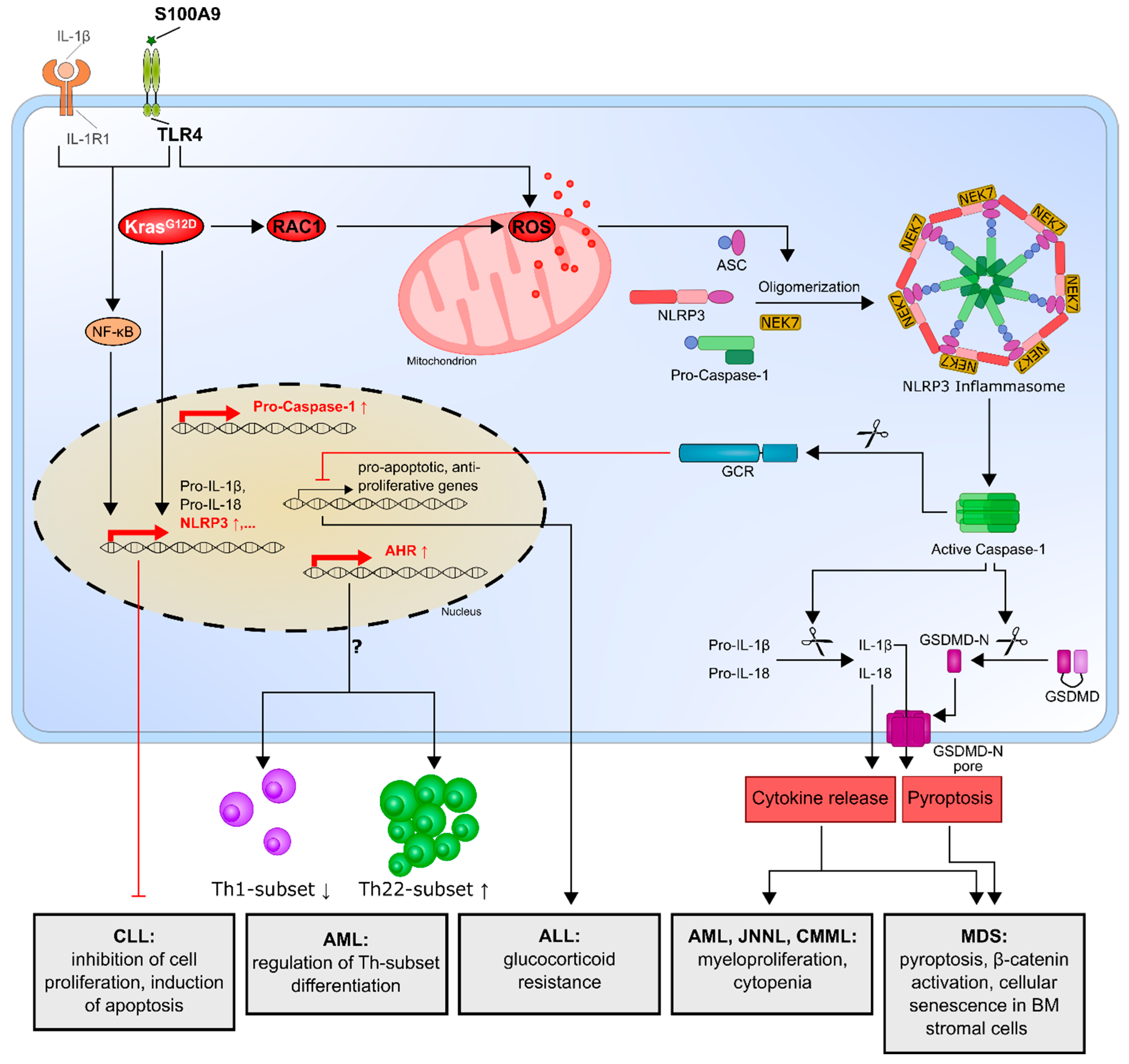
Figure 2.
The NLRP3 inflammasome and its effects in leukemia. DAMPs such as alarmin S100A9 induce the transcriptional upregulation of inflammasome components mainly via activation of the transcription factor NF-κB. DAMPs and/or MDS gene mutations lead to the production of ROS, which cause NLRP3 inflammasome activation and, consequently, pyroptotic cell death and altered inflammatory cytokine secretion in MDS patients. In CMML, JNNL and AML, the oncogenic KrasG12D mutation leads to NLRP3/ASC transcription and ROS production via the activation of RAC. Elevated NLRP3 and caspase-1 expression in ALL results in caspase-1-mediated cleavage of the glucocorticoid receptor. Enhanced NLRP3 expression in AML patients correlates with an increased expression of AHR and a shift in Th cell subsets, while NLRP3 overexpression inhibits cell proliferation and induces apoptotic cell death in CLL. ↓ = Activation; ⊥ = Inhibition. NLRP3, NOD-like receptor protein 3; ASC, apoptosis-associated speck-like protein; NEK7, NIMA-related kinase 7; GSDMD, Gasdermin-D; GSDMD-N, GSDMD amino-terminal cell death domain; DAMP, damage-associated molecular pattern; NF-κB, nuclear factor-κB; CLL, chronic lymphocytic leukemia; AML, acute myeloid leukemia; AHR, aryl hydrocarbon receptor; Th subset, T-helper cell subset; ALL, acute lymphocytic leukemia; CMML, chronic myelomonocytic leukemia; JNNL, juvenile myelomonocytic leukemia; RAC, Ras-related C3 botulinum toxin substrate 1; ROS, reactive oxygen species; IL-1β, interleukin-1β; MDS, myelodysplastic syndrome and S100A9, S100 calcium-binding protein A9.

Figure 3.
The NLRP3 inflammasome and its connection to mitophagy/autophagy. NLRP3 agonists promote the NF-κB-induced expression of inflammasome components but, also, the expression of p62, which negatively regulates caspase-1 activation by the elimination of mitochondria via mitophagy. NLRP3 activation can also lead to the ubiquitination of NLRP3 inflammasome components and destruction in the autophagosome, thereby limiting IL-1β secretion. In addition, NLRP3 inflammasome activation is able to attenuate autophagy via the caspase-1-mediated cleavage of TRIF, which enhances inflammasome activation. ↓ = Activation; ⊥ = Inhibition. NLRP3, NOD-like receptor protein 3; ASC, apoptosis-associated speck-like protein; NEK7, NIMA-related kinase 7; GSDMD, Gasdermin-D; GSDMD-N, GSDMD amino-terminal cell death domain; NF-κB, nuclear factor-κB; ROS, reactive oxygen species; Ub, ubiquitin; Parkin, E3 ubiquitin protein ligase; LC3, microtubule-associated proteins 1A/1B light chain 3B and TRIF, TIR-domain-containing adapter-inducing interferon-β.


Table 1.
Key findings of the role of the NLRP3 inflammasome in different hematologic malignancies. NLRP3, NOD-like receptor protein 3; MDS, myelodysplastic syndrome; IL-1β, interleukin-1β; TLR4, Toll-like receptor 4; KRAS, Kristen rat sarcoma viral oncogene homolog; RAC1, Ras-related C3 botulinum toxin substrate 1; ROS, reactive oxygen species; AML, acute myeloid leukemia; CMML, chronic myelomonocytic leukemia; JNNL, juvenile myelomonocytic leukemia; ALL, acute lymphocytic leukemia; and CLL, chronic lymphocytic leukemia.
| Type of Hematologic Malignancy | Key Findings | Reference |
|---|---|---|
| MDS | NLRP3 inflammasome activation in MDS disorders is responsible for the key biological features of MDS, which drive pyroptotic cell death and β-catenin activation. | [74,75] |
| Cellular senescence in bone marrow stromal cells from MDS patients is induced by increased S100A9 expression through TLR4, NLRP3 inflammasome activation and IL-1β secretion. | [76] | |
| AML, CMML, JNNL | Oncogenic KrasG12D mutation activates the KRAS/RAC1/ROS/NLRP3/IL-1β axis and promotes myeloproliferation and cytopenia. | [77] |
| AML | Enhanced NLRP3 expression correlates with an increased aryl hydrocarbon receptor and might influence T-helper cell differentiation. | [78] |
| ALL | Overexpression of NLRP3 and caspase-1 is responsible for glucocorticoid resistance through the cleavage of the glucocorticoid receptor by caspase-1. | [79] |
| CLL | NLRP3 negatively regulates the progression of CLL by promoting the expression of P2X7R, while NLRP3 overexpression inhibits cell proliferation and survival. | [80] |
Table 2.
NLRP3 inhibitors and their targets.
| Inhibitor | Inhibition Mechanism | Reference |
|---|---|---|
| MCC950 | Binds Walker B motif of the NLRP3 NACHT domain; NACHT ATPase inhibitor | [98,99] |
| CY-09 | Binds Walker A motif of the NLRP3 NACHT domain; NACHT ATPase inhibitor | [100] |
| OLT1177 | NACHT ATPase inhibitor | [101] |
| Tranilast | Binds the NLRP3 NACHT domain and inhibits NLRP3–NLRP3 interaction | [102] |
| Oridonin | Binds irreversibly to NLRP3 Cys279 and inhibits NLRP3–NEK7 interaction | [103] |
| Disulfiram | Blocks gasdermin D pore formation and inhibits pyroptosis and cytokine release | [104] |
| Necrosulfonamide (NSA) | Binds to gasdermin D and prevents pyroptosis | [105] |
Publisher’s Note: MDPI stays neutral with regard to jurisdictional claims in published maps and institutional affiliations. |
© 2021 by the authors. Licensee MDPI, Basel, Switzerland. This article is an open access article distributed under the terms and conditions of the Creative Commons Attribution (CC BY) license (http://creativecommons.org/licenses/by/4.0/).
Share and Cite
MDPI and ACS Style
Urwanisch, L.; Luciano, M.; Horejs-Hoeck, J. The NLRP3 Inflammasome and Its Role in the Pathogenicity of Leukemia. Int. J. Mol. Sci. 2021, 22, 1271. https://0-doi-org.brum.beds.ac.uk/10.3390/ijms22031271
AMA Style
Urwanisch L, Luciano M, Horejs-Hoeck J. The NLRP3 Inflammasome and Its Role in the Pathogenicity of Leukemia. International Journal of Molecular Sciences. 2021; 22(3):1271. https://0-doi-org.brum.beds.ac.uk/10.3390/ijms22031271
Chicago/Turabian StyleUrwanisch, Laura, Michela Luciano, and Jutta Horejs-Hoeck. 2021. "The NLRP3 Inflammasome and Its Role in the Pathogenicity of Leukemia" International Journal of Molecular Sciences 22, no. 3: 1271. https://0-doi-org.brum.beds.ac.uk/10.3390/ijms22031271
Note that from the first issue of 2016, this journal uses article numbers instead of page numbers. See further details here.