
The Role of Lipids in Legionella-Host Interaction


Department of Genetics and Microbiology, Institute of Biological Sciences, Faculty of Biology and Biotechnology, Maria Curie-Sklodowska University, Akademicka St. 19, 20-033 Lublin, Poland
*
Author to whom correspondence should be addressed.
Int. J. Mol. Sci. 2021, 22(3), 1487; https://0-doi-org.brum.beds.ac.uk/10.3390/ijms22031487
Submission received: 21 December 2020
/
Revised: 28 January 2021
/
Accepted: 28 January 2021
/
Published: 2 February 2021
(This article belongs to the Special Issue Host-Pathogen Interaction 2.0)
Abstract
:Legionella are Gram-stain-negative rods associated with water environments: either natural or man-made systems. The inhalation of aerosols containing Legionella bacteria leads to the development of a severe pneumonia termed Legionnaires’ disease. To establish an infection, these bacteria adapt to growth in the hostile environment of the host through the unusual structures of macromolecules that build the cell surface. The outer membrane of the cell envelope is a lipid bilayer with an asymmetric composition mostly of phospholipids in the inner leaflet and lipopolysaccharides (LPS) in the outer leaflet. The major membrane-forming phospholipid of Legionella spp. is phosphatidylcholine (PC)—a typical eukaryotic glycerophospholipid. PC synthesis in Legionella cells occurs via two independent pathways: the N-methylation (Pmt) pathway and the Pcs pathway. The utilisation of exogenous choline by Legionella spp. leads to changes in the composition of lipids and proteins, which influences the physicochemical properties of the cell surface. This phenotypic plasticity of the Legionella cell envelope determines the mode of interaction with the macrophages, which results in a decrease in the production of proinflammatory cytokines and modulates the interaction with antimicrobial peptides and proteins. The surface-exposed O-chain of Legionella pneumophila sg1 LPS consisting of a homopolymer of 5-acetamidino-7-acetamido-8-O-acetyl-3,5,7,9-tetradeoxy-l-glycero-d-galacto-non-2-ulosonic acid is probably the first component in contact with the host cell that anchors the bacteria in the host membrane. Unusual in terms of the structure and function of individual LPS regions, it makes an important contribution to the antigenicity and pathogenicity of Legionella bacteria.
1. Introduction
Bacteria from the family Legionellaceae are Gram-negative bacilli, which are part of the natural microflora of aquatic and soil environments, as well as anthropogenic ecosystems. From the microbiological point of view, the ecology of Legionella bacilli is highly complex. Legionella bacteria survive in a temperature range between 5 °C and 65 °C and at pH 7–9; however, due to their specific nutritional requirements and a narrow range of environmental conditions necessary for their growth, they are unable to compete with other bacteria [1]. The bacteria require specific physicochemical conditions for development, which can only be fulfilled inside the eukaryotic host. Protozoa, commonly found in natural ecosystems, are an important link in the food chain and exert a significant impact on bacterial populations. However, not all bacteria are ingested, killed, and digested by protozoa. The outcome of the Legionella–protozoa interaction is associated with the host species. In some cases, Legionella resists digestion and kills the host, and in others, the protist digests the bacterium [2]. A total of 61 different Legionella species and more than 70 serogroups have been identified [3]. An in-silico analysis of the Legionella pneumophila genome identified several genomic islands indispensable for bacterial growth within protozoa, which differ among the various amoeba species [4]. Free-living amoebae from the genus Acanthamoeba, Naegleria, and Vermamoeba (Hartmanella) and ciliates from the genus Tetrahymena provide an intracellular niche in which Legionella can proliferate [5,6]. The intracellular growth of the bacteria has been associated with the enhanced environmental survival, virulence, and antibiotic resistance of the bacteria [7,8]. L. pneumophila, i.e., the most pathogenic species of this family, use many molecular and cellular aspects of intracellular proliferation inside protozoa in patho-adaptation to human cells [9]. Legionella enter the human organism from the natural environment via contaminated aerosols generated by cooling towers, large air-conditioning systems, fountains, showers, groundwater used for sprinkler irrigation, and similar sources [10,11]. This mode of bacterial spread is regarded as the major infection route, although bacterial transmission from person to person has been reported as well [12]. In the human organism, the bacteria cause respiratory infections with varying severity: from a flu-like infection called Pontiac fever, which does not require specialised treatment, to an acute, multi-lobar pneumonia called Legionnaires’ disease, which may result in death [13]. The bacteria proliferate robustly in lung alveolar macrophages, leading to tissue damage and the subsequent development of the disease. The macrophage resistance of Legionella spp. is a prerequisite for their virulence. Bacterial survival in contact with eukaryotic cells—in particular, those predisposed for killing bacteria—depends on many determinants of adaptation to an environment that is extremely hostile but rich in nutrients. The bacteria overcome the killing mechanisms of phagocytes by means of specialised protein translocation systems: type II Lsp and type IVB Dot/Icm. The type II system controls the secretion of enzymes (lipases, protease, and RNase), thus promoting growth, intracellular replication, and virulence [14]. The Dot/Icm transporter delivers substrates that modulate multiple host cell processes, resulting in the biogenesis of the L. pneumophila-containing vacuole (LCV) permissive for intracellular bacterial replication. L. pneumophila achieve host infection by exporting approximately 300 substrates of the Dot/Icm system across one or two cell membranes to the site of action [15]. The precise delivery of the effectors to the host cell in a timely manner and space is possible thanks to an efficiently functioning bacterial cell envelope. The cell envelope of L. pneumophila is typical for Gram-negative bacteria and consists of two distinct membranes, the inner (IM) and outer membrane (OM), separated by the periplasm. The periplasm contains a relatively thin layer of strongly crosslinked peptidoglycan and various proteins. Peptidoglycan is composed of muramic acid, glucosamine, glutamic acid, alanine, and meso-diaminopimelic acid in a molar ratio of 0.8:0.8:1.1:1.7:1 [16]. The OM is asymmetric, with an inner leaflet mostly composed of phospholipids and an outer leaflet mostly comprising lipopolysaccharides (LPS). This asymmetry is critical for maintaining the OM permeability barrier. LPS consists of three regions: O-antigen, core, and lipid A. The lipid A region anchors LPS molecules to the outer membrane through hydrophobic interactions with the acyl chains of the phospholipids (PLs) constituting the inner layer of this membrane [17]. In addition to proteins, which have a fundamental importance for various aspects of cell physiology, including the infection of a host organism, lipids are involved in the highly specific interactions with the host cell. Three types of lipid-containing molecules are present in the cell envelope of L. pneumophila: phospholipids (PL), lipopolysaccharide (LPS), and lipoproteins (Figure 1).
2. Characteristics of Legionella Phospholipids
Most bacterial membranes are formed by amphiphilic lipids, mainly glycerophospholipids. Phospholipids are composed of a variable hydrophilic head group, two fatty acids esterified in the sn-1 and sn-2 positions of the glycerol moiety, which form a hydrophobic tail, and a phosphate group in the sn-3 position. Phospholipids fulfil a passive role as building blocks of the lipid bilayer and stimulate cell functions, e.g., targeted protein transport, DNA replication, or signal transduction. The composition and distribution of phospholipids in the membrane, their ability to undergo hydrolysis, and the modifications of the phospholipid “head-groups” constitute a specific “molecular language” in the interactions with other cell molecules and the environment [18].
2.1. Phospholipids of Legionella spp.
The most typical bacterial phospholipids include phosphatidylethanolamine (PE), phosphatidylglycerol (PG), and cardiolipin (CL). Zwitterionic PE is found in many groups of Gram-negative bacteria (e.g., Enterobacteriaceae) and in several Gram-positive bacteria, while anionic PG and CL are common in Gram-positive bacteria such as enterococci, streptococci, and staphylococci [19,20].
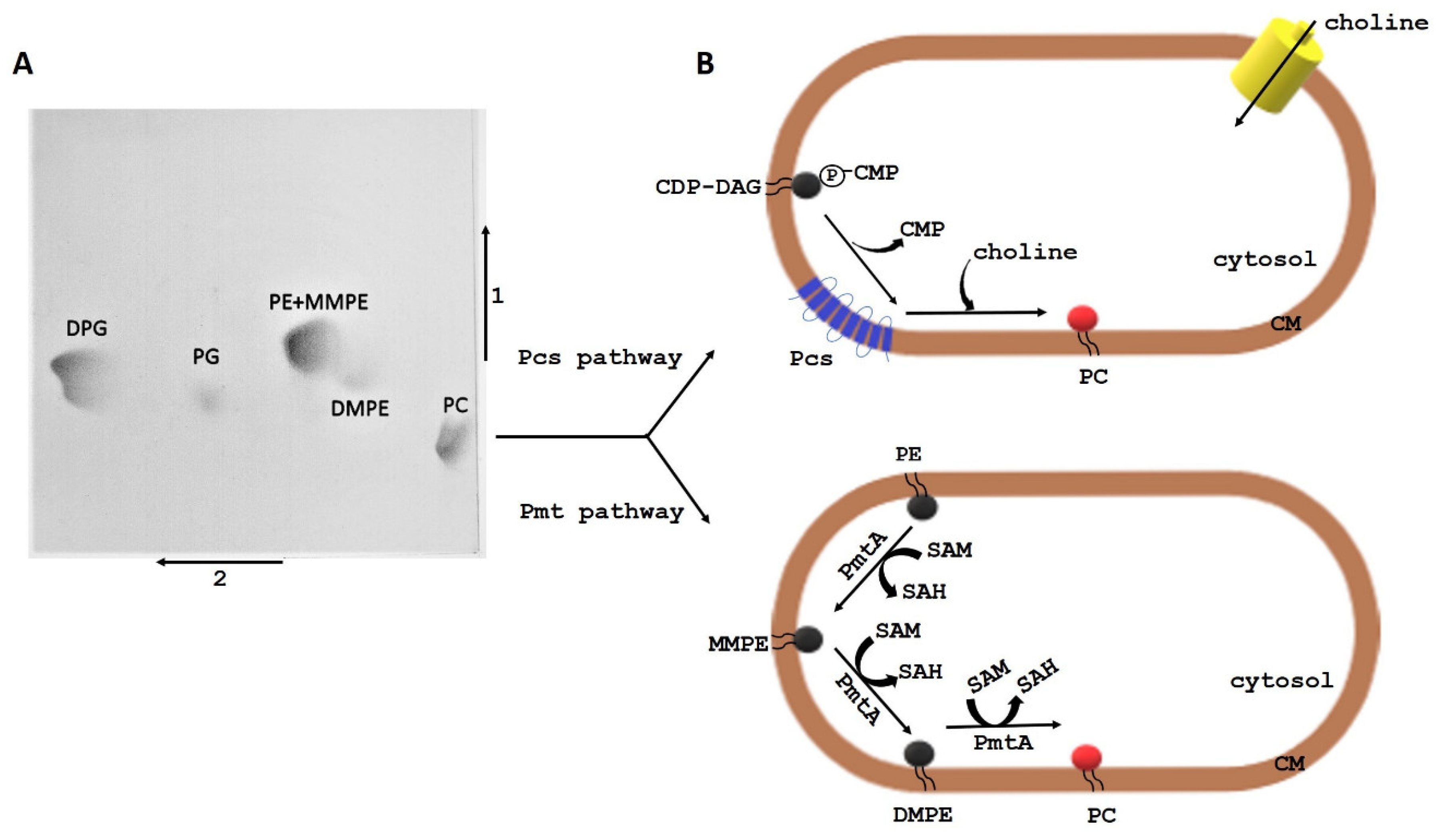
The following phospholipid classes have been identified in the membranes of L. pneumophila, L. lytica, L. bozemanae, L. dumoffii, L. anisa, L. gormanii, and L. longbeachae: phosphatidylcholine (PC), PE, CL, and PG [21,22,23,24,25] (Figure 2A). PC and PE are the major Legionella phospholipids. The relative amount of PC in the different species ranges from 30% to 50% of all PLs. PE dominates in the lipids of L. anisa, L. gormanii, and L. longbeachae [25]. The distribution of phospholipids in the particular layers of the L. pneumophila bacterial envelope is as follows: the outer layer of the cytoplasmic membrane (CM-1) contains ca. 43% PC, 45% PE, and 12% CL, while the inner layer (CM-2) has 35% PC, 42% PE, 14% CL, and 8% PG. PE dominates in the outer (OM-1) and inner layers (OM-2) (ca. 50% in each layer), whereas PC constitutes 27% and 33%, respectively, CL represents 10% and 6%, respectively, and PG accounts for ca. 13% [26].
2.2. Fatty Acid Composition of Legionella spp.
The major fatty acyl residues in Escherichia coli phospholipids are palmitic, stearic, cis-vaccenic, and cyclopropane-containing lactobacillic acids [19]. An analysis of the fatty acid (FA) composition of Legionella spp. showed the presence of cyclopropyl heptadecanoic acid and a high content of branched (iso and anteiso) FAs (40–90%). The cellular FAs are dominated by 14-methylpentadecanoic (i16:0), 12-methyltetradecanoic (a15:0), and 14-methylhexadecanoic (a17:0) acids. On the basis of the differences in the relative amounts of 14-methylpentadecanoic (il6:0), hexadecenoic (16:0), and 12-methyltetradecanoic (a15:0) acids, Lambert and Moss divided the investigated Legionella species into three major groups (16C, A15, and A15/16C). The 16C group includes species with a high content of i16:0 or 16:1 (with one unsaturated bond) or both fatty acids. The A15 group includes species with a high content of a15:0 at concentrations approximately twice that of i16:0. The third group comprises species with a15:0 and i16:0 as major fatty acids in approximately equal amounts [27]. An analysis of the fatty acid contents in the individual L. dumoffii lipid classes showed that PC and PE contained high amounts of a15:0 and cyclopropyl 17:0 fatty acids. The PC fraction was also characterised by the presence of unbranched hexadecenoic acid, while PE had a17:0 fatty acid. Similar to PC and PE, CL contained significant amounts of a15:0 and branched i16:0. Saturated and unbranched hexadecenoic and octadecanoic fatty acids dominated in the PG fraction [28].
Detailed information on the structure of phospholipids, i.e., the distribution of fatty acids in the sn-1 and sn-2 positions of the glycerol backbone, was provided by liquid chromatography (LC) coupled with mass spectrometry (MS). Besides components that were common to L. bozemanae, L. lytica, and L. dumoffii, e.g., PC16:0_15:0 and PE15:0_15:0, the species differed in the composition of fatty acids bound with different classes of phospholipids. In the phosphatidylcholine class, PC17:0_15:0 was characteristic for L. bozemanae, PC16:0_14:0 and PC18:0_16:1 for L. lytica, and PC16:0_17:1 (or cyclopropyl 17:0) for L. dumoffii. PE16:1_15:0 dominated in L. bozemanae, PE14:0_14:0 and PE15:0_14:0 in L. lytica, and PE15:0_15:1 in L. dumoffii.
The differences in the composition of fatty acids contained in the phospholipids are an important chemotaxonomic feature and may have practical relevance in the diagnostics of this bacterial group at the species level. Moreover, the aforementioned Legionella species synthesised straight-chain acids besides the branched ones, in contrast to L. pneumophila, whose phospholipid molecules contained only branched fatty acids [21,22,23,24,25]. The LC-MS/MS method based on the structural analysis of PLs can be applied in Legionella diagnostics, but a greater number of species should be examined to create PL databases allowing the identification of Legionella species.
L. pneumophila change their fatty acid compositions, depending on the growth phase. In the stationary growth phase, the proportion of branched-chain fatty acids rises to over 60%, and the average length of fatty acids in phospholipid molecules decreases, compared to the exponential growth. Since acyl chains attached to phospholipids determine various membrane properties, e.g., fluidity and sensitivity to antibiotics, Verdon et al. showed that changes in the pattern of L. pneumophila fatty acids between the growth phases led to an increased tolerance to the antimicrobial peptide warnericin RK [29].
PC is the main phospholipid of Legionella bacteria. This zwitterionic phospholipid, characteristic of eukaryotic cells and a narrow group of bacteria, is responsible for many biological functions, including interactions with the host cell.
2.3. PC Synthesis Pathways
Among the four experimentally confirmed PC synthesis pathways in bacterial cells, Legionella spp. use the phosphatidylethanolamine (PE) methylation (Pmt) pathway and the PC synthase (Pcs) pathway [30] (Figure 2B). In the N-methylation pathway, PE is N-methylated three times, which leads to the formation of PC. Monomethyl-N-phosphatidylethanolamine (MMPE) and dimethyl-N,N-phosphatidylethanolamine (DMPE) are intermediate compounds in this process. The pathway is catalysed by phospholipid N-methyltransferases (PmtA). The enzyme uses S-adenosylmethionine (SAM) as the methyl donor, converting it to S-adenosylhomocysteine (SAH) [31]. The Pcs pathway is a single-step reaction between choline and CDP-diacylglycerol to form PC [32]. The reaction is catalysed by the phosphatidylcholine synthase (Pcs) present exclusively in bacteria [33].
2.4. Genetic Diversity of Legionella pcs and pmtA Genes
As demonstrated by genetic analyses, Legionella species contain genes (pcs and pmtA) encoding enzymes involved in the synthesis of PC via two independent pathways. A comparative analysis of the nucleotide sequences of the pcs gene encoding Pcs has shown the high sequence identity of these genes among members of the Legionellaceae family (in a range from 64% to 98%). L. bozemanae, L. anisa, L. gormanii, L. parisiensis, and L. tusconensis were found to form one clade on the phylogenetic tree, which indicates that they are closely related. The three strains of L. pneumophila, L. fallonii, and L. micdadei exhibit a considerable phylogenetic distance from all non-L. pneumophila species [25]. The pcs genes in all analysed Legionella spp. exhibit a high conservation degree, although the genome of these bacteria is highly heterogeneous, with over 60% species-specific genes [34]. Legionella Pcs proteins encoded by pcs genes have a similar length of 254 amino acids the exceptions are L. pneumophila (255 aa) and L. longbeachae (253 aa). These proteins contain a highly conserved 27-aa-long motif DGX2ARX8PX3GX3DX3D, and the amino acid motif is shared with the CDP-alcohol phosphotransferase superfamily [35]. The Pcs proteins are highly hydrophobic. They contain up to eight transmembrane helices with N- and C-termini in the cytoplasm. Their N-terminal region has a domain responsible for enzymatic activity.
In comparison with the pcs genes, Legionella pmtA genes encoding PmtA enzymes exhibit higher sequence diversity and lower sequence identity to each other. As revealed by comparative sequence studies of the pmtA genes, L. longbeachae is closely related to L. sainthelensi (92% sequence identity), L. cincinnatiens (89%), L. santicrucis (91%), and L. gratiana (87%). However, no pmtA sequences for L. drancourtii, L. gormanii, L. dumoffii, and L. micdadei have been identified with the use of degenerative primer pairs homologous to Legionella, Rhodobacter, and Rhizobium bacteria. PmtA sequences have been found to diverge substantially among Legionella spp. at the amino acid level. The PmtA protein of L. pneumophila exhibited only 65–70% aa sequence identity to the proteins from other Legionella. The pmtA genes encode small 208–218-aa-long cytosolic proteins. The catalytic domain of these proteins includes a 9-aa-long motif: V/ILE/DXGXGXG. It is predicted to bind the methyl donor S-adenosylmethionine (SAM) and is characteristic for methyltransferases [36]. At least two PmtA families have been described in bacterial cells. One family is similar to the PmtA of Rhodobacter sphaeroides (Rs-PmtA), whereas the other type is similar to the PmtA of Ensifer meliloti (Sm-PmtA) [37]. The Legionella PmtA enzymes exhibit homology to the Rhodobacter Pmt-type enzyme, which, in turn, is homologous to UbiE (ubiquinone/menaquinone biosynthesis methyltransferase). The single PmtA enzyme in Legionella spp. catalyses successive PE N-methylation via the MMPE and DMPE intermediates to form PC. Interestingly, as shown by an analysis of the composition of L. dumoffii phospholipids, the bacterium produces methylated derivatives of PE, which provides evidence for the presence of the Pmt pathway in PC synthesis. However, the absence of a pmtA homologue in the L. dumoffii genome may suggest the presence of a new type of enzyme with PmtA activity, different from Sm-PmtA or Rs-PmtA.
A method for identification and discrimination between Legionella species in the set of duplex-PCR was specified based on the 16S rRNA gene and the pcs and pmtA genes encoding PC synthase and PmtA, respectively [38].
2.5. Utilisation of Exogenous Choline for PC Synthesis by Legionella spp.
The use of exogenous choline by Legionella spp. in the Pcs pathway leads to a change in the contents of their individual classes of phospholipids. 31P nuclear magnetic resonance (NMR) spectroscopy showed that L. gormanii cultured on a choline-supplemented medium synthesised by 21% and 12% higher PC and PE contents, respectively, and by 9% lower CL levels in comparison with bacteria grown on the medium without choline [25]. A comparative analysis of the contents of each PL class revealed that L. dumoffii grown on choline synthesised by 12% higher PC levels and by 12% lower PE amounts than bacteria cultured without the addition of choline [28]. L. pneumophila grown on the medium with choline produced by 6% and 3% higher quantities of PC and PE, respectively, and by 9% lower amounts of CL compared with bacteria grown on the medium without choline. However, the contents of individual classes of PLs in extracts from L. anisa and L. longbeachae cells were unchanged, irrespective of the culture conditions [25]. L. bozemanae and L. dumoffii grown on BCYE medium supplemented with N-nonadeuterotrimethyl-choline chloride synthesised labelled PC species. The occurrence of labelled PC such as d9-PC16:0_15:0, d9-PC15:0/15:0, d9-PC17:0_15:0, d9-PC16:1_15:0, d9-PC17:0_17:0 cyclopropyl, and d9-PC15:1_15:0 in L. bozemanae phospholipids, as well as d9-PC14:0_16:0, d9-PC16:0_17:1 (or 17:0 cyclopropyl), and d9-PC15:0_15:0 in L. dumoffii phospholipids, indicated the utilisation of exogenous choline by these bacteria. An analysis of the distribution of labelled PC in the outer (OM) and inner (IM) membranes in L. dumoffii revealed that phosphatidylcholines derived from the Pcs pathway are located in both membranes. The selective MRM (multiple reaction monitoring) method showed greater amounts of PC in the inner membrane. A comparison of ion intensities in the mass spectra (Matrix-Assisted Laser Desorption/Ionization/Time-of-Flight, MALDI-TOF) of PC (derived via the Pcs pathway) with unlabelled PC (derived via the Pmt pathway) indicated that both PC synthesis pathways operate simultaneously in L. bozemanae and L. dumoffii cells. The Pcs pathway was found to be a dominant, and probably more energy-efficient pathway [23,24,28]. The L. pneumophila pmtA mutant, in which the Pcs pathway depends on extracellular choline, synthesised almost all the PC, while the pcs mutant with the functioning Pmt pathway produced only 6% of PC [39].
The utilisation of extracellular choline was found to influence the composition of L. gormanii fatty acids. The bacterium cultured on choline nonsupplemented medium synthesised more a15:0 fatty acid and larger amounts of n16:0, 16:1, and cyclopropyl 17:0 fatty acids when grown on the medium with choline [25]. However, the addition of choline to the growth medium did not induce qualitative and quantitative changes in the fatty acid profiles in L. pneumophila, L. anisa, and L. longbeachae cells [25].
Exogenous choline induced changes not only in lipids and the fatty acid composition but, also, in proteins, as shown by FTIR (Fourier-Transform Infrared with Attenuated Total Reflection) spectroscopy. After choline supplementation, higher concentrations of proteins represented by the amide I and amide II bands in L. anisa, L. gormanii, and L. longbeachae were detected [25].
2.6. Biological Importance of Legionella PC
Legionella rods belong to the narrow group of bacteria synthesizing PC, which, in addition to membrane formation, plays important roles in bacterial–host interactions ranging from symbiosis to pathogenesis [40]. The presence of PC in cell membranes is specific to pneumonia-causing bacteria. The lung surfactant covering the small airways, bronchioles, and alveolar surface is composed of approximately 10% of protein and 90% of lipids, with approximately 80% of the lung surfactant lipids representing PC. P. aeruginosa utilises lung surfactant PC as a primary carbon and energy source, which facilitates high-cell density replication during lung infection [41]. In cystic fibrosis patients, in whom P. aeruginosa is a common cause of pulmonary infections, choline, i.e., a soluble product of PC and sphingomyelin hydrolysis, is a putative osmoprotectant in the high osmolarity environment of the lung [42]. The PC synthesis pathway (Pcs) can play the role of an environmental sensor, since the biosynthetic precursors present in lung tissue may exert an effect on L. pneumophila virulence via PC incorporation into the bacterial cell envelope [39]. Rapid PC synthesis might be required in Legionella spp. to quickly adjust their membrane physiology to new environmental conditions [43]. A PC-deficient L. pneumophila strain showed attenuated binding to macrophages and a defect in the functioning of the IV secretion system, which is essential for effector transport and formation of the intracellular replication niche in alveolar macrophages. The lack of PC in L. pneumophila membranes resulted in reduced cytotoxicity and impaired capability of the intracellular proliferation of these bacteria, which indicates an important function of this phospholipid in the interaction with the host cell [39]. The utilisation of extracellular choline and incorporation of PC into Legionella membranes lead to changes in the compositions (lipids and proteins) of cell membranes, which influences their physicochemical properties and interactions with external factors (other cells and antimicrobial peptides). Atomic force microscopy measurements revealed changes in the nanomechanical properties of the cell surface of L. dumoffii grown with extracellular choline, i.e., a two-fold increase in the Derjaguin–Muller–Toporov (DMT) modulus reflecting elasticity. A considerably more effective binding of apolipophorin III (apoLp-III, an insect homologue of human apolipoprotein E) to bilayers composed of PLs extracted from choline-grown L. dumoffii, thus enriched with PC, was observed [28]. This is in line with the study conducted by Zhang, which demonstrated a stronger binding of apoLp-III to PC compared to PE, indicating an important role of cell membrane PC in the interaction with apoLp-III [44]. Interestingly, the elasticity of the cell surface of choline-cultured L. dumoffii treated with apoLp-III did not change in contrast to the six-fold increase in the cell envelope elasticity of these bacteria grown without choline after incubation with apoLp-III [45]. Another possible explanation for the stronger binding of apoLp-III to lipids isolated from L. dumoffii grown on choline is the change in the ratio between the PC and PE contents in these bacteria and the resulting alterations in the membrane architecture. PC formed a bilayer structure, whereas PE made the membrane more rigid. The 12% reduction in the PE content in membranes of choline-cultured L. dumoffii may be the cause of the less-tight membrane packing and easier apoLp-III binding. Similar to apoLp-III, defensin isolated from Galleria mellonella haemolymph was over three times more active against L. dumoffii when the bacterial cells were cultured in the presence of exogenous choline [46]. However, the differences in the morphology of the cells visualised with the use of the electron microscope, resulting from the action of apoLp-III and defensin, indicate a different mechanism of their interaction with L. dumoffii.
PC may serve as a specific recognition molecule. L. pneumophila PC is required for the binding of these bacteria to macrophages via the platelet-activating factor receptor (PAF receptor), and the efficiency of this process depends on the content of PC. The adhesion of L. pneumophila to macrophages was successfully blocked by a PAF receptor antagonist [39]. Chemically, PAF is 1-O-alkyl-2-acetyl-sn-glycerol-3-phosphocholine, and, due to this structural similarity, L. pneumophila PC can mimic PAF and, by binding to its receptor, increase the chance of bacterial uptake by macrophages. L. dumoffii cells with increased PC contents underwent internalisation by macrophages of the THP-1 (the human monocytic leukemia cells) line more readily than bacteria with a reduced content of this phospholipid [24]. Legionella PC may influence the modulation of the inflammatory functions of macrophages measured by the level of proinflammatory cytokines (tumour necrosis factor, TNF-α and interleukin-6, IL-6). These cytokines are involved in the protective immune response controlling the early stages of L. pneumophila infection. The treatment of animals with either TNF-α or IL-6 resulted in a significant reduction in mortality caused by L. pneumophila infection.
Human phagocytic THP-1 cells, which differentiate into macrophages after treatment with phorbol 12-myristate 13 acetate (PMA), have been investigated. PMA-triggered THP-1 differentiation causes a rearrangement of the macrophage-specific kinome towards a more proinflammatory phenotype [47]. L. dumoffii grown on a choline-supplemented medium at a dose of 10 and 100 MOI (multiplication of infection) induced over a three-fold lower level of TNF-α in THP-1 macrophages than bacteria cultured without the addition of choline. Similarly, the inner membrane of L. dumoffii isolated from bacteria grown on the choline-supplemented medium was a weaker TNF-α inducer than the membrane of bacteria grown without choline. Statistically significant results were obtained in the presence of 100 ng/mL of the inner membrane. In turn, the same dose of the outer membrane of bacteria cultured with choline was a better cytokine inducer than the outer membrane of bacteria cultured without choline supplementation [24]. L. anisa, L. longbeachae, L. gormanii, and L. pneumophila grown on a medium with choline caused much lower TNF-α production, with the highest decrease in the TNF-α level for L. longbeachae, compared to those bacteria grown without the choline addition [25]. The supplementation of growth medium with choline was also the cause of a decrease in IL-6 production after the incubation of macrophages with L. pneumophila, L. longbeachae, and L. anisa, with the highest decrease by 52% in the level of IL-6 for L. anisa [25]. The exact mechanism of the weaker induction of proinflammatory cytokines by Legionella spp. grown on the choline-supplemented medium compared to those cultured without exogenous choline requires elucidation. A still open question is whether there are different mechanisms responsible for the attenuated production of proinflammatory cytokines, since there are differences among the various Legionella spp. in the PC structure and content, depending on culture conditions.
Changes in the contents and structures of membrane PCs may affect the contents and activity of other cell components (e.g., other classes of phospholipids and proteins), and the observed decrease in the proinflammatory cytokine production may result from the effect of various factors on the macrophages. PC synthesis in the Pcs pathway can lead to a change in the proportion between zwitterionic (PC and, PE) and anionic (CL and PG) lipids, resulting in an imbalance in the electrostatic charge of the cell membrane. Since such a balance is required for many integral membrane proteins to adopt the correct topology in the cell membrane, changes in mutual relations between PLs can lead to modifications in the structure of the transmembrane α-helices of membrane proteins, altering the packing of these helices [48,49]. All of this induces surface changes in bacterial ligands and may lead to altered interactions with macrophages.
The less potent proinflammatory cytokine induction under the influence of bacteria grown on a choline-supplemented medium, in comparison with that induced by bacteria grown without a choline addition, may be one of the ways for bacterial evasion of the host immune system. To survive and multiply in host cells, Legionella rods have developed sophisticated adaptation mechanisms such as the ability to utilise extracellular choline sources. Selective blocking of phosphatidylcholine synthase can become a target in the specific therapeutic effect supporting the treatment of Legionnaires’ disease. Legionella spp. are able to synthesise PC via two independent pathways, and potential drugs that block the activity of phosphatidylcholine synthase may contribute to the attenuation of the virulence trait.
3. Structure and Significance of Legionella Lipopolysaccharide
3.1. Chemical Structure of Legionella LPS
Lipopolysaccharide is a glycolipid composed of three covalently linked regions: lipid A, a core oligosaccharide featuring an outer and inner region, and an O-specific chain. Although LPS isolated from various Legionella species share common features in their basic architecture, the LPS of L. pneumophila differs significantly in the chemical structure and biological significance of the individual parts. The O-specific chain of the L. pneumophila Philadelphia strain consists of a homopolymer of α-(2-4)-linked 5-acetamidino-7-acetamido-8-O-acetyl-3,5,7,9-tetradeoxy-l-glycero-d-galacto-non-2-ulosonic acid [50]. This unusual sugar, termed legionaminic acid, is highly hydrophobic due to the presence of acetyl and acetimidoyl groups. The O-specific chain is bound to a seven-sugar outer core composed of rhamnose (Rha), mannose (Man), acetylquinovosamine (QuiNAc), and acetylglucosamine (GlcNAc) in the molar ratio of 2.1:1.1:1:1.4 [51]. The presence of N-acetyl groups (QuiNAc and GlcNAc); methyl groups of 6-deoxy sugars (Rha and QuiNAc); and O-acetyl groups at C2 of Rha, C4 of QuiNAc, and C3 of GlcNAc makes the outer core highly hydrophobic. The hydrophilic inner core consists of two molecules of 3-deoxy-D-manno-2-octulosonic acid bound by a 2→4 ketosidic linkage and one molecule of D-mannose connected to the C8 of Kdo present in the inner core [52]. A characteristic feature of the inner core of LPS from L. pneumophila is the absence of heptoses and phosphate residues. The carbohydrate backbone of L. pneumophila lipid A consists of a bisphosphorylated disaccharide of the 2,3 diamino-2,3-dideoxy-D-glucose (GlcN3N) amide linked with 3-hydroxy and 2,3-dihydroxy fatty acids [53,54]. The hydroxyl groups of the fatty acids linked directly to the sugar backbone are acylated by straight, branched (iso and anteiso), and long-chain fatty acids. Lipid A of L. pneumophila contains eight nonhydroxylated acids from 14 to 20 carbon atoms and five long-chain fatty acids (28:0(27-OH), 28:0(27-oxo), 30:0(29-oxo), 27-dioic, and 29-dioic) [55].
The high diversity among Legionella spp. is also expressed in the structures of the lipopolysaccharides, including the conserved region of lipid A. The lipid A moiety of LPS from most Legionella spp. comprises disaccharide GlcN3N. However, a mixed disaccharide backbone containing GlcN3N and glucosamine was found in L. israelensis and L. bozemanae [56,57]. An unusual feature of lipid A from Legionella spp. is the high content of non-hydroxy fatty acids (ester-linked) and long-chain (-ω)-oxo, (-ω)-hydroxy, and (ω)-dioic fatty acids (ester linked) [58]. 27-oxo-octacosanoic acid is generally present in all Legionella species studied; therefore, it serves as a useful marker of this bacterial group. An analysis of the sugar components of LPS from different Legionella species showed that all structurally characterised lipopolysaccharides displayed high contents of d-mannose and d-glucosamine. In addition, Legionella LPS contain species-specific sugars (3 amino-3, 6-dideoxy-mannose in L. israelensis, galacturonic acid and galactosamine in L. hackeliae, quinovose in L. feeleii, and a rare sugar yersinose in L. micdadei and L. maceachernii) [59,60]. Unlike L. pneumophila, LPS isolated from several other species, e.g., L. feeleii, L. jordanis, L. erythra, L. bozemanae, L. oakridensis, and L. micdadei contain d- and l-glycero-d-manno-heptose [56,60,61].
The chemical composition and structure of Legionella LPS, especially the presence of various decorative substituents, exerts a strong influence on its biological activity in all stages of host cell infection.
3.2. Biological Significance of Legionella LPS
L. pneumophila LPS is the main antigen recognised by antibodies contained in the serum of patients and the thermostable antigen excreted in urine [62]. The urinary antigen test is the most commonly used method for the diagnosis of Legionnaires’ disease [63]. The use of monoclonal antibodies (mAb) of the Dresden panel, recognising epitopes located in the highly heterogeneous O-specific chain of L. pneumophila LPS, facilitated distinguishing 15 serogroups and nine subgroups within serogroup 1 [64]. The two antibodies (mAb3/1 and mAb8/5) that recognise the virulent strains responsible for the majority of laboratory-confirmed cases of Legionnaires’ disease turned out to be valuable in epidemiological studies and for clinical purposes. The characterisation of the LPS biosynthesis loci of L. pneumophila serogroup 1 strains revealed two major regions: a specific 18-kb region and a conserved 15-kb region containing genes found in serogroup 1 and non-serogroup 1 strains. The most variable region is involved in O-antigen modification [65]. Antibody mAb3/1 recognises an epitope associated with the 8-O-acetyl group in legionaminic acid. The O-acetyl-transferase enzyme encoded by the lag-1 gene is responsible for the transfer of the O-acetyl group to legionaminic acid [66]. Studies carried out in Europe and Asia have shown that the lag-1 gene was harboured by a significantly higher number of clinical isolates of L. pneumophila sg1 compared with environmental isolates [67,68].
The LPS of L. pneumophila is highly hydrophobic due to the presence of deoxy groups and N- and O-acyl substituents in legionaminic acid, but the highest degree of hydrophobicity is exhibited by lag-1 strains producing 8-O-acetyl groups, which most likely contributes to the transmissibility of these bacteria in aerosols and adhesion to host cells. A TF3/1 mutant in the lag-1 gene defective in the synthesis of 8-O-acetyl substituents, not recognised by the mAb3/1 antibody, adhered less strongly to the macrophages of the THP-1 line and to A. castellanii cells, compared to the wild-type strain [69]. This mutant also failed to produce high-molecular-weight long-chain O-polysaccharide [70]. Comparative studies of the kinetics of interactions between the host cell and the L. pneumophila wild-type strain or the mutant showed a higher efficiency of binding to the amoeba surface in bacteria with the full-length O-chain and 8-O-acetyl groups. However, both strains multiplied inside the host cells successfully, irrespective of the differences in the length and structure of the polysaccharide part of LPS. Previous studies also showed that L. pneumophila strains lacking 8-O-acetyl substituents were as effective in infecting amoebae and macrophages as strains that expressed this LPS motif [70]. Thus, the LPS of L. pneumophila plays a critical role in the early stages of infection, anchoring the bacteria to the host cell’s membrane. Disturbances in the synthesis of the polysaccharide region of L. pneumophila LPS exert an effect on the composition and structure of phospholipids and proteins. The TF3/1 mutant showed differences in the structures of the PC and PG species, compared to the wild-type strain. Moreover, the mutant strain was synthesised by 11 mol% lower amounts of branched fatty acids and approximately two-fold higher amounts of long-chain fatty acid (20:0) than the wild-type strain. The changes in the surface components determined the cell surface topography of these bacteria and their nanomechanical properties. The TF3/1 mutant had grooves on the cell surface and did not produce as many outer membrane vesicles (OMV) as the wild-type strain [69]. Spherical bilayers consisting of LPS, phospholipids, outer membrane proteins, and periplasmic components are naturally secreted from the cell envelope of L. pneumophila. OMVs play a significant role in the pathogenesis of these bacteria, simultaneously delivering multiple virulence factors to host cells and tissues [71]. The release of the vesicles from L. pneumophila is developmentally regulated, i.e., the vesicles are connected to the cell wall but shed LPS in the replicative phase and are profusely released in the transmissive phase [72]. One of the functions of L. pneumophila OMVs is the inhibition of phagosome–lysosome fusion during the intracellular infection of macrophages. This capability was correlated with growth phase-dependent modifications of the composition of glycoconjugates contained in the vesicles. The LPS of the bacteria in the transmissive phase of growth is deacetylated and elongated to a form that effectively blocks the fusion between phagosomes and lysosomes and, thus, independently of the effectors of the IV secretion system, inhibits the maturation of macrophages [73]. In turn, during the replicative phase, the degree of acetylation of the LPS O-chain increases, which probably contributes to the increased tolerance to the hostile conditions of the intracellular environment of macrophages [73]. Seeger et al. showed that LPS fractions below 300 kDa, not associated with OMV, significantly delayed phagolysosomal maturation one hour after phagocytosis, regardless of the bacterial growth phase [74].
LPS expressed by L. pneumophila in the transmissive phase binds to a sialic acid-specific lectin more strongly than LPS from bacteria in the replicative phase of growth. The ability of the bacteria to bind to this receptor correlates with the effectiveness of macrophage infection [73]. Legionaminic acid of L. pneumophila sg1 shares the same d-glycero-d-galacto absolute configuration as 5-acetamido neuraminic acid (Neu5Ac, sialic acid). Neu5Ac, located on the surface of mammalian cells, is involved in cell–cell interactions and the immune response [75]. The molecular mimicry of eukaryotic cell macromolecules is one of the strategies used by Legionella bacteria to colonise host cells. The structural similarity of legionaminic and neuraminic acid may be an example of mimicry to the host cell used not only in the adhesion process but, also, in modulation of the immune response to infection. The LPS of L. pneumophila is less toxic and less potent in its ability to induce proinflammatory cytokines (IL-1β, IL-6, IL-8, and TNF-α) from Mono Mac 6 cells, compared to the highly pyrogenic LPS from Enterobacteriaceae members [76]. The bioactive centre of LPS is lipid A, whose toxicity is directly influenced by the length and number of groups of the fatty acids attached to its glycosidic backbone. The presence of fatty acid chains twice the length of the corresponding chains found in the majority of toxic lipids A containing C12, C12OH, C14, and C14OH account for the low endotoxic activity of L. pneumophila LPS due to a failure to interact with receptor CD14 (a glycosylphosphatidylinositol-anchored protein) and with its soluble form [76]. Host pattern recognition receptors such as Toll-like receptors (TLRs) are involved in the process of recognition of LPS. TLR4 functions as a sensor of LPS on the OM in Gram-negative bacteria, promptly inducing the production of antibacterial cytokines. The lipid A region of L. pneumophila LPS was a weak TLR4 agonist [77]. Additionally, macrophages of TLR4-deficient mice infected by L. pneumophila were not defective in the production of cytokines, and the rate of bacterial clearance from the lungs of these mice was similar to that in wild-type mice [78,79]. However, the TLR2-deficient mice were more sensitive to L. pneumophila than TLR2-sufficient mice [79,80]. These results indicated that LPS from L. pneumophila was recognised by TLR2, which is a typical receptor for peptidoglycan [77]. This unusual detection pattern of switching TLR4 to TLR2 is related to the presence of substituent (a ketone group at C27) or branch on the penultimate carbon of different fatty acids of L. pneumophila lipid A [77]. Upon recognition by TLR2, LPS triggers signalling pathways controlled by the MyD88 adaptor protein, which results in the production of inflammatory cytokines and subsequent clearance of L. pneumophila from the lungs [81]. The level of inflammatory cytokine production was reduced in TLR2 and MyD88 gene knockdown macrophages of the U937 cell line infected by L. pneumophila [82]. Additionally, human macrophages induced by L. pneumophila OMVs synthesised IL-8 relying on TLR2-dependent signalling pathways [71].
The structure of lipid A composed of GlcN3N and long-chain fatty acids also provides a protective mechanism against the degradation of lipid A/LPS by amidases and/or esterases of amoebae during the intracellular growth of L. pneumophila [54].
L. pneumophila LPS is subject to phase variations correlated with the attenuation of virulence traits such as the ability to multiply within macrophage-like HL60 cells or A. castellanii [83]. The molecular mechanism responsible for this variability consists in the chromosomal insertion and excision of an unstable 30-kb genetic element, which does not harbour genes related to LPS biosynthesis [84]. Phase variation in the RC1 strain of the L. pneumophila sg 1 subgroup OLDA influences the O-specific chain structure and the fatty acid profile of lipid A. The phase-variant strain 811 with the 29-kb element excised from the chromosome is devoid of N-methylation in legionaminic acid and contains shorter 3-hydroxy (16:0 and 18:0) fatty acids in lipid A [85,86].
L. pneumophila strain PtVFX/2014, associated with the first evidence of person-to-person transmission, i.e., a strain that was able to overcome the transmission barrier of human innate immunity, carried eight horizontally transferred regions encompassing genes involved in, e.g., LPS biosynthesis [87]. Epidemiological studies showed that the genes of the LPS cluster determining sg 1 of L. pneumophila were present in highly diverse genomic backbones of the strains responsible for the largest outbreaks of Legionnaires’ disease described so far and that it probably constitutes a major determinant of human disease itself [87].
On the one hand, the LPS composed of three distinct regions in terms of structure and biological properties is an important factor in the virulence of Legionella bacteria, participating in complex mechanisms of the induction of lesions. On the other hand, LPS is a ligand recognised by proteins involved in the response to infection. During the infection of macrophages with L. pneumophila, host guanylate binding proteins (GBPs), triggered by cytoplasmic LPS derived from the bacteria, induce caspase-11-dependent pyroptosis [88,89]. The LPS of L. pneumophila is a ligand for collectins, which play an important role in the innate immunity of the lung. After the direct binding of LPS, hydrophilic proteins A and D promote the localisation of L. pneumophila in the acidic environment of the lysosomes, thus attenuating the intracellular multiplication of the phagocytosed bacteria [90]. Additionally, human apolipoprotein E binds to L. pneumophila LPS, which results in disturbances in the normal structure of the bacterial surface, and these changes may lead to impaired penetration of the host cells [91]. Two molecules of apoLp-III bind to a single micelle of L. dumoffii LPS formed from 12 to 29 monomeric LPS molecules, pointing to new strategies for anti-Legionella therapies [45].
Legionella spp. are important aetiological agents of pneumonia. They are responsible for 2–8% of community-acquired pneumonia cases [92]. A review of 46 CAP (community acquired pneumonia) studies from European countries has indicated that Legionella spp. are particularly frequent among patients who require admission to an intensive care unit (ICU) [93]. In the USA, the reported cases of Legionnaires’ disease increased from 2.301 in 2005 to 7.104 in 2018. In Europe, the number of known cases of Legionnaires’ disease has almost doubled (from 1.3/100,000 people in 2014 to 2.3/100,000 people in 2018) [94]. The increase in cases may be related to the changing environmental conditions, which favour the growth of Legionella bacteria, as well as the increasing number of people susceptible to infection, such as the elderly and immunocompromised subjects. The closure of public buildings related to the SARS-CoV-2 pandemic has led to the long-term stagnation of water in installations and created optimal conditions for intensive multiplication of the bacteria, which substantially increased the risk of Legionella infections [95]. L. pneumophila produces a variety of cytosolic and cell envelope-associated lipids with a unique structure that plays an important role in its physiology and promotes bacterial adhesion and adaptation to the host’s intracellular environment.
Although more than 60 different Legionella species are known, with approximately half of the number of species isolated from clinical specimens, the complete structure of LPS is only known in L. pneumophila. The analysis of the genomes of various Legionella species has shown that these bacteria contain genes whose products are involved in the synthesis of lipids characteristic for eukaryotic cells, such as PC, or, e.g., hopanoids in L. falonii [96]. Lipid components, especially the unique ones, can serve as important biomarkers used in diagnostics and in taxonomic studies of this bacterial group. Bacterial lipid membranes are targets for the development of new antimicrobial drugs and a promising object of research in the fight against resistant bacteria [97]. Therefore, the elucidation of the complex lipid structure of the cell envelope of various Legionella spp. using advanced lipidomic technologies may be important for the development of new strategies for the prevention and treatment of Legionella infections.
Author Contributions
M.P.-S.: conceptualization, writing, and review and editing; B.K.: writing and review and editing; and E.C.: writing and review and editing. All authors have read and agreed to the published version of the manuscript.
Funding
This work was supported by the research grant from the National Science Center of Poland (NCN, No. 2017/27/B/NZ6/01544).
Institutional Review Board Statement
Not applicable.
Informed Consent Statement
Not applicable.
Data Availability Statement
Not applicable.
Conflicts of Interest
The authors declare no conflict of interest.
References
- Fields, B.S.; Benson, R.F.; Besser, R.E. Legionella and Legionnaires’ disease: 25 years of investigation. Clin. Microbiol. Rev. 2002, 15, 506–526. [Google Scholar] [CrossRef] [PubMed] [Green Version]
- Amaro, F.; Wang, W.; Gilbert, J.A.; Anderson, O.R.; Shuman, H.A. Diverse protist grazers select for virulence-related traits in Legionella. ISME J. 2015, 9, 1607–1618. [Google Scholar] [CrossRef] [PubMed]
- Euzeby, J.P. List of Prokaryotic Names with Standing in Nomenclature Genus Legionella. Available online: https://www.bacterio.net/legionella.html (accessed on 27 January 2021).
- O’Connor, T.J.; Adepoju, Y.; Boyd, D.; Isberg, R.R. Minimization of the Legionella pneumophila genome reveals chromosomal regions involved in host range expansion. Proc. Natl. Acad. Sci. USA 2011, 108, 14733–14740. [Google Scholar] [CrossRef] [PubMed] [Green Version]
- Shaheen, M.; Ashbolt, N.J. Differential bacterial predation by free-living amoebae may result in blooms of Legionella in drinking water systems. Microorganisms 2021, 15, 174. [Google Scholar] [CrossRef] [PubMed]
- Brieland, J.; McClain, M.; LeGendre, M.; Engleberg, C. Intrapulmonary Hartmannella vermiformis: A potential niche for Legionella pneumophila replication in a murine model of legionellosis. Infect. Immun. 1997, 65, 4892–4896. [Google Scholar] [CrossRef] [PubMed] [Green Version]
- Personnic, N.; Striednig, B.; Lezan, E.; Manske, C.; Welin, A.; Schmidt, A.; Hilbi, H. Quorum sensing modulates the formation of virulent Legionella persisters within infected cells. Nat. Commun. 2019, 10, 5216. [Google Scholar] [CrossRef] [Green Version]
- Brieland, J.; McClain, M.; Heath, L.; Chrisp, C.; Huffnagle, G.; LeGendre, M.; Hurley, M.; Fantone, J.C.; Engleberg, C. Coinoculation with Hartmanella vermiformis enhances replicative Legionella pneumophila lung infection in a murine model of Legionnaires’ disease. Infect. Immun. 1996, 64, 2449–2456. [Google Scholar] [CrossRef] [Green Version]
- Al-Quadan, T.; Price, C.T.; Kwaik, Y.A. Exploitation of evolutionarily conserved amoeba and mammalian processes by Legionella. Trends Microbiol. 2012, 20, 299–306. [Google Scholar] [CrossRef] [Green Version]
- De Giglio, O.; Fasano, F.; Diella, G.; Lopuzzo, M.; Napoli, C.; Apollonio, F.; Brigida, S.; Calia, C.; Campanale, C.; Marzella, A.; et al. Legionella and legionellosis in touristic-recreational facilities: Influence of climate factors and geostatistical analysis in Southern Italy (2001–2017). Environ. Res. 2019, 178, 108721. [Google Scholar] [CrossRef]
- De Giglio, O.; Napoli, C.; Apollonio, F.; Brigida, S.; Marzella, A.; Diella, G.; Calia, C.; Scrascia, M.; Pacifico, C.; Pazzani, C.; et al. Occurrence of Legionella in groundwater used for sprinkler irrigation in Southern Italy. Environ. Res. 2019, 170, 215–221. [Google Scholar] [CrossRef]
- Correia, A.M.; Ferreira, J.S.; Borges, V.; Nunes, A.; Gomes, B.; Capucho, R.; Gonçalves, J.; Antunes, D.M.; Almeida, S.; Mendes, A.; et al. Probable person-to-person transmission of Legionnaires’ disease. N. Engl. J. Med. 2016, 374, 497–498. [Google Scholar] [CrossRef] [PubMed] [Green Version]
- Cunha, B.A.; Burillo, A.; Bouza, E. Legionnaires’ disease. Lancet 2016, 387, 376–385. [Google Scholar] [CrossRef]
- Portlock, T.J.; Tyson, J.Y.; Dantu, S.C.; Rehman, S.; White, R.C.; McIntire, I.E.; Sewell, L.; Richardson, K.; Shaw, R.; Pandini, A.; et al. Structure, dynamics and cellular insight into novel substrates of the Legionella pneumophila type II secretion system. Front. Mol. Biosci. 2020, 11, 112. [Google Scholar] [CrossRef] [PubMed]
- Bärlocher, K.; Welin, A.; Hilbi, H. Formation of the Legionella replicative compartment at the crossroads of retrograde trafficking. Front. Cell. Infect. Microbiol. 2017, 7, 482. [Google Scholar] [CrossRef] [PubMed]
- Shevchuk, O.; Jäger, J.; Steinert, M. Virulence properties of the Legionella pneumophila cell envelope. Front. Microbiol. 2011, 25, 74. [Google Scholar] [CrossRef] [PubMed] [Green Version]
- Caroff, M.; Novikov, A. LPS structure, function, and heterogeneity. In Endotoxin Detection and Control in Pharma, Limulus, and Mammalian Systems; Williams, K.L., Ed.; Springer Nature: Berlin/Heidelberg, Germany, 2019; pp. 53–93. [Google Scholar] [CrossRef]
- Bohdanowich, M.; Grinstein, S. Role of phospholipids in endocytosis, phagocytosis, and macropinocytosis. Physiol. Rev. 2013, 93, 69–106. [Google Scholar] [CrossRef] [Green Version]
- Zhang, Y.M.; Rock, C.O. Membrane lipid homeostasis in bacteria. Nat. Rev. Microbiol. 2008, 6, 222–233. [Google Scholar] [CrossRef]
- Geiger, O. Biogenesis of Fatty Acids, Lipids and Membranes; Springer: Berlin/Heidelberg, Germany, 2019; pp. 1–886. [Google Scholar] [CrossRef]
- Finnerty, W.R.; Makula, R.A.; Feeley, J.C. Cellular lipids of the Legionnaires’ disease bacterium. Ann. Intern. Med. 1979, 90, 631–634. [Google Scholar] [CrossRef]
- Palusinska-Szysz, M.; Kalityński, R.; Russa, R.; Dawidowicz, A.L.; Drożański, W.J. Cellular envelope phospholipids from Legionella lytica. FEMS Microbiol. Lett. 2008, 283, 239–246. [Google Scholar] [CrossRef] [Green Version]
- Palusinska-Szysz, M.; Janczarek, M.; Kalityński, R.; Dawidowicz, A.L.; Russa, R. Legionella bozemanae synthesizes phosphatidylcholine from exogenous choline. Microbiol. Res. 2011, 166, 87–98. [Google Scholar] [CrossRef]
- Palusinska-Szysz, M.; Szuster-Ciesielska, A.; Kania, M.; Janczarek, M.; Chmiel, E.; Danikiewicz, W. Legionella dumoffii utilizes exogenous choline for phosphatidylcholine synthesis. Int. J. Mol. Sci. 2014, 15, 8256–8279. [Google Scholar] [CrossRef] [PubMed] [Green Version]
- Palusinska-Szysz, M.; Szuster-Ciesielska, A.; Janczarek, M.; Wdowiak-Wróbel, S.; Schiller, J.; Reszczyńska, E.; Gruszecki, W.I.; Fuchs, B. Genetic diversity of Legionella pcs and pmtA genes and the effect of utilization of choline by Legionella spp. on induction of proinflammatory cytokines. Pathog. Dis. 2019, 77, ftz065. [Google Scholar] [CrossRef] [PubMed]
- Hindahl, M.S.; Iglewski, B.H. Isolation and characterization of the Legionella pneumophila outer membrane. J. Bacteriol. 1984, 159, 107–113. [Google Scholar] [CrossRef] [PubMed] [Green Version]
- Lambert, M.A.; Moss, C.W. Cellular fatty acid compositions and isoprenoid quinine contents of 23 Legionella species. J. Clin. Microbiol. 1989, 27, 465–473. [Google Scholar] [CrossRef] [Green Version]
- Palusinska-Szysz, M.; Zdybicka-Barabas, A.; Reszczyńska, E.; Luchowski, R.; Kania, M.; Gisch, N.; Waldow, F.; Mak, P.; Danikiewicz, W.; Gruszecki, W.I.; et al. The lipid composition of Legionella dumoffii membrane modulates the interaction with Galleria mellonella apolipophorin III. Biochim. Biophys. Acta 2016, 1861, 617–629. [Google Scholar] [CrossRef]
- Verdon, J.; Labanowski, J.; Sahar, T.; Ferreira, T.; Lacombe, C.; Buchrieser, C.; Berjeaud, J.M.; Hechard, Y. Fatty acid composition modulates sensitivity of Legionella pneumophila to warnericin RK, an antimicrobial peptide. Biochim. Biophys. Acta 2011, 1808, 1146–1153. [Google Scholar] [CrossRef]
- López-Lara, I.M.; Geiger, O. Novel pathway for phosphatidylcholine biosynthesis in bacteria associated with eukaryotes. J. Biotechnol. 2001, 91, 211–221. [Google Scholar] [CrossRef]
- Sohlenkamp, C.; López-Lara, M.I.; Geiger, O. Biosynthesis of phosphatidylcholine in bacteria. Prog. Lipid Res. 2003, 42, 115–162. [Google Scholar] [CrossRef]
- de Rudder, K.E.E.; Sohlenkamp, C.; Geiger, O. Plant-exudated choline is used for rhizobial membrane lipid biosynthesis by phosphatidylcholine synthase. J. Biol. Chem. 1999, 274, 20011–20016. [Google Scholar] [CrossRef] [Green Version]
- Martinez-Morales, F.; Schobert, M.; Lopez-Lara, I.M.; Geiger, O. Pathways for phosphatidylcholine biosynthesis in bacteria. Microbiology 2003, 149, 3461–3471. [Google Scholar] [CrossRef]
- Gomez-Valero, L.; Rusniok, C.; Rolando, M.; Neou, M.; Dervins-Ravault, D.; Demirtas, J.; Rouy, Z.; Moore, R.J.; Chen, H.; Petty, N.K.; et al. Comparative analyses of Legionella species identifies genetic features of strains causing Legionnaires’ disease. Genome Biol. 2014, 15, 505. [Google Scholar] [CrossRef] [PubMed]
- Williams, J.G.; McMaster, C.R. Scanning alanine mutagenesis of the CDP-alcohol phosphotransferase motif of Saccharomyces cerevisiae cholinephosphotransferase. J. Biol. Chem. 1998, 273, 13482–13487. [Google Scholar] [CrossRef] [PubMed] [Green Version]
- de Rudder, K.E.; López-Lara, I.M.; Geiger, O. Inactivation of the gene for phospholipid N-methyltransferase in Sinorhizobium meliloti: Phosphatidylcholine is required for normal growth. Mol. Microbiol. 2000, 37, 763–772. [Google Scholar] [CrossRef] [PubMed]
- Arondel, V.; Benning, C.; Somerville, C. Isolation and functional expression in Escherichia coli of a gene encoding phosphatidylethanolamine methyltransferase (EC 2.1.1.17) from Rhodobacter sphaeroides. J. Biol. Chem. 1993, 268, 16002–16008. [Google Scholar] [CrossRef]
- Janczarek, M.; Palusinska-Szysz, M. PCR method for the rapid detection and discrimination of Legionella spp. based on the amplification of pcs, pmtA, and 16S rRNA genes. J. Appl. Genet. 2016, 57, 251–261. [Google Scholar] [CrossRef]
- Conover, G.M.; Martinez-Morales, F.; Heidtman, M.I.; Luo, Z.Q.; Tang, M.; Chen, C.; Geiger, O.; Isberg, R.R. Phosphatidylcholine synthesis required for optimal function of Legionella pneumophila virulence determinants. Cell. Microbiol. 2008, 10, 514–528. [Google Scholar] [CrossRef] [Green Version]
- Sohlenkamp, C.; Geiger, O. Bacterial membrane lipids: Diversity in structures and pathways. FEMS Microbiol. Rev. 2016, 1, 133–159. [Google Scholar] [CrossRef] [Green Version]
- Sun, Z.; Kang, Y.; Norris, M.H.; Troyer, R.M.; Son, M.S.; Schweizer, H.P.; Dow, S.W.; Hoang, T.T. Blocking phosphatidylcholine utilization in Pseudomonas aeruginosa, via mutagenesis of fatty acid, glycerol and choline degradation pathways, confirms the importance of this nutrient source in vivo. PLoS ONE 2014, 7, e103778. [Google Scholar] [CrossRef] [Green Version]
- Kondakova, T.; D’Heygère, F.; Feuilloley, M.J.; Orange, N.; Heipieper, H.J.; Duclairoir Poc, C. Glycerophospholipid synthesis and functions in Pseudomonas. Chem. Phys. Lipids 2015, 190, 27–42. [Google Scholar] [CrossRef]
- Fozo, E.M.; Rucks, E.A. The making and taking of lipids: The role of bacterial lipid synthesis and the harnessing of host lipids in bacterial pathogenesis. Adv. Microb. Physiol. 2016, 69, 51–155. [Google Scholar] [CrossRef]
- Zhang, Y.; Lewis, R.N.; McElhaney, R.N.; Ryan, R.O. Calorimetric and spectroscopic studies of the interaction of Manduca Sexta apolipophorin III with zwitterionic, anionic, and nonionic lipids. Biochemistry 1993, 32, 3942–3952. [Google Scholar] [CrossRef] [PubMed]
- Palusińska-Szysz, M.; Zdybicka-Barabas, A.; Luchowski, R.; Reszczyńska, E.; Śmiałek, J.; Mak, P.; Gruszecki, W.I.; Cytryńska, C. Choline supplementation sensitizes Legionella dumoffii to Galleria mellonella apolipophorin III. Int. J. Mol. Sci. 2020, 21, 5818. [Google Scholar] [CrossRef] [PubMed]
- Palusińska-Szysz, M.; Zdybicka-Barabas, A.; Pawlikowska-Pawlęga, B.; Mak, P.; Cytryńska, M. Anti-Legionella dumoffii activity of Galleria mellonella defensin and apolipophorin III. Int. J. Mol. Sci. 2012, 13, 17048–17064. [Google Scholar] [CrossRef] [Green Version]
- Richter, E.; Ventz, K.; Harms, M.; Mostertz, J.; Hochgräfe, F. Induction of macrophage function in human THP-1 cells is associated with rewiring of MAPK signaling and activation of MAP3K7 (TAK1) protein kinase. Front. Cell Dev. Biol. 2016, 4, 21. [Google Scholar] [CrossRef] [PubMed] [Green Version]
- Zhang, W.; Campbell, H.A.; King, S.C.; Dowhan, W. Phospholipids as determinants of membrane protein topology. Phosphatidylethanolamine is required for the proper topological organization of the γ-aminobutyric acid permease (GabP) of Escherichia coli. J. Biol. Chem. 2005, 280, 26032–26038. [Google Scholar] [CrossRef] [Green Version]
- Dowhan, W. Molecular basis for membrane phospholipid diversity: Why are there so many lipids? Annu. Rev. Biochem. 1997, 66, 199–232. [Google Scholar] [CrossRef] [Green Version]
- Knirel, Y.A.; Rietschel, E.T.; Marre, R.; Zähringer, U. The structure of the O-specific chain of Legionella pneumophila serogroup 1 lipopolysaccharide. Eur. J. Biochem. 1994, 221, 239–245. [Google Scholar] [CrossRef]
- Knirel, Y.A.; Moll, H.; Zähringer, U. Structural study of a highly O-acetylated core of Legionella pneumophila serogroup 1 lipopolysaccharide. Carbohydr. Res. 1996, 293, 223–234. [Google Scholar] [CrossRef]
- Moll, H.; Knirel, A.Y.; Helbig, H.J.; Zähringer, U. Identification of an ɑ-d-Manp-(1→8)-Kdo disaccharide in the inner core region and the structure of the complete core region of the Legionella pneumophila serogroup 1 lipopolysaccharide. Carbohydr. Res. 1997, 304, 91–95. [Google Scholar] [CrossRef]
- Moll, H.; Sonesson, A.; Jantzen, E.; Marre, R.; Zähringer, U. Identification of 27-oxo-octacosanoic acid and heptacosane-1,27-dioic acid in Legionella pneumophila. FEMS Microbiol. Lett. 1992, 76, 1–6. [Google Scholar] [CrossRef]
- Zähringer, U.; Knirel, A.Y.; Lindner, B.; Helbig, J.H.; Sonesson, A.; Marre, R.; Rietschel, E.T. The lipopolysaccharide of Legionella pneumophila (strain Philadelphia 1): Chemical structure and biological significance. Prog. Clin. Biol. Res. 1995, 392, 113–139. [Google Scholar] [PubMed]
- Sonesson, A.; Jantzen, E.; Bryn, K.; Larsson, L.; Eng, J. Chemical composition of a lipopolysaccharide from Legionella pneumophila. Arch. Microbiol. 1989, 153, 72–78. [Google Scholar] [CrossRef] [PubMed]
- Sonesson, A.; Jantzen, E.; Tangen, T.; Zähringer, U. Chemical composition of lipopolysaccharides from Legionella bozemanii and Legionella longbeachae. Arch. Microbiol. 1994, 162, 215–221. [Google Scholar] [CrossRef]
- Sonesson, A.; Jantzen, E.; Bryn, K.; Tangen, T.; Eng, J.; Zähringer, U. Composition of 2,3-dihydroxy fatty acids-containing lipopolysaccharides from Legionella israelensis, Legionella maceachernii and Legionella micdadei. Microbiology 1994, 140, 1261–1271. [Google Scholar] [CrossRef] [PubMed] [Green Version]
- Palusinska-Szysz, M.; Russa, R. Chemical structure and biological significance of lipopolysaccharide from Legionella. Recent Pat. Anti-Infect. Drug Discov. 2009, 4, 96–107. [Google Scholar] [CrossRef]
- Sonesson, A.; Jantzen, A. The branched-chain octose yersiniose A is a lipopolysaccharide constituent of Legionella micdadei and Legionella maceachernii. J. Microbiol. Methods 1992, 15, 241–248. [Google Scholar] [CrossRef]
- Sonesson, A.; Jantzen, E.; Tangen, T.; Zähringer, U. Chemical characterization of lipopolysaccharides from Legionella feeleii, Legionella hackeliae and Legionella jordanis. Microbiology 1994, 140, 2663–2671. [Google Scholar] [CrossRef] [Green Version]
- Sonesson, A.; Jantzen, E.; Tangen, T.; Zähringer, U. Lipopolysaccharides of Legionella erythra and Legionella oakridgensis. Can. J. Microbiol. 1994, 40, 666–671. [Google Scholar] [CrossRef]
- Ciesielski, C.A.; Blaser, M.J.; Wang, W.L. Serogroup specificity of Legionella pneumophila is related to lipopolysaccharide characteristics. Infect. Immun. 1986, 51, 397–404. [Google Scholar] [CrossRef] [Green Version]
- Helbig, J.H.; Jacobs, E.; Lück, C. Legionella pneumophila urinary antigen subtyping using monoclonal antibodies as a tool for epidemiological investigations. Eur. J. Clin. Microbiol. Infect. Dis. 2012, 31, 1673–1677. [Google Scholar] [CrossRef]
- Joly, J.R.; McKinney, R.M.; Tobin, J.O.; Bibb, W.F.; Watkins, I.D.; Ramsay, D. Development of a standardized subgrouping scheme for Legionella pneumophila serogroup 1 using monoclonal antibodies. J. Clin. Microbiol. 1986, 23, 768–771. [Google Scholar] [CrossRef] [PubMed] [Green Version]
- Petzold, M.; Thürmer, A.; Menzel, S.; Mouton, J.W.; Heuner, K.; Lück, C. A structural comparison of lipopolysaccharide biosynthesis loci of Legionella pneumophila serogroup 1 strains. BMC Microbiol. 2013, 13, 198. [Google Scholar] [CrossRef] [PubMed] [Green Version]
- Zou, H.C.; Knirel, Y.A.; Helbig, H.J.; Zähringer, U.; Mintz, C.S. Molecular cloning and characterization of a locus responsible for O-acetylation of the O polysaccharide of Legionella pneumophila serogroup 1 lipopolysaccharide. J. Bacteriol. 1999, 181, 4137–4141. [Google Scholar] [CrossRef] [PubMed] [Green Version]
- Helbig, J.H.; Lück, P.C.; Knirel, Y.A.; Witzleb, W.; Zähringer, U. Molecular characterization of a virulence-associated epitope on the lipopolysaccharide of Legionella pneumophila serogroup 1. Epidemiol. Infect. 1995, 115, 71–78. [Google Scholar] [CrossRef] [PubMed] [Green Version]
- Jiang, L.; Amemura-Maekawa, J.; Ren, H.; Li, Y.; Sakata, M.; Zhou, H.; Murai, M.; Chang, B.; Ohnishi, M.; Qin, T. Distribution of lag-1 alleles, ORF7, and ORF8 genes of lipopolysaccharide and sequence-based types among Legionella pneumophila serogroup 1 isolates in Japan and China. Front. Cell. Infect. Microbiol. 2019, 9, 274. [Google Scholar] [CrossRef] [PubMed] [Green Version]
- Palusinska-Szysz, M.; Luchowski, R.; Gruszecki, W.I.; Choma, A.; Szuster-Ciesielska, A.; Lück, C.; Petzold, M.; Sroka-Bartnicka, A.; Kowalczyk, B. The role of Legionella pneumophila serogroup 1 lipopolysaccharide in host-pathogen interaction. Front. Microbiol. 2019, 10, 2890. [Google Scholar] [CrossRef]
- Lück, P.C.; Freier, T.; Steudel, C.; Knirel, Y.A.; Lûneberg, E.; Zähringer, U.; Helbig, J.H. A point mutation in the active site of Legionella pneumophila O-acetyltransferase results in modified lipopolysaccharide but does not influence virulence. Int. J. Med. Microbiol. 2001, 291, 345–352. [Google Scholar] [CrossRef]
- Jäger, J.; Keese, S.; Roessle, M.; Steinert, M.; Schromm, A.B. Fusion of Legionella pneumophila outer membrane vesicles with eukaryotic membrane systems is a mechanism to deliver pathogen factors to host cell membranes. Cell. Microbiol. 2015, 5, 607–620. [Google Scholar] [CrossRef]
- Helbig, J.H.; Fernandez-Moreira, E.; Jacobs, E.; Luck, P.C.; Witt, M. Lipopolysaccharide architecture of Legionella pneumophila grown in broth and host cells. In Legionella: State of the Art 30 Years After its Recognition; Cianciotto, N.P., Ed.; ASM Press: Washington, DC, USA, 2006; pp. 261–264. [Google Scholar]
- Fernandez-Moreira, E.; Helbig, J.H.; Swanson, M.S. Membrane vesicles shed by Legionella pneumophila inhibit fusion of phagosomes with lysosomes. Infect. Immun. 2006, 74, 3285–3295. [Google Scholar] [CrossRef] [Green Version]
- Seeger, E.M.; Thuma, M.; Fernandez-Moreira, E.; Jacobs, E.; Schmitz, M.; Helbig, J.H. Lipopolysaccharide of Legionella pneumophila shed in a liquid culture as a nonvesicular fraction arrests phagosome maturation in amoeba and monocytic host cells. FEMS Microbiol. Lett. 2010, 307, 113–119. [Google Scholar] [CrossRef] [Green Version]
- Ghosh, S. Sialic acid and biology of life: An introduction. In Sialic Acids and Sialoglycoconjugates in the Biology of Life, Health and Disease, 1st ed.; Academic Press: Cambridge, MA, USA, 2020; pp. 1–61. [Google Scholar] [CrossRef]
- Neumeister, B.; Faigle, M.; Sommer, M.; Zähringer, U.; Stelter, F.; Menzel, R.; Schütt, C.; Northoff, H. Low endotoxic potential of Legionella pneumophila lipopolysaccharide due to failure of interaction with the monocyte lipopolysaccharide receptor CD14. Infect. Immun. 1998, 66, 4151–4157. [Google Scholar] [CrossRef] [PubMed]
- Girard, R.; Pedron, T.; Uematsu, S.; Balloy, V.; Chignard, M.; Akira, S.; Chaby, R. Lipopolysaccharides from Legionella and Rhizobium stimulate mouse bone marrow granulocytes via Toll-like receptor 2. J. Cell Sci. 2003, 116, 293–302. [Google Scholar] [CrossRef] [PubMed] [Green Version]
- Lettinga, K.D.; Florquin, S.; Speelman, P.; van Ketel, R.; van der Poll, T.; Verbon, A. Toll-like receptor 4 is not involved in host defense against pulmonary Legionella pneumophila infection in a mouse model. J. Infect. Dis. 2002, 186, 570–573. [Google Scholar] [CrossRef] [PubMed] [Green Version]
- Archer, K.A.; Roy, C.R. MyD88-dependent responses involving toll-like receptor 2 are important for protection and clearance of Legionella pneumophila in a mouse model of Legionnaires’ disease. Infect. Immun. 2006, 74, 3325–3333. [Google Scholar] [CrossRef] [Green Version]
- Hawn, T.R.; Smith, K.D.; Aderem, A.; Skerrett, S.J. Myeloid differentiation primary response gene (88)-and toll-like receptor 2-deficient mice are susceptible to infection with aerosolized Legionella pneumophila. J. Infect. Dis. 2006, 193, 1693–1702. [Google Scholar] [CrossRef] [Green Version]
- Archer, K.A.; Alexopoulou, L.; Flavell, R.A.; Roy, C.R. Multiple MyD88-dependent responses contribute to pulmonary clearance of Legionella pneumophila. Cell. Microbiol. 2009, 11, 21–36. [Google Scholar] [CrossRef] [Green Version]
- Mallama, C.A.; McCoy-Simandle, K.; Cianciotto, N.P. The type II secretion system of Legionella pneumophila dampens the MyD88 and Toll-like receptor 2 signaling pathway in infected human macrophages. Infect. Immun. 2017, 85, e00897-16. [Google Scholar] [CrossRef] [Green Version]
- Lüneberg, E.; Zähringer, U.; Knirel, Y.A.; Steinmann, D.; Hartmann, M.; Steinmetz, I.; Rohde, M.; Köhl, J.; Frosch, M. Phase-variable expression of lipopolysaccharide contributes to the virulence of Legionella pneumophila. J. Exp. Med. 1998, 188, 49–60. [Google Scholar] [CrossRef]
- Lüneberg, E.; Mayer, B.; Daryab, N.; Kooistra, O.; Zähringer, U.; Rohde, M.; Swanson, J.; Frosch, M. Chromosomal insertion and excision of a 30 kb unstable genetic element is responsible for phase variation of lipopolysaccharide and other virulence determinants in Legionella pneumophila. Mol. Microbiol. 2001, 39, 1259–1271. [Google Scholar] [CrossRef]
- Kooistra, O.; Lüneberg, E.; Knirel, Y.A.; Frosch, M.; Zähringer, U. N-Methylation in polylegionaminic acid is associated with the phase-variable epitope of Legionella pneumophila serogroup 1 lipopolysaccharide. Identification of 5-(N,N-dimethylacetimidoyl) amino and 5-acetimidoyl(N-methyl)amino-7-acetamido-3,5,7,9-tetradeoxynon-2-ulosonic acid in the O-chain polysaccharide. Eur. J. Biochem. 2002, 269, 560–572. [Google Scholar] [CrossRef]
- Kooistra, O.; Knirel, Y.A.; Lüneberg, E.; Frosch, M.; Zähringer, U. Phase variation in Legionella pneumophila serogroup 1, subgroup OLDA, strain RC1 influences lipid A structure. In Proceedings of the 5th International Conference on Legionella, Ulm, Germany, 26–29 September 2000; Marre, R., Abu Kwaik, Y., Bartlett, C., Cianciotto, N.P., Fields, B.S., Frosch, M., Hacker, J., Lück, P.C., Eds.; ASM Press: Washington, DC, USA, 2002; pp. 68–73. [Google Scholar] [CrossRef]
- Borges, V.; Nunes, A.; Sampaio, D.A.; Vieira, L.; Machado, J.; Simões, M.J.; Gonçalves, P.; Gomes, J.P. Legionella pneumophila strain associated with the first evidence of person-to-person transmission of Legionnaires’ disease: A unique mosaic genetic backbone. Sci. Rep. 2016, 6, 26261. [Google Scholar] [CrossRef] [PubMed]
- Case, C.L.; Kohler, L.J.; Lima, J.B.; Strowig, T.; de Zoete, M.R.; Flavell, R.A.; Zamboni, D.S.; Roy, C.R. Caspase-11 stimulates rapid flagellin-independent pyroptosis in response to Legionella pneumophila. Proc. Natl. Acad. Sci. USA 2013, 110, 1851–1856. [Google Scholar] [CrossRef] [PubMed] [Green Version]
- Pilla, D.M.; Hagar, J.A.; Haldar, A.K.; Mason, A.K.; Degrandi, D.; Pfeffer, K.; Ernst, R.K.; Yamamoto, M.; Miao, E.A.; Coers, J. Guanylate binding proteins promote caspase-11-dependent pyroptosis in response to cytoplasmic LPS. Proc. Natl. Acad. Sci. USA 2014, 111, 6046–6051. [Google Scholar] [CrossRef] [PubMed] [Green Version]
- Sawada, K.; Ariki, S.; Kojima, T.; Saito, A.; Yamazoe, M.; Nishitani, C.; Shimizu, T.; Takahashi, M.; Mitsuzawa, H.; Yokota, S.; et al. Pulmonary collectins protect macrophages against pore-forming activity of Legionella pneumophila and suppress its intracellular growth. J. Biol. Chem. 2010, 285, 8434–8443. [Google Scholar] [CrossRef] [PubMed] [Green Version]
- Palusinska-Szysz, M.; Zdybicka-Barabas, A.; Cytryńska, M.; Wdowiak-Wróbel, S.; Chmiel, E.; Gruszecki, W.I. Analysis of cell surface alterations in Legionella pneumophila cells treated with human apolipoprotein E. Pathog. Dis. 2015, 73, 1–8. [Google Scholar] [CrossRef] [PubMed] [Green Version]
- Viasus, D.; Calatayud, L.; McBrown, M.V.; Ardanuy, C.; Carratalà, J. Urinary antigen testing in community-acquired pneumonia in adults: An update. Expert Rev. Anti-Infect. Ther. 2019, 17, 107–115. [Google Scholar] [CrossRef]
- Welte, T.; Torres, A.; Nathwani, D. Clinical and economic burden of community-acquired pneumonia among adults in Europe. Thorax 2012, 67, 71–79. [Google Scholar] [CrossRef] [Green Version]
- Nisar, M.A.; Ross, K.E.; Brown, M.H.; Bentham, R.; Whiley, H. Legionella pneumophila and protozoan hosts: Implications for the control of hospital and potable water systems. Pathogens 2020, 9, 286. [Google Scholar] [CrossRef]
- Palazzolo, C.; Maffongelli, G.; D’Abramo, A.; Lepore, L.; Mariano, A.; Vulcano, A.; Bartoli, T.A.; Bevilacqua, N.; Giancola, M.L.; Di Rosa, E.; et al. Legionella pneumonia: Increased risk after COVID-19 lockdown? Italy, May to June 2020. Euro Surveill. 2020, 25, 1–3. [Google Scholar] [CrossRef]
- Gomez-Valero, L.; Rusniok, C.; Carson, D.; Mondino, S.; Pérez-Cobas, A.E.; Rolando, M.; Pasricha, S.; Reuter, S.; Demirtas, J.; Crumbach, J.; et al. More than 18,000 effectors in the Legionella genus genome provide multiple, independent combinations for replication in human cells. Proc. Natl. Acad. Sci. USA 2019, 5, 2265–2273. [Google Scholar] [CrossRef] [Green Version]
- Chatterjee, R.; Chowdhury, A.R.; Mukherjee, D.; Chakravortty, D. Lipid larceny: Channelizing host lipids for establishing successful pathogenesis by bacteria. Virulence 2021, 12, 195–216. [Google Scholar] [CrossRef] [PubMed]
Figure 1.
Scanning electron microscopy image of Legionella pneumophila, and model of the L. pneumophila cell envelope. Structure of lipopolysaccharide (LPS) (modified from [16]). Leg., legionaminic acid; Rha, rhamnose; OAc, O-acetyl; QuiNAc, acetylquinovosamine; GlcNAc, acetylglucosamine; Man, mannose; Kdo, 3-deoxy-d-manno-oct-2-ulosonic acid; P, phosphate; and GlcN3N, 2,3 diamino-2,3-dideoxy-d-glucose.

Figure 2.
(A) Two-dimensional thin-layer chromatogram of phospholipids from Legionella gormanii. PC—phosphatidylcholine, DMPE—phosphatidyl-N,N-dimethylethanolamine, PE—phosphatidylethanolamine, MMPE—phosphatidyl-N-monomethylethanolamine, PG—phosphatidylglycerol, and DPG—diphosphatidylglycerol (cardiolipin). Solvent system: (1) first dimension—chloroform/methanol/water (14:6:1, v/v/v) and (2) second dimension—chloroform/methanol/glacial acetic acid (13:5:2, v/v/v). (B) PC biosynthesis pathways in L. pneumophila. CDP, diacylglycerol; Pcs, phosphatidylcholine synthase; PmtA, phospholipid N-methyltransferase; SAM, S-adenosylmethionine; SAH, S-adenosylhomocysteine; and CM, cytoplasmic membrane.

Publisher’s Note: MDPI stays neutral with regard to jurisdictional claims in published maps and institutional affiliations. |
© 2021 by the authors. Licensee MDPI, Basel, Switzerland. This article is an open access article distributed under the terms and conditions of the Creative Commons Attribution (CC BY) license (http://creativecommons.org/licenses/by/4.0/).
Share and Cite
MDPI and ACS Style
Kowalczyk, B.; Chmiel, E.; Palusinska-Szysz, M. The Role of Lipids in Legionella-Host Interaction. Int. J. Mol. Sci. 2021, 22, 1487. https://0-doi-org.brum.beds.ac.uk/10.3390/ijms22031487
AMA Style
Kowalczyk B, Chmiel E, Palusinska-Szysz M. The Role of Lipids in Legionella-Host Interaction. International Journal of Molecular Sciences. 2021; 22(3):1487. https://0-doi-org.brum.beds.ac.uk/10.3390/ijms22031487
Chicago/Turabian StyleKowalczyk, Bozena, Elzbieta Chmiel, and Marta Palusinska-Szysz. 2021. "The Role of Lipids in Legionella-Host Interaction" International Journal of Molecular Sciences 22, no. 3: 1487. https://0-doi-org.brum.beds.ac.uk/10.3390/ijms22031487
Note that from the first issue of 2016, this journal uses article numbers instead of page numbers. See further details here.