
Arsenoplatin-Ferritin Nanocage: Structure and Cytotoxicity

 , , , , , and
Abstract
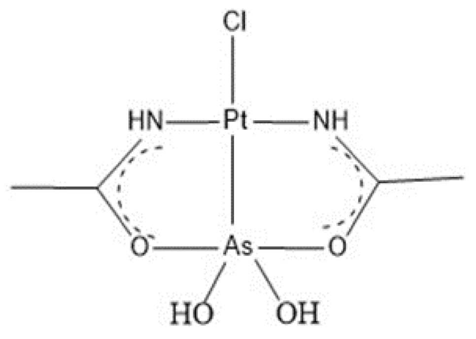
:1. Introduction
2. Results
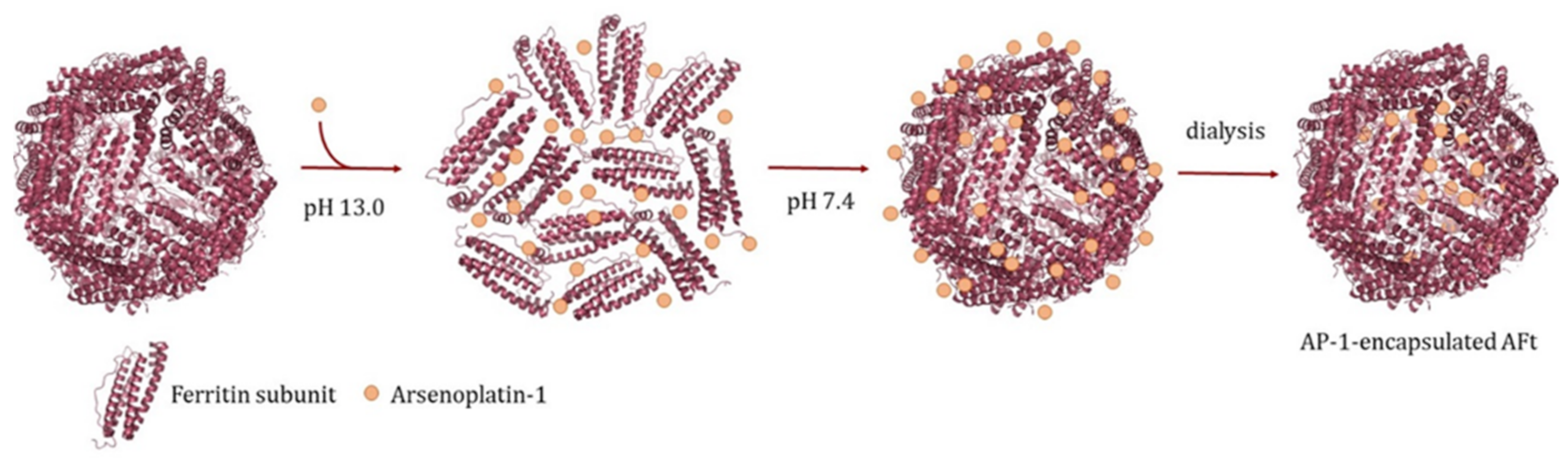
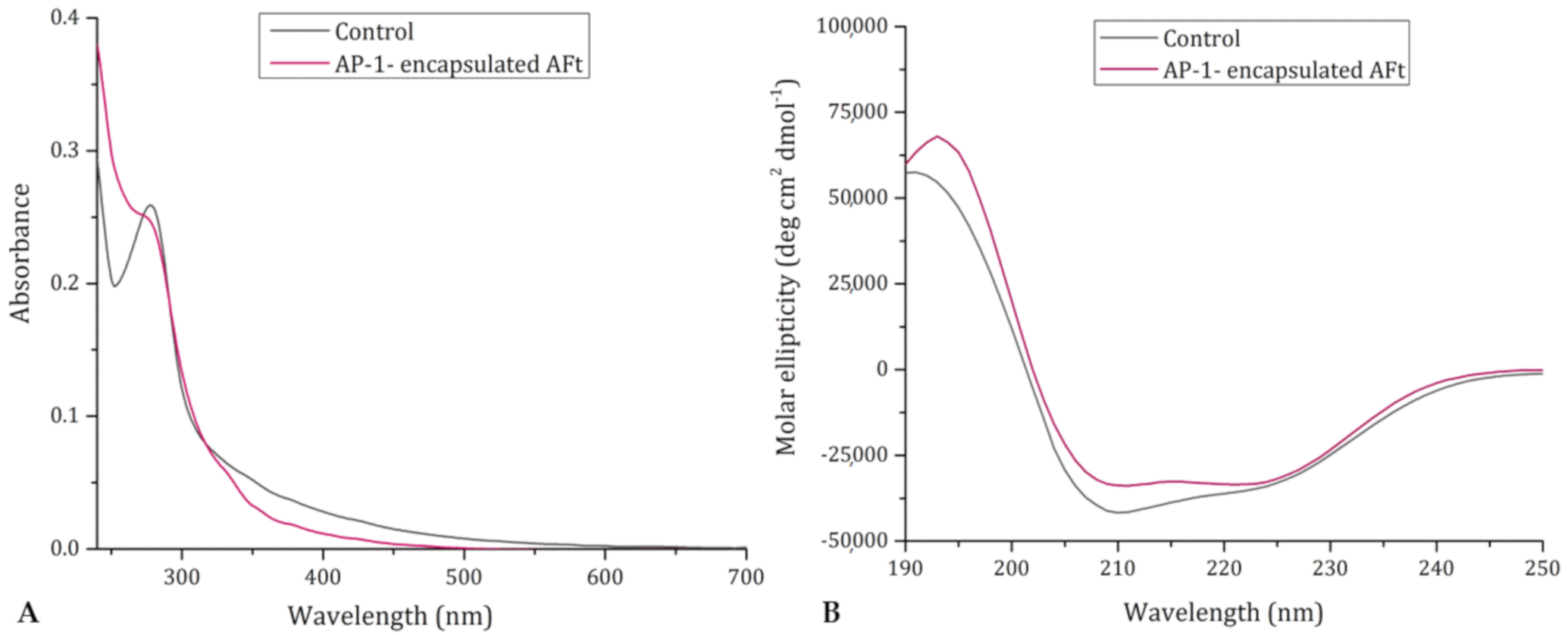
2.1. Preparation and Characterization of AP-1-Encapsulated Ferritin
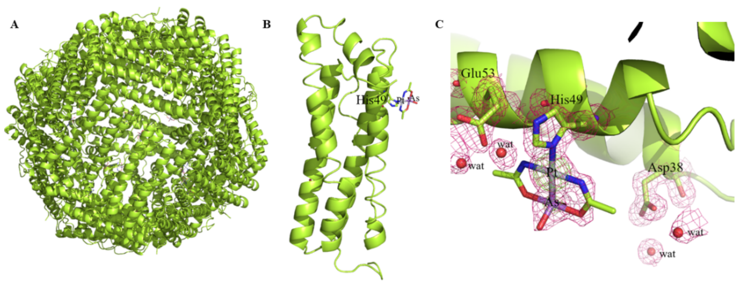
2.2. X-ray Structure of AP-1-Encapsulated AFt
2.3. Drug Release from AP-1 Encapsulated AFt
2.4. Cytotoxicity Studies
2.5. Cellular Uptake Studies
3. Discussion
4. Materials and Methods
4.1. Preparation and Spectroscopic Characterization of AP-1-Encapsulated AFt
4.2. ICP-AES Measurements
4.3. Crystallization and X-ray Diffraction Data Collection
4.4. Structure Solution and Refinement
4.5. Cytotoxicity and Uptake Experiments
5. Conclusions
Author Contributions
Funding
Institutional Review Board Statement
Informed Consent Statement
Data Availability Statement
Acknowledgments
Conflicts of Interest
Abbreviations
| A431 | Human epidermoid carcinoma |
| AFt | Apoferritin |
| AP-1 | Arsenoplatin-1 |
| CD | Circular Dichroism |
| Cisplatin | cis-Pt(NH3)2Cl2 |
| CNS | Central Nervous System |
| DNA | Deoxyribonucleic acid |
| FDA | Food and Drug Administration |
| Ft | Ferritin |
| HEWL | Hen Egg White Lysozyme |
| ICP-AES | Inductively Coupled Plasma-Optical Emission Spectrometry |
| MTT | 3-(4,5-dimethylthiazol-2-yl)-2,5-diphenyltetrazolium bromide |
| PDB | Protein Data Bank |
| PML | Acute Promyelocytic Leukemia |
| UV | Ultraviolet |
| UV-vis | Ultraviolet-visible |
References
- Rottenberg, S.; Disler, C.; Perego, P. The rediscovery of platinum-based cancer therapy. Nat. Rev. Cancer 2021, 21, 37–50. [Google Scholar] [CrossRef]
- Ghosh, S. Cisplatin: The first metal based anticancer drug. Bioorg. Chem. 2019, 88, 102925. [Google Scholar] [CrossRef] [PubMed]
- Boros, E.; Dyson, P.J.; Gasser, G. Classification of Metal-Based Drugs according to Their Mechanisms of Action. Chem 2020, 6, 41–60. [Google Scholar] [CrossRef] [PubMed]
- Ramaekers, B.L.T.; Riemsma, R.; Grimm, S.; Fayter, D.; Deshpande, S.; Armstrong, N.; Witlox, W.; Pouwels, X.; Duffy, S.; Worthy, G.; et al. Arsenic Trioxide for Treating Acute Promyelocytic Leukaemia: An Evidence Review Group Perspective of a NICE Single Technology Appraisal. PharmacoEconomics 2019, 37, 887–894. [Google Scholar] [CrossRef] [Green Version]
- De Thè, H.; Pandolfi, P.P.; Chen, Z. Acute Promyelocytic Leukemia: A Paradigm for Oncoprotein-Targeted Cure. Cancer Cell 2017, 32, 552–560. [Google Scholar] [CrossRef] [PubMed] [Green Version]
- Lallemand-Breitenbach, V.; Zhu, J.; Chen, Z.; De Thé, H. Curing APL through PML/RARA degradation by As2O3. Trends Mol. Med. 2012, 18, 36–42. [Google Scholar] [CrossRef]
- Kaiming, C.; Sheng, Y.; Zheng, S.; Yuan, S.; Huang, G.; Liu, Y. Arsenic trioxide preferentially binds to the ring finger protein PML: Understanding target selection of the drug. Metallomics 2018, 10, 1564–1569. [Google Scholar] [CrossRef] [PubMed]
- Zhang, X.W.; Yan, X.J.; Zhou, Z.R.; Yang, F.F.; Wu, Z.Y.; Sun, H.B.; Liang, W.X.; Song, A.X.; Lallemand-Breitenbach, V.; Jeanne, M.; et al. Arsenic trioxide controls the fate of the PML-RARα oncoprotein by directly binding PML. Science 2010, 328, 240–243. [Google Scholar] [CrossRef]
- Zhang, H.; Yanga, L.; Lingd, J.; Czajkowsky, D.M.; Wanga, J.F.; Zhang, X.W.; Zhou, Y.M.; Ge, F.; Yang, M.; Xiong, Q.; et al. Systematic identification of arsenic-binding proteins reveals that hexokinase-2 is inhibited by arsenic. Proc. Natl. Acad. Sci. USA 2015, 112, 15084–15089. [Google Scholar] [CrossRef] [PubMed] [Green Version]
- Zhou, X.; Sun, X.; Mobarak, C.; Gandolfi, A.J.; Burchiel, S.W.; Hudson, L.G.; Liu, K.J. Differential binding of monomethylarsonous acid compared to arsenite and arsenic trioxide with zinc finger peptides and proteins. Chem. Res. Toxicol. 2014, 27, 690–698. [Google Scholar] [CrossRef]
- Mathews, V.; George, B.; Lakshmi, K.M.; Viswabandya, A.; Bajel, A.; Balasubramanian, P.; Shaji, R.V.; Srivastava, V.M.; Srivastava, A.; Chandy, M. Single-agent arsenic trioxide in the treatment of newly diagnosed acute promyelocytic leukemia: Durable remissions with minimal toxicity. Blood 2006, 107, 2627–2632. [Google Scholar] [CrossRef] [PubMed]
- Wang, W.; Qin, S.K.; Chen, B.A.; Chen, H.Y. Experimental study on antitumor effect of arsenic trioxide in combination with cisplatin or doxorubicin on hepatocellular carcinoma. World J. Gastroenterol. 2001, 7, 702–705. [Google Scholar] [CrossRef] [Green Version]
- Zhang, N.; Wu, Z.M.; McGowan, E.; Shi, J.; Hong, Z.B.; Ding, C.W.; Xia, P.; Di, W. Arsenic trioxide and cisplatin synergism increase cytotoxicity in human ovarian cancer cells: Therapeutic potential for ovarian cancer. Cancer Sci. 2009, 100, 2459. [Google Scholar] [CrossRef] [PubMed]
- Li, H.; Zhu, X.L.; Zhang, Y.; Xiang, J.; Chen, H. Arsenic trioxide exerts synergistic effects with cisplatin on non-small cell lung cancer cells via apoptosis induction. J. Exp. Clin. Cancer Res. 2009, 28, 110. [Google Scholar] [CrossRef] [Green Version]
- Miodragović, Đ.U.; Quentzel, J.A.; Kurutz, J.W.; Stern, C.L.; Ahn, R.W.; Kandela, I.; Mazar, A.; O’Halloran, T.V. Robust structure and reactivity of aqueous arsenous acid-platinum(II) anticancer complexes. Angew. Chem. Int. 2013, 52, 10749. [Google Scholar] [CrossRef] [Green Version]
- Miodragović, Ð.; Swindell, E.P.; Waxali, Z.S.; Bogachkov, A.; O’Halloran, T.V. Beyond cisplatin: Combination therapy with arsenic trioxide. Inorg. Chim. Acta 2019, 496, 119030. [Google Scholar] [CrossRef]
- Miodragović, Đ.U.; Merlino, A.; Swindell, E.P.; Bogachkov, A.; Ahn, R.W.; Abuhadb, S.; Ferraro, G.; Marzo, T.; Mazar, A.P.; Messori, L.; et al. Arsenoplatin-1 Is a Dual Pharmacophore Anticancer Agent. J. Am. Chem. Soc. 2019, 141, 6453. [Google Scholar] [CrossRef]
- Falvo, E.; Tremante, E.; Arcovito, A.; Papi, M.; Elad, N.; Boffi, A.; Morea, V.; Conti, G.; Toffoli, G.; Fracasso, G.; et al. Improved doxorubicin encapsulation and pharmacokinetics of ferritin fusion protein Nanocarriers bearing proline, serine, and alanine elements. Biomacromolecules 2016, 17, 514–522. [Google Scholar] [CrossRef]
- Fracasso, G.; Falvo, E.; Colotti, G.; Fazi, F.; Ingegnere, T.; Amalfitano, A.; Doglietto, B.G.; Alfieri, S.; Boffi, A.; Morea, V.; et al. Selective delivery of doxorubicin by novel stimuli-sensitive nanoferritins overcomes tumor refractoriness. J. Control Release 2016, 239, 10–18. [Google Scholar] [CrossRef] [PubMed]
- Chen, H.; Zhang, S.; Xu, C.; Zhao, G. Engineering protein interfaces yields ferritin disassembly and reassembly under benign experimental conditions. Chem. Commun. (Camb) 2016, 52, 7402–7405. [Google Scholar] [CrossRef]
- Monti, D.; Ferraro, G.; Merlino, M. Ferritin-based anticancer metallodrug delivery: Crystallographic, analytical and cytotoxicity studies. Nanomed. NBM 2019, 20, 101997. [Google Scholar] [CrossRef] [PubMed]
- Hogemann-Savellano, D.; Bos, E.; Blondet, C.; Sato, F.; Abe, T.; Josephson, L.; Weissleder, R.; Gaudet, J.; Sgroi, D.; Peters, P.J.; et al. The Transferrin Receptor: A Potential Molecular Imaging Marker for Human Cancer. Neoplasia 2003, 5, 495–506. [Google Scholar] [CrossRef] [Green Version]
- Mendes-Jorge, L.; Ramos, D.; Valenca, A.; Lopez-Luppo, M.; Pires, V.M.R.; Catita, J.; Nacher, V.; Navarro, M.; Carretero, A.; Rodriguez-Baeza, A.; et al. L-Ferritin Binding to Scara5: A New Iron Traffic Pathway Potentially Implicated in Retinopathy. PLoS ONE 2014, 9, 106974. [Google Scholar] [CrossRef] [PubMed] [Green Version]
- Li, J.Y.; Paragas, N.; Ned, R.M.; Qiu, A.D.; Viltard, M.; Leete, T.; Drexler, I.R.; Chen, X.; Sanna-Cherchi, S.; Mohammed, F.; et al. Scara5 is a ferritin receptor mediating non-transferrin iron delivery. Dev. Cell 2009, 16, 35–46. [Google Scholar] [CrossRef] [PubMed] [Green Version]
- Aime, S.; Frullano, L.; Crich, S.G. Compartmentalization of a Gadolinium Complex in the Apoferritin Cavity: A Route to Obtain High Relaxivity Contrast Agents for Magnetic Resonance Imaging. Angew. Chem. Int. Ed. 2002, 41, 1017–1019. [Google Scholar] [CrossRef]
- Ji, X.T.; Huang, L.; Huang, H.Q. Construction of nanometer cisplatin core ferritin(NCC-F) and proteomic analysis of gastric cancer cell apoptosis induced with cisplatin released from the NCC-F. J. Proteom. 2012, 75, 3145–3157. [Google Scholar] [CrossRef]
- Xing, R.M.; Wang, X.Y.; Zhang, C.L.; Zhang, Y.M.; Wang, Q.; Yang, Z.; Guo, Z. Characterization and cellular uptake of platinum anticancer drugs encapsulated in apoferritin. J. Inorg. Biochem. 2009, 103, 1039–1044. [Google Scholar] [CrossRef]
- Zhu, B.; Huang, L.; Huang, H.Q. Cloning analysis of ferritin and the cisplatin-subunit for cancer cell apoptosis in Aplysia juliana hepatopancreas. Comp. Biochem. Phys. C 2012, 156, 95–103. [Google Scholar] [CrossRef] [PubMed]
- Falvo, E.; Tremante, E.; Fraioli, R.; Leonetti, C.; Zamparelli, C.; Boffi, A.; More, V.; Ceci, P.; Giacomini, P. Antibody-drug conjugates: Targeting melanoma with cisplatin encapsulated in protein-cage nanoparticles based on human ferritin. Nanoscale 2013, 5, 12278–12285. [Google Scholar] [CrossRef] [Green Version]
- Pontillo, N.; Pane, F.; Messori, L.; Amoresano, A.; Merlino, A. Cisplatin encapsulation within a ferritin nanocage: A high-resolution crystallographic study. Chem. Commun. 2016, 52, 4136. [Google Scholar] [CrossRef]
- Kaszuba, M.; McKnight, D.; Connah, M.T.; McNeil-Watson, F.K.; Nobbmann, U. Measuring sub nanometre sizes using dynamic light scattering. J. Nanopart. Res. 2008, 10, 823–829. [Google Scholar] [CrossRef] [Green Version]
- Mitróová, Z.; Melníková, L.; Kovác, J.; Timko, M.; Kopcanský, P. Synthesis and Characterization of Magnetoferritin. Acta Phys. Pol. A 2012, 121, 1318–1320. [Google Scholar] [CrossRef]
- Bhattacharjee, S. DLS and zeta potential—What they are and what they are not? J. Control Release 2016, 235, 337–351. [Google Scholar] [CrossRef]
- Tannock, I.F.; Rotin, D. Acid pH in tumors and its potential for therapeutic exploitation. Cancer Res. 1989, 49, 4373–4384. [Google Scholar]
- Gerweck, L.E.; Seetharaman, K. Cellular pH gradient in tumor versus normal tissue: Potential exploitation for the treatment of cancer. Cancer Res. 1996, 56, 1194–1198. [Google Scholar] [PubMed]
- Stockert, J.C.; Blazquez-Castro, A.; Canete, M.; Horobin, R.W.; Villanueva, A. MTT assay for cell viability: Intracellular localization of the formazan product is in lipid droplets. Acta Histochem. 2012, 114, 785–796. [Google Scholar] [CrossRef] [PubMed]
- Zhang, Y.; Orner, B.P. Self-Assembly in the Ferritin Nano-Cage Protein Superfamily. Int. J. Mol. Sci. 2011, 12, 5406–5421. [Google Scholar] [CrossRef] [PubMed] [Green Version]
- Ferraro, G.; Petruk, G.; Maiore, L.; Pane, F.; Amoresano, A.; Cinellu, M.A.; Monti, D.M.; Merlino, A. Caged noble metals: Encapsulation of a cytotoxic platinum(II)-gold(I) compound within the ferritin nanocage. Int. J. Biol. Macromol. 2018, 115, 1116–1121. [Google Scholar] [CrossRef]
- Ferraro, G.; Cirri, D.; Marzo, T.; Pratesi, A.; Messori, L.; Merlino, A. The first step of arsenoplatin-1 aggregation in solution unveiled by solving the crystal structure of its protein adduct. Dalton Trans. 2021, 50, 68–71. [Google Scholar] [CrossRef]
- Monti, D.M.; Ferraro, G.; Petruk, G.; Maiore, L.; Pane, F.; Amoresano, A.; Cinellu, M.A.; Merlino, A. Ferritin nanocages loaded with gold ions induce oxidative stress and apoptosis in MCF-7 human breast cancer cells. Dalton Trans. 2017, 46, 15354–15362. [Google Scholar] [CrossRef] [Green Version]
- Ferraro, G.; Pica, A.; Petruk, G.; Pane, F.; Amoresano, A.; Cilibrizzi, A.; Vilar, R.; Monti, D.M.; Merlino, A. Preparation, structure, cytotoxicity and mechanism of action of ferritin-Pt(II) terpyridine compound nanocomposites. Nanomedicine 2018, 13, 2995–3007. [Google Scholar] [CrossRef] [PubMed] [Green Version]
- Petruk, G.; Monti, D.M.; Ferraro, G.; Pica, A.; D’Elia, L.; Pane, F.; Amoresano, A.; Ferrer, J.; Kowalski, K.; Merlino, A. Encapsulation of the dinuclear trithiolato-bridged arene ruthenium complex diruthenium-1 in an apoferritin nanocage: Structure and cytotoxicity. ChemMedChem 2019, 14, 594–602. [Google Scholar] [CrossRef] [PubMed]
- Kim, M.; Rho, Y.; Jin, K.S.; Ahn, B.; Jung, S.; Kim, H.; Ree, M. pH-Dependent Structures of Ferritin and Apoferritin in Solution: Disassembly and Reassembly. Biomacromolecules 2011, 12, 1629–1640. [Google Scholar] [CrossRef]
- Ciambellotti, S.; Pratesi, A.; Severi, M.; Ferraro, G.; Alessio, M.; Merlino, A.; Messori, L. The NAMI A—human ferritin system: A biophysical characterization. Dalton Trans. 2018, 47, 11429–11437. [Google Scholar] [CrossRef]
- Otwinowski, Z.; Minor, W. Processing of X-ray diffraction data collected in oscillation mode. Methods Enzymol. 1997, 276, 307–326. [Google Scholar] [PubMed]
- Battye, T.G.G.; Kontogiannis, L.; Johnson, O.; Powell, H.R.; Leslie, A.G.W. iMOSFLM: A new graphical interface for diffraction-image processing with MOSFLM. Acta Crystallogr. D Biol. Crystallogr. 2011, 67, 271–281. [Google Scholar] [CrossRef] [PubMed] [Green Version]
- Evans, P.R. An introduction to data reduction: Space-group determination, scaling and intensity statistics. Acta Crystallogr. D Biol. Crystallogr. 2011, 67, 282–292. [Google Scholar] [CrossRef] [Green Version]
- McCoy, A.J.; Grosse-Kunstleve, R.W.; Adams, P.D.; Winn, M.D.; Storoni, L.C.; Read, R.J. Phaser crystallographic software. J. Appl. Crystallogr. 2007, 40, 658–674. [Google Scholar] [CrossRef] [Green Version]
- Murshudov, G.N.; Skubak, P.; Lebedev, A.A.; Pannu, N.S.; Steiner, R.A.; Nicholls, R.A.; Winn, M.D.; Long, F.; Vagin, A.A. REFMAC5 for the refinement of macromolecular crystal structures. Acta Crystallogr. D Biol. Crystallogr. 2011, 67, 355–367. [Google Scholar] [CrossRef] [Green Version]
- Emsley, P.; Lohkamp, B.; Scott, W.G.; Cowtan, K. Features and development of coot. Acta Crystallogr. D Biol. Crystallogr. 2010, 66, 486–501. [Google Scholar] [CrossRef] [Green Version]
- Pontillo, N.; Ferraro, G.; Helliwell, H.H.R.; Amoresano, A.; Merlino, A. X-ray structure of the carboplatin-loaded apo-ferritin nanocage. ACS Med. Chem. Lett. 2017, 8, 433–437. [Google Scholar] [CrossRef] [PubMed] [Green Version]
- Annunziata, A.; Amoresano, A.; Cucciolito, M.E.; Esposito, R.; Ferraro, G.; Iacobucci, I.; Imbimbo, P.; Lucignano, R.; Melchiorre, M.; Monti, M.; et al. Pt (II) versus Pt (IV) in carbene glycoconjugate antitumor agents: Minimal structural variations and great performance changes. Inorg. Chem. 2020, 59, 4002–4014. [Google Scholar] [CrossRef] [PubMed]


| 48 h Incubation | ||||
|---|---|---|---|---|
| AP-1 (μM) | AP-1-Encapsulated AFt (μM/Ft chain) | AP-1-Encapsulated AFt (μM/AP-1) | ||
| Non cancer cell lines | HaCaT | 2.60 ± 0.26 | 0.38 ± 0.02 | 10.91 ± 0.73 |
| Balb/c-3T3 | 3.60 ± 0.10 | 0.95 ± 0.00 | 27.80 ± 0.14 | |
| Cancer cell lines | A431 | 4.04 ± 0.92 | 0.16 ± 0.01 | 4.66 ± 0.29 |
| SVT2 | 3.87 ± 1.01 | 0.40 ± 0.01 | 11.64 ± 0.29 | |
| Data Collection Statistics | |
|---|---|
| X-ray source | Synchrotron |
| Wavelength | 0.9677 Å |
| Space group | F432 |
| Unit cell parameters a = b = c (Å) | 180.91 |
| Molecules per asymmetric unit | 1 |
| Observed reflections | 357664 (18387) |
| Unique reflections | 40897 (2028) |
| Resolution (Å) | 41.50–1.50 (1.53–1.50) |
| Completeness (%) | 100.0 (100.0) |
| Rmerge | 0.113 (0.840) |
| Rpim | 0.040 (0.293) |
| Rmeas | 0.120 (0.891) |
| I/σ(I) | 12.4 (2.6) |
| Multiplicity | 8.7 (9.1) |
| CC1/2 | 0.999 (0.563) |
| Refinement Statistics | |
| Resolution (Å) | 41.50–1.50 |
| N° reflections in working set | 38816 |
| N° reflections in test set | 2059 |
| N° non-H atoms in the refinement | 1659 |
| R factor/Rfree (%) | 0.167/0.185 |
| Estimated occupancy of Pt | 0.40 |
| Estimated occupancy of As | 0.40 |
| B-factor overall (Å2) | 17.10 |
| B-factor of Pt (Å2) | 27.98 |
| B-factor of As (Å2) | 37.03 |
| Ramachandran values (%) | |
| Most favored/ Additional allowed | 0 |
| Generously allowed/ Disallowed | 3 |
| R.m.s.d. from ideality | |
| R.m.s.d. bonds (Å) | 0.014 |
| R.m.s.d. angles (°) | 1.63 |
Publisher’s Note: MDPI stays neutral with regard to jurisdictional claims in published maps and institutional affiliations. |
© 2021 by the authors. Licensee MDPI, Basel, Switzerland. This article is an open access article distributed under the terms and conditions of the Creative Commons Attribution (CC BY) license (http://creativecommons.org/licenses/by/4.0/).
Share and Cite
Ferraro, G.; Pratesi, A.; Cirri, D.; Imbimbo, P.; Maria Monti, D.; Messori, L.; Merlino, A. Arsenoplatin-Ferritin Nanocage: Structure and Cytotoxicity. Int. J. Mol. Sci. 2021, 22, 1874. https://0-doi-org.brum.beds.ac.uk/10.3390/ijms22041874
Ferraro G, Pratesi A, Cirri D, Imbimbo P, Maria Monti D, Messori L, Merlino A. Arsenoplatin-Ferritin Nanocage: Structure and Cytotoxicity. International Journal of Molecular Sciences. 2021; 22(4):1874. https://0-doi-org.brum.beds.ac.uk/10.3390/ijms22041874
Chicago/Turabian StyleFerraro, Giarita, Alessandro Pratesi, Damiano Cirri, Paola Imbimbo, Daria Maria Monti, Luigi Messori, and Antonello Merlino. 2021. "Arsenoplatin-Ferritin Nanocage: Structure and Cytotoxicity" International Journal of Molecular Sciences 22, no. 4: 1874. https://0-doi-org.brum.beds.ac.uk/10.3390/ijms22041874