Article

Characterization and Comparative Analyses of Mitochondrial Genomes in Single-Celled Eukaryotes to Shed Light on the Diversity and Evolution of Linear Molecular Architecture

1
Institute of Evolution & Marine Biodiversity and College of Fisheries, Ocean University of China, Qingdao 266003, China
2
Key Laboratory of Mariculture (OUC), Ministry of Education, Qingdao 266003, China
3
Centre for Comparative Genomics and Evolutionary Bioinformatics, Department of Biochemistry and Molecular Biology, Dalhousie University, Halifax, NS B3H 4R2, Canada
4
Laboratory for Marine Biology and Biotechnology, Qingdao National Laboratory for Marine Science and Technology, Qingdao 266033, China
*
Author to whom correspondence should be addressed.
Int. J. Mol. Sci. 2021, 22(5), 2546; https://0-doi-org.brum.beds.ac.uk/10.3390/ijms22052546
Submission received: 2 February 2021 / Revised: 23 February 2021 / Accepted: 26 February 2021 / Published: 3 March 2021
(This article belongs to the Special Issue Mitochondrial DNA and RNA)

Abstract

:
Determination and comparisons of complete mitochondrial genomes (mitogenomes) are important to understand the origin and evolution of mitochondria. Mitogenomes of unicellular protists are particularly informative in this regard because they are gene-rich and display high structural diversity. Ciliates are a highly diverse assemblage of protists and their mitogenomes (linear structure with high A+T content in general) were amongst the first from protists to be characterized and have provided important insights into mitogenome evolution. Here, we report novel mitogenome sequences from three representatives (Strombidium sp., Strombidium cf. sulcatum, and Halteria grandinella) in two dominant ciliate lineages. Comparative and phylogenetic analyses of newly sequenced and previously published ciliate mitogenomes were performed and revealed a number of important insights. We found that the mitogenomes of these three species are linear molecules capped with telomeric repeats that differ greatly among known species. The genomes studied here are highly syntenic, but larger in size and more gene-rich than those of other groups. They also all share an AT-rich tandem repeat region which may serve as the replication origin and modulate initiation of bidirectional transcription. More generally we identified a split version of ccmf, a cytochrome c maturation-related gene that might be a derived character uniting taxa in the subclasses Hypotrichia and Euplotia. Finally, our mitogenome comparisons and phylogenetic analyses support to reclassify Halteria grandinella from the subclass Oligotrichia to the subclass Hypotrichia. These results add to the growing literature on the unique features of ciliate mitogenomes, shedding light on the diversity and evolution of their linear molecular architecture.

1. Introduction

Mitochondria are double-membraned semiautonomous organelles that contain their own genomes (mitogenomes) that originated from an endosymbiotic α-proteobacterium that evolved prior to the last eukaryotic common ancestor [1,2,3]. Although they are best known for generating ATP by aerobic respiration to supply the energy needs of eukaryotic cells, these organelles perform a much wider range of functions that vary substantially among diverse eukaryotic lineages [1]. Mitochondria also have biomedical significance, as mutations affecting mitochondrial function are implicated in ageing and diseases, including Parkinson’s, Alzheimer’s, and Huntington’s diseases [4,5].
Mitogenomes usually evolve more rapidly than their nuclear counterparts in most species [6,7]. Additionally, each cell possesses multiple mitogenome copies and mitogenome recombination is a widespread process occurring in plants, fungi, protists, and animals [8,9,10]. In general, unicellular protists possess the most gene-rich and structurally varied mitogenomes [11]. Ciliates are a diverse and well-studied protistan assemblage with unusual cellular and molecular features (nuclear dimorphism and conjugation) [12,13,14,15,16,17,18,19,20,21,22,23]. Their mitogenomes were the first amongst protists to be characterized providing novel insights into the structure, function, and evolution of organellar genomes [24]. The mitogenomes of the ciliates Tetrahymena and Paramecium were the first confirmed to be linear double-stranded DNA molecules and to have their telomeric sequences identified, which provided novel insights into the replication of linear mitochondrial DNA [25,26,27].
In the years since those seminal discoveries, more ciliate mitogenomes have been characterized, including 20 mitogenomes from species in the class Oligohymenophorea (Tetrahymena pyriformis, T. thermophila, T. malaccensis, T. paravorax, T. pigmentosa, T. rostrata, Ichthyophthirius multifiliis, Uronema marinum, Paramecium caudatum, P. aurelia, P. tetraurelia, P. sexaurelia, P. multimicronucleatum, P. biaurelia, P. octaurelia, P. novaurelia, P. decaurelia, P. dodecaurelia, P. quadecaurelia, and P. jenningsi) [28,29,30,31,32,33,34,35,36], eight mitogenomes of species in the class Spirotrichea (Oxytricha trifallax, Laurentiella strenua, Stylonychia lemnae, Paraurostyla sp., Urostyla grandis, Pseudourostyla cristata, Euplotes minuta, and E. crassus) [37,38,39], two mitogenomes in the class Heterotrichea (Stentor coeruleus and Gruberia lanceolata) [20,40], as well as seven hydrogenosomal genomes of anerobic ciliates (Nyctotherus ovalis, Metopus contortus, Metopus es, Metopid sp., Heterometopus sp., Parablepharisma sp., and Muranothrix gubernata) [41,42,43]. Ciliate mitogenomes are generally in size of 20–70 kb, with high A+T content (58.53% for N. ovalis ~81.51% for I. multifiliis) and relatively large gene complements (20–30 protein-coding genes) [39,41,44]. Mitochondrial DNA has been proven extremely useful in phylogenetic analyses to determine ciliate relationships and species delimitation [45].
In this study, we report the analysis of complete mitogenomes of three ciliates: two species of the subclass Oligotrichia (Strombidium sp. and Strombidium cf. sulcatum) and a representative species of Halteriidae (Halteria grandinella) whose phylogenetic position is controversial (i.e., morphological data contradicts molecular data [46,47,48]). These species all belong to the class Spirotrichea, which is a highly differentiated and species-rich assemblage among ciliates [49]. We annotated and compared these three genomes with the previously published ciliate mitogenomes, particularly the mitogenomes among Spirotrichea, shedding light on the diversity and evolution of mitochondrial genomes within this group. Phylogenetic analysis based on 14 mitochondrial ortholog proteins of 34 ciliates was performed in order to further investigate and clarify the evolutionary relationships among Oligotrichia, Halteriidae, and Hypotrichia.

2. Results

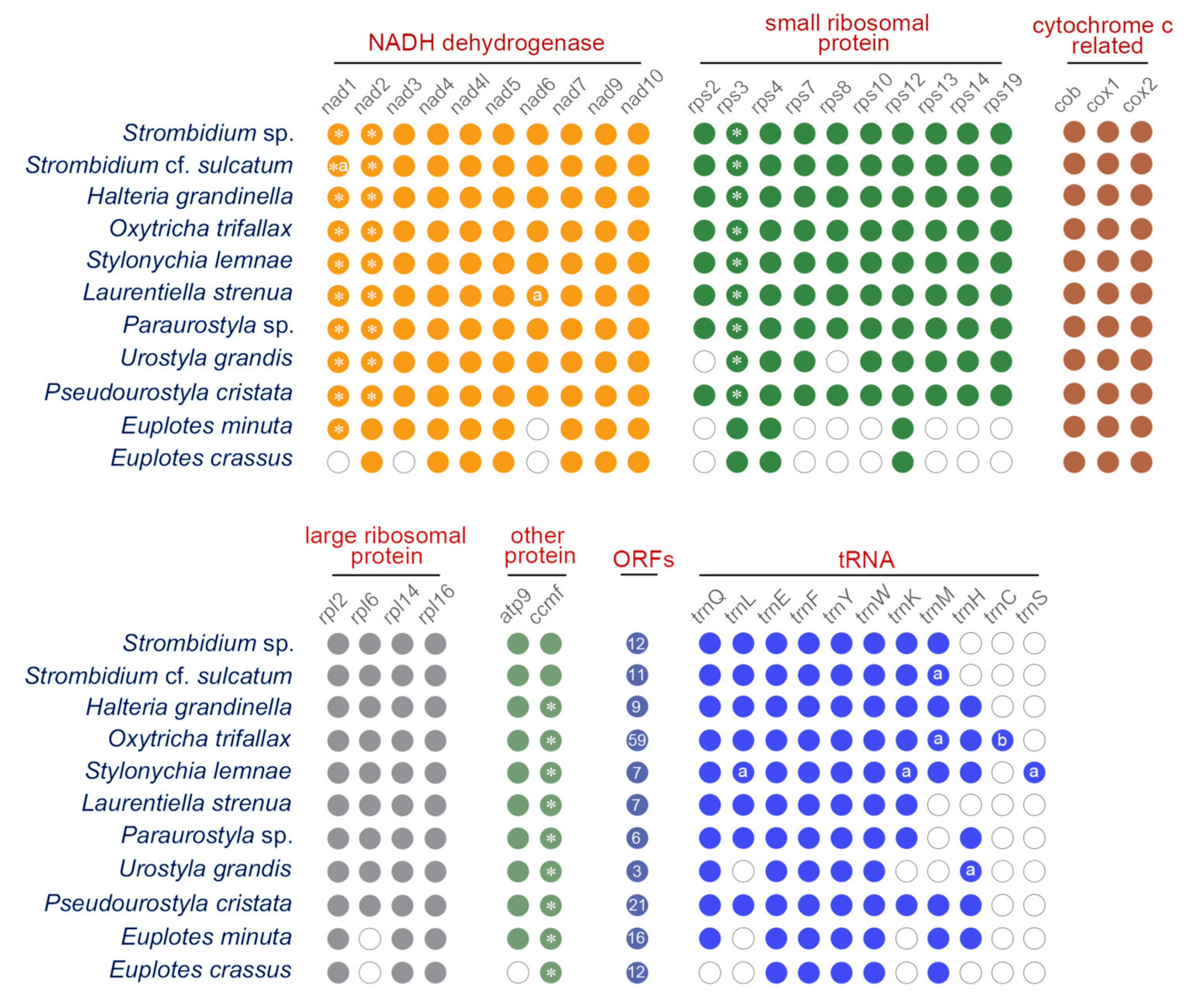
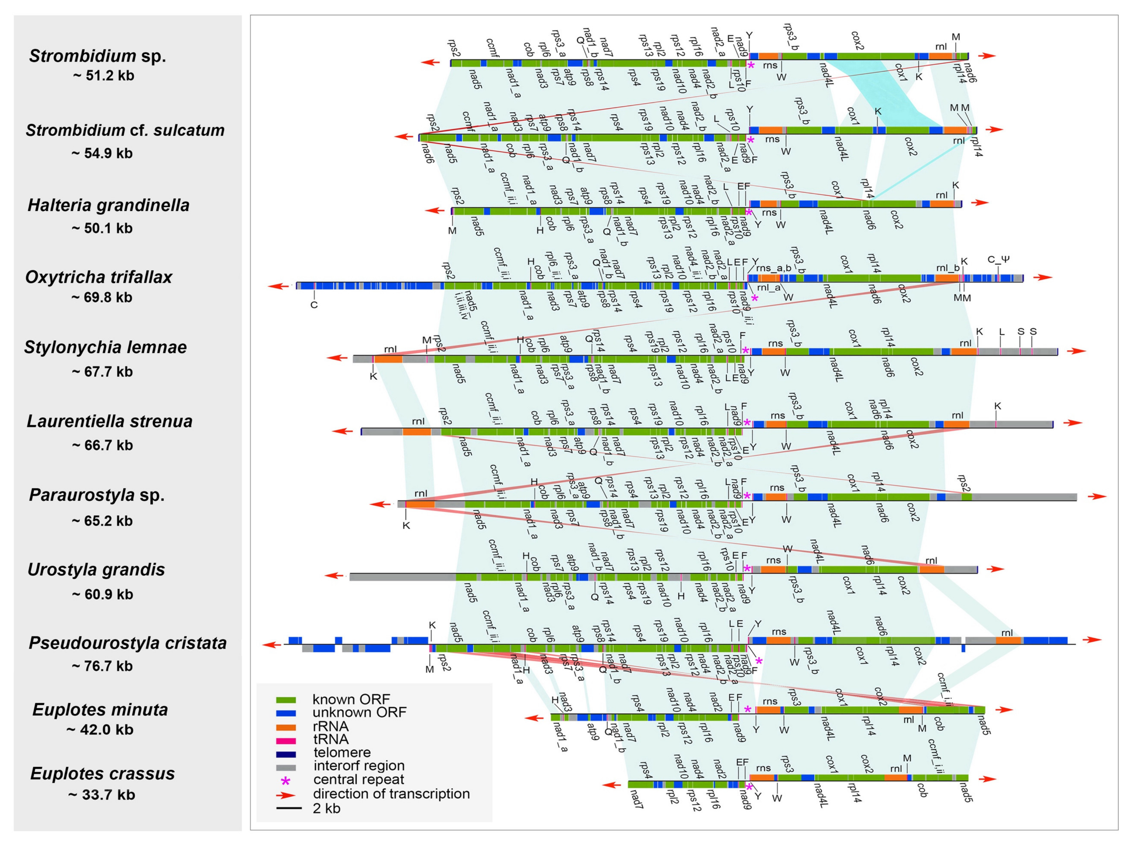
2.1. Mitogenome Overview

For each species, mitochondrial contigs (Strombidium sp.: one contig; Halteria grandinella: one contig; Strombidium cf. sulcatum: 9 contigs) are recovered from the assembled mitochondrial or genomic data by BLASTN searches. They are assembled into linear molecules for the mitogenomes of Strombidium sp., S. cf. sulcatum, and H. grandinella with coverage of 194.2, 492.2–564.8, and 40.2 respectively (Figures S1 and S2). The lengths of the mitogenomes are 51,232 bp for Strombidium sp., 54,912 bp for S. cf. sulcatum, and 50,085 bp for H. grandinella with the A+T contents of 77.32%, 71.68%, and 80.20%, respectively (Table 1). Furthermore, the three mitogenomes are all capped with telomeric tandem repeats at both ends, which are reverse complements of each other. The telomeric repeat sequences are divergent among the three species (18 bp in Strombidium sp.: CTC CCT TAT CTA GTC TTT; 34 bp in S. cf. sulcatum: TTA TAT CCT TTC TCC CCT ATA TCT CTA TAG TAC T; and 31 bp in H. grandinella: AAA ACA GCT CCG TTC CAA TAC TAC TAA CTA A) (Table 2). A central repeat region found in all three genomes is made up of tandem repeat units, and occurs at the same position between tRNA_Phe (trnF) and tRNA_Tyr (trnY) in each genome. However, the central repeats differ from one another in terms of length and sequence. In Strombidium sp., the central repeat region is 170 bp long, composed of 11 repeats of the unit ATA ATA TAA TAA TAT. This region is 142 bp long in S. cf. sulcatum, composed of two repeats of the unit ATA AAT TTA ATT TTA and an irregular sequence for the rest. In H. grandinella, this region is 168 bp long and composed of nine repeats of the unit TAT ACA TAT AAT ATA TA (Table 2). Diverging and starting from this central repeat region, all the genes in the three mitogenomes are arranged in two opposite transcriptional directions (Figure 1).
For Strombidium sp., 94.89% of the mitogenome is coding sequence, including 29 known protein genes, 8 tRNA genes, 2 rRNA genes, and 12 open reading frames (ORFs) with unknown function. The mitogenome of S. cf. sulcatum also contains 29 known protein genes, but instead has 9 tRNA genes, 2 rRNA genes, and 11 ORFs, with the coding region comprising 95.09%. The proportion of coding sequence is 90.65% in the H. grandinella mitogenome, the lowest among the three taxa, which comprises 29 known protein genes, 9 tRNA genes, 2 rRNA genes, and 9 ORFs (Table 1, Figure 1 and Figure 2). The mitochondrial genes of the three species are tightly packed and some genes are partially overlapping (Strombidium sp.: seven cases (10–46 bp, avg. 24 bp), S. cf. sulcatum: seven cases (7–82 bp, avg. 46 bp), H. grandinella: fifteen cases (4–190 bp, avg. 39 bp)) (Table S3, Figure 1). Three genes of nad1, nad2 (NADH dehydrogenase genes), and rps3 (small subunit ribosomal protein gene) are separated into two parts for all the three mitogenomes, while the ccmf (cytochrome c maturation-related gene) is split only for H. grandinella. Two duplicated genes of nad1_a and tRNA_Met (trnM) are found in S. cf. sulcatum mitogenomes (Figure 1 and Figure 2). No introns are detected in any of the three mitogenomes.

2.2. Mitogenome Comparison among Species in the Class Spirotrichea

We compared the three newly sequenced mitogenomes with eight other representative mitogenomes available in the class Spirotrichea. Among these mitogenomes, only the three that were newly sequenced and those of Oxytricha trifallax and Laurentiella strenua are complete, with both ends capped by telomeres (Table 2, Figure 3). To our knowledge, Pseudourostyla cristata possesses the longest mitogenome (76,660 bp) among the available mitogenomes in ciliates. Within the class of Spirotrichea, the A+T content of the mitogenome in Paraurostyla sp. is the highest (80.59%), while that of Urostyla grandis is the lowest (61.12%, Table 1). It is noteworthy that all mitogenomes of Spirotrichea contain a central tandem repeat that has a high A+T content (83.36–100%, avg. 93.31%) (Table 2). Besides, the central repeats are always located between trnF and trnY (Figure 3). Strikingly, the mitochondrial central repeat regions contain potential palindrome sequences, such as TA repeats. The repeat unit sequence is identical between two species of Euplotia, while it varies among species in oligotrichs and hypotrichs (Table 2). According to the secondary structures of central repeats of species in Spirotrichea (apart from Euplotes minuta), the tandem repeat region shows similar stem-loop structure: the sun-shape structure containing 4–17 helices with similar length for P. cristata, H. grandinella, U. grandis, O. trifallax, E. crassus, Strombidium sp., and Stylonychia lemnae, but with divergent length for the remaining three species. (Figure 4A–J). Moreover, a general model of the secondary structure of mitogenomes in the class Spirotrichea is proposed based on these structures (Figure 4K).
The three newly characterized mitogenomes share largely the same complement of known protein-coding genes with the other mitogenomes of the class Spirotrichea (Figure 2). The two Euplotes species lack many mitochondrial genes, probably due to the incompleteness of the mitogenomes. The protein-coding genes include NADH dehydrogenase genes (nad1–7, nad9–10, nad4L), cytochrome c reductase and oxidase genes (cob, cox1, cox2), ATP synthase gene (atp9), ribosomal protein genes (rps2–4, rps7–8, rps10, rps12–14, rps19, rpl2, rpl6, rpl14, rpl16), and cytochrome c maturation-related gene (ccmf). Nevertheless, rps2 and rps8 are not detected in the mitogenome of U. grandis. In addition, the ccmf gene is split into ccmf_i and ccmf_ii in the mitogenomes of all Spirotrichea species apart from two oligotrichous species, which will be discussed in detail below (Figure 2, Figure 3, Figure 5 and Figure 6). According to the transmembrane profile predictions, the ccmf protein of H. grandinella exhibits similar transmembrane helices with those of hypotrichs and euplotids where ccmf_i contains 11–16 transmembrane structures and ccmf_ii comprises 2–3 transmembrane helices and a long non-transmembrane region (Figure 5). Duplications are detected in mitogenomes of the class Spirotrichea. For instance, nad1_a of S. cf. sulcatum, nad6 of L. strenua, mitochondrial large subunit ribosomal RNA gene (rnl) of S. lemnae and L. strenua, trnM of S. cf. sulcatum and O. trifallax, tRNA_Leu (trnL), tRNA_Lys (trnK), and tRNA_Ser (trnS) of S. lemnae, and tRNA_His (trnH) of U. grandis (Figure 2 and Figure 3). Notably, tRNA_Glu (trnE), trnF, trnY, and tRNA_Trp (trnW) are present in all these eleven mitogenomes, while trnS is only detected in S. lemnae (Figure 2). Besides, a pseudogene of tRNA_Cys (trnC) is detected in the mitogenome of O. trifallax (Figure 2). Except for the known protein genes, some homologous ORFs with unknown functions are also detected in mitogenomes of the class Spirotrichea according to BLASTP results and gene location on the mitogenome map, such as orf535, orf546, orf578, orf583, and orf592. Interestingly, seven ORFs (orf_s1–s7) in mitogenomes of two Strombidium species and six ORFs (orf259, orf163, orf187, orf96, orf111, and orf155) in two Euplotes mitogenomes are unique and no homolog is found in mitogenomes of other species (Table S4).
Gene order comparison shows that the gene synteny is highly conserved among mitogenomes within the class Spirotrichea (Figure 3). For the two Strombidium species, there is collinearity for most genes, with the exceptions of two translocations (cox2 and orf_s6) and one inversion (nad6). The mitogenomes of S. cf. sulcatum and H. grandinella are largely collinear, except that two terminal genes of nad6 and rpl14 in S. cf. sulcatum are moved to the position between cox1 and cox2 in H. grandinella. The gene orders in H. grandinella and O. trifallax mitogenomes are identical. The mitogenome synteny is also significant among the hypotrichs, except for the inversion of rps2 in Paraurostyla sp. and the gene duplication of rnl in S. lemnae, L. strenua, and Paraurostyla sp. The two Euplotes mitogenomes are collinear and are largely collinear with those of the hypotrichs in the core region, from nad3 to rnl, except for the inversion of nad5, ccmf, and cob. Besides, the gene order of tRNA, tRNA_Gln (trnQ)-trnL-trnE-trnF-trnY-trnW, is conserved in all the species except for the missing trnQ in E. crassus and trnL in U. grandis, E. minuta, and E. crassus.
Similar to the extensive collinearity within the class Spirotrichea, the mitogenomes are largely collinear within the class Oligohymenophorea (mainly represented by Tetrahymena and Paramecium), with the exception of one large inversion and translocation from cob to rnl [39]. The collinearity between the classes Spirotrichea and Oligohymenophorea is lower than within classes, and no collinearity is observed between the classes Spirotrichea and Heterotrichea, which is consistent with the higher ciliate taxonomic classification [39].

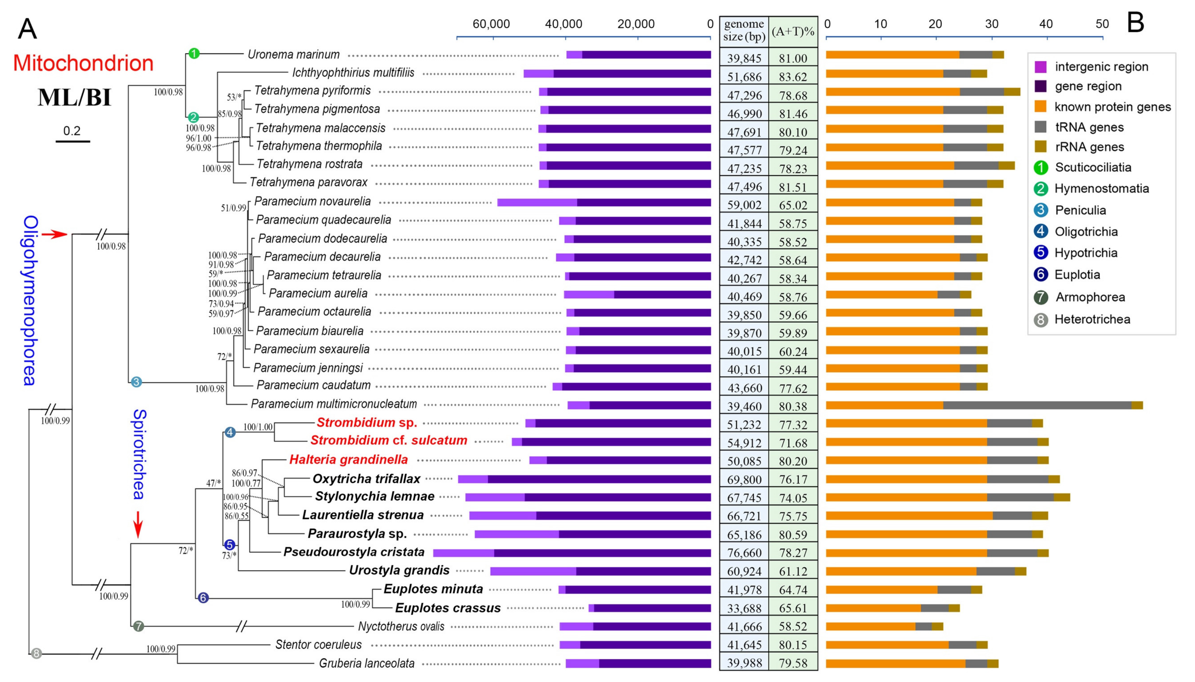
2.3. Phylogenetic Analyses Based on Mitochondrial Proteins

The phylogenetic trees based on mitochondrial proteins reveal a concordant topology with that inferred from nuclear small subunit ribosomal DNA (nSSU-rDNA) data, thus only the topology based on mitochondrial protein with support values generated from both maximum likelihood (ML) and Bayesian inference (BI) analyses is presented (Figure 7). According to the phylogenetic trees in our work, all the classes (Spirotrichea and Oligohymenophorea) and the subclasses (Oligotrichia, Euplotia, Hypotrichia, Peniculia, and Hymenostomatia) are monophyletic. Moreover, H. grandinella clusters with four species of the family Oxytrichidae (S. lemnae, O. trifallax, L. strenua, and Paraurostyla sp.) based on the mitochondrial data (ML/BI: 100/0.77), which is consistent with previous studies [48,49]. According to the phylogenetic trees based on multiple nuclear gene markers (nSSU-rDNA, internal transcribed spacer (ITS), 5.8 S rDNA, nuclear large subunit ribosomal DNA (nLSU-rDNA), α-tubulin, and actin I genes), H. grandinella was always clustered within core hypotrichs [49,50,51,52]. Recently, phylogenomic analyses based on more than 130 orthologs also implied the close relationship between H. grandinella and Hypotrichia [19,48]. In our work, H. grandinella nests within hypotrichs, while the subclass Oligotrichia, represented by two Strombidium species because of a limitation of data, forms a monophyletic clade that is sister to hypotrichs (Figure 7). In order to test the possibility of previous assignment of H. grandinella into Oligotrichia, the constrained ML tree with two Strombidium species clustering with H. grandinella was compared with the non-constrained ML topology using the approximately unbiased (AU) test. However, the clustering of H. grandinella and Strombidium taxa is rejected on the basis of mitochondrial data in the present work (p = 0.0024).
The main features of the 34 mitogenomes in ciliates, including all the published mitogenomes of aerobic ciliates and a representative hydrogenosomal genome of anaerobic ciliates, are also compared and mapped on the phylogenetic trees (Figure 7). Except the incomplete assembly of two Euplotes mitogenomes, the mitogenomes of the species in Spirotrichea are generally both gene-rich and relatively large. For example, Spirotrichea mitogenomes are generally 50–70 kb, containing about 30 known protein genes and 7–12 tRNA genes, whereas the Oligohymenophorea mitogenomes are generally 40–50 kb, with about 20 known protein genes and 3–8 tRNA genes.

3. Discussion

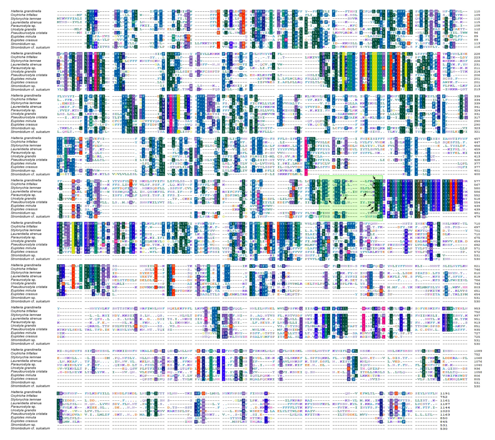
3.1. Ccmf and Other Split Protein-Coding Genes in Mitogenome

The ccmf protein is very important for aerobic eukaryote mitochondria, given that it is associated with maturation of cytochrome c which binds to heme and transfers electrons between respiratory chains [53]. In the present work, all mitogenomes of Spirotrichea species contain the ccmf gene. According to previous studies, the ccmf gene is suspected to be split into two parts in the mitogenomes of O. trifallax, L. strenua, U. grandis, and P. cristata [37,39,54]. According to De Graaf et al. 2009 [38], ccmf could also be a split gene for E. minuta and E. crassus, but have three parts (an additional split of ccmf_i for E. crassus and an additional split of ccmf_ii for E. minuta). Nevertheless, we found that the split of ccmf_i for E. crassus results from the insertion of an adenine nucleotide based on the comparison of our mitogenomes of E. crassus and E. vannus (unpublished) with the published data. Similarly, many insertions and deletions of ccmf_ii of E. minuta were detected. Although the insertion and deletion might come from the interspecific divergence, we think the additional split of ccmf_ii of E. minuta might not be a true split but instead is the product of sequencing errors. This speculation is consistent with the length, transmembrane (TM) profile, and sequence alignment of ccmf proteins as discussed below (Figure 5 and Figure 6).
The ccmf genes of H. grandinella, S. lemnae, and Paraurostyla sp. might also be split into two parts, with the following four findings supporting this hypothesis. Firstly, for the mitogenome in each species, two adjacent ORFs align with two parts of the ccmf protein (ccmf_i and ccmf_ii, respectively) based on a prediction with the NCBI Open Reading Frame Finder and SmartBLAST, with the best BLAST hit generally coming from one of the ciliate mitogenomes. Secondly, each of the two hypothetical split parts (ccmf_i and ccmf_ii) possess similar lengths among Hypotrichia and Euplotia mitogenomes, respectively: the lengths of ccmf_i are 543 amino acids for H. grandinella, 480–548 (avg. 524) amino acids for Hypotrichia, and 405 amino acids for Euplotia; the lengths of ccmf_ii are 648 amino acids for H. grandinella, 219–645 (avg. 547) amino acids for Hypotrichia, and 440–445 (avg. 442) amino acids for Euplotia. Thirdly, ccmf is a mitochondrial integral membrane protein containing multiple transmembrane helices [55]. In our work based on TM profiles, the transmembrane helix structure of hypothetical ccmf split proteins is similar among H. grandinella, Hypotrichia, and Euplotia (Figure 5). Fourthly, according to the alignment result of ccmf proteins in Spirotrichea, the split sites of ccmf_i and ccmf_ii are almost identical (Figure 6). All of these suggest that the ccmf gene is split into two parts in the mitogenomes of H. grandinella, Hypotrichia, and the Euplotia species described here.
However, as for the two Strombidium species, the ccmf gene is non-split because we only find one ORF that is homologous to cytochrome c maturation-related genes (Figure 1, Figure 2 and Figure 3 and Figure 5). According to the TM profiles, the ccmf of the two Strombidium species possesses 13–16 transmembrane helices, which is similar to that of other spirotrichous species (14–19 transmembrane helices). Besides, the split phenomenon was not detected based on the ccmf sequence of two Strombidium species, nearby the conserved split positions of Hypotrichia and Euplotia ccmf proteins (Figure 6). Hence, it appears that the mitochondrial ccmf gene is likely split for Hypotrichia and Euplotia species but corresponds to a single polypeptide for Oligotrichia species.
According to previous studies [37,39], the protein-coding genes nad1, nad2 and rps3 were reported as split genes in most sequenced ciliate mitogenomes. These three genes are also found split in the newly characterized three mitogenomes in the present study. This split appears to be ancestral and shared by the phylum Ciliophora. The non-split of the genes nad2 and rps3 in Euplotes mitogenomes could be due to sequencing and annotation errors or they were fused again in the Euplotes species. By contrast, the ccmf gene is found split only in Hypotrichia and Euplotia species, while it is not split in Oligotrichia and Oligohymenophorea, which indicates that the split of ccmf gene in Hypotrichia and Euplotia species is a derived character and probably evolved independently in these two groups. Notably, the protein-coding genes nad4, nad5, nad9, and rpl6 were also predicted as potential split genes in O. trifallax [39]. However, these genes are not split in other ciliate mitogenomes. The split of nad4, nad5, nad9, and rpl6 in O. trifallax may be not accurate due to sequencing errors, because of the fact that this mitogenome was sequenced with error-prone 454 sequencing technology. In conclusion, the split in genes nad1, nad2, and rps3 could be an ancestral characteristic among all ciliates. Meanwhile the split in ccmf is a specific feature within the subclasses Hypotrichia and Euplotia, which could be used for clarifying the phylogenetic relationships for these groups.

3.2. ORFs with Unknown Functions in Mitogenome

The ORFs with unknown functions have always been a problem for protist mitogenomes, and result in the trouble of annotation for mitochondrial genes. However, the high evolutionary rate of mitochondrial genes and low sequence identity between ORFs and known genes make it difficult to define these ORFs with unknown functions by BLAST searches [7,31]. Specifically, compared to known protein-coding genes, the ORFs with unknown functions have significantly higher Ka/Ks values and lower sequence similarities, suggesting a lower selective pressure on these ORFs [31,37]. Nevertheless, there are some ORFs that are homologous and reside in the same relative positions among the Spirotrichea mitogenomes (e.g., orf535, orf546, orf578, orf583, and orf592 in Table S4).
Except for the ORFs that are homologous among ciliates, there are also some taxon or group-specific ORFs. For example, no homologous sequence was detected in other ciliate mitogenomes for the orf_s1–s7 in the two Strombidium mitogenomes or for the six ORFs in the two Euplotes mitogenomes based on BLASTP searches (Figure 1, Table S4). Orf192 is only detected in the mitogenomes of H. grandinella and P. cristata, and orf549 is only detected in H. grandinella, O. trifallax, and S. lemnae. These taxon or group-specific ORFs might represent novel mitochondrial proteins or perform similar functions but be too divergent to find homologies [33,39].

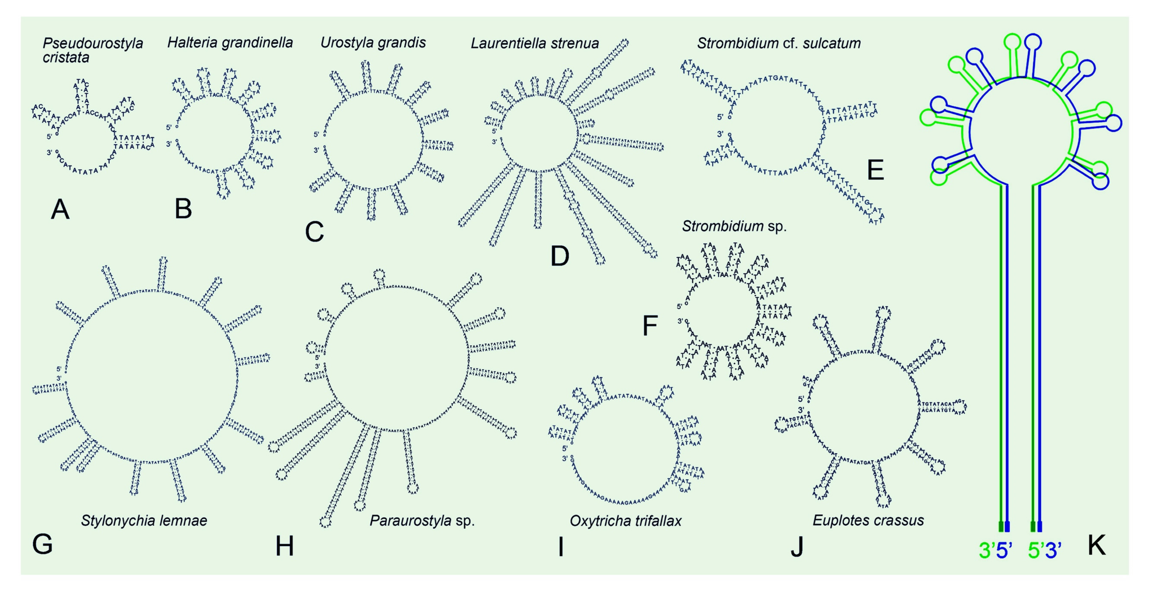
3.3. Repeat Regions in Mitogenomes

In ciliate mitogenomes, there are three kinds of repeat regions: terminal inverted repeats (TIR) (repeat sequences with opposite directions at each end of the linear molecule), telomeric repeats (a telomeric sequence composed of repeat unit that is usually repeated several times), and central repeats (a sequence comprised of repeat units at approximately a central position of the linear molecule), though some mitogenomes may contain one, two, or all of them. TIR is a common characteristic of linear mitogenomes from diverse eukaryotes as reviewed in [39] and is proposed as a solution of the incomplete 5′ end replication problem for linear molecules [56]. For ciliate mitogenomes, the TIR is only detected in several ciliate species, including Tetrahymena species, O. trifallax, S. lemnae, and L. strenua, with the size ranging from ~1800–3100 bp (Table 2). Notably, the TIR region in O. trifallax mitogenome appears to be largely comprised of non-annotated ORFs, while the TIR in S. lemnae’s and L. strenua’s is largely comprised of the large subunit ribosomal RNAs and tRNAs, which is similar to that in Tetrahymena species. Coincidentally, a high frequency of structural rearrangement events is also observed within the mitogenomes of Spirotrichea, i.e., the existence of similar gene sets in both ends of the mitogenomes in O. trifallax, S. lemnae, and L. strenua (Figure 3). These suggest that, instead of as a solution of the 5′ end replication problem, the TIR in Spirotrichea mitogenomes may play a role in its structural rearrangements, which may result from terminal inverted gene duplications and subsequent successive gene losses through homologous recombination between both termini.
According to Nosek and Tomáška [56], we speculate that the telomeric repeats in the Spirotrichea mitogenomes may play important roles in solving the end replication problem. Most Spirotrichea mitogenomes are capped by telomeric repeats, with the same sequence at both ends and an unknown repeat number (Table 2). The undetected telomeric repeat in some Spirotrichea mitogenomes is probably due to the incompleteness of the mitogenomes. However, compared to the highly conserved telomeric sequences in the nuclear genome, the telomeric repeats in the Spirotrichea mitogenomes are highly variable, ranging from 13–36 bp, even among closely related species (e.g., two Strombidium species). The telomeric repeats are also found in Tetrahymena mitogenomes (31–53 bp), but even different at each end in the T. pigmentosa mitogenome [28]. It was proposed that mitochondrial telomeres are derived from mobile elements (transposons or plasmids) [56]. A linear mitochondrial plasmid with the 3′ end containing the same telomeric repeats as the primary mitogenome was found in O. trifallax, which supports the previous proposal of the telomere transfer between mitogenomes and linear plasmids [39].
An AT-rich central tandem repeat is discovered in all the available Spirotrichea mitogenomes, where a bidirectional transcription initiates from this region. The repeat region comprises a palindromic sequence and could be predicted to form stable stem-loop structures (Figure 1, Figure 3 and Figure 4). Similarly, Tetrahymena mitogenomes (Cl.: Oligohymenophorea) also contain a conserved putative GC-box control region located in the highly AT-rich stretch, which is speculated to be significant to bidirectional transcription [28]. Except for the ciliated protists, tandem repeat and stem-loop structures are also reported in mitogenomes of numerous organisms, for instance, unicellular cryptophyte algae (Rhodomonas salina [57] and Hemiselmis andersenii [58]), plants (Chondrus crispus [59] and Porphyra purpurea [60]) and animals (chickens [61] and lagomorphs [62]). According to previous studies, the tandem repeat and hairpin structure may be associated with a promoter and involved in the regulation of transcription initiation [57,58,61]. Additionally, the transcription control region of mitogenomes was demonstrated to be AT-rich [28,61]. All of these points mentioned above imply that the central tandem repeat may play an important role in the bidirectional transcription of the mitochondrial genes in Spirotrichea species.
Like the mitogenomes of diverse organisms [28,30,57,58], the position of transcription initiation also corresponds to origin of DNA replication, which means that the central repeat regions in the Spirotrichea mitogenomes could also be the replication origin. In Paramecium mitogenomes, the possession of covalently-closed single-stranded hairpins (consisting of direct tandem repeats) at one end and an open structure at the opposite side was found. The hairpin structure was demonstrated to be associated with a replication origin [27,63]. In these structural features, replication is initiated through the hairpin loop generating a dimer molecule, which is then processed to two cross-linked monomers [27,56]. Recently, more Paramecium mitogenomes were published [33]. We found that, instead of an open structure at the opposite end, a linear molecule terminates with telomeric repeats ranging from 23–33 bp. For the Spirotrichea mitogenomes, if we fold them from the central regions, their structures are surprisingly analogous to that in Paramecium mitogenomes: with covalently-closed sun-shape central repeat at one end and telomeric repeats at the other end (Figure 4K). As a consequence, we speculate that the central repeat region may serve as the replication initiation in Spirotrichea mitogenomes. More molecular information and experimental evidence are needed to test this speculation.

3.4. Phylogenetic Position of Halteria grandinella

The family Halteriidae, represented by H. grandinella, was assigned into the subclass of Oligotrichia in consideration of its similar morphological characteristics with oligotrichs, for example, the globular/ovoid cell shape, planktonic life style, the reduced somatic ciliature, apical oral membranelles, de novo-derived undulating membrane, and enantiotropy pattern for cell division [46,47]. All these characteristics are divergent with those in hypotrichs, such as mostly dorsoventrally compressed, mostly benthic life style, well-developed compound ventral cirri and dorsal kineties, oral membranelles on left-anterior portion of the ventral surface, as well as a synclastic pattern for cell division [48]. However, H. grandinella was always closely related to hypotrichs based on phylogenetic trees inferred from nuclear data [19,48,49,50,51,52]. This is concordant with our phylogenetic analyses based on mitochondrial data (Figure 7).
Except for the phylogenetic analyses, the characteristics of the mitogenome also support the close relationship of H. grandinella and hypotrichs. In the present study, the mitogenome of H. grandinella exhibits more extensive synteny with those of hypotrichs than oligotrichs, especially the complete collinearity with that of O. trifallax (Figure 3). Furthermore, the ccmf of H. grandinella is split into two parts, which is consistent with hypotrichs but different from the two oligotrichs whose ccmf is non-split (Figure 5). It is noteworthy that the mitogenomes of Strombidium species contain specific ORFs (orf_s1–s7) which are not detected in H. grandinella. The foregoing findings demonstrate that Halteria has a closer relationship with hypotrichs than oligotrichs.
According to Lynn and Kolisko [19] and Wang et al. 2019 [48], the morphological features shared between halteriids and oligotrichs are likely convergent characteristics for planktonic lifestyle and H. grandinella might be a “oligotrich-like” hypotrich. Both our mitogenome information and phylogenetic analyses support this hypothesis, though limited mitochondrial data in the subclass Oligotrichia is available now. Besides, as discussed in Wang et al. 2019 [48], H. grandinella shared more nuclear orthologous genes with oxytrichids than with oligotrichs. Considering that Halteria is the type genus of the family Halteriidae, we agree with Lynn [64] that Halteriidae should be classified into the subclass Hypotrichia and it has a closer relationship with the family Oxytrichidae than other families within Hypotrichia.

4. Materials and Methods

4.1. Cell Cultures

Halteria grandinella was collected from the pond of the Baihuayuan Park in Qingdao (36°04′ N, 120°22′ E), China. Several cells were isolated by glass micropipette under microscope and washed 4–5 times, and then were cultured in filtered and autoclaved in situ water at room temperature, with Escherichia coli as food source. Strombidium sp. was kindly supplied by Dr. Sheng-Fang Tsai of the Institute of Environmental Biology and Fisheries Science in National Taiwan Ocean University. Strombidium cf. sulcatum was sampled from Daya Bay in Guangdong (22°37′ N, 114°38′ E), China. Both Strombidium species were cultured in filtered and autoclaved marine water at room temperature with E. coli as food source. All of the three morphospecies were identified by live microscopic observation and silver impregnations [65].

4.2. Mitochondrial DNA, Genomic DNA Extraction, and High-Throughput Sequencing

For the cultures of H. grandinella and Strombidium sp., cells were starved and treated with antibiotic-antimycotic (2 mL for 1 L, ThermoFisher Scientific, Waltham, MA, USA; Cat No. 15,240,062) for about 24 h before mitochondria isolation. Samples were collected by centrifugation at 200× g for 3 min. The total numbers of cells were approximately 3 × 10⁴ for H. grandinella and 5 × 10⁴ for Strombidium sp. Mitochondria were isolated using a Qproteome Mitochondria Isolation Kit (Qiagen, Hilden, Germany; Cat No. 37,612), following the manufacturer’s protocol without washing with 0.9% sodium chloride solution. Subsequently, the DNA extraction was performed by DNeasy Blood & Tissue Kit (Qiagen, Hilden, Germany; Cat No. 69,506). Finally, the mitochondrial DNA was amplified using a REPLI-g® Mitochondrial DNA Kit (Qiagen, Hilden, Germany; Cat No. 151,023) with 10 µL template DNA.
For the culture of S. cf. sulcatum, cells were starved and treated with the antibiotic-antimycotic as described above. After that, cells were harvested by filtering through a 5 µm membrane (Shanghai Xin Ya Purification Equipment, Shanghai, China). Subsequently, cells (ca. 2 × 10⁵) were lysed using Proteinase K solution from DNeasy Blood & Tissue Kit (Qiagen, Hilden, Germany; Cat No. 69,506) and a lysis buffer (10 mM Tris-HCl, 25 mM EDTA, 0.5% SDS) at 56 °C in a water bath for 3 h. Genomic DNA was then extracted by Phenol-Chloroform method. RNA was eliminated with RNase A (Omega Bio-tek, Norcross, GA, USA; Lot No. L10RG).
Three illumina libraries (350 bp) were constructed using NEBNext® Ultra™ DNA Library Prep Kit (NEB, Ipswich, MA, USA; Cat No. E7370L) according to the manufacturer’s instructions: mitochondrial DNA of H. grandinella, mitochondrial DNA of Strombidium sp., and genomic DNA of S. cf. sulcatum. Paired-end sequencing (150 bp reads length) was performed using an Illumina HiSeq4000 sequencer (Novogene, Beijing, China). Sequencing adapters and low-quality reads (more than 50% bases with Q value = < 5) were removed by FASTX-Toolkit with the parameters of -q 5 -p 0.5 [66].

4.3. Mitogenome Assembly and Annotation

Mitogenomes of H. grandinella and Strombidium sp. as well as the whole genome of S. cf. sulcatum, were assembled using SPAdes v.3.11 with default parameters varying k-values: 21, 33, 55, and 77 [13,67]. Afterwards, assemblies were screened by BLASTN (e-value = < 1 × 10⁻⁵) using eight mitogenomes downloaded from the National Center for Biotechnology Information (NCBI) as a reference database, including Oxytricha trifallax (acc. no. JN383843), Tetrahymena pyriformis (acc. no. AF160864), Tetrahymena thermophila (acc. no. AF396436), Paramecium aurelia (acc. no. NC001324), Paramecium caudatum (acc. no. FN424190), Euplotes minuta (acc. no. GQ903130), Euplotes crassus (acc. no. GQ903131), and Nyctotherus ovalis (acc. no. GU057832).
For H. grandinella and Strombidium sp., one resulting contig (i.e., mitogenome) possessing high similarity with the reference data was found in each species (similarity: >72.24% in H. grandinella; >72.73% in Strombidium sp.). For S. cf. sulcatum, three contigs were recovered (similarity: >72.82%). These three contigs were subsequently used as references for the second search in the genomic data with BLASTN (using an e-value threshold of = < 1 × 10⁻⁵) and another six contigs were obtained. Nine mitochondrial contigs of S. cf. sulcatum with sizes ranging from about 1.2 kb to 19 kb were assembled into two scaffolds by SeqMan v.7.1.0 (DNAStar). Polymerase chain reactions (PCRs) were performed to confirm the junctions of these contigs and link the two scaffolds together into a linear mitogenome (Figure S1). The PCR amplifications were performed using the 2× EasyTaq® PCR SuperMix (Transgen Biotech, Beijing, China; Cat No. AS111–14), with primers designed according to the nine mitochondrial contigs (Table S1). PCR products were purified by EasyPure Quick Gel Extraction Kit (Transgen Biotech, Beijing, China; Cat No. EG101–01) and cloned using a pEASY-T1 Cloning Kit (Transgen Biotech, Beijing, China; Cat No. CT101–01), or directly sequenced bidirectionally in Tsingke Biological Technology Company (Beijing, China). Raw reads data of the three taxa were respectively remapped onto the mitogenome to calculate the coverage through HISAT2 v.2.1.0 [68] and SAMtools [69]. Integrative Genomics Viewer v.2.4.6 [70] was employed to visualize coverage results (Figure S2).
Three mitogenomes were annotated firstly using both MITOS and MFannot with genetic code 4 of “the mold, protozoan, and coelenterate mitochondrial code and the mycoplasma/spiroplasma code” [71,72]. Then, the protein-coding genes were further verified by the NCBI Open Reading Frame Finder (https://0-www-ncbi-nlm-nih-gov.brum.beds.ac.uk/orffinder (accessed on 23 February 2021)) and annotated by searching against the NCBI non-redundant protein sequences (nr) database with SmartBLAST or BLASTP. The ribosomal RNA (rRNA) genes were confirmed by BLASTN searches against the NCBI nucleotide collection (nr/nt) database. The transfer RNA (tRNA) genes were also predicted using tRNAscan-SE v.2.0 with default mode and other mitogenomes as reference sources [73]. Mitogenome maps were drawn using OGDRAW [74]. The secondary structures of the central repeated portion of the mitogenomes were predicted using the Mfold web server (http://www.unafold.org/mfold/applications/rna-folding-form.php (accessed on 23 February 2021)) with default parameters. RnaViz v.2.0 [75] was used for aesthetic purposes. The transmembrane (TM) structure of the ccmf protein which is involved in cytochrome c maturation in mitochondria was predicted by TMHMM v.2.0 [76].

4.4. Phylogenetic Analyses and Topology Testing

Fourteen conserved mitochondrial protein sequences were used to construct phylogenetic trees. Newly characterized sequences were combined with relevant orthologs of the other 31 ciliates downloaded from the GenBank database, resulting in fourteen mitochondrial datasets (Table S2). Stentor coeruleus and Gruberia lanceolata were selected as the outgroup taxa. The fourteen datasets were aligned using the MAFFT algorithm on the GUIDANCE2 Server (http://guidance.tau.ac.il/ver2/ (accessed on 23 February 2021)) and trimmed by trimAl v.1.4 with default parameters [77]. Subsequently, fourteen mitochondrial alignments were concatenated using SeaView v.4 [78]. The final alignments used for phylogenetic analyses included 3730 sites. Maximum likelihood (ML) analysis was performed by IQ-tree v.1.5.5 [79] with the model LG+C60+F+G4. In total, 100 bootstrap replicates were run to calculate the standard nonparametric bootstrap supports. Bayesian inference (BI) analysis was carried out by PhyloBayes v.4.1 [80] under the model CAT+GTR. Four Markov chain Monte Carlo (MCMC) simulations were run for 1,000,000 generations. The bpcomp program of PhyloBayes was performed to compare the discrepancy of bipartition frequencies, generating a consensus tree. The tree topologies were visualized using FigTree v.1.4.4 (http://tree.bio.ed.ac.uk/software/figtree/ (accessed on 23 February 2021)).
To test the monophyly of H. grandinella and the subclass Oligotrichia, the constrained ML tree was generated with a constraint block by enforcing the monophyly of H. grandinella and two Strombidium species. Subsequently, the approximately unbiased (AU) test [81] was performed using IQ-tree v.1.5.5 [79].

Supplementary Materials

Author Contributions

Conceptualization, supervision, and funding acquisition, F.G.; investigation, T.Z., C.L., X.Z., and C.W.; writing—review and editing, T.Z., C.L., X.Z., C.W., A.J.R., and F.G. All authors have read and agreed to the published version of the manuscript.

Funding

This research was funded by the National Natural Science Foundation of China (Project No. 31922013, 32030015 and 31772428), and the Natural Science Foundation of Shandong Province (ZR2020JQ13).

Institutional Review Board Statement

Not applicable.

Informed Consent Statement

Not applicable.

Data Availability Statement

Three new mitogenome assemblies and annotation data in this study have been submitted to the GenBank database under accession numbers as follows: MT471315 for mitogenome of Strombidium sp., MT471316 for mitogenome of Strombidium cf. sulcatum, and MT471317 for mitogenome of Halteria grandinella.

Acknowledgments

We are grateful to Sheng-Fang Tsai at the Institute of Environmental Biology and Fisheries Science, National Taiwan Ocean University, who kindly supplied us the isolate of Strombidium sp. We thank Weibo Song (OUC) for the helpful suggestions in the manuscript and illustrations. Our thanks are also given to Wen Song (PhD graduate of OUC) for sample collection and species identification, Liping Lv (graduate student of OUC) for his help with data analyses and Shelby Williams (PhD student of Dalhousie University) for her help with the IQ-tree. We acknowledge the computing resources provided on IEMB-1, a high-performance computing cluster operated by the Institute of Evolution and Marine Biodiversity, OUC.

Conflicts of Interest

The authors declare no conflict of interest.

References

  1. Roger, A.J.; Muñoz-Gómez, S.A.; Kamikawa, R. The origin and diversification of mitochondria. Curr. Biol. 2017, 27, R1177–R1192. [Google Scholar] [CrossRef] [PubMed] [Green Version]
  2. Hampl, V.; Čepička, I.; Eliáš, M. Was the mitochondrion necessary to start eukaryogenesis? Trends Microbiol. 2019, 27, 96–104. [Google Scholar] [CrossRef] [PubMed]
  3. Gray, M.W. Rickettsia, typhus and the mitochondrial connection. Nature 1998, 396, 109–110. [Google Scholar] [CrossRef] [PubMed]
  4. Mottis, A.; Herzig, S.; Auwerx, J. Mitocellular communication: Shaping health and disease. Science 2019, 366, 827–832. [Google Scholar] [CrossRef]
  5. Hämäläinen, R.H.; Landoni, J.C.; Ahlqvist, K.J.; Goffart, S.; Ryytty, S.; Rahman, M.O.; Brilhante, V.; Icay, K.; Hautaniemi, S.; Wang, L. Defects in mtDNA replication challenge nuclear genome stability through nucleotide depletion and provide a unifying mechanism for mouse progerias. Nat. Metab. 2019, 1, 958–965. [Google Scholar] [CrossRef] [PubMed] [Green Version]
  6. Avise, J.C.; Arnold, J.; Ball, R.M.; Bermingham, E.; Lamb, T.; Neigel, J.E.; Reeb, C.A.; Saunders, N.C. Intraspecific phylogeography: The mitochondrial DNA bridge between population genetics and systematics. Annu. Rev. Ecol. Syst. 1987, 18, 489–522. [Google Scholar] [CrossRef]
  7. Rand, D.M. The units of selection on mitochondrial DNA. Annu. Rev. Ecol. Syst. 2003, 32, 415–448. [Google Scholar] [CrossRef]
  8. Cole, L.W.; Guo, W.; Mower, J.P.; Palmer, J.D. High and variable rates of repeat-mediated mitochondrial genome rearrangement in a genus of plants. Mol. Biol. Evol. 2018, 35, 2773–2785. [Google Scholar] [CrossRef] [PubMed]
  9. Fritsch, E.S.; Chabbert, C.D.; Klaus, B.; Steinmetz, L.M. A genome-wide map of mitochondrial DNA recombination in yeast. Genetics 2014, 198, 755–771. [Google Scholar] [CrossRef] [Green Version]
  10. Ladoukakis, E.D.; Zouros, E. Direct evidence for homologous recombination in mussel (Mytilus galloprovincialis) mitochondrial DNA. Mol. Biol. Evol. 2001, 18, 1168–1175. [Google Scholar] [CrossRef] [Green Version]
  11. Gray, M.W.; Lang, B.F.; Burger, G. Mitochondria of protists. Annu. Rev. Genet. 2004, 38, 477–524. [Google Scholar] [CrossRef]
  12. Li, X.; Huang, J.; Filker, S.; Stoeck, T.; Bi, Y.; Yu, Y.; Song, W. Spatio-temporal patterns of zooplankton in a main-stem dam affected tributary: A case study in the Xiangxi River of the Three Gorges Reservoir, China. Sci. China Life Sci. 2019, 62, 1058–1069. [Google Scholar] [CrossRef] [PubMed]
  13. Chen, X.; Jiang, Y.; Gao, F.; Zheng, W.; Krock, T.J.; Stover, N.A.; Lu, C.; Katz, L.A.; Song, W. Genome analyses of the new model protist Euplotes vannus focusing on genome rearrangement and resistance to environmental stressors. Mol. Ecol. Resour. 2019, 19, 1292–1308. [Google Scholar] [CrossRef]
  14. Cheng, T.; Wang, Y.; Huang, J.; Chen, X.; Zhao, X.; Gao, S.; Song, W. Our recent progress in epigenetic research using the model ciliate, Tetrahymena thermophila. Mar. Life Sci. Technol. 2019, 1, 4–14. [Google Scholar] [CrossRef] [Green Version]
  15. Wang, Y.Y.; Sheng, Y.; Liu, Y.; Zhang, W.; Cheng, T.; Duan, L.; Pan, B.; Qiao, Y.; Liu, Y.; Gao, S. A distinct class of eukaryotic MT-A70 methyltransferases maintain symmetric DNA N6-adenine methylation at the ApT dinucleotides as an epigenetic mark associated with transcription. Nucleic Acids Res. 2019, 47, 11771–11789. [Google Scholar] [CrossRef] [PubMed]
  16. Wang, Y.R.; Wang, C.; Jiang, Y.; Katz, A.L.; Gao, F.; Yan, Y. Further analyses of variation of ribosome DNA copy number and polymorphism in ciliates provide insights relevant to studies of both molecular ecology and phylogeny. Sci. China Life Sci. 2019, 62, 203–214. [Google Scholar] [CrossRef] [PubMed]
  17. Yan, Y.; Maurer-Alcalá, X.X.; Knight, R.; Pond, S.L.K.; Katz, L.A. Single-cell transcriptomics reveal a correlation between genome architecture and gene family evolution in ciliates. mBio 2019, 10, e02524-19. [Google Scholar] [CrossRef] [Green Version]
  18. Zhao, X.; Xiong, J.; Mao, F.; Sheng, Y.; Chen, X.; Feng, L.; Dui, W.; Yang, W.; Kapusta, A.; Feschotte, C. RNAi-dependent Polycomb repression controls transposable elements in Tetrahymena. Genes Dev. 2019, 33, 348–364. [Google Scholar] [CrossRef] [PubMed] [Green Version]
  19. Lynn, D.H.; Kolisko, M. Molecules illuminate morphology: Phylogenomics confirms convergent evolution among ‘oligotrichous’ ciliates. Int. J. Syst. Evol. Microbiol. 2017, 67, 3676–3682. [Google Scholar] [CrossRef] [PubMed]
  20. Slabodnick, M.M.; Ruby, J.G.; Reiff, S.B.; Swart, E.C.; Gosai, S.; Prabakaran, S.; Witkowska, E.; Larue, G.E.; Fisher, S.; Freeman, R.M. The macronuclear genome of Stentor coeruleus reveals tiny introns in a giant cell. Curr. Biol. 2017, 27, 569–575. [Google Scholar] [CrossRef] [PubMed] [Green Version]
  21. Sheng, Y.; Duan, L.; Cheng, T.; Qiao, Y.; Stover, N.A.; Gao, S. The completed macronuclear genome of a model ciliate Tetrahymena thermophila and its application in genome scrambling and copy number analyses. Sci. China Life Sci. 2020, 63, 1534–1542. [Google Scholar] [CrossRef]
  22. Wang, Y.R.; Jiang, Y.; Liu, Y.; Li, Y.; Katz, L.A.; Gao, F.; Yan, Y. Comparative studies on the polymorphism and copy number variation of mtSSU rDNA in ciliates (Protista, Ciliophora): Implications for phylogenetic, environmental, and ecological research. Microorganisms 2020, 8, 316. [Google Scholar] [CrossRef] [PubMed] [Green Version]
  23. Ma, M.; Li, Y.; Ma, H.; Al-Rasheid, K.A.; Warren, A.; Wang, Y.; Yan, Y. Morphology and molecular phylogeny of four trachelocercid ciliates (Protozoa, Ciliophora, Karyorelictea) found in marine coastal habitats of northern China, with description of a new genus, two new species and a new combination. Front. Mar. Sci. 2021, 7, 615903. [Google Scholar] [CrossRef]
  24. Gray, M.W.; Lang, B.F.; Cedergren, R.; Golding, G.B.; Lemieux, C.; Sankoff, D.; Turmel, M.; Brossard, N.; Delage, E.; Littlejohn, T.G. Genome structure and gene content in protist mitochondrial DNAs. Nucleic Acids Res. 1998, 26, 865–878. [Google Scholar] [CrossRef] [PubMed]
  25. Suyama, Y.; Miura, K. Size and structural variations of mitochondrial DNA. Proc. Natl. Acad. Sci. USA 1968, 60, 235–242. [Google Scholar] [CrossRef] [Green Version]
  26. Morin, G.B.; Cech, T.R. The telomeres of the linear mitochondrial DNA of Tetrahymena thermophila consist of 53 bp tandem repeats. Cell 1986, 46, 873–883. [Google Scholar] [CrossRef]
  27. Pritchard, A.E.; Cummings, D.J. Replication of linear mitochondrial DNA from Paramecium: Sequence and structure of the initiation-end crosslink. Proc. Natl. Acad. Sci. USA 1981, 78, 7341–7345. [Google Scholar] [CrossRef] [PubMed] [Green Version]
  28. Moradian, M.M.; Beglaryan, D.; Skozylas, J.M.; Kerikorian, V. Complete mitochondrial genome sequence of three Tetrahymena species reveals mutation hot spots and accelerated nonsynonymous substitutions in Ymf genes. PLoS ONE 2007, 2, e650. [Google Scholar] [CrossRef] [PubMed]
  29. Burger, G.; Zhu, Y.; Littlejohn, T.G.; Greenwood, S.J.; Schnare, M.N.; Lang, B.F.; Gray, M.W. Complete sequence of the mitochondrial genome of Tetrahymena pyriformis and comparison with Paramecium aurelia mitochondrial DNA. J. Mol. Biol. 2000, 297, 365–380. [Google Scholar] [CrossRef] [Green Version]
  30. Brunk, C.F.; Lee, L.C.; Tran, A.B.; Li, J. Complete sequence of the mitochondrial genome of Tetrahymena thermophila and comparative methods for identifying highly divergent genes. Nucleic Acids Res. 2003, 31, 1673–1682. [Google Scholar] [CrossRef] [Green Version]
  31. Barth, D.; Berendonk, T.U. The mitochondrial genome sequence of the ciliate Paramecium caudatum reveals a shift in nucleotide composition and codon usage within the genus Paramecium. BMC Genom. 2011, 12, 272. [Google Scholar] [CrossRef] [PubMed] [Green Version]
  32. Valverde, J.R.; Marco, R.; Garesse, R. A conserved heptamer motif for ribosomal RNA transcription termination in animal mitochondria. Proc. Natl. Acad. Sci. USA 1994, 91, 5368–5371. [Google Scholar] [CrossRef] [PubMed] [Green Version]
  33. Johri, P.; Marinov, G.K.; Doak, T.G.; Lynch, M. Population genetics of Paramecium mitochondrial genomes: Recombination, mutation spectrum, and efficacy of selection. Genome Biol. Evol. 2019, 11, 1398–1416. [Google Scholar] [CrossRef] [Green Version]
  34. Johri, P.; Krenek, S.; Marinov, G.K.; Doak, T.G.; Berendonk, T.U.; Lynch, M. Population genomics of Paramecium species. Mol. Biol. Evol. 2017, 34, 1194–1216. [Google Scholar] [CrossRef] [PubMed]
  35. Li, R.; Gao, Y.; Hou, Y.; Ye, S.; Wang, L.; Sun, J.; Li, Q. Mitochondrial genome sequencing and analysis of scuticociliates (Uronema marinum) isolated from Takifugu rubripes. Mitochondrial DNA Part B 2018, 3, 736–737. [Google Scholar] [CrossRef] [PubMed] [Green Version]
  36. Watt, A.; Haites, R.; Young, N.; Billman-Jacobe, H. The mitochondrial genome of Tetrahymena rostrata. Mitochondrial DNA Part B 2020, 5, 53–54. [Google Scholar] [CrossRef]
  37. Chen, X. Genome Architecture, Rearrangement and Evolution in the Ciliate Oxytricha Trifallax. Ph.D. Thesis, Princeton University, Princeton, NJ, USA, 2015. [Google Scholar]
  38. De Graaf, R.M.; Van Alen, T.A.; Dutilh, B.E.; Kuiper, J.W.; Van Zoggel, H.J.; Huynh, M.B.; Görtz, H.-D.; Huynen, M.A.; Hackstein, J.H. The mitochondrial genomes of the ciliates Euplotes minuta and Euplotes crassus. BMC Genom. 2009, 10, 514. [Google Scholar] [CrossRef] [Green Version]
  39. Swart, E.C.; Nowacki, M.; Shum, J.; Stiles, H.; Higgins, B.P.; Doak, T.G.; Schotanus, K.; Magrini, V.J.; Minx, P.; Mardis, E.R. The Oxytricha trifallax mitochondrial genome. Genome Biol. Evol. 2011, 4, 136–154. [Google Scholar] [CrossRef] [Green Version]
  40. Park, M.-H.; Min, G.-S. The complete mitochondrial genome of Gruberia lanceolata (Gruber, 1884) Kahl, 1932 (Ciliophora: Heterotrichea). Mitochondrial DNA Part B 2019, 4, 3443–3445. [Google Scholar] [CrossRef]
  41. De Graaf, R.M.; Ricard, G.; Van Alen, T.A.; Duarte, I.; Dutilh, B.E.; Burgtorf, C.; Kuiper, J.W.; Van Der Staay, G.W.; Tielens, A.G.; Huynen, M.A. The organellar genome and metabolic potential of the hydrogen-producing mitochondrion of Nyctotherus ovalis. Mol. Biol. Evol. 2011, 28, 2379–2391. [Google Scholar] [CrossRef] [PubMed] [Green Version]
  42. Rotterová, J.; Salomaki, E.; Pánek, T.; Bourland, W.; Žihala, D.; Táborský, P.; Edgcomb, V.P.; Beinart, R.A.; Kolísko, M.; Čepička, I. Genomics of new ciliate lineages provides insight into the evolution of obligate anaerobiosis. Curr. Biol. 2020, 30, 2037–2050. [Google Scholar] [CrossRef]
  43. Lewis, W.H.; Lind, A.E.; Sendra, K.M.; Onsbring, H.; Williams, T.A.; Esteban, G.F.; Hirt, R.P.; Ettema, T.J.; Embley, T.M. Convergent evolution of hydrogenosomes from mitochondria by gene transfer and loss. Mol. Biol. Evol. 2020, 37, 524–539. [Google Scholar] [CrossRef] [PubMed]
  44. Coyne, R.S.; Hannick, L.; Shanmugam, D.; Hostetler, J.B.; Brami, D.; Joardar, V.S.; Johnson, J.; Radune, D.; Singh, I.; Badger, J.H. Comparative genomics of the pathogenic ciliate Ichthyophthirius multifiliis, its free-living relatives and a host species provide insights into adoption of a parasitic lifestyle and prospects for disease control. Genome Biol. 2011, 12, R100. [Google Scholar] [CrossRef] [PubMed] [Green Version]
  45. Zhang, T.; Fan, X.; Gao, F.; Al-Farraj, S.A.; El-Serehy, H.A.; Song, W. Further analyses on the phylogeny of the subclass Scuticociliatia (Protozoa, Ciliophora) based on both nuclear and mitochondrial data. Mol. Phylogenet. Evol. 2019, 139, 106565. [Google Scholar] [CrossRef]
  46. Agatha, S. A cladistic approach for the classification of oligotrichid ciliates (Ciliophora: Spirotricha). Acta Protozool. 2004, 43, 201–217. [Google Scholar]
  47. Foissner, W.; Muller, H.; Agatha, S. A comparative fine structural and phylogenetic analysis of resting cysts in oligotrich and hypotrich Spirotrichea (Ciliophora). Eur. J. Protistol. 2007, 43, 295–314. [Google Scholar] [CrossRef] [PubMed] [Green Version]
  48. Wang, C.; Yan, Y.; Chen, X.; Al-Farraj, S.A.; El-Serehy, H.A.; Gao, F. Further analyses on the evolutionary “key-protist” Halteria (Protista, Ciliophora) based on transcriptomic data. Zool. Scr. 2019, 48, 813–825. [Google Scholar] [CrossRef]
  49. Gao, F.; Warren, A.; Zhang, Q.; Gong, J.; Miao, M.; Sun, P.; Xu, D.; Huang, J.; Yi, Z.; Song, W. The all-data-based evolutionary hypothesis of ciliated protists with a revised classification of the phylum Ciliophora (Eukaryota, Alveolata). Sci. Rep. 2016, 6, 24874. [Google Scholar] [CrossRef] [PubMed] [Green Version]
  50. Hewitt, E.A.; Muller, K.M.; Cannone, J.; Hogan, D.J.; Gutell, R.; Prescott, D.M. Phylogenetic relationships among 28 spirotrichous ciliates documented by rDNA. Mol. Phylogenet. Evol. 2003, 29, 258–267. [Google Scholar] [CrossRef]
  51. Paiva, T.S.; Borges, B.N.; Harada, M.L.; Silva-Neto, I.D. Comparative phylogenetic study of Stichotrichia (Alveolata: Ciliophora: Spirotrichea) based on 18S-rDNA sequences. Genet. Mol. Res. 2009, 8, 223–246. [Google Scholar] [CrossRef]
  52. Zheng, W.; Wang, C.; Lynch, M.; Gao, S. The compact macronuclear genome of the ciliate Halteria grandinella: A transcriptome-like genome with 23,000 nanochromosomes. mBio 2021, 12, e01964-20. [Google Scholar] [CrossRef]
  53. Nishimura, Y.; Tanifuji, G.; Kamikawa, R.; Yabuki, A.; Hashimoto, T.; Inagaki, Y. Mitochondrial genome of Palpitomonas bilix: Derived genome structure and ancestral system for cytochrome c maturation. Genome Biol. Evol. 2016, 8, 3090–3098. [Google Scholar] [CrossRef] [Green Version]
  54. Park, K.M.; Min, G.S.; Kim, S. The mitochondrial genome of the ciliate Pseudourostyla cristata (Ciliophora, Urostylida). Mitochondrial DNA Part B 2019, 4, 66–67. [Google Scholar] [CrossRef] [Green Version]
  55. Allen, J.W.; Jackson, A.P.; Rigden, D.J.; Willis, A.C.; Ferguson, S.J.; Ginger, M.L. Order within a mosaic distribution of mitochondrial c-type cytochrome biogenesis systems? FEBS J. 2008, 275, 2385–2402. [Google Scholar] [CrossRef] [PubMed]
  56. Nosek, J.; Tomáška, Ľ. Mitochondrial genome diversity: Evolution of the molecular architecture and replication strategy. Curr. Genet. 2003, 44, 73–84. [Google Scholar] [CrossRef]
  57. Hauth, A.M.; Maier, U.G.; Lang, B.F.; Burger, G. The Rhodomonas salina mitochondrial genome: Bacteria-like operons, compact gene arrangement and complex repeat region. Nucleic Acids Res. 2005, 33, 4433–4442. [Google Scholar] [CrossRef] [PubMed] [Green Version]
  58. Kim, E.; Lane, C.E.; Curtis, B.A.; Kozera, C.; Bowman, S.; Archibald, J.M. Complete sequence and analysis of the mitochondrial genome of Hemiselmis andersenii CCMP644 (Cryptophyceae). BMC Genom. 2008, 9, 215. [Google Scholar] [CrossRef] [PubMed] [Green Version]
  59. Richard, O.; Bonnard, G.; Grienenberger, J.M.; Kloareg, B.; Boyen, C. Transcription initiation and RNA processing in the mitochondria of the red alga Chondrus crispus: Convergence in the evolution of transcription mechanisms in mitochondria. J. Mol. Biol. 1998, 283, 549–557. [Google Scholar] [CrossRef] [PubMed]
  60. Burger, G.; Saint-Louis, D.; Gray, M.W.; Lang, B.F. Complete sequence of the mitochondrial DNA of the red alga Porphyra purpurea: Cyanobacterial introns and shared ancestry of red and green algae. Plant Cell 1999, 11, 1675–1694. [Google Scholar] [CrossRef] [PubMed] [Green Version]
  61. L’abbé, D.; Duhaime, J.F.; Lang, B.F.; Morais, R. The transcription of DNA in chicken mitochondria initiates from one major bidirectional promoter. J. Biol. Chem. 1991, 266, 10844–10850. [Google Scholar] [CrossRef]
  62. Casane, D.; Dennebouy, N.; De Rochambeau, H.; Mounolou, J.; Monnerot, M. Nonneutral evolution of tandem repeats in the mitochondrial DNA control region of lagomorphs. Mol. Biol. Evol. 1997, 14, 779–789. [Google Scholar] [CrossRef] [PubMed] [Green Version]
  63. Pritchard, A.; Laping, J.; Seilhamer, J.; Cummings, D. Inter-species sequence diversity in the replication initiation region of Paramecium mitochondrial DNA. J. Mol. Biol. 1983, 164, 1–15. [Google Scholar] [CrossRef]
  64. Lynn, D.H. The Ciliated Protozoa: Characterization, Classification and Guide to the Literature, 3rd ed.; Springer: Dordrecht, The Netherlands, 2008. [Google Scholar]
  65. Wilbert, N. Eine verbesserte Technik der Protargolimprä gnation für Ciliaten. Mikrokosmos 1975, 64, 171–179. [Google Scholar]
  66. Gordon, A.; Hannon, G. Fastx-toolkit. FASTQ/A short-reads preprocessing tools. 2010, unpublished. [Google Scholar]
  67. Chen, X.; Wang, C.; Pan, B.; Lu, B.; Li, C.; Shen, Z.; Warren, A.; Li, L. Single-cell genomic sequencing of three peritrichs (Protista, Ciliophora) reveals less biased stop codon usage and more prevalent programmed ribosomal frameshifting than in other ciliates. Front. Mar. Sci. 2020, 7, 602323. [Google Scholar] [CrossRef]
  68. Kim, D.; Langmead, B.; Salzberg, S.L. HISAT: A fast spliced aligner with low memory requirements. Nat. Methods 2015, 12, 357–360. [Google Scholar] [CrossRef] [Green Version]
  69. Li, H.; Handsaker, B.; Wysoker, A.; Fennell, T.; Ruan, J.; Homer, N.; Marth, G.; Abecasis, G.; Durbin, R. The sequence alignment/map format and SAMtools. Bioinformatics 2009, 25, 2078–2079. [Google Scholar] [CrossRef] [Green Version]
  70. Robinson, J.T.; Thorvaldsdóttir, H.; Wenger, A.M.; Zehir, A.; Mesirov, J.P. Variant review with the integrative genomics viewer. Cancer Res. 2017, 77, e31–e34. [Google Scholar] [CrossRef] [PubMed] [Green Version]
  71. Valach, M.; Burger, G.; Gray, M.W.; Lang, B.F. Widespread occurrence of organelle genome-encoded 5S rRNAs including permuted molecules. Nucleic Acids Res. 2014, 42, 13764–13777. [Google Scholar] [CrossRef] [Green Version]
  72. Bernt, M.; Donath, A.; Jühling, F.; Externbrink, F.; Florentz, C.; Fritzsch, G.; Pütz, J.; Middendorf, M.; Stadler, P.F. MITOS: Improved de novo metazoan mitochondrial genome annotation. Mol. Phylogenet. Evol. 2013, 69, 313–319. [Google Scholar] [CrossRef]
  73. Lowe, T.M.; Chan, P.P. tRNAscan-SE On-line: Integrating search and context for analysis of transfer RNA genes. Nucleic Acids Res. 2016, 44, W54–W57. [Google Scholar] [CrossRef]
  74. Lohse, M.; Drechsel, O.; Bock, R. OrganellarGenomeDRAW (OGDRAW): A tool for the easy generation of high-quality custom graphical maps of plastid and mitochondrial genomes. Curr. Genet. 2007, 52, 267–274. [Google Scholar] [CrossRef] [PubMed]
  75. De Rijk, P.; De Wachter, R. RnaViz, a program for the visualisation of RNA secondary structure. Nucleic Acids Res. 1997, 25, 4679–4684. [Google Scholar] [CrossRef] [Green Version]
  76. Krogh, A.; Larsson, B.; Von Heijne, G.; Sonnhammer, E.L. Predicting transmembrane protein topology with a hidden Markov model: Application to complete genomes. J. Mol. Biol. 2001, 305, 567–580. [Google Scholar] [CrossRef] [Green Version]
  77. Capella-Gutiérrez, S.; Silla-Martínez, J.M.; Gabaldón, T. trimAl: A tool for automated alignment trimming in large-scale phylogenetic analyses. Bioinformatics 2009, 25, 1972–1973. [Google Scholar] [CrossRef] [PubMed]
  78. Gouy, M.; Guindon, S.; Gascuel, O. SeaView version 4: A multiplatform graphical user interface for sequence alignment and phylogenetic tree building. Mol. Biol. Evol. 2010, 27, 221–224. [Google Scholar] [CrossRef] [PubMed] [Green Version]
  79. Nguyen, L.-T.; Schmidt, H.A.; Von Haeseler, A.; Minh, B.Q. IQ-TREE: A fast and effective stochastic algorithm for estimating maximum-likelihood phylogenies. Mol. Biol. Evol. 2015, 32, 268–274. [Google Scholar] [CrossRef] [PubMed]
  80. Lartillot, N.; Lepage, T.; Blanquart, S. PhyloBayes 3: A Bayesian software package for phylogenetic reconstruction and molecular dating. Bioinformatics 2009, 25, 2286–2288. [Google Scholar] [CrossRef] [Green Version]
  81. Shimodaira, H. An approximately unbiased test of phylogenetic tree selection. Syst. Biol. 2002, 51, 492–508. [Google Scholar] [CrossRef] [PubMed] [Green Version]
Figure 1. Mitochondrial and morphological information of the three species with newly characterized mitogenomes. (A) Mitogenome map of Strombidium sp. (B) Mitogenome map of Strombidium cf. sulcatum. (C) Mitogenome map of Halteria grandinella. (D) Photomicrographs of the three species in vivo and the legend of mitochondrial maps. Genes are represented by different colored blocks as indicated in the legend of diagram D. Outside and inside blocks of each mitochondrial map indicate the genes on the positive and reverse strands, respectively.
Ijms 22 02546 g001
Figure 2. Protein and tRNA genes encoded in mitogenomes of the class Spirotrichea. The solid circles represent presence and the hollow circles represent absence. The asterisk “*” indicates a split gene that is split into two parts. The letter “a” indicates the gene is duplicated into two copies, while “b” represents that one copy of tRNA_Cys (trnC) is a pseudogene. The number indicates the number of unknown ORFs.
Ijms 22 02546 g002
Figure 3. Mitogenome comparison of representatives within Spirotrichea (the subclass Oligotrichia: Strombidium sp., MT471315; Strombidium cf. sulcatum, MT471316; the subclass Hypotrichia: Halteria grandinella, MT471317; Oxytricha trifallax, JN383843; Stylonychia lemnae, KX524144; Laurentiella strenua, KX529838; Paraurostyla sp., KX524143; Urostyla grandis, KX494929; Pseudourostyla cristata, MH888186; the subclass Euplotia: Euplotes minuta, GQ903130; Euplotes crassus, GQ903130). Collinearity between the genomes is indicated by pale-green shades, while the structural rearrangements are indicated by pale-blue or pink shades. tRNA genes are represented by abbreviations of capital letters.
Ijms 22 02546 g003
Figure 4. Secondary structures of central repeat regions of mitogenomes in the subclass Hypotrichia (AD, GI), Oligotrichia (E,F), and Euplotia (J) based on single-stranded DNA. The general model of mitogenomes of the class Spirotrichea (K) inferred from double-stranded DNA is based on the structures of AJ.
Ijms 22 02546 g004
Figure 5. Transmembrane (TM) profiles for the concatenated ccmf proteins of eleven representative mitogenomes in the class Spirotrichea. The x and y axes denote amino acid length and posterior probabilities detected by TMHMM v.2.0, respectively. The green blocks represent TM regions. The arrow indicates concatenation point of ccmf_i and ccmf_ii. The double arrowheads denote the pseudo split of ccmf_ii in Euplotes minuta.
Ijms 22 02546 g005
Figure 6. Sequence alignment of ccmf proteins of mitogenomes in Spirotrichea. Regions with substantial sequence similarity are indicated in color shade. Arrow indicates the concatenation points of ccmf_i and ccmf_ii.
Ijms 22 02546 g006
Figure 7. Phylogenetic analyses and main information of all the available mitogenomes in aerobic ciliates and a representative hydrogenosome genome in anaerobic ciliates. (A) Maximum likelihood (ML) tree based on the concatenated protein sequences of 14 mitochondrial orthologs of 34 available ciliates mitogenomes. Numbers at the nodes represent the standard nonparametric bootstrap values of ML out of 100 replicates and the posterior probability values of Bayesian analysis (BI). The asterisk “*” indicates the disagreement between ML and BI. The scale bar corresponds to 20 substitutions per 100 nucleotide positions. (B) Mitogenome and hydrogenosome information in 34 ciliate morphospecies. The left block indicates the length of the gene and intergenic regions, and the right block shows the number of the three kinds of genes, respectively.
Ijms 22 02546 g007
Table 1. Main features of all the available mitogenomes of aerobic ciliates and a representative hydrogenosome genome of anaerobic ciliates. Newly characterized mitogenomes are in bold.
SpeciesAccession No.Genome Size (bp)Gene Region (bp)Intergenic Region (bp)Overall A+T Content (%)Known Protein GenestRNA GenesrRNA Genes
Strombidium sp.MT47131551,23248,614261877.322982
Strombidium cf. sulcatumMT47131654,91252,218269471.682992
Halteria grandinellaMT47131750,08545,401468480.202992
Oxytricha trifallaxJN38384369,80061,685811576.1729112
Stylonychia lemnaeKX52414467,74551,50116,24474.0529123
Laurentiella strenuaKX52983866,72148,26618,45575.753073
Paraurostyla sp.KX52414365,18642,14923,03780.592982
Urostyla grandisKX49492960,92437,32723,59761.122772
Pseudourostyla cristataMH88818676,66059,95216,70878.272992
Euplotes minutaGQ90313041,97840,257172164.742062
Euplotes crassusGQ90313133,68832,273141565.611752
Tetrahymena pyriformisAF16086447,29645,285201178.682483
Tetrahymena thermophilaAF39643647,57745,619195879.242183
Tetrahymena malaccensisDQ92730347,69145,528216380.102183
Tetrahymena paravoraxDQ92730447,49644,812268481.512183
Tetrahymena pigmentosaDQ92730546,99044,889210181.462183
Tetrahymena rostrataMN02542747,23545,336189978.232383
Ichthyophthirius multifiliisJN22708651,68643,469821783.622153
Uronema marinumMG27226239,84535,544430181.002462
Paramecium caudatumFN42419043,66041,091256977.622432
Paramecium aureliaNC00132440,46926,80813,66158.762042
Paramecium tetraurelia-40,26739,204106358.342332
Paramecium sexaurelia-40,01537,474254160.242432
Paramecium multimicronucleatum-39,46033,539592180.3821342
Paramecium biaurelia-39,87036,297357359.892432
Paramecium octaurelia-39,85037,784206659.662332
Paramecium novaurelia-59,00236,99622,00665.022332
Paramecium decaurelia-42,74237,779496358.642432
Paramecium dodecaurelia-40,33537,947238858.522332
Paramecium quadecaurelia-41,84437,450439458.752332
Paramecium jenningsi-40,16138,010215159.442432
Nyctotherus ovalisGU05783241,66632,511915558.521632
Stentor coeruleusMPUH0100065241,64536,154549180.152252
Gruberia lanceolataMK30117739,98830,910907879.582542
- indicates the mitogenome data of ten Paramecium species which are not available on the NCBI database but can be accessed on Zenodo (https://0-doi-org.brum.beds.ac.uk/10.5281/zenodo.2539699 (accessed on 23 February 2021)).
Table 2. Information of mitogenome repeats within the class Spirotrichea. * Denotes absence.
SpeciesCentral RepeatTelomeric Repeat (5′–3′)Terminal Inverted Repeat
Length (bp)A+T ContentRepeat Unit (Number of Repeats)
Strombidium sp.170 100.00%ATAATATAATAATAT (11)CTCCCTTATCTAGTCTTT
(both ends)
*
Strombidium cf. sulcatum14296.48%ATAAATTTAATTTTA (2) +
irregular sequence for the rest
TTATATCCTTTCTCCCCTATATCTCTATAGTACT (both ends)*
Halteria grandinella168 94.05%TATACATATAATATATA (9)AAAACAGCTCCGTTCCAATACTACTAACTAA (both ends)*
Oxytricha trifallax~28596.76%TATATAAA (11) +
TATAAATAAA (3) +
AAAAAG (5)
CGACTCCTCTATCCTCATCCTAGACTCCGCTTACT (both ends)~1800 bp
Stylonychia lemnae~60792.29%TATARTAGTTATATTATA (27)TTCATACCTTTACTAGATACCCGCCTCCGGCTCTCC (3′ end)~3100 bp
Laurentiella strenua73398.91%ATATAAATGTATATAA (7) +
ATAAA(TA)nT (49) +
TTT(AT)n (4), n = 0–8
CCTACTACGCTTCATACGCTAAA (partial) (both ends)~2400 bp
Paraurostyla sp.~802 98.86%ATATAACAAATA (7) +
AAATAA(TA)nAT (20), n = 2–29
**
Urostyla grandis~279 95.91%ATATATTTATTAATATATAGTAT (10)GTAGCACATGTAG
(3′ end)
*
Pseudourostyla cristata8086.25%TATATATACATATAC (3) +
(TA)nC (3), n = 3 or 5
**
Euplotes minuta~1596 83.36%ATAGTATATAATGTATAC (63) + ATAGTATATAATGTTAC (1) + ATAGTATATAATTGTTAC (18)**
Euplotes crassus~41683.54%ATAGTATATAATGTATAC (15)**
Publisher’s Note: MDPI stays neutral with regard to jurisdictional claims in published maps and institutional affiliations.

