Article

A Genomic Blueprint of Flax Fungal Parasite Fusarium oxysporum f. sp. lini

1
Institute of Translational Biomedicine, Saint Petersburg State University, 199034 Saint Petersburg, Russia
2
Mathematical Biology & Bioinformatics Laboratory, Peter the Great Saint Petersburg Polytechnic University, 195251 St. Petersburg, Russia
3
Laboratory of Breeding Technologies, Federal Research Center for Bast Fiber Crops, 172002 Torzhok, Russia
*
Author to whom correspondence should be addressed.
These authors contributed equally to this work.
Int. J. Mol. Sci. 2021, 22(5), 2665; https://0-doi-org.brum.beds.ac.uk/10.3390/ijms22052665
Submission received: 19 February 2021 / Revised: 2 March 2021 / Accepted: 3 March 2021 / Published: 6 March 2021
(This article belongs to the Special Issue Host-Pathogen Interaction 2.0)

Abstract

:
Fusarium wilt of flax is an aggressive disease caused by the soil-borne fungal pathogen Fusarium oxysporum f. sp. lini. It is a challenging pathogen presenting a constant threat to flax production industry worldwide. Previously, we reported chromosome-level assemblies of 5 highly pathogenic F. oxysporum f. sp. lini strains. We sought to characterize the genomic architecture of the fungus and outline evolutionary mechanisms shaping the pathogen genome. Here, we reveal the complex multi-compartmentalized genome organization and uncover its diverse evolutionary dynamics, which boosts genetic diversity and facilitates host adaptation. In addition, our results suggest that host of functions implicated in the life cycle of mobile genetic elements are main contributors to dissimilarity between proteomes of different Fusaria. Finally, our experiments demonstrate that mobile genetics elements are expressed in planta upon infection, alluding to their role in pathogenicity. On the whole, these results pave the way for further in-depth studies of evolutionary forces shaping the host–pathogen interaction.

1. Introduction

Fusarium oxysporum species complex (FOSC) is a group of soil-borne pathogens causing economically important blights, crown or root rots, and vascular wilts. Although the fungus attacks a wide range of hosts, individual strains exhibit surprising host selectivity towards one species. Strains with the same host range are classified into one forma specialis (f. sp.) [1].
Genomics have fueled the studies of F. oxysporum ff. spp. genome architecture, revealing remarkable similarities in its organization and sparking an on-going debate on genomic composition and evolvability. The current, prevalent hypothesis suggests a dual compartmentalization of FOSC genomes, dividing them into core and accessory (e.g., lineage-specific (LS)) parts that differ in gene density, composition, and evolutionary dynamics. The core compartment is evolutionary stable both between and within FOSC strains, whereas the variable part is linage-specific and is allegedly solely responsible for host adaptation. The variable compartment features sparse gene density, high transposon abundance, specific repertoire of protein-coding genes. Additionally, in contrast to the core, it carries a large number of variants and exhibits rapid evolutionary dynamics [2,3,4].
These observations regarding genome architecture gave momentum to the concept of a “two-speed genome”, as a driver of adaptive evolution [5,6,7]. A horizontal transfer of lineage-specific chromosomes between strains, both pathogenic and non-pathogenic, is feasible and may prove instrumental in host switches, as evidentiated by experiments with pathogens infecting tomatoes and cucurbits [2,8,9,10]. Furthermore, the two-speed model finds an indirect support in polyphyletic host preference of FOSC strains [4] and in grouping of pathogenic strains with non-pathogenic strains in population genomics studies [11,12,13].
Over a past decade, however, the two-speed genome model faced criticism as over-simplistic and loosely-defined [7]. Studies of F.oxysporim f. sp. cepae and F.oxysporim f. sp. lycopersici (Fol) have clearly demonstrated that three chromosomes attributed to the core compartment of the genome, are more divergent, express a greater number of genes encoding secreted proteins and cell wall degrading enzymes, i.e., genes associated with cell defense and virulence [4,14]. These and other observations challenge the original two-speed hypotheses, paving the way for more sophisticated models based on the notion of the multi-compartmentalized genome reflective of a constant “arms race” between host and parasite.
Plant cell wall being the main frontline of host defense against pathogen is attacked by a wide range of pathogen-secreted enzymes (CAZYmes, carbohydrate-active enzymes), namely pectinases, cellulases, and hemicellulases [2]. The host fights back with the antifungal compounds, such as phytoalexins, small antifungal peptides, reactive oxygen species, and plant-resistant proteins [15]. The latter respond to fungal effectors, also known as avirulence factors (Avr) [16,17]. Perhaps the most well studied among Avrs are secreted in xylem (SIX) proteins encompassing 14 distinct families of small cysteine-rich secreted molecules [2]. The combinatorics of SIX genes repertoire is complex and elusive, as their composition is a result of host adaptation or host evasion and is purposely tuned to maximize infection potential [16,18,19].
F. oxysporum f. sp. lini is a cause of a wilt disease in Linum usitatissimum (flax), limiting the crop cultivation in almost all flax and linseed growing countries. Recently, we reported genome assemblies of five highly pathogenic Fusarium oxysporum f. sp. lini (FOLINI) strains: the reference hybrid assembly of monoisolate 39 (MI39) sequenced with SMRT technology and Illumina-based assemblies of strains F329, F324, F282, F287 [20]. Syntenic analyses of MI39 assembly and the reference Fol genome facilitated provisional attribution of FOLINI chromosomes into two genomic compartments. Specifically, chromosomes 1–11 and 16–19 were attributed to core part, whereas chromosomes 12–15 and 20 were classified into accessory or variable compartment.
The study provides a detailed annotation of Fusarium oxysporum f. sp. lini genome architecture and presents its comparison with genomes of other FOSC strains, thus putting the flax pathogen into the global context of fungal plant diseases.

2. Results

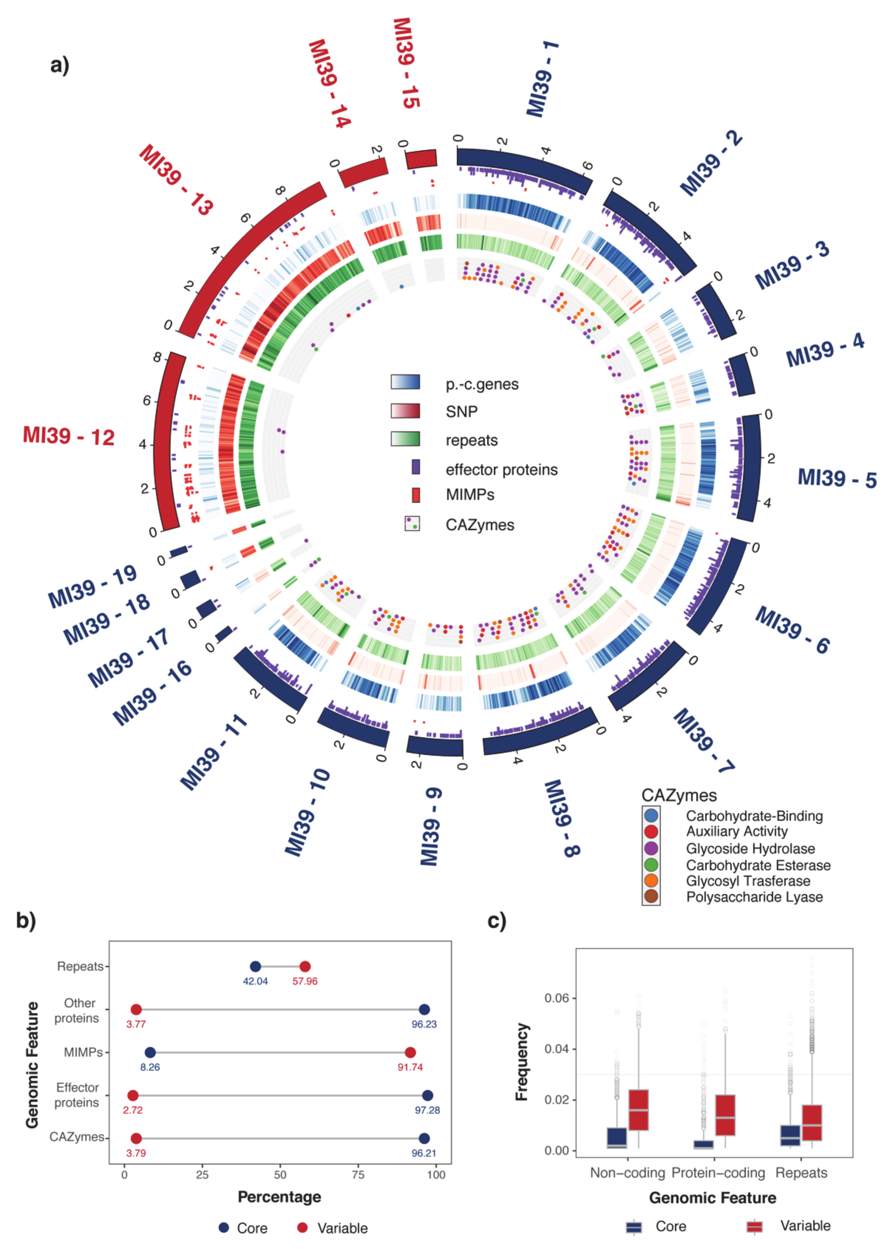
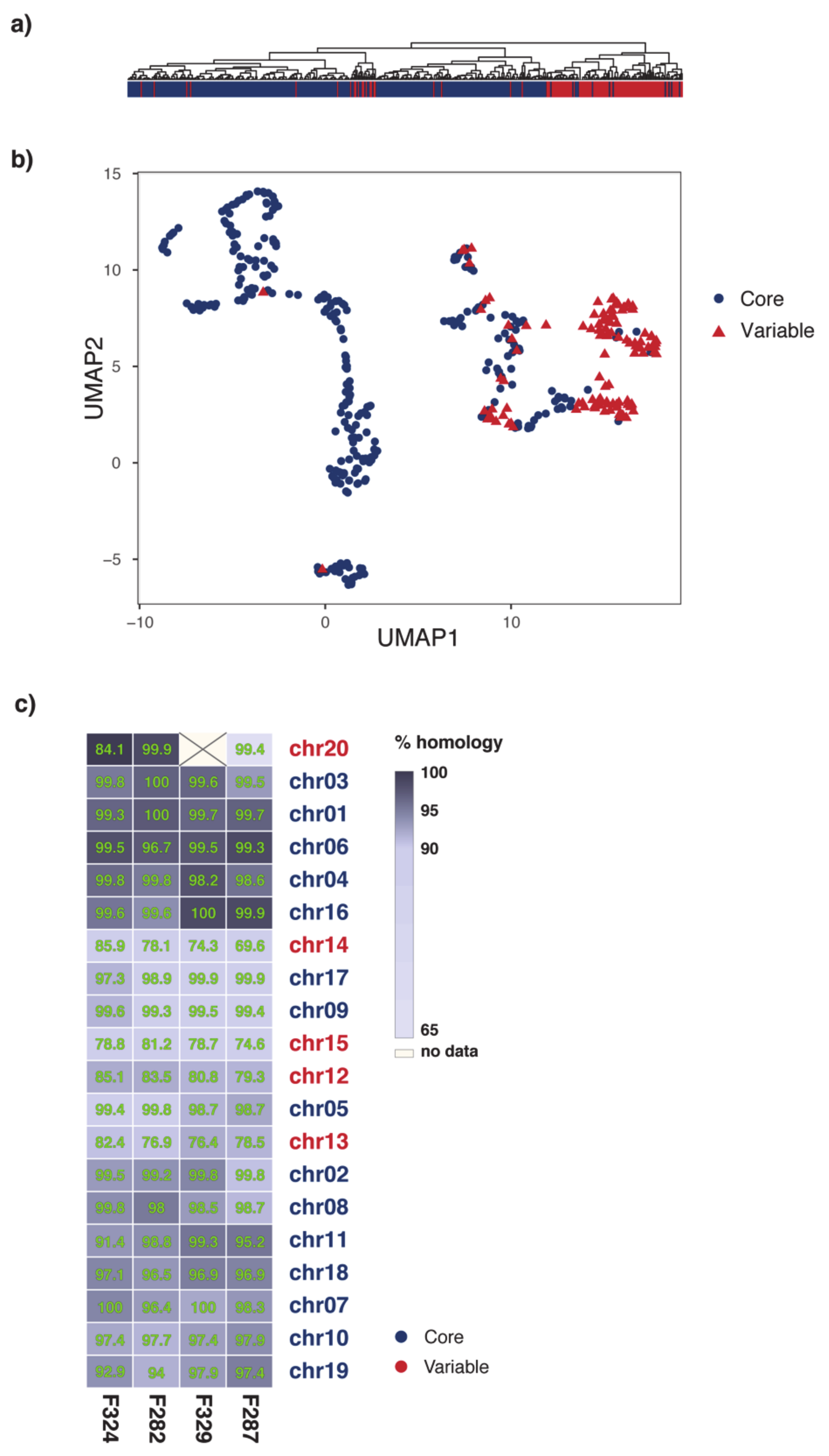
2.1. Organization of MI39 Reference Genome

Previously, MI39 whole genome assembly chromosomes were assigned to core and variable (auxiliary, lineage-specific) compartments of the size of 47.55 and 21.77 Mb, correspondingly [20]. Chromosomes were assigned to the core compartment based on high levels of homology and synteny with the core chromosomes of F. oxysporum f. sp. lycopersici. The synteny-based partitioning is further supported by statistically significant differences in density of protein coding genes, single nucleotide polymorphisms (SNPs), and other genomic features (Figure 1 and Table 1). Thus, in contrast to the variable part, core chromosomes are characterized with much higher density of genes, lower densities of variants, and repetitive regions. In addition, the vast majority of CAZYmes and effector genes are also localized to the core part, whereas mobile DNA (and other repeats including miniature impala repetitive elements (MIMPs) is concentrated in the variable part (Figure 1b and Table 1). The partitioning of the MI39 genome into the core and variable compartments are further supported by significant differences in SNP density (Figure 1c), regardless of its origin e.g., protein-coding, non-coding, and repeats (the associated p-values are 2.5 × 10−225, 9.5 × 10−129, and 5.8 × 10−110, correspondingly).
In contrast to the core part, the lineage-specific chromosomes are characterized by low abundance of genes responsible for pathogenicity and virulence of the fungus, e.g., effectors and CAZYmes (p-values 0.002 and 0.02, correspondingly), as shown in Figure 1a, in Figure S1 and Table 1. It has been shown that MIMPs are often found in proximity to genes encoding effector proteins [4]. Moreover, it has been proposed that their proximity could be a predictor for discovery of effector genes [17]. MI39 genome features 230 MIMPs, of which 211 are located in the LS compartment. We did not find statistically significant evidence in support of co-localization of MIMPs and effector genes, as only 11 of them were found within 2 Mb upstream of the nearest effector gene.
MI39 genome contains 3 SIX gene families, namely SIX1, SIX7, and SIX13 as identified by homology (Figure S1). The majority of them reside on chromosome 12, which contains SIX1 gene, 2 almost identical copies of SIX 13 and 2 copies of SIX7. At the same time, chromosome 15 features an extra copy of SIX7 clearly dissimilar to the rest of the family (Figure S2).
Fusarium produces a diverse spectrum of secondary metabolites of different biosynthetic origin. Genes encoding proteins responsible for synthesis of these metabolites are often clustered on core chromosomes. Using the antiSMASH tool, we predicted 30 secondary metabolite clusters in the MI39 genome, all of which are located in the core compartment. These clusters contain genes encoding non-ribosomal peptide synthase, polyketide synthase, and terpene synthase (Table 1 and Table 2).
Interestingly, a noticeable difference in gene density is observed within core compartment between chromosomes 3, 4, and 9 and others constituents of the core part (p-value = 6.8 × 10⁻²⁸) (Figure 1). The aforementioned chromosomes are syntenic to F. oxysporum f. sp. lycopesici (Foli) chromosomes 11, 12, and 13, which are distinguished from the rest of Fol4287 core chromosomes for different evolutionary properties (gene density, etc.) [4,14].
An in-depth analysis carried out using various quantitative features related to genome stability and variability hinted at the possibility of non-binary partitioning of FOLINI genome. Clustering of density values computed over SNPs and repeats in 4Kb windows, dN/dS values associated with 2326 protein-coding genes, as well as local genome integrity score (Figure 2a) reveals at least three types of genomic regions. The observed multi-compartmentalization of the genome is further supported by application of uniform manifold approximation and projection (UMAP), a non-linear dimension reduction tool, to the data (Figure 2b). In tune with the above, the extended analysis of chromosome synteny between MI39 and 4 other FOLINI strains shown on a heatmap in Figure 2c, corroborates the evidence for existence of at least three genome partitions.

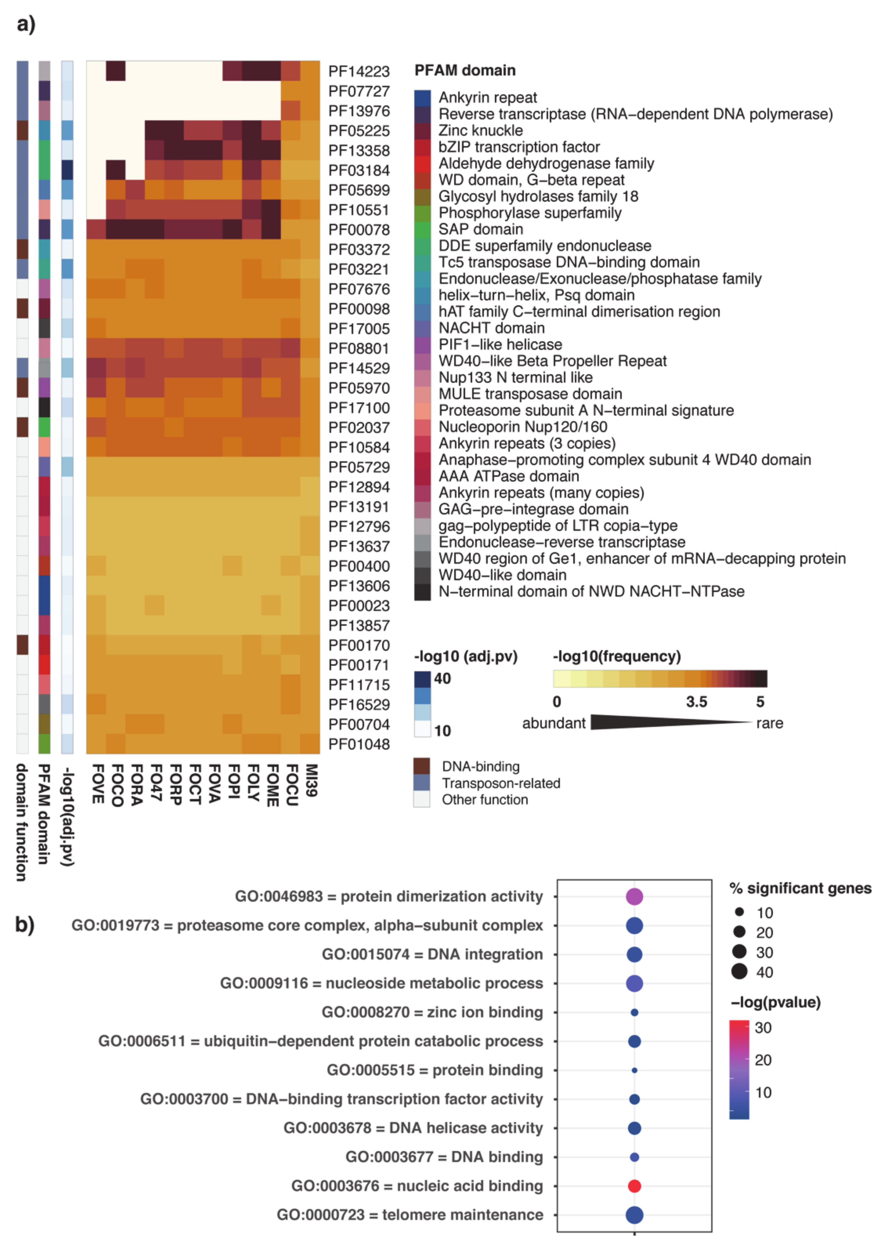
2.2. Functional Annotation of FOLINI Proteome
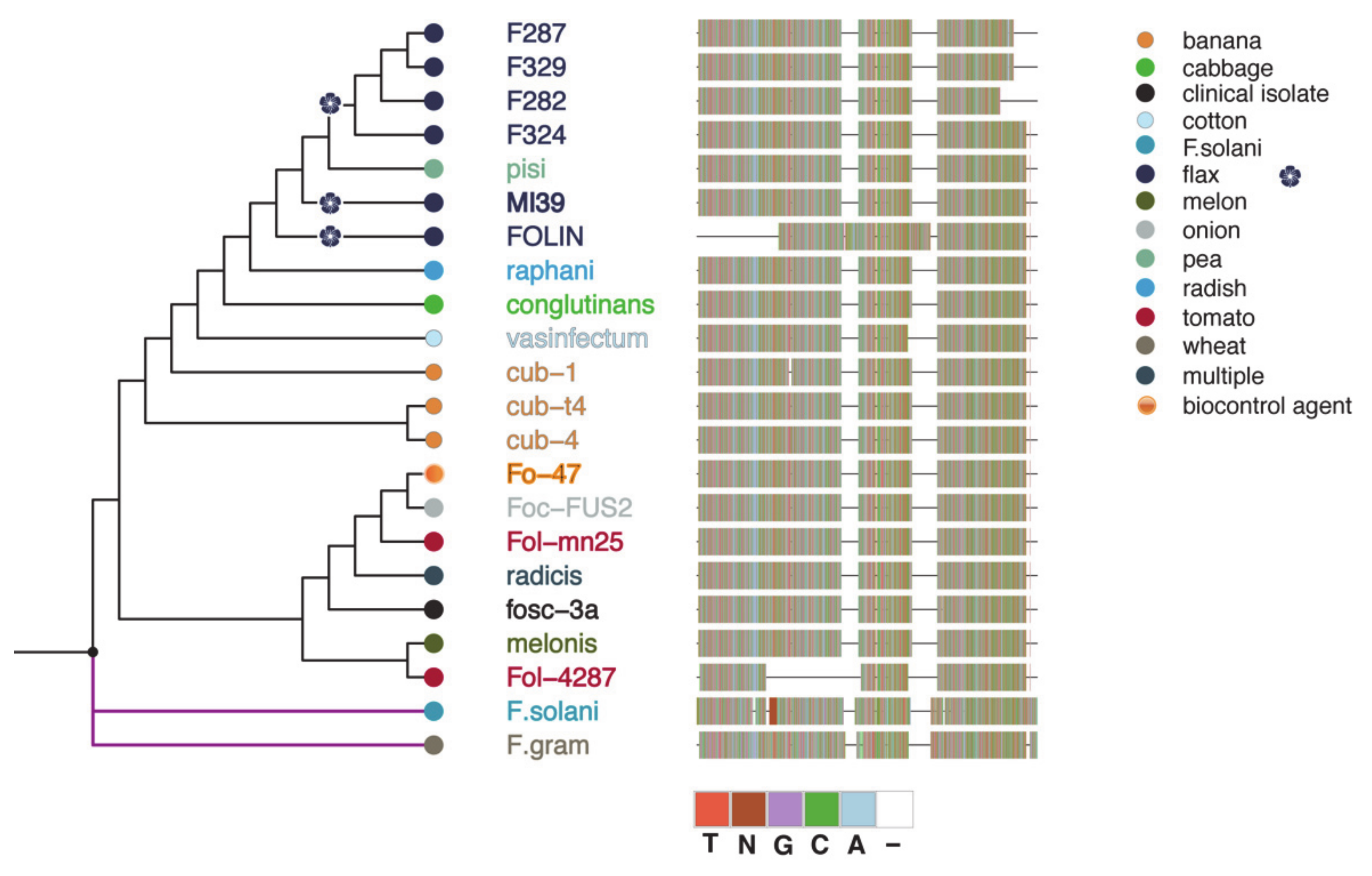
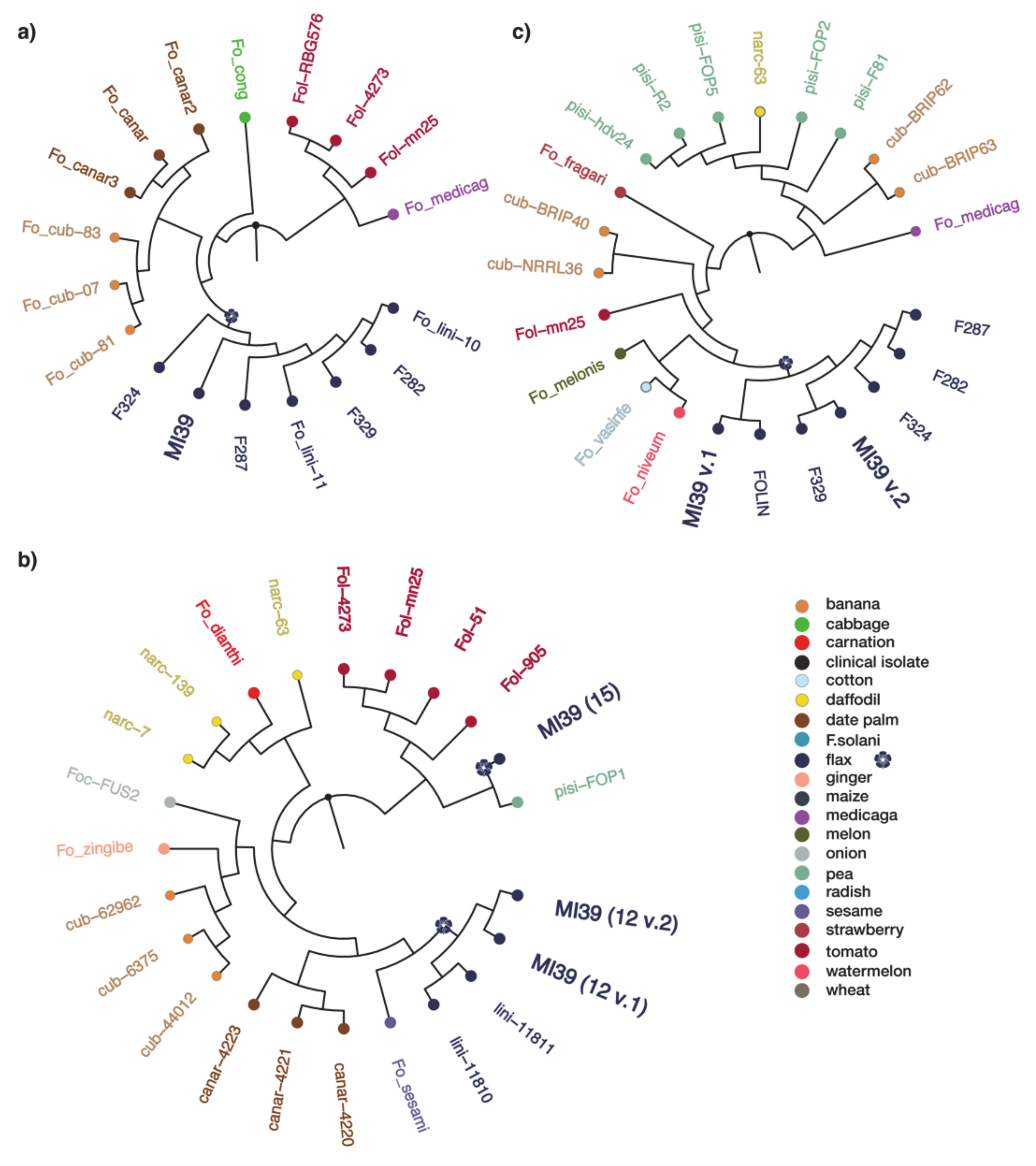
A comprehensive genome-wide annotation of FOLINI was performed using the PFAM database, a collection of protein families based on functional domains. PFAM domain frequencies were computed for proteomes of 11 Fusarium species obtained from Ensembl Fungi (i.e., F. oxysporum f. sp. cubense, F. oxysporum f. sp. meloni, F. oxysporum f. sp. lycopersici, F. oxysporum f. sp. pisi, F. oxysporum f. sp. vasinfectum, F. oxysporum f. sp. cotton, F. oxysporum f. sp. raphani, FO47 strain, F. oxysporum f. sp. radicis, F. oxysporum f. sp. conglutinans, Fusarium verticilloides) and MI39 proteome. Domain enrichment analysis revealed a group of 35 PFAMs significantly over-represented in LS compartment of MI39. Unsurprisingly, the core part is quite homogeneous in terms of domain repertoire, whereas the LS compartment shows noticeable differences between various formae specialis (Figure 3a and Figure S3), thus indirectly underpinning a theory regarding a pivotal role of variable compartment in host adaptation. Interestingly, domain spectrum of MI39 variable proteome is most similar to that of banana pathogen (F.oxysporum f. sp. cubense race 4, GCA_000350365). Both proteomes are characterized with relatively high frequency of domains associated with activity of transposable elements apparently absent in other f. spp.
To get further insight into biological roles of proteins specific to the variable compartment of MI39, we searched the respective proteome for significantly enriched Gene Ontology (GO) terms (FDR < 0.001). The results encompassing terms such as nucleic acid binding, DNA integration, DNA binding, DNA helicase activity, DNA binding transcription factor activity presented in Figure 3b are suggestive of a toolbox for genome reorganization to potentiate host adaptation processes.

2.3. Phylogeny

To determine whether the 5 sequenced strains are monophyletic, we inferred RPB2-TUB2-EF1α maximum likelihood phylogeny (Figure 4). The 5 strains of f. sp. lini are clustered together with another strain FOLIN of the same formae specialis and with strain hdv-247 which belongs to f. sp. pisi.
Further in-depth analysis was carried out to confirm whether f. sp. lini at all has polyphyletic or monophyletic origin. We tested F. oxysporum f. sp. lini strains collected from different locations and constructed a phylogenetic tree based on alignment of EF1α sequences (Figure S4). The resulting tree exhibits at least 4 distinct clonal lineages distributed in different F. oxysporum clades, confirming that f. sp. lini cannot be defined as monophyletic group. This finding is in good agreement with previous phylogenetic analyses of Fusaria from flax [21].
We also constructed separate phylogenetic trees for SIX1, SIX7, and SIX13 genes to determine their modes of origin (Figure 5). The SIX1 dataset included 18 F. oxysporum isolates (Table S1) belonging to six formae specialis. SIX1 genes found in f. sp. lini cluster together with ff. spp. cubense and canariensis, and form a well-supported clade with f. sp. conglutinans. The cluster is distinct from the clade which includes ff. spp. lycopersici and medicaginis. Taken together, these observations clearly attest to the hypothesis that f. sp. lini SIX1 genes are of monophyletic origin.
The SIX13 gene tree consists of 22 isolates (Table S1) and its monophyletic origin is supported for at least 6 strains of f. sp. lini used in this analysis. However, the SIX7 gene phylogeny, consisting of 21 isolates from 10 ff. spp. (Table S1), shows that two copies of this gene located on 12 chromosome form one clade with other f. sp. lini strains, while the third copy on 15 chromosome is phylogenetically distinct and is more related to isolates obtained from S.lycopersicum and P.sativum.
We conclude that f. sp. lini is of polyphyletic origin as confirmed by housekeeping genes phylogeny. SIX7 gene homologs are of polyphyletic origin as well. In contrast, SIX1 and SIX13 genes detected in the 5 strains of f. sp. lini are most likely of single origin as demonstrated in respective phylogenetic trees.
Multiple sequence alignments of SIX proteins (Figure S2) clearly demonstrate high level of conservation between different strains. Phylogenetic analyses using SIX7 protein sequences show two distinct groups of proteins located on chromosomes 12 and 15. Interestingly, a copy situated on chromosome 15 is quite different from the rest of SIX7 protein sequences analyzed here.

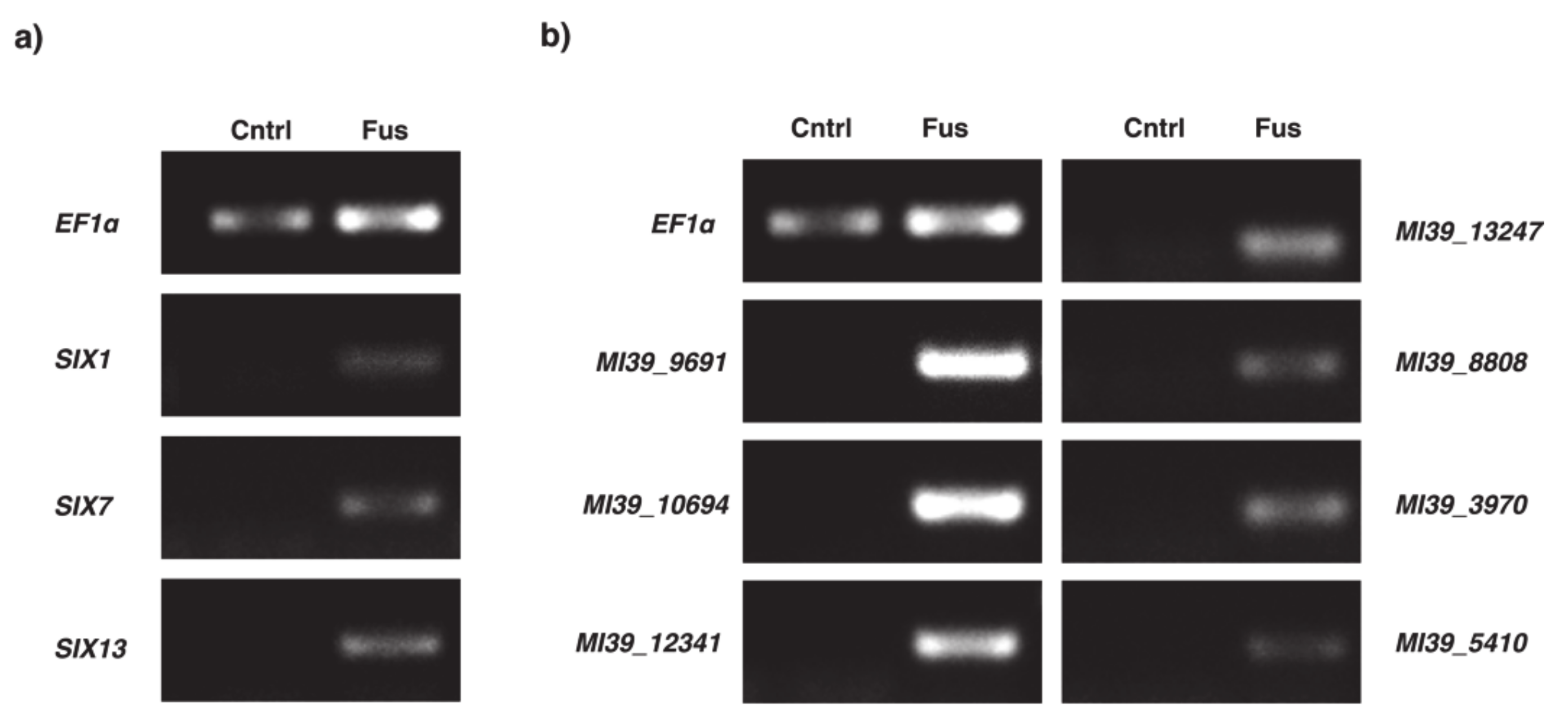
2.4. Expression Analysis of Selected Genes within Variable Compartment

In view of the results of the comparative domain enrichment analysis carried out in 12 FOSC proteomes that demonstrated a significant overrepresentation of domains associated with transposable elements, we hypothesized that expression of proteins harboring those domains could play a significant role in pathogenicity. Specifically, we concentrated on protein domains directly implicated in the life cycle of DNA transposons and retrotransposable elements. Thus, proteins containing gag and RT (reverse transcriptase) domains were obvious candidates for exploratory analyses. From the list of proteins containing the aforementioned domains 10 candidates were selected at random and their expression profiles were assessed in planta. In a like manner, 5 candidates were chosen from the list of proteins containing DDE and endonuclease domains.
To explore their potential role in infection, gene expression levels were assessed with RT-PCR. Furthermore, expression levels of all SIX genes identified in FOLINI were profiled as infection markers, whereas E2F1 and CYC were used as reference controls. All 11 genes tested have shown clear expression in planta, suggesting their role in the infection process (Figure 6).

3. Discussion

An extensive genome analysis of 5 strains of Fusarium oxysporum f. sp. lini revealed a remarkable genome architecture challenging the “two-speed” genome model in the case of this particular pathogen. Stunningly and in sharp contrast to current perception of a “two-speed” genome model, our results show significant differences in numbers of effector proteins, CAZYmes and secondary metabolite clusters found on stable, core chromosomes in vast quantities (Table 1). Evaluation of genomic features, such as densities of SNP, repetitive regions, protein-coding genes etc., commonly used as a proxy for genome plasticity and susceptibility to variation points rather at a feasibility of a “three-speed” or even “multi-speed” evolutionary model, thus questioning the rationale behind over-simplistic theories. Our findings evidence in favor of at least three genomic compartments (Figure 1a and Figure 2), presumably with different evolutionary properties.
Thus, a compartment distinguished by a relatively low number of genes and repeats, high level of intra-strain synteny, and low level of genetic variation (chromosomes 1, 3, 4, 6, 16) is referred to as a conserved “core” part of the genome. The so-called “variable”, dynamic compartment is just the opposite of the core and includes chromosomes 12–15. This part of the genome is highly syntenic with LS chromosomes of Fol [20] and features low number of protein-coding genes, as well as both high rates of intra-strain variation and density of repeats. Genomic regions comprising rest of the genome (chromosomes 2, 5, 7, 8–11, 17, and 18) exhibit a mixed profile of features which complicate their attribution to either of the compartments. This observation is in tune with recent studies [4,22], suggesting a crosstalk between core and accessory components of the genome.
Pathogen- and virulence-specific genes are not solely bound to the variable genome, hinting at the existence of complex genetic and epigenetic mechanisms in charge of pathogenicity and host adaptation. For instance, two homologs of cerato-plantanin gene FocCP1 [23], responsible for host penetration and virulence in f. sp. cubense is located on chromosomes 1 and 5. Conversely, all copies of SIX genes belong to the dynamic genome compartment (chromosomes 12 and 15). Undoubtedly, in-depth analysis involving other omics data modalities is essential to refine the proposed genome compartmentalization scheme to decipher the evolutionary model of fungal pathogenicity.
In contrast to housekeeping genes, members of SIX1 and SIX13 gene families demonstrate host-related clustering as revealed by phylogenetic analyses (Figure 5 and Figure S4), thus being indicative of strong adaptation to host. In this context, SIX7 gene poses a particular interest, as one of its copies is located on different chromosome, is absent from other 4 FOLINI strains sequenced and is more related to SIX7 homologs from f. sp. lycopersici strains, strongly suggesting a relatively recent horizontal transfer event.
Functional analysis of F. oxysporum f. spp. protein repertoire reveals the dominating role of the variable compartment in diversification of the formae specialis. Remarkably, the main contributors to proteome divergence are PFAM domains associated with DNA-binding and mobilization of transposable elements, possibly suggestive of active transcription and ongoing genome reshuffling in variable compartment (Figure 3).
Previous research has shown that active transposable elements populate lineage specific compartment of fungal pathogens [24]. It has been hypothesized that variable genome compartment evolves through rearrangements triggered by transposable element activity, which impacts the evolution of pathogen virulence. We were able to observe simultaneous expression of pathogenicity-related genes and genes directly involved in a life cycle of both DNA transposons and retroelements. This may support the aforementioned hypothesis that evolution of Fusarium genomes and remarkable adaptability of the fungus to the host are for the most part shaped and driven by the activity of mobile elements. Contrarily, the abundant expression of transposable elements RNA could be a consequence of extensive chromatin re-modeling concomitant to the infection process. Thus, further insight into chromatin biology of F. oxysporum f. sp. lini will be instrumental to enhance our perception of the evolution of its multi-compartmentalized genome and on its pathogenicity.
Parasitic genome is arguably a center-point of adaptation destined to counterweight the host immune system and environmental challenges. A new genomic blueprint of a fungal pathogen lays a solid foundation for in-depth studies of molecular mechanisms of species divergence, adaptation, and pathogenicity, thus boosting our efforts to fight and control fungal infections.

4. Materials and Methods

4.1. Data Sources and Sequence Analysis

The nucleotide sequences assembly, de novo prediction, and annotation of coding regions of monoisolate 39 and 4 strains (282, 289, 324, 327) have been described in our previous publication [20]. De novo prediction of repetitive sequences was made using RepeatScout [25] version 1.0.6 MIMPs have been predicted using custom Perl scripts and regular expressions/CAGTGGG.GCAA[TA] AA/and/TT[TA]TTGC.CCCACTG/, described in [4]. Short reads of 4 FO strains have been aligned to MI39 reference genome using bwa-mem [26] with default parameters. Variant calling for the alignments have been done using NGSEP [27] version 4.0. Synteny analysis was done using Satsuma2 program [28]. Genome stability metrics was computed as a local aberration score with spector R package (https://github.com/anasrana/spector, accessed 1 December 2020).

4.2. Proteins Annotation

Protein sequences predicted by BUSCO [29] as described earlier (Table S3). Secondary metabolite clusters analysis was done using Fungal antismash server version 5.1.2 [30] with the following types of analysis: KnownClusterBlast, ClusterBlast, SubClusterBlast, ActiveSiteFinder, Cluster Pfam analysis, and Pfam-based GO term annotation. CAZYme proteins have been predicted with dbCAN meta server [31] using HMMER, DIAMOND, and Hotpep prediction methods. Effector proteins prediction was made using EffectorP package version 2.0 [32]. Search for PFAM domains in the protein sequences was done for PFAM database version 33.1 using HMMER version 3.3 [33]. Search for orthologous groups of proteins was made with orthoMCL version 2.0.9 [34]. Orthology analysis identified genes common between Foli strains and 16 other proteomes from FOC, 97.1% proteins were clustered into 23,975 orthogroups, 7239 orthogroups were shared between all 16 proteomes and represented 187,648 proteins, while 3065 orthogroups contained proteins in a 1:1 relationship between MI39 proteome and others FOSC.

4.3. Phylogenetic Analysis

For the phylogenetic analysis of housekeeping genes, we used publicly available sequences of the genes on NCBI: RPB2 (GB: KX434985.1), EF1α (GB: KC889026.1), and TUB2 (GB: KP964895.1) as queries for BLASTN search. Databases were created using makeblastdb script for our published genomes of Fo f. sp. lini, 12 genomes from Ensembl Fungi (Table S1), Fo f. sp. lycopersici Fol-4287, and Fo f. sp. cepae Foc-FUS2 genomes from NCBI. BLASTN search was performed with default parameters. Sequences with identity more than 90% (1 sequence for each of 3 genes of each species) were extracted from BLAST databases using blastdbcmd script. Sequences of the same genes of Fusarium graminearum and F. solani were downloaded from NCBI and used as outgroup. Each of 3 gene datasets were aligned using ClustalX [35] and then 3 resulting alignments were concatenated together with MEGA X software [36]. Maximum likelihood phylogeny was inferred in MEGA X by using Tamura-Nei model [37] with discrete Gamma distribution (4 categories, G-parameter = 1.0613) and 1000 bootstrap replicates. The phylogenetic trees were visualized in FigTree and rooted on the outgroup. The resulting trees are presented as cladograms (see Figure 4, Figure 5 and Figure S4), for clarity of presentation reasons only.
For the phylogenetic analysis of EF1α gene sequences were selected as described above. We also added 15 sequences of EF1α from other strains of f. sp. lini available on NCBI and one strain XJ37 belonging to f. sp. cubense (Table S2). Sequences of EF1α from F. graminearum, F. verticelloides, and F. solani from NCBI were used as outgroup. Then we aligned 37 sequences using ClustalX and manually curated the alignment. Maximum likelihood phylogeny was estimated and tree was constructed with IQ-TREE software [38]. We used Tamura-Nei + G4 substitution model and 1000 bootstrap replicates. Resulting tree was curated with FigTree as described above. The phylogenetic analysis of SIX1, SIX7, and SIX13 genes was carried out using available sequences of these genes for different FOSC ff. spp. obtained either from NCBI or from Ensembl Fungi (see Table S2). To infer tree structure for each gene, we performed a multiple sequence alignment with ClustalX and subsequent phylogeny analysis with IQ-TREE. Substitution models were chosen in accordance with ModelTest as implemented in IQ-TREE software. Next, the consensus trees from 1000 bootstrap replicates were built. The resulting trees were midpoint rooted and curated using FigTree. The phylogeny of protein sequences of SIX genes products was inferred from multiple sequence alignments as implemented in MAFFT software (L-INS-i algorithm, 1000 iterations). Trees visualization was created with ggtree R package [39].

4.4. RNA Isolation, Reverse Transcript, and PCR

For gene expression analysis, flax roots after inoculation with MI39 isolate for 3 days were sampled. As a control, we used non-infected plants. The root was washed clean, cut from the plants, and used for RNA extraction with RNeasy Mini Kit (QIAGEN, Germany) as per manufacturer’s instructions. After quantification using NanoDrop 8000, the first strand of cDNA was synthesized with MMLV-RT commercial kit (Evrogen, Russia). We used Oligo(dT)17-primer for first strand cDNA synthesis, as per the manufacturer’s instructions. The PCR was carried out using the Veriti 9000 instrument (Applied Biosystems) in technical triplicate using the indicated primer pairs (see Table S4). Agarose gel electrophoresis was used to separate the amplified fragments of cDNA. We used Quantum-ST gel-documenting system and software (Vilber Lourmat) to visualize the results of PCR and curate images.

Supplementary Materials

The following are available online at https://0-www-mdpi-com.brum.beds.ac.uk/1422-0067/22/5/2665/s1, Figure S1: MI39 F. oxysporim f.sp. lini variable chromosomes, Figure S2: Multiple sequence alignments and phylogeny of SIX proteins, Figure S3: PFAM domain repertorie in the core part of F.oxysporum genomes, Figure S4: Phylogeny of Fusarium oxysporum isolates based on alignment of EF1α gene sequences, Table S1: Identifiers of SIX genes sequences in public databases, Table S2: Identifiers of EF1α genes sequences in public databases, Table S3: Annotation of predicted protein coding genes in a genome of MI39 monoisolate of F. oxysporum f.sp. lini, Table S4: Primer sequences used in PCR experiments.

Author Contributions

Conceptualization, A.S., A.K., M.S.; Investigation, M.B., T.R., A.L.; Software, A.K., M.G.; Formal Analysis, A.K., A.S., A.L.; Resources, M.S.; Writing—Original Draft Preparation, A.S., A.K., M.S., A.L.; Writing—Review & Editing, A.S., A.K., M.S.; Visualization, A.S.; Funding Acquisition, M.S. All the authors have read and agreed to the published version of the manuscript.

Funding

This work was supported by RSF fund, grant 19-16-00030 to A.K., T.R., M.B. and M.S. (data processing, genome annotation, comparative proteomics, plant inoculation, plant and fungi handling, RNA extraction). A.S. and M.G. (functional genomics, genome integrity analyses) were supported by RSF fund, grant 20-14-00072.

Institutional Review Board Statement

Not applicable.

Informed Consent Statement

Not applicable.

Data Availability Statement

The data that support the findings of this study are openly available in NCBI GenBank at https://0-www-ncbi-nlm-nih-gov.brum.beds.ac.uk/genbank/, reference numbers: JABJUA000000000, JABJUB000000000, JABJUC000000000, JABJUD000000000, JABJUE000000000, accessed 03.09.2020.

Conflicts of Interest

The authors declare no conflict of interest.

References

  1. Zhang, Y.; Ma, L.-J. Deciphering Pathogenicity of Fusarium oxysporum From a Phylogenomics Perspective. Adv. Genet. 2017, 100, 179–209. [Google Scholar]
  2. Ma, L.-J.; van der Does, H.C.; Borkovich, K.A.; Coleman, J.J.; Daboussi, M.-J.; Di Pietro, A.; Dufresne, M.; Freitag, M.; Grabherr, M.; Henrissat, B.; et al. Comparative genomics reveals mobile pathogenicity chromosomes in Fusarium. Nature 2010, 464, 367–373. [Google Scholar] [CrossRef]
  3. Williams, A.H.; Sharma, M.; Thatcher, L.F.; Azam, S.; Hane, J.K.; Sperschneider, J.; Kidd, B.N.; Anderson, J.P.; Ghosh, R.; Garg, G.; et al. Comparative genomics and prediction of conditionally dispensable sequences in legume–infecting Fusarium oxysporum formae speciales facilitates identification of candidate effectors. BMC Genom. 2016, 17, 311–324. [Google Scholar] [CrossRef] [Green Version]
  4. Armitage, A.D.; Taylor, A.; Sobczyk, M.K.; Baxter, L.; Greenfield, B.P.J.; Bates, H.J.; Wilson, F.; Jackson, A.C.; Ott, S.; Harrison, R.J.; et al. Characterisation of pathogen-specific regions and novel effector candidates in Fusarium oxysporum f. sp. cepae. Sci. Rep. 2018, 8, 13530. [Google Scholar] [CrossRef] [PubMed]
  5. Dong, S.; Raffaele, S.; Kamoun, S. The two-speed genomes of filamentous pathogens: Waltz with plants. Curr. Opin. Genet. Dev. 2015, 35, 57–65. [Google Scholar] [CrossRef] [PubMed]
  6. Frantzeskakis, L.; Kusch, S.; Panstruga, R. The need for speed: Compartmentalized genome evolution in filamentous phytopathogens. Mol. Plant Pathol. 2018, 20, 3–7. [Google Scholar] [CrossRef] [Green Version]
  7. Torres, D.E.; Oggenfuss, U.; Croll, D.; Seidl, M.F. Genome evolution in fungal plant pathogens: Looking beyond the two-speed genome model. Fungal Biol. Rev. 2020, 34, 136–143. [Google Scholar] [CrossRef]
  8. Vlaardingerbroek, I.; Beerens, B.; Rose, L.; Fokkens, L.; Cornelissen, B.J.C.; Rep, M. Exchange of core chromosomes and horizontal transfer of lineage-specific chromosomes in Fusarium oxysporum. Environ. Microbiol. 2016, 18, 3702–3713. [Google Scholar] [CrossRef] [PubMed]
  9. Li, J.; Fokkens, L.; Conneely, L.J.; Rep, M. Partial pathogenicity chromosomes in Fusarium oxysporumare sufficient to cause disease and can be horizontally transferred. Environ. Microbiol. 2020, 12, 2033. [Google Scholar]
  10. Li, J.; Fokkens, L.; van Dam, P.; Rep, M. Related mobile pathogenicity chromosomes in Fusarium oxysporumdetermine host range on cucurbits. Mol. Plant Pathol. 2020, 21, 761–776. [Google Scholar] [CrossRef] [Green Version]
  11. Kistler, H.C. Genetic Diversity in the Plant-Pathogenic Fungus. Phytopathology 1997, 87, 474–479. [Google Scholar] [CrossRef] [PubMed] [Green Version]
  12. Ellis, M.L.; Jimenez, D.R.C.; Leandro, L.F.; Munkvold, G.P. Genotypic and Phenotypic Characterization of Fungi in the Fusarium oxysporum Species Complex from Soybean Roots. Phytopathology 2014, 104, 1329–1339. [Google Scholar] [CrossRef] [PubMed]
  13. Nirmaladevi, D.; Venkataramana, M.; Srivastava, R.K.; Uppalapati, S.R.; Gupta, V.K.; Yli-Mattila, T.; Clement Tsui, K.M.; Srinivas, C.; Niranjana, S.R.; Chandra, N.S. Molecular phylogeny, pathogenicity and toxigenicity of Fusarium oxysporum f. sp. lycopersici. Sci. Rep. 2016, 6, 167. [Google Scholar] [CrossRef] [PubMed] [Green Version]
  14. Fokkens, L.; Shahi, S.; Connolly, L.R.; Stam, R.; Schmidt, S.M.; Smith, K.M.; Freitag, M.; Rep, M. The multi-speed genome of Fusarium oxysporumreveals association of histone modifications with sequence divergence and footprints of past horizontal chromosome transfer events. BioRxiv 2020, 701, 315–362. [Google Scholar]
  15. Husaini, A.M.; Sakina, A.; Cambay, S.R. Host–Pathogen Interaction in Fusarium oxysporum Infections: Where Do We Stand? Mol. Plant Microbe Interact. 2018, 31, 889–898. [Google Scholar] [CrossRef] [Green Version]
  16. Houterman, P.M.; Cornelissen, B.J.C.; Rep, M. Suppression of Plant Resistance Gene-Based Immunity by a Fungal Effector. PLoS Pathog. 2008, 4, e1000061. [Google Scholar] [CrossRef] [PubMed] [Green Version]
  17. Schmidt, S.M.; Lukasiewicz, J.; Farrer, R.; van Dam, P.; Bertoldo, C.; Rep, M. Comparative genomics of Fusarium oxysporum f. sp. melonisreveals the secreted protein recognized by the Fom-2resistance gene in melon. New Phytol. 2015, 209, 307–318. [Google Scholar] [CrossRef] [Green Version]
  18. Houterman, P.M.; Ma, L.; van Ooijen, G.; de Vroomen, M.J.; Cornelissen, B.J.C.; Takken, F.L.W.; Rep, M. The effector protein Avr2 of the xylem-colonizing fungus Fusarium oxysporum activates the tomato resistance protein I-2 intracellularly. Plant J. 2009, 58, 970–978. [Google Scholar] [CrossRef] [PubMed]
  19. Rep, M.; van der Does, H.C.; Meijer, M.; van Wijk, R.; Houterman, P.M.; Dekker, H.L.; de Koster, C.G.; Cornelissen, B.J.C. A small, cysteine-rich protein secreted by Fusarium oxysporum during colonization of xylem vessels is required for I-3-mediated resistance in tomato. Mol. Microbiol. 2004, 53, 1373–1383. [Google Scholar] [CrossRef]
  20. Kanapin, A.; Samsonova, A.; Rozhmina, T.; Bankin, M.; Logachev, A.; Samsonova, M. The Genome Sequence of Five Highly Pathogenic Isolates of Fusarium oxysporum f. sp. lini. Mol. Plant Microbe Interact. 2020, 33, 1112–1115. [Google Scholar] [CrossRef] [PubMed]
  21. Baayen, R.P.; O’Donnell, K.; Bonants, P.J.; Cigelnik, E.; Kroon, L.P.; Roebroeck, E.J.; Waalwijk, C. Gene Genealogies and AFLP Analyses in the Fusarium oxysporum Complex Identify Monophyletic and Nonmonophyletic Formae Speciales Causing Wilt and Rot Disease. Phytopathology 2000, 90, 891–900. [Google Scholar] [CrossRef] [Green Version]
  22. Yang, H.; Yu, H.; Ma, L.-J. Accessory Chromosomes in Fusarium oxysporum. Phytopathology 2020, 110, 1488–1496. [Google Scholar] [CrossRef] [PubMed]
  23. Liu, S.; Wu, B.; Yang, J.; Bi, F.; Dong, T.; Yang, Q.; Hu, C.; Xiang, D.; Chen, H.; Huang, H.; et al. A Cerato-Platanin Family Protein FocCP1 Is Essential for the Penetration and Virulence of Fusarium oxysporum f. sp. cubense Tropical Race 4. Int. J. Mol. Sci. 2019, 20, 3785. [Google Scholar] [CrossRef] [Green Version]
  24. Faino, L.; Seidl, M.F.; Shi-Kunne, X.; Pauper, M.; van den Berg, G.C.M.; Wittenberg, A.H.J.; Thomma, B.P.H.J. Transposons passively and actively contribute to evolution of the two-speed genome of a fungal pathogen. Genome Res. 2016, 26, 1091–1100. [Google Scholar] [CrossRef] [PubMed] [Green Version]
  25. Price, A.L.; Jones, N.C.; Pevzner, P.A. De novo identification of repeat families in large genomes. Bioinformatics 2005, 21 (Suppl. 1), i351–i358. [Google Scholar] [CrossRef] [Green Version]
  26. Li, H.; Durbin, R. Fast and accurate short read alignment with Burrows-Wheeler transform. Bioinformatics 2009, 25, 1754–1760. [Google Scholar] [CrossRef] [Green Version]
  27. Tello, D.; Gil, J.; Loaiza, C.D.; Riascos, J.J.; Cardozo, N.; Duitama, J. NGSEP3: Accurate variant calling across species and sequencing protocols. Bioinformatics 2019, 35, 4716–4723. [Google Scholar] [CrossRef] [PubMed] [Green Version]
  28. Grabherr, M.G.; Russell, P.; Meyer, M.; Mauceli, E.; Alföldi, J.; Di Palma, F.; Lindblad-Toh, K. Genome-wide synteny through highly sensitive sequence alignment: Satsuma. Bioinformatics 2010, 26, 1145–1151. [Google Scholar] [CrossRef] [PubMed] [Green Version]
  29. Seppey, M.; Manni, M.; Zdobnov, E.M. BUSCO: Assessing Genome Assembly and Annotation Completeness. Methods Mol. Biol. 2019, 1962, 227–245. [Google Scholar]
  30. Blin, K.; Shaw, S.; Steinke, K.; Villebro, R.; Ziemert, N.; Lee, S.Y.; Medema, M.H.; Weber, T. antiSMASH 5.0: Updates to the secondary metabolite genome mining pipeline. Nucleic Acids Res. 2019, 47, W81–W87. [Google Scholar] [CrossRef] [PubMed] [Green Version]
  31. Zhang, H.; Yohe, T.; Huang, L.; Entwistle, S.; Wu, P.; Yang, Z.; Busk, P.K.; Xu, Y.; Yin, Y. dbCAN2: A meta server for automated carbohydrate-active enzyme annotation. Nucleic Acids Res. 2018, 46, W95–W101. [Google Scholar] [CrossRef] [Green Version]
  32. Sperschneider, J.; Dodds, P.N.; Gardiner, D.M.; Singh, K.B.; Taylor, J.M. Improved prediction of fungal effector proteins from secretomes with EffectorP 2.0. Mol. Plant Pathol. 2018, 19, 2094–2110. [Google Scholar] [CrossRef] [Green Version]
  33. Eddy, S.R. A new generation of homology search tools based on probabilistic inference. Genome Inf. 2009, 23, 205–211. [Google Scholar]
  34. Fischer, S.; Brunk, B.P.; Chen, F.; Gao, X.; Harb, O.S.; Iodice, J.B.; Shanmugam, D.; Roos, D.S.; Stoeckert, C.J. Using OrthoMCL to assign proteins to OrthoMCL-DB groups or to cluster proteomes into new ortholog groups. Curr. Protoc. Bioinform. 2011, 35, 6–12. [Google Scholar]
  35. Larkin, M.A.; Blackshields, G.; Brown, N.P.; Chenna, R.; McGettigan, P.A.; McWilliam, H.; Valentin, F.; Wallace, I.M.; Wilm, A.; Lopez, R.; et al. Clustal W and Clustal X version 2.0. Bioinformatics 2007, 23, 2947–2948. [Google Scholar] [CrossRef] [PubMed] [Green Version]
  36. Stecher, G.; Tamura, K.; Kumar, S. Molecular Evolutionary Genetics Analysis (MEGA) for macOS. Mol. Biol. Evol. 2020, 37, 1237–1239. [Google Scholar] [CrossRef] [PubMed]
  37. Tamura, K.; Nei, M. Estimation of the number of nucleotide substitutions in the control region of mitochondrial DNA in humans and chimpanzees. Mol. Biol. Evol. 1993, 10, 512–526. [Google Scholar] [PubMed]
  38. Nguyen, L.-T.; Schmidt, H.A.; Haeseler von, A.; Minh, B.Q. IQ-TREE: A fast and effective stochastic algorithm for estimating maximum-likelihood phylogenies. Mol. Biol. Evol. 2015, 32, 268–274. [Google Scholar] [CrossRef]
  39. Yu, G. Using ggtree to Visualize Data on Tree-Like Structures. Curr. Protoc. Bioinform. 2020, 69, e96. [Google Scholar] [CrossRef] [PubMed]
Figure 1. Genomic architecture and properties of Fusarium oxysporum f. sp. lini. (a) Genomic architecture of Fusarium oxysporum f. sp. lini MI39 isolate visualized with Circos software. The outermost ring depicts chromosome ideogram where chromosomes were assigned to either Core (blue) or Variable (red) compartments of the genome. The next two rings (violet and red ticks) show effector protein loci and MIMPs genomic positions, respectively. Protein-coding genes (p.-c genes), SNP, and repetitive element densities computed in 50 K bins along the chromosomes are presented as blue, red, and green tracks. Color intensity gradient reflects changes in density. The darkest color shade corresponds to maximum density values. The most inner track (grey with circle glyphs) depicts location of CAZymes in MI39 F. oxysporum f. sp. lini genome. CAZyme types are color-coded as follows: blue—carbohydrate-binding, red—auxillary activity, violet—glycoside hydrolase, green—carbohydrate esterase, orange—glycosyl transferase, brown—polysaccharide lyase. (b) Fraction of genomic features associated with either Core (blue) or Variable (red) parts of the genome. Values in a dumbbell chart correspond to percentage of features attributed to either of them. (c) Distribution of SNP frequency values computed in various genomic regions (as described in (a)) and parts of the genome. Core and variable part is shown in red and blue, correspondingly.
Ijms 22 02665 g001
Figure 2. Compartmentalization of F. oxysporum f. sp. lini genome. (a) Hierarchical clustering of genomic regions in accordance with genomic characteristics associated with genome stability. Regions attributed to the Core part are marked with blue, whereas loci from the variable part are shown in red. (b) Visualization of genomic region clustering with uniform manifold approximation and projection (UMAP) reveals three groups of loci. (c). Homology heatmap for comparisons of the percentage identity between flax strains. The percentage identity is indicated by different box colors, whereas the numbers shown is the boxes correspond to percentage of overlap between chromosomes. Chromosome labels are highlighted according to genome compartmentalization.
Ijms 22 02665 g002
Figure 3. Functional annotation of F. oxysporum genomes. Heatmap of PFAM domain frequencies observed in variable part of F. oxysporum genomes. (a) The heatmap shows 35 PFAM domains significantly overrepresented (adj. p-value < 10−6) in variable genome of MI39 F. oxysporum f. sp. lini isolate. Frequency values associated with each domain were mean-centered by rows. Each row of the heatmap represents log10-transformed frequency values of one PFAM domain across all F. oxysporum formae speciales (yellow, high frequency; brown, low frequency). Ivory color represents missing data points i.e., situations when a domain has not been detected in a pathogen’s genome. Domain enrichment significance is indicated in shades of blue on a separate panel on the left of the heatmap. The next two panels present domain name for each PFAM accession and information regarding domain function and its relevance to genome mobilization. The abbreviated F. oxysporum formae specialis names shown at the bottom of the heatmap stand for: MI39, FOCU—f. sp. cubense, FOME—f. sp. meloni, FOLY—f. sp. lycopersici, FOPI—f. sp. pisi, FOVA—f. sp. vasinfectum, FOCT—f. sp. cotton, FORP—f. sp. raphani, FO47—FO47 strain, FORA—f. sp. radicis, FOCO—f. sp. conglutinans, FOVE—Fusarium verticilloides. (b) Significantly overrepresented GO terms encompassing processes associated with proteins in the variable compartment of the MI39 genome. Spheres represent GO term enrichment with size and color as indicated in the inset.
Ijms 22 02665 g003
Figure 4. Bayesian phylogeny of Fusarium oxysporum isolates from flax and other hosts. The phylogeny is inferred from the maximum likelihood analysis of concatenated alignment of RPB2-TUB2-EF1α and is rooted on the outgroup comprised of F. solani and F. graminearum branch). Tree tips and label colors reflect the pathogen’s host. The color-coded nucleotide sequence alignments are shown to the right of the tree.
Ijms 22 02665 g004
Figure 5. Consensus trees from the Bayesian phylogenetic analysis of SIX genes from the Fusarium clade. Tree tips and label colors reflect the pathogen’s host organism. List of hosts is shown separately. (a) SIX1 consensus tree consensus tree, (b) SIX7 consensus tree, and (c) SIX13 consensus tree.
Ijms 22 02665 g005
Figure 6. PCR analyses showing the presence of F.oxysporum f. sp. lini gene products. (a) PCR amplification of E1Fα (reference), SIX1, SIX7, and SIX13; (b) PCR amplification of genes involved in a life cycle of transposable elements. Control cDNA, extracted from non-infected plant is labeled with Cntrl, whereas Fus marks cDNA obtained from infected flax roots as described in the Materials and Methods section.
Ijms 22 02665 g006
Table 1. Characteristics of F. oxysporum f. sp. lini monoisolate 39 reference genome. Densities of genomic elements per 1 Mb and p-values in support of significant difference in the densities between core and variable parts of the genome.
Genomic Elements
ChromosomeLength (Mb)ProteinsMIMPsSecondary Metabolites ClustersCAZYmesEffector GenesRepeatsSNPsTFs
MI39-16.5372.930.6150.3076.7636.00263.39186.003.076
MI39-25.28343.921.7040.9465.8729.54254.34505.652.651
MI39-32.6493.3300.3792.659.86257.61335.010
MI39-41.9149.64008.9518.96186.53245.010
MI39-54.99315.1000.4007.8034.61279.09328.500.601
MI39-64.82389.170.4140.6228.5040.22244.24101.592.489
MI39-74.23301.7000.4724.2427.38269.36147.782.364
MI39-85.51316.3900.1818.8931.23221.22475.861.633
MI39-92.62101.660.7640.3823.8213.37218.22491.870
MI39-103.36273.75005.9325.23295.12404.391.785
MI39-113.9312.49006.4028.94323.51404.711.282
MI39-128.419.8617.8400.351.784830.759296.250.238
MI39-139.723.184.8400.821.957828.969924.000.103
MI39-142.2135.660.90200.450.902803.506367.080
MI39-151.4119.818.49000.707834.915314.450.709
MI39-160.3345.330048.359.06507.72456.340
MI39-170.5651.520030.2010.66483.25577.410
MI39-180.6129.483.271.63701.637804.195929.120
MI39-190.2825.340003.6201035.397247.740
MI39-200.03500000688.17200.710
p-value (core/variable) 0.0020.0030.0580.0240.0020.0090.0030.677
Table 2. Secondary metabolites clusters.
Gene IDChromosomeStartEndTypeMost Similar Known ClusterSimilarity
MI39_11795MI39-1396,772398,794NRPS
MI39_7258MI39-1592,293593,294saccharide
MI39_7862MI39-1967,185970,941NRPS-like
MI39_1628MI39-12,347,8252,349,019saccharide
MI39_10036MI39-2298,534305,960T1PKS
MI39_231MI39-22,684,0912,688,260NRPS-like
MI39_13258MI39-22,684,0912,688,694NRPS-like
MI39_2278MI39-23,261,3683,262,567saccharide
MI39_14038MI39-23,765,3743,773,009fatty_acid
MI39_2573MI39-23,765,3773,768,012fatty_acid
MI39_9444MI39-23,765,8493,768,012fatty_acid
MI39_12401MI39-24,574,0444,577,898NRPS-like
MI39_11789MI39-24,575,7934,577,898NRPS-like
MI39_1311MI39-31,671,6381,674,973NRPS-like
MI39_9164MI39-5232,718235,765NRPS-like
MI39_1436MI39-52,035,0262,036,282terpene
MI39_10721MI39-6699,152703,444T1PKSnaphthopyronePolyketide 100%
MI39_2845MI39-61,385,2161,386,634terpene
MI39_12234MI39-61,385,2161,386,765terpene
MI39_7678MI39-62,432,5772,438,251fatty_acid
MI39_10493MI39-62,432,5772,435,482fatty_acid
MI39_10754MI39-62,432,5772,436,631fatty_acid
MI39_13479MI39-7383,162391,130T1PKS
MI39_5620MI39-72,604,5812,606,040T3PKS
MI39_8099MI39-83,062,9633,066,332NRPS-like
MI39_5810MI39-91,725,2351,737,100T1PKS, NRPSACT-Toxin IIPolyketide 100%
MI39_602MI39-101,433,1011,435,614saccharide
MI39_9909MI39-101,433,1011,434,782saccharide
MI39_4863MI39-112,099,2102,100,758saccharide
MI39_8184MI39-18373,737383,484T1PKS, NRPS
Publisher’s Note: MDPI stays neutral with regard to jurisdictional claims in published maps and institutional affiliations.

