Article

Structural Studies of the Lipopolysaccharide of Aeromonas veronii bv. sobria Strain K133 Which Represents New Provisional Serogroup PGO1 Prevailing among Mesophilic Aeromonads on Polish Fish Farms

by
Katarzyna Dworaczek
1,
Maria Kurzylewska
1,
Magdalena Laban
1,
Dominika Drzewiecka
2,
Agnieszka Pękala-Safińska
3 and
Anna Turska-Szewczuk
1,*
1
Department of Genetics and Microbiology, Institute of Biological Sciences, Maria Curie-Skłodowska University, Akademicka 19 St., 20-033 Lublin, Poland
2
Laboratory of General Microbiology, Department of Biology of Bacteria, Faculty of Biology and Environmental Protection, University of Łódź, Banacha 12/16 St., 90-237 Łódź, Poland
3
Department of Fish Diseases, National Veterinary Research Institute, Partyzantów 57 St., 24-100 Puławy, Poland
*
Author to whom correspondence should be addressed.
Int. J. Mol. Sci. 2021, 22(8), 4272; https://0-doi-org.brum.beds.ac.uk/10.3390/ijms22084272
Submission received: 31 March 2021 / Revised: 16 April 2021 / Accepted: 16 April 2021 / Published: 20 April 2021

Abstract

:
In the present work, we performed immunochemical studies of LPS, especially the O-specific polysaccharide (O-PS) of Aeromonas veronii bv. sobria strain K133, which was isolated from the kidney of carp (Cyprinus carpio L.) during an outbreak of motile aeromonad infection/motile aeromonad septicemia (MAI/MAS) on a Polish fish farm. The structural characterization of the O-PS, which was obtained by mild acid degradation of the LPS, was performed with chemical methods, MALDI-TOF mass spectrometry, and 1H and 13C NMR spectroscopy. It was revealed that the O-PS has a unique composition of a linear tetrasaccharide repeating unit and contains a rarely occurring sugar 2,4-diamino-2,4,6-trideoxy-D-glucose (bacillosamine), which may determine the specificity of the serogroup. Western blotting and ELISA confirmed that A. veronii bv. sobria strain K133 belongs to the new serogroup PGO1, which is one of the most commonly represented immunotypes among carp and trout isolates of Aeromonas sp. in Polish aquacultures. Considering the increase in the MAI/MAS incidences and their impact on freshwater species, also with economic importance, and in the absence of an effective immunoprophylaxis, studies of the Aeromonas O-antigens are relevant in the light of epidemiological data and monitoring emergent pathogens representing unknown antigenic variants and serotypes.

Graphical Abstract

1. Introduction

The world production of farmed food fish relies increasingly on inland aquaculture, which is typically practiced in a freshwater environment in most countries. It is estimated that freshwater species, such as carp, rainbow trout, catfish, and tilapia, are expected to represent about 62% of total world aquaculture production in 2030, compared with 58 percent in 2016 [1,2]. However, a worrying phenomenon is the occurrence of health disorders and infectious diseases in freshwater fish species, which contribute to large economic losses exceeding $10 billion worldwide. Diseases in aquacultures can be caused by various factors, however the most important among bacterial infections are those caused by motile Aeromonas representatives [3,4].
Aeromonads are Gram-negative, rod-shaped bacteria occurring ubiquitously in freshwater and marine environments, including drinking-water distribution systems, even when the water supply is chlorinated [5,6,7], and wastewater systems [8,9]. Aeromonas bacteria are especially known as opportunistic pathogens of fish. Outbreaks in cultured fish are caused by stressful environmental conditions such as sudden water temperature changes, excessive stocking density, or poor water quality [10]. The infections observed in freshwater fish comprise a wide variety of clinical manifestations, from skin ulceration including gill and fin lesions known as MAI (motile aeromonad infection) to the acute form of the disease, which quickly leads to sepsis, called MAS (motile aeromonad septicemia). In both the MAI and MAS modes of the disease, a high mortality rate of approximately 80% of stock is observed [3].
The pathogenicity of Aeromonas is associated with the production and/or secretion of numerous virulence factors, such as aerolysin, haemolysin, enterotoxins, proteases, and hemagglutinins. Moreover, these proteins and enzymes help to distinguish between potentially pathogenic and non-pathogenic strains [11,12]. The mechanisms of iron acquisition known as metallostasis, have also been described as important factors in the development of aeromonad infections [13,14,15]. In addition, components of the Gram-negative bacterial cell envelope, i.e., the capsule, S-layer, lipopolysaccharide (LPS), outer membrane proteins, and structures involved in the colonization process, e.g., polar flagella and pili, play a significant role in the pathogenicity of Aeromonas [11,16].
The lipopolysaccharide molecule is a glycolipid and a key trigger of innate immune responses, ranging from local inflammation to disseminated sepsis [17,18]. LPS contains three structural regions: the hydrophobic lipid A (endotoxin), a phosphorylated and nonrepetitive core oligosaccharide (core OS), and an O-specific polysaccharide (O-polysaccharide, O-PS, O-antigen) [19]. The O-polysaccharide is a polymer with highly antigenically variable oligosaccharide repeating subunits (a heteropolymeric variant of the O-antigen) and, according to the structure and composition of this surface polysaccharide, bacteria can be classified into different serotypes and serovars [20], which is important for diagnostics and epidemiological monitoring.
Regarding the Aeromonas spp. serotyping, the original classification scheme proposed by Sakazaki and Shimada, which included 44 different O-serogroups (the NIH scheme; National Institute of Health, Japan), was later supplemented by addition of new provisional serogroups, and it currently consists of 97 O-serogroups [21,22]. Serotyping of pathogenic bacteria leads to recognition of the etiological agent associated with specific disease syndrome [23]. Since there are differences in the distribution of the dominant O-serogroups responsible for the onset of various diseases depending on geographical regions [24], as evidenced by epidemiological studies carried out in various areas, including Polish aquacultures indicating to the emergence of new O-antigen variants, there are reasons to extend further or modify the classical serogrouping scheme [25,26,27].
In the present work, we have shown the structural characterization of LPS (including the O-specific polysaccharide) isolated from A. veronii bv. sobria strain K133, which has been classified into the new provisional serogroup PGO1, i.e., the most commonly represented immunotype among isolates pathogenic to carp and trout in Polish aquacultures. We have demonstrated that the O-chain is composed of a linear tetrasaccharide repeating unit and contains a rarely occurring sugar 2,4-diamino-2,4,6-trideoxy-D-glucose (bacillosamine), which may determine the specificity of this new serogroup. To the best of our knowledge, this is the first paper describing the occurrence of bacillosamine as a component of Aeromonas O-antigens. Moreover, the composition and structure of the O-PS repeating unit elucidated in the paper is unique among known bacterial polysaccharides.

2. Results

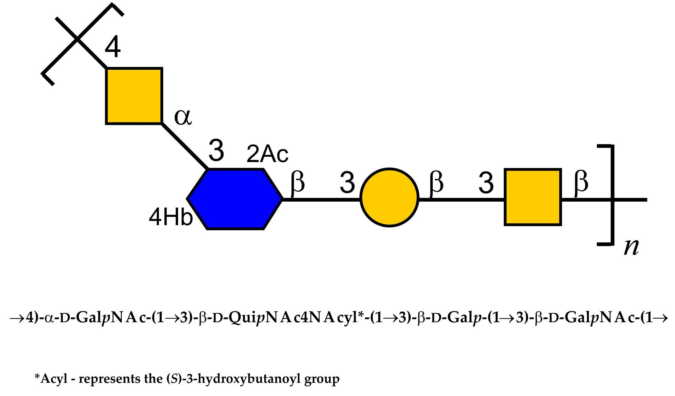
2.1. Bacterial Cultivation, Isolation of LPS, and SDS-PAGE Study

Aeromonas veronii bv. sobria K133 bacteria were cultivated in TSB (tryptic soy broth) at 28 °C for 72 h, and the cell biomass obtained was enzymatically treated, as described in Section 4, and then subjected to lipopolysaccharide extraction using the hot phenol-water method [28]. The HMW (high molecular weight) S-LPS species were recovered only from the phenol phase in a yield of 1.2% of the dry bacterial cell mass. After silver nitrate gel staining, the SDS-PAGE analysis of the phenol-soluble LPS revealed a profile characteristic for glycoforms isolated from smooth bacterial cells composed of both fast-migrating LMW (low molecular weight) species typical of rough R- and semi rough SR-LPS, and slow-migrating species representing S-LPS glycoforms with different chain lengths of the O-antigen polysaccharide (O-PS) (Figure 1a).

2.2. Serological Studies of the Aeromonas veronii bv. sobria Strain K133 O-PS

A. veronii bv. sobria strain K133 was serologically typed by agglutination tests using heat-inactivated bacteria and antisera for 44 defined Aeromonas spp. O-serogroups (NIH system) and 20 provisional serogroups (PGO1—PGO20) for selected Polish isolates. Based on these tests, strain K133 was classified into the serogroup PGO1 [27,29]. The affiliation to the serogroup was further confirmed by the reactivity of the PGO1 reference antiserum with the phenol-soluble LPS and with both the whole bacterial cells and the LPS of A. veronii bv. sobria strain K133 in the Western blotting and ELISA experiments, respectively.
The Western blot with the PGO1 antiserum revealed positive reaction in the region of S-LPS and SR-LPS species (Figure 1b). In addition, the stained bands corresponding to the fast-migrating R-LPS may also indicate the presence of immunoglobulins in the antiserum, which recognize structural determinants in the core oligosaccharide region.
Accordingly, in the ELISA experiment (Table 1), the rabbit polyclonal reference antiserum reacted with whole bacterial cells and the LPS of A. veronii bv. sobria strain K133 to the titer of 128,000 and 64,000, respectively. However, the reactions were weaker than that in the homologous system with the PGO1 cells. Adsorption of the PGO1 reference antiserum with the A. veronii bv. sobria K133 cells decreased its reactivity in the homologous system, while there was no reaction of the adsorbed PGO1 antiserum with both the whole bacterial cells and the LPS of A. veronii bv. sobria K133. The latter findings indicated that the adsorption process was complete and resulted in removal of anti-K133 antibodies from the PGO1 reference antiserum. Moreover, these data also demonstrated that the PGO1 antiserum contained additional immunoglobulins recognizing structural determinants that were not found in the A. veronii bv. sobria K133 O-antigen.
Therefore, detailed chemical analyses were performed to establish the structure of the O-PS from A. veronii bv. sobria strain K133, which represents the PGO1 serogroup.

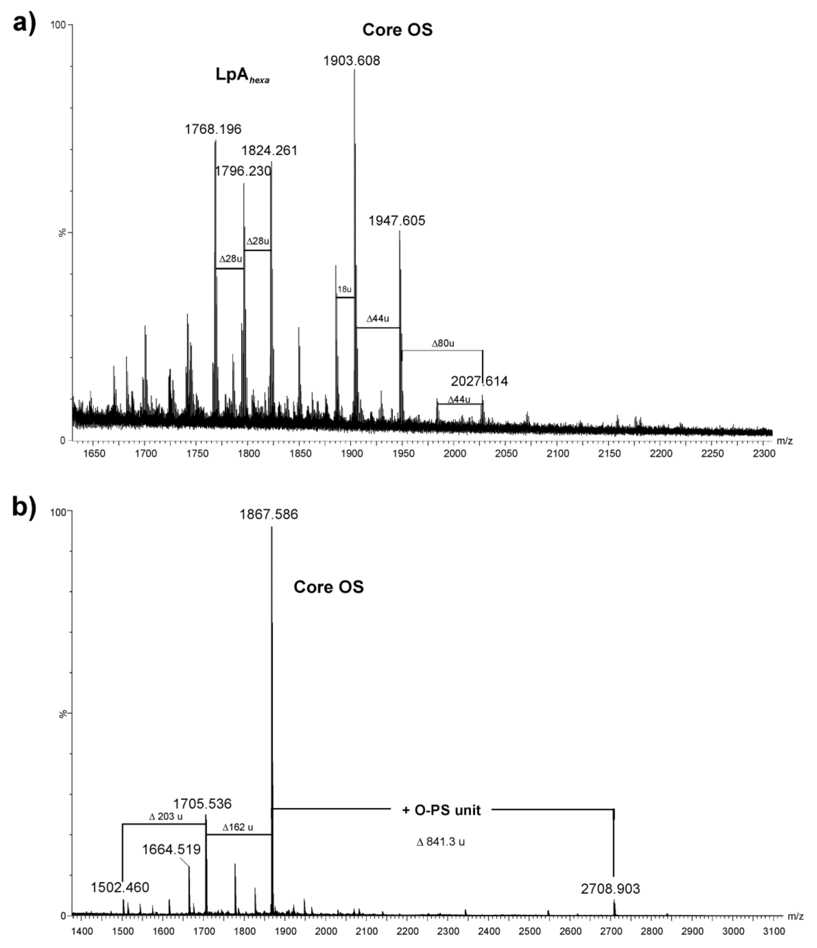
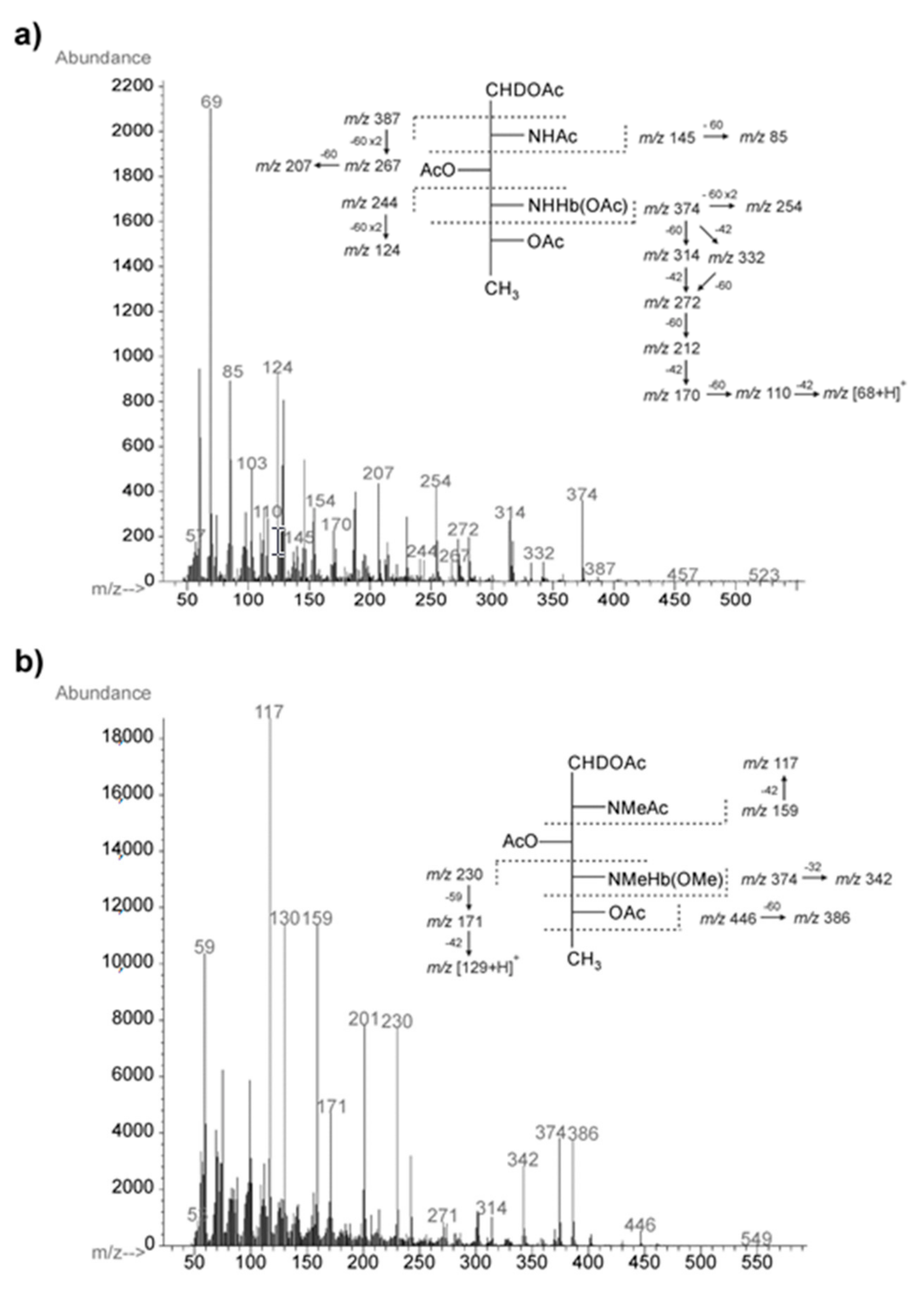
2.3. Chemical and Mass Spectrometry Analyses of LPS

The compositional analysis of the degraded polysaccharide (dgPS) fraction released from the phenol-soluble LPS after mild acid hydrolysis was performed using GLC-MS of alditol acetates. The analysis showed the presence of d-glucose (d-Glc), d-galactose (d-Gal), 2-amino-2-deoxy-d-glucose (d-GlcN), 2-amino-2-deoxy-d-galactose (d-GalN), d-glycero-d-manno-heptose (d,d-Hep), and l-glycero-d-manno-heptose (l,d-Hep) in a ratio of 2.0:5.0:1.0:6.2:5.3:4.2. Kdo (3-deoxy-d-manno-2-octulosonic acid)—the only acidic sugar—was found in the LPS after treatment of the LPS with 48% aqueous HF (hydrofluoric acid), which suggested its phosphorylation [30,31,32]. In addition, the GLC-MS analysis of fatty acids as methyl esters and O-TMS derivatives revealed 3-hydroxytetradecanoic C14:0(3-OH), 3-hydroxyisopentadecanoic Ci15:0(3-OH), dodecanoic C12:0, and tetradecanoic C14:0 acids as the major components in a ratio of 5.5:1.4:2.6:1.0. GlcN was identified as a constituent of the lipid A disaccharide.
The negative ion (MALDI-TOF) mass spectrum of the A. veronii bv. sobria K133 lipopolysaccharide (Figure 2a) showed the most intensive signals in the m/z range 1600–2000, which were attributable to the lipid A and core oligosaccharide species (Y- and B-type fragment ions) arising from in-source fragmentation at the ketosidic bond between the core oligosaccharide and the lipid A [33]. The ions at m/z 1768.19, 1796.23, and 1824.26 corresponded to hexaacylated lipid A species (Y-fragment ions) [25] composed of a bisphosphorylated glucosaminyl disaccharide and substituted by different chain-length fatty acids. The ion at m/z 1796.23 represented the lipid A species substituted by three 3-hydroxytetradecanoic acids C14:0(3-OH), one 3-hydroxyisopentadecanoic acid Ci15:0(3-OH), and two tetradecanoic acids C14:0. In turn, the ion at m/z 1768.19 contained a sugar backbone acylated by four 3-hydroxytetradecanoic acids and two dodecanoic acids, instead of two tetradecanoic acids, compared with the ion at m/z 1824.26. The composition of the ions is shown in Table 2.
The signals at m/z 2027.6, 1947.6, and 1903.6 (B-fragment ions) were assigned to the core oligosaccharide with the following composition: HexNAc1HexN1Hex2Hep5KdoanhP. The mass difference between the first two ions of 80 amu corresponded to bisphosphorylated and monophosphorylated core oligosaccharides, respectively. In turn, the most abundant ion at m/z 1903.6 represented a core decasaccharide with loss of the carboxyl group of Kdo.
The negative ion mass spectrum of the core oligosaccharide fraction (core OS), which was liberated from LPS after mild acid hydrolysis and separation by GPC, showed the major ion at m/z 1867.58 corresponding to the dephosphorylated core decasaccharide with the composition HexNAc1HexN1Hex2Hep5Kdoanh (calcd monoisotopic mass = 1868.629 amu, calcd mass of deprotonated molecule = 1867.621 amu) (Figure 2b, Table 2). The proposed structure of the core oligosaccharide corresponded to the compositional analysis of the fraction performed by GLC-MS of alditol acetate derivatives.
In addition, the ion at m/z 2708.9, observed at a higher mass range of the MALDI-TOF mass spectrum, was attributed to the core oligosaccharide with one O-antigen repeating unit attached. The mass difference of 841.31 amu, corresponding to the following composition: [6dHexNAcNAcyl1HexNAc2Hex1]-H2O (calcd monoisotopic mass: 841.332), was in complete agreement with the structure of the O-PS repeating unit established based on the NMR experiments (see Section 2.4).

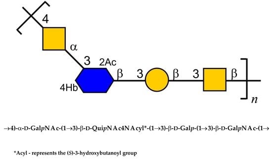
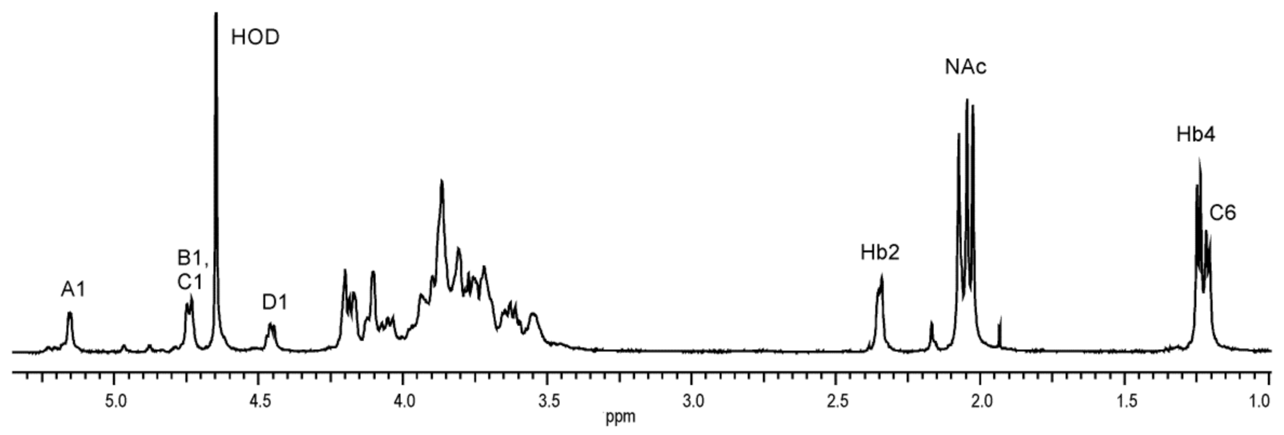
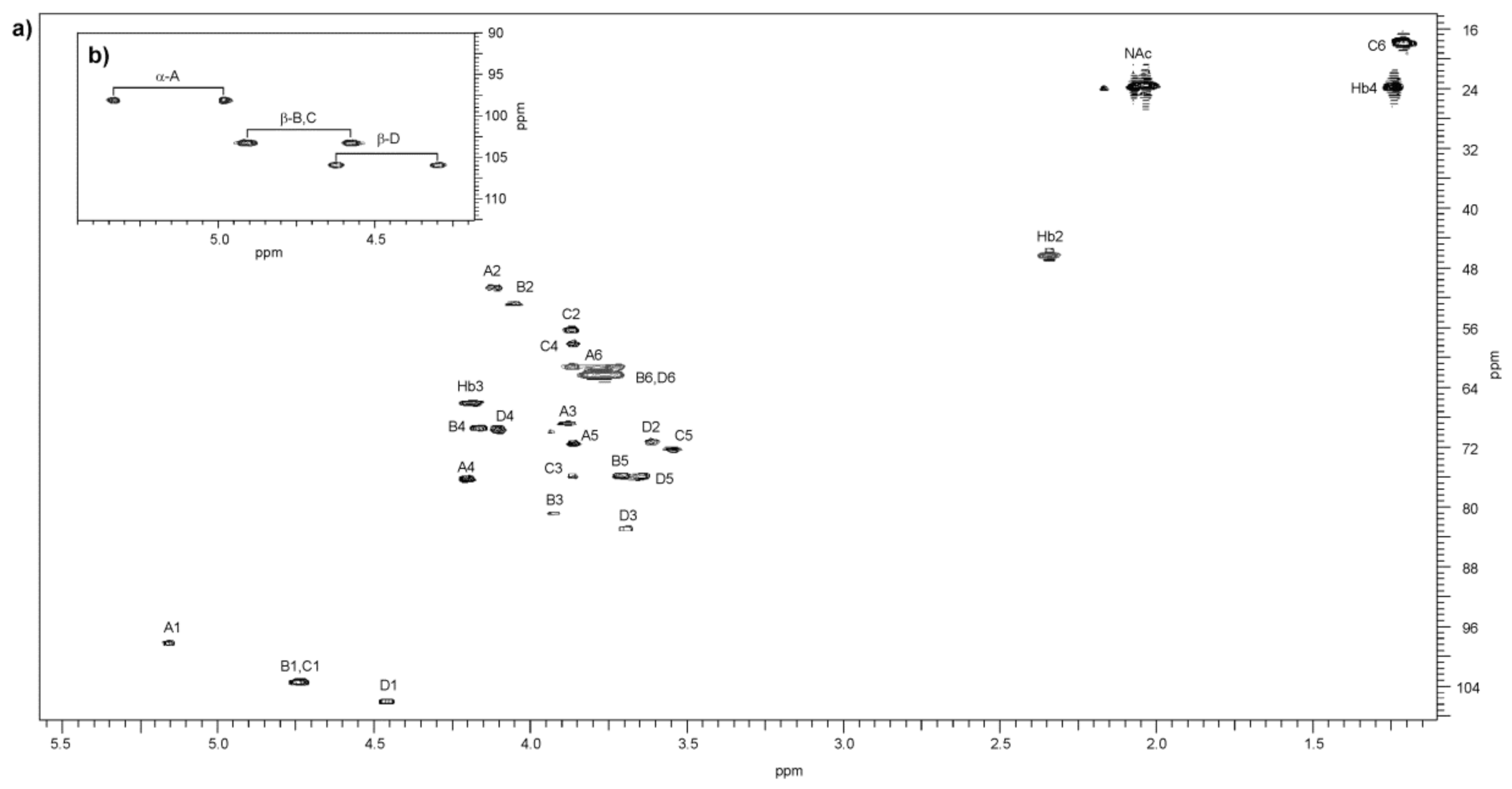
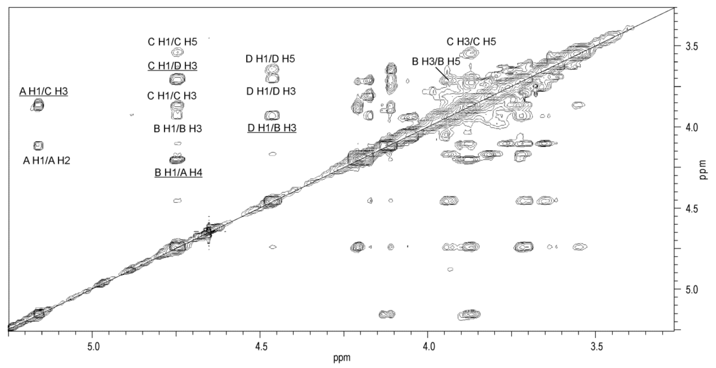
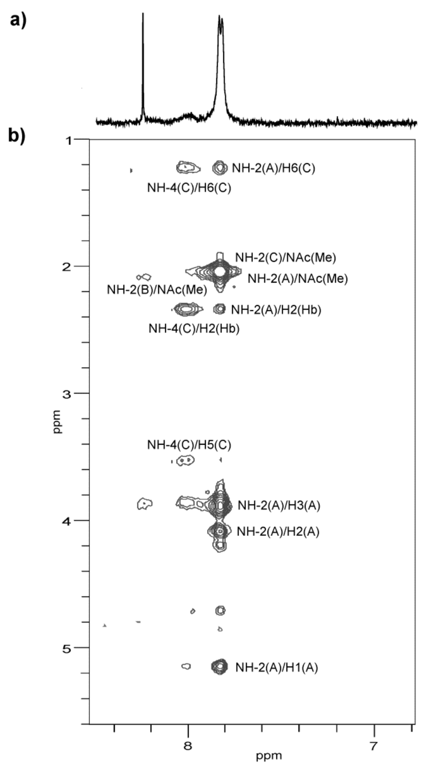
2.4. Structural Studies of O-Polysaccharide (O-PS)

The O-PS was released from the phenol-soluble LPS by mild-acid degradation followed by centrifugation of the lipid A precipitate and isolated, in a void volume, by gel-permeation-chromatography (GPC) on a Sephadex G50 Fine column. The yield of the high-molecular-mass O-PS fraction was 22% of the LPS mass. The GLC-MS sugar analysis of alditol acetates obtained after full acid hydrolysis of the O-PS with 2 M CF3CO2H showed the presence of galactose (Gal) and galactosamine (GalN) as the major components, in a peak area ratio of 1.0:1.6. There were also smaller amounts of glucose, two heptose isomers, and a component with a longer retention time identified as 2,4-diamino-2,4,6-trideoxyhexitol containing a 3-hydroxybutanoyl group (Hb) amido-linked at C-4. The electron impact EI mass spectrum (Figure 3a) of the latter compound as an alditol acetate derivative showed ions at m/z 145, 244, 374, and 387 for the (C-1 ÷ C-2), (C-4 ÷ C-6), (C-1 ÷ C-4) and (C-2 ÷ C-6) primary fragments, respectively, and the derived secondary fragment ions at m/z 85, 124, 254, and 207, which allowed to distinguish the location of the N-Hb group.
Moreover, when stronger conditions of the O-polysaccharide hydrolysis (4 M HCl, 100 °C, 16 h) were applied, 2,4-diamino-2,4,6-trideoxyhexitol (identified as QuiN4N, see below) was detected. The latter finding was confirmed by the NMR spectroscopic studies of the O-PS.
The determination of the absolute configuration of the monosaccharides by GLC of acetylated (S)-2-octyl glycosides [34] indicated the presence of d-Gal and d-GalN. The d-configuration of QuiN4N (bacillosamine) was inferred from the analysis of glycosylation effects on the 13C NMR resonances in the O-PS (see below). The S configuration of the Hb group was established by GLC-MS of the O-TMS (S)-2-octyl ester of 3-hydroxybutanoic acid released by strong acid hydrolysis of the O-PS [35].
The methylation analysis of the O-PS completed the compositional data and resulted in identification of 1,3,5-tri-O-acetyl-2,4,6-tri-O-methylhexitol-1-d (derived from 3-substituted Gal), 1,4,5-tri-O-acetyl-2-deoxy-3,6-di-O-methyl-2-(N-methyl)acetamidohexitol-1-d (derived from 4-substituted GalN), 1,3,5-tri-O-acetyl-2-deoxy-4,6-di-O-methyl-2-(N-methyl)acetamidohexitol-1-d (derived from 3-substituted GalN), and 1,3,5-tri-O-acetyl-2,4,6-trideoxy-2-(N-methylacetamido)-4-(3-methoxy-N-methylbutanoylamido)hexitol-1-d (derived from 3-substituted QuiN4NHb). The electron impact EI mass spectrum of the latter derivative contained ions at m/z 159, 230, and 374 characteristic of the (C-1 ÷ C2), (C-4 ÷ C6), and (C-1 ÷ C4) primary fragments, respectively, and confirmed the position of the 3-hydroxybutanoyl group at N-4 of QuiN4N (Figure 3b).
The O-polysaccharide structure of A. veronii bv. sobria strain K133 was then studied with the use of 1D and 2D NMR spectroscopy.
A low-field region of the 1H NMR spectrum (Figure 4) of the O-PS contained signals for anomeric protons at δ 5.16 (3J1,2~3.3 Hz), 4.74, and 4.46 (3J1,2~7.5 Hz) with an integral intensity ratio of 1.0:1.8:0.96. However, the signal at δ 4.74 corresponded in fact to two overlapping anomeric protons (both 3J1,2~8 Hz), which was demonstrated in the further 2D homonuclear 1H,1H DQF-COSY, TOCSY, and NOESY experiments.
A high-field region of the spectrum included signals for two CH3-CH groups: H-6 of QuiN4N and H-4 of Hb at δ 1.21–1.25, one CH2 group of Hb at δ 2.35, and three signals for N-acetyl groups at δ 2.03, 2.05, and 2.07. Additionally, the 1H NMR spectrum showed proton signals of CH2-OH of hexose and aminohexose residues and CH-OH of H-3 of the 3-hydroxybutanoyl group at δ 4.20, as well as the ring proton signals in the range of δ 3.54–4.21, some of which overlapped.
The analysis of the two dimensional homonuclear (1H,1H DQF-COSY, TOCSY, and NOESY) and heteronuclear (1H,13C HSQC, 1H,13C H2BC, and 1H,13C HMBC) NMR experiments resulted in the assignment of the 1H and 13C resonances to the O-PS of A. veronii bv. sobria strain K133. The 1H and 13C NMR chemical shifts are collected in Table 3.
The 1H,13C HSQC spectrum (Figure 5) contained four correlation signals at δ 4.12/50.6, 4.05/52.8, 3.87/56.2, and 3.87/58.0 of protons at the nitrogen-bearing carbons to the corresponding carbons and showed that the O-PS repeating unit contained N-acetamido sugars. Moreover, the absence of signals at the 13C coordinate in the region of δ 83.0–88.0 characteristics of the furanose ring demonstrated that all the sugars were pyranoses [36].
The 1H,1H TOCSY and DQF-COSY spectra revealed four spin systems for monosaccharide residues, which were labelled AD in the order of the decreasing chemical shifts of their H1/H2 protons, and an additional non-sugar spin system corresponding to the N-3-hydroxybutanoyl group (Hb).
The correlations in the two dimentional homonuclear 1H,1H TOCSY, DQF-COSY and NOESY (Supplementary Materials Figure S1) spectra revealed three spin systems for sugar residues having the galacto configuration and one monosaccharide with the gluco configuration (see below).
In the TOCSY spectrum, starting from the H-1 proton signal the correlations with H-2, H-3 and H-4 were visible for spin systems A, B, and D, indicating galacto-configured monosaccharides. The remaining resonances were assigned from the NOESY (Figure 6), DQF-COSY and heteronuclear experiments.
In the 1H,13C HMBC spectrum, correlations of the anomeric proton with carbons C-3 and C-5 were found for the spin system A, and then the proton resonances were assigned from the 1H,13C HSQC spectrum. In the NOESY spectrum, correlations of H-4/H-5 and H-4/H-6 were visible for spin systems A and D. In turn, for spin system B, the chemical shift of H-5 was assigned from the H-3/H-5 and H-4/H-5 intraresidue NOE contacts. The H-6 resonances would then be assigned from H-5/H-6 correlations in the DQF COSY spectrum. The corresponding 13C resonances for spin systems A, B, and D were inferred from the 1H,13C HSQC spectrum. Moreover, in the latter spectrum, the correlations of two H-2 protons at δ 4.12 and δ 4.05 with the corresponding nitrogen-bearing carbons at δ 50.6 and δ 52.8, respectively, indicated that two spin systems, i.e., A and B, were N-acetamido sugars. By including 3JH1,H2 coupling constant values, spin systems A, B, and D were assigned to α-GalpN, β-GalpN, and β-Galp, respectively.
In the DQF-COSY and TOCSY spectra, correlations of H-1/H-2 up to H-6 typical of monosaccharide having the gluco configuration were found for spin system C. The corresponding carbons were inferred from the 1H,13C HSQC spectrum. However, given the almost complete coincidence of the H-2,3,4 proton signals, the chemical shifts of C-2, C-3, and C-4 of spin system C were identified after consideration of the two-bond and long-range correlations in the H2BC and HMBC spectra, respectively, and the glycosylation effects on the 13C NMR resonances [37].
The 1H,13C HSQC spectrum at the 1H coordinate (Figure 5a) showed two correlation signals at δ 3.87/56.2 and 3.87/58.0 of protons at the nitrogen-bearing carbons to the corresponding carbons, which after including the high-field positions of H-6/C-6 at δ 1.21/17.1 indicated that spin system C was a diamino-6-deoxysugar. The latter finding was confirmed in the HMBC spectrum (Figure 7) by the presence of a long-range correlation between H-6 and C-4 at δ 1.21/58.0, which allowed identifying C-4 as the second nitrogen-bearing carbon. The high-field region of the HMBC spectrum at the 1H coordinate of the methyl protons (δ 1.21) also showed a correlation to C-5 of residue C at δ 72.3, whose low field position indicated β-linked sugar [32,38]. Based on these data and after including a relatively large 3JH1,H2 coupling constant value of ~8 Hz, spin system C was assigned to β-QuipN4N.
The 13C resonances of the two nitrogen-bearing carbons of QuiN4N were also verified by the two-bond correlations in the 1H,13C-H2BC experiment. In the spectrum, for the nitrogen-bearing carbon at δ 56.2, there were correlations with the protons at δ 3.87 and 4.74 (H-1 of residue C). In turn, for the second carbon at δ 58.0, a connection was found with signals at δ 3.87 and 3.54 (H-5 of residue C). These correlations confirmed the chemical shifts of C-2 and C-4 QuiN4N at δ 56.2 and 58.0, respectively.
Accordingly, the 1H,13C-H2BC spectrum also showed correlations for two other nitrogen-bearing carbons of residues A and B to H-1 and H-3 protons at δ 5.16/50.6;3.89/50.6 and at δ 4.74/52.8;3.93/52.8, respectively.
The 1JC1,H1 coupling constant values determined from the 1H,13C HSQC spectrum measured without 13C decoupling (Figure 5b) of the O-PS confirmed that one of the monosaccharides with C-1 at δ 98.2 (A) was α-linked (1JC1,H1 175.0 Hz) and three others with C-1 at δ 103.3 (B, C) and 106.1 (D) were β-linked (1JC1,H1 163–167 Hz) [39].
The anomeric configuration of the sugar residues was also supported by the NOE contacts of H-1 to H-2 for A and H-1 to both, H-3 and H-5 for the other three sugars (B, C, D) and confirmed the α- and β-anomeric configurations of monosaccharides, respectively. These intraresidue cross-peaks were informative especially for spin systems B and C with overlapping H-1 proton signals.
The low-field displacement of the signals for C-4 of A (δ 76.3) and C-3 of B (δ 80.8), C (δ 75.9), and D (δ 82.8), compared with their resonances in the corresponding non-substituted monosaccharides, indicated the linkage position in each sugar residue [37,39,40].
The substitution pattern and sequence of the monosaccharides in the O-PS repeating unit was established in the NOESY and HMBC experiments. The 1H,1H NOESY spectrum (Figure 6) showed interresidue cross-peaks between residues AC, CD, DB, and BA. The following strong correlations for the transglycosidic protons were observed: GalpN A H-1/QuipN4N C H-3 at δ 5.16/3.87, QuipN4N C H-1/Galp D H-3 at δ 4.74/3.70, Galp D H-1/GalpN B H-3 at δ 4.46/3.93, and GalpN B H-1/ GalpN A H-4 at δ 4.74/4.21.
The HMBC spectrum (Figure 7) demonstrated the following expected correlations between the anomeric protons and the linkage carbons: GalpN A H-1/QuipN4N C C-3 at δ 5.16/75.9, QuipN4N C H-1/Galp D C-3 at δ 4.74/82.8 (week), Galp D H-1/GalpN B C-3 at δ 4.46/80.8, and GalpN B H-1/ GalpN A C-4 at δ 4.74/76.3.
The 13C resonance of the NAc carbonyl signals were inferred from the correlations between the H-2 protons of residues A and B (δ 4.12 and δ 4.05) and the corresponding carbons in the HMBC spectrum (δ 175.5 and 176.4), and between the latter and the methyl proton signals at δ 2.05 and 2.07, respectively.
The distribution of the N-acyl (Hb) and N-acetyl groups was established by the NOESY experiment on the O-PS sample in a 90% H2O—10% D2O mixture (Figure 8), which enabled detection of nitrogen-linked protons. The 1H NMR spectrum revealed three NH protons at δ 7.83, 8.02 (a broad signal), and 8.24, which were assigned by a 2D NOESY experiment to NH-2 of both α-GalN (A) and QuiN4N (C), NH-4 of QuiN4N, and NH-2 of β-GalN (B), respectively. The spectrum showed correlations of NH-2 of α-GalN (A) and NH-2 of QuiN4N with the NAc methyl protons at δ 7.83/2.03–2.05, NH-4 of QuiN4N with H-2 of Hb at δ 8.02/2.35, and NH-2 of β-GalN (B) with the NAc methyl protons at δ 8.24/2.07, respectively. Moreover, the intraresidue NOE contacts between NH-4 of QuiN4N and both H-5, H-6 at δ 8.02/3.54, 1.21, and the interresidue NOE correlations for A(1→3)C between NH-2 of α-GalN (A) and H-2 (Hb), and H-6 of QuiN4N at δ 7.83/2.35 and 7.83/1.21, respectively, resulting from the spatial proximity of the residues, confirmed the N-acylation of QuiN4N at position 4.
The d configuration of QuipN4N in the O-PS of A. veronii bv. sobria strain K133 was established by the analysis of the glycosylation effects on the 13C NMR chemical shifts [37,41,42]. In the disaccharide fragment C-(1→3)-D, β-QuipN4N-(1→3)-β-d-Galp, the large positive α-effect on C-1 of residue C (+7.45 ppm) and C-3 of residue D (+9 ppm) and the small negative β-effect (−0.1 ppm) on C-4 of D indicated that the linked monosaccharides have the same d absolute configuration. In the case of different l-d absolute configurations, the positive α-effect on C-1 of residue C and C-3 of residue D would have been <4 ppm and <7 ppm, respectively, and the negative β-effect on C-4 of D would have been much higher (−3.0 ppm) [37,41].
In the disaccharide fragment A-(1→3)-C, α-d-GalpNAc-(1→3)-β-QuipN4N, the small β-effect of glycosylation on C-4 of β-QuipN4N C (<0.5 ppm) indicated that the linked monosaccharides had the same d absolute configuration. In the case of different d-l absolute configurations, the higher negative β-effect of ~1.4 ppm on C-4 would have been observed [41,42].
Based on the data obtained, it was concluded that the O-polysaccharide from the LPS of A. veronii bv. sobria strain K133 is composed of a tetrasaccharide repeating unit and has the structure presented below: Ijms 22 04272 i001
To our knowledge, the O-PS from A. veronii bv. sobria strain K133 is unique among bacterial polysaccharide structures as indicated by the Bacterial Carbohydrate Structure Database (http://glyco.ac.ru/bcsdb, (accessed on 10 March 2021)) [43].

3. Discussion

The inland aquaculture in Poland is focused on two fish species, i.e., carp and rainbow trout [44], the farming of which is endangered due to the stressful environmental conditions and various diseases, with dominance of infections caused by motile Aeromonas bacteria [3,4]. Studies on the occurrence of mesophilic Aeromonas species associated with outbreaks of MAI/MAS in Polish culture facilities showed the A. veronii bv. sobria species as one of the dominant isolates on carp farms, while representatives of A. veronii bv. sobria, A. bestiarum, and A. salmonicida were pathogenic for both carp and trout. Moreover, the veterinary data indicated that, despite their large diversity, only some Aeromonas sp. serogroups seem to be associated with virulence for freshwater fish species [26]. As demonstrated recently, the majority of isolates pathogenic to carp and trout in Polish cultures were positively classified based on the somatic O-antigen variants when the 44 antisera of the NIH scheme were extended to include those for 20 new provisional serogroups of local origin. Moreover, it was evidenced that the highest number of Aeromonas strains isolated from carp and trout represented the serogroup PGO1 [27].
Here, we established the structure of the LPS and the O-specific polysaccharide from A. veronii bv. sobria strain K133, which was isolated from the kidney of carp during an outbreak of MAI/MAS on a Polish fish farm. Strain K133 was classified to the serogroup PGO1, i.e., an immunotype that is common among aeromonads with pathogenicity to fish in Polish aquacultures.
The chemical and MALDI-TOF mass spectrometry analyses have revealed that the LPS of A. veronii bv. sobria strain K133 contains hexaacylated lipid A species with a conserved architecture of the backbone composed of a 1,4′-bisphosphorylated-β-(1→6)-linked-d-GlcN disaccharide, acylated by three or four residues of 3-hydroxytetradecanoic acids C14:0(3-OH) and saturated fatty acids, i.e., dodecanoic or tetradecanoic acids.
The sugar analysis of the core oligosaccharide revealed d,d-heptose and l,d-heptose residues indicating the type of the core OS shared by A. hydrophila [30,31,45] and A. bestiarum species [46]. This LPS core variant is different from those described for A. salmonicida subsp. salmonicida, in which there was only one heptose isomer (l,d-Hep) [47]. However, some differences were found in the structure of the core region studied here. The chemical and mass spectrometry experiments revealed the following composition of the core oligosaccharide: HexNAc1HexN1Hex2Hep5KdoanhP1, whose structure slightly differs from those described for A. hydrophila and A. bestiarum species by the presence of five instead of six heptose residues and an additional N-acetamido hexose (compare with the composition of the core OS of A. hydrophila HexN1Hex2Hep6Kdo1P1).
The SDS-PAGE analysis confirmed that the phenol-soluble LPS contained HMW S-LPS glycoforms and thus suggested a highly hydrophobic character of the O-polysaccharide chains. This finding was in agreement with structural analysis, which demonstrated the presence in the O-PS of deoxy-amino sugars and amino sugars with hydrophobic substituents i.e., N-acetyl and N-acyl groups.
The Western blotting and ELISA experiments supported the results of serotyping with the use of the agglutination test showing the classification of the strain K133 to the new provisional serogroup PGO1. As reported recently, using heat-inactivated bacteria and antisera for 44 defined Aeromonas O-serogroups of the NIH system and after complementing the classical scheme with 20 new antisera for provisional serogroups (PGO1—PGO20) of local origin, the positive classification of the Aeromonas sp. isolates based on appropriate somatic antigen increased from 53% (with the use of the NIH system) to nearly 90% after including the new antisera [27,29].
The structure of the O-PS of A. veronii bv. sobria strain K133 has been established and it has been found that the O-antigen of the strain is built up of linear tetrasaccharide repeating units. To the best of our knowledge, the composition of the O-PS is unique among O-chains of Aeromonas spp. and other bacterial polysaccharide structures (Bacterial Carbohydrate Structure Database: http://glyco.ac.ru/bcsdb, (accessed on 10 March 2021)) [43].
While both d-Galp and d-GalpNAc are commonly known as compounds building bacterial polysaccharides, the presence of 2,4-diamino-2,4,6-trideoxy-glucopyranose (bacillosamine) is still a peculiar feature. This sugar has been reported as a component of heteropolymeric O-chains of several bacteria, e.g., Pseudomonas fluorescens biovar B strain IMV 247 [48], Pseudoalteromonas haloplanktis strain ATCC 14393 [40], Acinetobacter haemolyticus strains 57 and 61 [49], Vibrio cholerae O5 [50], V. cholerae O8 [38], V. cholerae O100 [51], Shewanella japonica KMM 3299T [52], Plesiomonas shigelloides strain 302-73, serotype O1 [53], Pseudomonas chlororaphis subsp. aureofaciens UCM B-306 [54], and Idiomarina abyssalis KMM 227T [55], as well as capsular polysaccharides of Psychrobacter maritimus 3pS [56], Providencia rustigianii O11 [57], and Acinetobacter baumannii clinical isolate MG1 [58]. Additionally, in Vibrio anguillarum serotype O2, which affects salmonids and other marine fish species causing vibriosis, bacillosamine has been established as a component of tetrasaccharide repeating units of both the O-chain polysaccharide and acidic capsular polysaccharide [59].
Structural studies have indicated that QuiN4N in microbial polysaccharides usually carries N-acetyl groups located at both N-2 and N-4. Such a derivative of diamino-6-deoxyglucose, i.e., N,N′-diacetylbacillosamine, has been found in the O-PS repeating units of V. cholerae O5 and O8 [38,50], V. anguillarum O2 [59], P. haloplanktis [40], S. japonica KMM 3299T [52], P. chlororaphis subsp. aureofaciens UCM B-306 [54], and I. abyssalis KMM 227T [55]. As often as mentioned above, a bacillosamine residue substituted with an N-acetyl group at N-2 and a (S)-3-hydroxybutanoyl group at N-4 has been reported. This derivative was found as a component of the O-polysaccharides of P. fluorescens strain IMV 247 [48] and P. shigelloides strain 302-73 [32], as well as capsular polysaccharides of A. baumannii [58], P. rustigianii O11 [57], and P. maritimus 3pS [56]. In turn, a very unique derivative of QuiN4N with the presence of (R)-3,5-dihydroxyhexanoyl group N-linked at position 4 has been indicated only in two O-antigens of V. cholerae O3 and V. cholerae O100 [51,60].
The ELISA experiment with the reference PGO1 antiserum, carried out in this study, revealed the positive recognition of surface antigenic elements within the whole bacterial cells and the isolated LPS molecules of A. veronii bv. sobria strain K133, and thus confirmed affiliation of the strain to the PGO1 serogroup. Moreover, the assay also demonstrated that the reference antiserum contained immunoglobulins recognizing a wider range of structural epitopes that were not found in the A. veronii bv. sobria K133 O-polysaccharide, suggesting both similarities and possible differences in the PGO1 and K133 O antigens. It is worth mentioning, that the O-PS structure of the Aeromonas sp. reference strain for the PGO1 serogroup remains unknown. The structural fragment of the O-PS of A. veronii bv. sobria strain K133, containing β1→3-linked 2-acetamido-4-[(S)-3-hydroxybutanoyl]amino-6-deoxyglucose, seems to be one of the key antigenic determinants of the immunospecificity of the PGO1 serotype.
In conclusion, the immunochemical studies of the LPS of Aeromonas spp. bacteria, which are pathogenic to freshwater fish species and represent the dominant serogroups, will contribute to advancement in research targeted at development of an effective vaccine based on the antigenic profile of emergent pathogens and dedicated to specific fish farms as an alternative to antibiotic therapy. In fisheries, where bacterial diseases appear systematically and may bring large losses, immunoprophylaxis consisting of the use of preparations that increase non-specific and/or specific immunity in fish should play a significant role in preventing bacterial diseases. Due to the emerging difficulties of effective prophylaxis caused by the lack of commercial vaccines, the interest in the use of autovaccinations that can offer protection against defined serotypes from a specific region or geographic area is increasing in veterinary fields, including ichthyopathology [4].

4. Materials and Methods

4.1. Bacterial Strain, Growth Conditions, and LPS Isolation

A. veronii bv. sobria strain K133, serogroup PGO1, was isolated from the kidney of a common carp during an outbreak of MAS/MAI on a Polish fish farm and classified to the species level by restriction analysis of 16S rDNA amplified by PCR [27]. For the LPS studies, strain K133 was obtained from the collection of the Department of Fish Diseases, National Veterinary Research Institute (Puławy, Poland). The bacterium was cultivated with shaking (120 rpm) on tryptic soy broth (TSB) for 72 h at 28 °C. The cells were harvested by low speed centrifugation (8000× g, 20 min). The recovered bacterial cell pellet was washed twice with 0.85% saline and once more with distilled water.
The bacterial cells (5 g dry mass) were suspended in 50 mM phosphate buffer (pH 7.0) containing 5 mM MgCl2 and treated with lysozyme, RNAse, and DNAse (16 h, 0.6 mg/g) and then with Proteinase K (16 h, 0.6 mg/g). The enzymatically digested biomass was extracted three times with aqueous 45% phenol at 70 °C [35]. Layers separated by centrifugation (3000× g, 45 min, 4 °C) were dialyzed against tap and distilled water. LPS species recovered from the phenol and water layers were purified by ultracentrifugation at 105,000× g (4 h, 18 °C) and freeze-dried to give a yield of 3% of dry bacterial cell mass.

4.2. SDS-PAGE

The phenol-soluble LPS fraction (3 µg) of A. veronii bv. sobria strain K133 prepared in the sample buffer (2% SDS and 50 mM Tris/HCl (pH 6.8), 25% glycerol, 0.1% bromophenol blue) was separated in 12.5% SDS-Tricine polyacrylamide electrophoresis gel and the profile was visualized by staining with silver nitrate after oxidation with periodate according to the published method [61].

4.3. Serological Studies

Western blotting with the rabbit antiserum PGO1 was performed after transferring SDS-PAGE-separated phenol-soluble LPS of A. veronii bv. sobria strain K133 to Immobilon P (Millipore, St. Louis, MO, USA). The primary antibodies were detected using alkaline phosphatase-conjugated goat anti-rabbit antibodies (Sigma, St. Louis, MO, USA). The blot was developed with nitroblue tetrazolium and 5-bromo-4-chloro-3-indolylphosphate toluidine (Sigma) for 5 min, as described elsewhere [25]. Polyclonal rabbit O-antiserum against the Aeromonas sp. reference strain for the provisional serogroup PGO1 was the kind gift from Professor Alicja Kozińska (the National Veterinary Research Institute, Puławy, Poland) [29].
The enzyme-linked immunosorbent assay (ELISA) was performed as described previously [62] with some modifications. In short: 1–2 μg of the A. veronii bv. sobria strain K133 LPS or 10–20 μg of whole cell biomasses of A. veronii bv. sobria strain K133 or the Aeromonas sp. PGO1 reference strain per well were coated on flat-bottom F96 Maxisorp Nunc-Immuno plates (Thermo Fisher Scientific, Roskilde, Denmark); polyclonal rabbit PGO1 antiserum and rabbit-IgG specific peroxidase-conjugated goat antibodies (Jackson ImmunoResearch, West Grove, PA, USA) were used. 2,2′-Azino-bis(3-ethylbenzothiazoline-6-sulfonic acid) diammonium salt (ABTS) was used as a substrate for peroxidase; the absorbance (A405) was measured with the help of a Multiskan Go microplate reader (Thermo Fisher Scientific USA, Vantaa, Finland). The reference PGO1 antiserum diluted 1:50 in PBS (phosphate-buffered saline) was adsorbed on K133 cells during 0.5 h incubation on ice, in the ratio of 100 µL of wet biomass to 1 mL of the serum. The cells were removed by centrifugation and the process was repeated two more times [63].

4.4. Degradation of LPS and Isolation of O-Polysaccharide

The phenol-soluble LPS sample (100 mg) was heated in aq 2% acetic acid at 100 °C for 3 h, and the lipid A precipitate was removed by centrifugation (13,000× g, 30 min). The supernatant was concentrated and then fractionated by GPC on a column (1.8 × 80 cm) of Sephadex G-50 Fine (Pharmacia, Sweden) using 1% acetic acid as the eluent and monitoring with a differential refractometer (Knauer, Berlin, Germany). The yield of the O-PS fraction was 22% of the LPS portion subjected to hydrolysis.

4.5. Chemical Analyses

For neutral and amino sugar analysis, the degraded polysaccharide (dgPS) fraction released from the phenol-soluble LPS after mild acid hydrolysis, the O-PS, and the core oligosaccharide samples were hydrolyzed with 2 M CF3CO2H (120 °C, 2 h), reduced with NaBD4, and peracetylated with a 1:1 (v/v) mixture of acetic anhydride and pyridine (85 °C, 0.5 h). The O-PS was also hydrolyzed with 4 M HCl for 16 h at 100 °C to release QuiN4N, N-acetylated, reduced with NaBD4, and peracetylated.
To release acidic sugar (Kdo), LPS was dephosphorylated with 48% aqueous HF (4 °C, 18 h) and dried under vacuum over sodium hydroxide [32]. Methanolysis was performed with 1 M MeOH/HCl (85 °C, 1 h), and the sample was extracted with hexane. The methanol layer was concentrated and the residue was dried and peracetylated.
The absolute configuration of the monosaccharides was determined by GLC of acetylated (S)-(+)-2-octyl glycosides using authentic sugars as standards according to a published method [34], except for QuiN4N, whose configuration was determined upon analysis of glycosylation effects on 13C resonances in the O-PS.
The absolute configuration of 3-hydroxybutanoic acid (Hb) was determined according to the method of Kenne, et al. [35] with some modifications: after hydrolysis of the O-PS with 2 M CF3CO2H (120 °C, 4 h), the product was extracted three times with EtOAc, evaporated under nitrogen, and subjected to solvolysis with 2 M HCl in S(+)-2-octanol at 80 °C for 16 h. The sample was concentrated to dryness, and trimethylsilylated derivatives were analyzed by GLC-MS and compared with the retention time of O-TMS (S)-2-octyl esters of authentic (S)- and (R)-3-hydroxybutanoates as references.
Methylation of the O-PS (1.0 mg) was carried out with methyl iodide in dimethyl sulfoxide in the presence of powdered sodium hydroxide as described by Ciucanu and Kerek [64]. The products were recovered by extraction with chloroform/water (1:1, v/v), N-acetylated, hydrolyzed with 2 M CF3CO2H (120 °C, 2 h), N-acetylated, reduced with NaBD4 and peracetylated. The partially methylated alditol acetates derivatives were analyzed by GLC-MS.
For fatty acid analysis, a lipid A sample (1 mg) was subjected to methanolysis in 2 M methanolic HCl (85 °C, 12 h). The resulting fatty acid methyl esters were extracted with hexane and converted to their O-trimethylsilyl (O-TMS) derivatives, as described elsewhere [53,65]. The methanol layer containing methyl glycosides was dried and peracetylated with a pyridine-acetic anhydride mixture. The fatty acid derivatives and acetylated methyl glycosides were analyzed by GLC-MS as above.
All the sample derivatives were analyzed on an Agilent Technologies 7890A gas chromatograph (Agilent Technologies, Wilmington, DE, USA) connected to a 5975C MSD detector (inert XL EI/CI, Agilent Technologies, Wilmington, DE, USA). The chromatograph was equipped with an HP-5MS capillary column (Agilent Technologies, 30 m × 0.25 mm, flow rate of 1 mL/min, He as carrier gas). The temperature program for all the derivatives was as follows: 150 °C for 5 min, then 150 to 310 °C at 5 °C/min, and the final temperature was maintained for 10 min.

4.6. NMR Spectroscopy

The O-PS sample (7 mg) was deuterium-exchanged by freeze-drying from a 99.95% D2O solution and examined in 99.98% D2O. 1D and 2D NMR spectra were recorded at 42 °C on a 500 MHz NMR Varian Unity Inova instrument and calibrated with external acetone (δH 2.225, δC 31.45). Additionally, for detection of nitrogen-linked protons, 1H and 1H,1H NOESY spectra in a 90% H2O—10% D2O mixture were recorded at 20 °C on a 500 MHz NMR Varian instrument. Standard Varian software (Vnmrj V. 4.2 rev.) was used to acquire and process the NMR data. The homonuclear and heteronuclear two-dimensional experiments: 1H,1H DQF-COSY, 1H,1H TOCSY, 1H,1H NOESY, 1H,13C HSQC, 1H,13C H2BC, and 1H,13C HMBC were conducted for signal assignments and determination of the sugar sequence in the repeating unit. The mixing time of 90 and 200 ms was used in the TOCSY and NOESY experiments, respectively. The 1H,13C HSQC experiment (gHSQCAD) with CRISIS based multiplicity editing was optimized for a coupling constant of 146 Hz. The 1H,13C HSQC spectrum (band-selective gHSQCAD) measured without 13C decoupling was used to determine the 1JC1,H1 coupling constants for the anomeric carbons. The heteronuclear multiple-bond correlation (HMBC) experiment was optimized for JC,H = 8 Hz, with 2-step low-pass filter 130 and 165 Hz to suppress one-bond correlations.

4.7. MALDI-TOF Mass Spectrometry (MS)

The LPS and oligosaccharide samples were analyzed with matrix-assisted laser desorption/ionization time-of flight (MALDI-TOF) mass spectrometry (MS) using a Waters SYNAPT G2-Si HDMS instrument (Waters Corporation, Milford, MA, USA) equipped with a 1 kHz Nd:YAG laser system. Acquisition of the data was performed using MassLynx software version 4.1 SCN916 (Waters Corporation, Wilmslow, UK). Mass spectra were assigned with a multi-point external calibration using red phosphorous (Sigma) and recorded in the negative ion mode. Phenol-soluble LPS and oligosaccharide samples (both at a concentration of 15 µg/µL) were suspended in a water/methanol (1:1, v/v) solution (containing 2 mM EDTA for the LPS sample) and dissolved by ultrasonication. After desalting with the use of cation exchange beads (Dowex 50WX8-200; Sigma), one microliter of each sample was transferred onto a well plate covered with a thin matrix film and allowed to dry at room temperature. The matrix solution was prepared from 2′,4′,6′-trihydroxyacetophenone (THAP) (200 mg/mL in methanol) mixed with nitrocellulose (15 mg/mL) suspended in 2-propanol/acetone (1:1, v/v) in a proportion of 4:1 (v/v), according to the published method [65,66].

Supplementary Materials

The following are available online at https://0-www-mdpi-com.brum.beds.ac.uk/article/10.3390/ijms22084272/s1, Figure S1. 1H,1H NOESY (500 MHz) spectrum of the O-PS of A. veronii bv. sobria strain K133.

Author Contributions

Conceptualization, A.T.-S. and K.D.; methodology, A.T.-S., K.D., D.D., and A.P.-S.; investigation, K.D., M.L., M.K., and A.T.-S.; serotyping, A.P.-S.; ELISA experiments, D.D.; Western blotting studies, K.D., and M.K.; NMR analysis and interpretation, A.T.-S., K.D., and M.K.; data curation, A.T.-S. and K.D.; writing—original draft preparation, A.T.-S. and K.D.; writing—review and editing, All authors.; supervision, A.T.-S.; funding acquisition, K.D., M.K., and M.L. All authors have read and agreed to the published version of the manuscript.

Funding

This research was financially supported by the Polish Ministry of Science and Higher Education research funding for young scientists.

Institutional Review Board Statement

Not applicable.

Informed Consent Statement

Not applicable.

Data Availability Statement

The data presented in this study have been disclosed in the main text and the Supplementary Materials.

Acknowledgments

The authors wish to thank Paweł Sowiński for recording the NMR spectra and his valuable advice in resolving the spectra (Nuclear Magnetic Resonance Laboratory, Faculty of Chemistry, Gdańsk University of Technology, Poland) and Alicja Kozińska (the National Veterinary Research Institute, Puławy, Poland) for the kind gift of the PGO1 reference antiserum. The authors also highly appreciate the assistance and technical support of Hubert Pietras during LPS isolation and purification.

Conflicts of Interest

The authors declare no conflict of interest.

References

  1. Yahia, N.; Brown, C.A.; Rapley, M.; Chung, M. Level of nutrition knowledge and its association with fat consumption among college students. BMC Public Health 2016, 16, 1047. [Google Scholar] [CrossRef] [Green Version]
  2. The State of World Fisheries and Aquaculture 2018-Meeting the Sustainable Development Goals. Available online: http://www.fao.org/3/i9540en/i9540en.pdf (accessed on 10 March 2021).
  3. Pękala-Safińska, A. Contemporary Threats of Bacterial Infections in Freshwater Fish. J. Vet. Res. 2018, 62, 261–267. [Google Scholar] [CrossRef] [Green Version]
  4. Schulz, P.; Terech-Majewska, E.; Siwicki, A.K.; Kazuń, B.; Demska-Zakęś, K.; Rożyński, M.; Zakęś, Z. Effect of Different Routes of Vaccination against Aeromonas salmonicida on Rearing Indicators and Survival after an Experimental Challenge of Pikeperch (Sander lucioperca) in Controlled Rearing. Vaccines 2020, 8, 476. [Google Scholar] [CrossRef]
  5. Forn-Cuní, G.; Fulton, K.M.; Smith, J.C.; Twine, S.M.; Mendoza-Barberà, E.; Tomás, J.M.; Merino, S. Polar Flagella Glycosylation in Aeromonas: Genomic Characterization and Involvement of a Specific Glycosyltransferase (Fgi-1) in Heterogeneous Flagella Glycosylation. Front. Microbiol. 2020, 11, 595697. [Google Scholar] [CrossRef]
  6. Igbinosa, I.H.; Beshiru, A.; Odjadjare, E.E.; Ateba, C.N.; Igbinosa, E.O. Pathogenic potentials of Aeromonas species isolated from aquaculture and abattoir environments. Microb. Pathog. 2017, 107, 185–192. [Google Scholar] [CrossRef]
  7. Talagrand-Reboul, E.; Jumas-Bilak, E.; Lamy, B. The Social Life of Aeromonas through Biofilm and Quorum Sensing Systems. Front. Microbiol. 2017, 8, 1–19. [Google Scholar] [CrossRef]
  8. Harnisz, M.; Korzeniewska, E. The prevalence of multidrug-resistant Aeromonas spp. in the municipal wastewater system and their dissemination in the environment. Sci. Total Environ. 2018, 626, 377–383. [Google Scholar] [CrossRef]
  9. Piotrowska, M.; Przygodzińska, D.; Matyjewicz, K.; Popowska, M. Occurrence and Variety of β-Lactamase Genes among Aeromonas spp. Isolated from Urban Wastewater Treatment Plant. Front. Microbiol. 2017, 8, 1–12. [Google Scholar] [CrossRef] [Green Version]
  10. El-Gohary, F.A.; Zahran, E.; Abd El-Gawad, E.A.; El-Gohary, A.H.; Abdelhamid, F.M.; El-Mleeh, A.; Elmahallawy, E.K.; Elsayed, M.M. Investigation of the Prevalence, Virulence Genes, and Antibiogram of Motile Aeromonads Isolated from Nile Tilapia Fish Farms in Egypt and Assessment of their Water Quality. Animals 2020, 10, 1432. [Google Scholar] [CrossRef]
  11. Janda, J.M.; Abbott, S.L. The Genus Aeromonas: Taxonomy, Pathogenicity, and Infection. Clin. Microbiol. Rev. 2010, 23, 35–73. [Google Scholar] [CrossRef] [Green Version]
  12. Sinha, S.; Shimada, T.; Ramamurthy, T.; Bhattacharya, S.K.; Yamasaki, S.; Takeda, Y.; Nair, G.B. Prevalence, serotype distribution, antibiotic susceptibility and genetic profiles of mesophilic Aeromonas species isolated from hospitalized diarrhoeal cases in Kolkata, India. J. Med. Microbiol. 2004, 53, 527–534. [Google Scholar] [CrossRef]
  13. Beaz-Hidalgo, R.; Figueras, M.J. Aeromonas spp. whole genomes and virulence factors implicated in fish disease. J. Fish Dis. 2013, 36, 371–388. [Google Scholar] [CrossRef]
  14. Fernández-Bravo, A.; Figueras, M.J. An Update on the Genus Aeromonas: Taxonomy, Epidemiology, and Pathogenicity. Microorganisms 2020, 8, 129. [Google Scholar] [CrossRef] [Green Version]
  15. Waldron, K.J.; Rutherford, J.C.; Ford, D.; Robinson, N.J. Metalloproteins and metal sensing. Nature 2009, 460, 823–830. [Google Scholar] [CrossRef]
  16. Rasmussen-Ivey, C.R.; Figueras, M.J.; McGarey, D.; Liles, M.R. Virulence Factors of Aeromonas hydrophila: In the Wake of Reclassification. Front. Microbiol. 2016, 7, 1–10. [Google Scholar] [CrossRef]
  17. Raetz, C.R.; Whitfield, C. Lipopolysaccharide endotoxins. Ann. Rev. Biochem. 2002, 71, 635–700. [Google Scholar] [CrossRef] [Green Version]
  18. Tomás, J.M. The main Aeromonas pathogenic factors. ISRN Microbiol. 2012, 2012, 256261. [Google Scholar] [CrossRef] [Green Version]
  19. Ebbensgaard, A.; Mordhorst, H.; Aarestrup, F.M.; Hansen, E.B. The Role of Outer Membrane Proteins and Lipopolysaccharides for the Sensitivity of Escherichia coli to Antimicrobial Peptides. Front. Microbiol. 2018, 9, 1–13. [Google Scholar] [CrossRef] [Green Version]
  20. Anwar, M.A.; Choi, S. Gram-Negative Marine Bacteria: Structural Features of Lipopolysaccharides and Their Relevance for Economically Important Diseases. Mar. Drugs 2014, 12, 2485–2514. [Google Scholar] [CrossRef] [Green Version]
  21. Sakazaki, R.; Shimada, T. O-serogrouping scheme for mesophilic Aeromonas strains. Jpn. J. Med. Sci. Biol. 1984, 37, 247–255. [Google Scholar] [CrossRef] [PubMed] [Green Version]
  22. Thomas, L.V.; Gross, R.J.; Cheasty, T.; Rowe, B. Extended serogrouping scheme for motile, mesophilic Aeromonas species. J. Clin. Microbiol. 1990, 28, 980–984. [Google Scholar] [CrossRef] [Green Version]
  23. Cao, H.; Wang, M.; Wang, Q.; Xu, T.; Du, Y.; Li, H.; Qian, C.; Yin, Z.; Wang, L.; Wei, Y.; et al. Identifying genetic diversity of O antigens in Aeromonas hydrophila for molecular serotype detection. PLoS ONE 2018, 13, e0203445. [Google Scholar] [CrossRef] [PubMed]
  24. Alavandi, S.V.; Ananthan, S. Biochemical characteristics, serogroups, and virulence factors of Aeromonas species isolated from cases of diarrhoea and domestic water samples in Chennai. Indian J. Med. Microbiol. 2003, 21, 233–238. [Google Scholar] [PubMed]
  25. Dworaczek, K.; Drzewiecka, D.; Pękala-Safińska, A.; Turska-Szewczuk, A. Structural and Serological Studies of the O6-Related Antigen of Aeromonas veronii bv. sobria Strain K557 Isolated from Cyprinus carpio on a Polish Fish Farm, which Contains l-perosamine (4-amino-4,6-dideoxy-l-mannose), a Unique Sugar Characteristic for Aeromonas Serogroup O6. Mar. Drugs 2019, 17, 399. [Google Scholar] [CrossRef] [Green Version]
  26. Kozińska, A.; Pękala, A. Characteristics of disease spectrum in relation to species, serogroups, and adhesion ability of motile aeromonads in fish. Sci. World J. 2012, 2012, 949358. [Google Scholar] [CrossRef] [Green Version]
  27. Kozińska, A.; Pękala, A. Serotyping of Aeromonas species isolated from Polish fish farms in relation to species and virulence phenotype of the bacteria. Bull. Vet. Inst. Pulawy 2010, 54, 315–320. Available online: http://agro.icm.edu.pl/agro/element/bwmeta1.element.dl-catalog-a1889583-5c2c-4f57-9f69-92857b6ffa38 (accessed on 10 March 2021).
  28. Westphal, O.; Jann, K. Bacterial lipopolysaccharide. Extraction with phenol-water and further applications of the procedure. Methods Carbohydr. Chem. 1965, 5, 83–91. [Google Scholar]
  29. Kozińska, A. Genotypic and Serological Analysis of Domestic Mesophilic Isolates Aeromonas sp. in Terms of Pathogenicity and the Type of Disease Symptoms Caused by Them in Fish. Habilitation Thesis, The National Veterinary Institute—The National Research Institute, Pulawy, Poland, 2009. [Google Scholar]
  30. Dworaczek, K.; Kurzylewska, M.; Karas, M.A.; Janczarek, M.; Pekala-Safinska, A.; Turska-Szewczuk, A. A Unique Sugar l-Perosamine (4-Amino-4,6-dideoxy-l-mannose) Is a Compound Building Two O-Chain Polysaccharides in the Lipopolysaccharide of Aeromonas hydrophila Strain JCM 3968, Serogroup O6. Mar. Drugs 2019, 17, 254. [Google Scholar] [CrossRef] [Green Version]
  31. Knirel, Y.A.; Vinogradov, E.; Jimenez, N.; Merino, S.; Tomás, J.M. Structural studies on the R-type lipopolysaccharide of Aeromonas hydrophila. Carbohydr. Res. 2004, 339, 787–793. [Google Scholar] [CrossRef]
  32. Pieretti, G.; Corsaro, M.M.; Lanzetta, R.; Parrilli, M.; Nicolaus, B.; Gambacorta, A.; Lindner, B.; Holst, O. Structural Characterization of the Core Region of the Lipopolysaccharide from the Haloalkaliphilic Halomonas pantelleriensis: Identification of the Biological O-Antigen Repeating Unit. Eur. J. Organ. Chem. 2008, 2008, 721–728. [Google Scholar] [CrossRef]
  33. Domon, B.; Costello, C.E. A systematic nomenclature for carbohydrate fragmentations in FAB-MS/MS spectra of glycoconjugates. Glycoconjug. J. 1988, 5, 397–409. [Google Scholar] [CrossRef]
  34. Leontein, K.; Lindberg, B.; Lōnngren, J. Assignment of absolute configuration of sugars by g.l.c. of their acetylated glycosides formed from chiral alcohols. Carbohydr. Res. 1978, 62, 359–362. [Google Scholar] [CrossRef]
  35. Kenne, L.; Lindberg, B.; Rahman, M.M.; Mosihuzzaman, M. Structural studies of the Vibrio mimicus W-26768 O-antigen polysaccharide. Carbohydr. Res. 1993, 243, 131–138. [Google Scholar] [CrossRef]
  36. Bock, K.; Pedersen, C. Carbon-13 Nuclear Magnetic Resonance Spectroscopy of Monosaccharides. In Advances in Carbohydrate Chemistry and Biochemistry; Tipson, R.S., Horton, D., Eds.; Academic Press: Cambridge, MA, USA, 1983; Volume 41, pp. 27–66. [Google Scholar] [CrossRef]
  37. Lipkind, G.M.; Shashkov, A.S.; Knirel, Y.A.; Vinogradov, E.V.; Kochetkov, N.K. A computer-assisted structural analysis of regular polysaccharides on the basis of 13C-n.m.r. data. Carbohydr. Res. 1988, 175, 59–75. [Google Scholar] [CrossRef]
  38. Kocharova, N.A.; Perepelov, A.V.; Zatonsky, G.V.; Shashkov, A.S.; Knirel, Y.A.; Jansson, P.E.; Weintraub, A. Structural studies of the O-specific polysaccharide of Vibrio cholerae O8 using solvolysis with triflic acid. Carbohydr. Res. 2001, 330, 83–92. [Google Scholar] [CrossRef]
  39. Jansson, P.E.; Kenne, L.; Widmalm, G. Computer-assisted structural analysis of polysaccharides with an extended version of CASPER using 1H- and 13C-n.m.r. data. Carbohydr. Res. 1989, 188, 169–191. [Google Scholar] [CrossRef]
  40. Hanniffy, O.M.; Shashkov, A.S.; Senchenkova, S.N.; Tomshich, S.V.; Komandrova, N.A.; Romanenko, L.A.; Knirel, Y.A.; Savage, A.V. Structure of an acidic O-specific polysaccharide of Pseudoalteromonas haloplanktis type strain ATCC 14393 containing 2-acetamido-2-deoxy-D- and -L-galacturonic acids and 3-(N-acetyl-D-alanyl)amino-3,6-dideoxy-D-glucose. Carbohydr. Res. 1999, 321, 132–138. [Google Scholar] [CrossRef]
  41. Shashkov, A.S.; Lipkind, G.M.; Knirel, Y.A.; Kochetkov, N.K. Stereochemical factors determining the effects of glycosylation on the 13C chemical shifts in carbohydrates. Magnet. Reson. Chem. 1988, 26, 735–747. [Google Scholar] [CrossRef]
  42. Perepelov, A.V.; Babicka, D.; Senchenkova, S.N.; Shashkov, A.S.; Moll, H.; Rozalski, A.; Zähringer, U.; Knirel, Y.A. Structure of the O-specific polysaccharide of Proteus vulgaris O4 containing a new component of bacterial polysaccharides, 4,6-dideoxy-4-{N-[(R)-3-hydroxybutyryl]-l-alanyl}amino-d-glucose. Carbohydr. Res. 2001, 331, 195–202. [Google Scholar] [CrossRef]
  43. Toukach, P.V.; Egorova, K.S. Carbohydrate structure database merged from bacterial, archaeal, plant and fungal parts. Nucleic Acids Res. 2016, 44, D1229–D1236. [Google Scholar] [CrossRef]
  44. Lirski, A.; Myszkowski, L. Polish aquaculture in 2016 based on the analysis of questionnaire RRW-22. Part 1. Komun. Ryb. 2017, 6, 20–27. Available online: https://www.infish.com.pl/article/polska-akwakultura-w-2016-roku-na-podstawie-analizy-kwestionariuszy-rrw-22-cz%C4%99%C5%9B%C4%87-1 (accessed on 10 March 2021).
  45. Jimenez, N.; Canals, R.; Lacasta, A.; Kondakova, A.N.; Lindner, B.; Knirel, Y.A.; Merino, S.; Regué, M.; Tomás, J.M. Molecular analysis of three Aeromonas hydrophila AH-3 (serotype O34) lipopolysaccharide core biosynthesis gene clusters. J. Bacteriol. 2008, 190, 3176–3184. [Google Scholar] [CrossRef] [Green Version]
  46. Turska-Szewczuk, A.; Lindner, B.; Komaniecka, I.; Kozinska, A.; Pekala, A.; Choma, A.; Holst, O. Structural and immunochemical studies of the lipopolysaccharide from the fish pathogen, Aeromonas bestiarum strain K296, serotype O18. Mar. Drugs 2013, 11, 1235–1255. [Google Scholar] [CrossRef] [PubMed] [Green Version]
  47. Wang, Z.; Li, J.; Vinogradov, E.; Altman, E. Structural studies of the core region of Aeromonas salmonicida subsp. salmonicida lipopolysaccharide. Carbohydr. Res. 2006, 341, 109–117. [Google Scholar] [CrossRef] [PubMed]
  48. Shashkov, A.S.; Paramonov, N.A.; Veremeychenko, S.P.; Grosskurth, H.; Zdorovenko, G.M.; Knirel, Y.A.; Kochetkov, N.K. Somatic antigens of pseudomonads: Structure of the O-specific polysaccharide of Pseudomonas fluorescens biovar B, strain IMV 247. Carbohydr. Res. 1998, 306, 297–303. [Google Scholar] [CrossRef]
  49. Pantophlet, R.; Haseley, S.R.; Vinogradov, E.V.; Brade, L.; Holst, O.; Brade, H. Chemical and antigenic structure of the O-polysaccharide of the lipopolysaccharides from two Acinetobacter haemolyticus strains differing only in the anomeric configuration of one glycosyl residue in their O-antigens. Eur. J. Biochem. 1999, 263, 587–595. [Google Scholar] [CrossRef]
  50. Hermansson, K.; Jansson, P.E.; Holme, T.; Gustavsson, B. Structural studies of the Vibrio cholerae O:5 O-antigen polysaccharide. Carbohydr. Res. 1993, 248, 199–211. [Google Scholar] [CrossRef]
  51. Perepelov, A.V.; Guo, X.; Filatov, A.V.; Shashkov, A.S.; Senchenkova, S.N.; Li, B. Structure elucidation and gene cluster annotation of the O-antigen of Vibrio cholerae O100 containing two rarely occurred amino sugar derivatives. Carbohydr. Res. 2019, 472, 98–102. [Google Scholar] [CrossRef]
  52. Kilcoyne, M.; Shashkov, A.S.; Perepelov, A.V.; Nazarenko, E.L.; Gorshkova, R.P.; Ivanova, E.P.; Widmalm, G.; Savage, A.V. Structure of the O-specific polysaccharide from Shewanella japonica type strain KMM 3299T containing the rare amino sugar Fuc4NAc. Carbohydr. Res. 2005, 340, 1557–1561. [Google Scholar] [CrossRef]
  53. Pieretti, G.; Corsaro, M.M.; Lanzetta, R.; Parrilli, M.; Vilches, S.; Merino, S.; Tomás, J.M. Structure of the Core Region from the Lipopolysaccharide of Plesiomonas shigelloides Strain 302-73 (Serotype O1). Eur. J. Organ. Chem. 2009, 2009, 1365–1371. [Google Scholar] [CrossRef]
  54. Zdorovenko, E.L.; Varbanets, L.D.; Shashkov, A.S.; Kiprianova, E.A.; Knirel, Y.A. Structure of the O-polysaccharide of the lipopolysaccharide of Pseudomonas chlororaphis subsp. aureofaciens UCM B-306. Carbohydr. Res. 2015, 410, 47–50. [Google Scholar] [CrossRef]
  55. Kokoulin, M.S.; Komandrova, N.A.; Kalinovskiy, A.I.; Tomshich, S.V.; Romanenko, L.A.; Vaskovsky, V.V. Structure of the O-specific polysaccharide from the deep-sea marine bacterium Idiomarina abyssalis KMM 227(T) containing a 2-O-sulfate-3-N-(4-hydroxybutanoyl)-3,6-dideoxy-d-glucose. Carbohydr. Res. 2015, 413, 100–106. [Google Scholar] [CrossRef] [PubMed]
  56. Kondakova, A.N.; Novototskaya-Vlasova, K.A.; Shashkov, A.S.; Drutskaya, M.S.; Senchenkova, S.N.; Shcherbakova, V.A.; Gilichinsky, D.A.; Nedospasov, S.A.; Knirel, Y.A. Structure of an acidic polysaccharide isolated from Psychrobacter maritimus 3pS containing a bacillosamine derivative. Carbohydr. Res. 2012, 359, 7–10. [Google Scholar] [CrossRef] [PubMed]
  57. Ovchinnikova, O.G.; Arbatsky, N.P.; Chizhov, A.O.; Kocharova, N.A.; Shashkov, A.S.; Rozalski, A.; Knirel, Y.A. Structure of a polysaccharide from Providencia rustigianii O11 containing a novel amide of 2-acetamido-2-deoxygalacturonic acid with L-glutamyl-L-alanine. Carbohydr. Res. 2012, 349, 95–102. [Google Scholar] [CrossRef]
  58. Fregolino, E.; Gargiulo, V.; Lanzetta, R.; Parrilli, M.; Holst, O.; Castro, C.D. Identification and structural determination of the capsular polysaccharides from two Acinetobacter baumannii clinical isolates, MG1 and SMAL. Carbohydr. Res. 2011, 346, 973–977. [Google Scholar] [CrossRef] [Green Version]
  59. Sadovskaya, I.; Brisson, J.R.; Altman, E.; Mutharia, L.M. Structural studies of the lipopolysaccharide O-antigen and capsular polysaccharide of Vibrio anguillarum serotype O:2. Carbohydr. Res. 1996, 283, 111–127. [Google Scholar] [CrossRef]
  60. Chowdhury, T.A.; Jansson, P.E.; Lindberg, B.; Lindberg, J.; Gustafsson, B.; Holme, T. Structural studies of the Vibrio cholerae O:3 O-antigen polysaccharide. Carbohydr. Res. 1991, 215, 303–314. [Google Scholar] [CrossRef]
  61. Tsai, C.M.; Frasch, C.E. A sensitive silver stain for detecting lipopolysaccharides in polyacrylamide gels. Anal. Biochem. 1982, 119, 115–119. [Google Scholar] [CrossRef]
  62. Sidorczyk, Z.; Zych, K.; Toukach, F.V.; Arbatsky, N.P.; Zablotni, A.; Shashkov, A.S.; Knirel, Y.A. Structure of the O-polysaccharide and classification of Proteus mirabilis strain G1 in Proteus serogroup O3. Eur. J. Biochem. 2002, 269, 1406–1412. [Google Scholar] [CrossRef]
  63. Drzewiecka, D.; Arbatsky, N.P.; Shashkov, A.S.; Staczek, P.; Knirel, Y.A.; Sidorczyk, Z. Structure and serological properties of the O-antigen of two clinical Proteus mirabilis strains classified into a new Proteus O77 serogroup. FEMS Immunol. Med. Microbiol. 2008, 54, 185–194. [Google Scholar] [CrossRef] [PubMed]
  64. Ciucanu, I.; Kerek, F. A simple and rapid method for the permethylation of carbohydrates. Carbohydr. Res. 1984, 131, 209–217. [Google Scholar] [CrossRef]
  65. Komaniecka, I.; Choma, A.; Lindner, B.; Holst, O. The structure of a novel neutral lipid A from the lipopolysaccharide of Bradyrhizobium elkanii containing three mannose units in the backbone. Chemistry 2010, 16, 2922–2929. [Google Scholar] [CrossRef] [PubMed]
  66. Silipo, A.; Molinaro, A.; Sturiale, L.; Dow, J.M.; Erbs, G.; Lanzetta, R.; Newman, M.A.; Parrilli, M. The elicitation of plant innate immunity by lipooligosaccharide of Xanthomonas campestris. J. Biol. Chem. 2005, 280, 33660–33668. [Google Scholar] [CrossRef] [PubMed] [Green Version]
Figure 1. (a) Silver-stained SDS-PAGE of phenol-soluble LPS from A. veronii bv. sobria strain K133 (3 μg were loaded per lane), and (b) Western blot with the PGO1 reference antiserum.
Ijms 22 04272 g001
Figure 2. MALDI-TOF mass spectra (negative ion mode) of the LPS (a) and low molecular mass fraction (b) of A. veronii bv. sobria strain K133. The notations indicate: 18 u—loss of H2O, 28 u—differences in (CH2)2 in the fatty acid chain length; 44 u—loss of CO2; 80 u—loss of phosphate; 162 u—loss of hexose, 203 u—loss of N-acetyl hexosamine, LpAhexa—hexaacylated lipid A; Core OS—core oligosaccharide.
Ijms 22 04272 g002
Figure 3. EI mass spectra and fragmentation pathways of the alditol acetates of 2,4,6-trideoxy-4-[3-hydroxybutanoylamido]-d-glucose (a) and 2,4,6-trideoxy-2-(N-methylacetamido)-4-(3-methoxy-N-methylbutanoylamido)-d-glucose (b) obtained from the O-PS of A. veronii bv. sobria strain K133. Diagnostic primary and secondary fragment ions are indicated. The mass difference 42, 59, 60, or 32 indicates loss of chetene, acetamide, acetic acid, or methanol, respectively.
Ijms 22 04272 g003
Figure 4. 1H NMR spectrum of the O-PS of A. veronii bv. sobria strain K133. The spectrum was recorded in D2O at 42 °C at 500 MHz. Capital letters and Arabic numerals refer to atoms in the sugar residues denoted as shown in Table 3. NAc—N-acetyl groups.
Ijms 22 04272 g004
Figure 5. 1H,13C HSQC spectra (500 × 125 MHz) of the O-PS of A. veronii bv. sobria strain K133. Overlay of (a) the 1H-detected HSQC spectrum with 13C decoupling during acquisition, and (b) the anomeric region of the HSQC spectrum measured without decoupling presenting the 1JC1,H1 coupling constant values of α- or β-anomeric configurations of monosaccharides. 1JC1,H1 for A (175 Hz), for B and C (163–167 Hz), and D (163 Hz). Capital letters and Arabic numerals refer to atoms in sugar residues denoted as shown in Table 3.
Ijms 22 04272 g005
Figure 6. A part of 1H,1H NOESY spectrum of the O-PS of A. veronii bv. sobria strain K133. The map shows NOE contacts between anomeric protons and protons at the glycosidic linkages (underlined). Some other H/H correlations are depicted as well. Capital letters and Arabic numerals refer to atoms in the sugars denoted as shown in Table 3.
Ijms 22 04272 g006
Figure 7. Regions of the 1H,13C HMBC spectrum of the O-PS of A. veronii bv. sobria strain K133. The maps show heteronuclear correlations for: (a) anomeric protons, and (b) H-6 protons. Interresidue correlations between anomeric protons and carbons at the glycosidic linkages are underlined. Some other correlations H/C are depicted as well. Capital letters and Arabic numerals refer to protons or carbons in the sugar residues denoted as shown in Table 3.
Ijms 22 04272 g007
Figure 8. Parts of 1H NMR (a) and 1H,1H NOESY (b) spectra of the NH region of the O-PS of A. veronii bv. sobria strain K133 recorded at 20 °C in a 90% H2O—10% D2O mixture. The map shows NOE contacts for the NH protons. Capital letters and Arabic numerals refer to atoms in the sugars denoted as shown in Table 3.
Ijms 22 04272 g008
Table 1. Reactivity (reciprocal titers) of the PGO1 antiserum (intact or adsorbed) with the LPS of A. veronii bv. sobria K133 and the cells of both K133 and PGO1 reference strains.
Type of PGO1 AntiserumPGO1 CellsK133 CellsK133 LPS
intact512,000128,00064,000
adsorbed on K133 cells64,000<1000<1000
Table 2. Composition of the main species present in the negative ion MALDI-TOF mass spectrum of the LPS of A. veronii bv. sobria strain K133.
Observed
Mass
[M − H]
Calculated
Mass
[M − H]
Monoisotopic
Mass
[M]
Composition
1768.1961768.1811769.188HexN2P2[14:0(3-OH)]4(12:0)2
1796.2301796.1391797.146HexN2P2[14:0(3-OH)]3[i15:0(3-OH)](12:0)2
1824.2611824.2431825.250HexN2P2[14:0(3-OH)]4(14:0)2
1903.6081903.5981904.605[HexNAc1HexN1Hex2Hep5KdoanhP]-COO
1947.6051947.5881948.595HexNAc1HexN1Hex2Hep5KdoanhP
2027.6142027.5542028.561HexNAc1HexN1Hex2Hep5KdoanhP2
1705.5361705.5681706.576HexNAc1HexN1Hex1Hep5Kdoanh
1867.5861867.6211868.629HexNAc1HexN1Hex2Hep5Kdoanh
2708.9032708.9532709.9616dHexNAcNAcyl1HexNAc3HexN1Hex3Hep5Kdoanh
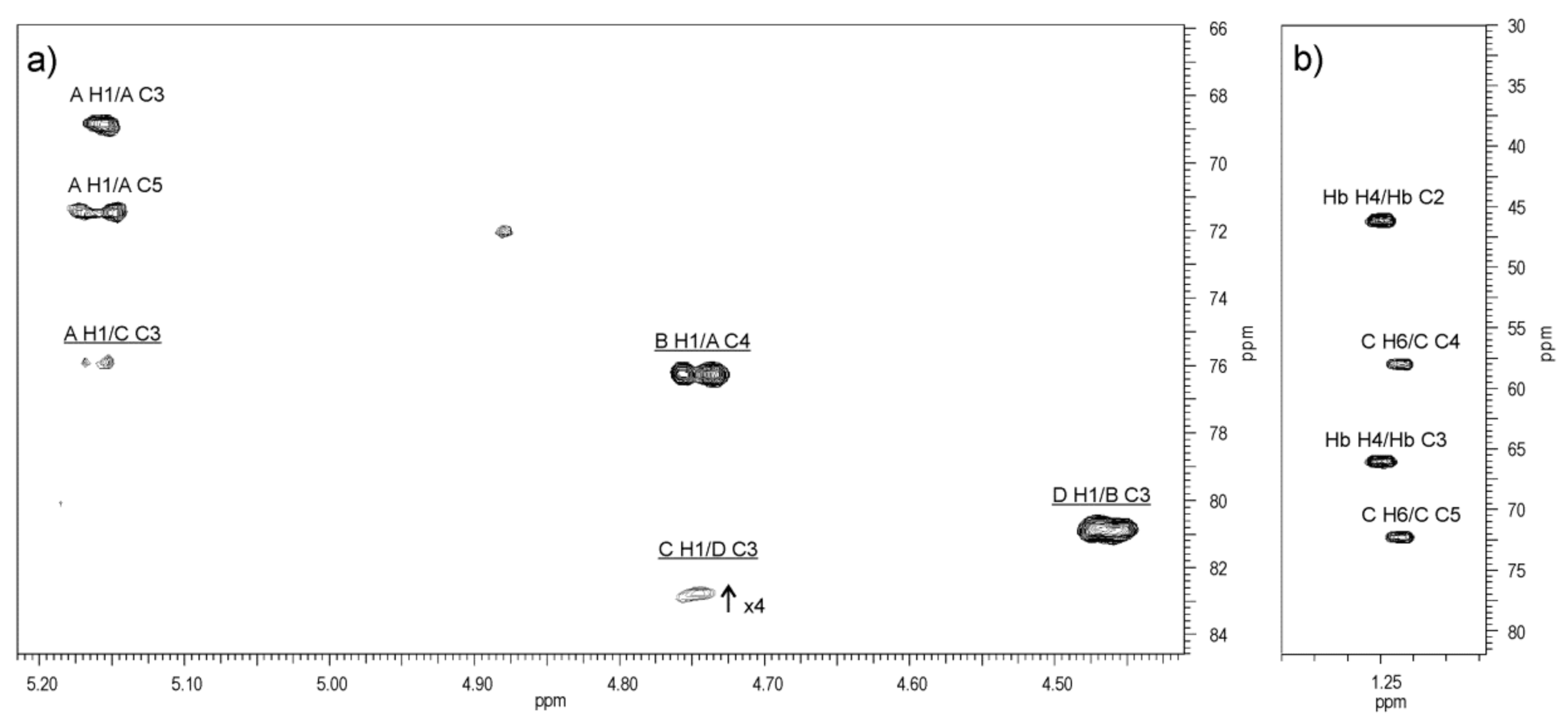
Table 3. 1H (500 MHz) and 13C NMR (125 MHz) data (δ, ppm) for the O-PS of A. veronii bv. sobria strain K133.
ResidueChemical Shifts (δ, ppm)
H-1
C-1
H-2
C-2
H-3
C-3
H-4
C-4
H-5
C-5
H-6
C-6
NAc
→4)-α-d-GalpNAc-(1→A5.16
98.2
4.12
50.6
3.89
68.8
4.21
76.3
3.87
71.4
3.73; 3.87
61.3
2.05
23.7; 175.5
→3)-β-d-GalpNAc-(1→B4.74
103.3
4.05
52.8
3.93
80.8
4.17
69.5
3.71
75.9
3.76; 3.81
62.3
2.07
23.7; 176.4
→3)-β-d-QuipNAc4NAcyl-(1→C4.74
103.3
3.87
56.2
3.87
75.9
3.87
58.0
3.54
72.3
1.21
17.7
2.03
23.7; 176.4
→3)-β-d-Galp-(1→D4.46
106.1
3.61
71.2
3.70
82.8
4.10
69.6
3.65
75.9
3.76; 3.81
62.3
(S)-3-hydroxybutanoylHb
175.0
2.35
46.2
4.20
66.0
1.25
23.7
Publisher’s Note: MDPI stays neutral with regard to jurisdictional claims in published maps and institutional affiliations.

