Review

Penicillin-Binding Proteins, β-Lactamases, and β-Lactamase Inhibitors in β-Lactam-Producing Actinobacteria: Self-Resistance Mechanisms

Departamento de Biología Molecular, Universidad de León, 24071 León, Spain
*
Author to whom correspondence should be addressed.
Int. J. Mol. Sci. 2022, 23(10), 5662; https://0-doi-org.brum.beds.ac.uk/10.3390/ijms23105662
Submission received: 10 April 2022 / Revised: 14 May 2022 / Accepted: 16 May 2022 / Published: 18 May 2022

Abstract

:
The human society faces a serious problem due to the widespread resistance to antibiotics in clinical practice. Most antibiotic biosynthesis gene clusters in actinobacteria contain genes for intrinsic self-resistance to the produced antibiotics, and it has been proposed that the antibiotic resistance genes in pathogenic bacteria originated in antibiotic-producing microorganisms. The model actinobacteria Streptomyces clavuligerus produces the β-lactam antibiotic cephamycin C, a class A β-lactamase, and the β lactamases inhibitor clavulanic acid, all of which are encoded in a gene supercluster; in addition, it synthesizes the β-lactamase inhibitory protein BLIP. The secreted clavulanic acid has a synergistic effect with the cephamycin produced by the same strain in the fight against competing microorganisms in its natural habitat. High levels of resistance to cephamycin/cephalosporin in actinobacteria are due to the presence (in their β-lactam clusters) of genes encoding PBPs which bind penicillins but not cephalosporins. We have revised the previously reported cephamycin C and clavulanic acid gene clusters and, in addition, we have searched for novel β-lactam gene clusters in protein databases. Notably, in S. clavuligerus and Nocardia lactamdurans, the β-lactamases are retained in the cell wall and do not affect the intracellular formation of isopenicillin N/penicillin N. The activity of the β-lactamase in S. clavuligerus may be modulated by the β-lactamase inhibitory protein BLIP at the cell-wall level. Analysis of the β-lactam cluster in actinobacteria suggests that these clusters have been moved by horizontal gene transfer between different actinobacteria and have culminated in S. clavuligerus with the organization of an elaborated set of genes designed for fine tuning of antibiotic resistance and cell wall remodeling for the survival of this Streptomyces species. This article is focused specifically on the enigmatic connection between β-lactam biosynthesis and β-lactam resistance mechanisms in the producer actinobacteria.

1. Introduction

Resistance to Antibiotics and Its Relation to Antibiotic-Producing Bacteria

Antibiotics have saved thousands of human lives since their clinical introduction in the fourth decade of last century [1,2]; however, at present, there is a serious problem of antibiotic resistance in many pathogenic bacteria, which is a challenge in the fight against bacterial diseases [3,4,5]. The problem includes resistance to β-lactams, aminoglycosides, tetracyclins, and macrolides [6,7], and is being also observed against antibiotics introduced more recently in clinical practice such as fluoroquinolones [8] and imipenem [9]. The appearance of antibiotic resistance was partially associated with the widespread and frequently indiscriminate use of antibiotics in medicine. In parallel, the finding of β-lactamases and other antibiotic resistance genes forming part of the antibiotic biosynthesis gene clusters (GC) led to the hypothesis that the modern resistance determinants might have originated from those that protect the antibiotic-producing bacteria [10,11,12]. Recent advances in molecular genetics, proteomics, and transcriptomic studies of antibiotic-producing strains indicate that although this hypothesis might be partially true, the evolution of antibiotic resistance genes is very complex and they respond to elaborate horizontal transfer mechanisms [13]. In this article, we focus on the available information about the complex and enigmatic interactions between β-lactam antibiotics, penicillin-binding proteins, β-lactamases, and β-lactamase inhibitors in actinobacteria.

2. Overview of Resistance Mechanisms to Antibiotics in Antibiotic-Producing Actinobacteria

Actinobacteria constitute a large group of Gram-positive bacteria that have a complex vegetive cycle, forming mycelium that usually differentiates into spores, although some actinobacteria do not form spores. Actinobacteria have been widely studied because they produce a large number of antibiotics and other pharmacologically active secondary metabolites.
There is great interest in understanding the molecular mechanism by which, in general, the antibiotic-producing bacteria avoid suicide [14,15,16]. An important mechanism of antibiotic resistance is the modification of the antibiotic target in the antibiotic-producing bacteria [17,18]. In β-lactam-producing actinobacteria, an intrinsic self-resistance mechanism has been attributed to modified penicillin-binding proteins (PBPs) [17] with low affinity for the β-lactam produced by the strain. A second mechanism is the breaking down or inactivation of the antibiotic, usually by enzyme-mediated cleavage or modification of the antibiotic molecule. This is the case of the cleavage of the β-lactam ring of penicillin and cephalosporin by β-lactamases [19,20]. A third frequent mechanism is the active secretion of the antibiotic, thus preventing its inhibitory effect on the growth of the producing strain [21,22]. Genes encoding β-lactam efflux pumps, putatively mediating β-lactam secretion, have been located in the cephamycin GCs of Nocardia lactamdurans [23] and S. clavuligerus, although there is still limited information of the effect of these β-lactam efflux pumps on the resistance mechanism [24].
Those actinobacteria that produce β-lactam antibiotics, e.g. S. clavuligerus, Streptomyces jumonjinensis, and Streptomyces cattleya, face the challenge of avoiding the toxicity of the antibiotics that they produce. S. clavuligerus, a producer of cephamycin C, is completely insensitive to cephalosporin or cefoxitin (MIC 7.0 mg/mL cephalosporin), while the cephamycin non-producers Streptomyces albus J1074, Streptomyces flavogriseus ATCC 33,331, and Streptomyces coelicolor M1146 are not able to grow in 2.0–3.0 mg/mL cephalosporin C [25]. When the cephamycin C gene cluster that contains antibiotic resistance genes is introduced into other Streptomyces strains, it increases the resistance to cephalosporin C in the transformants [25]. Figure 1 shows a model of the resistance mechanisms to β-lactam antibiotic in β-lactam-producing actinobacteria.
Similar mechanisms of resistance to antibiotics have been acquired either by mutation [26] or by horizontal gene transfer [27,28] in a variety of Gram-positive and Gram-negative bacteria. This resistance to antibiotics is exerted by distinct mechanisms that have been widely described [6,7,26,29]. However, in this article, we restrict the study to the relationship between β-lactamases, β-lactamase inhibitors, and penicillin-binding proteins as mechanisms of resistance in β-lactam-producing actinobacteria, which are discussed in detail in the following sections.

3. Peptidoglycan Biosynthesis and Penicillin-Binding Proteins

An important aspect that has been developed in recent decades is the concept of cell wall remodeling in actinobacteria to understand the role of PBPs and β-lactamases in the resistance to β-lactam antibiotics.
In actinobacteria, as well as in other Gram-positive and Gram-negative bacteria, there are genes encoding penicillin-binding proteins that are involved in the biosynthesis of the peptidoglycan polymer of the cell wall. These include transglycosylases, which bind the N-acetylmuramic acid and the N acetylglucosamine monomers of the peptidoglycan, and transpeptidases, which establish bonds between the parallel glycan chains, forming the crosslinked peptidoglycan. In addition, there are DD-carboxypeptidases that recognize the D-alanyl-D-alanyl terminus of the pentapeptide chain as a substrate and, finally, endopeptidases that cleave the peptide bridges between the glycan chains. These diverse PBPs differ in their molecular weight and also in the key amino acid motifs that line their active centers. The transglycosylases and some transpeptidases have high molecular weight, whereas other transpeptidases and, in particular, the D-alanyl-D-alanyl carboxypeptidases are low-molecular weight proteins [30]. The bacterial cell wall is remodeled during the distinct growth stages, which includes cell wall murein degradation and recycling of the released peptides for the synthesis of new cell wall. In Escherichia coli, it is known that the remodeling process involves the uptake of the tetrapeptide L-ala-D-glu-m-DAP-D-ala into the cytoplasm, where is trimmed down to the tripeptide L-ala-D-glu-m-DAP and incorporated into the cell wall. The tripeptide also serves as a signal that induces the formation of β-lactamases [31]. Recent studies in the model actinobacteria S. coelicolor indicate that there is an extensive degradation and reconstruction of the cell wall, resulting in its remodeling, particularly during the conversion of mycelium to spores [32]. Several enzymes act in the cell wall remodeling in actinobacteria, including autolysines, carboxypeptidases, and penicillin-binding proteins [33]. Chemical studies using high-performance liquid chromatography associated to mass spectrometry (HPLC-MS) on the changes of cell wall polymers in vegetative mycelium reveal large amounts of free disaccharide N-acetylglucosamine-N-acetylmuramic acid and free pentapeptide l-ala-d-glu-LL-DAP(or glycine)-d-ala-d-ala [32,34]. These results indicate that extensive changes in the crosslinking mediated by PBPs occur during growth and differentiation in Streptomyces species.

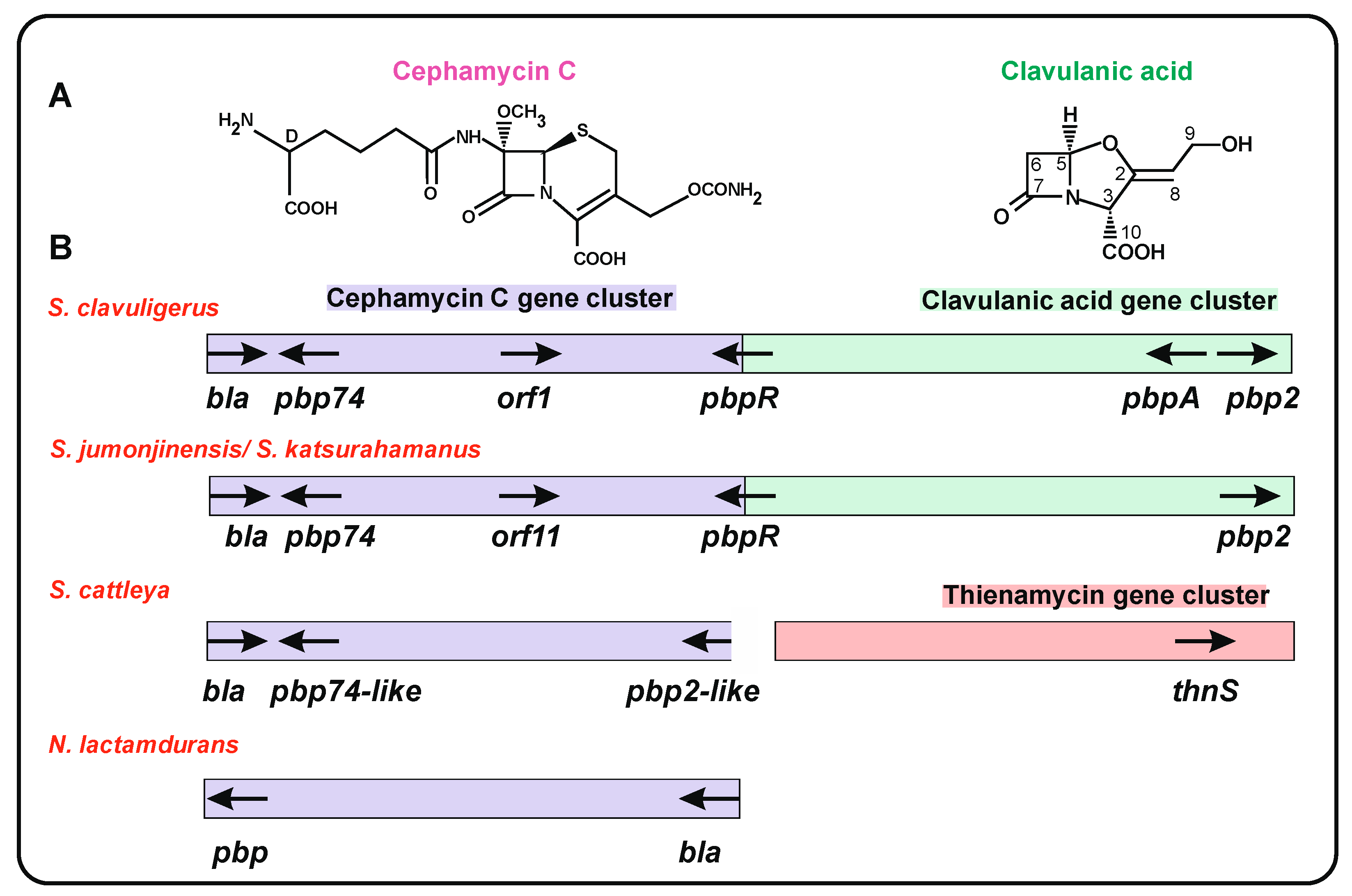
4. Clusters of Genes for β-Lactam Antibiotics in Actinobacteria

The β-lactam antibiotics cephamycin A, B, and C have a cephalosporin nucleus formed by a four-membered β-lactam ring and a six-membered dehydrothiazinic ring, modified at C-7 and C-3’ (Figure 2A, left). Cephamycins are produced by several actinobacteria, including S. clavuligerus, S. cattleya, Streptomyces griseus NRRL3851, Streptomyces lipmanii, and N. lactamdurans [35]. The cephamycin biosynthesis genes and the encoded enzymes were characterized in the 1990s [36,37]. An important finding was discovering that one of these species, S. clavuligerus, was able to produce, in addition to cephamycin C, β-lactamases and the β-lactamase inhibitor clavulanic acid [38,39]; this compound has a β-lactam ring and a five-membered oxazolidinic ring (Figure 2A, right). Since then, clavulanic acid was developed industrially as a potent pharmacological compound, acting synergistically with β-lactams against class A β-lactamases of clinically important bacteria [40]. Interestingly, Ward and Hodgson [41] observed in S. clavuligerus, S. jumonjinensis, and Streptomyces katsurahamanus that the clavulanic acid GC was adjacent to the cephamycin C GC, forming the so called β-lactam supercluster (CFM-CA) that also encodes genes for PBPs and β-lactamases (Table 1). Both the cephamycin and clavulanic acid GCs are coregulated by the activator protein CcaR [42,43,44]. In strains containing a CFM-CA supercluster, the ccaR gene is located in the cephamycin GC, but in strains lacking the cephamycin cluster, it is frequently located in the clavulanic acid cluster (see below).
Clavulanic acid clusters have been studied also in Streptomyces flavogriseus (also named Streptomyces pratensis) and Saccharomonospora viridis [45]. The organization of these clusters is different from that of S. clavuligerus, and the scientific bases for their lack of clavulanic acid production have been thoroughly studied [45].
Most actinobacteria have several copies of PBPs [17]; in particular, in β-lactam producers, the antibiotic biosynthesis clusters contain several PBPs (Figure 2B). In order to determine whether clavulanic acid and/or cephamycin C clusters in other Streptomyces strains carried genes for β-lactamases or PBP proteins, we searched in actinobacteria databases for proteins orthologous to those of S. clavuligerus, with percentages of identity higher than 60%. This analysis resulted in the finding of complete sets of clavulanic acid genes in different Streptomyces strains (see below), although the production of clavulanic acid has not yet been demonstrated in many of them.

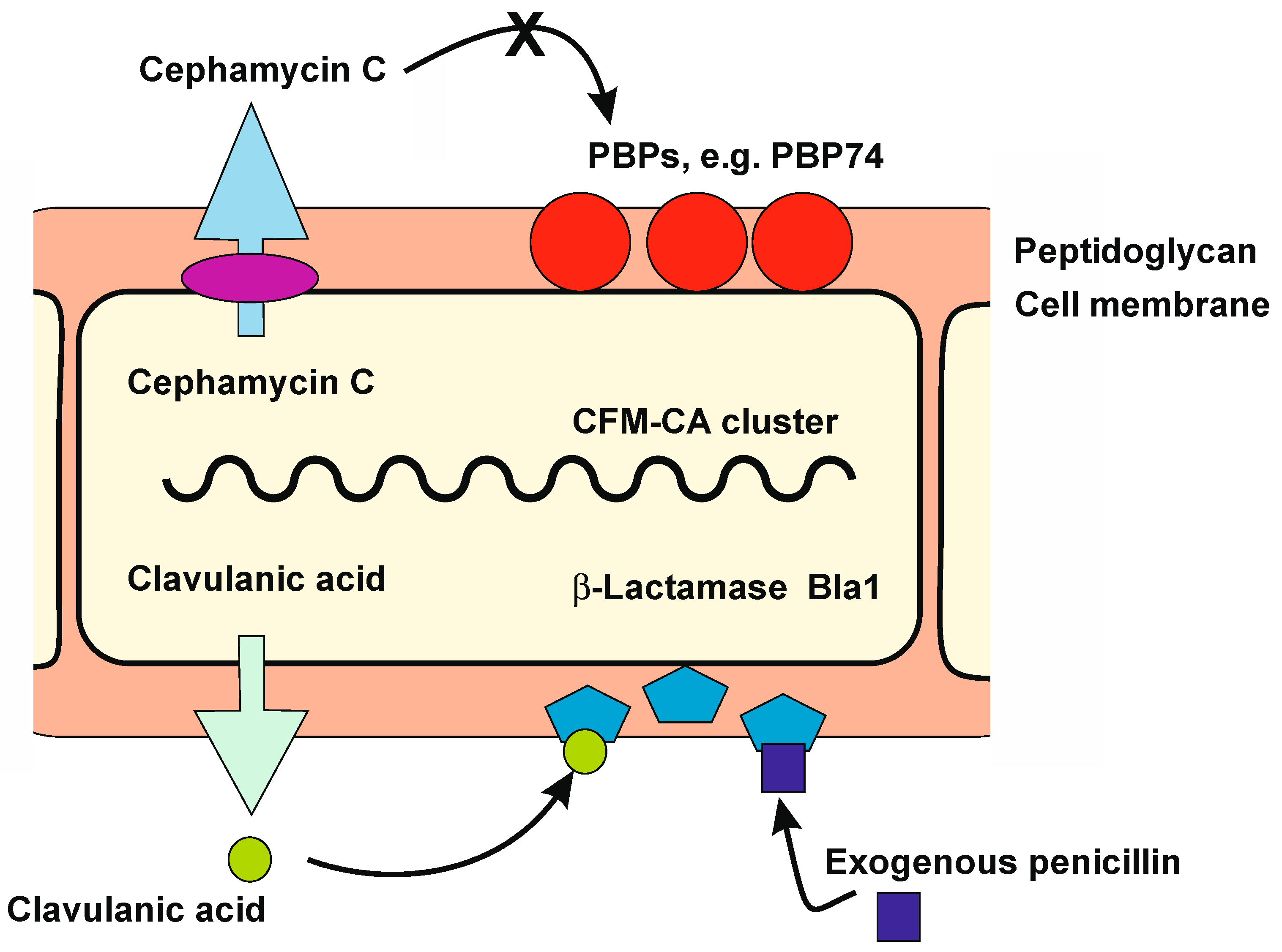
5. Genes Encoding PBP Proteins in S. clavuligerus and Other Actinobacteria β-Lactam Gene Clusters

For many years, it was suggested that β-lactam-producing actinobacteria are resistant to penicillin/cephalosporins due to evolutive modification of the PBPs [17,46]. Electrophoretic studies of penicillin-labelled proteins showed that both Streptomyces lividans and the cephamycin producer N. lactamdurans have eight PBPs. One of them, PBP-4, is encoded by a gene located in the N. lactamdurans cephamycin cluster. In vitro competition binding experiments showed that PBP-4 lacks affinity for cephamycin C [23], providing experimental support for the Ogawara [46] hypothesis.
The best studied PBPs in the β-lactam GC are those of S. clavuligerus. A bioinformatic analysis of the S. clavuligerus genome revealed the presence of at 8 eight PBPs [47]; in contrast, Ogawara et al. [17] listed 12: 3 of the class A and 9 of class B, not including PBP-74 [17]. Four S. clavuligerus PBPs are encoded by genes located in the CFM-CA supercluster. One of them, named pbp74, is located at one end in the cephamycin cluster [24]; the other two, named pbpA and pbp2, are located at the distal end in the clavulanic acid cluster (Figure 2B). In addition, the gene pbpR (also designated pcbR) is located between the cephamycin and the clavulanic acid GCs [48,49,50].
The characteristics of these four PBPs are summarized in Table 1.
Table 1. PBPs and β-lactamases encoded by genes in S. clavuligerus CFM-CA supercluster.
ProteinAmino
Acid
Number
CharacteristicsMotifsAccession
Number
PBP-74693Located at the CFM cluster. Amino acids 1–300 are proline rich. Amino acids 300–693 contain carboxypeptidase motifs. An α-helix trans-membrane motif (aa 286–309) separates the proline-rich N-terminal half and the carboxypeptidase moiety.The PBP-conserved motifs * STAK, SGN (instead of SDN), and KTG are present in the second half of the protein [24].WP_003952489
PBP-R551Located between the clavulanic acid and the cephamycin C cluster. The N-terminal end contains a hydrophobic stretch of 30–60 amino acids, which may act as a membrane anchor.The C-terminal domain has the penicillin-binding region; it contains the * STFK, SCN (instead of SDN), and KTG motifs of BPBs and β-lactamases [48].WP_003952508
PBP-A494Located at the end of the AC cluster. Contains a pfam0095 transpeptidase domain and an ATP/GTP-binding motif.Contains the * STFK, STN, and KTG motifs, but lacks the EPELN motif [49].WP_003952525
PBP-2717Located at the end of the AC cluster. Domain pbp2_mrdA.Contains SIFK and FTG (instead of KTG) [49].WP_003952526
Bla1332Located at the CFM cluster.Class A β-lactamase.Contain the active center 70 STFK and the motifs 130 SDG (instead of SDN), 166 EPELN, and 234 KTG of class A β-lactamases [51].WP_003952487
BlaB1338Located at the CFM cluster [42]. Class B β-lactamase [this work].Motif 117 HGHFD121.WP_003952502

5.1. The PBP-74 Protein

This protein is encoded by the pbp74 gene located in the cephamycin C cluster and adjacent to a gene encoding a β-lactamase (bla1). PBP-74 has an N-terminal domain rich in proline residues which is separated from the carboxyl terminal moiety of the protein by an α-helix transmembrane domain [24]. The 411 amino acids of the C-terminal moiety are 43/58% identical/similar to the same region of S. coelicolor SCO4439 and contain carboxypeptidase motifs (Table 1). Of note, the amino terminal region of PBP-74 lacks significant identity to the equivalent region of S. coelicolor SCO4439 that was reported to correspond to a putative transcriptional factor [30]. The N-terminal moieties of other PBPs similar to PBP-74 are diverse, suggesting that these genes/enzymes were formed by a combination of genes for a carboxypeptidase with the amino terminal moiety of a gene of different origin. Proteins similar to PBP-74 (71% identical) are encoded by genes in the cephamycin-clavulanic acid GCs of S. katsurahamanus and S. jumonjinensis. In S. cattleya, there is a gene for a PBP-74-like protein at one end of the cephamycin C GC, adjacent to a gene for a class A β-lactamase. This PBP is a carboxypeptidase of 419 amino acids, 59/67% identical/similar to the carboxyl moiety of S. clavuligerus PBP-74. Similar proteins are encoded by genes in other Streptomyces species (Figure 2B, Table 2).

5.2. The PBP-R Protein

A PBP of 551 amino acids is encoded by a pbpR gene that separates the cephamycin and clavulanic acid GCs. The carboxyl end of PBP-R contains a transpeptidase domain. The N-terminal moiety has a hydrophobic transmembrane domain, suggesting that this protein is membrane bound and, indeed, using anti-PBP-R antibodies it was located in the cell membrane [48]. A pbpR-disrupted mutant was not affected in clavulanic acid. S. clavuligerus is naturally more resistant to cephalosporin and cephalothin production. This mutant showed lower resistance to penicillin and cephalosporin than the parental strain [48]; this suggests that PBP-R is involved in antibiotic resistance/cell wall biosynthesis than to penicillin [25], which might be partially due to the lack of affinity of PBP-R towards cephalosporin. In Streptomyces strains carrying the clavulanic acid GCs, but not the adjacent cephamycin GC, the pbpR gene is located at the end of the clavulanic acid GC (Figure 2). Expression of pbpR in S. flavogriseus is low but greatly increases in S. flavogriseus strains transformed with the S. clavuligerus cephamycin GC, independently of the presence of the activator ccaR or the bla gene. This correlates well with the higher resistance to penicillin G and cefoxitin observed in these transformants [45], confirming that the pbpR gene present in the transformants confers resistance to β-lactam antibiotics.

5.3. PBP-A and PBP-2

Two other PBPs, encoded by genes located at the other end of S. clavuligerus clavulanic acid cluster (Table 1, Figure 2B), are named PBP-A and PBP-2 [49,52]. These two PBPs have 494 and 717 amino acids, respectively, and only share 20% overall identity. PBP-A contains a transpeptidase domain and an ATP/GTP-binding motif in addition to the classical PBP motifs STKF, STN, and KTG, but lacks the EPELN motif. In both PBP-A and PBP-2, a single hydrophobic region confers the ability to bind cell membranes, and the two proteins were found to be associated with membranes when expressed in E. coli. PBP-2 appears to confer more resistance to penicillin G than PBP-A [52]. The PBP-A amino acid sequence shows high similarity (82%) with the S. coelicolor PBP-A protein [49]; of note, the pbpA gene is not present in the supercluster of S. jumonjinensis and S. katsurahamanus. In S. cattleya, adjacent to the ccaR gene for the cluster positive regulator, there is a gene encoding a PBP protein of 695 amino acids with a transpeptidase domain which is 66/78% identical/similar to S. clavuligerus PBP-2. Genes encoding proteins similar to PBP-A and PBP-2 are frequent in Streptomyces species, but are rarely associated with β-lactam clusters (Table 2). As occurs with pbpR, the heterologous expression of S. clavuligerus pbpA in S. flavogriseus leads to higher resistance to penicillin G and cefoxitin [45], thus confirming that the PBPs are determinants for β-lactam resistance.

6. β-Lactamases

β-lactamases exist in Gram-negative and Gram-positive bacteria, including actinobacteria [53]. These enzymes cleave the β-lactam ring of penicillins and cephalosporins, and many of them may be inactivated by β-lactamase inhibitors. Bacterial β-lactamases are extracellular enzymes secreted to protect the cell against external β-lactam antibiotics produced by filamentous fungi or other microorganisms that live together with actinobacteria in their natural habitats.
β-lactamases are divided into four classes (from A to D) depending on their structure and the mechanism of their β-lactam bond hydrolysis.
Classes A, C, and D are serine hydrolases in which a serine residue in the active center (* STFK) of the protein exerts a nucleophilic attack on the β-lactam ring, forming an acyl–enzyme intermediate through the carbonyl group of the open β-lactam ring [54,55]. The acylated β-lactamase protein is then deacylated with the help of the highly conserved glutamic acid (166 E) residue that activates a water molecule for the hydrolysis. Indeed, mutation of this glutamic acid residue results in loss of the deacylation step, and this inactivates the β-lactamase [56,57]. Class A β-lactamases hydrolyze penicillins, a type of β-lactams containing a five-membered thiazolidine ring in addition to the four-membered β-lactam ring, and are inactivated by the β-lactamase inhibitor clavulanic acid (Section 7.1, Figure 2A). In contrast, class C β-lactamases cleave the β-lactam ring of cephalosporin and some semisynthetic derivatives, which contain the β-lactam ring fused to a six-membered dehydrothiazinic ring. Class D β-lactamases are mainly active towards oxacillins.
Of note, class B β-lactamases are metalloenzymes that have a different mechanism of action: they use a zinc-coordinated water molecule to attack and cleave the β-lactam ring.

6.1. Analysis and Role of β-Lactamases Located in the S. clavuligerus β-Lactam Supercluster

Genes encoding type A β-lactamases have been found in most of the cephamycin GCs in actinobacteria. An important question is whether the β-lactamases associated with these clusters are somehow different from other class A β-lactamases occurring in actinobacteria that do not produce β-lactams. A related question concerns the role of the β-lactamases produced by cephamycin-producing actinobacteria and whether they destroy the isopenicillin N and penicillin N intermediates of the cephamycin pathway. Of particular interest are the β-lactamases encoded in the genome of S. clavuligerus, which serves as a model of actinobacteria, due to its ability to simultaneously produce the β-lactamase inhibitor clavulanic acid. In S. clavuligerus, Ogawara [52] bioinformatically identified three class A β-lactamases, eight class B β-lactamases, and six class C β-lactamases (Table S1).

Class A β-Lactamases of S. clavuligerus

This class includes the enzyme encoded by the bla gene (hereafter named bla1), located in the cephamycin GC (see below), and the β-lactamase-like protein encoded by the orf12 gene, located in the clavulanic acid cluster [49,50].
The S. clavuligerus bla1 gene is located at the cephamycin end of the CFM-CA supercluster, contiguous to pbp74 [24]. The encoded protein has 332 amino acids and is a class A β-lactamase containing an active center at position 70 STFK73 and three additional conserved motifs of class A β-lactamases: SDG (instead of SDN), EPELN, and KTG [51]. The standard bacterial β-lactamases are secreted proteins and their activity is detected in the supernatant broth. However, there is no β-lactamase activity in the supernatant of S. clavuligerus cultures, and, as occurs with the β-lactamase of N. lactamdurans (see below), S. clavuligerus Bla1 remains located between the membrane and cell wall and is only released after protoplast formation. The purified enzyme has a Km of 11 μM for benzylpenicillin, but the Km for cephalosporin C or semisynthetic cephalosporins rises to 5000 μM. Bla1 binds and retains labelled [H3]-benzylpenicillin in contrast to the efficient release of the hydrolyzed penicillin by other bacterial β-lactamases; this behavior correlates with a low penicillin deacylation rate and suggests that this enzyme has some properties of penicillin-binding proteins that are known to retain the bound penicillin [57]. This indicates that the β-lactamase in the CFM-CA supercluster is somehow different from extracellular β-lactamases from other bacteria. This similarity suggests that S. clavuligerus Bla1 may have a role in the biosynthesis/regulation or remodeling of the cell wall.
The Orf12 protein, encoded in S. clavuligerus clavulanic CG, has a β-lactamase domain, but the pure protein lacks β-lactamase activity [58,59] and the orf12-disrupted mutants are unable to produce clavulanic acid. Therefore, the Orf12 protein appears to be a biosynthetic enzyme that is not included among the standard S. clavuligerus β-lactamases.

6.2. Class A β-Lactamases Associated with β-Lactam Clusters in Other Actinobacteria

The N. lactamdurans β-lactamase was the first of this type of enzyme reported to be encoded by a gene located in a cephamycin GC [23,60]. This β-lactamase has 302 amino acids and contains the classical active center * STFK and all the conserved motifs of class A β-lactamases. The N. lactamdurans Bla1 protein has an N-terminal putative leader peptide of 29 amino acids ending in AAA29, but surprisingly, there is no β-lactamase activity detectable in the supernatant of N. lactamdurans cultures. The enzyme is trapped between the membrane and the cell wall peptidoglycan, and the β-lactamase activity is only detected after protoplast formation. The enzyme released after this treatment is very active on benzylpenicillin and less active on isopenicillin N, an intermediate of cephamycin biosynthesis; in contrast, it is poorly active against cephalosporins and has no activity against cephamycin, the final product of its biosynthetic pathway. The N. lactamdurans Bla1 enzyme is inhibited by clavulanic acid (50% inhibition by 0.5 μg/ml CA). Mutants disrupted in bla1 are more sensitive to penicillin, but do not show significant differences in sensitivity to cephalosporin C or cephamycin. Conversely, N. lactamdurans transformants with a bla1 amplified copy number are more resistant to benzylpenicillin, indicating that the major role of this β-lactamase is the defense against exogenous benzylpenicillin rather than degradation of the cytosolic isopenicillin N/penicillin N synthesized by this strain [60].
A class A β-lactamase similar to S. clavuligerus Bla1 (65% identical) is encoded by a gene located in the S. cattleya cephamycin cluster, but this enzyme has not been characterized biochemically or functionally [61]. Using bioinformatic analysis, in several Streptomyces species, we have found genes associated with cephamycin GCs that encode class A β-lactamases (Table 3A).

6.3. Class B β-Lactamases

The six class B β-lactamases identified in S. clavuligerus by Ogawara [52] are encoded by genes scattered in the genome. In addition, a seventh gene, located in the cephamycin C cluster, encodes an MBL-fold hydrolase, hereafter named BlaB1 (acronym for β-lactamase class B). The blaB1 gene is contiguous to ccaR, a gene encoding the positive regulator that controls cephamycin C and clavulanic acid production. When this protein was initially discovered, no similarity to other proteins was found [42]; therefore, it was not further studied. It was only by recent searching in the databases that we found that this gene encodes a hydrolase of the metallo-β-lactamase family. This protein has the 117HGHFD121 motif and histidine in the expected positions (193H, 265H) for a class B β-lactamase (Table 1 and Table 3).
Searching in the databases shows the presence of genes encoding class B β-lactamases in most of the clavulanic acid GCs of other actinobacteria (Table 3B, Figure 3) suggesting that this gene may have a direct/indirect role in clavulanic acid biosynthesis or regulation. Unfortunately, no blaB1-disrupted mutants have ever been reported. The S. cattleya thnS gene, located in the thienamycin GC, encodes a class B β-lactamase which is 43/68% identical/similar to S. clavuligerus BlaB1. ThnS is involved in resistance to thienamycin in the producer strain [62].

6.4. Class C β-Lactamases

Six putative class C β-lactamases have been found bioinformatically in the genome of S. clavuligerus and annotated as cephalosporinases (Table S1) [52]. However, none of these genes are located in a β-lactam biosynthesis cluster. Most of these proteins contain all the conserved domains of classical type C β-lactamases, and one of them is truncated. However, S. clavuligerus lacks significant extracellular cephalosporinase activity.

7. β-Lactamase Inhibitors and β-Lactamase Inhibitory Proteins

Two distinct components of the β-lactamase regulatory system in actinobacteria are the β-lactamase inhibitors and the β-lactamase inhibitory proteins (BLIP).

7.1. β-Lactamases Inhibitors

An important strategy to improve the action of β-lactam antibiotics was the systematic search of β-lactamase inhibitors [40]. This search focused on finding inhibitors structurally related to penicillin that might acylate the β-lactamases but will not deacylate them easily [63]. This resulted in the finding of compounds that bind β-lactamase in an irreversible or partially reversible form, such as CA and the olivanic acid family [38]. The irreversible inhibitors permanently inactivated the β-lactamases by reaction with the active site of the cognate β-lactamases [64]. Clavulanic acid contains a β-lactam ring fused to an oxygen-containing five-membered oxazolidinic ring (Figure 2A right) that mimics the penicillin structure [39]. CA combined with amoxicillin or other penicillins has potent β-lactamase inhibitory activity against class A β-lactamases and low activity against classes B, C, and D [40,63]; this compound is the more frequently used in clinical practice. The CA molecule binds the serine in the active site of β-lactamases and performs the acylation step, but the acylated β-lactamase–CA complex is resistant to the subsequent deacylation; therefore, the acylated β-lactamases remain inactive. The biosynthesis of CA and its molecular genetics have been well studied [65,66,67]. The role of autogenous CA in β-lactam-producing actinobacteria is enigmatic; CA may act as a synergistic metabolite in the fight against soil bacterial. In addition, CA inactivates the purified class A β-lactamase Bla1 of S. clavuligerus or N. lactamdurans, which suggests that CA may modulate these enzymes’ activity in vivo [57,60]. Clavulanic acid has weak antibiotic activity and in E. coli interacts with the PBP2 protein [68]. This interaction affects the cell wall remodeling and results in alteration of the rod shape of E. coli cells. Similarly, in S. clavuligerus, CA may be also a fine modulator of the cell wall remodeling, although experimental work needs to be done to support this hypothesis.

7.2. The BLIP Protein of S. clavuligerus and Other Actinobacteria

Some β-lactamase-producing actinobacteria, such as S. clavuligerus, contain a separated gene, named blip, encoding a small protein that is a strong inhibitor of class A β-lactamases [69]. The blip gene is located far from the clavulanic acid GC [70] and encodes an 18 KDa protein that is found in the supernatant of S. clavuligerus cultures, in which it is a major protein. Proteins similar to BLIP (74% identical) are encoded in the genomes of S. jumonjinensis and S. katsurahamanus, but not in other Streptomyces species. Structural studies reveal that BLIP contains a tandem domain forming a polar concave protruding region that interacts with a conserved convex loop-helix region adjacent to the active center of class A β-lactamases, inhibiting its β-lactamase activity [71,72,73]. The BLIP protein may have a modulation role in the β-lactamase activity in S. clavuligerus.
Two other similar proteins, BLIP-I and BLIP-II, were subsequently discovered in culture supernatants of Streptomyces exfoliatus [74,75]. BLIP-II is a 28 KDa protein; it folds forming a seven-bladed β-propeller structure that interacts with the same region of β-lactamases adjacent to the active center as BLIP does. Thermodynamic studies have shown that BLIP-II has a very high affinity for the β-lactamases and an extremely low dissociation constant. It has been proposed that BLIP-II plays a role in the remodeling of the cell wall in S. exfoliatus [74].

8. Distinct Arrangement of the Clavulanic Acid Cluster in Actinobacteria

There are different arrangements of the genes in the β-lactam gene clusters in antibiotic-poducing actinobacteria [45,66,76].

8.1. Is a PBP Gene in the β-Lactam Gene Cluster Required for Self Resistance?

Since PBPs are determinants for resistance to the β-lactam antibiotics, but only some of them are located in the CFM-CA supercluster, an interesting question is whether any of the PBP genes in the clavulanic or cephamycin GC are required for self-resistance.
In our bioinformatic study for this article of the CFM-CA gene clusters in actinobacteria, we found considerable differences in the organization of these clusters. This search revealed 15 complete and 4 incomplete CA clusters (Figure 3). Only three of the studied strains (S. clavuligerus, S. katsurahamanus, and S. jumonjinensis) contain the complete CFM-CA supercluster [41]; all other strains lack an adjacent cephamycin cluster. Four strains (S. albiflavescens, Streptomyces sp. SID14446, Streptomyces sp. M41, and Streptomyces sp. SID288) have identical organization to the previous strains, but contain the blaB1 and ccaR genes adjacent to the CA cluster. In the third block of strains (Streptomyces flavovirens, S. flavogriseus, Streptomyces sp. S-325, Streptomyces sp. PAMC 2608, and Streptomyces sp. NRRL 2401), two blocks of genes (from oppA1 to gcaS and from oat2 to cas2) are conserved, and the blaB1 and ccaR genes are inserted between them. Finally, the gene organization is different in Streptomyces fulveorobeos, Streptomyces finlayi, and Sacc. viridis (Figure 3) and incomplete in the other four species, although in all of them, the four genes (ceaS-pah-bls-cas) for the early steps of the pathway leading to clavaminic acid formation are conserved. Regarding the presence of pbp genes associated with resistance to β-lactam antibiotics, the only gene that is present in most clavulanic acid clusters (89% of the cases) is pbpR (Table 2, Figure 3). The pbp74 gene is always associated with the cephamycin C cluster, either when the cluster is adjacent to the clavulanic acid cluster or when it stands alone; finally, the pbpA and pbp2 genes occur rarely and are associated either with the clavulanic GC or with the cephamycin GCs. These results suggest that PBP-R is the more important protein required for the maintenance of resistance in the clavulanic acid clusters, and that PBP-74 exerts a similar function in the cephamycin clusters.
Figure 3. Organization of the clavulanic acid gene clusters of different actinobacteria. The name of the strain is indicated in the left column. The genes are color-coded and their names are indicated. Genes in black color are not related to cephamycin or clavulanic acid biosynthesis. The initial search of CA gene clusters was made using the essential S. clavuligerus proteins GcaS and CeaS2 as probes. Subsequently, all the CA biosynthesis proteins were searched using the homologous proteins of S. clavuligerus as probe. The organization of the genes was obtained using the NCBI Genome Blast tool. The gene clusters of clavulanic acid in S. clavuligerus, S. jumonjinensis-S.katsurahamanus, S. flavogriseus, and Sacc. viridis have been previously reported [45,66,76]. The CA clusters of all other actinobacteria shown in Figure 3 are reported for the first time in this work.
Ijms 23 05662 g003

8.2. Distribution of β-Lactamase and Blip Genes in the Cephamycin and Clavulanic Acid GC

Genes for class A β-lactamases are always associated with cephamycin GCs, while the gene for the class B β-lactamase blaB1 is most frequently associated with the clavulanic acid gene cluster, suggesting that this gene may play a role, directly or indirectly, in clavulanic acid biosynthesis. Genes similar to either blip or blipII are very rare in actinobacteria [69,74], and only orthologous proteins with less than 40% identity have been found in some of the new strains carrying CA clusters indicated in this work (Figure 3).

8.3. Are Some Genes in the Clavams or Cephamycin Clusters Required for Biosynthesis of Clavulanic Acid

In addition to clavulanic acid and cephamycin C GCs, S. clavuligerus contains two different clusters for the biosynthesis of 3S, 5S antifungal clavams that share the initial intermediates with the clavulanic acid pathway up to clavaminic acid (Figure S1), but do not isomerize the 3S, 5S clavams to the 3R, 5R configuration characteristic of clavulanic acid [77,78].
It has been suggested that the clavulanic acid cluster was initially formed by duplication of a 3S, 5S clavam cluster [79]. In that article, the authors hypothesized that after the clavam cluster was duplicated, one of the copies acquired some genes to convert the 3S, 5S intermediates to the 3R, 5R final compound. The formation of clavulanic acid was also proposed to be a response to the presence and expression of a CFM cluster in the genome and to the production of cephamycin by the strains [79].
This raises the question of whether there are actinobacteria able to produce clavulanic acid without the presence of 3S, 5S clavam clusters. The clavam GC is present in some Streptomyces species, (e.g., Streptomyces antibioticus) which do not contain the CA gene cluster and therefore produce clavams but not clavulanic acid [80,81]. In a search for clavam biosynthesis genes in the novel strains carrying clavulanic acid clusters (Figure 3), we could not find evidence for clavam clusters. The lack of 3S, 5S clavam clusters in all the analyzed strains suggests that the clavam cluster was formed in the clavulanic producer strains to increase the supply of enzymes and intermediates for clavulanic acid production.
A different question is whether the genetic information encoded by the cephamycin GC is required for the occurrence or expression of the clavulanic acid genes. Here, we discuss novel information that sheds light on this intriguing question. Early fermentation studies indicated that it is possible to dissociate the cephamycin and clavulanic acid production [82], and that the lack of cephamycin formation does not affect significantly clavulanic acid production [83]. While S. clavuligerus, S. katsurahamanus, and S. jumonjinensis have the CFM-CA supercluster, in the novel strains analyzed here, only carrying the CA cluster, one of them, Streptomyces fulvorobeos, contains an incomplete truncated cephamycin GC. This truncated cluster lacks the genes for the medium and late cephamycin biosynthesis steps; therefore, the final product of this cluster should be penicillin N. The cephamycin cluster of S. fulvorobeos is separated by 1.75 Mb from the CA cluster.
In strains carrying the CFM-CA supercluster, the only gene located in the cephamycin GC required for CA formation is that encoding the activator CcaR, which regulates the expression of the genes in both subclusters. Of note, this gene has been preserved in strains that lack the cephamycin cluster and is located in the clavulanic acid GC (Figure 3), thus allowing expression of the CA biosynthesis genes without requiring a cephamycin GC. In summary, the complete cephamycin cluster is not required to produce clavulanic acid, but the presence of the ccaR gene is essential for triggering the production of clavulanic acid.

9. Conclusions and Future Outlook

The origin of antibiotic resistance in ancient times may be due to gene transfer from antibiotic-producing actinobacteria (in which these resistance genes occur) to sensitive bacteria; however, this is difficult to demonstrate experimentally. As reviewed in this article, some cephamycin-producing actinobacteria contain cephalosporin-resistant PBPs; in addition, they include class A β-lactamases, a gene cluster for the β-lactamase inhibitor clavulanic acid, and also genes for β-lactamase inhibitory proteins. All these components may have evolved to form an elaborate biosynthetic and regulatory system that prevents suicide of the producer strain and contributes to the fine tuning of the biosynthesis of cell wall components in the β-lactam-producing actinobacteria. Many actinobacteria contain the cephamycin gene cluster, and some contain the clavulanic acid gene cluster, but only a few have the integrated CFM-CA supercluster, which has likely evolved to coordinate the biosynthesis of these two compounds.
Regarding the molecular mechanism of resistance of β-lactam-producing actinobacteria to their own antibiotics, it is now evident that one of the major resistance determinants is the presence of modified PBPs. In addition, the efflux transport contributes to the protection against the toxic metabolite [84]. There is still limited information about the β-lactam antibiotic secretion process, although disruption of one of the putative transport systems indicates that the efflux of the antibiotic has an important role in the secretion to avoid suicide [50]. Further molecular genetics and protein structure/functional analysis are required to provide a better understanding of the complex resistance systems and their regulation in actinobacteria.

Supplementary Materials

The following supporting information can be downloaded at: www.mdpi.com/article/10.3390/ijms23105662/s1.

Author Contributions

Conceptualization, J.F.M. and P.L.; software, P.L.; writing, J.F.M. and P.L., supervision, J.F.M. and P.L.; bioinformatic analysis, P.L. and R.A.-A. All authors have read and agreed to the published version of the manuscript.

Funding

This research received no external funding.

Institutional Review Board Statement

Not applicable.

Informed Consent Statement

Not applicable.

Data Availability Statement

Not applicable.

Acknowledgments

J.F.M. and P.L. acknowledge the support of the University of León (Spain) for the use of its bioinformatic facilities.

Conflicts of Interest

The authors declare no conflict of interest.

Abbreviations

AC—Clavulanic acid; BLIP—β-lactamases inhibitory protein; CFM—cephamycin C; GC—Gene cluster, MRSA—Methicillin-resistant Staphylococcus aureus; MBL—Metallo β-lactamase fold hydrolase; PBP—Penicillin-binding-protein; Thn—thienamycin.

References

  1. Waksman, S.A.; Reilly, H.C.; Schatz, A. Strain specificity and production of antibiotic substances. V. Strain resistance of bacteria to antibiotic substances, especially streptomycin. Proc. Natl. Acad. Sci. USA 1945, 31, 157–164. [Google Scholar] [CrossRef] [Green Version]
  2. Demain, A.L. Antibiotics: Natural products essential to human health. Med. Res. Rev. 2009, 29, 821–842. [Google Scholar] [CrossRef]
  3. Davies, J.; Davies, D. Origins and evolution of antibiotic resistance. Microbiol. Mol. Biol. Rev. 2010, 74, 417–433. [Google Scholar] [CrossRef] [Green Version]
  4. Wells, V.; Piddock, L.J.V. Addressing antimicrobial resistance in the UK and Europe. Lancet Infect. Dis. 2017, 17, 1230–1231. [Google Scholar] [CrossRef] [Green Version]
  5. Cox, J.A.G.; Worthington, T. The ‘antibiotic apocalypse’—Scaremongering or scientific reporting? Trends Microbiol. 2017, 25, 167–169. [Google Scholar] [CrossRef] [Green Version]
  6. Wright, G.D. The antibiotic resistome: The nexus of chemical and genetic diversity. Nat. Rev. Microbiol. 2007, 5, 175–186. [Google Scholar] [CrossRef]
  7. D’Costa, V.M.; Griffiths, E.; Wright, G.D. Expanding the soil antibiotic resistome: Exploring environmental diversity. Curr. Opin. Microbiol. 2007, 10, 481–489. [Google Scholar] [CrossRef]
  8. Azargun, R.; Gholizadeh, P.; Sadeghi, V.; Hosainzadegan, H.; Tarhriz, V.; Memar, M.Y.; Pormohammad, A.; Eyvazi, S. Molecular mechanisms associated with quinolone resistance in Enterobacteriaceae: Review and update. Trans. R. Soc. Trop. Med. Hyg. 2020, 114, 770–781. [Google Scholar] [CrossRef]
  9. Gomis-Font, M.A.; Cabot, G.; Sánchez-Diener, I.; Fraile-Ribot, P.A.; Juan, C.; Moya, B.; Zamorano, L.; Oliver, A. In vitro dynamics and mechanisms of resistance development to imipenem and imipenem/relebactam in Pseudomonas aeruginosa. J. Antimicrob. Chemother. 2020, 75, 2508–2515. [Google Scholar] [CrossRef]
  10. Benveniste, R.; Davies, J. Mechanisms of antibiotic resistance in bacteria. Annu. Rev. Biochem. 1973, 42, 471–506. [Google Scholar] [CrossRef]
  11. Barlow, M.; Hall, B.G. Phylogenetic analysis shows that the OXA beta-lactamase genes have been on plasmids for millions of years. J. Mol. Evol. 2002, 55, 314–321. [Google Scholar] [CrossRef]
  12. Wencewicz, T.A. Crossroads of Antibiotic Resistance and Biosynthesis. J. Mol. Biol. 2019, 431, 3370–3399. [Google Scholar] [CrossRef]
  13. Jiang, X.; Ellabaan, M.M.H.; Charusanti, P.; Munck, C.; Blin, K.; Tong, Y.; Weber, T.; Sommer, M.O.A.; Lee, S.Y. Dissemination of antibiotic resistance genes from antibiotic producers to pathogens. Nat. Commun. 2017, 8, 15784. [Google Scholar] [CrossRef] [Green Version]
  14. Cundliffe, E. How antibiotic-producing organisms avoid suicide. Annu. Rev. Microbiol. 1989, 43, 207–233. [Google Scholar] [CrossRef]
  15. Cundliffe, E.; Demain, A.L. Avoidance of suicide in antibiotic-producing microbes. J. Ind. Microbiol. Biotechnol. 2010, 37, 643–672. [Google Scholar] [CrossRef]
  16. Hopwood, D.A. How do antibiotic-producing bacteria ensure their self-resistance before antibiotic biosynthesis incapacitates them? Mol. Microbiol. 2007, 63, 937–940. [Google Scholar] [CrossRef]
  17. Ogawara, H. Penicillin-binding proteins in Actinobacteria. J. Antibiot. 2015, 68, 223–245. [Google Scholar] [CrossRef] [Green Version]
  18. Ogawara, H. Comparison of Antibiotic Resistance Mechanisms in Antibiotic-Producing and Pathogenic Bacteria. Molecules 2019, 24, 3430. [Google Scholar] [CrossRef] [Green Version]
  19. Frére, J.M. β-Lactamases and bacterial resistance to antibiotics. Mol. Microbiol. 1995, 16, 385–395. [Google Scholar] [CrossRef]
  20. King, D.T.; Sobhanifar, S.; Strynadka, N.C. One ring to rule them all: Current trends in combating bacterial resistance to the β-lactams. Protein Sci. 2016, 25, 787–803. [Google Scholar] [CrossRef] [Green Version]
  21. Piddock, L.J. Multidrug-resistance efflux pumps—Not just for resistance. Nat. Rev. Microbiol. 2006, 4, 629–636. [Google Scholar] [CrossRef] [PubMed]
  22. Severi, E.; Thomas, G.H. Antibiotic export: Transporters involved in the final step of natural product production. Microbiology 2019, 165, 805–818. [Google Scholar] [CrossRef]
  23. Coque, J.J.; Liras, P.; Martín, J.F. Genes for a beta-lactamase, a penicillin-binding protein and a transmembrane protein are clustered with the cephamycin biosynthetic genes in Nocardia lactamdurans. EMBO J. 1993, 12, 631–639. [Google Scholar] [CrossRef]
  24. Pérez-Llarena, F.J.; Rodríguez-García, A.; Enguita, F.J.; Martín, J.F.; Liras, P. The pcd gene encoding piperideine-6-carboxylate dehydrogenase involved in biosynthesis of alpha-aminoadipic acid is located in the cephamycin cluster of Streptomyces clavuligerus. J. Bacteriol. 1998, 180, 4753–4756. [Google Scholar] [CrossRef] [Green Version]
  25. Martínez-Burgo, Y.; Álvarez-Álvarez, R.; Pérez-Redondo, R.; Liras, P. Heterologous expression of Streptomyces clavuligerus ATCC 27064 cephamycin C gene cluster. J. Biotechnol. 2014, 186, 21–29. [Google Scholar] [CrossRef]
  26. Munita, J.M.; Arias, C.A. Mechanisms of Antibiotic Resistance. Microbiol. Spectr. 2016, 4, 1–30. [Google Scholar] [CrossRef] [Green Version]
  27. Perry, J.A.; Wright, G.D. The antibiotic resistance “mobilome”: Searching for the link between environment and clinic. Front. Microbiol. 2013, 4, 138. [Google Scholar] [CrossRef] [Green Version]
  28. Forsberg, K.J.; Reyes, A.; Wang, B.; Selleck, E.M.; Sommer, M.O.; Dantas, G. The shared antibiotic resistome of soil bacteria and human pathogens. Science 2012, 337, 1107–1111. [Google Scholar] [CrossRef] [Green Version]
  29. Durão, P.; Balbontín, R.; Gordo, I. Evolutionary Mechanisms Shaping the Maintenance of Antibiotic Resistance. Trends Microbiol. 2018, 26, 677–691. [Google Scholar] [CrossRef] [Green Version]
  30. Rioseras, B.; Yagüe, P.; López-García, M.T.; González-Quiñonez, N.; Binda, E.; Marinelli, F.; Manteca, A. Characterization of SCO4439, a D-alanyl-D-alanine carboxypeptidase involved in spore cell wall maturation, resistance, and germination in Streptomyces coelicolor. Sci. Rep. 2016, 6, 21659. [Google Scholar] [CrossRef]
  31. Jacobs, C.; Huang, L.J.; Bartowsky, E.; Normark, S.; Park, J.T. Bacterial cell wall recycling provides cytosolic muropeptides as effectors for β-lactamase induction. EMBO J. 1994, 13, 4684–4694. [Google Scholar] [CrossRef] [PubMed]
  32. Van der Aart, L.T.; Spijksm, G.K.; Harms, A.; Vollmer, W.; Hankemeier, T.; van Wezel, G.P. High-Resolution Analysis of the Peptidoglycan Composition in Streptomyces coelicolor. J. Bacteriol. 2018, 200, e00290-18. [Google Scholar] [CrossRef] [PubMed] [Green Version]
  33. Haiser, H.J.; Yousef, M.R.; Elliot, M.A. Cell wall hydrolases affect germination, vegetative growth, and sporulation in Streptomyces coelicolor. J. Bacteriol. 2009, 191, 6501–6512. [Google Scholar] [CrossRef] [Green Version]
  34. Hugonnet, J.E.; Haddache, N.; Veckerle, C.; Dubost, L.; Marie, A.; Shikura, N.; Mainardi, J.L.; Rice, L.B.; Arthur, M. Peptidoglycan cross-linking in glycopeptide-resistant Actinomycetales. Antimicrob. Agents Chemother. 2014, 58, 1749–1756. [Google Scholar] [CrossRef] [PubMed] [Green Version]
  35. Stapley, E.O.; Jackson, M.; Hernández, S.; Zimmerman, S.B.; Currie, S.A.; Mochales, S.; Mata, J.M.; Woodruff, H.B.; Hendlin, D. Cephamycins, a new family of beta-lactam antibiotics I. Production by actinomycetes, including Streptomyces lactamdurans sp. Antimicrob. Agents Chemother. 1972, 2, 122–131. [Google Scholar] [CrossRef] [Green Version]
  36. Aharonowitz, Y.; Cohen, G.; Martín, J.F. Penicillin and cephalosporin biosynthetic genes: Structure, organization, regulation, and evolution. Annu. Rev. Microbiol. 1992, 46, 461–495. [Google Scholar] [CrossRef]
  37. Liras, P. Biosynthesis and Molecular Genetics of Cephamycins. Ant. Van Leeuwenhoek 1999, 75, 109–124. [Google Scholar] [CrossRef]
  38. Brown, A.G.; Butterworth, D.; Cole, M.; Hanscomb, G.; Hood, J.D.; Reading, C.; Rolinson, G.N. Naturally-occurring beta-lactamase inhibitors with antibacterial activity. J. Antibiot. 1976, 29, 668–669. [Google Scholar] [CrossRef]
  39. Reading, C.; Cole, M. Clavulanic acid: A beta-lactamase-inhibiting beta-lactam from Streptomyces clavuligerus. Antimicrob. Agents Chemother. 1977, 11, 852–857. [Google Scholar] [CrossRef] [Green Version]
  40. Drawz, S.M.; Bonomo, R.A. Three decades of beta-lactamase inhibitors. Clin. Microbiol. Rev. 2010, 23, 160–201. [Google Scholar] [CrossRef] [Green Version]
  41. Ward, J.M.; Hodgson, J.E. The biosynthetic genes for clavulanic acid and cephamycin production occur as a ‘super-cluster’ in three Streptomyces. FEMS Microbiol. Lett. 1993, 110, 239–242. [Google Scholar] [CrossRef] [PubMed]
  42. Pérez-Llarena, F.J.; Liras, P.; Rodríguez-García, A.; Martín, J.F. A regulatory gene (ccaR) required for cephamycin and clavulanic acid production in Streptomyces clavuligerus: Amplification results in overproduction of both beta-lactam compounds. J. Bacteriol. 1997, 179, 2053–2059. [Google Scholar] [CrossRef] [PubMed] [Green Version]
  43. Alexander, D.C.; Jensen, S.E. Investigation of the Streptomyces clavuligerus cephamycin C gene cluster and its regulation by the CcaR protein. J. Bacteriol. 1998, 180, 4068–4079. [Google Scholar] [CrossRef] [PubMed] [Green Version]
  44. Santamarta, I.; López-García, M.T.; Kurt, A.; Nárdiz, N.; Alvarez-Álvarez, R.; Pérez-Redondo, R.; Martín, J.F.; Liras, P. Characterization of DNA-binding sequences for CcaR in the cephamycin-clavulanic acid supercluster of Streptomyces clavuligerus. Mol. Microbiol. 2011, 81, 968–981. [Google Scholar] [CrossRef]
  45. Álvarez-Álvarez, R.; Martínez-Burgo, Y.; Pérez-Redondo, R.; Braña, A.F.; Martín, J.F.; Liras, P. Expression of the endogenous and heterologous clavulanic acid cluster in Streptomyces flavogriseus: Why a silent cluster is sleeping. Appl. Microb. Biotechnol. 2013, 97, 9451–9463. [Google Scholar] [CrossRef]
  46. Ogawara, H. Antibiotic resistance in pathogenic and producing bacteria, with special reference to β-lactam antibiotics. Microbiol. Rev. 1981, 45, 591–619. [Google Scholar] [CrossRef]
  47. Medema, M.H.; Trefzer, A.; Kovalchuk, A.; van den Berg, M.; Müller, U.; Heijne, W.; Wu, L.; Alam, M.T.; Ronning, C.M.; Nierman, W.C.; et al. The sequence of a 1.8-mb bacterial linear plasmid reveals a rich evolutionary reservoir of secondary metabolic pathways. Genome Biol. Evol. 2010, 2, 212–224. [Google Scholar] [CrossRef]
  48. Paradkar, A.S.; Aidoo, K.A.; Wong, A.; Jensen, S.E. Molecular analysis of a beta-lactam resistance gene encoded within the cephamycin gene cluster of Streptomyces clavuligerus. J. Bacteriol. 1996, 178, 6266–6274. [Google Scholar] [CrossRef] [Green Version]
  49. Mellado, E.; Lorenzana, L.M.; Rodríguez-Sáiz, M.; Díez, B.; Liras, P.; Barredo, J.L. The clavulanic acid biosynthetic cluster of Streptomyces clavuligerus: Genetic organization of the region upstream of the car gene. Microbiology 2002, 148, 1427–1438. [Google Scholar] [CrossRef] [Green Version]
  50. Jensen, S.E.; Paradkar, A.S.; Mosher, R.H.; Anders, C.; Beatty, P.H.; Brumlik, M.J.; Griffin, A.; Barton, B. Five additional genes are involved in clavulanic acid biosynthesis in Streptomyces clavuligerus. Antimicrob. Agents Chemother. 2004, 48, 192–202. [Google Scholar] [CrossRef] [Green Version]
  51. Pérez-Llarena, F.; Martín, J.F.; Galleni, M.; Coque, J.J.; Fuente, J.L.; Frère, J.M.; Liras, P. The bla gene of the cephamycin cluster of Streptomyces clavuligerus encodes a class A beta-lactamase of low enzymatic activity. J. Bacteriol. 1997, 179, 6035–6040. [Google Scholar] [CrossRef] [PubMed] [Green Version]
  52. Ishida, K.; Hung, T.V.; Liou, K.; Lee, H.C.; Shin, C.-H.; Sohng, J.K. Characterization of pbpA and pbp2 encoding penicillin-binding proteins located on the downstream of clavulanic acid gene cluster in Streptomyces clavuligerus. Biotechnol. Lett. 2006, 28, 409–417. [Google Scholar] [CrossRef] [PubMed]
  53. Ogawara, H. Self-resistance in Streptomyces, with Special Reference to β-Lactam Antibiotics. Molecules 2016, 10, 605. [Google Scholar] [CrossRef] [PubMed] [Green Version]
  54. Christensen, H.; Martin, M.T.; Waley, S.G. Beta-lactamases as fully efficient enzymes. Determination of all the rate constants in the acyl-enzyme mechanism. Biochem. J. 1990, 266, 853–861. [Google Scholar] [PubMed]
  55. Strynadka, N.C.; Adachi, H.; Jensen, S.E.; Johns, K.; Sielecki, A.; Betzel, C.; Sutoh, K.; James, M.N. Molecular structure of the acyl-enzyme intermediate in beta-lactam hydrolysis at 1.7 A resolution. Nature 1992, 359, 700–705. [Google Scholar] [CrossRef]
  56. Adachi, H.; Ohta, T.; Matsuzawa, H. Site-directed mutants, at position 166, of RTEM-1 beta-lactamase that form a stable acyl-enzyme intermediate with penicillin. J. Biol. Chem. 1991, 266, 3186–3191. [Google Scholar] [CrossRef]
  57. Guillaume, G.; Vanhove, M.; Lamotte-Brasseur, J.; Ledent, P.; Jamin, M.; Joris, B.; Frère, J.M. Site-directed mutagenesis of glutamate 166 in two beta-lactamases: Kinetic and molecular modelling studies. J. Biol. Chem. 1997, 272, 5438–5444. [Google Scholar] [CrossRef] [Green Version]
  58. Valegård, K.; Iqbal, A.; Kershaw, N.J.; Ivison, D.; Généreux, C.; Dubus, A.; Blikstad, C.; Demetriades, M.; Hopkinson, R.J.; Lloyd, A.J.; et al. Structural and mechanistic studies of the orf12 gene product from the clavulanic acid biosynthesis pathway. Acta Crystallogr. Sect. D Biol. Crystallogr. 2013, 69, 1567–1579. [Google Scholar] [CrossRef]
  59. Srivastava, S.K.; King, K.S.; AbuSara, N.F.; Malayny, C.J.; Piercey, B.M.; Wilson, J.A.; Tahlan, K. In vivo functional analysis of a class A β-lactamase-related protein essential for clavulanic acid biosynthesis in Streptomyces clavuligerus. PLoS ONE 2019, 14, e0215960. [Google Scholar] [CrossRef] [Green Version]
  60. Kumar, V.; de la Fuente, J.L.; Leitão, A.L.; Liras, P.; Martín, J.F. Effect of amplification or targeted disruption of the beta-lactamase gene of Nocardia lactamdurans on cephamycin biosynthesis. Appl. Microbiol. Biotechnol. 1996, 45, 621–628. [Google Scholar] [CrossRef]
  61. Barbe, V.; Bouzon, M.; Mangenot, S.; Badet, B.; Poulain, J.; Segurens, B.; Vallenet, D.; Marlière, P.; Weissenbach, J. Complete genome sequence of Streptomyces cattleya NRRL 8057, a producer of antibiotics and fluorometabolites. J. Bacteriol. 2011, 193, 5055–5056. [Google Scholar] [CrossRef] [PubMed] [Green Version]
  62. Diene, S.M.; Pinault, L.; Baron, S.A.; Azza, S.; Armstrong, N.; Hadjadj, L.; Chabrière, E.; Rolain, J.M.; Pontarotti, P.; Raoult, D. A metallo-beta-lactamase enzyme for internal detoxification of the antibiotic thienamycin. Sci. Rep. 2021, 11, 10062. [Google Scholar] [CrossRef] [PubMed]
  63. Fisher, J.; Belasco, J.G.; Charnas, R.L.; Khosla, S.; Knowles, J.R. β-Lactamase inactivation by mechanism-based reagents. Philos. Trans. R. Soc. Lond. B Biol. Sci. 1980, 289, 309–319. [Google Scholar]
  64. Bush, K. β-Lactamase inhibitors from laboratory to clinic. Clin. Microbiol. Rev. 1988, 1, 109–123. [Google Scholar] [CrossRef] [PubMed]
  65. Jensen, S.E.; Paradkar, A.S. Biosynthesis and molecular genetics of clavulanic acid. Ant. Van Leeuwenhoek 1999, 75, 125–133. [Google Scholar] [CrossRef]
  66. Liras, P.; Rodríguez-García, A. Clavulanic acid, a β-lactamase inhibitor: Biosynthesis and molecular genetics. Appl. Microbiol. Biotechnol. 1999, 54, 467–475. [Google Scholar] [CrossRef]
  67. Liras, P.; Martín, J.F. Streptomyces clavuligerus: The Omics Era. J. Ind. Microbiol. Biotechnol. 2021, 48, kuab072. [Google Scholar] [CrossRef]
  68. Spratt, B.G.; Jobanputra, V.; Zimmermann, W. Binding of thienamycin and clavulanic acid to the penicillin-binding proteins of Escherichia coli K-12. Antimicrob. Agents Chemother. 1977, 12, 406–409. [Google Scholar] [CrossRef] [Green Version]
  69. Strynadka, N.C.; Jensen, S.E.; Johns, K.; Blanchard, H.; Page, M.; Matagne, A.; Frère, J.M.; James, M.N. Structural and kinetic characterization of a beta-lactamase-inhibitor protein. Nature 1994, 368, 657–660. [Google Scholar] [CrossRef]
  70. Doran, J.L.; Leskiw, B.K.; Aippersbach, S.; Jensen, S.E. Isolation and characterization of a beta-lactamase-inhibitory protein from Streptomyces clavuligerus and cloning and analysis of the corresponding gene. J. Bacteriol. 1990, 172, 4909–4918. [Google Scholar] [CrossRef] [Green Version]
  71. Zhang, Z.; Palzkill, T. Dissecting the protein-protein interface between beta-lactamase inhibitory protein and class A beta-lactamases. J. Biol. Chem. 2004, 279, 42860–42866. [Google Scholar] [CrossRef] [PubMed] [Green Version]
  72. Yuan, J.; Huang, W.; Chow, D.-C.; Palzkill, T. Fine mapping of the sequence requirements for binding of beta-lactamase inhibitory protein (BLIP) to TEM-1 beta-lactamase using a genetic screen for BLIP function. J. Mol. Biol. 2009, 389, 401–412. [Google Scholar] [CrossRef] [PubMed] [Green Version]
  73. Gretes, M.; Lim, D.C.; de Castro, L.; Jensen, S.E.; Kang, S.G.; Lee, K.J.; Strynadka, N.C. Insights into positive and negative requirements for protein-protein interactions by crystallographic analysis of the beta-lactamase inhibitory proteins BLIP, BLIP-I, and BLP-II. J. Mol. Biol. 2009, 389, 289–305. [Google Scholar] [CrossRef] [PubMed]
  74. Park, H.U.; Lee, K.J. Cloning and heterologous expression of the gene for BLIP-II, a β-lactamase-inhibitory protein from Streptomyces exfoliatus SMF19. Microbiology 1998, 144, 2161–2167. [Google Scholar] [CrossRef] [PubMed] [Green Version]
  75. Lim, D.; Park, H.U.; De Castro, L.; Kang, S.G.; Lee, H.S.; Jensen, S.; Lee, K.J.; Strynadka, N.C. Crystal structure and kinetic analysis of beta-lactamase inhibitor protein-II in complex with TEM-1 beta-lactamase. Nat. Struct. Biol. 2001, 8, 848–852. [Google Scholar] [CrossRef]
  76. AbuSara, N.F.; Piercey, B.M.; Moore, M.A.; Shaikh, A.A.; Nothias, L.F.; Srivastava, S.K.; Cruz-Morales, P.; Dorrestein, P.C.; Barona-Gómez, F.; Tahlan, K. Comparative Genomics and Metabolomics Analyses of Clavulanic Acid-Producing Streptomyces Species Provides Insight Into Specialized Metabolism. Front. Microbiol. 2019, 10, 2550. [Google Scholar] [CrossRef] [PubMed]
  77. Tahlan, K.; Park, H.U.; Jensen, S.E. Three unlinked gene clusters are involved in clavam metabolite biosynthesis in Streptomyces clavuligerus. Can. J. Microbiol. 2004, 50, 803–810. [Google Scholar] [CrossRef]
  78. Tahlan, K.; Park, H.U.; Wong, A.; Beatty, P.H.; Jensen, S.E. Two sets of paralogous genes encode the enzymes involved in the early stages of clavulanic acid and clavam metabolite biosynthesis in Streptomyces clavuligerus. Antimicrob. Agents Chemother. 2004, 48, 930–939. [Google Scholar] [CrossRef] [Green Version]
  79. Challis, G.L.; Hopwood, D.A. Synergy and contingency as driving forces for the evolution of multiple secondary metabolite production by Streptomyces species. Proc. Nat. Acad. Sci. USA 2003, 100, 14555–14561. [Google Scholar] [CrossRef] [Green Version]
  80. Egan, L.A.; Busby, R.W.; Iwata-Reuyl, D.; Townsend, C.A. Probable role of clavaminic acid as the terminal intermediate in the common pathway to clavulanic acid and the antipodal clavam metabolites. J. Am. Chem. Soc. 1997, 119, 2348–2355. [Google Scholar] [CrossRef]
  81. Jensen, S.E. Biosynthesis of clavam metabolites. J. Ind. Microbiol. Biotechnol. 2012, 39, 1407–1419. [Google Scholar] [CrossRef] [PubMed]
  82. Romero, J.; Liras, P.; Martín, J.F. Dissociation of cephamycin and clavulanic acid biosynthesis in Streptomyces clavuligerus. Appl. Microbiol. Biotechnol. 1984, 20, 318–325. [Google Scholar] [CrossRef]
  83. Pérez-Redondo, R.; Rodríguez-García, A.; Martín, J.F.; Liras, P. The claR gene of Streptomyces clavuligerus, encoding a LysR-type regulatory protein controlling clavulanic acid biosynthesis, is linked to the clavulanate-9-aldehyde reductase (car) gene. Gene 1998, 211, 311–321. [Google Scholar] [CrossRef]
  84. Martín, J.F.; Casqueiro, J.; Liras, P. Secretion systems for secondary metabolites: How producer cells send out messages of intercellular communication. Curr. Opin. Microbiol. 2005, 8, 282–293. [Google Scholar] [CrossRef] [PubMed]
Figure 1. Proposed model of the major resistance mechanisms to β-lactam antibiotics in β-lactam-producing S. clavuligerus. A S. clavuligerus cell (part of the mycelium) is shown in yellow color; the cell wall peptidoglycan is indicated in pink. PBPs (e.g., PBP74) are located in the cell wall (red circles). Cephamycin C encoded by the CFM-CA cluster is secreted through an efflux pump (purple ellipse). The secreted cephamycin C does not bind to the insensitive PBPs of S. clavuligerus (indicated by an X symbol). The β-lactamase Bla1 is secreted and retained in the cell wall matrix (blue pentagons). Clavulanic acid encoded by the CFM-CA cluster is also secreted (green circle), although the nature of the efflux mechanism is uncertain. The Bla1 β-lactamase inactivates the exogenous penicillin produced by filamentous fungi living in the same habitat (dark blue squares). The secreted clavulanic acid binds the Bla1 β-lactamase, modulating its activity. A similar mechanism occurs in other β-lactam-producing actinobacteria (see text for details).
Ijms 23 05662 g001
Figure 2. Location of genes related to β-lactam resistance in β-lactam gene clusters. (A) Structure of cephamycin C (left side) and clavulanic acid (right side). (B) Location of genes encoding β-lactamases or PBPs in the cephamycin C gene clusters (purple), clavulanic acid gene clusters (green), and thienamycin gene cluster (pink) of S. clavuligerusm S, jumonjinensis, S. katsurahamanus, S. cattleya, and N. lactamdurans.
Ijms 23 05662 g002
Table 2. Genes encoding PBPs located in actinobacteria β-lactam gene clusters.
StrainAmino Acid
Number 1
Identity/
Similarity 2 (%)
LocationAccession Number
A. Proteins with identity/similarity to S. clavuligerus PBP-74
S. clavuligerus693100 CFM clusterWP_003952489
S. jumonjinensis43767/77CFM clusterMQT02641
S. katsurahamanus86271/81CFM cluster MQS35996
S. sulfonofaciens84362CFM clusterWP_229924744
S. megasporus38868CFM clusterWP_031505760
S. cattleya41963/74CFM clusterCCB78378
B. Proteins with identity/similarity to S. clavuligerus PBP-R
Streptomyces clavuligerus551100 to PBP-RCFM-CA 3WP_003952508
Streptomyces jumonjinensis55076/87CFM-CA 3MQT02628
Streptomyces katsurahamanus55076/87CFM-CA 3MQS35981
Streptomyces fulvorobeus55566/77CA clusterWP_173313670
Streptomyces albiflavescens54871/83CA clusterWP_189188591
Streptomyces sp. M4154872/83CA clusterWP_081218566
Streptomyces sp. SID1444654871/82CA clusterWP_164372891
Streptomyces sp. B9354971/83CA clusterWP_210923884
Streptomyces sp. SID288854767/79CA clusterWP_161240914
Streptomyces sp. NRRL 2405155361/74CA clusterWP_014152699
Streptomyces sp. S-32555361/74CA clusterWP_014152699
Streptomyces sp. SM1055661/74CA clusterWP_103513573
Streptomyces sp. PMAC260855361/74CA clusterWP_014152699
Streptomyces flavovirens55360/74CA clusterWP_030636769
Streptomyces flavogriseus55161/74CA clusterMBD28344891
Streptomyces finlayi55067/79CA clusterWP_185300304
Streptomyces sulfonofaciens54970/80CFM clusterWP_189933512
C. Proteins with identity/similarity to S. clavuligerus PBP-A
Streptomyces clavuligerus494100CA clusterWP_003952525
Streptomyces sp. B9349478/89CA clusterWP_210923906
D. Proteins with identity/similarity to S. clavuligerus PBP-2
Streptomyces clavuligerus764100CA clusterWP_003952526
Streptomyces jumonjinensis72180CA clusterMQ535963
Streptomyces katsurahamanus72180CA clusterMQT02611
Streptomyces cattleya69566/78CFM clusterCCB78364
1 The identity is calculated for the carboxyl terminal end of PBP-74 (i.e., amino acids 330–693); 2 The % of identity/similarity was obtained using the NCBI global alignment Needleman–Wunsch Program; 3 Gene located between the CA and the CFM clusters.
Table 3. Genes for β-lactamases located in actinobacteria β-lactam biosynthesis GC.
StrainAmino Acid
Number
Identity/
Similarity 1
(%)
LocationAccession
Number
A. Genes encoding class A β-lactamases
Streptomyces clavuligerus332100CFM clusterWP_003952487
Streptomyces jumonjinensis33273CFM clusterWP_153524219
Streptomyces katsurahamanus33276CFM clusterWP_153482554
Streptomyces megasporus31263/73CFM clusterWP_031505759
Streptomyces fulvorobeus33242/54CFM clusterWP_179764187
Nocardia lactamdurans30246/59CFM clusterZ13971
Streptomyces cattleya31060/74CFM clusterCCB78379.1
B. Genes encoding class B MBL fold Metallo-hydrolases
Streptomyces clavuligerus BlaB1338100CFM clusterWP_003952502
Streptomyces SID288833173/81CA clusterWP_161240912
Streptomyces albiflavescens33271/92CA clusterWP_189188593
Streptomyces M4133270/78CA clusterWP_081218564
Streptomyces SID144633270/78CA clusterWP_164372889
Streptomyces fulvorobeus34252/65CA clusterWP_173313669
Streptomyces sp. SM1033452/65CA clusterWP_103513567
Streptomyces sp. PMAC260833552/66CA clusterWP_014152693
Saccharomonospora viridis31539/52CA clusterWP_015787604
Streptomyces flavovirens33553/67CA clusterWP_030636771
Streptomyces sp. S-32533553/67CA clusterWP_030636771
Streptomyces sp. NRRL-2405133553/66CA clusterWP_030124616
Streptomyces sulfonofaciens33841/52CFM clusterWP_189933518
Streptomyces flavogriseus ATCC 3333135458/67THN clusterADW01616
Streptomyces cattleya ThnS32943/68THN clusterCCB71864
1 Comparison to S. clavuligerus BlaB1 was made using the NCBI Protein Blast Needleman–Wunsch global alignment tool.
Publisher’s Note: MDPI stays neutral with regard to jurisdictional claims in published maps and institutional affiliations.

