Article

Screening of Differentially Expressed Genes and Localization Analysis of Female Gametophyte at the Free Nuclear Mitosis Stage in Pinus tabuliformis Carr.

1
College of Biological Sciences and Technology, Beijing Forestry University, Beijing 100083, China
2
College of Horticulture, Jilin Agriculture University, Changchun 130118, China
*
Authors to whom correspondence should be addressed.
Int. J. Mol. Sci. 2022, 23(3), 1915; https://0-doi-org.brum.beds.ac.uk/10.3390/ijms23031915
Submission received: 30 December 2021 / Revised: 27 January 2022 / Accepted: 3 February 2022 / Published: 8 February 2022
(This article belongs to the Collection Recent Advances in Plant Molecular Science in China 2021)

Abstract

:
Female sterility is a common phenomenon in the plant world, and systematic research has not been carried out in gymnosperms. In this study, the ovules of No. 28 sterile line and No. 15 fertile line Pinus tabuliformis were used as materials, and a total of 18 cDNA libraries were sequenced by the HiSeqTM 4000 platform to analyze the differentially expressed genes (DEGs) and simple sequence repeats (SSRs) between the two lines. In addition, this study further analyzed the DEGs involved in the signal transduction of plant hormones, revealing that the signal pathways related to auxin, cytokinin, and gibberellin were blocked in the sterile ovule. Additionally, real-time fluorescent quantitative PCR verified that the expression trend of DEGs related to plant hormones was consistent with the results of high-throughput sequencing. Frozen sections and fluorescence in situ hybridization (FISH) were used to study the temporal and spatial expression patterns of PtRab in the ovules of P. tabuliformis. It was found that PtRab was significantly expressed in female gametophytes and rarely expressed in the surrounding diploid tissues. This study further explained the molecular regulation mechanism of female sterility in P. tabuliformis, preliminarily mining the key factors of ovule abortion in gymnosperms at the transcriptional level.

1. Introduction

Sexual reproduction is the most evolved reproductive mode of plants. The development of embryos and reproductive organs is the key step to completing sexual reproduction. However, in the plant kingdom, female sterility is widespread during sexual reproduction, which might be induced by incomplete megagametophyte development, abnormal ovule development, the wrong number of polar nuclei, meiosis mitotic disorder, and some other factors. For example, the meiosis of a megaspore mother cell was abnormal in soybean, which led to damaged megaspore formation and female sterility [1]. Chen et al. reported that ovule development was damaged after the dysplasia of the inner integument primordium in Punica granatum L. [2]. Awasthi et al. found that the number of polar nuclei in megagametophytes was reduced in rice, and the unusual cell cycle progression caused disordered mitotic division during the formation of ovules [3]. These results showed that the mechanism of female sterility in plants is complicated. Furthermore, revealing the molecular regulation mechanism of female sterility is of great significance for the improvement of germplasm resources and the breeding of plus trees.
In the past few decades, many genes related to plant hormones participating in the regulation of ovule development have been reported, such as the ectopic expression of YUC1 damaging the synthesis of auxin and disrupting cell specificity in the female gametophyte (FG) [4,5]. The PIN-FORMED (PIN) auxin protein family is the main efflux carrier, and in Arabidopsis, low expression of PIN1 might lead to mitosis arrest and female sterility [6]. Cytokinin oxidases/dehydrogenases (CKX) are related to hormone concentrations and the degradation of cytokinin, whose overexpression would reduce ovule abortion in Arabidopsis [7,8]. Most of the studies about plant sterility are focused on plants with a short life cycle [9], while studies of woody plants, especially gymnosperms, are limited.
Gymnosperms combine some features of spore plants and angiosperms, which plays an important role in the plant evolutionarily, thus benefiting the study of their reproductive development [10]. Conifers dominate the world’s forest ecosystems and are the most widely planted tree species, accounting for more than 70% of timber in the world [11]. Pinus tabuliformis Carr. is a unique evergreen conifer species in China and has important ecological and garden ornamental value [12]. In addition, P. tabuliformis takes 13 months to complete the process from the beginning of free nuclear mitosis of megagametophyte (FNMM) to complete cellularization; therefore, P. tabuliformis is a suitable material for the study of molecular regulation in gymnosperm reproductive development [13]. Our previous research found that line No. 28 was a female sterile mutated line in the P. tabuliformis seed orchard in Xingcheng, Liaoning, China, of which the appearance of the cone was normal, but FNMM was terminated and resulted in ovule abortion [14]. To reveal the reason behind female sterility in P. tabuliformis, we have carried out a series of systematic studies based on anatomy [15], physiology and biochemistry [16], and proteome level [17] and obtained a series of meaningful results. However, because the factors of ovule abortion were polyphyletic and the available techniques were limited, the regulatory mechanisms of ovule development are still unclear and need to be further researched.
In the present study, we used high-throughput sequencing and bioinformatics technologies to analyze the gene expression levels in different samples to reveal the mechanism of P. tabuliformis female sterility during the process of free nuclear mitosis (FNM). Furthermore, we also screened out potential differentially expressed genes (DEGs) participating in the regulation of ovule abortion during the FNM process by cluster analysis. In addition, the distribution characteristics and composition of SSRs in the ovules were also counted. Fluorescence in situ hybridization (FISH) was used in this study to determine the temporal and spatial distribution of PtRab (Rab small GTP-binding protein gene) in different lines during ovule development of P. tabuliformis. In summary, we further revealed the molecular regulation mechanism of ovule development half-stopping in a female sterile mutated line in P. tabuliformis in this study, preliminarily mining the key factors of ovule abortion in gymnosperms at the transcriptional level.

2. Results

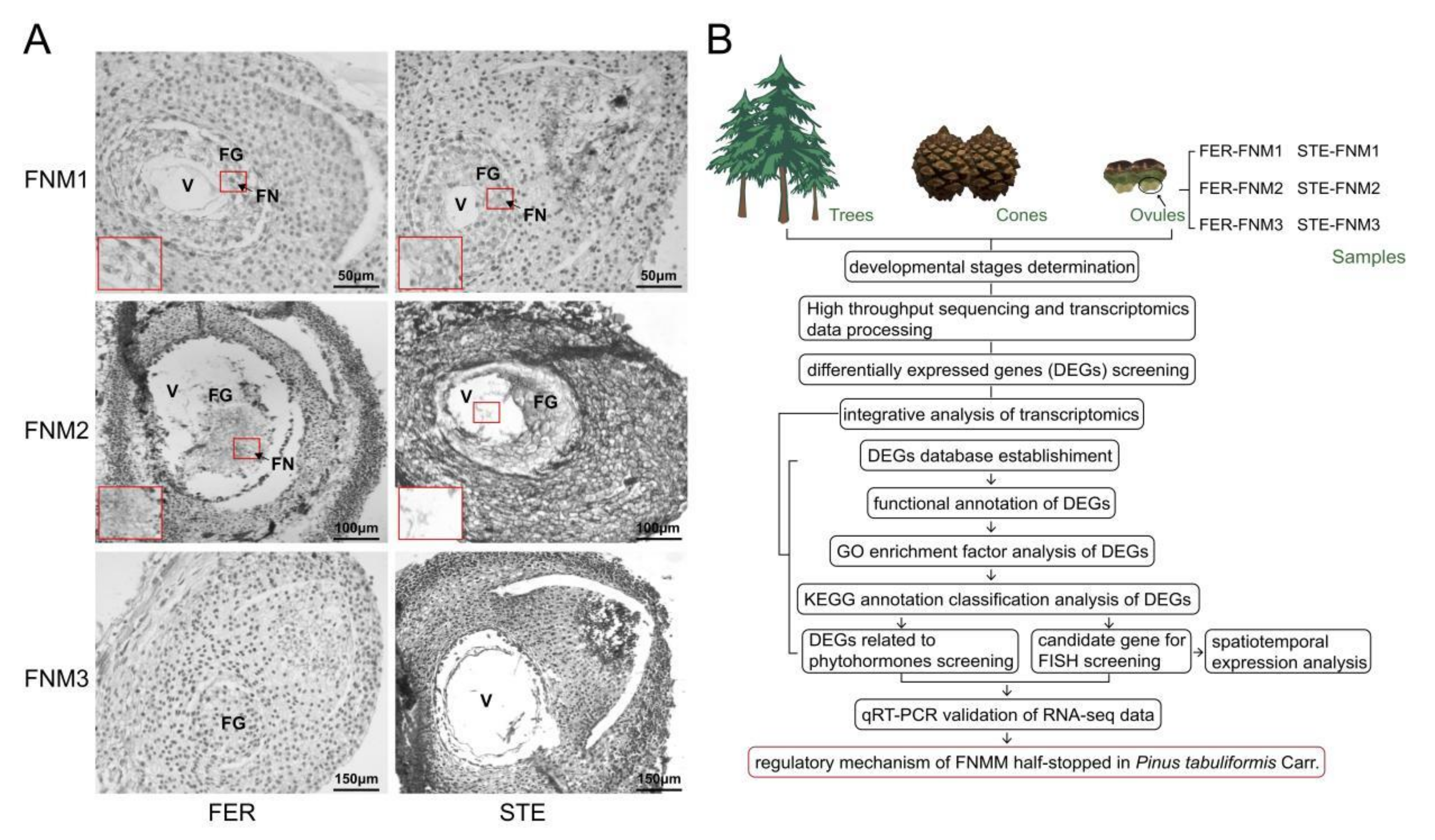
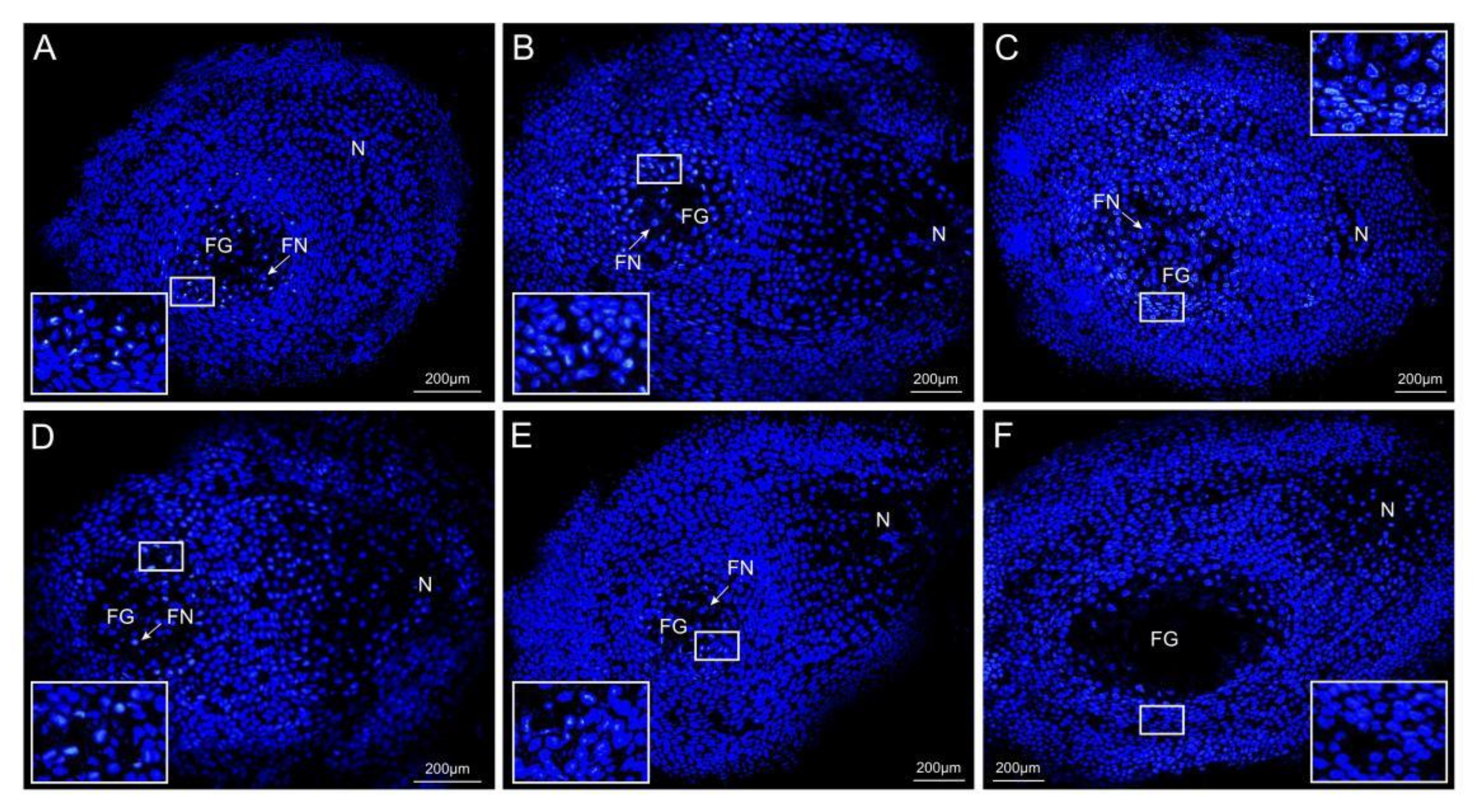
2.1. Ovule Phenotypes of P. tabuliformis

The phenotypes of P. tabuliformis ovules were shown in Figure 1A. In the early stage of FNMM, the anatomical structures of female sterile line (STE) and female fertile line (FER) ovules were similar. The center of FG was a large vacuole surrounded by several dozen free nuclei. In the middle stage, the free nuclei were still increased in FER; however, this trend was arrested in STE. Up to the late stage, the number of free nuclei was sharply increased and filled the center in FER ovules, while the free nuclei in STE ovules began to disappear and their centers were empty.

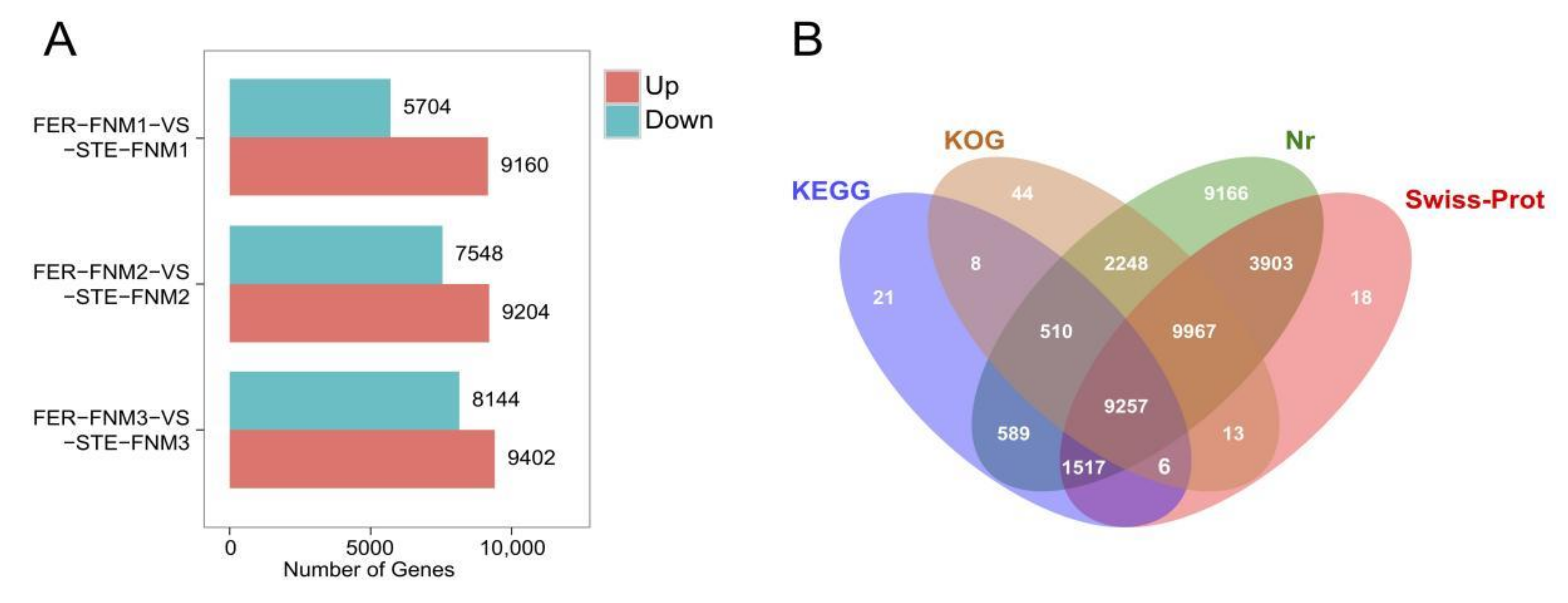
2.2. Sequencing, Read Assembly and Global Data Analysis

To explore the process of FNM and reveal the reasons of the development of FNMM half-stopped in the female sterile lines of P. tabuliformis, we selected three key time points: the early, middle, and late periods of the FNM process. FER ovule samples at the collection periods described above were marked as FER-FNM1, FER-FNM2, and FER-FNM3, respectively, while STE ovule samples were marked as STE-FNM1, STE-FNM2, and STE-FNM3, respectively, and each sample from the above periods was in triplicate (Supplementary Figure S1). A total of 18 mRNA libraries were paired-end (2 × 150 bp) sequenced using the HiSeqTM 4000 platform, resulting in 142.7 gigabase clean reads. The Q30 percentage of each sample was between 97.32% to 97.51%, and the length of N50 was 1767 bp. Given the assembled sequences, a total of 63,449 unigenes were generated from 18 libraries.
The reads mapped to de novo assembly were used for further research, and the mapping ratios of each sample were all beyond 84% (Supplementary Table S1). DEGs (differentially expressed genes) were screened with the FDR value <0.01 and fold change >2. From Figure 2A, we can find that there were more DEGs up-regulated than down-regulated in FER during the whole FNM process. In the FNM1 period, compared with STE, there were 9160 DEGs up-regulated in FER and 5704 down-regulated; in the FNM2 period, 9204 DEGs up-regulated and 7548 down-regulated in FER; and in the FNM3 period, there were 9402 DEGs up-regulated and 8144 down-regulated in FER.
For annotation of the generated unigenes, alignment searches were conducted against public bioinformatics databases. As shown in Figure 2B, among these unigenes, 37,270 unigenes had hits in public databases, including the NCBI non-redundant (NR) database, the Swiss-Prot protein database, Gene Ontology (GO), and the Kyoto Encyclopedia of Genes and Genomes (KEGG). The results indicated that 37,157 unigenes had strong similarity to proteins in the NR database. Similarly, up to 24,684 unigenes had Swiss-Prot annotation, 22,053 unigenes had KOG annotation, and 11,911 unigenes were matched in the KEGG database.

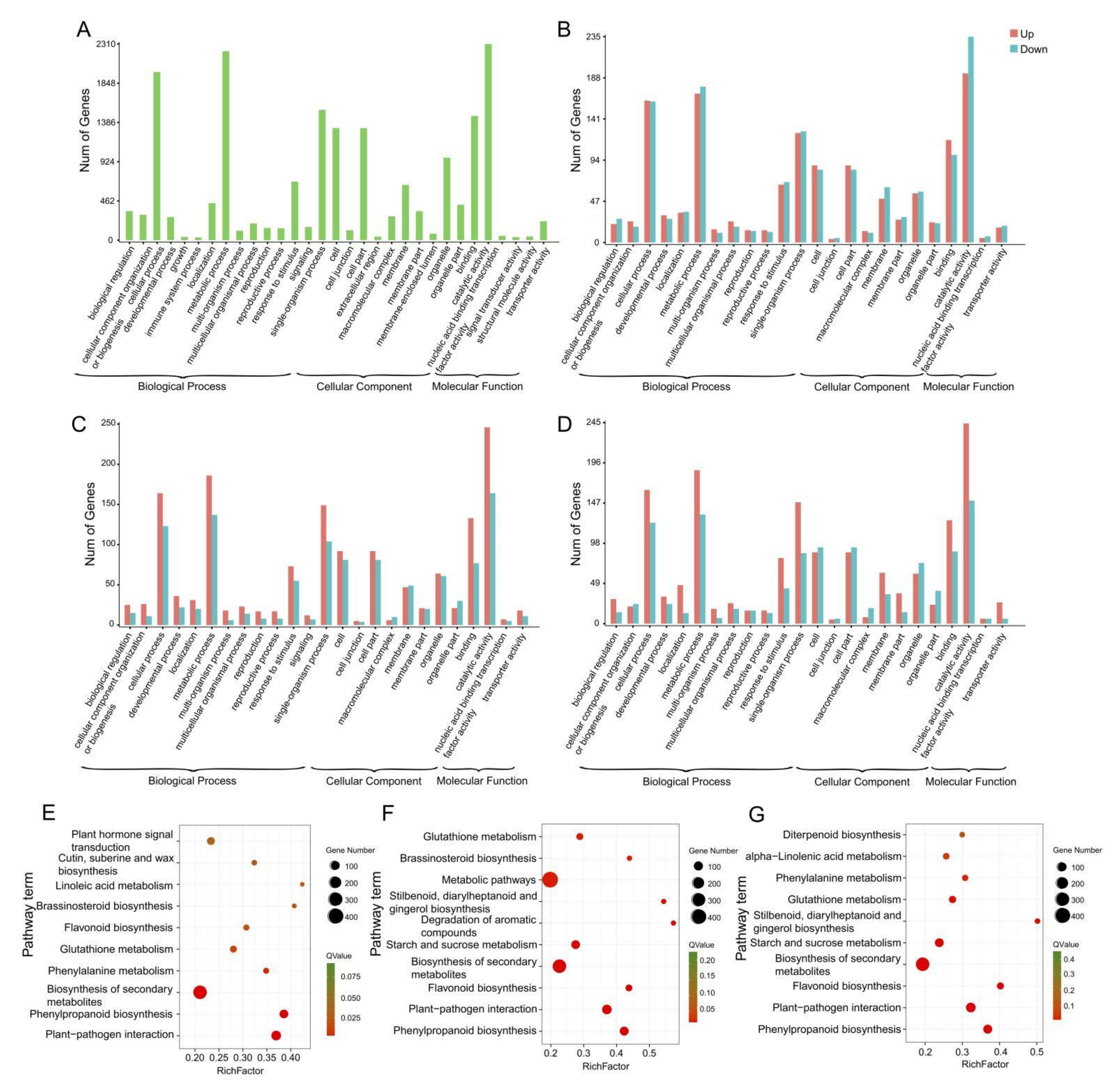
2.3. Gene Annotation and Functional Classification

To investigate the expression differences of unigenes between FER and STE, we used reads per kb per million reads (RPKM) to evaluate the expression level of DEGs during the FNM process in two lines, as well as to further reveal the function distribution of unigenes, which were classified by Gene Ontology (GO) annotation (Figure 3A). Annotated unigenes were divided into three categories, “biological process”, “cellular component”, and “molecular function”. In the biological process category, annotated unigenes were significantly enriched in “metabolic processes” (2225), “cellular processes” (1980), and “single-organism processes” (1535). Furthermore, “cell” (1320) and “cell part” (1320) were more obviously enriched in the cellular component categories; in the molecular function category, the unigenes were mainly enriched in “catalytic activity” (2310) and “binding” (1462). To characterize the function distribution of unigenes, we have analyzed GO terms in each ovule development stage in FER and STE (Figure 3B–D, Tables S2–S4). In each period of the FNM process, DEGs between two lines were all mainly enriched in the “cellular process”, “metabolic process”, and “response to stimulus” terms in the biological process category; in the cellular component category, “cell”, “cell part”, and “membrane” terms were dominant; “catalytic activity” and “binding” were still enriched with the most DEGs in the molecular function category.
KEGG was also used to investigate the pathway and network of the molecular regulation in each sample. From Figure 3E–G we found that from the FNM1 to the FNM3 period, DEGs were highly enriched in “phenylpropanoid biosynthesis”, “plant-pathogen interaction”, “biosynthesis of secondary metabolites”, and “flavonoid biosynthesis”. Furthermore, many DEGs were enriched in the “glutathione metabolism” and “brassinosteroid biosynthesis” pathways in the FNM1 and FNM2 periods and were also extremely enriched in “starch and sucrose metabolism” and “stilbenoid, diarylheptanoid and gingerol biosynthesis” in the middle and late periods of FNM.

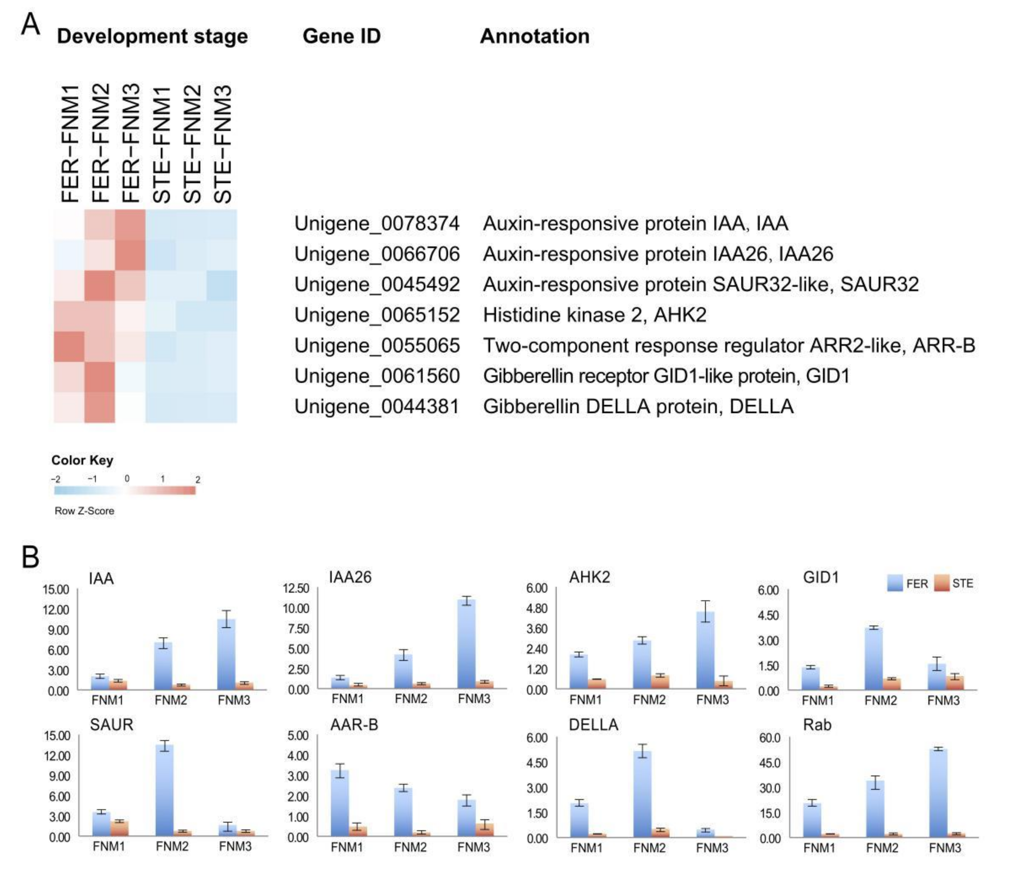
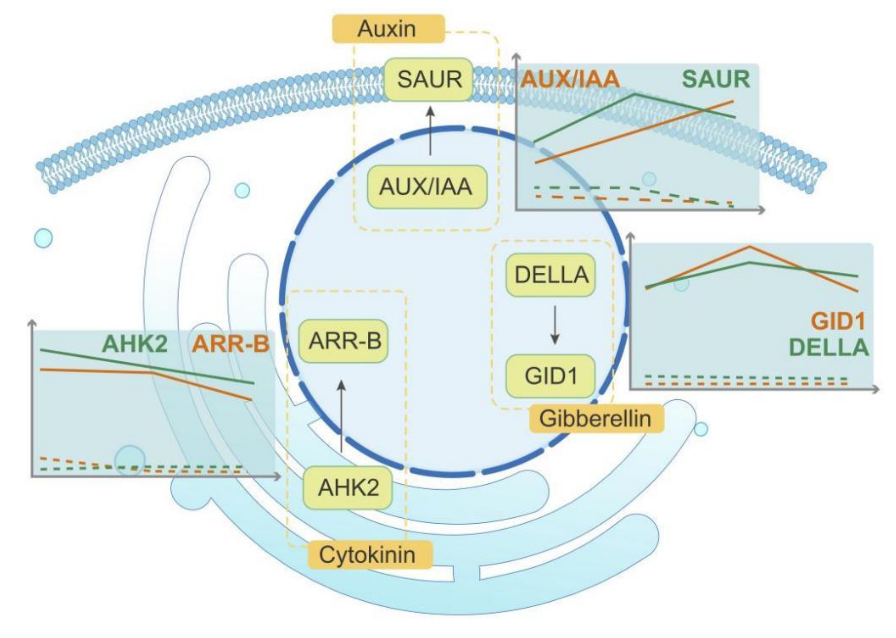
2.4. DEGs Related to Phytohormones in Comparisons between Fertile and Sterile Ovules during FNMM

According to the sequencing data and the analysis results of the pathways, we found that many genes participating in plant hormone transport were significantly differently expressed in FER and STE ovules. As shown in Figure 4A, many DEGs related to hormone transport pathways were up-regulated in FER ovules in the FNM process. Specifically, the transcript levels of auxin-related genes (IAA, Unigene_0078374, Unigene_0066706; SAUR, Unigene_0045492), cytokinin-related genes (AHK2, Unigene_0065152; ARR-B, Unigene_0071228), and gibberellin-related genes (GID1, Unigene_0061560; DELLA, Unigene_0044381) were markedly higher in FER than STE.

2.5. Verification of the Gene Expression Profile by qRT-PCR

To confirm the transcriptomic analysis results, the DEGs listed above were selected for quantitative real-time PCR (qRT-PCR) validation using the same type of samples compared with formerly used samples in high-throughput sequencing analysis. The primers of these DEGs are listed in Supplementary Table S5. The expression profiles of the candidate DEGs revealed by qRT-PCR data were consistent with those derived from sequencing (Figure 4B). We found that DEGs related to hormones were highly expressed in FER, and the expression levels of SAUR and DELLA were significantly high in the FNM2 stage. In addition, Rab was also up-regulated and expressed more highly in FER. The expression profiles of the qRT-PCR data were consistent with those derived from sequencing.

2.6. Expression Patterns of DEGs between STE and FER during the FNM Process

Based on the gene expression levels during the ovule development process in FER and STE, we used Short Time-series Expression Miner (STEM) software to cluster DEGs into eight profiles. These model profiles have been chosen to summarize the expression patterns of the genes. Furthermore, four patterns of genes showed very significant p-values among the eight patterns, marked as colored boxes (Supplementary Figure S2), which contained two up-regulated patterns (profile 6 and 7) and two down-regulated patterns (profile 0 and 1). These DEGs showed contrary expression trends between FER and STE during the FNM process, which might play an indispensable role in the ovule development of P. tabuliformis. There were total 775 DEGs up-regulated in FER but down-regulated in STE (Figure 5A,B), and we subjected these DEGs to GO term analysis, which also classified them into three main categories of “cellular component”, “molecular function”, and “biological process” (Figure 5C). Moreover, these DEGs were subjected to KEGG pathway enrichment analysis (Figure 5D), and “pantothenate and CoA biosynthesis”, “plant–pathogen interaction” and “spliceosome” were the top three pathways in which the DEGs were highly enriched.

2.7. Spatiotemporal Expression of PtRab in the Ovules

Based the expression trend analysis and the result of the qRT-PCR, we found that PtRab (Unigene_0044338) was highly expressed in the FER ovules and weakly expressed in STE (Figure 4B). The Rab family is the biggest subfamily of small GTP-binding proteins and participates in a variety of life activities, including protein synthesis, vesicle transport, and cytoskeleton assembly in plant cells. A digoxigenin-labeled DNA probe of PtRab was prepared for in situ hybridization of the frozen sections to display the temporal and spatial differences between two lines during the FNM process. As shown in Figure 6, in the FER ovules, PtRab was mainly expressed in the female gametophyte and kept being up-regulated as the number of free nuclei increased. However, the expression level of PtRab was significantly lower in the STE ovules and kept being down-regulated during ovule development up until the FNM3 stage, where the fluorescence signal was nearly undetectable. A fluorescence signal of PtRab was also detected in the surrounding tissue in two lines, but the signal was too scattered, which cannot directly reflect the expression trend in the ovules.

2.8. Development and Characterization of SSR Markers

Transcriptome sequencing provided a simple and effective method to develop a large number of unigene-based SSR markers, which was essential for mining a large number of functional SSR markers [18]. To further investigate the differences in regulatory mechanisms between FER and STE ovules, microsatellites were identified in this study. We used a microsatellite (MISA, http://pgrc.ipk-gatersleben.de/misa/, accessed on 23 March 2021) to analyze the transcriptome data, and a total of 3756 potential simple sequence repeat (SSR) markers were identified in 3288 unigenes. 374 unigenes contained more than one SSR; and the frequency of SSRs was 3.83%. As shown in Table 1, potential microsatellites were discovered and defined as di- to hexa-nucleotide motifs. The dinucleotide and trinucleotide repeats ranked first (49.95% and 30.91%, respectively), while the sum of the other type of nucleotide repeats accounted for less than 20%. The most represented repeat units of potential SSRs were 4–12, up to 3724 (99.15%), and 6 repeats and 5 repeats were the most significant (29.47% and 24.2%, respectively). Based on Supplementary Figure S3, we analyzed the SSR motifs in P. tabuliformis. The AT/TA di-nucleotide repeat was the most abundant motif (24.8%), followed by AG/CT (16.6%), AC/GT (8.6%), AAG/CTT (6.6%), and AAG/CTT (6.4%). These five types of nucleotide repeats mentioned above represented about 60%.

3. Discussion

The development of FG was the key process of plant gametophyte and sporophyte generation alternation, which also affected the development of seeds. So far, most studies on FG development were focused on angiosperms, such as Arabidopsis thaliana [19], although there were a few studies on female sterility in woody plants, such as Ulmus pumila Mill. and Xanthoceras sorbifolia Bunge [20,21]. Nevertheless, due to the fact that the natural female sterile mutants of gymnosperms were scarce and the volume of the ovules was too small, the molecular mechanisms of ovule development in gymnosperms were still unclear. In this study, we used high-throughput sequencing to analyze the transcripts in different periods of FER and STE ovules in P. tabuliformis. A total of 18 cDNA libraries were created (Supplementary Figure S1), and we obtained 142.7 G clean data and 98,038 unigenes in total. Analysis results of GO and KEGG indicated that the GO terms’ and KEGG pathways’ DEG enrichment were highly similar in varying lines, demonstrating that the transcriptome at the pathway level was generally conserved in the ovules of P. tabuliformis (Figure 3).
SSR markers have been used to determining protein function, genetic development, and gene expression regulation [21]. For example, SSR markers have been used to identify the position of the gene causing female sterility in wheat and soybean [21,22]. As shown in Table 1, we obtained 3756 SSR markers from 3288 unigenes, of which the frequency was 3.83%. The frequency of SSRs was diverse in different species, such as Codonopsis pilosula (12.2%) [23], Populus L. (15.3%) [24], and Robinia pseudoacacia L. (13.79%) [25]. This rate being lower in P. tabuliformis might be because of the lack of a reference genome, leading to a lack of information; another reason is that the gymnosperms were relatively primitive, with genes more stable than other species in the long-term evolution process, and not prone to mutation. The frequency of SSRs was also low in some other gymnosperm species, such as Taxaceae (2.24%) [26] and Ginkgo biloba L. (5.09%) [27]. Similarly to the other plants, excluding the mono-nucleotide repeats, di-nucleotide repeats were the most abundant type, followed by tri-nucleotide repeats [28].
As signal molecules in plants, hormones regulate a variety of biological processes. In this investigation, the plant hormone signal transduction pathway enriched many DEGs, and the exploration of the expression patterns of DEGs related to plant hormones was helpful to further reveal the internal mechanism of the cessation of FNMM in FG. Auxin is a mobile signaling molecule regulating various processes in plant development which also influences the development of ovules [29,30]. Previous studies have shown that a low concentration of auxin may lead to a loss of embryo sac in Arabidopsis [31]. GH3, SAUR, and AUX/IAA are the auxin primary response genes [32]. Genes related to auxin response showed obvious differential expression in FER and STE ovules at different stages of FNMM, suggesting that correct expression of auxin-response-related genes is crucial to the development of FG. In our investigation, with FNMM development in FER, AUX/IAA and SAUR were significantly up-regulated; on the contrary, they were all down-regulated in the STE ovules. SAUR was especially highly expressed in the FNM2 period, and the expression levels of AUX/IAA were significantly higher in the FNM2 and FNM3 periods. It is suggested that auxin accumulation in the STE ovules may be insufficient, which hindered the development of FG. Moreover, DEGs may further affect the distribution of auxin and the development of ovules (Figure 4).
Cytokinin is involved in cell division and differentiation, and a decrease in cytokinin concentration in plants might lead to female sterility and a decrease in the number of ovules [33,34]. AHK2 is the receptor protein of cytokinin which undergoes autophosphorylation of histidine after sensing cytokinin [35,36]. Phosphorylated histidine transfer protein has been shown to enter the nucleus, transfer the phosphate group to type-A or type-B response regulators, and then perform a series of regulation on the plants [37,38]. In the present study, many genes associated with the cytokinin pathway showed different expression patterns during ovule development in FER compared with STE. In the FER ovules, AHK2 and ARR-B were up-regulated during the whole FNM process and significantly highly expressed in the FNM1 and FNM2 stages, but in the STE ovules, they were all weakly expressed. This indicated that until the FNM2 period, the free nuclei in the FER ovules maintained vigorous division, whereas the concentration of cytokinin might have been insufficient and affected the nuclear division in the STE ovules (Figure 4). Gibberellin is an important plant hormone which plays an important role in the whole life process of plants and participates in the regulation of plant reproductive growth [39,40]. GID1 is the receptor protein of gibberellin, and DELLA is another important element protein of gibberellin signal transduction pathway which is also the negative regulator of gibberellin [41,42]. DELLA mainly exists in the nucleus and participated in the regulation of plant growth and reproduction [43,44]. In our investigation, with FNMM development, GID1 and DELLA were significantly up-regulated and highly expressed in the FNM2 period in FER; on the contrary, they were all down-regulated in the STE ovules. It is suggested that the gibberellin-related pathway may have been blocked, which affected the normal development of ovules and induced ovule abortion (Figure 7).
In line with the analysis results of the transcriptome data and expression trends of DEGs, PtRab showed obviously different expression between FER and STE. Rab plays a pivotal role in protein synthesis, substance transport, and cell division [45]. Previous research showed that Rab was abundantly expressed in tissues with cell division, and elongation was vigorous in rice [46]; in Pinus pinaster Ait., Rab was high expressed in the early stage of embryo development, and the expression level increased when the seed germinated [47]; Gutkowska found that the pollen tube development of Rab was abnormal and induced male sterility in Arabidopsis [48]. These studies indicated that the expression level of Rab perhaps plays a role in the reproductive growth of plants. In this study, we analyzed the temporal and spatial differences of PtRab between two lines during the FNM process. We found that in the FER ovules, the expression level of PtRab was markedly increased and dominantly expressed in FG; on the contrary, the development of FNMM was disrupted in the FNM2 period in STE, leading to the number of free nuclei decreasing, and PtRab was down-regulated without a fluorescence signal detected (Figure 5). Thus, we suspected that PtRab might be expressed as FG develops and that a low expression level of PtRab might be one the of the causes of female sterility in P. tabuliformis.

4. Materials and Methods

4.1. Plant Materials

In line with the previous analyses from our research group, the FER ovule and STE ovule were genetically closely related by the DNA marker technique [49]. The ovules were selected from the P. tabuliformis Seed Garden, Xingcheng, Liaoning Province, China. We selected three time points to collect the samples: the early, middle, and late periods of the FNM process (in early February, March, and April, respectively), and the same size cones were selected and collected from the middle of the crown. The ovules were removed from the scales under a dissecting microscope, and ovules from five cones were collected as one sample. The samples were placed into liquid nitrogen overnight and then stored at −80 °C. Paraffin sections were made according to Zhang et al. [50], and the developmental stages of ovules were observed under a microscope (Leica, Weztlar, Germany).

4.2. RNA Sample Preparation and RNA-Seq

We used Plant RNA Assistant Kit (Kebaiao, Beijing, China) to extract the total RNA from ground samples, according to the manufacturer’s instructions, and quantified the integrity of the RNA by agarose gel electrophoresis and Agilent 2100 Bioanalyzer. The purity and concentration of the RNA were determined by Nanodrop 2000 (Thermo Specific, USA).
The RNA-Seq samples were used to construct 18 libraries (FER-FNM1, FER-FNM2, FER-FNM3, STE-FNM1, STE-FNM2, and STE-FNM3; each sample had three replicates). After total RNA was extracted, mRNA was enriched by Oligo (dT) beads and then fragmented into short fragments using fragmentation buffer and reverse transcripted into cDNA with random primers. Second-strand cDNA was synthesized by DNA polymerase I, RNase H, dNTP, and buffer. Then, the cDNA fragments were purified with QiaQuick PCR extraction kit (QIAGEN, Valencia, CA, USA), were end-repaired, had poly (A) added, and were ligated to Illumina sequencing adapters. The ligation products were size-selected by agarose gel electrophoresis, PCR-amplified, and sequenced using Illumina HiSeqTM 4000 (Illumina, San Diego, CA, USA).

4.3. De Novo Assembly

The raw reads were cleaned by removing adapter sequences, low-quality sequences (reads with ambiguous bases “N”), and reads with more than 10% Q < 20 bases. De novo assembly of the clean reads was performed using Trinity [51] program to build the reference-free full-length transcription. In the present study, K-mer value 25 was chosen in Trinity, with group-pairs-distance =500 and other default parameters. Next, Trinity connected the contigs between each pair of contigs, using “N” to represent unknown bases, thus generating scaffolds. Paired-end reads were used again for scaffold gap filling to obtain sequences with the fewest Ns and those that could not be extended at either end. Such sequences were defined as unigenes. Finally, the potential transcript sequences were clustered using the TGI Clustering tool to obtain uni-transcripts [52].

4.4. Functional Annotation

We BLAST the genes based on four databases to annotate the unigenes: the Nr database (http://www.ncbi.nlm.nih.gov, accessed on 3 October 2020), the KEGG database (http://www.genome.jp/kegg, accessed on 3 October 2020), the Swiss-Prot protein database (http://www.expasy.ch/sprot, accessed on 6 October 2020), and the COG/KOG database (http://0-www-ncbi-nlm-nih-gov.brum.beds.ac.uk/COG, accessed on 6 October 2020), with BLASTx program (http://0-www-ncbi-nlm-nih-gov.brum.beds.ac.uk/BLAST/, accessed on 6 October 2020) to evaluate sequence similarity with genes of other species at an e-value threshold of 1×10⁻⁵. GO annotation was analyzed by Blast2GO software [53] with Nr annotation results, and WEGO software was used to perform the functional classification of unigenes [54]. The KEGG database was used to explore the genes involved in the biological pathways, and the pathway was defined as significantly enriched with p-value <0.05. All raw data of high-throughput sequencing have been deposited to the National Genomics Data Center (https://bigd.big.ac.cn, accessed on 25 January 2022) with the dataset accession number PRJCA004609.

4.5. Analysis of Differentially Expressed Genes (DEGs)

We estimate the gene expression level by the reads per kilobase of exon model per million mapped reads (RPKM) [55,56]. To identify DEGs across samples, the edgeR package (http://www.r-project.org/, accessed on 27 October 2020) was used. We identified genes with a fold change ≥2 and a false discovery rate (FDR) <0.01 in a comparison as significant DEGs.

4.6. Simple Sequence Repeat (SSR) Marker Prediction

We used MISA (http://pgrc.ipk-gatersleben.de/misa/, accessed on 30 October 2020) to discover the SSRs in the whole transcriptome. The minimum number of repeat units was as follows: six for di-nucleotides, five for tri-nucleotides, four for tetra-, penta-, and hexa-nucleotides. The shortest distance between two SSRs was 100 bp, or else they would be seen as one SSR.

4.7. Fluorescence In Situ Hybridization

4.7.1. Preparation and Labeling of DNA Probes

Primer for PtRab was 5′-primer CTTCTAAAGGCAACATTC, 3′-primer TCTATCCCTACACTGGTT. The cDNA of P. tabuliformis ovule was used as template for PCR and purified by Cycle Pure Kit (Biomiga, San Diego, CA, USA). DNA probe was marked by PCR DIG Probe Synthesis Kit.

4.7.2. Preparation of Frozen Sections

Successive sections of 10 μm were made at −20 °C with Leica CM1950 freezing microtome (Leica, Weztlar, Germany), cutterbed angle is 8°. The sections were picked up with an anatomical needle and adhered to the slide; the glass slide was specially used for FISH (Superfrost® Plus Slide, Hatfield, PA, USA). Dried at room temperature for 1–2 h.

4.7.3. Pretreatment of Frozen Sections before the Fluorescence In Situ Hybridization

The dried frozen sections were put into the hybridization tank, hydration of 10 mM sodium citrate buffer 5 min, and then transferred to equal volume 2 × SSC solution for 5 min; finally, sections were transferred to 50% formamide prepared by 2 × SSC for 1 h.

4.7.4. Fluorescence In Situ Hybridization

Hydrated frozen sections with 10 mM sodium citrate buffer for 5 min, immersed in 2 × SSC solution for 5 min, stood in 50% formamide for 1 h, and then put into a slice box containing 100% ethanol, avoiding dry slices. Attached the hybridization chamber to the slide and protected from light strictly with the following steps: add 20 µL of pre-hybridization solution without probe and pre-hybridize at 37 °C for 2 h; remove the probe and heat it at 85 °C for 5 min to denature the probe; add probe to the small hole left in the hybridization chamber and hybridize at 37 °C 12—24 h.

4.7.5. Elution and Detection after Hybridization

Removed the hybridization chamber, rinsed in 2 × SSC solution at 37 °C (5 min/time, 3 times), 0.1 × SSC rinsing solution, (5 min/time, 2 times), 2 × SSC solution isotonic for 2 min. After natural drying, 0.05 μg/mL DAPI was added dropwise to cover the tissue section and counterstained for 30 min. Wiped the slide and diluted the anti-digoxin antibody at 1:1000, and then, added Antifade Mounting Medium (Absin, Shanghai, China) and mounted. Finally, observed with a fluorescence microscope by ultraviolet fluorescence.

4.8. Real-Time Quantitative PCR Analysis

The Plant RNA Assistant Kit was used to isolate the total RNA in this research (Kebaiao, Beijing, China). Reverse transcription into cDNA was performed with SuperReal PreMix Plus (TIANGEN Biotech, Beijing, China), performed following the protocols included with the kits. Primers were designed using Primer Premier 6. The qRT-PCR was performed using a MiniOpticon Real-Time PCR Detection System (Bio-Rad, Hercules, CA, USA). The PCR condition was 94 °C for 35 s, then 45 cycles of 95 °C for 3 s for denaturation, and 55 °C for 30 s for annealing and extension, repeated three times. Relative expression levels of the target genes were normalized and then calculated using comparative Ct (2−ΔΔ Ct) method.

5. Conclusions

In this study, we analyzed transcriptome features based on high-throughput sequencing in different FNM periods of the FER and STE ovules in P. tabuliformis. Our findings determined the DEGs associated with plant hormone, suggesting that the synthesis of auxin, cytokinin, and gibberellin might be abnormal in STE ovules. In addition, the spatiotemporal expression of PtRab was different between FER and STE, indicating that PtRab might play an important role during ovule development. These findings may provide new strategies for improving our understanding of the molecular mechanisms involved in the FNM process and ovule development in gymnosperms (Figure 7).

Supplementary Materials

The following supporting information can be downloaded at: https://0-www-mdpi-com.brum.beds.ac.uk/article/10.3390/ijms23031915/s1.

Author Contributions

Conceptualization, C.Z. and Y.Z.; methodology, C.Z.; validation, H.H. and L.X.; formal analysis, H.H.; data curation, Z.G.; writing—original draft preparation, Z.G.; writing—review and editing, Z.G.; supervision, Y.Z. and C.Z.; funding acquisition, C.Z. All authors have read and agreed to the published version of the manuscript.

Funding

This work was supported by the National Natural Science Foundation of China (31870571).

Institutional Review Board Statement

Not applicable.

Informed Consent Statement

Not applicable.

Data Availability Statement

All raw data of high-throughput sequencing have been deposited to the National Genomics Data Center (https://bigd.big.ac.cn, accessed on 25 January 2022) with the dataset accession number CRA004027.

Conflicts of Interest

The authors declare no conflict of interest. The funders had no role in the design of the study; in the collection, analyses, or interpretation of data; in the writing of the manuscript, or in the decision to publish the results.

References

  1. Hu, Q.; Gao, S.; Li, F. Advances of research on plant female sterility. J. Beijing For. Univ. 2004, 26, 87–91. [Google Scholar]
  2. Guo, A.; Zheng, C.X. Female gametophyte development. J. Plant Biol. 2013, 56, 345–356. [Google Scholar] [CrossRef]
  3. Chen, L.; Zhang, J.; Li, H.; Niu, J.; Xue, H.; Liu, B.; Wang, Q.; Luo, X.; Zhang, F.; Zhao, D.; et al. Transcriptomic Analysis Reveals Candidate Genes for Female Sterility in Pomegranate Flowers. Front. Plant Sci. 2017, 8, 1430. [Google Scholar] [CrossRef] [PubMed] [Green Version]
  4. Awasthi, A.; Paul, P.; Kumar, S.; Verma, S.K.; Prasad, R.; Dhaliwal, H.S. Abnormal endosperm development causes female sterility in rice insertional mutant OsAPC6. Plant Sci. 2012, 183, 167–174. [Google Scholar] [CrossRef]
  5. Rosellini, D.; Ferranti, F.; Barone, P.; Veronesi, F. Expression of female sterility in alfalfa (Medicago sativa L.). Sex. Plant Reprod. 2003, 15, 271–279. [Google Scholar] [CrossRef]
  6. Clark, S.E.; Williams, R.W.; Meyerowitz, E.M. The CLAVATA1 gene encodes a putative receptor kinase that controls shoot and floral meristem size in Arabidopsis. Cell 1997, 89, 575–585. [Google Scholar] [CrossRef] [Green Version]
  7. Alvarez, J.M.; Cortizo, M.; Bueno, N.; Rodríguez, A.; Ordás, R.J. CLAVATA1-LIKE, a leucine-rich-repeat protein receptor kinase gene differentially expressed during adventitious caulogenesis in Pinus pinaster and Pinus pinea. Plant Cell Tissue Organ Cult. PCTOC 2013, 112, 331–342. [Google Scholar] [CrossRef]
  8. Subbaiah, C.C.; Huber, S.C.; Sachs, M.M.; Rhoads, D. Sucrose synthase: Expanding protein function. Plant Signal Behav. 2007, 2, 28–29. [Google Scholar]
  9. Kong, J.; Li, Z.; Tan, Y.; Wan, C.; Li, S.; Zhu, Y. Different gene expression patterns of sucrose-starch metabolism during pollen maturation in cytoplasmic male-sterile and male-fertile lines of rice. Physiol. Plant. 2007, 130, 136–147. [Google Scholar] [CrossRef]
  10. Qiu, Y.; Taylor, A.B.; McManus, H.A. Evolution of the life cycle in land plants. J. Syst. Evol. 2012, 50, 171–194. [Google Scholar] [CrossRef] [Green Version]
  11. Jin, W.; Gernandt, D.; Wehenkel, C.; Xia, X.; Wei, X.; Wang, X. Phylogenomic and ecological analyses reveal the spatiotemporal evolution of global pines. Proc. Natl. Acad. Sci. USA 2021, 118, e2022302118. [Google Scholar] [CrossRef] [PubMed]
  12. Zhang, M.; Zhang, W.; Gong, Z.; Zheng, C. Morphologic and anatomical observations in the process of ovulate strobilus generation and development in Pinus tabuliformis. J. Beijing For. Univ. 2017, 39, 1–12. [Google Scholar]
  13. Chen, K.; Abbott, R.J.; Milne, R.I.; Tian, X.; Liu, J. Phylogeography of Pinus tabulaeformis Carr. (Pinaceae), a dominant species of coniferous forest in northern China. Mol. Ecol. 2008, 17, 4276–4288. [Google Scholar] [CrossRef] [PubMed]
  14. Li, F.; Shen, X.; Li, Y. Discovery of female sterility in Pinus tabulaeformis and analysis of its causes. J. Hebei For. Coll. 1992, 7, 93–98. [Google Scholar]
  15. Bao, R. Primary Study on the Mechanism of Ovule Abortion of Female-Sterile Clone in Pinus Tabulaeformis Carr; Beijing Forestry University: Beijing, China, 2006. [Google Scholar]
  16. He, Y.; Zhang, C. Relationship between metabolism in developmental ovule and femalesterile in Pinus tabulaeformis Carr. J. Beijing For. Univ. 2007, 29, 88–93. [Google Scholar]
  17. Ding, S.; Zheng, C.; Bao, R. Analyses of peroxidase isozyme and protein polypeptides in female sterile Pinus tabulaeformis. Acta Bot. Boreal.-Occident. Sin. 2004, 24, 17–20. [Google Scholar]
  18. Chen, X.; Li, Q.; Li, Y.; Qian, J.; Han, J. Chloroplast genome of Aconitum barbatum var. puberulum (Ranunculaceae) derived from CCS reads using the PacBio RS platform. Front. Plant Sci. 2015, 6, 42. [Google Scholar] [CrossRef] [PubMed] [Green Version]
  19. Wang, H.; Liu, R.; Wang, J.; Wang, P.; Shen, Y.; Liu, G. The Arabidopsis kinesin gene AtKin-1 plays a role in the nuclear division process during megagametogenesis. Plant Cell Rep. 2014, 33, 819–828. [Google Scholar] [CrossRef]
  20. López-Almansa, J.C.; Pannell, J.R.; Gil, L. Female sterility in Ulmus minor (Ulmaceae): A hypothesis invoking the cost of sex in a clonal plant. Am. J. Bot. 2003, 90, 603–609. [Google Scholar] [CrossRef]
  21. Yan, M.; Dai, X.; Li, S.; Yin, D. Sequence Analysis and Comparison of EST-SSRs in Pine, Poplar and Eucalyptus. Genom. Appl. Biol. 2011, 30, 103–109. [Google Scholar]
  22. Dou, B.; Hou, B.; Xu, H.; Lou, X.; Chi, X.; Yang, J.; Wang, F.; Ni, Z.; Sun, Q. Efficient mapping of a female sterile gene in wheat (Triticum aestivum L.). Genet Res. 2009, 91, 337–343. [Google Scholar] [CrossRef] [PubMed] [Green Version]
  23. Wang, D.; Cao, L.; Gao, J. Data mining of simple sequence repeats in Codonopsis pilosula transcriptome. Chin. Tradit. Herb. Drugs 2014, 45, 2390–2394. [Google Scholar]
  24. Jun, T.; Michel, A.P.; Mian, M.A.R. Development of soybean aphid genomic SSR markers using next generation sequencing. Genome 2011, 54, 360–367. [Google Scholar] [CrossRef]
  25. Yang, X.; Zhang, K.; Wang, J.; Jia, H.; Li, Y.; Duan, J.; Ma, L. Analysis of Genetic Diversity and Construction of Core Collections of Black Locust Based on SSR Markers. Mol. Plant Breed. 2020, 18, 3086–3097. [Google Scholar]
  26. Li, Y.; Yang, X.; Zhang, J.; Huang, S.; Xiong, X. Studies on SSR Molecular Markers Based on Transcriptome of Taxus chinensis var. mairei. Acta Hortic. Sin. 2014, 41, 735–745. [Google Scholar]
  27. Fan, H.; Li, T.; Li, Z.; Lin, Y.; Cai, Y. Characteristics of EST-SSR Distribution in Ginkgo ESTs. Genom. Appl. Biol. 2009, 28, 869–873. [Google Scholar]
  28. Wu, J.; Cai, C.; Cheng, F.; Cui, H.; Zhou, H. Characterisation and development of EST-SSR markers in tree peony using transcriptome sequences. Mol. Breed. 2014, 34, 1853–1866. [Google Scholar] [CrossRef]
  29. Fu, W.; Zhao, Z.; Ge, X.; Ding, L.; Li, Z. Anatomy and transcript profiling of gynoecium development in female sterile Brassica napus mediated by one alien chromosome from Orychophragmus violaceus. BMC Genom. 2014, 15, 61. [Google Scholar] [CrossRef] [PubMed] [Green Version]
  30. Robert, H.S.; Park, C.; Gutièrrez, C.L.; Wójcikowska, B.; Pěnčík, A.; Novák, O.; Chen, J.; Grunewald, W.; Dresselhaus, T.; Friml, J.; et al. Maternal auxin supply contributes to early embryo patterning in Arabidopsis. Nat. Plants 2018, 4, 548–553. [Google Scholar] [CrossRef]
  31. Pagnussat, G.C.; Alandete-Saez, M.; Bowman, J.L.; Sundaresan, V. Auxin-Dependent Patterning and Gamete Specification in the Arabidopsis Female Gametophyte. Science 2009, 324, 1684–1689. [Google Scholar] [CrossRef] [Green Version]
  32. Panoli, A.; Martin, M.V.; Alandete-Saez, M.; Simon, M.; Neff, C.; Swarup, R.; Bellido, A.; Yuan, L.; Pagnussat, G.C.; Sundaresan, V. Auxin Import and Local Auxin Biosynthesis Are Required for Mitotic Divisions, Cell Expansion and Cell Specification during Female Gametophyte Development in Arabidopsis thaliana. PLoS ONE 2015, 10, e0126164. [Google Scholar] [CrossRef] [PubMed]
  33. Kinoshita-Tsujimura, K.; Kakimoto, T. Cytokinin receptors in sporophytes are essential for male and female functions inArabidopsis thaliana. Plant Signal. Behav. 2014, 6, 66–71. [Google Scholar] [CrossRef] [Green Version]
  34. Cucinotta, M.; Manrique, S.; Cuesta, C.; Benkova, E.; Novak, O.; Colombo, L. CUP-SHAPED COTYLEDON1 (CUC1) and CUC2 regulate cytokinin homeostasis to determine ovule number in Arabidopsis. J. Exp. Bot. 2018, 69, 5169–5176. [Google Scholar] [CrossRef]
  35. Cheng, C.; Mathews, D.E.; Eric Schaller, G.; Kieber, J.J. Cytokinin-dependent specification of the functional megaspore in the Arabidopsis female gametophyte. Plant J. 2013, 73, 929–940. [Google Scholar] [CrossRef]
  36. Feng, J.; Wang, C.; Chen, Q.; Chen, H.; Ren, B.; Li, X.; Zuo, J. S-nitrosylation of phosphotransfer proteins represses cytokinin signaling. Nat. Commun. 2013, 4, 1529. [Google Scholar] [CrossRef] [PubMed] [Green Version]
  37. Agostino, I.B.D.; Kieber, J.J. Molecular Mechanisms of Cytokinin Action; Elsevier Ltd.: London, UK, 1999; Volume 2, pp. 359–364. [Google Scholar]
  38. Liu, Z.; Dai, X.; Li, J.; Liu, N.; Liu, X.; Li, S.; Xiang, F. The Type-B Cytokinin Response Regulator ARR1 Inhibits Shoot Regeneration in an ARR12-Dependent Manner in Arabidopsis. Plant Cell 2020, 32, 2271–2291. [Google Scholar] [CrossRef] [PubMed]
  39. Hedden, P.; Sponsel, V. A Century of Gibberellin Research. J. Plant Growth Regul. 2015, 34, 740–760. [Google Scholar] [CrossRef] [Green Version]
  40. Peng, J.; Carol, P.; Richards, D.E.; King, K.E.; Cowling, R.J.; Murphy, G.P.; Harberd, N.P. The Arabidopsis GAI gene defines a signaling pathway that negatively regulates gibberellin responses. Gene Dev. 1997, 11, 3194–3205. [Google Scholar] [CrossRef] [Green Version]
  41. Li, D.; Duan, H.; Liu, H.; Wang, S.; Peng, W.; Yang, X. Progress on gibberellins, gibberellins functional analogues and gibberellins receptor. Chin. J. Pestic. Sci. 2013, 15, 601–608. [Google Scholar]
  42. Itoh, H.; Sasaki, A.; Ueguchi-Tanaka, M.; Ishiyama, K.; Kobayashi, M.; Hasegawa, Y.; Minami, E.; Ashikari, M.; Matsuoka, M. Dissection of the Phosphorylation of Rice DELLA Protein, SLENDER RICE1. Plant Cell Physiol. 2005, 46, 1392–1399. [Google Scholar] [CrossRef]
  43. Harberd, N.P. Botany. Relieving DELLA restraint. Science 2003, 299, 1853–1854. [Google Scholar] [CrossRef] [PubMed]
  44. Harberd, N.P.; Belfield, E.; Yasumura, Y. The Angiosperm Gibberellin-GID1-DELLA Growth Regulatory Mechanism: How an “Inhibitor of an Inhibitor” Enables Flexible Response to Fluctuating Environments. Plant Cell 2009, 21, 1328–1339. [Google Scholar] [CrossRef] [Green Version]
  45. Kirchhelle, C.; Chow, C.; Foucart, C.; Neto, H.; Stierhof, Y.; Kalde, M.; Walton, C.; Fricker, M.; Smith, R.S.; Jérusalem, A.; et al. The Specification of Geometric Edges by a Plant Rab GTPase Is an Essential Cell-Patterning Principle During Organogenesis in Arabidopsis. Dev. Cell 2016, 36, 386–400. [Google Scholar] [CrossRef] [PubMed] [Green Version]
  46. Kim, H.Y.; Seo, H.S.; Jeong, J.Y.; Lee, S.Y.; Cho, M.J.; Bahk, J.D. Molecular characterization of a rab-related small GTP binding protein cDNA from rice (Oryza sativa L. IR-36). Mol. Cells 1997, 7, 226–230. [Google Scholar] [PubMed]
  47. Gonçalves, S.; Cairney, J.; Rodríguez, M.P.; Cánovas, F.; Oliveira, M.; Miguel, C. PpRab1, a Rab GTPase from maritime pine is differentially expressed during embryogenesis. Mol. Genet Genom. 2007, 278, 273–282. [Google Scholar] [CrossRef]
  48. Gutkowska, M.; Wnuk, M.; Nowakowska, J.; Lichocka, M.; Stronkowski, M.M.; Swiezewska, E. Rab geranylgeranyl transferase β subunit is essential for male fertility and tip growth in Arabidopsis. J. Exp. Bot. 2015, 66, 213–224. [Google Scholar] [CrossRef]
  49. Li, M.; Zheng, C. RAPD analysis in female sterility clone 28 of Pinus tabulae- formis Carr. J. Beijing Forest. Univ. 2002, 24, 35–38. [Google Scholar]
  50. Zhang, W.; Zheng, C.; Zhao, N.; Li, F. Observation on the formation of ovule tapetum in Pinus tabulaeformis Carr. J. Beijing Forest. Univ. 2015, 37, 53–57. [Google Scholar]
  51. Grabherr, M.G.; Haas, B.J.; Yassour, M.; Levin, J.Z.; Thompson, D.A.; Amit, I.; Adiconis, X.; Fan, L.; Raychowdhury, R.; Zeng, Q.; et al. Full-length transcriptome assembly from RNA-seq data without a reference genome. Nat. Biotechnol. 2011, 29, 644–652. [Google Scholar] [CrossRef] [Green Version]
  52. Pertea, G.; Huang, X.; Liang, F.; Antonescu, V.; Sultana, R.; Karamycheva, S.; Lee, Y.; White, J.; Cheung, F.; Parvizi, B.; et al. TIGR gene indices clustering tools (TGICL): A software system for fast clustering of large EST datasets. Bioinformatics 2003, 19, 651–652. [Google Scholar] [CrossRef] [Green Version]
  53. Conesa, A.; Gotz, S.; Garcia-Gomez, J.M.; Terol, J.; Talon, M.; Robles, M. Blast2GO: A universal tool for annotation, visualization and analysis in functional genomics research. Bioinformatics 2005, 21, 3674–3676. [Google Scholar] [CrossRef] [Green Version]
  54. Ye, J.; Fang, L.; Zheng, H.; Zhang, Y.; Chen, J.; Zhang, Z.; Wang, J.; Li, S.; Li, R.; Bolund, L.; et al. WEGO: A web tool for plotting GO annotations. Nucleic Acids Res. 2006, 34, W293–W297. [Google Scholar] [CrossRef] [PubMed]
  55. Mortazavi, A.; Williams, B.A.; McCue, K.; Schaeffer, L.; Wold, B. Mapping and quantifying mammalian transcriptomes by RNA-seq. Nat. Methods 2008, 5, 621–628. [Google Scholar] [CrossRef] [PubMed]
  56. Audic, S.; Claverie, J.M. The significance of digital gene expression profiles. Genome Res. 1997, 7, 986–995. [Google Scholar] [CrossRef] [PubMed]
Figure 1. Background description of tissues and workflow. (A) Microscopic observations of ovules; FER: female fertile line, STE: female sterile line, FNM: the process of free nuclear mitosis, FG: female gametophyte, FN: free nucleus, V: vacuole. (B) Schematic overview of transcriptome data generation and analyses workflow.
Ijms 23 01915 g001
Figure 2. RNA sequencing data from ovules in P. tabuliformis. (A) Differentially expressed genes between FER and STE in FNM1, FNM2, and FNM3. (B) Venn diagram of unigene annotation in P. tabuliformis ovules.
Ijms 23 01915 g002
Figure 3. GO and KEGG annotation of unigenes of different free nuclear mitosis stages in ovules of P. tabuliformis. (A) GO annotation of unigenes of the ovule of P. tabuliformis. (BD) GO annotation of differentially expressed genes between FER and STE in the stage of FNM1, FNM2, and FNM3 of P. tabuliformis. (EG) KEGG pathway enrichment of differentially expressed genes in the stage of FNM1, FNM2, and FNM3 of P. tabuliformis.
Ijms 23 01915 g003
Figure 4. Analysis of expression levels of the differentially expressed genes. Expression patterns of differentially expressed genes related to the plant hormone. Data for the gene expression level were normalized to Z-score (A). Candidate DEGs expression levels revealed by qRT-PCR (B). Auxin-responsive protein IAA (IAA); auxin-responsive protein IAA26 (IAA26); auxin-responsive protein SAUR32-like (SAUR32); histidine kinase 2 (AHK2); two-component response regulator ARR2-like (ARR-B); gibberellin receptor GID1-like protein (GID1); gibberellin DELLA protein (DELLA); Rab-type small GTP-binding protein (Rab).
Ijms 23 01915 g004
Figure 5. DEGs with opposite expression patterns during the FNM process in STE and FER; DEGs up-regulated in FER and down-regulated in STE, Venn diagram (A), and heat map (B). GO classification of DEGs with opposite expression levels (C). KEGG pathway enrichment of DEGs with opposite expression levels (D).
Ijms 23 01915 g005
Figure 6. Fluorescence in situ hybridization of PtRab in P. tabuliformis ovules. (AC) The spatiotemporal expression of PtRab in ovules from FNM1 to FNM3 period in fertile line. (DF) The spatiotemporal expression of PtRab in ovules from FNM1 to FNM3 period in sterile line. FG: female gametophyte, FN: free nucleus, I: integument.
Ijms 23 01915 g006
Figure 7. Speculative network model revealing the ovule abortion in the female sterile line of Pinus tabuliformis Carr.
Ijms 23 01915 g007
Table 1. Occurrence frequency of different microsatellite repeat motifs of P. tabuliformis ovule transcriptome.
Repeat Motif LengthRepeat NumberTotalPercentage
456789101112>12
Di-nucleotide 795362294193128571532187649.95%
Tri-nucleotide 82121292228303 116130.91%
Tetra-nucleotide3495011 1 40210.7%
Penta-nucleotide132101 1 1443.83%
Hexa-nucleotide14028212 1734.61%
Total62190910114563192011325718323756100%
Percentage16.53%24.2%29.47%12.14%4.49%5.51%3.61%1.52%0.48%0.85%
Publisher’s Note: MDPI stays neutral with regard to jurisdictional claims in published maps and institutional affiliations.

