Review

Toxicological Impact of Rare Earth Elements (REEs) on the Reproduction and Development of Aquatic Organisms Using Sea Urchins as Biological Models

by
Chiara Martino
1,
Teresa Chianese
2,
Roberto Chiarelli
1,
Maria Carmela Roccheri
1 and
Rosaria Scudiero
2,*
1
Department of Biological, Chemical and Pharmaceutical Sciences and Technologies (STEBICEF), University of Palermo, Viale delle Scienze, Building 16, 90128 Palermo, Italy
2
Department of Biology, University Federico II, Via Cintia 21, 80126 Napoli, Italy
*
Author to whom correspondence should be addressed.
Int. J. Mol. Sci. 2022, 23(5), 2876; https://0-doi-org.brum.beds.ac.uk/10.3390/ijms23052876
Submission received: 9 February 2022 / Revised: 2 March 2022 / Accepted: 4 March 2022 / Published: 6 March 2022
(This article belongs to the Special Issue Molecular Research on Reproductive Toxicity)

Abstract

:
The growing presence of lanthanides in the environment has drawn the attention of the scientific community on their safety and toxicity. The sources of lanthanides in the environment include diagnostic medicine, electronic devices, permanent magnets, etc. Their exponential use and the poor management of waste disposal raise serious concerns about the quality and safety of the ecosystems at a global level. This review focused on the impact of lanthanides in marine organisms on reproductive fitness, fertilization and embryonic development, using the sea urchin as a biological model system. Scientific evidence shows that exposure to lanthanides triggers a wide variety of toxic insults, including reproductive performance, fertilization, redox metabolism, embryogenesis, and regulation of embryonic gene expression. This was thoroughly demonstrated for gadolinium, the most widely used lanthanide in diagnostic medicine, whose uptake in sea urchin embryos occurs in a time- and concentration-dependent manner, correlates with decreased calcium absorption and primarily affects skeletal growth, with incorrect regulation of the skeletal gene regulatory network. The results collected on sea urchin embryos demonstrate a variable sensitivity of the early life stages of different species, highlighting the importance of testing the effects of pollution in different species. The accumulation of lanthanides and their emerging negative effects make risk assessment and consequent legislative intervention on their disposal mandatory.

1. Introduction

The aquatic environment is a sink for many anthropogenic contaminants, the diversity of which and their concentrations are rapidly increasing. Therefore, understanding the impact of the human-derived waste on aquatic animal species is a major issue for the scientific community, with a major aim to translate this understanding to global awareness. Despite the relevance of this emergency, few studies have been carried out so far to understand the complex interactions between environmental stressors and their biological effects on the whole ecosystem. A major challenge in impact and risk assessment of environment is to link the harmful effects of pollutants in individual sentinel animals and their ecological consequences.

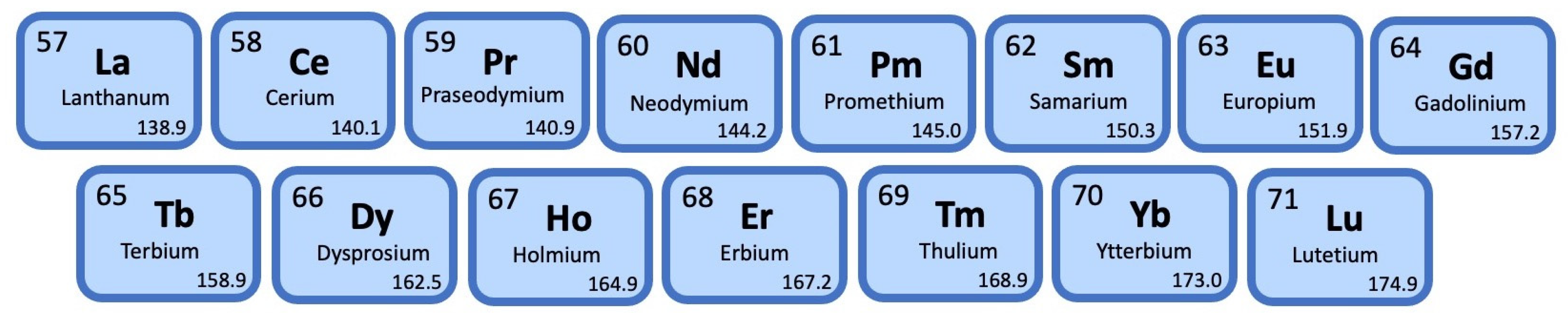
2. Rare Earth Elements

Among the contaminants of emerging interest, rare earth elements (REEs) play a fundamental role; they consist of a group of 15 trivalent elements of the lanthanide family with atomic numbers of 57 to 71 (IUPAC, 2005) (Figure 1) and are used in various fields of technology such as medicine (diagnostics and drugs), electronic devices (cell phones and plasma screens), and permanent magnets [1].
REE extraction and processing into commercial products have grown exponentially in recent years and have raised environmental concerns about their release into aquatic and terrestrial ecosystems [2,3,4,5,6]. Increased concentrations of REEs have been reported in lakes, rivers, and marine environments, making the removal of REEs from wastewater almost impossible [2,5,7]. The ratio of the concentration of a chemical in biota to that in ambient water is called the bioconcentration factor (BCF) and depends on many parameters; in the case of lanthanides, it depends on the type, the exposure concentration, and the microorganism considered [8]. BCF is a very common parameter to describe the accumulation of pollutants in biota relative to water [9]. It can be expressed as the ratio of the concentration of a chemical in an organism to the concentration of the chemical in the surrounding environment, so BCF is expressed in units of liter per kilogram. The BCF of lanthanides in algae reaches values up to 3,000,000 L/kg [10]. The importance of the chemical mobility of a REE is its link to its bioavailability. In physiological studies, lanthanides are often used to inhibit the uptake of divalent calcium ions (Ca2+, which have a similar ionic radius but a lower charge density than the trivalent lanthanide ion [11,12]. However, in addition to the health risks associated with their applications, ecotoxicological and epidemiological evidence are directly linked to environmental exposure to REEs and adverse health conditions remain weak [2].

3. Gadolinium

Among the lanthanides, gadolinium is widely used as a contrasting agent in the diagnostic procedures of magnetic resonance imaging (MRI) due to its paramagnetic properties [13]. For this reason, Gd is becoming a new powerful contaminant of aquatic environments, and the most common pollutant of the lanthanide family [3,14]. Gadolinium, like other REEs, is naturally occurring in the environment as a result of the dissolution of minerals. The background Gd level only minimally affects the total Gd measured in fresh and marine water, since most of the Gd+3 ions determined in aquatic environments derive from gadolinium-based contrast agents (GBCAs) excreted via the kidney by MRI patients [15]. GBCAs are eliminated unmetabolized. Being at first generally considered safe, the use of Gd in medicine has grown exponentially in the last decades, leading to a consequent increase in its emission into the environment, although its use has later been linked to nephrogenic systemic fibrosis (NSF) disease [16,17]. Indeed, in 2016, an annual emission of 4 tons of Gd has been estimated in Germany and to 19 tons in Europe [5,7,18]. Increasing concentrations of Gd have been found in rivers, where it passed from the geogenic background of a few ng/L to about 100 µg, with peaks of 1 mg, per liter [5,7,18,19]. High Gd concentrations have also been found in drinking water [3]. An increase in Gd concentrations has also been demonstrated in coastal seawaters, as occurring in Nagoya City near sewage treatment plants [20]; in areas of San Francisco Bay surrounded by hospitals and research centers using GBCA where the Gd concentration passed from 8.27 to 112 pmol kg−1 in the last 20 years [21]; and on the of northeastern coast of Brazil, near the city of Salvador, where it is estimated that between 698 and 2021 g Gd per day are discharged into the oceans due to a submarine outfall sewage along the Brazilian coasts [22]. In the future, GBCA concentrations in the aquatic environment are likely to increase due to the constant use of MRI [23].
The presence of Gd has been confirmed in aquatic organisms, such as lichens, algae, plants, invertebrates and vertebrates [3,14,24,25]. However, like other REEs, detailed studies on metabolization, bioaccumulation, and the environmental fate of Gd complexes and their toxicological effects on living organisms are lacking.
In this framework, it is important to develop appropriate methodologies to gain a deeper understanding of the impact of REEs on several important processes, including fertilization and embryonic development. In particular, the use of biological model systems should provide valuable information on this fundamental issue.

4. The Sea Urchin Embryo

The sea urchin (Echinodermata: Echinoidea) embryo has long been used as a model organism for biological developmental studies [26,27,28,29]. Several factors make this system suitable for conducting a wide range of biological tests. These include low maintenance costs; a small size; high fecundity; simple artificial spawning; fertilization and rearing; rapid synchronous development; optical transparency of the embryo, allowing for direct observation of cell division and movement within the embryos and larvae; and well understood embryogenesis. Furthermore, as invertebrate species, sea urchins are not subject to animal welfare concerns. This trait satisfies the strategy for developing alternative approaches to the use of vertebrates in biological testing [27].
Sea urchin embryos have been used successfully to study many cellular processes, such as adhesion [30], differentiation, survival, and death [29,30,31,32,33]. They are also recognized as an excellent model system for eco- and geno-toxicological studies [34,35,36].
Sea urchin embryos are being used as a model to elucidate the role of cellular and molecular mechanisms involved in human health and disease. This is because general cellular properties are common to many organisms [37,38]. Complete sequencing of the sea urchin genome also revealed that sea urchins are more closely related to vertebrates, with which they share the superphylum group of deuterostomes, than other invertebrates that are commonly used as models of human disease, e.g., Drosophila melanogaster and Caenorhabditis elegans [27].

5. Rare Earth Elements and the Sea Urchin Embryo

Regarding toxicological studies, it is well documented that echinoderm early life stages exhibit a high sensitivity to several toxicants, including heavy metals, persistent organic pollutants and microplastics [39,40,41,42,43,44,45]. The effects of lanthanides on sea urchins have also been evaluated; in particular, the potential damage of seawater contamination on gamete viability, fertilization, and larval development [46,47,48,49,50,51,52,53,54,55]. In these studies, many sea urchin species are utilized, in consideration of their availability in the different areas interested in ecotoxicological studies.
In the Sphaerechinus granularis (family Toxopneustidae), a sea urchin family inhabiting the Mediterranean Sea and eastern Atlantic Ocean, sperm exposure to REEs induces an overall inhibition of fertilization success and developmental defects. Of REEs, 10−5 M Gd3+ ions show the greatest sperm toxicity [55]. Cytogenetic analysis of REE-exposed S. granularis embryos reveals the induction of mitotic aberrations to different extents by all tested REEs, with the highest aberration frequencies being induced by 10−6 M Gd3+. The inhibition of mitotic activity and the frequency of mitotic aberrations were observed in embryos exposed to REEs at concentrations ranging from 10−6 to 10−4 [54,55].
Interestingly, experiments performed on different sea urchin species demonstrated different REE sensitivities. S. granularis displayed significantly greater sensitivity than the other two species with which it shares a habitat, Arbacia lixula (family Arbaciidae) and Paracentrotus lividus (family Parechinidae), both following embryo and sperm exposure.
In P. lividus, sperm exposure to REEs (10−5 to 10−4 M) resulted in a concentration-related decrease in fertilization success along with increase in offspring damage. Following sperm exposure to 10−4 M trivalent REE salts, a significant loss of fertilizing capacity was exhibited by all tested REEs (La, Ce, Nd, Sm, Eu, Dy, Gd, Yb). Eu ions displayed the most severe spermiotoxicity. The induction of transmissible damage presented as developmental defects in the offspring was significantly increased following sperm exposure to REEs, with Yb and La cause the most severe developmental defects. REE-exposed sperm caused offspring showing cytogenetic anomalies and aberrations, along with significant inhibition of mitotic activity. Exposure of P. lividus embryos and/or larvae to REE salts in the micromolar range from 10−6 to 10−4 M determined developmental defects, lipid peroxidation and redox anomalies. The most severe effects were recorded with Gd, followed by Yb, La, Nd, Eu, Ce, and then Sm. Gd and La were also effective in exerting cytogenetic defects [47,49].
Exposures of A. lixula spermatozoa to REEs did not affect the fertilization success. On the other hand, the offspring of A. lixula REEs-treated spermatozoa suffered significant developmental defects, as recorded in P. lividus offspring, particularly after exposure of the spermatozoa to Dy, Er and Yb. These three elements were found to be highly toxic to embryos and larvae. Minor defects were recorded in embryos/larvae exposed to Ce and Lu [47,54].

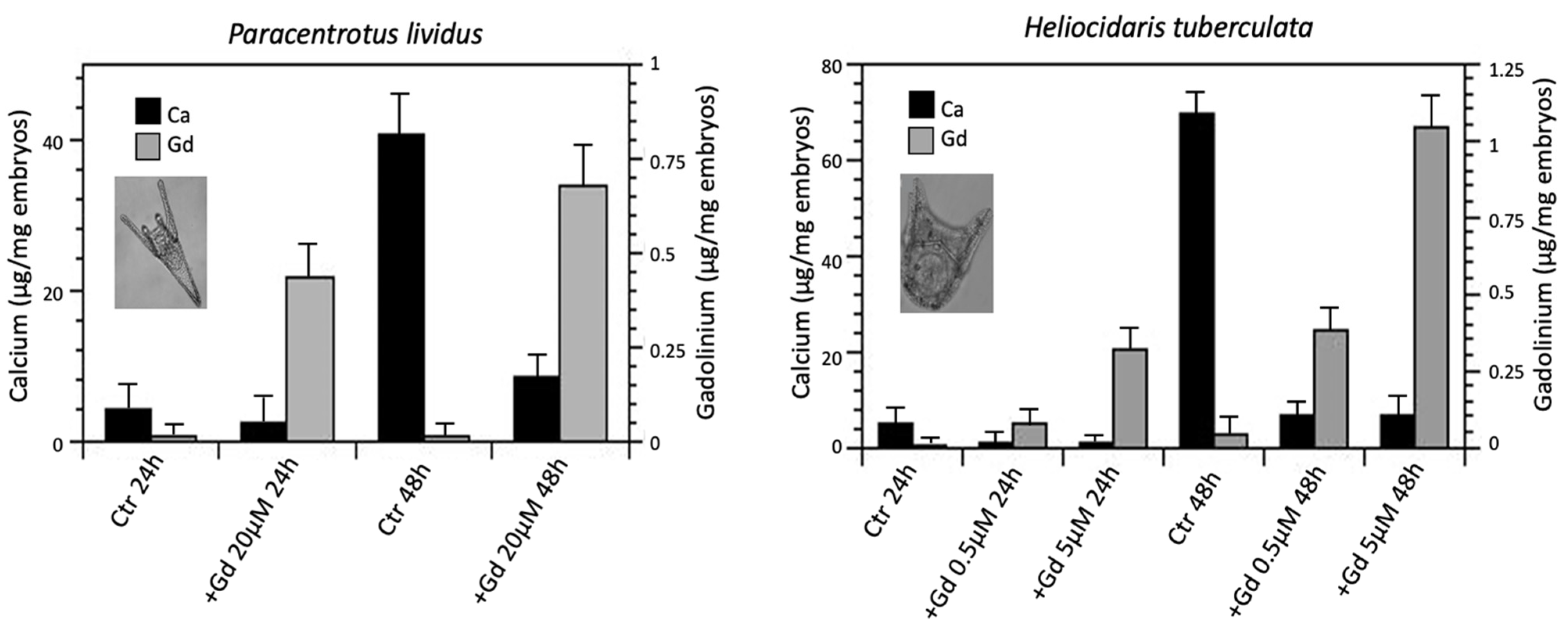
6. Gadolinium and the Sea Urchin Embryos

Considering the massive use of Gd in diagnostics, the toxicity of this REE on embryonic development in sea urchins was particularly studied. The responses to different Gd concentrations during sea urchin development in A. lixula and P. lividus, two species belonging to the same habitat (north western coast of Sicily, Mediterranean Sea), have been compared with those recorded in two phylogenetically and geographically distant species, Heliocidaris tuberculata (family Echinometridae) and Centrostephanus rodgersii (family Diadematidae), both from the Pacific Ocean (eastern Australia), that share similar larval phenotypes to theMediterranean species [50]. Indeed, 48 h post fertilization (hpf) these four species go through a feeding echinopluteus larva that show a tripartite gut and a complex three-dimensional skeleton.
These studies retrieved, for the two Mediterranean species, similar values of EC50 (1.18 mM for P. lividus and 2.1 mM for A. lixula) and an identical NOEC value of 250 nM, whereas the two oceanic species showed great differences in sensitivity. There was a difference of several orders of magnitude in the EC50 and NOEC value between H. tuberculata (56 nM and 1 nM, respectively) and C. rodgersii (132 mM and 1 mM, respectively) [50]. Based on this, the authors suggested that the different Gd sensitivity shown by the two Australian species may be due to the different phylogenetic history of these sea urchins [50]. Indeed, phylogenetic analysis indicated C. rodgersii as the oldest sea urchin lineage among the four examined, and H. tuberculata, the most Gd sensitive species, as the more recent and more closely related to the Mediterranean species [56]. Fascinating is the hypothesis that C. rodgersii is the most resistant to Gd insult because it is from an older lineage. This feature might have contributed to the adaptive capacity and resilience of C. rodgersii in a changing world [57]. However, exposure to Gd led to greater alterations or inhibitions of skeleton growth for the species examined at the final endpoint (48 hpf); the phenotypic response to Gd of the altered skeleton formation was similar in the four species, indicating a similar response mechanism, although with different levels of sensitivity with respect to the concentrations used. Indeed, in all the four species examined, exposure to Gd had no major effect on early development up to gastrulation (24 hpf); the ingression and migration of the PMCs and the invagination of the vegetal plate occurred with the right timing; and at 48 hpf, the differentiation of the ectodermal territories in the columnar epithelium at the animal pole and the in the squamous epithelium at the vegetal pole also appeared normal, as well as the development of the tripartite intestine. On the contrary, at 48 hpf, impaired skeleton development with several skeletal anomalies was evident [50,51].
Similar evidence for a conserved mechanism of Gd toxicity for sea urchin embryos was found in three Japanese species, Hemicentrotus pulcherrimus, Heliocidaris crassispina and Pseudocentrotus depressus [48]. Even in these species the ingression of PMCs into the blastocoel in the late blastula stage, their migration along the inner surface of the blastocoel, and the formation of both ventrolateral clusters at the gastrula stage were not inhibited by Gd-containing seawater; however, the progress of archenteron invagination was accompanied by the formation of abnormal and asymmetrical spicules. The degree of sensitivity to Gd ions also varied in these species. A concentration of 1 µmol/L Gd exerted greater inhibitory effects on spicule formation in A. crassispina and P. depressus than in H. pulcherrimus. Since this Gd level is considered insufficient to block divalent calcium ion (Ca2+) channels [58,59], the authors excluded that the inhibition of spicule formation may be caused by the inhibition of Ca2+ channels and suggest that trivalent Gd ions exerts their inhibitory effects on spicule formation specifically during the late blastula stage through different molecular pathways, rather than during the gastrula stage when spicule formation effectively occurs.
However, measurements of Ca and Gd content in sea urchin embryos using flame atomic absorption spectrometry (FAAS) clearly demonstrated a correlation between Ca and Gd uptake, with a reduction in the amount of Ca in parallel with an increase in the Gd content [51]. In control embryos, Ca uptake increases significantly during development, the total amount of Ca being 10-fold and 13-fold greater in 48 hpf larvae than in 24 hpf embryos in P. lividus and H. tuberculata, respectively (Figure 2). It should be considered that the production of spicules in 48 hpf larvae is at its highest, resulting in the formation of a more complex three-dimensional skeleton in H. tuberculata. In P. lividus embryos developed in Gd-contaminated seawater, there is an approximately 45% and a 78% reduction in the amount of Ca after 24 and 48 hpf, respectively. Gd uptake reaches about 0.70 µg/mg in embryos (Figure 2).
In H. tuberculata embryos exposed to Gd, the reduction in the amount of Ca is about 80% at 24 hpf and 90% at 48 hpf, regardless of the concentration of Gd in water, thus demonstrating that even a low concentration of Gd (0.5 µM) is sufficient to block calcium absorption. Conversely, concentration-dependent uptake of Gd was observed, with a greater Gd amount at the highest concentration (5 µM Gd) at both 24 and 48 hpf (approximately 0.4 and 1 µg/mg embryos, respectively) (Figure 2).
The results showed that Gd interfered with Ca uptake, probably because Gd competes with Ca to bind to the same ion channels. As a trivalent ion, Gd bound with higher affinity than the divalent Ca ions [60]. As expected, the FAAS results also showed that Gd accumulated in sea urchin embryos in a time- and dose-dependent manner.
The different sensitivity of embryos belonging to different species of sea urchins has also been demonstrated for other contaminants, such as nanoparticles [61], antibiotics, and disinfectants [62]. The different sensitivity to REEs depended on the species considered, but also on the type of lanthanide. Indeed, Gd3+ ions induced high severe effects in P. lividus embryos than in S. granularis and A. lixula embryos. This result was in contrast with the findings of the other REEs, where S. granularis and A. lixula appeared more sensitive than P. lividus. Table 1 summarizes the effects of REEs on sperm and embryonic development evaluated on various sea urchin species. This paradoxical finding deserves further investigation. Moreover, it suggests that in studies of biomonitoring, toxicity, and conservation of biodiversity, organisms deriving from different species within the same phyletic group should be used, even when performing a widely consolidated experimental model such as that of the sea urchin embryo.

7. Synergistic Effects of Gadolinium Exposure and Increased Temperature on Sea Urchin Embryos

Climate change has severe effects on coastal areas, as global warming has been raising the sea surface temperature since the late 19th century [63]. Understanding the combined effects of water pollution and global warming has therefore become an urgent issue, as this can generate additive, antagonistic, or synergistic effects. The impact of ocean warming and polluting nanoparticles, such as nano zinc oxide (nZnO), was studied on the larvae of the tropical sea urchin, Tripneustes gratilla. Both stressors had strong interactive effects on fertilization and development of these larvae. In particular, larval growth was strongly influenced by the combined stress of high temperature and nZnO [64]. More recently, microplastics have been shown to aggravate the effect of an increase in water temperature by generating additional stress on P. lividus larvae, manifested in a lower growth rate and impaired development [65]. Regarding a possible combined effect of temperature and lanthanides on sea urchin embryos, to date only one published article focuses on this topic. In this study, the interactive effects of increased temperature and Gd pollution during P. lividus development were investigated [53]. Using combined treatments of three temperatures (control: 18 °C; near-future projections: 21 °C; today’s marine heatwave conditions: 24 °C), the authors demonstrated that a lower thermal increase (21 °C) reduced the negative effects of Gd exposure (20 μM) on development, with embryos and larvae having a lower percentage of abnormalities and better skeletal growth, while the higher thermal increase (24 °C) had a negative synergistic effect when combined with Gd exposure, causing a lower percentage of embryos reaching advanced larval stages. At the molecular level, the combined treatments induced the expression of the Hsp60 protein at 24 h, and elicited autophagic and apoptotic processes at 48 h. These apoptotic events were found to be selective, probably to eliminate the most damaged cells. Overall, the authors concluded that a moderate increase in seawater temperature (+3 °C, i.e., 21 °C) will mitigate the negative effects of Gd pollution on embryonic development, while a major increase (+6 °C, i.e., 24 °C) would have negative synergistic effects [53].

8. Gadolinium and Embryonic Gene Expression in Sea Urchins

The mechanisms underlying the induced toxicity in sea urchin embryos have been investigated by gene expression analyses. In particular, the possible changes in the expression of genes that are involved in the early stages of development, in spiculogenesis, and in cell signaling were determined in embryos of P. lividus and H. tuberculata exposed to Gd [52].
The choice fell on these two species because, as mentioned before, they exhibit a similar morphological response to Gd, although H. tuberculata shows greater sensitivity. In both species, the PMC ingression and archenteron invagination occurred on schedule, but Gd-exposed embryos lacked the two triradiate spicules, thus demonstrating that Gd exposure primarily affects skeleton formation. In both species, some embryos showed supernumerary rods resulting in the inability to achieve the pluteus shape, while others had shorter spicules leading to a reduction in arm length, while still others had an asymmetric skeletal pattern [50]. The developmental success of sea urchin larvae depends on their ability to swim and feed, which is determined by the length of their arms [64]. Therefore, it is likely that Gd exposure can reduces survival in nature. In addition, in both species the uptake of Gd into embryos occurs in a time- and concentration-dependent manner and correlates with the decrease in calcium uptake [52].
Densitometric analysis of the gel bands obtained by the One-Step RT-PCR kit was used by Martino et al. to perform comparative gene expression assays and to detect differences between controls and treated embryos. Starting with an equal amount of total RNA from control and Gd-exposed embryos, they amplified the selected RNA targets reported in Table 2 and the reference genes (ribosomal protein S24 for P. lividus and zinc-finger transcription factor Z12 per H. tuberculata) used to normalize amplicon band intensities [52]. The authors determined the temporal expression of the genes that are a part of the skeletogenic gene regulatory network, that controls the expression levels of downstream genes involved in mesenchyme differentiation and biomineralization. In particular, they analysed genes expressed earlier during development (e.g., alx-1, nodal, lefty, and bmp), and genes involved in molecular signaling and skeletogenesis, such as univin, vegf, vegf-r, fgf, msp130, sm30, p16, and p19. Table 2 summarizes the expression profiles of these genes in the two species considered, at different concentrations of Gd and hours of development.
Alx1 is a gene encoding a key transcription factor selectively expressed in the large micromere lineage. It is essential for PMC specification, [66] and the protein regulates the expression levels of many skeletogenic genes, as demonstrated in echinoderm embryos [67,68]. Nodal and bone morphogenetic protein (Bmp) signaling pathways are integral to embryonic axis determination, and their opposite signals regulate left–right asymmetry in the sea urchin larvae [69,70]. Lefty (left–right determination factor) expression begins at the 128-cell stage immediately after that of nodal, and is rapidly limited to the presumptive oral ectoderm and then shifted to the right side after gastrulation. In the sea urchin embryo, it is essential for the formation of the oral–aboral axis acting as a long-range inhibitor of Nodal signalling [71].
The PCR analysis showed a significant decrease in the mRNA levels of all these genes in Gd-exposed embryos. In particular, P. lividus showed the greatest reduction of Alx1 and nodal transcripts which were observed at 48 and 24 hpf, respectively. In H. tuberculata, nodal, lefty and bmp transcript levels decreased at 24 hpf, regardless of the Gd dose, whereas at 48 hpf a significant reduction was observed only for the nodal transcripts in embryos exposed to the highest Gd dose.
As for the other genes investigated, it is known that the establishment of a left–right asymmetry in sea urchin embryos involves reciprocal signaling between the ectoderm expressing nodal and a left–right organizer of endodermal origin and requires different signaling pathways including the fgf-erk and univin/vg1 [72], as well as the vegf/vegf-r and fgf pathways, which are essential for providing guidance and differentiation cues to PMCs during sea urchin gastrulation and controlling the directional migration of PMCs and the formation of the embryonic skeleton [73]. The latter is in turn strongly influenced by the expression of the genes encoding the mineralization-related proteins msp130, sm30, p16, and p19 [74].
In P. lividus embryos, vegf and vegf-r mRNAs levels were not significantly different to those of controls, either at 24 or 48 hpf, demonstrating that the expression of these signal molecule genes was not affected by Gd exposure. In contrast, in H. tuberculata, the vegf expression was significantly up-regulated at 24 hpf, and then reverted to the transcription levels of control embryos at 48 hfp. Significant down-regulation of univin and fgf transcripts was observed in Gd-treated P. lividus embryos at 48 hpf. In H. tuberculate, the fgf gene expression was down-regulated by Gd treatment, regardless the time and dose of exposure.
Concerning skeletal genes, the analysis performed in P. lividus on msp130, p16 and p19 transcripts demonstrated a reduction of the Gd-mediated msp 130 expression only at 48 hfp, while p16 and p19 Gd-mediated expression increased at 24 hpf and decreased at 48 hpf. In H. tuberculata the trend was opposite, as the gene expression of msp 130 responded to Gd only at 24 hpf and returned to control levels at 48 hpf. sm30 gene expression undergoes a Gd-mediated down-regulation during the entire development period.
Although the development of sea urchin larval skeleton requires a conserved and rather complex program of genomic regulation [75], several differences in gene expression have been found in response to Gd treatment in P. lividus and H. tuberculata. However, it must be considered that these two species are phylogenetically distant and have a structurally different skeleton. H. tuberculata produces a robust extended skeleton with fenestrated arm rods, while P. lividus produces elongated and thin non-fenestrated rods, typical of Echinidae [76]. This difference could explain the greater amount of calcium in H. tuberculata larvae and the different expression of skeletal-specific transcripts between these two species.
Down-regulation of fgf expression in Gd-exposed P. lividus embryos at 48 hpf can inhibit spicule elongation. On the other hand, the downregulation of fgf observed at 24 hpf and the great up-regulation of vegf in Gd-exposed H. tuberculata embryos can result in abnormal and excessive branching of the skeleton, as observed in many embryos.
Taken together, the results obtained by studying the harmful effects of Gd exposure on the development of two geographically and phylogenetically distant sea urchin species showed that Gd mainly affected skeletal growth, with an incorrect regulation of the skeletogenic gene regulatory network in both species, albeit with some species-specific differences. Comparative gene expression analysis provided valuable insights to understanding the impact of regulatory changes on the development of differences in specific structures such as the larval skeleton of P. lividus and H. tuberculate, and the potential mechanisms by which toxic agents such as Gd operate at the genetic and cellular level. The data collected can also provide insight into ecotoxicological issues, to better understand how evolutionary differences affect sensitivity to environmental pollution [52].

9. Conclusions

Pollution due to the continuous introduction of human-derived contaminants into the marine environment poses a threat to all marine species. Chemical pollutants represent a constant source of evolutionary challenges for living organisms, strenuously eliciting their adaptive potential [77,78]. A comparative investigation of the toxicity associated with REEs in the early life stages of sea urchins provided evidence for different effects of individual REEs. These effects affect processes such as fertilization, redox metabolism, embryogenesis, skeletogenesis, and the regulation of embryonic gene expression. Early damage in life stages, along with redox, cytogenetic anomalies, and dysregulation of gene expression should be the focus of future REE toxicity studies, as these factors have a major impact on wildlife survival.
It is known that the phenotypic responses to environmental changes can be a key factor in species survival, buffering developmental processes via altered morphology, gene expression, and stress response mechanisms [79,80]. The results demonstrate a variable sensitivity in the early life stages of different sea urchin species, highlighting the importance of testing the effects of pollution for risk assessment in different species, even within the same taxonomic group [48,50,61].
Paradigmatic, as far as sea urchins are concerned, is the case of S. granularis, whose greater sensitivity to REEs could indicate a more general sensitivity of this species to environmental pollution, given the notable decrease in S. granularis populations observed in the last years in the Mediterranean Sea.

Author Contributions

C.M., M.C.R. and R.S. developed the outline for the review; T.C. carried out the thorough literature search; R.S. undertook the original draft preparation; C.M., M.C.R., R.C. and R.S. all contributed to the review and editing of the manuscript. All authors have read and agreed to the published version of the manuscript.

Funding

This research received no external funding.

Data Availability Statement

Data supporting the findings of this study are available within the article.

Conflicts of Interest

The authors declare no conflict of interest.

References

  1. Balaram, V. Rare earth elements: A review of applications, occurrence, exploration, analysis, recycling, and environmental impact. Geosci. Front. 2019, 10, 1285–1303. [Google Scholar] [CrossRef]
  2. Gwenzi, W.; Mangori, L.; Danha, C.; Chaukura, N.; Dunjana, N.; Sanganyado, E. Sources, behaviour, and environmental and human health risks of high-technology rare earth elements as emerging contaminants. Sci. Total Environ. 2018, 636, 299–313. [Google Scholar] [CrossRef]
  3. Rogowska, J.; Olkowska, E.; Ratajczyk, W.; Wolsk, L. Gadolinium as a new emerging contaminant of aquatic environments. Environ. Toxicol. Chem. 2018, 37, 1523–1534. [Google Scholar] [CrossRef] [PubMed]
  4. Vukojević, V.; Đurđić, S.; Stefanović, V.; Trifković, J.; Čakmak, D.; Perović, V.; Mutić, J. Scandium, yttrium, and lanthanide contents in soil from Serbia and their accumulation in the mushroom Macrolepiota procera (Scop.) Singer. Environ. Sci. Pollut. Res. Int. 2019, 26, 5422–5434. [Google Scholar] [CrossRef] [PubMed]
  5. Brünjes, R.; Hofmann, T. Anthropogenic gadolinium in freshwater and drinking water systems. Water Res. 2020, 182, 115966. [Google Scholar] [CrossRef] [PubMed]
  6. Blinova, I.; Muna, M.; Heinlaan, M.; Lukjanova, A.; Kahru, A. Potential Hazard of Lanthanides and Lanthanide-Based Nanoparticles to Aquatic Ecosystems: Data Gaps, Challenges and Future Research Needs Derived from Bibliometric Analysis. Nanomaterials 2020, 10, 328. [Google Scholar] [CrossRef] [PubMed] [Green Version]
  7. Trapasso, G.; Chiesa, S.; Freitas, R.; Pereira, E. What do we know about the ecotoxicological implications of the rare earth element gadolinium in aquatic ecosystems? Sci. Total Environ. 2021, 781, 146273. [Google Scholar] [CrossRef] [PubMed]
  8. Herrmann, H.; Nolde, J.; Berger, S.; Heise, S. Aquatic ecotoxicity of lanthanum—A review and an attempt to derive water and sediment quality criteria. Ecotoxicol. Environ. Saf. 2016, 124, 213–238. [Google Scholar] [CrossRef] [Green Version]
  9. Arnot, J.A.; Gobas, F.A.P.C. A review of bioconcentration factor (BCF) and bioaccumulation factor (BAF) assessments for organic chemicals in aquatic organisms. Environ. Rev. 2006, 14, 257–297. [Google Scholar] [CrossRef]
  10. Ferreira, N.; Ferreira, A.; Viana, T.; Lopes, C.B.; Costa, M.; Pinto, J.; Soares, J.; Pinheiro-Torres, J.; Henriques, B.; Pereira, E. Assessment of marine macroalgae potential for gadolinium removal from contaminated aquatic systems. Sci. Total Environ. 2020, 20, 749–141488. [Google Scholar] [CrossRef] [PubMed]
  11. Boucek, M.M.; Snyderman, R. Calcium influx requirement for human neutrophil chemotaxis: Inhibition by lanthanum chloride. Science 1976, 193, 905–907. [Google Scholar] [CrossRef] [PubMed]
  12. Pałasz, A.; Czekaj, P. Toxicological and cytophysiological aspects of lanthanides action. Acta Biochim. Pol. 2000, 47, 1107–1114. [Google Scholar] [CrossRef] [PubMed] [Green Version]
  13. Mitsumori, L.M.; Bhargava, P.; Essig, M.; Maki, J.H. Magnetic resonance imaging using gadolinium-based contrast agents. J. Magn. Reson. Imaging 2014, 23, 51–69. [Google Scholar] [CrossRef] [PubMed]
  14. Trapasso, G.; Coppola, F.; Queirós, V.; Henriques, B.; Soares, A.M.V.M.; Pereira, E.; Chiesa, S.; Freitas, R. How Ulva lactuca can influence the impacts induced by the rare earth element Gadolinium in Mytilus galloprovincialis? The role of macroalgae in water safety towards marine wildlife. Ecotoxicol. Environ. Saf. 2021, 215, 112101. [Google Scholar] [CrossRef] [PubMed]
  15. Hao, D.; Ai, T.; Goerner, F.; Hu, X.; Runge, V.M.; Tweedle, M. MRI contrast agents: Basic chemistry and safety. J. Magn. Reson. Imaging 2012, 36, 1060–1071. [Google Scholar] [CrossRef] [PubMed]
  16. Cowper, S.E.; Robin, H.S.; Steinberg, S.M.; Su, L.D.; Gupta, S.; LeBoit, P.E. Scleromyxoedema-like cutaneous diseases in renal-dialysis patients. Lancet 2000, 356, 1000–1001. [Google Scholar] [CrossRef]
  17. Grobner, T. Gadolinium—A specific trigger for the development of nephrogenic fibrosing dermopathy and nephrogenic systemic fibrosis? Nephrol. Dial. Transplant. 2006, 21, 1104–1108. [Google Scholar] [CrossRef] [PubMed] [Green Version]
  18. Bau, M.; Dulski, P. Anthropogenic origin of positive gadolinium anomalies in river waters. Earth Planet. Sci. Lett. 1996, 143, 245–255. [Google Scholar] [CrossRef]
  19. Parant, M.; Perrat, E.; Wagner, P.; Rosin, C.; Py, J.S.; Cossu-Leguille, C. Variations of anthropogenic gadolinium in rivers close to waste water treatment plant discharges. Environ. Sci. Pollut. Res. Int. 2018, 25, 36207–36222. [Google Scholar] [CrossRef] [PubMed]
  20. Zhu, Y.; Hoshino, M.; Yamada, H.; Itoh, A.; Haraguchi, H. Gadolinium anomaly in the distributions of rare earth elements observed for coastal seawater and river waters around Nagoya. City Bull. Chem. Soc. Jpn. 2004, 77, 1835–1842. [Google Scholar] [CrossRef]
  21. Hatje, V.; Bruland, K.W.; Flegal, A.R. Increases in Anthropogenic Gadolinium Anomalies and Rare Earth Element Concentrations in San Francisco Bay over a 20 Year Record. Environ. Sci. Technol. 2016, 50, 4159–4168. [Google Scholar] [CrossRef]
  22. Pedreira, R.M.A.; Pahnke, K.; Böning, P.; Hatje, V. Tracking hospital effluent-derived gadolinium in Atlantic coastal waters off Brazil. Water Res. 2018, 145, 62–72. [Google Scholar] [CrossRef] [PubMed]
  23. Zabrecky, J.M.; Liu, X.; Wu, Q.; Cao, C. Evidence of Anthropogenic Gadolinium in Triangle Area Waters, North Carolina, USA. Water 2021, 13, 1895. [Google Scholar] [CrossRef]
  24. Lingott, J.; Lindner, U.; Telgmann, L.; Esteban-Fernández, D.; Jakubowski, N.; Panne, U. Gadolinium-uptake by aquatic and terrestrial organisms-distribution determined by laser ablation inductively coupled plasma mass spectrometry. Environ. Sci. Process Impacts 2016, 18, 200–207. [Google Scholar] [CrossRef]
  25. Le Goff, S.; Barrat, J.A.; Chauvaud, L.; Paulet, Y.M.; Gueguen, B.; Ben Salem, D. Compound-specific recording of gadolinium pollution in coastal waters by great scallops. Sci. Rep. 2019, 29, 8015. [Google Scholar] [CrossRef]
  26. McClay, D.R. Evolutionary crossroads in developmental biology: Sea urchins. Development 2011, 138, 2639–2648. [Google Scholar] [CrossRef] [PubMed] [Green Version]
  27. Di Bernardo, M.; Di Carlo, M. The Sea Urchin Embryo: A Model for Studying Molecular Mechanisms Involved in Human Diseases and for Testing Bioactive Compounds. In Sea Urchin—From Environment to Aquaculture and Biomedicine; IntechOpen: London, UK, 2017. [Google Scholar] [CrossRef] [Green Version]
  28. Ettensohn, C.A. Sea Urchins as a Model System for Studying Embryonic Development. Ref. Modul. Biomed. Sci. 2017. [Google Scholar] [CrossRef]
  29. Adonin, L.; Drozdov, A.; Barlev, N.A. Sea Urchin as a Universal Model for Studies of Gene Networks. Front. Genet. 2021, 11, 627259. [Google Scholar] [CrossRef] [PubMed]
  30. Zito, F.; Costa, C.; Sciarrino, S.; Cavalcante, C.; Poma, V.; Matranga, V. Cell adhesion and communication: A lesson from echinoderm embryos for the exploitation of new therapeutic tools. Prog. Mol. Subcell. Biol. 2005, 39, 7–44. [Google Scholar] [CrossRef] [PubMed]
  31. Livingston, B.T.; Wilt, F.H. Determination of cell fate in sea urchin embryos. Bioessays 1990, 12, 115–119. [Google Scholar] [CrossRef] [PubMed]
  32. Bodnar, A.G. Marine invertebrates as models for aging research. Exp. Gerontol. 2009, 44, 477–484. [Google Scholar] [CrossRef] [PubMed]
  33. Chiarelli, R.; Agnello, M.; Bosco, L.; Roccheri, M.C. Sea urchin embryos exposed to cadmium as an experimental model for studying the relationship between autophagy and apoptosis. Mar. Environ. Res. 2014, 93, 47–55. [Google Scholar] [CrossRef] [PubMed]
  34. Le Bouffant, R.; Cormier, P.; Cueff, A.; Bellé, R.; Mulner-Lorillon, O. Sea urchin embryo as a model for analysis of the signaling pathways linking DNA damage checkpoint, DNA repair and apoptosis. Cell. Mol. Life Sci. 2007, 64, 1723–1734. [Google Scholar] [CrossRef] [PubMed]
  35. Pinsino, A.; Matranga, V.; Trinchella, F.; Roccheri, M.C. Sea urchin embryos as an in vivo model for the assessment of manganese toxicity: Developmental and stress response effects. Ecotoxicology 2010, 19, 555–562. [Google Scholar] [CrossRef] [PubMed]
  36. Di Natale, M.; Bennici, C.; Biondo, G.; Masullo, T.; Monastero, C.; Tagliavia, M.; Torri, M.; Costa, S.; Ragusa, M.A.; Cuttitta, A.; et al. Aberrant gene expression profiles in Mediterranean sea urchin reproductive tissues after metal exposures. Chemosphere 2019, 216, 48–58. [Google Scholar] [CrossRef] [PubMed]
  37. Qiao, D.; Nikitina, L.A.; Buznikov, G.A.; Lauder, J.M.; Seidler, F.J.; Slotkin, T.A. The sea urchin embryo as a model for mammalian developmental neurotoxicity: Ontogenesis of the high-affinity choline transporter and its role in cholinergic trophic activity. Environ. Health Perspect. 2003, 111, 1730–1735. [Google Scholar] [CrossRef] [PubMed] [Green Version]
  38. Drummond-Barbosa, D.; Tennessen, J.M. Reclaiming Warburg: Using developmental biology to gain insight into human metabolic diseases. Development 2020, 14, 189340. [Google Scholar] [CrossRef] [PubMed]
  39. Kobayashi, N.; Okamura, H. Effects of new antifouling compounds on the development of sea urchin. Mar. Pollut. Bull. 2002, 44, 748–751. [Google Scholar] [CrossRef]
  40. Schröder, H.C.; Di Bella, G.; Janipour, N.; Bonaventura, R.; Russo, R.; Müller, W.E.; Matranga, V. DNA damage and developmental defects after exposure to UV and heavy metals in sea urchin cells and embryos compared to other invertebrates. Prog. Mol. Subcell. Biol. 2005, 39, 111–137. [Google Scholar] [CrossRef] [PubMed]
  41. Anselmo, H.M.; Koerting, L.; Devito, S.; van den Berg, J.H.; Dubbeldam, M.; Kwadijk, C.; Murk, A.J. Early life developmental effects of marine persistent organic pollutants on the sea urchin Psammechinus miliaris. Ecotoxicol. Environ. Saf. 2011, 74, 2182–2192. [Google Scholar] [CrossRef] [PubMed]
  42. Chiarelli, R.; Martino, C.; Roccheri, M.C. Cadmium stress effects indicating marine pollution in different species of sea urchin employed as environmental bioindicators. Cell Stress Chaperones 2019, 24, 675–687. [Google Scholar] [CrossRef] [PubMed]
  43. Chiarelli, R.; Martino, C.; Roccheri, M.C.; Cancemi, P. Toxic effects induced by vanadium on sea urchin embryos. Chemosphere 2021, 274, 129843. [Google Scholar] [CrossRef] [PubMed]
  44. Trifuoggi, M.; Pagano, G.; Oral, R.; Pavičić-Hamer, D.; Burić, P.; Kovačić, I.; Siciliano, A.; Toscanesi, M.; Thomas, P.J.; Paduano, L.; et al. Microplastic-induced damage in early embryonal development of sea urchin Sphaerechinus granularis. Environ. Res. 2019, 179, 108815. [Google Scholar] [CrossRef]
  45. Rendell-Bhatti, F.; Paganos, P.; Pouch, A.; Mitchell, C.; D’Aniello, S.; Godley, B.J.; Pazdro, K.; Arnone, M.I.; Jimenez-Guri, E. Developmental toxicity of plastic leachates on the sea urchin Paracentrotus lividus. Environ. Pollut. 2021, 15, 115744. [Google Scholar] [CrossRef] [PubMed]
  46. Oral, R.; Bustamante, P.; Warnau, M.; D’Ambra, A.; Guida, M.; Pagano, G. Cytogenetic and developmental toxicity of cerium and lanthanum to sea urchin embryos. Chemosphere 2010, 81, 194–198. [Google Scholar] [CrossRef] [PubMed] [Green Version]
  47. Oral, R.; Pagano, G.; Siciliano, A.; Gravina, M.; Palumbo, A.; Castellano, I.; Migliaccio, O.; Thomas, P.J.; Guida, M.; Tommasi, F.; et al. Heavy rare earth elements affect early life stages in Paracentrotus lividus and Arbacia lixula sea urchins. Environ. Res. 2017, 154, 240–246. [Google Scholar] [CrossRef] [PubMed]
  48. Saitoh, M.; Kuroda, R.; Muranaka, Y.; Uto, N.; Murai, J.; Kuroda, H. Asymmetric inhibition of spicule formation in sea urchin embryos with low concentrations of gadolinium ion. Dev. Growth Differ. 2010, 52, 735–746. [Google Scholar] [CrossRef] [PubMed]
  49. Pagano, G.; Guida, M.; Siciliano, A.; Oral, R.; Koçbaş, F.; Palumbo, A.; Castellano, I.; Migliaccio, O.; Thomas, P.J.; Trifuoggi, M. Comparative toxicities of selected rare earth elements: Sea urchin embryogenesis and fertilization damage with redox and cytogenetic effects. Environ. Res. 2016, 147, 453–560. [Google Scholar] [CrossRef] [PubMed] [Green Version]
  50. Martino, C.; Bonaventura, R.; Byrne, M.; Roccheri, M.; Matranga, V. Effects of exposure to gadolinium on the development of geographically and phylogenetically distant sea urchins species. Mar. Environ. Res. 2017, 128, 98–106. [Google Scholar] [CrossRef] [PubMed] [Green Version]
  51. Martino, C.; Chiarelli, R.; Bosco, L.; Roccheri, M.C. Induction of skeletal abnormalities and autophagy in Paracentrotus lividus sea urchin embryos exposed to gadolinium. Mar. Environ. Res. 2017, 130, 12–20. [Google Scholar] [CrossRef] [PubMed]
  52. Martino, C.; Costa, C.; Roccheri, M.C.; Koop, D.; Scudiero, R.; Byrne, M. Gadolinium perturbs expression of skeletogenic genes, calcium uptake and larval development in phylogenetically distant sea urchin species. Aquat. Toxicol. 2018, 194, 57–66. [Google Scholar] [CrossRef] [PubMed]
  53. Martino, C.; Byrne, M.; Roccheri, M.C.; Chiarelli, R. Interactive effects of increased temperature and gadolinium pollution in Paracentrotus lividus sea urchin embryos: A climate change perspective. Aquat. Toxicol. 2021, 21, 105750. [Google Scholar] [CrossRef] [PubMed]
  54. Trifuoggi, M.; Pagano, G.; Guida, M.; Palumbo, A.; Siciliano, A.; Gravina, M.; Lyons, D.M.; Burić, P.; Levak, M.; Thomas, P.J.; et al. Comparative toxicity of seven rare earth elements in sea urchin early life stages. Environ. Sci. Pollut. Res. Int. 2017, 24, 20803–20810. [Google Scholar] [CrossRef] [Green Version]
  55. Gravina, M.; Pagano, G.; Oral, R.; Guida, M.; Toscanesi, M.; Siciliano, A.; Di Nunzio, A.; Burić, P.; Lyons, D.M.; Thomas, P.J.; et al. Heavy Rare Earth Elements Affect Sphaerechinus granularis Sea Urchin Early Life Stages by Multiple Toxicity Endpoints. Bull. Environ. Contam. Toxicol. 2018, 100, 641–646. [Google Scholar] [CrossRef] [PubMed]
  56. Littlewood, D.T.; Smith, A.B. A combined morphological and molecular phylogeny for sea urchins (Echinoidea: Echinodermata). Phil. Trans. R. Soc. Lond. B 1995, 347, 213–234. [Google Scholar] [CrossRef]
  57. Foo, S.A.; Dworjanyn, S.A.; Poore, A.G.; Byrne, M. Adaptive capacity of the habitat modifying sea urchin Centrostephanus rodgersii to ocean warming and ocean acidification: Performance of early embryos. PLoS ONE 2012, 7, 42497. [Google Scholar] [CrossRef]
  58. Yang, X.C.; Sachs, F. Block of stretch-activated ion channels in Xenopus oocytes by gadolinium and calcium ions. Science 1989, 243, 1068–1071. [Google Scholar] [CrossRef]
  59. Lansman, J.B. Blockade of current through single calcium channels by trivalent lanthanide cations. Effect of ionic radius on the rates of ion entry and exit. J. Gen. Physiol. 1990, 95, 679–696. [Google Scholar] [CrossRef] [Green Version]
  60. Sherry, A.D.; Caravan, P.; Lenkinski, R.E. Primer on gadolinium chemistry. J. Magn. Reson. Imag. 2009, 30, 1240–1248. [Google Scholar] [CrossRef] [PubMed] [Green Version]
  61. Burić, P.; Jaksić, Z.; Stajner, L.; Dutour Sikirić, M.; Jurasin, D.; Cascio, C.; Calzolai, L.; Lyons, D.M. Effect of silver nanoparticles on Mediterranean sea urchin embryonal development is species specific and depends on moment of first exposure. Mar. Environ. Res. 2015, 111, 50–59. [Google Scholar] [CrossRef] [PubMed] [Green Version]
  62. Carballeira, C.; De Orte, M.R.; Viana, I.G.; Delvalls, T.A.; Carballeira, A. Assessing the toxicity of chemical compounds associated with land-based marine fish farms: The sea urchin embryo bioassay with Paracentrotus lividus and Arbacia lixula. Arch. Environ. Contam. Toxicol. 2012, 63, 249–261. [Google Scholar] [CrossRef] [PubMed]
  63. IPCC. Special Report on the Ocean and Cryosphere in a Changing Climate; Pörtner, H.-O., Roberts, D.C., Masson-Delmotte, V., Zhai, P., Tignor, M., Poloczanska, E., Mintenbeck, K., Alegría, A., Nicolai, M., Okem, A., et al., Eds.; IPCC: Geneva, Switzerland, 2019; in press. [Google Scholar]
  64. Mos, B.; Kaposi, K.L.; Rose, A.L.; Kelaher, B.; Dworjanyn, S.A. Moderate ocean warming mitigates, but more extreme warming exacerbates the impacts of zinc from engineered nanoparticles on a marine larva. Environ. Pollut. 2017, 228, 190–200. [Google Scholar] [CrossRef] [Green Version]
  65. Bertucci, J.I.; Bellas, J. Combined effect of microplastics and global warming factors on early growth and development of the sea urchin (Paracentrotus lividus). Sci. Total Environ. 2021, 782, 146888. [Google Scholar] [CrossRef] [PubMed]
  66. Soars, N.A.; Prowse, T.A.A.; Byrne, M. Overview of phenotypic plasticity in echinoid larvae, Echinopluteus transversus’ type versus typical echinoplutei. Mar. Ecol. Prog. Ser. 2009, 383, 113–125. [Google Scholar] [CrossRef] [Green Version]
  67. Ettensohn, C.A.; Illies, M.R.; Oliveri, P.; De Jong, D.L. Alx1, a member of the Cart1/Alx3/Alx4 subfamily of Paired-class homeodomain proteins, is an essential component of the gene network controlling skeletogenic fate specification in the sea urchin embryo. Development 2003, 130, 2917–2928. [Google Scholar] [CrossRef] [Green Version]
  68. Czarkwiani, A.; Dylus, D.V.; Oliveri, P. Expression of skeletogenic genes during arm regeneration in the brittle star Amphiura filiformis. Gene Expr. Patterns 2013, 13, 464–472. [Google Scholar] [CrossRef] [PubMed] [Green Version]
  69. Koga, H.; Fujitani, H.; Morino, Y.; Miyamoto, N.; Tsuchimoto, J.; Shibata, T.F.; Nozawa, M.; Shigenobu, S.; Ogura, A.; Tachibana, K.; et al. Experimental Approach Reveals the Role of alx1 in the Evolution of the Echinoderm Larval Skeleton. PLoS ONE 2016, 11, 149067. [Google Scholar] [CrossRef] [PubMed] [Green Version]
  70. Luo, Y.J.; Su, Y.H. Opposing nodal and BMP signals regulate left-right asymmetry in the sea urchin larva. PLoS Biol. 2012, 10, 1001402. [Google Scholar] [CrossRef] [PubMed] [Green Version]
  71. Koop, D.; Cisternas, P.; Morris, V.B.; Strbenac, D.; Yang, J.Y.H.; Wray, G.A.; Byrne, M. Nodal and BMP expression during the transition to pentamery in the sea urchin Heliocidaris erytrogramma: Insight into patterning the enigmatic echinoderm body plan. BMC Dev. 2017, 17, 4. [Google Scholar] [CrossRef] [Green Version]
  72. Duboc, V.; Röttinger, E.; Lapraz, F.; Besnardeau, L.; Lepage, T. Left-right asymmetry in the sea urchin embryo is regulated by nodal signaling on the right side. Dev. Cell. 2005, 9, 147–158. [Google Scholar] [CrossRef] [PubMed] [Green Version]
  73. Bessodes, N.; Haillot, E.; Duboc, V.; Röttinger, E.; Lahaye, F.; Lepage, T. Reciprocal signaling between the ectoderm and a mesendodermal left-right organizer directs left-right determination in the sea urchin embryo. PLoS Genet. 2012, 8, 1003121. [Google Scholar] [CrossRef] [PubMed] [Green Version]
  74. Adomako-Ankomah, A.; Ettensohn, C.A. Growth factors and early mesoderm morphogenesis: Insights from the sea urchin embryo. Genesis 2014, 52, 158–172. [Google Scholar] [CrossRef] [PubMed]
  75. Veis, A. Organic matrix-related mineralization of sea urchin spicules, spines, test and teeth. Front. Biosci. Landmark 2011, 16, 2540–2560. [Google Scholar] [CrossRef] [PubMed] [Green Version]
  76. Rafiq, K.; Shashikant, T.; McManus, C.J.; Ettensohn, C.A. Genome-wide analysis of the skeletogenic gene regulatory network of sea urchins. Development 2014, 141, 950–961. [Google Scholar] [CrossRef] [PubMed] [Green Version]
  77. Zito, F.; Koop, D.; Byrne, M.; Matranga, V. Carbonic anhydrase inhibition blocks skeletogenesis and echinochrome production in Paracentrotus lividus and Heliocidaris tuberculata embryos and larvae. Dev. Growth Differ. 2015, 57, 507–514. [Google Scholar] [CrossRef]
  78. Whitehead, A. Evolutionary genomics of environmental pollution. Adv. Exp. Med. Biol. 2014, 781, 321–337. [Google Scholar] [CrossRef]
  79. Rodríguez-Verdugo, A.; Buckley, J.; Stapley, J. The genomic basis of eco-evolutionary dynamics. Mol. Ecol. 2017, 26, 1456–1464. [Google Scholar] [CrossRef] [Green Version]
  80. Chiarelli, R.; Martino, C.; Agnello, M.; Bosco, L.; Roccheri, M.C. Autophagy as a defense strategy against stress: Focus on Paracentrotus lividus sea urchin embryos exposed to cadmium. Cell Stress Chaperones 2016, 21, 19–27. [Google Scholar] [CrossRef] [Green Version]
Figure 1. List of the 15 lanthanoid elements with their atomic numbers, symbols and molecular masses.
Ijms 23 02876 g001
Figure 2. Calcium (Ca) and gadolinium (Gd) content determined by flame atomic absorption spectrometry in P. lividus and H. tuberculata embryos at 24 and 48 h post fertilization. The bars represent the metal content (mean ± SD) (n = 3) determined in embryo pools from three separate fertilizations. Determinations were performed in triplicate. Data were analysed by a one-way analysis of variance (ANOVA).
Ijms 23 02876 g002
Table 1. Toxic effects of REEs on sperm and embryonic development evaluated on various sea urchin species.
Sea Urchin SpeciesObserved AlterationREE(s) ConcentrationReference
S. granularisSperm toxicity
Developmental defects
10 µM Gd; 100µM La, Nd, Eu
1–100 µM Yb, La, Ce, Nd, Sm, Eu
[54]
[55]
P. lividusSperm toxicity
Impaired larval skeletogenesis
Developmental defects
100 µM La, Ce, Nd, Sm, Eu, Dy, Gd, Yb
20 µM Gd
1–100 µM Ho, Gd, Yb, La, Nd, Eu, Ce, Sm
[49]
[47]
[50,51,52]
A. lixulaSperm toxicity
Developmental defects
100 µM La, Sm, Eu
1–100 µM Yb, La, Ce, Nd, Sm, Eu
[47]
[54]
[50]
H. pulcherrimusImpaired larval skeletogenesis3 µM Gd[48]
A. crassispinaImpaired larval skeletogenesis1 µM Gd[48]
P. depressusImpaired larval skeletogenesis1 µM Gd[48]
H. tuberculataImpaired larval skeletogenesis0.5 µM Gd[50,52]
C. rodgersiiImpaired larval skeletogenesis150 µM Gd[50]
Table 2. Variations (in percent) of gene expression between control and Gd-treated sea urchin embryos.
Gene NameGd-Interference in Expression of Embryonic Skeletogenic Gene Regulatory Network
Paracentrotus lividusHeliocidaris tuberculata
20 µM Gd0.5 µM Gd5 µM Gd
24 hpf48 hpf24 hpf48 hpf24 hpf48 hpf
alx-1 −43 ± 0.07%−57 ± 0.06%n.dn.d.n.d.n.d.
nodal −59 ± 0.07%−20 ± 0.08%−33 ± 0.01%=−33 ± 0.04%−20 ± 0.01%
lefty n.d.n.d.−29 ± 0.04%=−43 ± 0.11%=
bmp n.d.n.d.−19 ± 0.05%=−29 ± 0.09%=
univin =−48 ± 0.05%n.d.n.d.n.d.n.d.
vegf ==+100 ± 8%=+179 ± 15%=
vegf-r ==n.d.n.d.n.d.n.d.
fgf =−58 ± 0.04%−24 ± 0.03%−30 ± 0.09%−19 ± 0.08%−30 ± 0.1%
msp130 =−52 ± 0.12%−34 ± 0.07%=−30 ± 0.01%=
sm30 n.dn.d−37 ± 0.06%=−35 ± 0.06%−29 ± 0.03%
p16 −28 ± 0.05%−49 ± 0.06%n.d.n.d.n.d.n.d.
p19 +35 ± 0.08%−23 ± 0.05%n.d.n.d.n.d.n.d.
Legends: =, unchanged; n.d., not determined. The results are the mean of 9 independent reactions ± SD (from embryos harvested from 9 wells) for the two developmental endpoints. Data were analysed by ANOVA, Levene’s test, and post-hoc Tukey’s HSD test. All the variations were significant (p < 0.05).
Publisher’s Note: MDPI stays neutral with regard to jurisdictional claims in published maps and institutional affiliations.

