
Comparative Analysis of Bacterial Cellulose Membranes Synthesized by Chosen Komagataeibacter Strains and Their Application Potential


Abstract
:1. Introduction
2. Results
2.1. The Choice of Komagataeibacter Strains Producing Most Adequate Membranes
2.1.1. Biosynthesis Yield
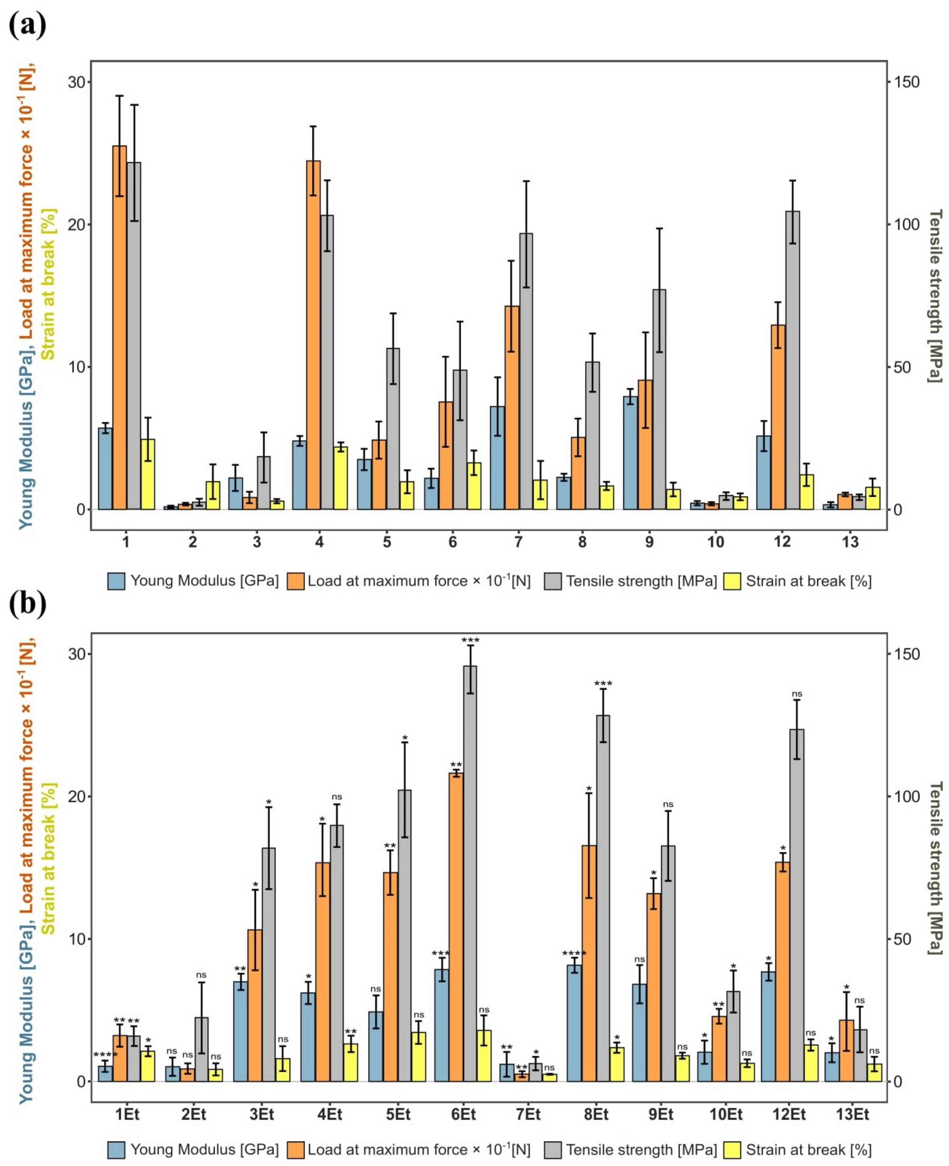
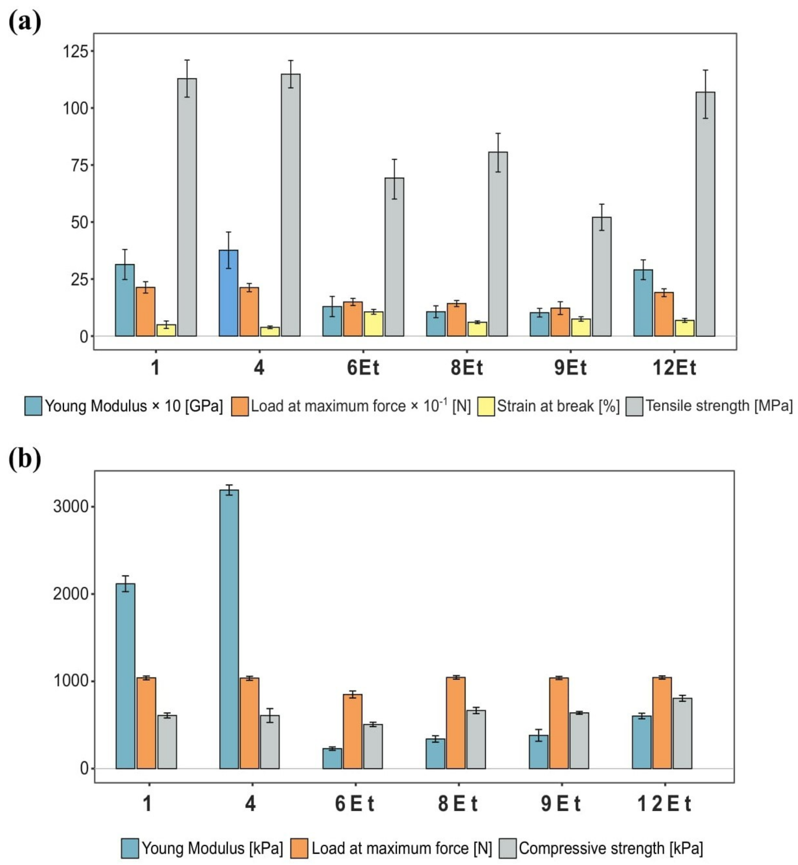
2.1.2. Mechanical Properties
2.2. The Properties of BNC Synthesized by Chosen Strains
2.2.1. Physical and Physico-Chemical Parameters
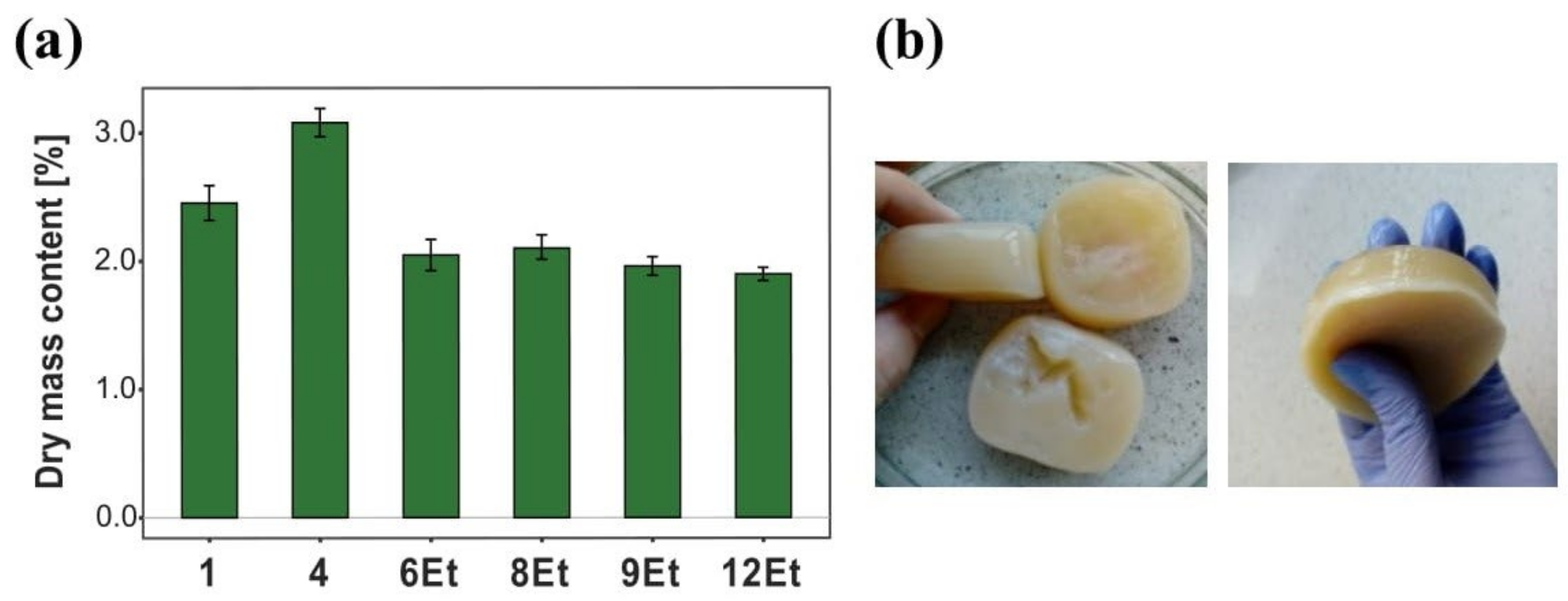
Dry Mass Content
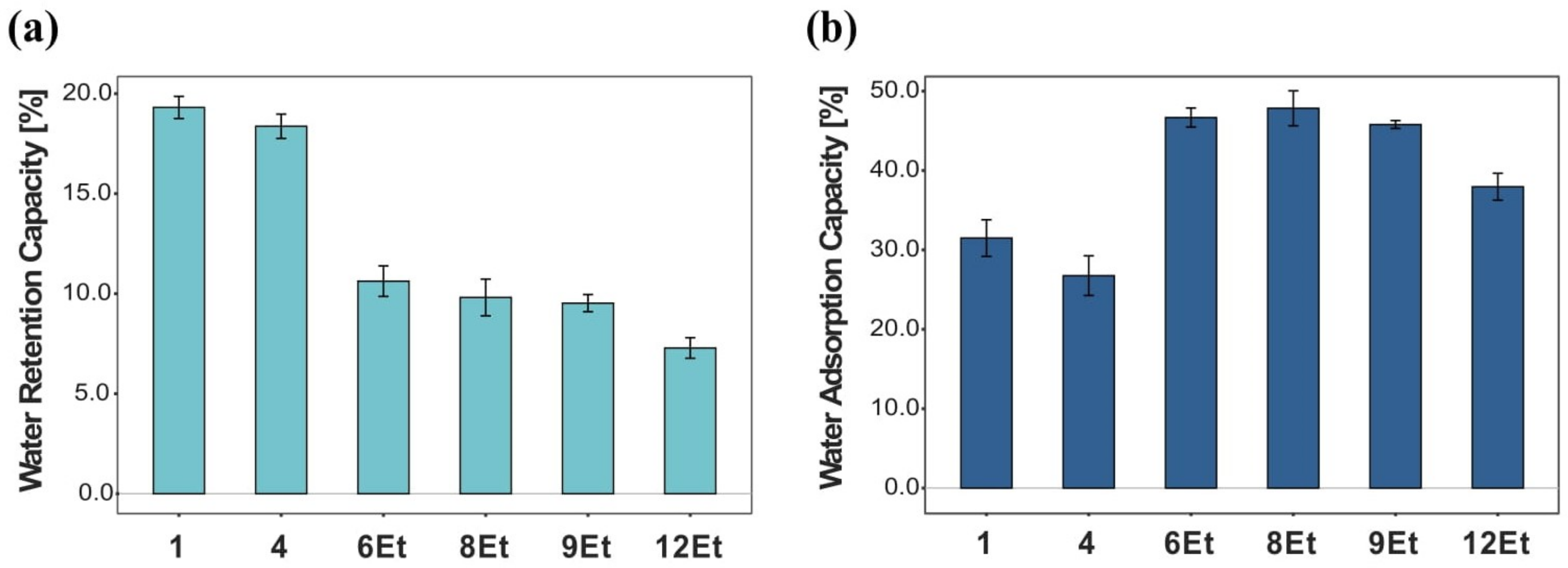
Water Retention and Adsorption Capacity
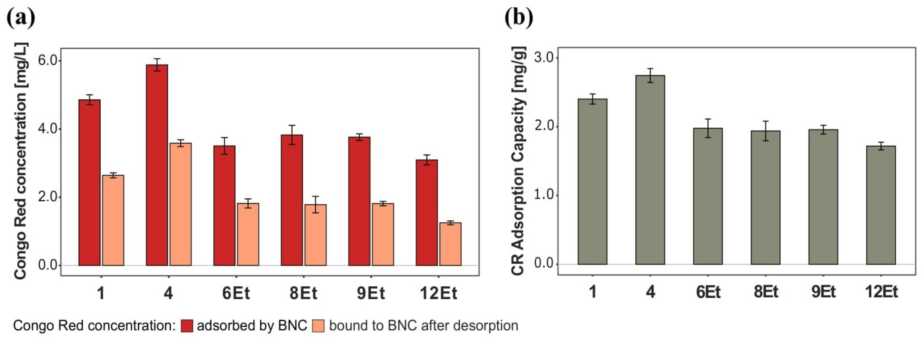
Dye Adsorption
2.2.2. Mechanical Properties
2.3. Composites of BNC with Selected Additives
2.3.1. Physical and Physico-Chemical Parameters
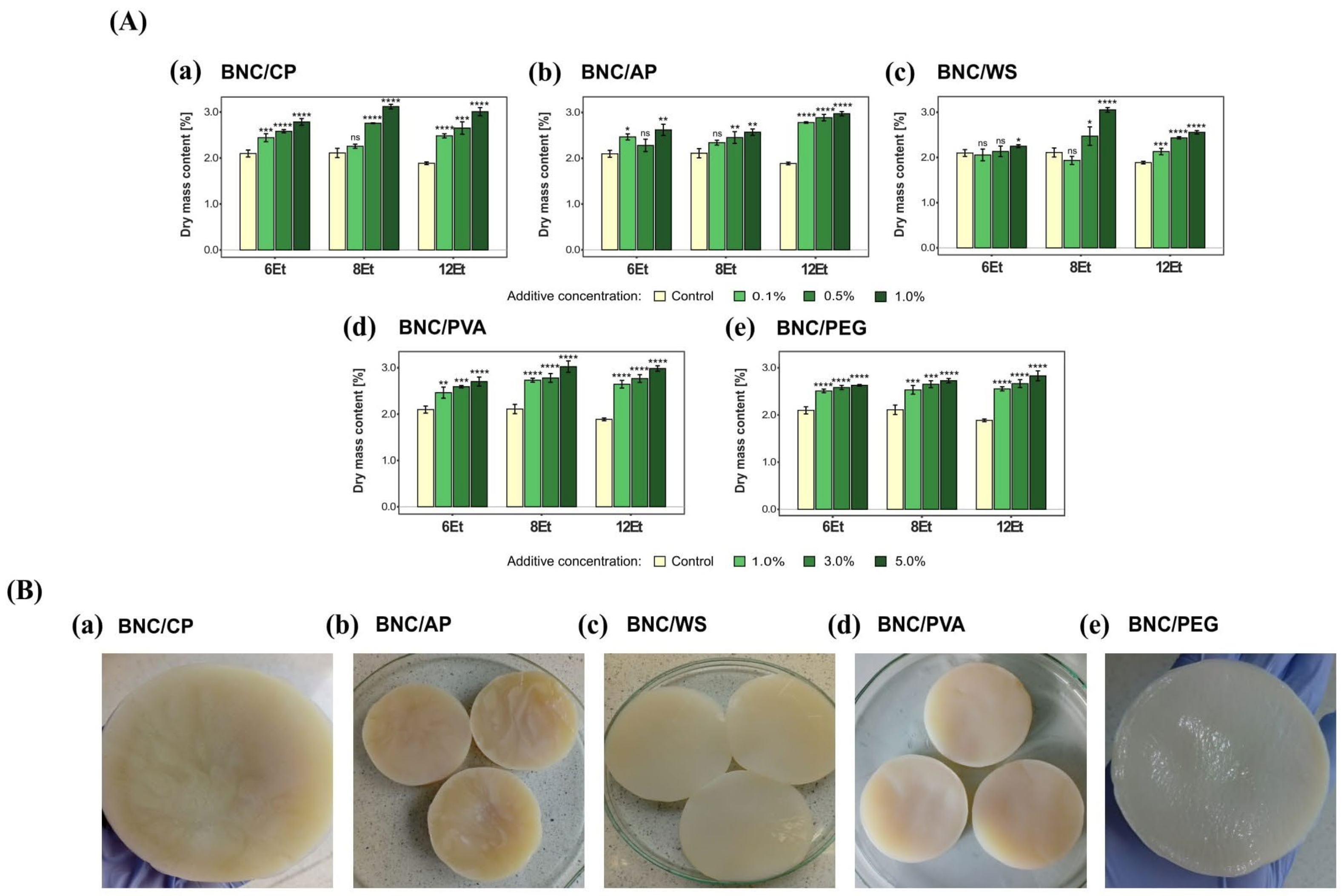
Dry Mass Content
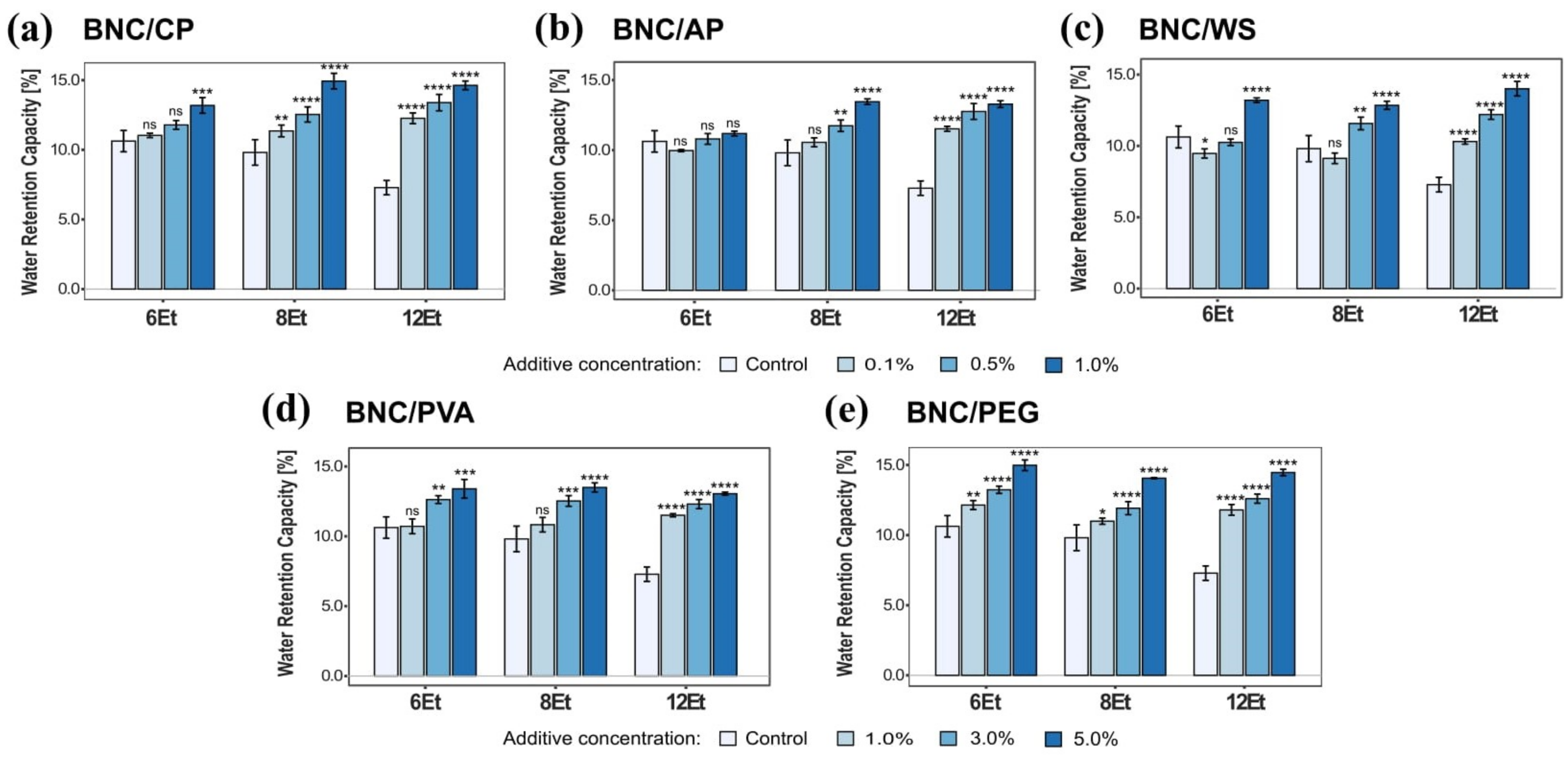
Water Retention Capacity (WRC)
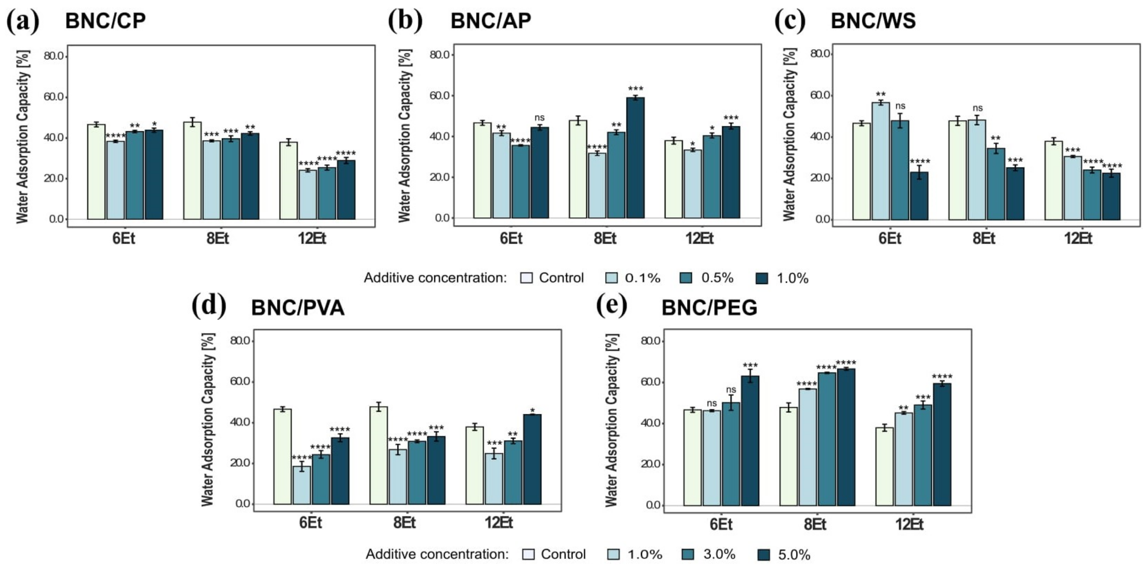
Water Adsorption Capacity (WAC)
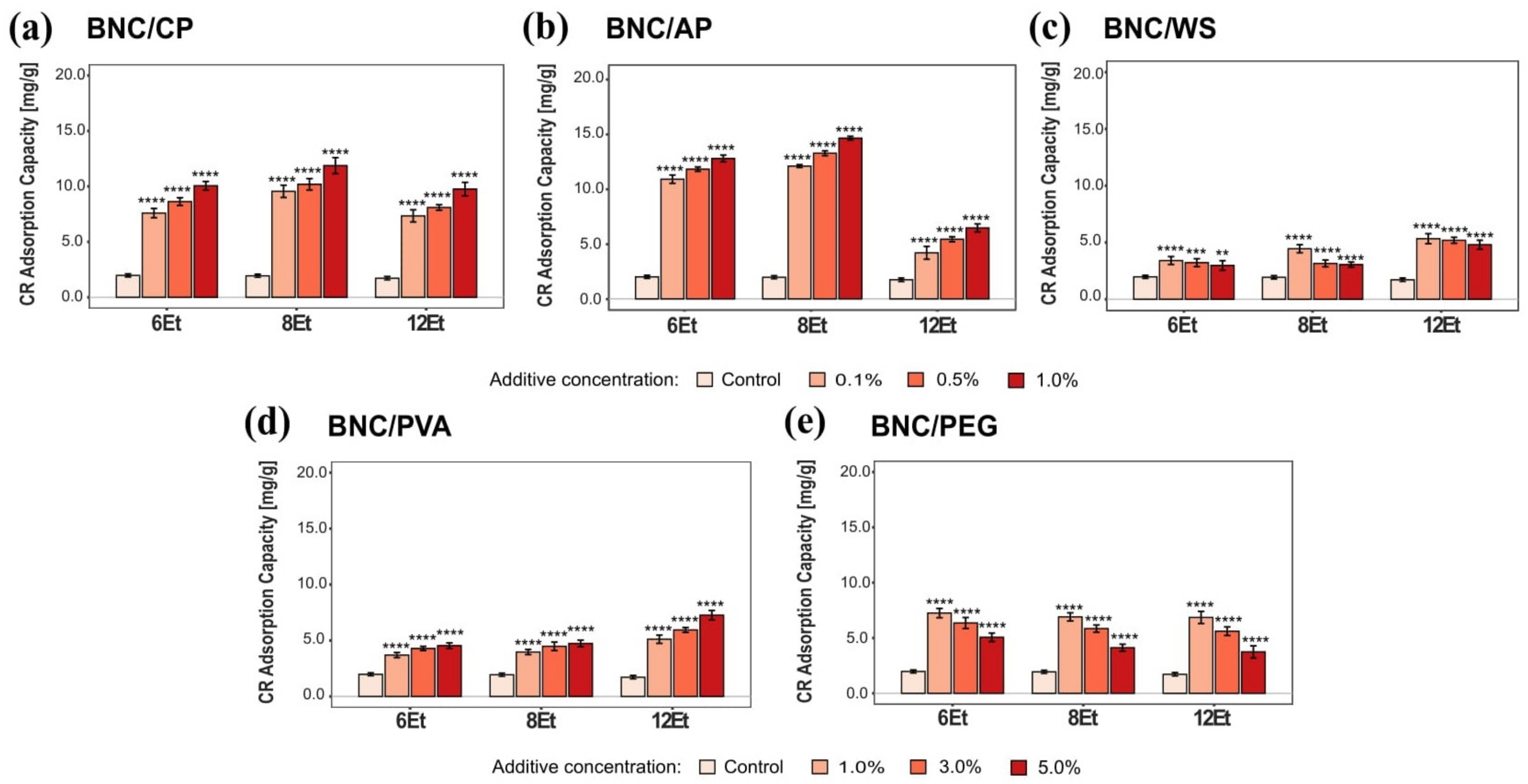
Dye Adsorption Capacity (CRC)
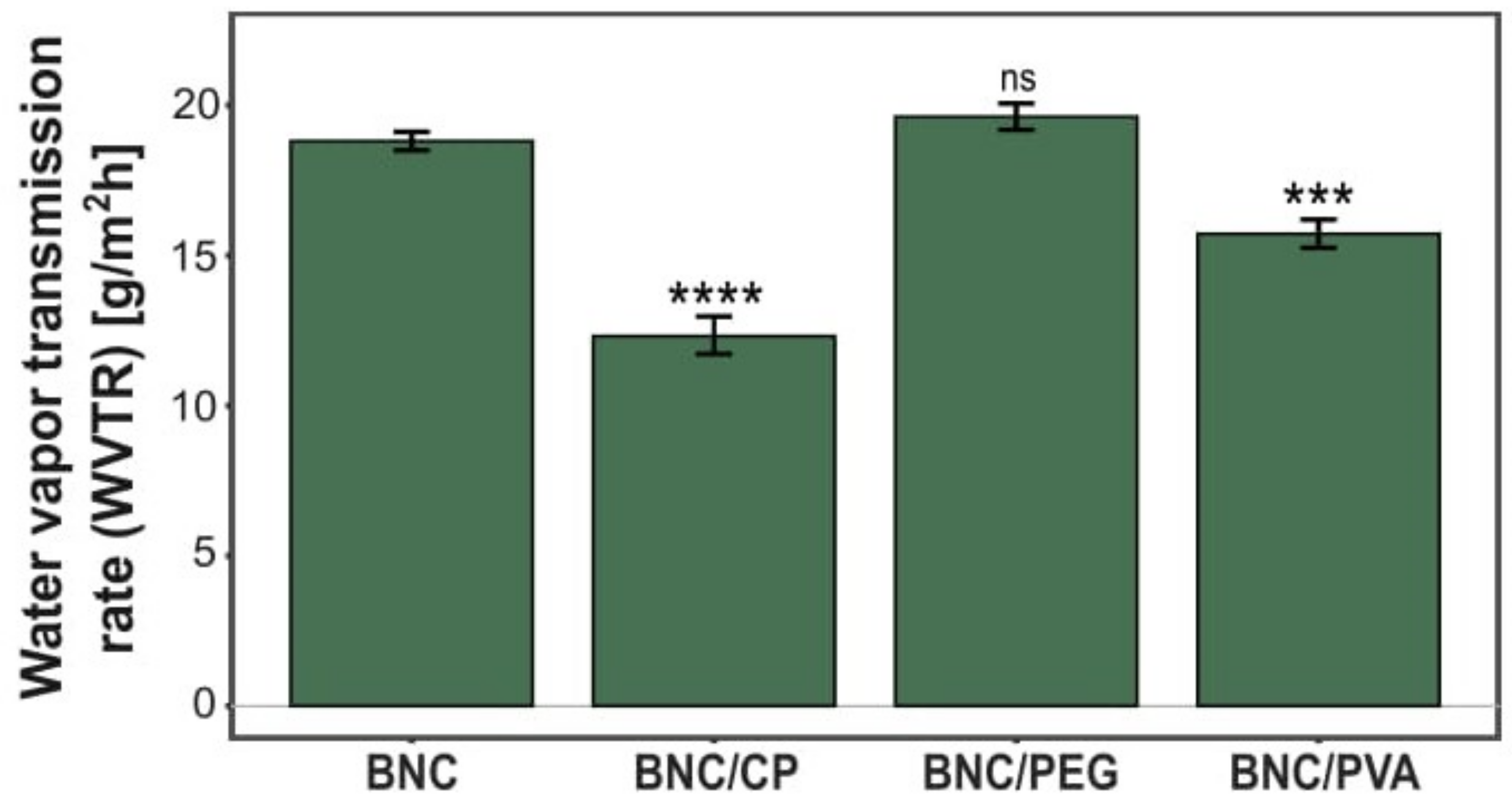
Water Vapor Permeability
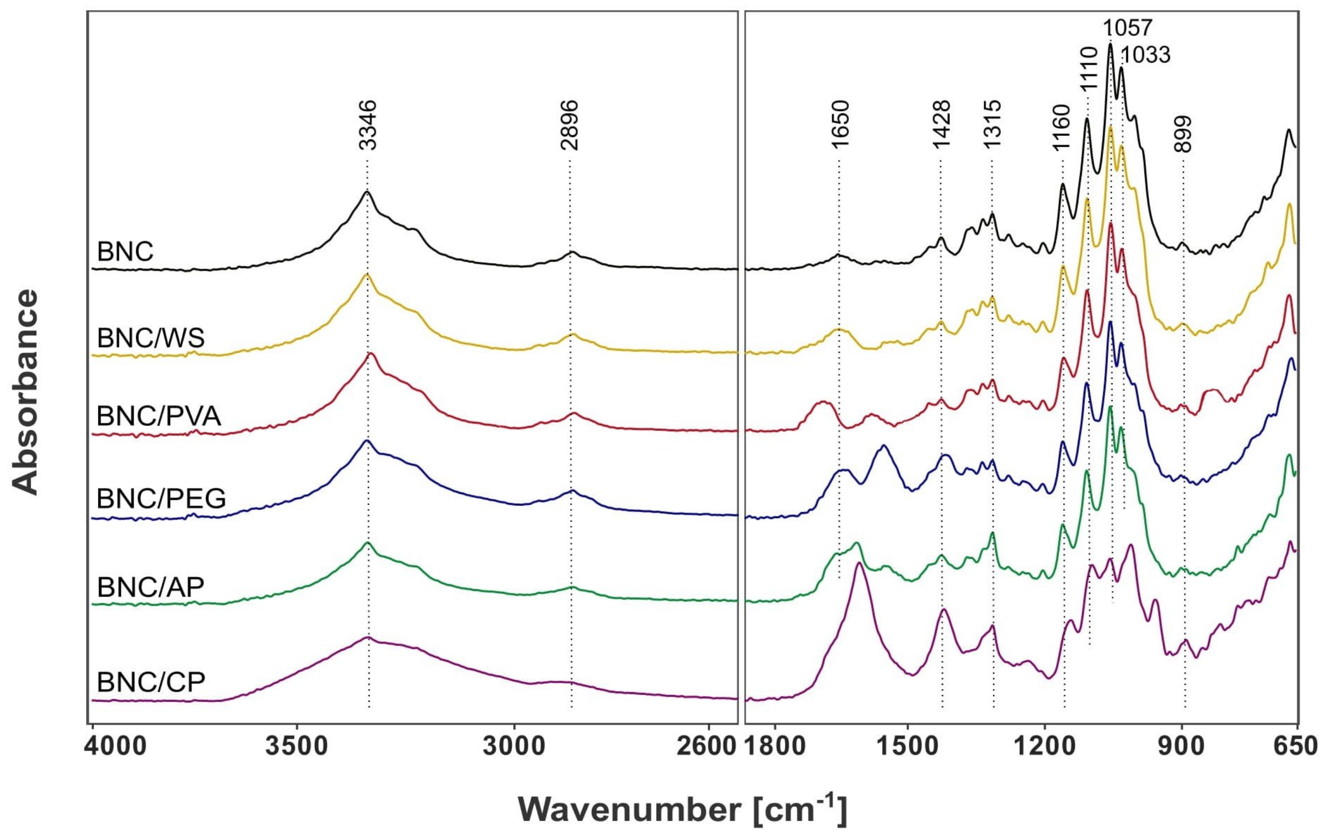
Fourier Transform Infrared Spectroscopy (ATR-FTIR)
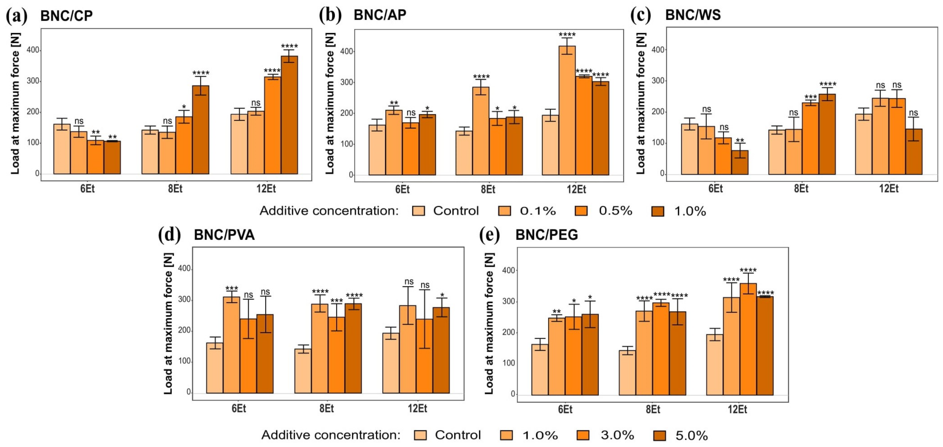
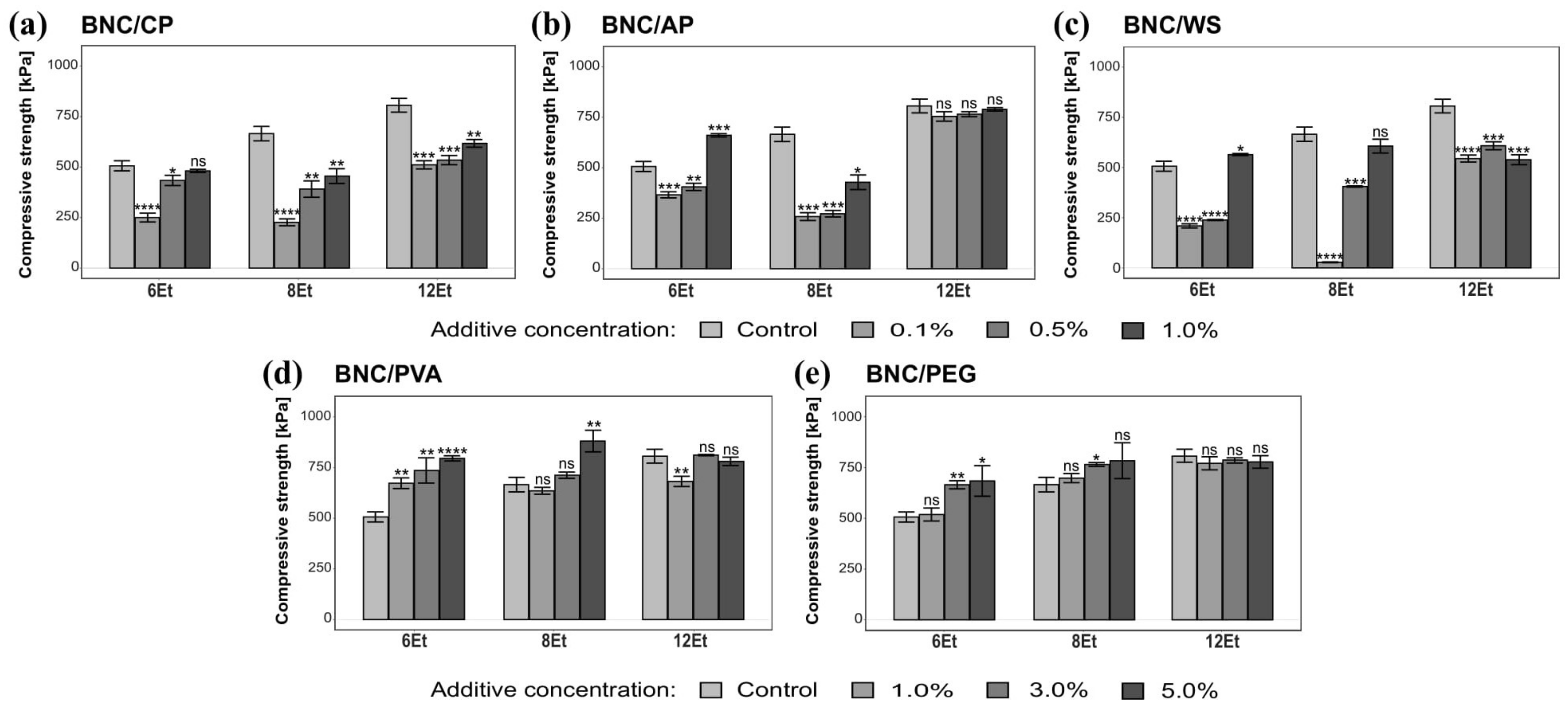
2.3.2. Mechanical Properties
2.3.3. Bacteriostatic Properties
3. Discussion
4. Materials and Methods
4.1. Komagataeibacter Strains and Cultivation Methods
4.2. Composites Preparation Methods
4.3. Physical Parameters Evaluation of BNC Membranes and Their Composites
4.3.1. Yield of Biosynthesis Process
4.3.2. Dry Mass Content
4.4. Physico-Chemical Parameter-Based Evaluation of BNC Membranes and Their Composites
4.4.1. Water Retention Capacity (WRC)
4.4.2. Water Adsorption Capacity (WAC)
4.4.3. Water Vapor Permeability
4.4.4. Congo Red Adsorption Capacity
4.4.5. Fourier Transform Infrared Spectroscopy (FTIR)
4.5. Mechanical Properties Testing
4.5.1. Breaking Strength Measurements
4.5.2. Compression Tests
4.6. Bacteriostatic Properties Analysis
4.7. Statistical Analysis
5. Conclusions
Supplementary Materials
Author Contributions
Funding
Institutional Review Board Statement
Informed Consent Statement
Data Availability Statement
Acknowledgments
Conflicts of Interest
References
- Chen, C.; Ding, W.; Zhang, H.; Zhang, L.; Huang, Y.; Fan, M.; Yang, J.; Sun, D. Bacterial Cellulose-Based Biomaterials: From Fabrication to Application. Carbohydr. Polym. 2022, 278, 118995. [Google Scholar] [CrossRef] [PubMed]
- Gorgieva, S.; Trček, J. Bacterial Cellulose: Production, Modification and Perspectives in Biomedical Applications. Nanomaterials 2019, 9, 1352. [Google Scholar] [CrossRef] [PubMed] [Green Version]
- Ludwicka, K.; Kaczmarek, M.; Białkowska, A. Bacterial Nanocellulose—A Biobased Polymer for Active and Intelligent Food Packaging Applications: Recent Advances and Developments. Polymers 2020, 12, 2209. [Google Scholar] [CrossRef] [PubMed]
- Azeredo, H.M.C.; Barud, H.; Farinas, C.S.; Vasconcellos, V.M.; Claro, A.M. Bacterial Cellulose as a Raw Material for Food and Food Packaging Applications. Front. Sustain. Food Syst. 2019, 3, 7. [Google Scholar] [CrossRef] [Green Version]
- Choi, S.M.; Shin, E.J. The Nanofication and Functionalization of Bacterial Cellulose and Its Applications. Nanomaterials 2020, 10, 406. [Google Scholar] [CrossRef] [Green Version]
- Swingler, S.; Gupta, A.; Gibson, H.; Kowalczuk, M.; Heaselgrave, W.; Radecka, I. Recent Advances and Applications of Bacterial Cellulose in Biomedicine. Polymers 2021, 13, 412. [Google Scholar] [CrossRef]
- Szustak, M.; Gendaszewska-Darmach, E. Nanocellulose-Based Scaffolds for Chondrogenic Differentiation and Expansion. Front. Bioeng. Biotechnol. 2021, 9, 733. [Google Scholar] [CrossRef]
- Jankau, J.; Błażyńska-Spychalska, A.; Kubiak, K.; Jędrzejczak-Krzepkowska, M.; Pankiewicz, T.; Ludwicka, K.; Dettlaff, A.; Pęksa, R. Bacterial Cellulose Properties Fulfilling Requirements for a Biomaterial of Choice in Reconstructive Surgery and Wound Healing. Front. Bioeng. Biotechnol. 2022, 9, 805053. [Google Scholar] [CrossRef]
- Ossowicz-Rupniewska, P.; Rakoczy, R.; Nowak, A.; Konopacki, M.; Klebeko, J.; Świątek, E.; Janus, E.; Duchnik, W.; Wenelska, K.; Kucharski, Ł.; et al. Transdermal Delivery Systems for Ibuprofen and Ibuprofen Modified with Amino Acids Alkyl Esters Based on Bacterial Cellulose. Int. J. Mol. Sci. 2021, 22, 6252. [Google Scholar] [CrossRef]
- Troncoso, O.P.; Torres, F.G. Bacterial Cellulose—Graphene Based Nanocomposites. Int. J. Mol. Sci. 2020, 21, 6532. [Google Scholar] [CrossRef]
- Campano, C.; Balea, A.; Blanco, A.; Negro, C. Enhancement of the Fermentation Process and Properties of Bacterial Cellulose: A Review. Cellulose 2015, 23, 57–91. [Google Scholar] [CrossRef]
- Ryngajłło, M.; Kubiak, K.; Jędrzejczak-Krzepkowska, M.; Jacek, P.; Bielecki, S. Comparative Genomics of the Komagataeibacter Strains—Efficient Bionanocellulose Producers. Microbiologyopen 2019, 8, e00731. [Google Scholar] [CrossRef] [PubMed] [Green Version]
- Cielecka, I.; Ryngajłło, M.; Maniukiewicz, W.; Bielecki, S. Response Surface Methodology-Based Improvement of the Yield and Differentiation of Properties of Bacterial Cellulose by Metabolic Enhancers. Int. J. Biol. Macromol. 2021, 187, 584–593. [Google Scholar] [CrossRef]
- Cielecka, I.; Ryngajłło, M.; Maniukiewicz, W.; Bielecki, S. Highly Stretchable Bacterial Cellulose Produced by Komagataeibacter Hansenii SI1. Polymers 2021, 13, 4455. [Google Scholar] [CrossRef] [PubMed]
- Ryngajłło, M.; Jacek, P.; Cielecka, I.; Kalinowska, H.; Bielecki, S. Effect of Ethanol Supplementation on the Transcriptional Landscape of Bionanocellulose Producer Komagataeibacter Xylinus E25. Appl. Microbiol. Biotechnol. 2019, 103, 6673–6688. [Google Scholar] [CrossRef] [Green Version]
- Jaroennonthasit, W.; Lam, N.T.; Sukyai, P. Evaluation of Carbon Sources from Sugar Industry to Bacterial Nanocellulose Produced by Komagataeibacter Xylinus. Int. J. Biol. Macromol. 2021, 191, 299–304. [Google Scholar] [CrossRef]
- Krystynowicz, A.; Czaja, W.; Wiktorowska-Jezierska, A.; Gonçalves-Miśkiewicz, M.; Turkiewicz, M.; Bielecki, S. Factors Affecting the Yield and Properties of Bacterial Cellulose. J. Ind. Microbiol. Biotechnol. 2002, 29, 189–195. [Google Scholar] [CrossRef]
- Naritomi, T.; Kouda, T.; Yano, H.; Yoshinaga, F. Effect of Ethanol on Bacterial Cellulose Production from Fructose in Continuous Culture. J. Ferment. Bioeng. 1998, 85, 598–603. [Google Scholar] [CrossRef]
- Nascimento, F.X.; Torres, C.A.V.; Freitas, F.; Reis, M.A.M.; Crespo, M.T.B. Functional and Genomic Characterization of Komagataeibacter Uvaceti FXV3, a Multiple Stress Resistant Bacterium Producing Increased Levels of Cellulose. Biotechnol. Rep. 2021, 30, e00606. [Google Scholar] [CrossRef]
- Chen, S.Q.; Mikkelsen, D.; Lopez-Sanchez, P.; Wang, D.; Martinez-Sanz, M.; Gilbert, E.P.; Flanagan, B.M.; Gidley, M.J. Characterisation of Bacterial Cellulose from Diverse Komagataeibacter Strains and Their Application to Construct Plant Cell Wall Analogues. Cellulose 2017, 24, 1211–1226. [Google Scholar] [CrossRef]
- Vigentini, I.; Fabrizio, V.; Dellacà, F.; Rossi, S.; Azario, I.; Mondin, C.; Benaglia, M.; Foschino, R. Set-Up of Bacterial Cellulose Production From the Genus Komagataeibacter and Its Use in a Gluten-Free Bakery Product as a Case Study. Front. Microbiol. 2019, 10, 1953. [Google Scholar] [CrossRef] [PubMed]
- Blanco, F.G.; Hernández, N.; Rivero-Buceta, V.; Maestro, B.; Sanz, J.M.; Mato, A.; Hernández-Arriaga, A.M.; Prieto, M.A. From Residues to Added-Value Bacterial Biopolymers as Nanomaterials for Biomedical Applications. Nanomaterials 2021, 11, 1492. [Google Scholar] [CrossRef] [PubMed]
- Gullo, M.; La China, S.; Falcone, P.M.; Giudici, P. Biotechnological Production of Cellulose by Acetic Acid Bacteria: Current State and Perspectives. Appl. Microbiol. Biotechnol. 2018, 102, 6885–6898. [Google Scholar] [CrossRef]
- Jacek, P.; da Silva, F.A.G.S.; Dourado, F.; Bielecki, S.; Gama, M. Optimization and Characterization of Bacterial Nanocellulose Produced by Komagataeibacter Rhaeticus K3. Carbohydr. Polym. Technol. Appl. 2021, 2, 100022. [Google Scholar] [CrossRef]
- Portela, R.; Leal, C.R.; Almeida, P.L.; Sobral, R.G. Bacterial Cellulose: A Versatile Biopolymer for Wound Dressing Applications. Microb. Biotechnol. 2019, 12, 586–610. [Google Scholar] [CrossRef]
- Andree, V.; Niopek, D.; Müller, C.; Eiselt, J.P.; Foh, N.; Rzany, A.; Hensel, B. Influence of Drying Methods on the Physical Properties of Bacterial Nanocellulose. Mater. Res. Express 2021, 8, 025402. [Google Scholar] [CrossRef]
- Ul-Islam, M.; Khan, T.; Park, J.K. Water Holding and Release Properties of Bacterial Cellulose Obtained by in Situ and Ex Situ Modification. Carbohydr. Polym. 2012, 88, 596–603. [Google Scholar] [CrossRef]
- Gelin, K.; Bodin, A.; Gatenholm, P.; Mihranyan, A.; Edwards, K.; Strømme, M. Characterization of Water in Bacterial Cellulose Using Dielectric Spectroscopy and Electron Microscopy. Polymer 2007, 48, 7623–7631. [Google Scholar] [CrossRef]
- Fijałkowski, K.; Zywicka, A.; Drozd, R.; Kordas, M.; Rakoczy, R. Effect of Gluconacetobacter Xylinus Cultivation Conditions on the Selected Properties of Bacterial Cellulose. Polish J. Chem. Technol. 2016, 18, 117–123. [Google Scholar] [CrossRef] [Green Version]
- Yakupova, E.I.; Bobyleva, L.G.; Vikhlyantsev, I.M.; Bobylev, A.G. Congo Red and Amyloids: History and Relationship. Biosci. Rep. 2019, 39, BSR20181415. [Google Scholar] [CrossRef] [Green Version]
- Mazeau, K.; Wyszomirski, M. Modelling of Congo Red Adsorption on the Hydrophobic Surface of Cellulose Using Molecular Dynamics. Cellulose 2012, 19, 1495–1506. [Google Scholar] [CrossRef] [Green Version]
- Yamaki, S.B.; Barros, D.S.; Garcia, C.M.; Socoloski, P.; Oliveira, O.N.; Atvars, T.D.Z. Spectroscopic Studies of the Intermolecular Interactions of Congo Red and Tinopal CBS with Modified Cellulose Fibers. Langmuir 2005, 21, 5414–5420. [Google Scholar] [CrossRef] [PubMed]
- ASTM E96: Standard Test Methods for Water Vapor Transmission of Materials. Annu. Books ASTM Stand. 1995, 552, 785–792.
- ISO 2528:2017; Sheet Materials—Determination of Water Vapour Transmission Rate (WVTR)—Gravimetric (Dish) Method. International Organization for Standardization: Geneva, Switzerland, 2017; pp. 1–15.
- Xu, Y.; Liu, X.; Jiang, Q.; Yu, D.; Xu, Y.; Wang, B.; Xia, W. Development and Properties of Bacterial Cellulose, Curcumin, and Chitosan Composite Biodegradable Films for Active Packaging Materials. Carbohydr. Polym. 2021, 260, 117778. [Google Scholar] [CrossRef]
- Tipachan, C.; Gupta, R.K.; Kajorncheappunngam, S. Water Vapor Barrier Property of PLA Nanocomposites Using Rice Husk Ash and Layered Double Hydroxides as Fillers. Eng. Appl. Sci. Res. 2019, 46, 285–291. [Google Scholar]
- Sun, Y.; Meng, C.; Zheng, Y.; Xie, Y.; He, W.; Wang, Y.; Qiao, K.; Yue, L. The Effects of Two Biocompatible Plasticizers on the Performance of Dry Bacterial Cellulose Membrane: A Comparative Study. Cellulose 2018, 25, 5893–5908. [Google Scholar] [CrossRef]
- Cichosz, S.; Masek, A. IR Study on Cellulose with the Varied Moisture Contents: Insight into the Supramolecular Structure. Materials 2020, 13, 4573. [Google Scholar] [CrossRef]
- Jacek, P.; Kubiak, K.; Ryngajłło, M.; Rytczak, P.; Paluch, P.; Bielecki, S. Modification of Bacterial Nanocellulose Properties through Mutation of Motility Related Genes in Komagataeibacter Hansenii ATCC 53582. New Biotechnol. 2019, 52, 60–68. [Google Scholar] [CrossRef]
- Hospodarova, V.; Singovszka, E.; Stevulova, N. Characterization of Cellulosic Fibers by FTIR Spectroscopy for Their Further Implementation to Building Materials. Am. J. Anal. Chem. 2018, 9, 303–310. [Google Scholar] [CrossRef] [Green Version]
- Wang, S.S.; Han, Y.H.; Ye, Y.X.; Shi, X.X.; Xiang, P.; Chen, D.L.; Li, M. Physicochemical Characterization of High-Quality Bacterial Cellulose Produced by Komagataeibacter Sp. Strain W1 and Identification of the Associated Genes in Bacterial Cellulose Production. RSC Adv. 2017, 7, 45145–45155. [Google Scholar] [CrossRef] [Green Version]
- Agustin, S.; Wahyuni, E.T.; Suparmo, S.; Supriyadi, S.; Cahyanto, M.N. Incorporation of Pectin during Biosynthesis of Bacterial Cellulose by Gluconacetobacter Xylinus InaCC B404: Possibility for Producing Green Food Packaging. Biodiversitas J. Biol. Divers. 2021, 22, 2548–2553. [Google Scholar] [CrossRef]
- Szymanska-Chargot, M.; Zdunek, A. Use of FT-IR Spectra and PCA to the Bulk Characterization of Cell Wall Residues of Fruits and Vegetables Along a Fraction Process. Food Biophys. 2013, 8, 29–42. [Google Scholar] [CrossRef] [Green Version]
- Długa, A.; Kowalonek, J.; Kaczmarek, H. Bionanocellulose/Poly(Vinyl Alcohol) Composites Produced by In-Situ Method and Ex-Situ/Impregnation or Sterilization Methods. Materials 2021, 14, 6340. [Google Scholar] [CrossRef] [PubMed]
- Characterisation of Composites of Bacterial Cellulose and Poly(Vinyl Alcohol) Obtained by Different Methods. Fibres Text. East. Eur. 2014, 22, 69–74.
- Phruksaphithak, N.; Kaewnun, C.; O-Thong, S. Characterization of Bacterial Cellulose from Oil Palm Shoot Juices and Coconut Juice/Poly(Ethylene Glycol) Biocomposite. J. Renew. Mater. 2019, 7, 493–504. [Google Scholar] [CrossRef] [Green Version]
- Cai, Z.; Kim, J. Bacterial Cellulose/Poly(Ethylene Glycol) Composite: Characterization and First Evaluation of Biocompatibility. Cellulose 2010, 17, 83–91. [Google Scholar] [CrossRef]
- Yin, N.; Chen, S.Y.; Cao, Y.M.; Wang, H.P.; Wu, Q.K. Improvement in Mechanical Properties and Biocompatibility of Biosynthetic Bacterial Cellulose/Lotus Root Starch Composites. Chin. J. Polym. Sci. 2017, 35, 354–364. [Google Scholar] [CrossRef]
- Aydın, Y.A.; Aksoy, N.D. Isolation and Characterization of an Efficient Bacterial Cellulose Producer Strain in Agitated Culture: Gluconacetobacter Hansenii P2A. Appl. Microbiol. Biotechnol. 2014, 98, 1065–1075. [Google Scholar] [CrossRef]
- Volova, T.G.; Prudnikova, S.V.; Kiselev, E.G.; Nemtsev, I.V.; Vasiliev, A.D.; Kuzmin, A.P.; Shishatskaya, E.I. Bacterial Cellulose (BC) and BC Composites: Production and Properties. Nanomaterials 2022, 12, 192. [Google Scholar] [CrossRef]
- Chen, S.Q.; Lopez-Sanchez, P.; Wang, D.; Mikkelsen, D.; Gidley, M.J. Mechanical Properties of Bacterial Cellulose Synthesised by Diverse Strains of the Genus Komagataeibacter. Food Hydrocoll. 2018, 81, 87–95. [Google Scholar] [CrossRef] [Green Version]
- Abol-Fotouh, D.; Hassan, M.A.; Shokry, H.; Roig, A.; Azab, M.S.; Kashyout, A.E.H.B. Bacterial Nanocellulose from Agro-Industrial Wastes: Low-Cost and Enhanced Production by Komagataeibacter Saccharivorans MD1. Sci. Rep. 2020, 10, 3491. [Google Scholar] [CrossRef] [Green Version]
- Ferreira, F.V.; Otoni, C.G.; De France, K.J.; Barud, H.S.; Lona, L.M.F.; Cranston, E.D.; Rojas, O.J. Porous Nanocellulose Gels and Foams: Breakthrough Status in the Development of Scaffolds for Tissue Engineering. Mater. Today 2020, 37, 126–141. [Google Scholar] [CrossRef]
- Kumar, A.; Han, S.S. Efficacy of Bacterial Nanocellulose in Hard Tissue Regeneration: A Review. Materials 2021, 14, 4777. [Google Scholar] [CrossRef]
- Mautner, A.; Bismarck, A. Bacterial Nanocellulose Papers with High Porosity for Optimized Permeance and Rejection of Nm-Sized Pollutants. Carbohydr. Polym. 2021, 251, 117130. [Google Scholar] [CrossRef] [PubMed]
- Maleki, H. Recent Advances in Aerogels for Environmental Remediation Applications: A Review. Chem. Eng. J. 2016, 300, 98–118. [Google Scholar] [CrossRef]
- Guan, F.; Guo, C.F. Flexible, High-Strength, and Porous Nano-Nano Composites Based on Bacterial Cellulose for Wearable Electronics: A Review. Soft Sci. 2021, 1, 16. [Google Scholar] [CrossRef]
- Sämfors, S.; Karlsson, K.; Sundberg, J.; Markstedt, K.; Gatenholm, P. Biofabrication of Bacterial Nanocellulose Scaffolds with Complex Vascular Structure. Biofabrication 2019, 11, 045010. [Google Scholar] [CrossRef]
- Murizan, N.I.S.; Mustafa, N.S.; Ngadiman, N.H.A.; Yusof, N.M.; Idris, A. Review on Nanocrystalline Cellulose in Bone Tissue Engineering Applications. Polymers 2020, 12, 2818. [Google Scholar] [CrossRef]
- Zhong, C. Industrial-Scale Production and Applications of Bacterial Cellulose. Front. Bioeng. Biotechnol. 2020, 8, 605374. [Google Scholar] [CrossRef]
- Szymańska-Chargot, M.; Chylińska, M.; Cybulska, J.; Kozioł, A.; Pieczywek, P.M.; Zdunek, A. Simultaneous Influence of Pectin and Xyloglucan on Structure and Mechanical Properties of Bacterial Cellulose Composites. Carbohydr. Polym. 2017, 174, 970–979. [Google Scholar] [CrossRef]
- Xue, G.; Ren, D.; Zhou, C.; Zheng, H.; Cao, W.; Lin, H.; Qin, X.; Zhang, C. Comparative Study on the Functional Properties of the Pearl Oyster (Pinctada Martensii) Protein Isolates and Its Electrostatic Complexes with Three Hydrophilic Polysaccharides. Int. J. Food Prop. 2020, 23, 1256–1271. [Google Scholar] [CrossRef]
- Tan, M.; Zheng, S.; Lv, H.; Wang, B.; Zhao, Q.; Zhao, B. Rational Design and Synthesis of Chitosan–Quinoa Polysaccharide Composite Aerogel and Its Adsorption Properties for Congo Red and Methylene Blue. New J. Chem. 2021, 45, 9829–9837. [Google Scholar] [CrossRef]
- Shak, K.P.Y.; Pang, Y.L.; Mah, S.K. Nanocellulose: Recent Advances and Its Prospects in Environmental Remediation. Beilstein J. Nanotechnol. 2018, 9, 2479–2498. [Google Scholar] [CrossRef] [PubMed]
- Almeida, T.; Silvestre, A.J.D.; Vilela, C.; Freire, C.S.R. Bacterial Nanocellulose toward Green Cosmetics: Recent Progresses and Challenges. Int. J. Mol. Sci. 2021, 22, 2836. [Google Scholar] [CrossRef] [PubMed]
- Da Silva, R.; Sierakowski, M.R.; Bassani, H.P.; Zawadzki, S.F.; Pirich, C.L.; Ono, L.; de Freitas, R.A. Hydrophilicity Improvement of Mercerized Bacterial Cellulose Films by Polyethylene Glycol Graft. Int. J. Biol. Macromol. 2016, 86, 599–605. [Google Scholar] [CrossRef]
- Xu, Z.; Li, J.; Zhou, H.; Jiang, X.; Yang, C.; Wang, F.; Pan, Y.; Li, N.; Li, X.; Shi, L.; et al. Morphological and Swelling Behavior of Cellulose Nanofiber (CNF)/Poly(Vinyl Alcohol) (PVA) Hydrogels: Poly(Ethylene Glycol) (PEG) as Porogen. RSC Adv. 2016, 6, 43626–43633. [Google Scholar] [CrossRef]
- Cielecka, I.; Szustak, M.; Kalinowska, H.; Gendaszewska-Darmach, E.; Ryngajłło, M.; Maniukiewicz, W.; Bielecki, S. Glycerol-Plasticized Bacterial Nanocellulose-Based Composites with Enhanced Flexibility and Liquid Sorption Capacity. Cellulose 2019, 26, 5409–5426. [Google Scholar] [CrossRef] [Green Version]
- Gama, M.; Dourado, F.; Bielecki, S. Bacterial Nanocellulose: From Biotechnology to Bio-Economy, 1st ed.; Elsevier Inc.: Amsterdam, The Netherlands, 2016; ISBN 9780444634665. [Google Scholar]
- Lin, D.; Liu, Z.; Shen, R.; Chen, S.; Yang, X. Bacterial Cellulose in Food Industry: Current Research and Future Prospects. Int. J. Biol. Macromol. 2020, 158, 1007–1019. [Google Scholar] [CrossRef]
- Popa, L.; Ghica, M.V.; Tudoroiu, E.-E.; Ionescu, D.-G.; Dinu-Pîrvu, C.-E. Bacterial Cellulose—A Remarkable Polymer as a Source for Biomaterials Tailoring. Materials 2022, 15, 1054. [Google Scholar] [CrossRef]
- Jacek, P.; Ryngajłło, M.; Bielecki, S. Structural Changes of Bacterial Nanocellulose Pellicles Induced by Genetic Modification of Komagataeibacter Hansenii ATCC 23769. Appl. Microbiol. Biotechnol. 2019, 103, 5339–5353. [Google Scholar] [CrossRef] [Green Version]
- Ludwicka, K.; Kolodziejczyk, M.; Gendaszewska-Darmach, E.; Chrzanowski, M.; Jedrzejczak-Krzepkowska, M.; Rytczak, P.; Bielecki, S. Stable Composite of Bacterial Nanocellulose and Perforated Polypropylene Mesh for Biomedical Applications. J. Biomed. Mater. Res. B Appl. Biomater. 2019, 107, 978–987. [Google Scholar] [CrossRef] [PubMed]
- Ciechańska, D. Multifunctional Bacterial Cellulose/Chitosan Composite Materials for Medical Applications. Fibres Text East Eur. 2004, 12, 69–72. [Google Scholar]
- Lopez-Sanchez, P.; Martinez-Sanz, M.; Bonilla, M.R.; Wang, D.; Gilbert, E.P.; Stokes, J.R.; Gidley, M.J. Cellulose-Pectin Composite Hydrogels: Intermolecular Interactions and Material Properties Depend on Order of Assembly. Carbohydr. Polym. 2017, 162, 71–81. [Google Scholar] [CrossRef] [PubMed]
- Tomer, G.; Patel, H.; Podczeck, F.; Newton, J.M. Measuring the Water Retention Capacities (MRC) of Different Microcrystalline Cellulose Grades. Eur. J. Pharm. Sci. 2001, 12, 321–325. [Google Scholar] [CrossRef]


| Strain Number | Systematic Name | Description |
|---|---|---|
| 1 | K. hansenii ATCC 53582 | ATCC collection [39] |
| 2 | K. hansenii ATCC 23769 | ATCC collection [72] |
| 3 | K. xylinus E25 | Strain belonging to Bowil Biotech Ltd. [73] |
| 4 | K. xylinus E26 | In-house collection of IMIB [12] |
| 5 | K. xylinus BCRC 12334 | Received from Jyh Ming Wu, Department of Chemical and Materials Engineering, Chinese Culture University, Taipei, Taiwan [12] |
| 6 | K. hansenii H3 | Received from Jose Fontana, Universidade Federal do Paraná (UFPR), Curitiba, Brazil |
| 7 | K. hansenii H4 | Received from Jose Fontana, Universidade Federal do Paraná (UFPR), Curitiba, Brazil |
| 8 | K. rhaeticus K4 | Isolated from Kombucha at IMIB |
| 9 | K. rhaeticus K3 | Isolated from Kombucha at IMIB [24] |
| 10 | K. hansenii H5 | Isolated from vinegar at IMIB |
| 11 | A. xylinum ŁOCK | Collection of Pure Cultures of the Institute of Microbiology and Fermentation, Lodz University of Technology [74] |
| 12 | Komagataeibacter sp. | Isolated from balsamic vinegar at IMIB |
| 13 | K. hansenii SI1 | Isolated from Kombucha at IMIB [14] |
| No. | Type of Additive | Concentration [% w/v] |
|---|---|---|
| 1. | Citrus pectin (CP) + 12.5 mM CaCl2 | 0.1 0.5 1.0 |
| 2. | Apple pectin (AP) + 12.5 mM CaCl2 | 0.1 0.5 1.0 |
| 3. | Wheat starch (WS) | 0.1 0.5 1.0 |
| 4. | Polyvinyl alcohol (PVA) | 1.0 3.0 5.0 |
| 5. | Polyethylene glycol (PEG) | 1.0 3.0 5.0 |
Publisher’s Note: MDPI stays neutral with regard to jurisdictional claims in published maps and institutional affiliations. |
© 2022 by the authors. Licensee MDPI, Basel, Switzerland. This article is an open access article distributed under the terms and conditions of the Creative Commons Attribution (CC BY) license (https://creativecommons.org/licenses/by/4.0/).
Share and Cite
Kaczmarek, M.; Jędrzejczak-Krzepkowska, M.; Ludwicka, K. Comparative Analysis of Bacterial Cellulose Membranes Synthesized by Chosen Komagataeibacter Strains and Their Application Potential. Int. J. Mol. Sci. 2022, 23, 3391. https://0-doi-org.brum.beds.ac.uk/10.3390/ijms23063391
Kaczmarek M, Jędrzejczak-Krzepkowska M, Ludwicka K. Comparative Analysis of Bacterial Cellulose Membranes Synthesized by Chosen Komagataeibacter Strains and Their Application Potential. International Journal of Molecular Sciences. 2022; 23(6):3391. https://0-doi-org.brum.beds.ac.uk/10.3390/ijms23063391
Chicago/Turabian StyleKaczmarek, Monika, Marzena Jędrzejczak-Krzepkowska, and Karolina Ludwicka. 2022. "Comparative Analysis of Bacterial Cellulose Membranes Synthesized by Chosen Komagataeibacter Strains and Their Application Potential" International Journal of Molecular Sciences 23, no. 6: 3391. https://0-doi-org.brum.beds.ac.uk/10.3390/ijms23063391