Review

Molecular Processes in Stress Urinary Incontinence: A Systematic Review of Human and Animal Studies

1
Department of Obstetrics and Gynecology, Radboud University Medical Center, 6525 GA Nijmegen, The Netherlands
2
Department of Human Genetics, Radboud University Medical Center, 6525 GA Nijmegen, The Netherlands
3
Radboud Institute of Health Sciences, Department of Human Genetics, Radboud University Medical Center, 6525 GA Nijmegen, The Netherlands
4
Department of Urology, Radboud University Medical Center, 6525 GA Nijmegen, The Netherlands
5
Department of Health Evidence, Radboud University Medical Center, 6525 GA Nijmegen, The Netherlands
*
Author to whom correspondence should be addressed.
These authors contributed equally to this study.
Int. J. Mol. Sci. 2022, 23(6), 3401; https://0-doi-org.brum.beds.ac.uk/10.3390/ijms23063401
Submission received: 21 February 2022 / Revised: 10 March 2022 / Accepted: 18 March 2022 / Published: 21 March 2022
(This article belongs to the Section Biochemistry)

Abstract

:
Stress urinary incontinence (SUI) is a common and burdensome condition. Because of the large knowledge gap around the molecular processes involved in its pathophysiology, the aim of this review was to provide a systematic overview of genetic variants, gene and protein expression changes related to SUI in human and animal studies. On 5 January 2021, a systematic search was performed in Pubmed, Embase, Web of Science, and the Cochrane library. The screening process and quality assessment were performed in duplicate, using predefined inclusion criteria and different quality assessment tools for human and animal studies respectively. The extracted data were grouped in themes per outcome measure, according to their functions in cellular processes, and synthesized in a narrative review. Finally, 107 studies were included, of which 35 used animal models (rats and mice). Resulting from the most examined processes, the evidence suggests that SUI is associated with altered extracellular matrix metabolism, estrogen receptors, oxidative stress, apoptosis, inflammation, neurodegenerative processes, and muscle cell differentiation and contractility. Due to heterogeneity in the studies (e.g., in examined tissues), the precise contribution of the associated genes and proteins in relation to SUI pathophysiology remained unclear. Future research should focus on possible contributors to these alterations.

1. Introduction

Stress urinary incontinence (SUI) is a common and burdensome condition, particularly among parous women [1]. Patients with SUI experience the involuntary leakage of urine synchronous with effort or physical exertion, or on sneezing or coughing [2]. It is the most occurring type of urinary incontinence (UI) in women [3] and has a peak prevalence around the fifth decade of life [4]. Known important environmental risk factors contributing to SUI are increasing age, increased BMI, (multiple) childbirth and other ‘traumatic’ events affecting the endopelvic fascia, and constipation [5,6]. As for menopausal status as a possible risk factor for developing SUI, no consensus has been reached [7,8,9,10]. The current therapeutic strategies consist of pelvic floor physiotherapy, vaginal pessaries, operative repair, or the periurethral injection of bulking agents. These treatments all have different downsides, including general lack of efficacy, poor long term efficacy, or complications due to foreign body reactions [11,12].
SUI is considered a multifactorial trait with bladder neck and urethral incompetence as well as impaired urethral support and levator ani function as contributors [13,14]. In women, SUI often coexists with pelvic organ prolapse (POP), and both are due to lack of support of connective tissues [15,16]. A systematic review of Isali et al. [17] on gene expression differences in SUI that was conducted through September 2017 described 13 genes that were differentially expressed in SUI patients. Those genes are involved in intermediate filament cytoskeleton and extracellular matrix organization. The review did not summarize evidence on animal models and protein expression changes. Several studies indicate a genetic predisposition to SUI [18,19,20] and the molecular pathways involved in SUI have been widely studied but a consensus on the relation to the precise pathophysiology is still lacking. To allow for risk stratification and to develop new preventive or therapeutic strategies more insight into the pathophysiology of SUI is required.
The aim of this systematic review is to synthesize and discuss evidence related to molecular processes in SUI, including genetic variants, gene expression and protein expression differences in both human and animal studies.

2. Results

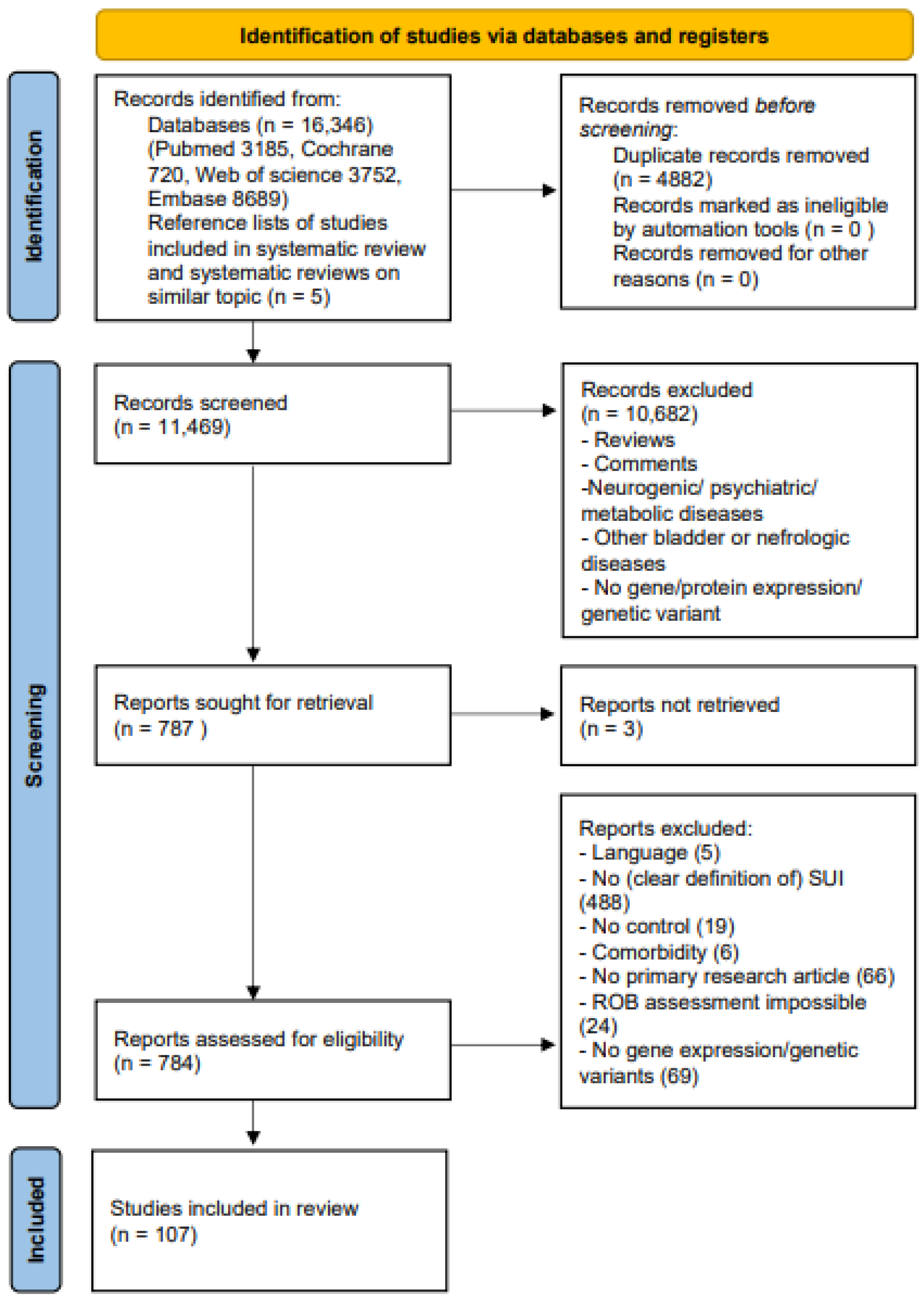
2.1. Study Selection

Figure 1 shows the PRISMA flow chart of the review [21]. Of 11,469 retrieved articles, a total of 107 studies (72 human, 35 animal) were included.

2.2. Study Characteristics

Supplementary Tables S1 and S2 show the study characteristics of the human and animal studies, respectively. All studies included women and female animals only. Of the human studies, SUI alone was investigated in 52 human studies [22,23,24,25,26,27,28,29,30,31,32,33,34,35,36,37,38,39,40,41,42,43,44,45,46,47,48,49,50,51,52,53,54,55,56,57,58,59,60,61,62,63,64,65,66,67,68,69,70,71,72,73], and SUI with the co-occurrence of POP in 23 studies [62,72,73,74,75,76,77,78,79,80,81,82,83,84,85,86,87,88,89,90,91,92,93], of which three studies investigated both SUI and SUI with POP [62,72,73], All animal studies focused on SUI alone [94,95,96,97,98,99,100,101,102,103,104,105,106,107,108,109,110,111,112,113,114,115,116,117,118,119,120,121,122,123,124,125,126,127,128]. The animal studies used rats (Sprague-Dawley, Lewis, Wistar, and undefined strain) or mice (C57BL/6 strain). The age of the mice and rats used, if reported, ranged from six weeks to six months old. All rats and mice, if mentioned, were virgins or primiparous. SUI induction was mostly performed by vaginal dilatation with various diameters, objects, and durations of dilatation, and with or without bilateral ovariectomy and/or previous spontaneous delivery. Other methods were pudendal nerve crush or transection, electrocauterization, surgically opening of paravesical space, and surgically detaching urethra from the anterior wall. The timeline of the gene or protein expression assessment post-induction of SUI, if reported, ranged from one week until nine months.
Eight human studies discussed genetic variants [48,49,52,62,68,69,84,90], 25 gene expression [25,26,29,32,36,37,38,40,42,44,45,46,47,50,53,61,65,66,67,70,71,73,78,81,87], and 62 protein expression [22,23,24,25,26,27,28,30,31,32,33,34,35,36,37,38,39,40,41,42,43,44,45,46,47,50,51,53,54,55,56,57,58,59,60,61,63,64,65,66,67,70,72,73,74,75,76,77,78,79,80,81,82,83,85,86,87,88,89,91,92,93]. Four animal studies examined genetic variants [107,112,121,122], 11 gene expression [96,101,103,104,111,112,113,115,116,118,120], and 32 protein expression [94,95,96,97,98,99,100,101,102,103,105,106,107,108,109,110,111,112,113,114,116,117,119,120,121,122,123,124,125,126,127,128].
Supplementary Table S3 shows all candidate genes and protein(-related product)s. In total, 179 unique genes/protein(-related product)s were examined (104 by human studies, 96 by animal studies, and 21 by both), and 12 studies focused on the whole genome (n = 1) [68], transcriptome (n = 5) [36,46,53,71,96] and/or (tissue, serum, or urinary) proteome (n = 6) [50,54,63,89,100,107]. One mouse study [107] examined protein expression differences between an SUI model based on Esr1 knockout and a wild-type control. All other animal studies examined either the gene or protein expression changes after mechanical SUI induction compared to control animals or described the clinical effect of an SUI model based on a specific gene knockout compared to a wild-type animal, without further examining the effect on expression changes.

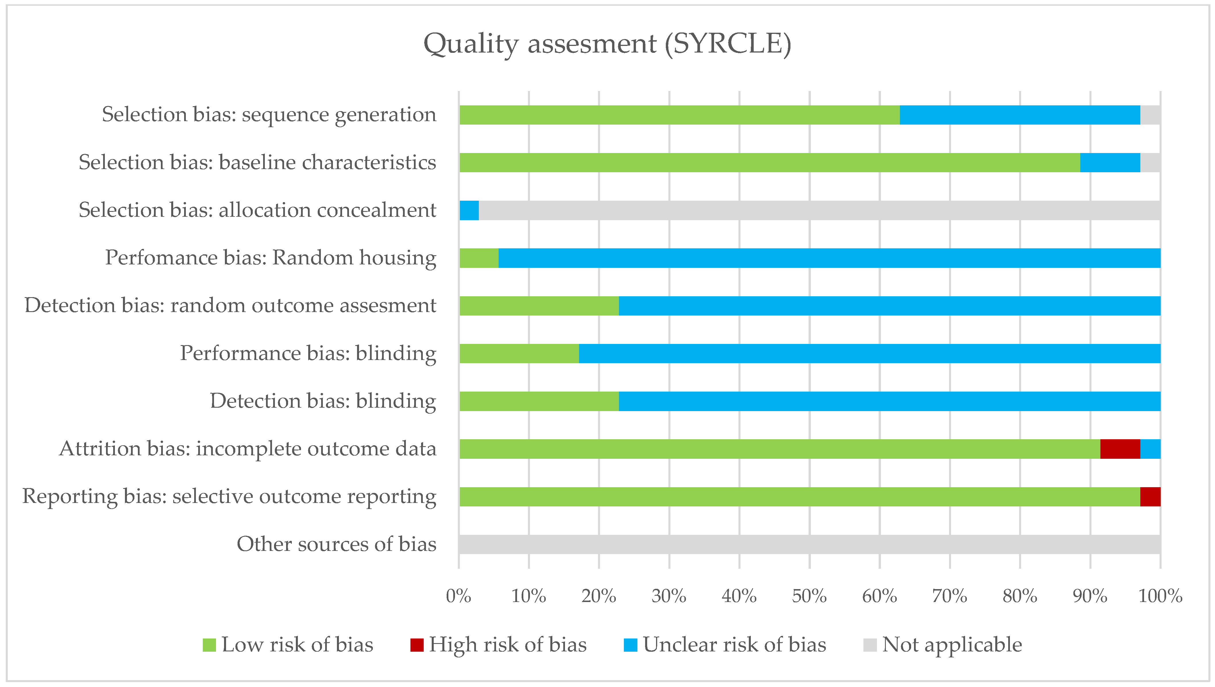
2.3. Risk of Bias of Included Studies

The summary of the risk of bias assessment of the human and animal studies is shown in Figure 2 and Figure 3; Supplementary Tables S4 and S5 show a detailed overview per study. Human studies generally scored low risk of bias on the classification of interventions, bias due to deviations from intended interventions, bias due to missing data and bias in the selection of the reported result. Poorer scoring results were assessed in reporting or matching BMI status, mode of delivery and other possibly confounding factors. Overall, 57% scored low or medium low risk of bias in the final judgment of risk of bias. The animal studies generally scored low risk of bias on baseline characteristics, attrition bias and reporting bias. However, many animal studies lacked in reporting housing conditions, detection bias and blinding, resulting in an unclear risk of bias.

2.4. Synthesis of the Results

The themes that could be distinguished as overarching for the majority of the results were extracellular matrix (ECM) remodeling, reproduction-related endocrinology, inflammation, oxidative stress, apoptosis, and cell-specific markers and processes. The results were further subdivided between these themes. Table 1 and Table 2 show a summary of analyses of the whole genome, transcriptome and proteome from human and animal studies, respectively. Some of these studies also examined candidate genes; these results are not included in these tables but are discussed below and/or in Supplementary Table S3. Genes and protein(-related products) differentially expressed or associated as indicated by these studies also directed towards these prementioned themes. Genes and/or protein(-related products) not fitting these themes and examined in less than three studies were left out of further discussion.

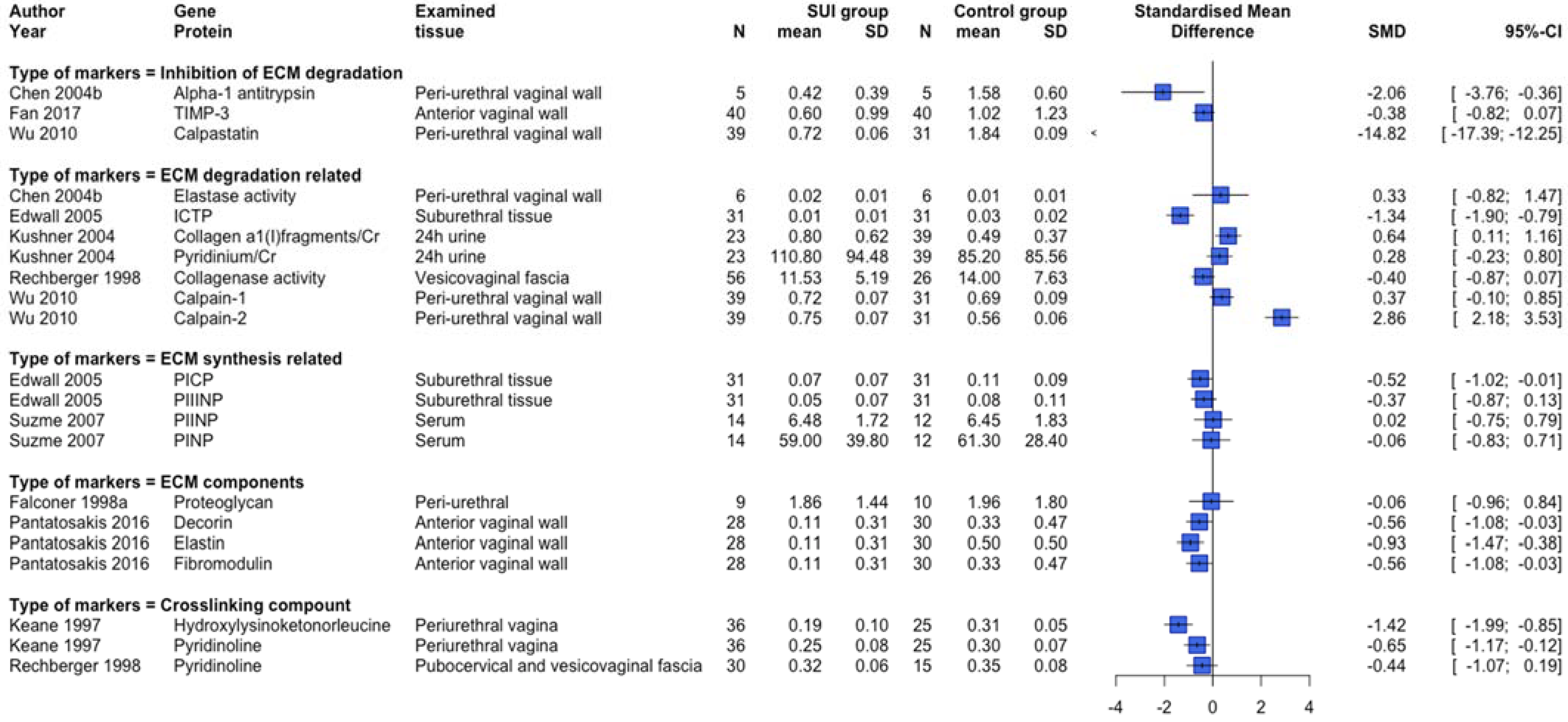
2.4.1. ECM Remodeling

The most studied topic of research encompasses the ECM. Concerning genetic variants, two [49,84] out of three human studies examining rs1800012 of COL1A1 in SUI patients and controls showed a significant association between the variant and SUI, the third [69] did not confirm this association. Specific variants in human FBLN5 [62] and LOXL1 [52,62] were also associated with SUI. No significant association of specific variants of human ELN [62] or MMP1 [69,90] was shown.
Several differentially expressed genes and proteins in human [36,50,71,89] and animal [96,107] studies examining the whole transcriptome or proteome pointed towards the involvement of the ECM metabolism.
Twenty studies examined ECM-related candidate gene expression [25,26,32,36,37,38,42,45,47,65,67,70,73,78,81,96,111,113,115,116]. Regarding collagen (type 1 and 3) gene expression, both human [25,26,32,45,67,70] and animal [111,113,115] studies show conflicting results irrespective of menopausal status, with the largest human study [32] showing no statistically significant differences in gene expression for both collagen type 1 and 3. Moreover, human studies examining gene expression of the proteoglycans biglycan, decorin, fibromodulin, lumican, and versican show conflicting results of which the majority shows no statistically significant differences [25,26,42,45,73]. Two human studies [38,81] show a lower gene expression of elastase inhibitor alpha-1 antitrypsin, corresponding with another human study [36] showing a higher expression of Elafin (elastase-specific protease inhibitor). Conflicting results were present regarding human FBN1/2 expression [37,45]. The studies examining matrix metalloproteases (MMPs) gene expression show an increase in human MMP1 expression [70,78,123] in the SUI group and no statistically significant differences in human [78] and animal [115] MMP2/Mmp2, human MMP3 [78] and animal Mmp13 [96] expression when compared to controls. Conflicting results in human [78] and animal [115] MMP9/Mmp9 expression were observed, showing no significant change in the human study and increased expression in the animal study comparing SUI subjects against controls. Regarding Tissue Inhibitor of Metalloproteases (TIMP/Timp) studies, TIMP1 expression in human studies was decreased [70,78], while no significant difference was seen in animal studies of Timp1 [115], or human [78] or animal [115] studies of TIMP2/Timp2 [78,115] and human TIMP3 [78]. As for other proteases, one human study found no significant difference in Calpain-1 expression, however, a higher expression of both Calpain-2 and Calpastatin was seen [47].
ECM-related protein expression and serum or urinary biomarkers were examined in 51 studies [22,23,24,25,26,27,28,30,32,33,34,35,36,37,38,39,41,42,45,47,56,59,65,67,70,73,74,75,76,77,78,81,82,85,93,94,96,97,98,99,108,109,111,112,113,114,119,121,122,126,127]. Figure 4 shows an overview of human [23,24,25,27,30,32,56,73,74,75,77,82,85,93] studies providing quantitative data and examining collagen-related protein expression subdivided per menopausal status group. In line with Figure 4, the majority of human studies not providing quantitative data showed a lower collagen-related protein expression in SUI patients compared to controls [22,26,38,41,45,65,67,70,76,78]. Further, six out of eight animal studies [94,97,111,112,113,114,121,127] showed a lower collagen-related protein expression in the SUI group compared to controls, while the other two [94,114] showed increased expression. Figure 5 displays an overview of the quantitative data provided by human studies examining other ECM-related protein expression and serum/urinary biomarkers. There seems to be a trend towards lower protein expression of ECM macromolecules, crosslinking compounds, and ‘inhibition of degradation’-related proteins. However, no clear difference in ECM synthesis-related protein expression or serum biomarkers was shown. There were, furthermore, some conflicting results regarding degradation-related markers, but the majority of degradation-related protein expression markers and levels of urinary biomarkers show a relatively higher expression in SUI patients versus controls. These results are in line with the studies providing only non-quantitative data [25,26,28,30,36,38,39,41,42,45,65,70,78]. The animal studies examining ECM-related protein expression show conflicting results and no clear trend could be determined (data not shown) [97,98,112,121,126,127].

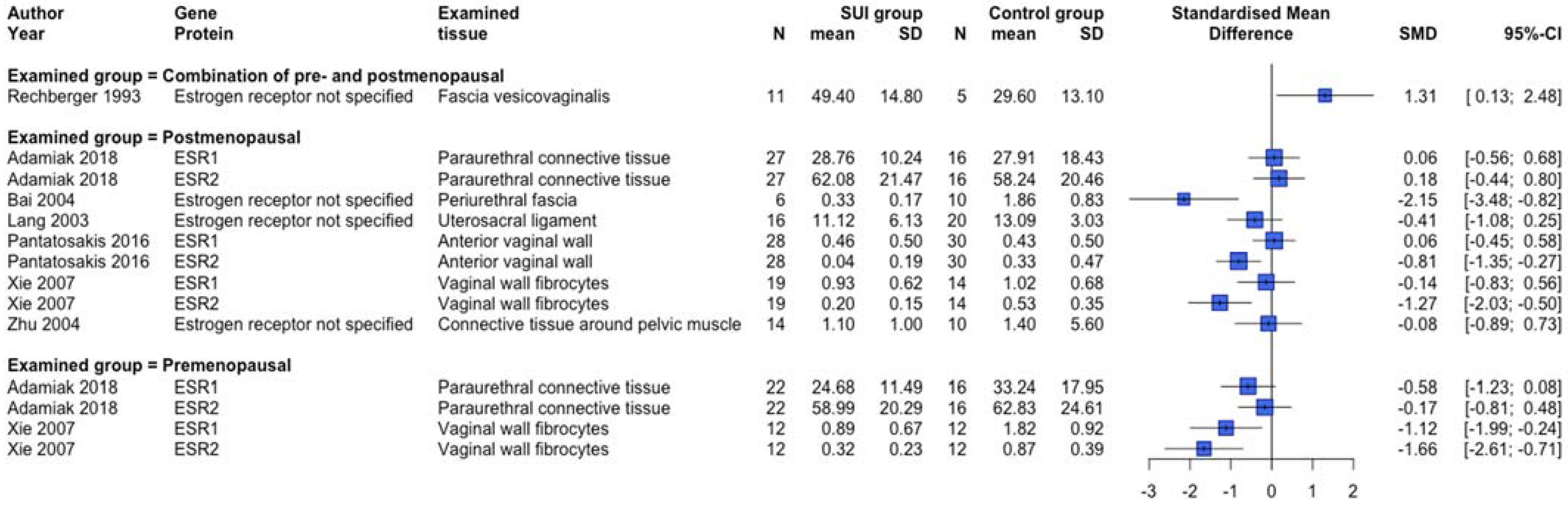
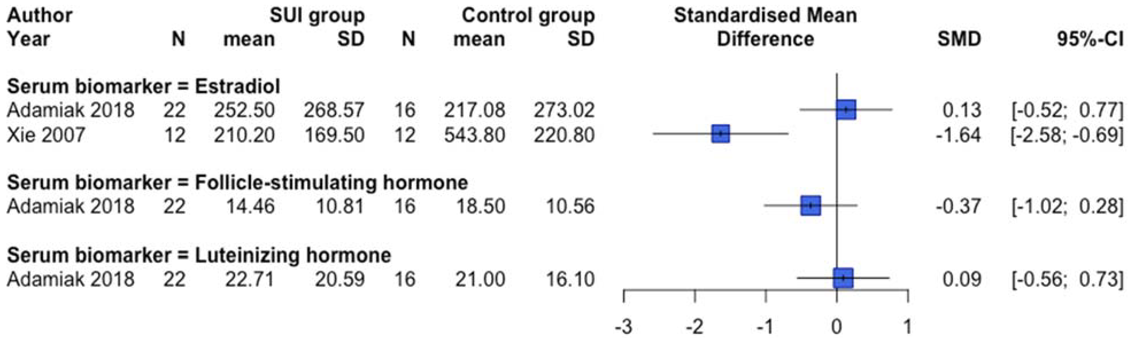
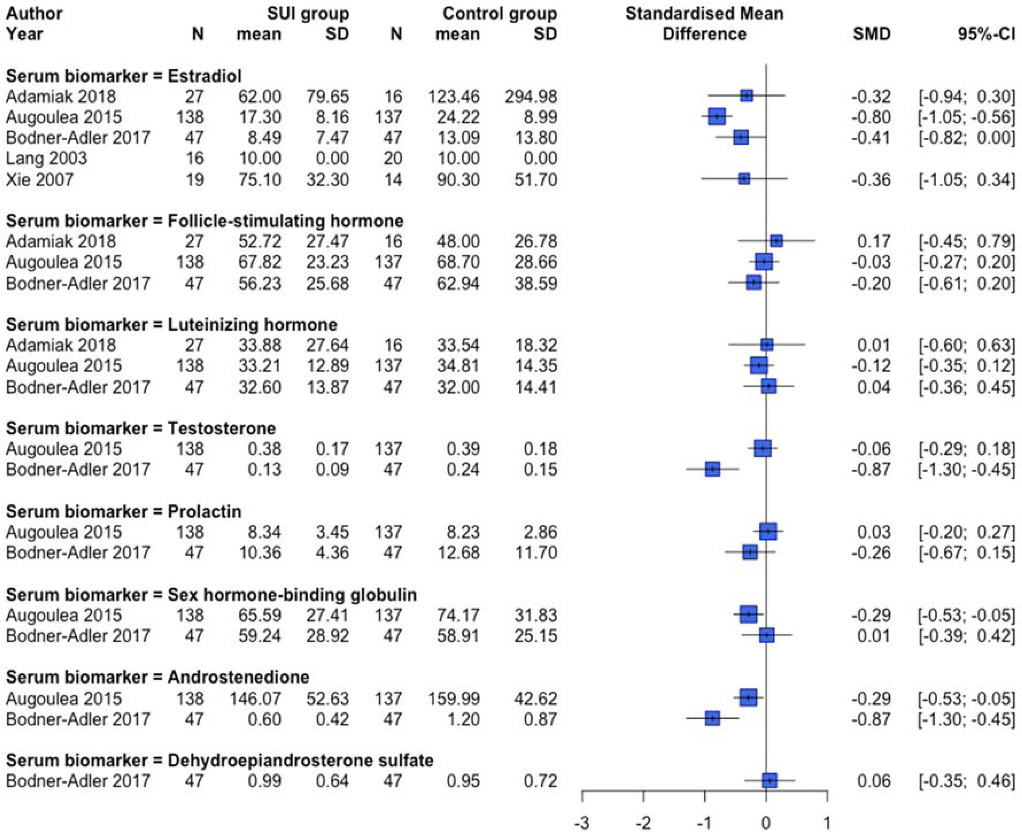
2.4.2. Reproduction-Related Endocrinology

Expression changes or genetic variants of the reproduction-related endocrinology system were examined by 19 studies [31,39,40,43,48,56,57,58,61,70,74,79,83,99,104,105,107,116,120]. A human study [48] examining polymorphisms of ESR1 and AR showed no statistically significant association. An animal Esr1 knockout study using an unmentioned strain of mice to create Esr1 knockout and wild-type mice via the Cre-loxP system, showed a significantly decreased leak-point-pressure (LPP) in the urodynamics of knockout mice when compared to controls, suggesting an association of this gene with SUI [107].
Gene expression changes were examined in three human studies. Only one [61] out of two [40,61] human studies investigating ESR1 showed a lower gene expression in premenopausal SUI women. No aberrant gene expression changes were shown regarding ESR1 in studies with postmenopausal women [40,61]. Furthermore, no statistically significant changes were shown in gene expression of ESR2 [61,70], AR [40], and PR [40] in studies examining premenopausal women [61], postmenopausal women [61], a combined group [40], or women with undefined menopausal status [70].
Protein expression changes and/or serum biomarkers of the reproduction-related endocrinology system were examined in 12 human [31,39,40,43,56,57,58,61,70,74,79,83] and two animal [105,107] studies. Lower protein expression of ESR1 and ESR2 was observed in premenopausal SUI women (Figure 6), but clear differences dependent on the postmenopausal status or a combined menopausal status group were absent. In another human study not providing quantitative data, no difference in ESR1 protein expression between SUI subjects and controls in both pre and postmenopausal women was found, but conversely, an increased ESR2 protein expression in premenopausal SUI women was detected [40]. An animal study showed increased protein expression of Esr1 among vaginal distended C57BL/6 mice and no changes regarding Esr2 expression [105]. Protein expression of the progesterone receptor was not different between SUI patients compared to controls [31,40,56]. Figure 7 and Figure 8 show an overview of studies examining serum biomarkers in pre and postmenopausal women, respectively. Only serum androstenedione [57,58] (examined in postmenopausal women only) and estradiol in postmenopausal women seem to be lower in SUI patients compared to controls, all other markers show conflicting results or no clear differences. An animal study showed no significant differences in estradiol plasma levels between vaginally dilated C57BL/6 mice versus controls [105].

2.4.3. Oxidative Stress and Apoptosis

Oxidative stress and apoptosis-related genes and proteins were examined in 18 studies [31,64,66,94,103,105,106,112,113,114,115,117,119,122,123,126,127,128]. One animal study showed that Nfe2l2-knockout mice had lower LPPs after vaginal dilatation compared to wild-type C57BL/6J mice and this suggests that this antioxidant gene inducer is a potential protective factor in the development of SUI [122].
Several differentially expressed genes and proteins in human [46,63,89] and animal [96,100] studies examining the whole transcriptome or proteome pointed towards the involvement of oxidative stress and/or apoptosis.
Concerning candidate gene expression, a human study showed increased expression of PERK and CHOP in SUI patients compared to controls [66], suggesting an involvement of endoplasmic reticulum stress and apoptosis in SUI. Two animal studies using Sprague-Dawley rats show conflicting results, namely a lower gene expression of Casp3 [103], no apparent changes in Tnfa [115] and Tnfr2 [115] expression, and increased expression of Tnfr1 [115] in the SUI models versus controls. A study using Lewis rat SUI models showed an increased gene expression of Hif1α [104].
In line with the gene expression data, PERK protein expression was also increased in human SUI patients [66]. Animal studies showed an increased protein expression of several oxidative stress markers (4-HNE [122], 8-OHdG [122], HO-1 [114], MDA [114,119,122]) in SUI models, except for LDH [103] and p38 [123]. However, multiple animal studies report conflicting results regarding several antioxidative markers [106,113,114,122,127]. Two mice studies [113,119] show a decrease in Nfe2l2 protein expression in SUI models, but a rat study [114] shows no statistically significant change. The majority [64,94,117,128] of the human [64] and animal [94,105,117,128] studies examining neuronal nitric oxide synthase (NOS) expression show a decreased expression in SUI subjects compared to controls. One animal study [117] examining endothelial Nos shows a decrease and two [105,117] out of three [105,106,117] animal studies examining inducible Nos show an increase in SUI models. Animal studies examining Bax and Bcl2 show an increase in Bax(/Bcl2 ratio) [114,122] and both human [66] and animal [122] studies show a decrease in BCL2/Bcl2 protein expression in SUI subjects compared to controls. Several pro-apoptotic markers (Casp3 [122] and Casp9 [122] in mice, Tnfα in rats [126], and CHOP in humans [66]) were increased in SUI subjects, while p53 [31] was decreased in one human study.

2.4.4. Inflammation

Inflammation-related genetic variants, gene and protein expression changes were examined in 21 studies [36,37,54,67,69,85,86,96,98,101,102,104,108,111,112,113,115,123,126,127,128]. One human study showed a non-significant trend towards an association of rs885786 in SERPINA5 with SUI [69].
Several differentially expressed genes and proteins in human [46,63] and animal [107] studies examining the whole transcriptome or proteome pointed towards the involvement of inflammation. Animal studies examining other gene expression differences show a higher gene expression of Tnfr1 [115], several cytokines (Il6 [115], Ccl7 [101,104], Ccr1 [104], Ccr2 [104], Ccr3 [104]), and cytokine receptor Cxcr4 [104], and no aberrant changes in Tnfa [115], Tnfr2 [115], and the cytokines Ccr5 [104], Cxcl12 [104], Il1b [115], and Il8 [104]. Conflicting results were present in human [37,67] and animal [111,115] studies examining TGFB1/Tgfb1, and animal Smad2 [96,111] and Smad3 [111] gene expression. One study examining Acta2 [113] expression in mice showed lower gene expression in the SUI group.
Human studies examining TGFB1 protein expression differences showed lower [67] and no aberrant protein expression differences [37], and a serum measurement study of TGFB1 also showed no aberrant differences [85] in SUI patients compared to controls. (Phosphorylated) Smad2/3/7 were examined in animal studies only, without providing quantitative data, and with conflicting results [96,98,108,111,112,113,127]. Animal studies showed increased protein expression of Tnf-α [126], Il-1 [126], Ccl7 [101] and conflicting results for Tgfb1 [98,111,112,113,127] in SUI models compared to controls. In addition, α-SMA protein expression was examined in animal studies only and was consistently downregulated in SUI subjects compared to controls [98,99,102,113,123,127,128]. A human study showed an increased urinary level of SERPINA5 in SUI patients [54].

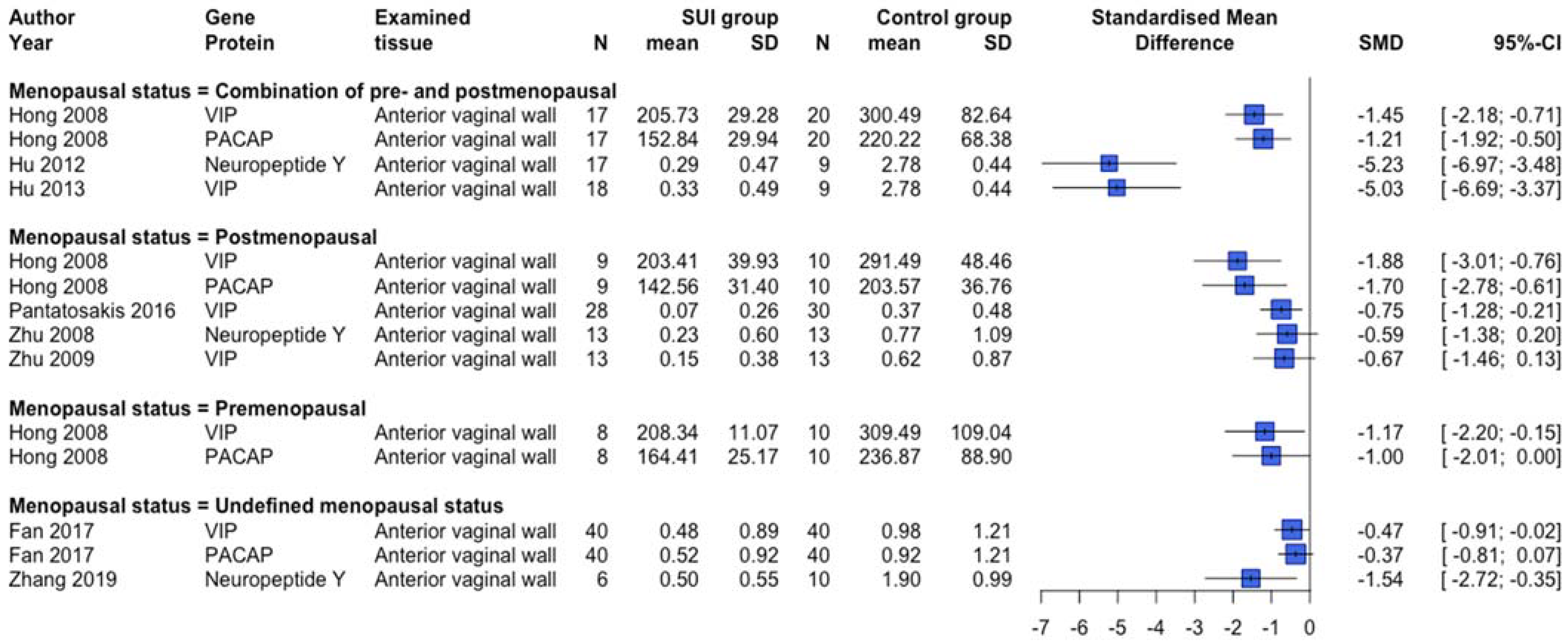
2.4.5. Cell-Specific Markers and Processes

Two human studies examining transcriptomics showed differentially expressed genes and micro-RNAs associated with neurodegeneration [46,53]. In line with these results, an animal transcriptomics study pointed towards neuronal involvement [96]. Generally, a lower protein expression of neuron-specific markers was shown in human SUI patients compared to controls (Figure 9). This is in agreement with human [80] and animal [102,109,124,128] studies not providing quantitative data, except for human neuron specific enolase [80] and Protein S100 [80] that showed no significant changes in protein expression.
Human [50,71,89] and animal [96,100,107] studies study examining transcriptomics and proteomics showed involvement of smooth muscle cell differentiation and/or contraction associated with SUI. Genes and proteins related to muscle cells were examined in animal studies only. Conflicting results were shown for myosin subtypes [106,116,123,128] and desmin [116]. A higher protein expression of the muscle regeneration protein Pax7 was shown in SUI rat models compared to controls [123].

2.4.6. Other

Three animal studies examined Vegf expression [104,116,120] and showed no significant changes in rats induced with SUI compared to controls. However, Vegf protein expression as examined by one rat study [99] was lower in induced-SUI Sprague-Dawley rats compared to controls.
The certainty of evidence assessment according to the GRADE method is shown in Table 3.

3. Discussion

To our knowledge, this is the first systematic review of genetic variants, gene and protein expression differences associated with SUI. This review shows that the molecular background of SUI is diverse, which was not unexpected in view of the general consensus that SUI is a multifactorial condition. Nevertheless, through this systematic review, common themes important for SUI were defined at the molecular level.

3.1. Principal Findings

SUI is associated with an aberrant ECM metabolism, most likely resulting in a lower gene and protein expression of ECM macromolecules (collagen, elastin, proteoglycans) and crosslinking compounds, and altered degradation, when compared to controls. ECM metabolism is a complex process that continuously adjusts to new conditions based on mechanical cues and cell–cell interactions. The collective results and forest plot (Figure 3) indicated a higher expression of a number of ECM degradation-related markers and lower expression of inhibitors of ECM degradation in SUI, but their relative contribution and role in disturbing the ECM metabolism cannot be judged. Based on this systematic review, there is insufficient evidence to conclude that these markers are the main contributors to the lower expression of ECM macromolecules.
A number of human [43,61] studies as well as an Esr1 KO animal study [107] suggest an association of ESR1 and ESR2 with SUI emphasizing the importance of hormonal status. It is unclear if this has an effect on ECM metabolism, which should be further examined.
The clinical value of serum hormonal biomarkers in SUI patients remains unclear: the contribution of menopause and its accompanying hormonal changes in association with SUI could not be confirmed in this review. Clear initiators of this process, rather than a previous trauma (e.g., childbirth), prolonged pressure and aging, still need to be determined in order to devise new prophylactic or therapeutic strategies.
In addition to the aberrant ECM metabolism, increased expression of several pro-apoptotic, oxidative stress-related, and inflammation-related markers and the association of the Nfe2l2 gene in one animal knockout study [112] imply the association of an aberrant wound healing process in SUI. The lower expression of many neuronal cell-specific proteins and association with neurodegeneration-related proteins in SUI subjects is also in line with this and is possibly due to partly irreversible neurological damage after trauma or a neurodegenerative process involved in SUI. As for muscle cells, involvement of smooth muscle cell differentiation and/or contraction seems to be associated with SUI. The molecular mechanisms underlying these processes and their role in the pathogenesis of SUI should be further examined in depth.
The overarching theme appears to be an altered micromilieu and metabolism compared to controls. Which factors are most important for the development or continuation of SUI cannot be concluded. Furthermore, it should be further investigated how the important environmental risk factors that contribute to SUI—e.g., aging and trauma to the endopelvic fascia—impact on and interact with the identified (putative) molecular processes.

3.2. Comparison with Existing Literature

The findings of the current systematic review are in line with and expand the knowledge of previously published reviews [17,129,130,131,132]. The systematic review on gene expression differences of Isali et al., included studies until September 2017. They showed that 13 genes were differentially expressed in SUI and indicated the probable involvement of intermediate filament cytoskeleton organization and ECM organization in the pathogenesis of SUI [17]. The current systematic review adds a suggestive association of ESR1 and ESR2 with SUI, increased expression of pro-apoptotic, oxidative stress, and inflammation-related markers, lower protein expression of neuron-specific markers, and an association with a neurodegenerative process as well as impaired muscle cell differentiation and contractility, in addition to aberrant ECM metabolism being involved in SUI. Cartwright et al., performed a meta-analysis of rs1799750 of MMP1 and rs1800013 of COL1A1 with SUI and did not confirm a clear association [129]. They included two study abstracts we excluded based on the impossibility to assess the risk of bias, but this did not result in different findings. Campeau et al., examined the ECM and showed an association with SUI and suggested the involvement of sex hormones altering ECM metabolism [130]. Chen et al., examined ECM metabolism and reproductive hormones and showed a genetic predisposition to abnormal ECM remodeling, modulated by reproductive hormones, trauma, mechanical stress load and aging [131]. McKenzie et al., examined genetic influences on SUI and showed there was increasing evidence suggesting a genetic basis for the development of SUI, and that candidate genes related to ECM remodeling might be associated. They suggested that future research should focus on the influence of estrogen and progesterone on ECM proteins [132].

3.3. Strengths and Limitations

The strength of this review lies in the broadly examined area and inclusion of both human and animal studies. Despite the broadly examined area, the numbers of studies per selected outcome are still rather low. Moreover, the considerable heterogeneity between the included studies has prevented us from drawing firm conclusions based on the results of this systematic review, and below, we will discuss the main heterogeneity-related issues per study type.
Generally, in human studies many different definitions and methods for diagnosing SUI were used, ranging from self-reported to diagnosed via urodynamics. Subsequently, the definitions of the controls used were rather different, ranging from “continent controls” or “women without SUI” to women with other benign gynecological complaints or with cervical neoplasms. This variation in definition impacts negatively on the homogeneity of the studies. In addition, multiple different tissues/fluids were examined, e.g., ligaments, periurethral biopsies, vaginal wall, skin biopsies, urine, and serum. Studies often selected only one specific tissue, e.g., vaginal wall tissue in their assessment, which may only explain part of the SUI etiology or with unclear direct relevance in relation to SUI. Finally, some patients had comorbidities or used hormonal replacement therapy, and many studies did not mention (probable) medication use or comorbidities. It is likely that all this may have influenced the data by over or underestimating expression differences or genetic associations.
We included articles regardless of the co-existence with POP. When evaluating the data, we did not correct for the additional effect of POP on our outcomes. We concluded that excluding these studies would lead to introducing more (selection) bias because SUI so often co-exists with POP and because many studies did not mention the (possible) co-existence.
Thirty-five animal studies, using rats and mice, were included in this review. In particular, animal studies using specific gene knockout models appeared to be useful as they showed a direct causative association of certain genes with SUI. Although we believe that animal models can provide a valuable contribution to the knowledge of the pathophysiology of SUI, there are some pitfalls when evaluating the results: the studies generally lacked reporting of important topics of risk of bias, generally used young and healthy animals, and measured expression changes within a broad range after inducing SUI—of which the majority focused on a relatively short time frame post-induction. This contrasts starkly with SUI in humans where SUI usually develops in the years after childbirth [133]. This may explain conflicting results and questions whether translation to humans is feasible. Furthermore, it is well established that these rodents usually functionally recover within a relatively short period of time after induction of SUI and research on the long-term effects of SUI induction by various methods is scarce [134]. Therefore, for future research in animals related to gene and protein expression changes in SUI, especially these long-term effects should be considered in order to avoid the bias of examining a (normal) wound healing process instead of the actual development of SUI.
In addition to summarizing the data of studies without quantitative data, the forest plots were used to visualize differences in expression when comparing SUI subjects with controls. However, a pooled summary result of the calculated SMDs per examined gene/protein could not be determined, mainly because of the heterogeneity in the tissues examined, the low number of studies per examined gene/protein (as there was often data from only one study), and the different assay methods.
Very few studies included in this review were based on a hypothesis-free approach. Examining candidate genes and proteins is important and necessary in determining an exact contribution of a specific marker, but this kind of hypothesis-driven focus could lead to missing important contributors.
Of the human studies, only 57% scored low or medium low on the total judgment of risk of bias. Future studies should report on and match participants in age, parity, BMI, and possibly menopausal status and mode of delivery and should define and diagnose SUI in a valid and consistent manner. This will greatly improve the quality of the results. In terms of the risk of bias of animal studies, future studies using animal models to mimic SUI need to improve transparency and reporting standards.

4. Materials and Methods

4.1. Eligibility Criteria

This systematic review includes human and animal studies (domain) on genetic variants, gene and/or protein expression differences (outcome) in relation to SUI with or without POP (determinant) compared to controls. We used our prospectively registered protocol in Prospero (registration number CRD42019120202) on UI in general (Supplement S6) narrowing our selection process to SUI instead of UI in general. This beforementioned protocol was also used for our review on urgency urinary incontinence [135]. The inclusion criteria were studies with primary research data of affected cases (SUI) and controls of humans or animals (all species and sexes), examining genetic variants, gene expression, or protein expression differences, with sufficient information to determine the risk of bias.

4.2. Information Sources and Search Strategy

On 5 January 2021, a systematic search was performed in Pubmed, Embase, Web of Science, and the Cochrane library to identify available studies using the search strategy described in Supplement S7. The terms used were related to UI in general and a broad spectrum of genetic and protein expression terms and assays. References of reviews and included studies were cross-checked for studies not retrieved by the database search.

4.3. Study Selection

Studies were screened by two independent reviewers (WMP screened all articles, JW and HG shared the second screening) in two phases (title/abstract and full text phase) using the predefined inclusion criteria. In case only one of the reviewers assessed a study as eligible for full text screening, the study was screened full text by both reviewers. Potential discrepancies were discussed until consensus was reached, ultimately with the help of another author (KBK) when required. Studies with either solely SUI or both SUI and POP were included. Studies with SUI due to cancer-related surgical interventions were excluded from our review, due to possible confounding effects, e.g., of the malignancy.

4.4. Data Extraction

Extraction of study characteristics was performed by one and verified by a second reviewer. Characteristics extracted from the human studies were: first author, year of publication, number of cases/controls, definition and diagnostic-method of SUI, assessed material, assay method, investigated gene/protein/genetic variant, and results of the (value) differences between the groups (SUI and controls). From the animal studies, first author, year of publication, type of animal, sex, number of subjects, SUI induction method and confirmation of diagnosis, assessed material, assay method, investigated gene/protein/genetic variant, and results of the (value) differences between the groups (SUI and controls), were extracted. During data extraction, data of animals receiving treatment were not included due to possible confounding effects of this treatment on gene and protein expression. In addition, data with (naïve) cell-cultures being exposed to specific conditions (e.g., mechanical stress) without defining a clear SUI phenotype were not included. Only data of (induced) SUI versus controls was included. Therefore, for some studies in this review only part of their data was collected and reviewed. In the case of (animal) studies with measurements on multiple timepoints after induction of SUI, the data of the last timepoint was collected.

4.5. Assessment of Risk of Bias

The assessment process was carried out by one and checked by a second reviewer. Potential discrepancies were discussed until consensus was reached, ultimately with the help of another author when required. The Cochrane ROBINS-I [136] and SYRCLE risk of bias [137] tools were used for human and animal studies, respectively. In addition to the basic aspects of risk of bias assessment, the following topics were added: differences between cases and controls in age, BMI, menopausal status, parity, and mode of delivery. Similar to our review of urgency urinary incontinence [135], we added seven signaling questions to the risk of bias tools, addressing the risk of bias for in-vitro aspects of studies (Supplement S8), to be used when applicable. These questions were developed based on a tool for in vitro studies by the National Toxicology Program [138].

4.6. Data Synthesis

Due to the broad research topic of genetic variants, gene and protein expression changes in humans and animals related to SUI and including both quantitative and non-quantitative studies, it was not justifiable to estimate pooled effects using meta-analysis in this systematic review. The extracted data per outcome measure were grouped in themes, according to their functions in cellular processes. The genetic variants and changes in gene and protein expression were summarized within these themes. The results and differences between human and animal study outcomes were discussed, and differences per menopausal status were discussed when applicable. We did not make a distinction between menstrual phases. When applicable and in order to show probable trends in gene or protein expression differences, forest plots of (semi-)quantitative data showing standardized mean differences (SMDs) were drawn using the statistical software R version 4.1.0 [139] and package meta version 4.18-2 [140]. It was decided to present SMDs instead of mean differences because of the different ways of measuring the expression changes and different outcome measures.
For the forest plots, only quantitative or semi-quantitative data were taken into account. Semi-quantitative data (e.g., from scoring systems for staining) was converted into quantitative data in a standardized manner (by assigning the values 0/1/2 to −/+/++ or 1.1/1.2/1.3 to +/++/+++). Proportions were included, considering the proportion values as means. The standard deviations (SDs) were then calculated with the following formula SD = sqrt ((proportion) × (1-proportion)). If unclear whether SDs or standard errors (SEs) were shown, a conservative approach was taken assuming the provided numbers were SEs, and SDs were calculated using the following formula: SD = SE × sqrt (numbers of participants). If data needed to be combined, e.g., in case of studies distinguishing between menstrual phase (proliferative and secretory) using different participants, it was combined using the formulas from the Cochrane Handbook for Systematic Reviews of Interventions Version 6.2 [141].
Data presented in figures without providing the exact values and/or variability measures were excluded from the forest plots. When studies examined multiple tissues of one subject, the data of the most relevant tissue according to the authors of this review was included in the forest plot, in order to prevent multiplicity issues.
Publication bias was not evaluated because the data included were far too heterogeneous, and therefore, no meta-analysis could be performed. The certainty of the evidence was assessed using the Grading of Recommendations, Assessment, Development and Evaluation (GRADE) approach [142]. Given the nature of this systematic review, a modification was used, for which observational studies start with high certainty evidence.

5. Conclusions

This systematic review provides an overview of the current knowledge of genetic variants and gene and protein expression differences associated with SUI. The results suggest that altered ECM metabolism with a lower protein expression of ECM macromolecules and crosslinking compounds, estrogen receptors, oxidative stress, apoptosis, inflammation, neurodegenerative processes, and muscle cell differentiation and contractility seem to be associated with SUI.
Future research should focus on possible contributors to these alterations. The authors recommend the use of hypothesis-free approaches in order to find possible new links and candidate genes/proteins.

Supplementary Materials

The following supporting information can be downloaded at: www.mdpi.com/article/10.3390/ijms23063401/s1.

Author Contributions

Conceptualization, W.M.P., E.O. and K.B.K.; methodology, W.M.P., J.I., E.O. and K.B.K.; formal analysis, W.M.P. and J.I.; investigation, W.M.P.; data curation, W.M.P., J.W. and H.G.; writing original draft, W.M.P.; visualization, W.M.P.; Validation, J.W., H.G. and K.B.K.; writing—review and editing, J.W., H.G., M.J.H.C., F.M.J.M., D.A.W.J., J.I., G.P., E.O. and K.B.K.; Supervision, J.I., G.P., E.O. and K.B.K. All authors have read and agreed to the published version of the manuscript.

Funding

This study was funded by the European Fund for Regional Development (EFRD), Grant/Award Number: PROJ00787 (DIABIP), and by the ‘More Knowledge with Fewer Animals project’ of ZonMw under Project No. 114024125.

Institutional Review Board Statement

Not applicable.

Informed Consent Statement

Not applicable.

Data Availability Statement

No new data were created or analyzed in this study. Data sharing is not applicable to this article.

Acknowledgments

The authors acknowledge OnYing Chan, librarian at the Medical Library of Radboud University, Nijmegen, for her help in developing the search strategy used for this review. Furthermore, we acknowledge Alejandra Ruiz-Zapata for her help with the data curation. While helping with the review, she was employed as a post-doc at the Department of Gynecology and Obstetrics at the RadboudUMC, Nijmegen, and was funded by the European Fund for Regional Development (EFRD), Grant/Award Number: PROJ00787 (DIABIP). Currently, she is working at the Upstream processing Department of Byondis BV, Nijmegen. Finally, Rob de Vries, working at the Health Evidence department of the Radboudumc, Nijmegen, is acknowledged for his help with the methodology. Rob de Vries was funded by the More Knowledge with Fewer Animals project of ZonMw under Project No. 114024125. All acknowledged persons report no further conflict of interest.

Conflicts of Interest

The authors declare no conflict of interest. The funders had no role in the design of the study; in the collection, analyses, or interpretation of data; in the writing of the manuscript, or in the decision to publish the results.

References

  1. Groutz, A.; Gordon, D.; Keidar, R.; Lessing, J.B.; Wolman, I.; David, M.P.; Chen, B. Stress urinary incontinence: Prevalence among nulliparous compared with primiparous and grand multiparous premenopausal women. Neurourol. Urodyn. 1999, 18, 419–425. [Google Scholar] [CrossRef]
  2. Haylen, B.T.; de Ridder, D.; Freeman, R.M.; Swift, S.E.; Berghmans, B.; Lee, J.; Monga, A.; Petri, E.; Rizk, D.E.; Sand, P.K.; et al. An International Urogynecological Association (IUGA)/International Continence Society (ICS) joint report on the terminology for female pelvic floor dysfunction. Neurourol. Urodyn. 2010, 29, 4–20. [Google Scholar] [CrossRef] [PubMed]
  3. Irwin, D.E.; Milsom, I.; Hunskaar, S.; Reilly, K.; Kopp, Z.; Herschorn, S.; Coyne, K.; Kelleher, C.; Hampel, C.; Artibani, W.; et al. Population-based survey of urinary incontinence, overactive bladder, and other lower urinary tract symptoms in five countries: Results of the EPIC study. Eur. Urol. 2006, 50, 1306–1314, discussion 1314–1305. [Google Scholar] [CrossRef] [PubMed]
  4. Hunskaar, S.; Lose, G.; Sykes, D.; Voss, S. The prevalence of urinary incontinence in women in four European countries. BJU Int. 2004, 93, 324–330. [Google Scholar] [CrossRef]
  5. Ninomiya, S.; Naito, K.; Nakanishi, K.; Okayama, H. Prevalence and Risk Factors of Urinary Incontinence and Overactive Bladder in Japanese Women. Low Urin. Tract Symptoms 2018, 10, 308–314. [Google Scholar] [CrossRef]
  6. Chermansky, C.J.; Moalli, P.A. Role of pelvic floor in lower urinary tract function. Auton. Neurosci. 2016, 200, 43–48. [Google Scholar] [CrossRef]
  7. Thangarajah, F.; Hartmann-Wobbe, J.; Ratiu, D.; Pahmeyer, C.; Radosa, J.C.; Mallmann, P.; Ludwig, S. The Onset of Urinary Incontinence in Different Subgroups and its Relation to Menopausal Status: A Hospital-based Study. In Vivo 2020, 34, 923–928. [Google Scholar] [CrossRef] [Green Version]
  8. Mishra, G.D.; Cardozo, L.; Kuh, D. Menopausal transition and the risk of urinary incontinence: Results from a British prospective cohort. BJU Int. 2010, 106, 1170–1175. [Google Scholar] [CrossRef] [Green Version]
  9. Jueng-Anuwat, P.; Roongruangsilp, U.; Kochakarn, W.; Ratana-Olarn, K. Risk factors for stress urinary incontinence in middle aged and elderly Thai women. J. Med. Assoc. Thail. 2001, 84, 1121–1125. [Google Scholar]
  10. Singh, U.; Agarwal, P.; Verma, M.L.; Dalela, D.; Singh, N.; Shankhwar, P. Prevalence and risk factors of urinary incontinence in Indian women: A hospital-based survey. Indian J. Urol. 2013, 29, 31–36. [Google Scholar] [CrossRef]
  11. Jun, T.; Yoon, H.S.; Kim, H.S.; Lee, J.W.; Bae, J.; Lee, H.W. Recurrence rate of stress urinary incontinence in females with initial cure after transobturator tape procedure at 3-year follow-up. Investig Clin. Urol. 2017, 58, 54–60. [Google Scholar] [CrossRef] [PubMed] [Green Version]
  12. Kirchin, V.; Page, T.; Keegan, P.E.; Atiemo, K.O.; Cody, J.D.; McClinton, S.; Aluko, P. Urethral injection therapy for urinary incontinence in women. Cochrane Database Syst. Rev. 2017, 7, CD003881. [Google Scholar] [CrossRef]
  13. Falah-Hassani, K.; Reeves, J.; Shiri, R.; Hickling, D.; McLean, L. The pathophysiology of stress urinary incontinence: A systematic review and meta-analysis. Int. Urogynecol. J. 2021, 32, 501–552. [Google Scholar] [CrossRef] [PubMed]
  14. Aoki, Y.; Brown, H.W.; Brubaker, L.; Cornu, J.N.; Daly, J.O.; Cartwright, R. Urinary incontinence in women. Nat. Rev. Dis. Primers 2017, 3, 17042. [Google Scholar] [CrossRef] [PubMed] [Green Version]
  15. Khadzhieva, M.B.; Kolobkov, D.S.; Kamoeva, S.V.; Salnikova, L.E. Expression changes in pelvic organ prolapse: A systematic review and in silico study. Sci. Rep. 2017, 7, 7668. [Google Scholar] [CrossRef] [PubMed] [Green Version]
  16. Herschorn, S. Female pelvic floor anatomy: The pelvic floor, supporting structures, and pelvic organs. Rev. Urol. 2004, 6 (Suppl. 5), S2–S10. [Google Scholar] [PubMed]
  17. Isali, I.; Mahran, A.; Khalifa, A.O.; Sheyn, D.; Neudecker, M.; Qureshi, A.; Conroy, B.; Schumacher, F.R.; Hijaz, A.K.; El-Nashar, S.A. Gene expression in stress urinary incontinence: A systematic review. Int. Urogynecol. J. 2020, 31, 1–14. [Google Scholar] [CrossRef]
  18. Hannestad, Y.S.; Lie, R.T.; Rortveit, G.; Hunskaar, S. Familial risk of urinary incontinence in women: Population based cross sectional study. BMJ 2004, 329, 889–891. [Google Scholar] [CrossRef] [Green Version]
  19. Wennberg, A.L.; Altman, D.; Lundholm, C.; Klint, A.; Iliadou, A.; Peeker, R.; Fall, M.; Pedersen, N.L.; Milsom, I. Genetic influences are important for most but not all lower urinary tract symptoms: A population-based survey in a cohort of adult Swedish twins. Eur. Urol. 2011, 59, 1032–1038. [Google Scholar] [CrossRef] [Green Version]
  20. Altman, D.; Forsman, M.; Falconer, C.; Lichtenstein, P. Genetic influence on stress urinary incontinence and pelvic organ prolapse. Eur. Urol. 2008, 54, 918–922. [Google Scholar] [CrossRef]
  21. Page, M.J.; McKenzie, J.E.; Bossuyt, P.M.; Boutron, I.; Hoffmann, T.C.; Mulrow, C.D.; Shamseer, L.; Tetzlaff, J.M.; Akl, E.A.; Brennan, S.E.; et al. The PRISMA 2020 statement: An updated guideline for reporting systematic reviews. Int. J. Surg. 2021, 88, 105906. [Google Scholar] [CrossRef]
  22. Ulmsten, U.; Ekman, G.; Giertz, G.; Malmstrom, A. Different biochemical composition of connective tissue in continent and stress incontinent women. Acta Obstet. Gynecol. Scand. 1987, 66, 455–457. [Google Scholar] [CrossRef] [PubMed]
  23. Falconer, C.; Ekman, G.; Malmstrom, A.; Ulmsten, U. Decreased collagen synthesis in stress-incontinent women. Obstet. Gynecol. 1994, 84, 583–586. [Google Scholar] [PubMed]
  24. Keane, D.P.; Sims, T.J.; Abrams, P.; Bailey, A.J. Analysis of collagen status in premenopausal nulliparous women with genuine stress incontinence. Br. J. Obstet. Gynaecol. 1997, 104, 994–998. [Google Scholar] [CrossRef] [PubMed]
  25. Falconer, C.; Blomgren, B.; Johansson, O.; Ulmsten, U.; Malmstrom, A.; Westergren-Thorsson, G.; Ekman-Ordeberg, G. Different organization of collagen fibrils in stress-incontinent women of fertile age. Acta Obstet. Gynecol. Scand. 1998, 77, 87–94. [Google Scholar] [CrossRef]
  26. Falconer, C.; Ekman-Ordeberg, G.; Blomgren, B.; Johansson, O.; Ulmsten, U.; Westergren-Thorsson, G.; Malmstrom, A. Paraurethral connective tissue in stress-incontinent women after menopause. Acta Obstet. Gynecol. Scand. 1998, 77, 95–100. [Google Scholar] [CrossRef]
  27. Rechberger, T.; Postawski, K.; Jakowicki, J.A.; Gunja-Smith, Z.; Woessner, J.F., Jr. Role of fascial collagen in stress urinary incontinence. Am. J. Obstet. Gynecol. 1998, 179, 1511–1514. [Google Scholar] [CrossRef]
  28. Chen, B.; Wen, Y.; Wang, H.; Polan, M.L. Differences in estrogen modulation of tissue inhibitor of matrix metalloproteinase-1 and matrix metalloproteinase-1 expression in cultured fibroblasts from continent and incontinent women. Am. J. Obstet. Gynecol. 2003, 189, 59–65. [Google Scholar] [CrossRef]
  29. Chen, B.; Wen, Y.; Zhang, Z.; Wang, H.; Warrington, J.A.; Polan, M.L. Menstrual phase-dependent gene expression differences in periurethral vaginal tissue from women with stress incontinence. Am. J. Obstet. Gynecol. 2003, 189, 89–97. [Google Scholar] [CrossRef]
  30. Cor, A.; Barbic, M.; Kralj, B. Differences in the quantity of elastic fibres and collagen type I and type III in endopelvic fascia between women with stress urinary incontinence and controls. Urol. Res. 2003, 31, 61–65. [Google Scholar] [CrossRef]
  31. Bai, S.W.; Jung, Y.W.; Kwon, H.S.; Yoon, J.M.; Shin, J.S.; Kim, S.K.; Park, K.H.; Park, J.H. The role of estrogen receptor, progesterone receptor and p53 in development of stress urinary incontinence. Yonsei Med. J. 2004, 45, 885–890. [Google Scholar] [CrossRef] [Green Version]
  32. Bakas, P.G.; Liapis, A.E.; Zervolea, I.; Voutsinas, G.; Kletsas, D.; Creatsas, G. mRNA assessment for procollagen production in women with genuine stress urinary incontinence. Int. Urogynecol. J. 2004, 15, 429–431; discussion 431. [Google Scholar] [CrossRef] [PubMed]
  33. Kushner, L.; Mathrubutham, M.; Burney, T.; Greenwald, R.; Badlani, G. Excretion of collagen derived peptides is increased in women with stress urinary incontinence. Neurourol. Urodyn. 2004, 23, 198–203. [Google Scholar] [CrossRef] [PubMed]
  34. Edwall, L.; Carlstrom, K.; Jonasson, A.F. Markers of collagen synthesis and degradation in urogenital tissue from women with and without stress urinary incontinence. Neurourol. Urodyn. 2005, 24, 319–324. [Google Scholar] [CrossRef] [PubMed]
  35. Radziszewski, P.; Borkowski, A.; Torz, C.; Bossowska, A.; Gonkowski, S.; Majewski, M. Distribution of collagen type VII in connective tissues of postmenopausal stress-incontinent women. Gynecol. Endocrinol. 2005, 20, 121–126. [Google Scholar] [CrossRef] [PubMed]
  36. Chen, B.; Wen, Y.; Zhang, Z.; Guo, Y.; Warrington, J.A.; Polan, M.L. Microarray analysis of differentially expressed genes in vaginal tissues from women with stress urinary incontinence compared with asymptomatic women. Hum. Reprod. 2006, 21, 22–29. [Google Scholar] [CrossRef] [PubMed] [Green Version]
  37. Wen, Y.; Polan, M.L.; Chen, B. Do extracellular matrix protein expressions change with cyclic reproductive hormones in pelvic connective tissue from women with stress urinary incontinence? Hum. Reprod. 2006, 21, 1266–1273. [Google Scholar] [CrossRef] [Green Version]
  38. Chen, B.; Wen, Y.; Yu, X.; Polan, M.L. The role of neutrophil elastase in elastin metabolism of pelvic tissues from women with stress urinary incontinence. Neurourol. Urodyn. 2007, 26, 274–279. [Google Scholar] [CrossRef]
  39. Edwall, L.; Carlstrom, K.; Jonasson, A.F. Endocrine status and markers of collagen synthesis and degradation in serum and urogenital tissue from women with and without stress urinary incontinence. Neurourol. Urodyn. 2007, 26, 410–415. [Google Scholar] [CrossRef]
  40. Soderberg, M.W.; Johansson, B.; Masironi, B.; Bystrom, B.; Falconer, C.; Sahlin, L.; Ordeberg, G.E. Pelvic floor sex steroid hormone receptors, distribution and expression in pre- and postmenopausal stress urinary incontinent women. Acta Obstet. Gynecol. Scand. 2007, 86, 1377–1384. [Google Scholar] [CrossRef]
  41. Trabucco, E.; Soderberg, M.; Cobellis, L.; Torella, M.; Bystrom, B.; Ekman-Ordeberg, G.; Petraglia, F.; Colacurci, N. Role of proteoglycans in the organization of periurethral connective tissue in women with stress urinary incontinence. Maturitas 2007, 58, 395–405. [Google Scholar] [CrossRef] [PubMed]
  42. Wen, Y.; Zhao, Y.Y.; Li, S.; Polan, M.L.; Chen, B.H. Differences in mRNA and protein expression of small proteoglycans in vaginal wall tissue from women with and without stress urinary incontinence. Hum. Reprod. 2007, 22, 1718–1724. [Google Scholar] [CrossRef] [PubMed] [Green Version]
  43. Xie, Z.W.; Shi, H.Y.; Zhou, C.Y.; Dong, M.Y.; Hong, L.H.; Jin, H.M. Alterations of estrogen receptor-α and -β in the anterior vaginal wall of women with urinary incontinence. Eur. J. Obstet. Gynecol. Reprod. Biol. 2007, 134, 254–258. [Google Scholar] [CrossRef]
  44. Wen, Y.; Man, W.C.; Sokol, E.R.; Polan, M.L.; Chen, B.H. Is α2-macroglobulin important in female stress urinary incontinence? Hum. Reprod. 2008, 23, 387–393. [Google Scholar] [CrossRef] [PubMed] [Green Version]
  45. Soderberg, M.W.; Bystrom, B.; Hammarstrom, M.; Malmstrom, A.; Ekman-Ordeberg, G. Decreased gene expression of fibrillin-1 in stress urinary incontinence. Neurourol. Urodyn. 2010, 29, 476–481. [Google Scholar] [CrossRef]
  46. Tong, J.; Lang, J.; Zhu, L. Microarray analysis of differentially expressed genes in vaginal tissues in postmenopausal women. The role of stress urinary incontinence. Int. Urogynecol. J. 2010, 21, 1545–1551. [Google Scholar] [CrossRef]
  47. Wu, Y.; Zhang, L.; Jin, H.; Zhou, J.; Xie, Z. The role of calpain-calpastatin system in the development of stress urinary incontinence. Int. Urogynecol. J. 2010, 21, 63–68. [Google Scholar] [CrossRef]
  48. Cornu, J.N.; Merlet, B.; Cussenot, O.; Cancel-Tassin, G.; Ciofu, C.; Amarenco, G.; Haab, F. Genetic susceptibility to urinary incontinence: Implication of polymorphisms of androgen and oestrogen pathways. World J. Urol. 2011, 29, 239–242. [Google Scholar] [CrossRef]
  49. Sioutis, D.; Economou, E.; Lambrinoudaki, I.; Tsamadias, V.; Creatsa, M.; Liapis, A. Sp1 collagen I A1 polymorphism in women with stress urinary incontinence. Int. Urogynecol. J. 2011, 22, 835–839. [Google Scholar] [CrossRef]
  50. Wen, Y.; Whitin, J.; Yu, T.; Cohen, H.; Polan, M.L.; Chen, B. Identification of protein marker in vaginal wall tissues of women with stress urinary incontinence by protein chip array. J. Obstet. Gynaecol. Res. 2012, 38, 89–96. [Google Scholar] [CrossRef] [Green Version]
  51. Huang, Q.; Jin, H.; Xie, Z.; Wang, M.; Chen, J.; Zhou, Y. The role of the ERK1/2 signalling pathway in the pathogenesis of female stress urinary incontinence. J. Int. Med. Res. 2013, 41, 1242–1251. [Google Scholar] [CrossRef] [PubMed] [Green Version]
  52. Ozbek, E.; Polat, E.C.; Ozcan, L.; Otunctemur, A.; Emrence, Z.; Ustek, D. TT polymorphism in rs2165241 and rs1048661 region in lysyl oxidase like-1 gene may have a role in stress urinary incontinence physiopathology. J. Obstet. Gynaecol. Res. 2013, 39, 237–242. [Google Scholar] [CrossRef] [PubMed]
  53. Liu, X.; Lang, J.; Wu, S.; Cheng, L.; Wang, W.; Zhu, L. Differential expression of microRNAs in periurethral vaginal wall tissues of postmenopausal women with and without stress urinary incontinence. Menopause 2014, 21, 1122–1128. [Google Scholar] [CrossRef]
  54. Koch, M.; Mitulovic, G.; Hanzal, E.; Umek, W.; Seyfert, S.; Mohr, T.; Koelbl, H.; Laterza, R.M. Urinary proteomic pattern in female stress urinary incontinence: A pilot study. Int. Urogynecol. J. 2016, 27, 1729–1734. [Google Scholar] [CrossRef] [PubMed] [Green Version]
  55. Ozbek, E.; Otunctemur, A.; Sahin, S.; Ozcan, L.; Dursun, M.; Polat, E.; Tulubas, F.; Cekmen, M. Low serum Insulin Like Growth Factor-1 in patients with Stress Urinary Incontinence. Int. Braz. J. Urol. 2016, 42, 787–792. [Google Scholar] [CrossRef]
  56. Pantatosakis, E.; Karandrea, D.; Liapis, E.; Kondi-Pafiti, A.; Liapis, A. Immunohistochemical expression of hormonal receptors, collagen, elastin, and proteoglycans in genuine urinary incontinence. Clin. Exp. Obstet. Gynecol. 2016, 43, 849–852. [Google Scholar] [CrossRef]
  57. Augoulea, A.; Sioutis, D.; Rizos, D.; Panoulis, C.; Triantafyllou, N.; Armeni, E.; Deligeoroglou, E.; Chrelias, C.; Creatsa, M.; Liapis, A.; et al. Stress urinary incontinence and endogenous sex steroids in postmenopausal women. Neurourol. Urodyn. 2017, 36, 121–125. [Google Scholar] [CrossRef]
  58. Bodner-Adler, B.; Bodner, K.; Kimberger, O.; Halpern, K.; Rieken, M.; Koelbl, H.; Umek, W. Role of serum steroid hormones in women with stress urinary incontinence: A case-control study. BJU Int. 2017, 120, 416–421. [Google Scholar] [CrossRef] [Green Version]
  59. Fan, B.; Jin, X.; Shi, Y.; Zhu, H.; Zhou, W.; Tu, W.; Ding, L. Expression and significance of TIMP-3, PACAP and VIP in vaginal wall tissues of patients with stress urinary incontinence. Exp. Ther. Med. 2017, 13, 624–628. [Google Scholar] [CrossRef] [Green Version]
  60. Zhang, Z.; Xu, P.; Xie, Z.; Shen, F.; Chen, N.; Yu, L.; He, R. Downregulation of AQP2 in the anterior vaginal wall is associated with the pathogenesis of female stress urinary incontinence. Mol. Med. Rep. 2017, 16, 3503–3509. [Google Scholar] [CrossRef] [Green Version]
  61. Adamiak-Godlewska, A.; Tarkowski, R.; Winkler, I.; Romanek-Piva, K.; Skorupska, K.; Jakimiuk, A.J.; Rechberger, T. Stress urinary incontinent women, the influence of age and hormonal status on estrogen receptor alpha and beta gene expression and protein immunoexpression in paraurethral tissues. J. Physiol. Pharmacol. 2018, 69, 53–59. [Google Scholar] [CrossRef]
  62. Khadzhieva, M.B.; Kamoeva, S.V.; Ivanova, A.V.; Salnikova, L.E. Genetic Factors of Comorbidity of Pelvic Organ Prolapse, Stress Urinary Incontinence, and Chronic Venous Insufficiency of the Lower Limbs in Women. Russ. J. Genet. 2018, 54, 1479–1486. [Google Scholar] [CrossRef]
  63. Koch, M.; Umek, W.; Hanzal, E.; Mohr, T.; Seyfert, S.; Koelbl, H.; Mitulovic, G. Serum proteomic pattern in female stress urinary incontinence. Electrophoresis 2018, 39, 1071–1078. [Google Scholar] [CrossRef] [PubMed]
  64. Ozcan, L.; Polat, E.C.; Onen, E.; Otunctemur, A.; Ozbek, E.; Somay, A.; Ozbay, N. Neuronal Nitric Oxide Synthase Expression in the Anterior Vaginal Wall of Patients with Stress Urinary Incontinence. Urol. J. 2018, 15, 280–284. [Google Scholar] [CrossRef] [PubMed]
  65. Yang, S.H.; Wang, J.; Xu, J.; Bai, Y.; Guo, Z.J. MiR-93-mediated collagen expression in stress urinary incontinence via calpain-2. Mol. Med. Rep. 2018, 17, 624–629. [Google Scholar] [CrossRef] [Green Version]
  66. Zhou, Y.; Liu, X.; Li, W.; Sun, X.; Xie, Z. Endoplasmic reticulum stress contributes to the pathogenesis of stress urinary incontinence in postmenopausal women. J. Int. Med. Res. 2018, 46, 5269–5277. [Google Scholar] [CrossRef] [PubMed] [Green Version]
  67. Li, Y.; Li, B.S.; Liu, C.; Hong, S.S.; Min, J.; Hu, M.; Tang, J.M.; Li, S.T.; Wang, T.T.; Zhou, H.X.; et al. Effect of integrin β1 in the treatment of stress urinary incontinence by electrical stimulation. Mol. Med. Rep. 2019, 19, 4727–4734. [Google Scholar] [CrossRef] [Green Version]
  68. Penney, K.L.; Townsend, M.K.; Turman, C.; Glass, K.; Staller, K.; Kraft, P.; Grodstein, F.; Minnassian, V.A. Genome-Wide Association Study for Urinary and Fecal Incontinence in Women. J. Urol. 2019, 203, 978–983. [Google Scholar] [CrossRef]
  69. Reischer, T.; Balendran-Braun, S.; Liebmann-Reindl, S.; Streubel, B.; Umek, W.; Koelbl, H.; Koch, M. Genetic association in female stress urinary incontinence based on proteomic findings: A case-control study. Int. Urogynecol. J. 2019, 31, 117–122. [Google Scholar] [CrossRef] [Green Version]
  70. Wang, M.; Wang, C.; Huang, X.; Ye, G. The alteration of ERB5 and collagen metabolism is relevant to the development of stress urinary incontinence. Int. J. Clin. Exp. Med. 2019, 12, 5154–5161. [Google Scholar]
  71. Wei, A.; Wang, R.; Wei, K.; Dai, C.; Huang, Y.; Xu, P.; Xu, J.; Tang, H.; Zhang, Y.; Fan, Y. LncRNA and mRNA Expression Profiling in the Periurethral Vaginal Wall Tissues of Postmenopausal Women with Stress Urinary Incontinence. Reprod. Sci. 2020, 27, 1490–1501. [Google Scholar] [CrossRef] [PubMed]
  72. Zhang, L.M.; Luo, X. Clinical significance of neuropeptide Y expression in pelvic tissue in patients with pelvic floor dysfunction. Open Life Sci. 2019, 14, 126–132. [Google Scholar] [CrossRef] [PubMed] [Green Version]
  73. Song, Y.; Hong, X.; Yu, Y.; Lin, Y. Changes of collagen type III and decorin in paraurethral connective tissue from women with stress urinary incontinence and prolapse. Int. Urogynecol. J. 2007, 18, 1459–1463. [Google Scholar] [CrossRef] [PubMed]
  74. Rechberger, T.; Donica, H.; Baranowski, W.; Jakowicki, J. Female urinary stress incontinence in terms of connective tissue biochemistry. Eur. J. Obstet. Gynecol. Reprod. Biol. 1993, 49, 187–191. [Google Scholar] [CrossRef]
  75. Bergman, A.; Elia, G.; Cheung, D.; Perelman, N.; Nimni, M.E. Biochemical composition of collagen in continent and stress urinary incontinent women. Gynecol. Obstet. Investig. 1994, 37, 48–51. [Google Scholar] [CrossRef]
  76. Liapis, A.; Bakas, P.; Pafiti, A.; Hassiakos, D.; Frangos-Plemenos, M.; Creatsas, G. Changes in the quantity of collagen type I in women with genuine stress incontinence. Urol. Res. 2000, 28, 323–326. [Google Scholar] [CrossRef]
  77. Liapis, A.; Bakas, P.; Pafiti, A.; Frangos-Plemenos, M.; Arnoyannaki, N.; Creatsas, G. Changes of collagen type III in female patients with genuine stress incontinence and pelvic floor prolapse. Eur. J. Obstet. Gynecol. Reprod. Biol. 2001, 97, 76–79. [Google Scholar] [CrossRef]
  78. Chen, B.H.; Wen, Y.; Li, H.; Polan, M.L. Collagen metabolism and turnover in women with stress urinary incontience and pelvic prolapse. Int. Urogynecol. J. 2002, 13, 80–87. [Google Scholar] [CrossRef]
  79. Lang, J.H.; Zhu, L.; Sun, Z.J.; Chen, J. Estrogen levels and estrogen receptors in patients with stress urinary incontinence and pelvic organ prolapse. Int. J. Gynecol. Obstet. 2003, 80, 35–39. [Google Scholar] [CrossRef]
  80. Busacchi, P.; Perri, T.; Paradisi, R.; Oliverio, C.; Santini, D.; Guerrini, S.; Barbara, G.; Stanghellini, V.; Corinaldesi, R.; De Giorgio, R. Abnormalities of somatic peptide-containing nerves supplying the pelvic floor of women with genitourinary prolapse and stress urinary incontinence. Urology 2004, 63, 591–595. [Google Scholar] [CrossRef]
  81. Chen, B.; Wen, Y.; Polan, M.L. Elastolytic activity in women with stress urinary incontinence and pelvic organ prolapse. Neurourol. Urodyn. 2004, 23, 119–126. [Google Scholar] [CrossRef] [PubMed]
  82. Chen, Y.; DeSautel, M.; Anderson, A.; Badlani, G.; Kushner, L. Collagen synthesis is not altered in women with stress urinary incontinence. Neurourol. Urodyn. 2004, 23, 367–373. [Google Scholar] [CrossRef] [PubMed]
  83. Zhu, L.; Lang, J.; Feng, R.; Chen, J.; Wong, F. Estrogen receptor in pelvic floor tissues in patients with stress urinary incontinence. Int. Urogynecol. J. 2004, 15, 340–343. [Google Scholar] [CrossRef] [PubMed]
  84. Skorupski, P.; Krol, J.; Starega, J.; Adamiak, A.; Jankiewicz, K.; Rechberger, T. An α-1 chain of type I collagen Sp1-binding site polymorphism in women suffering from stress urinary incontinence. Am. J. Obstet. Gynecol. 2006, 194, 346–350. [Google Scholar] [CrossRef]
  85. Suzme, R.; Yalcin, O.; Gurdol, F.; Gungor, F.; Bilir, A. Connective tissue alterations in women with pelvic organ prolapse and urinary incontinence. Acta Obstet. Gynecol. Scand. 2007, 86, 882–888. [Google Scholar] [CrossRef] [PubMed]
  86. Hong, X.; Huang, L.; Song, Y. Role of vasoactive intestinal peptide and pituitary adenylate cyclase activating polypeptide in the vaginal wall of women with stress urinary incontinence and pelvic organ prolapse. Int. Urogynecol. J. 2008, 19, 1151–1157. [Google Scholar] [CrossRef]
  87. Zhu, L.; Lang, J.; Jiang, X.; Jiang, F.; Chen, J.; Wong, F. Neuropeptide Y expression in vaginal epithelium of women with pelvic organ prolapse and stress urinary incontinence. Int. J. Gynaecol. Obstet. 2008, 102, 65–68. [Google Scholar] [CrossRef]
  88. Zhu, L.; Lang, J.; Jiang, F.; Jiang, X.; Chen, J. Vasoactive intestinal peptide in vaginal epithelium of patients with pelvic organ prolapse and stress urinary incontinence. Int. J. Gynaecol. Obstet. 2009, 105, 223–225. [Google Scholar] [CrossRef]
  89. Athanasiou, S.; Lymberopoulos, E.; Kanellopoulou, S.; Rodolakis, A.; Vlachos, G.; Antsaklis, A. Proteomic analysis of pubocervical fascia in women with and without pelvic organ prolapse and urodynamic stress incontinence. Int. Urogynecol. J. 2010, 21, 1377–1384. [Google Scholar] [CrossRef]
  90. Skorupski, P.; Miotla, P.; Jankiewicz, K.; Rechberger, T. MMP-1 and MMP-3 gene encoding polymorphism and the risk of the development of pelvic organ prolapse and stress urinary incontinence. Ginekol. Pol. 2010, 81, 594–599. [Google Scholar]
  91. Hu, J.M.; Wang, L.; Cheng, X.; Zhou, L.H.; Li, Z.G. Neuropeptide Y innervation in the vaginal mucosa among patients with pelvic organ prolapse. Mol. Med. Rep. 2012, 5, 444–448. [Google Scholar] [CrossRef] [PubMed] [Green Version]
  92. Hu, J.M.; Cheng, X.; Wang, L.; Zhu, J.N.; Zhou, L.H. Vasoactive intestinal peptide expression in the vaginal anterior wall of patients with pelvic organ prolapse. Taiwan. J. Obstet. Gynecol. 2013, 52, 233–240. [Google Scholar] [CrossRef] [PubMed] [Green Version]
  93. Han, L.; Wang, L.; Wang, Q.; Li, H.; Zang, H. Association between pelvic organ prolapse and stress urinary incontinence with collagen. Exp. Ther. Med. 2014, 7, 1337–1341. [Google Scholar] [CrossRef] [PubMed] [Green Version]
  94. Sievert, K.D.; Emre Bakircioglu, M.; Tsai, T.; Dahms, S.E.; Nunes, L.; Lue, T.F. The effect of simulated birth trauma and/or ovariectomy on rodent continence mechanism. Part I: Functional and structural change. J. Urol. 2001, 166, 311–317. [Google Scholar] [CrossRef]
  95. Resplande, J.; Gholami, S.S.; Graziottin, T.M.; Rogers, R.; Lin, C.S.; Leng, W.; Lue, T.F. Long-term effect of ovariectomy and simulated birth trauma on the lower urinary tract of female rats. J. Urol. 2002, 168, 323–330. [Google Scholar] [CrossRef]
  96. Lin, G.; Shindel, A.W.; Banie, L.; Deng, D.; Wang, G.; Hayashi, N.; Lin, C.S.; Lue, T.F. Molecular mechanisms related to parturition-induced stress urinary incontinence. Eur. Urol. 2009, 55, 1213–1222. [Google Scholar] [CrossRef] [Green Version]
  97. Chen, H.Y.; Lin, Y.N.; Chen, Y.H.; Chen, W.C. Stress urinary incontinence following vaginal trauma involves remodeling of urethral connective tissue in female mice. Eur. J. Obstet. Gynecol. Reprod. Biol. 2012, 163, 224–229. [Google Scholar] [CrossRef]
  98. Li, G.Y.; Cui, W.S.; Zhou, F.; Gao, Z.Z.; Xin, H.; Liu, T.; Li, W.R.; Gong, Y.Q.; Bai, G.Y.; Guo, Y.L.; et al. Pathology of urethral fibromuscular system related to parturition-induced stress urinary incontinence and TGF-β1/Smad pathway. Mol. Cell. Biochem. 2012, 364, 329–335. [Google Scholar] [CrossRef]
  99. Li, G.Y.; Zhou, F.; Gong, Y.Q.; Cui, W.S.; Yuan, Y.M.; Song, W.D.; Xin, H.; Liu, T.; Li, W.R.; Gao, Z.Z.; et al. Activation of VEGF and ERK1/2 and improvement of urethral function by adipose-derived stem cells in a rat stress urinary incontinence model. Urology 2012, 80, 953.E1–953.E8. [Google Scholar] [CrossRef]
  100. Chen, H.Y.; Chen, C.J.; Lin, Y.N.; Chen, Y.H.; Chen, W.C.; Chen, C.M. Proteomic analysis related to stress urinary incontinence following vaginal trauma in female mice. Eur. J. Obstet. Gynecol. Reprod. Biol. 2013, 171, 171–179. [Google Scholar] [CrossRef]
  101. Hijaz, A.K.; Grimberg, K.O.; Tao, M.; Schmotzer, B.; Sadeghi, Z.; Lin, Y.H.; Kavran, M.; Ozer, A.; Xiao, N.; Daneshgari, F. Stem cell homing factor, CCL7, expression in mouse models of stress urinary incontinence. Female Pelvic Med. Reconstr. Surg. 2013, 19, 356–361. [Google Scholar] [CrossRef] [PubMed] [Green Version]
  102. Hong, S.H.; Piao, S.; Kim, I.G.; Lee, J.Y.; Cho, H.J.; Kim, S.W.; Hwang, T.K.; Lee, J.Y. Comparison of three types of stress urinary incontinence rat models: Electrocauterization, pudendal denervation, and vaginal distension. Urology 2013, 81, 465.E1–465.E6. [Google Scholar] [CrossRef] [PubMed]
  103. Huang, J.; Cheng, M.; Ding, Y.; Chen, L.; Hua, K. Modified vaginal dilation rat model for postpartum stress urinary incontinence. J. Obstet. Gynaecol. Res. 2013, 39, 256–263. [Google Scholar] [CrossRef] [PubMed]
  104. Lenis, A.T.; Kuang, M.; Woo, L.L.; Hijaz, A.; Penn, M.S.; Butler, R.S.; Rackley, R.; Damaser, M.S.; Wood, H.M. Impact of parturition on chemokine homing factor expression in the vaginal distention model of stress urinary incontinence. J. Urol. 2013, 189, 1588–1594. [Google Scholar] [CrossRef] [PubMed] [Green Version]
  105. Chen, H.Y.; Chen, W.C.; Lin, Y.N.; Chen, Y.H. Synergistic effect of vaginal trauma and ovariectomy in a murine model of stress urinary incontinence: Upregulation of urethral nitric oxide synthases and estrogen receptors. Mediat. Inflamm. 2014, 2014, 314846. [Google Scholar] [CrossRef]
  106. Chen, Y.H.; Lin, Y.N.; Chen, W.C.; Hsieh, W.T.; Chen, H.Y. Treatment of stress urinary incontinence by ginsenoside Rh2. Am. J. Chin. Med. 2014, 42, 817–831. [Google Scholar] [CrossRef]
  107. Chen, Y.H.; Chen, C.J.; Lin, Y.N.; Wu, Y.C.; Hsieh, W.T.; Wu, B.T.; Ma, W.L.; Chen, W.C.; Tsai, K.S.; Wu, S.Y.; et al. Proteomic analysis of urethral protein expression in an estrogen receptor α-deficient murine model of stress urinary incontinence. World J. Urol. 2015, 33, 1635–1643. [Google Scholar] [CrossRef]
  108. Wang, H.; Liu, J.; Zeng, J.; Zeng, C.; Zhou, Y. Expression of TβR−2, Smad3 and Smad7 in the vaginal anterior wall of postpartum rats with stress urinary incontinence. Arch. Gynecol. Obstet. 2015, 291, 869–876. [Google Scholar] [CrossRef]
  109. Song, Q.X.; Balog, B.M.; Lin, D.L.; Jiang, H.H.; Gurel, V.; Spaulding, J.; Tobacyk, J.; McMichael, J.; Damaser, M.S. Combination histamine and serotonin treatment after simulated childbirth injury improves stress urinary incontinence. Neurourol. Urodyn. 2016, 35, 703–710. [Google Scholar] [CrossRef]
  110. Sumino, Y.; Yoshikawa, S.; Mori, K.I.; Mimata, H.; Yoshimura, N. IGF-1 as an Important Endogenous Growth Factor for Recovery from Impaired Urethral Continence Function in Rats with Simulated Childbirth Injury. J. Urol. 2016, 195, 1927–1935. [Google Scholar] [CrossRef]
  111. Min, J.; Li, B.; Liu, C.; Hong, S.; Tang, J.; Hu, M.; Liu, Y.; Li, S.; Hong, L. Therapeutic Effect and Mechanism of Electrical Stimulation in Female Stress Urinary Incontinence. Urology 2017, 104, 45–51. [Google Scholar] [CrossRef]
  112. Tang, J.; Li, B.; Liu, C.; Li, Y.; Li, Q.; Wang, L.; Min, J.; Hu, M.; Hong, S.; Hong, L. Mechanism of Mechanical Trauma-Induced Extracellular Matrix Remodeling of Fibroblasts in Association with Nrf2/ARE Signaling Suppression Mediating TGF-β1/Smad3 Signaling Inhibition. Oxid. Med. Cell. Longev. 2017, 2017, 8524353. [Google Scholar] [CrossRef] [PubMed] [Green Version]
  113. Tang, J.; Liu, C.; Min, J.; Hu, M.; Li, Y.; Hong, L. Potential therapeutic role of punicalagin against mechanical-trauma-induced stress urinary incontinence via upregulation of Nrf2 and TGF-β1 signaling: Effect of punicalagin on mechanical trauma induced SUI. Int. Urogynecol. J. 2017, 28, 947–955. [Google Scholar] [CrossRef] [PubMed] [Green Version]
  114. Wan, X.; Liu, C.; Chen, Y.B.; Gu, M.; Cai, Z.K.; Chen, Q.; Wang, Z. Sulforaphane Treatment of Stress Urinary Incontinence Via the Nrf2-ARE Pathway in a Rat Model. Cell. Physiol. Biochem. 2017, 44, 1912–1922. [Google Scholar] [CrossRef] [PubMed]
  115. Yoshikawa, S.; Sumino, Y.; Kwon, J.; Suzuki, T.; Kitta, T.; Miyazato, M.; Yoshimura, N. Effects of multiple simulated birth traumas on urethral continence function in rats. Am. J. Physiol. 2017, 313, F1089–F1096. [Google Scholar] [CrossRef] [PubMed] [Green Version]
  116. Bilhar, A.P.M.; Bortolini, M.A.T.; Se, A.B.; Feitosa, S.M.; Salerno, G.R.F.; Zanoteli, E.; Simoes, M.J.; Castro, R.A. Molecular and immunohistochemical analysis of the urethra of female rats after induced trauma and intravenous therapy with muscle derived stem cells. Neurourol. Urodyn. 2018, 37, 2151–2159. [Google Scholar] [CrossRef]
  117. Iatsyna, O.; Vernygorodskyi, S.; Kostyev, F. Morphological assessment of NO-synthase distribution in overactive bladder and stress urine incontinence in animal models administered with experimental pharmacocorrection regimens. Georgian Med. News 2018, 279, 143–150. [Google Scholar]
  118. Jiang, H.H.; Song, Q.X.; Gill, B.C.; Balog, B.M.; Juarez, R.; Cruz, Y.; Damaser, M.S. Electrical stimulation of the pudendal nerve promotes neuroregeneration and functional recovery from stress urinary incontinence in a rat model. Am. J. Physiol. 2018, 315, F1555–F1564. [Google Scholar] [CrossRef]
  119. Li, Q.; Li, B.; Liu, C.; Wang, L.; Tang, J.; Hong, L. Protective role of Nrf2 against mechanical-stretch-induced apoptosis in mouse fibroblasts: A potential therapeutic target of mechanical-trauma-induced stress urinary incontinence. Int. Urogynecol. J. 2018, 29, 1469–1477. [Google Scholar] [CrossRef]
  120. Wu, A.K.; Zhang, X.; Wang, J.; Ning, H.; Zaid, U.; Villalta, J.D.; Wang, G.; Banie, L.; Lin, G.; Lue, T.F. Treatment of stress urinary incontinence with low-intensity extracorporeal shock wave therapy in a vaginal balloon dilation induced rat model. Transl. Androl. Urol. 2018, 7, S7–S16. [Google Scholar] [CrossRef] [Green Version]
  121. Li, Y.; Liu, C.; Li, B.; Hong, S.; Min, J.; Hu, M.; Tang, J.; Wang, T.; Yang, L.; Hong, L. Electrical stimulation activates calpain 2 and subsequently upregulates collagens via the integrin β1/TGF-β1 signaling pathway. Cell. Signal. 2019, 59, 141–151. [Google Scholar] [CrossRef]
  122. Tang, J.; Liu, C.; Li, B.; Hong, S.; Li, Q.; Wang, L.; Min, J.; Hu, M.; Li, Y.; He, S.; et al. Protective Role of Nuclear Factor Erythroid-2-Related Factor 2 against Mechanical Trauma-Induced Apoptosis in a Vaginal Distension-Induced Stress Urinary Incontinence Mouse Model. Oxidative Med. Cell. Longev. 2019, 2019, 2039856. [Google Scholar] [CrossRef]
  123. Yang, B.; Li, M.; Lei, H.; Xu, Y.; Li, H.; Gao, Z.; Guan, R.; Xin, Z. Low Intensity Pulsed Ultrasound Influences the Myogenic Differentiation of Muscle Satellite Cells in a Stress Urinary Incontinence Rat Model. Urology 2019, 123, 297.e1–297.e8. [Google Scholar] [CrossRef]
  124. Zeng, J.; Liu, J.; Zeng, C.; Chen, D. Biological effects of electrical stimulation on pelvic floor muscle strength and neuropeptide y expression. Int. J. Clin. Exp. Med. 2019, 12, 2427–2434. [Google Scholar]
  125. Zordani, A.; Pisciotta, A.; Bertoni, L.; Bertani, G.; Vallarola, A.; Giuliani, D.; Puliatti, S.; Mecugni, D.; Bianchi, G.; de Pol, A.; et al. Regenerative potential of human dental pulp stem cells in the treatment of stress urinary incontinence: In vitro and in vivo study. Cell Prolif. 2019, 52, e12675. [Google Scholar] [CrossRef] [Green Version]
  126. Lo, T.S.; Lin, Y.H.; Uy-Patrimonio, M.C.; Chu, H.C.; Hsieh, W.C.; Chua, S. Dissecting of the paravesical space associated with lower urinary tract dysfunction—A rat model. Sci. Rep. 2020, 10, 1718. [Google Scholar] [CrossRef] [Green Version]
  127. Zhang, H.K.; Huang, J.Y.; Liu, J.Y.; Li, Y.H.; Gao, Y. BMMSC-sEV-derived miR-328a-3p promotes ECM remodeling of damaged urethral sphincters via the Sirt7/TGFβ signaling pathway. Stem Cell Res. Ther. 2020, 11, 286. [Google Scholar] [CrossRef]
  128. Zhang, X.; Ruan, Y.; Wu, A.K.; Zaid, U.; Villalta, J.D.; Wang, G.; Banie, L.; Reed-Maldonado, A.B.; Lin, G.; Lue, T.F. Delayed Treatment With Low-intensity Extracorporeal Shock Wave Therapy in an Irreversible Rat Model of Stress Urinary Incontinence. Urology 2020, 141, e181–e187. [Google Scholar] [CrossRef]
  129. Cartwright, R.; Kirby, A.C.; Tikkinen, K.A.; Mangera, A.; Thiagamoorthy, G.; Rajan, P.; Pesonen, J.; Ambrose, C.; Gonzalez-Maffe, J.; Bennett, P.; et al. Systematic review and metaanalysis of genetic association studies of urinary symptoms and prolapse in women. Am. J. Obstet. Gynecol. 2015, 212, 199.e1–199.e24. [Google Scholar] [CrossRef]
  130. Campeau, L.; Gorbachinsky, I.; Badlani, G.H.; Andersson, K.E. Pelvic floor disorders: Linking genetic risk factors to biochemical changes. BJU Int. 2011, 108, 1240–1247. [Google Scholar] [CrossRef]
  131. Chen, B.; Yeh, J. Alterations in connective tissue metabolism in stress incontinence and prolapse. J. Urol. 2011, 186, 1768–1772. [Google Scholar] [CrossRef] [PubMed]
  132. McKenzie, P.; Rohozinski, J.; Badlani, G. Genetic influences on stress urinary incontinence. Curr. Opin. Urol. 2010, 20, 291–295. [Google Scholar] [CrossRef]
  133. Gregory, W.T.; Nygaard, I. Childbirth and pelvic floor disorders. Clin. Obstet. Gynecol. 2004, 47, 394–403. [Google Scholar] [CrossRef] [PubMed]
  134. Song, Q.X.; Balog, B.M.; Kerns, J.; Lin, D.L.; Sun, Y.; Damaser, M.S.; Jiang, H.H. Long-term effects of simulated childbirth injury on function and innervation of the urethra. Neurourol. Urodyn. 2015, 34, 381–386. [Google Scholar] [CrossRef] [Green Version]
  135. Post, W.M.; Ruiz-Zapata, A.M.; Grens, H.; de Vries, R.B.M.; Poelmans, G.; Coenen, M.J.H.; Janssen, D.A.W.; Heesakkers, J.; Oosterwijk, E.; Kluivers, K.B. Genetic variants and expression changes in urgency urinary incontinence: A systematic review. Neurourol. Urodyn. 2020, 39, 2089–2110. [Google Scholar] [CrossRef] [PubMed]
  136. Sterne, J.A.; Hernan, M.A.; Reeves, B.C.; Savovic, J.; Berkman, N.D.; Viswanathan, M.; Henry, D.; Altman, D.G.; Ansari, M.T.; Boutron, I.; et al. ROBINS-I: A tool for assessing risk of bias in non-randomised studies of interventions. BMJ 2016, 355, i4919. [Google Scholar] [CrossRef] [Green Version]
  137. Hooijmans, C.R.; Rovers, M.M.; de Vries, R.B.; Leenaars, M.; Ritskes-Hoitinga, M.; Langendam, M.W. SYRCLE’s risk of bias tool for animal studies. BMC Med. Res. Methodol. 2014, 14, 43. [Google Scholar] [CrossRef] [Green Version]
  138. National Toxicology Program. Monograph on Immunotoxicity Associated with Exposure to Perfluorooctanoic acid (PFOA) and perfluorooctane sulfonate (PFOS). Natl. Toxicol. Program 2016. [Google Scholar]
  139. R Core Team. R: A Language and Environment for Statistical Computing; R Foundation for Statistical Computing: Vienna, Austria, 2021. [Google Scholar]
  140. Balduzzi, S.; Rücker, G.; Schwarzer, G. How to perform a meta-analysis with R: A practical tutorial. Evid.-Based Ment. Health 2019, 22, 153–160. [Google Scholar] [CrossRef] [Green Version]
  141. Higgins, J.P.T.; Thomas, J.; Chandler, J.; Cumpston, M.; Li, T.; Page, M.J.; Welch, V.A. (Eds.) Cochrane Handbook for Systematic Reviews of Interventions, Version 6.2; (Updated February 2021). Cochrane 2021, Chapter 6, Section 6.5.2.10; Available online: www.training.cochrane.org/handbook (accessed on 17 November 2021).
  142. Schünemann, H.B.J.; Guyatt, G.; Oxman, A. (Eds.) GRADE Handbook for Grading Quality of Evidence and Strength of Recommendations. 2013. Updated October 2013. Available online: https://gdt.gradepro.org/app/handbook/handbook.html (accessed on 7 December 2021).
Figure 1. Prisma flow chart [21]. Systematic selection of articles and main reasons for exclusion based on the Prisma 2020 statement. Abbreviations: SUI stress urinary incontinence, ROB risk of bias.
Ijms 23 03401 g001
Figure 2. Quality assessment of human studies using the ROBINS-I tool. Abbreviations: BMI body mass index.
Ijms 23 03401 g002
Figure 3. Quality assessment of animal studies using the SYRCLE tool.
Ijms 23 03401 g003
Figure 4. Forest plot of collagen-related protein expression in human studies. This forest plot shows the collagen-related protein expression differences between patients with SUI and controls, subdivided per menopausal status group. Abbreviations: CI confidence interval, N number, SD standard deviation, SMD standardized mean difference, SUI stress urinary incontinence.
Ijms 23 03401 g004
Figure 5. Forest plot of other extracellular matrix related protein expression and biomarker levels in human studies. This forest plot shows other extracellular matrix-related protein expression and biomarker level differences between patients with SUI and controls, subdivided per extracellular matrix metabolism-related theme. Abbreviations: CI confidence interval, N number, SD standard deviation, SMD standardized mean difference, SUI stress urinary incontinence.
Ijms 23 03401 g005
Figure 6. Forest plot of estrogen receptor-related protein expression in human studies. This forest plot shows estrogen receptor-related protein expression differences between patients with SUI and controls, subdivided per menopausal status group. Abbreviations: CI confidence interval, N number, SD standard deviation, SMD standardized mean difference, SUI stress urinary incontinence.
Ijms 23 03401 g006
Figure 7. Forest plot of hormonal biomarkers in the serum of premenopausal women. This forest plot shows differences in hormonal biomarkers in the serum between premenopausal patients with SUI and controls, subdivided per hormone. Abbreviations: CI confidence interval, N number, SD standard deviation, SMD standardized mean difference, SUI stress urinary incontinence.
Ijms 23 03401 g007
Figure 8. Forest plot of hormonal biomarkers in the serum of postmenopausal women. This forest plot shows differences in hormonal biomarkers in the serum between postmenopausal patients with SUI and controls, subdivided per hormone. Abbreviations: CI confidence interval, N number, SD standard deviation, SMD standardized mean difference, SUI stress urinary incontinence.
Ijms 23 03401 g008
Figure 9. Forest plot of neuron-specific marker-related protein expression in human studies. This forest plot shows differences in the protein expression of neuron-specific markers between patients with SUI and controls, subdivided per menopausal status group. Abbreviations: CI confidence interval, N number, SD standard deviation, SMD standardized mean difference, SUI stress urinary incontinence.
Ijms 23 03401 g009
Table 1. Human studies with hypothesis-free approach.
Author, YearSUI/SUI + POPGenome/Transcriptome/ProteomeAssay MethodsNo. of
Patients
No. of
Controls
Summary of Findings
Penney et al., 2019SUIGenomeGWAS18094811No genome-wide significant SNPs, after adjusting for known risk factors, the top ranked SNP in the unadjusted SUI analysis became genome-wide significant (rs7607995, p = 4.5 × 10⁻⁸, chromosome 2p13.1, WDR54)
Chen et al., 2006SUITranscriptomeMicroarray, RT-PCR, WB, QC-PCR, immunofluorescence1719Differential expression of 79 genes. Up-regulated genes (involved in ECM metabolism): skin-derived protease inhibitor 3 (elafin); IL-1RA; keratin 6, 14 and 16; and psoriasin 1. Downregulated genes: α2 actin; actin depolymerizing factor; smooth muscle myosin; light polypeptide kinase; RAMP-1; tropomyosin 1; microfibril-associated glycoprotein-2; insulinlike growth factor binding protein 7; collagen type IV α chain, several large cDNA genes (named KIAA).
Tong et al., 2010SUITranscriptomeMicroarray, RT-PCR, and IHC98Differential expression of 75 genes. The four most related pathways: solutable N-ethylmaleimidesensitive factor attachment protein receptor (SNARE) interactions in vesicular transport containing STX10, GOSR1 genes; neurodegenerative disorders containing GRB2, APOE genes; fructose and mannose metabolism containing TPl1, TSTA3 genes; and inositol metabolism containing GBA gene.
Liu et al., 2014SUITranscriptomeMicroarray, RT-PCR, WB1313Differential expression of 12 miRNAs, three miRNA-mRNA pairs. Target genes are associated with neurodegenerative conditions
Wei et al., 2020SUITranscriptomeMicroarray, qRT-PCR1111Differential expression of 8840 lncRNAs and 7102 mRNAs. Several lncRNAs are involved in the lysosome pathway associated with extracellular matrix (ECM) remodeling. Several mRNAs are involved in fibroblast pseudopodia formation, fibroblast growth, and the regulation of smooth muscle cell differentiation in the urinary tract.
Athanasiou et al., 2010SUI+POPProteome2-DE, MS and WB43Differential expression of seven proteins (more than two-fold): Overexpressed: Transgelin, Smooth muscle gamma-actin, myosin light polypeptide 6, precursor of alpha-1 antitrypsin, galectin-1. Underexpressed: two isoforms of transgelin. Only detected in patient group: type I keratin cytoskeletal 10 (CK10) and two isoforms of transgelin. These proteins are related to muscle contraction, cytoskeleton, cell maintenance, stability, and motility, smooth muscle differentiation, inhibition of extracellular matrix degradation, apoptosis.
Wen et al., 2012SUIProteomeSELDI-TOF MS, IHC, WB, RT-PCR1010Differential expression of SM-22a. Associated with Fibroblast-to-myofibroblast differentiation, wound healing.
Koch et al., 2016SUIProteomeHPLCS and MS2020828 proteins identified six significant differences. Higher in SUI: plasma serine protease inhibitor (SERPINA5), leucine-rich alpha-2-glycoprotein (LRG1), lysosomal alpha-glucosidase (GAA), and peptidyl-prolyl cis- trans isomerase A (PPIA), associated with inflammation, degradation of glycogen to glucose. Lower in SUI: uromodulin and TALPID3, associated with prevention of urinary tract infection, water/electrolyte balance, and kidney innate immunity, and ciliogenesis and sonic hedgehog/SHH signaling
Koch et al., 2018SUIProteomeMS19197012 proteins identified, 33 proteins were detected in SUI, not in controls, involved in inflammatory response, response to cellular stress, coagulation and cytoskeleton stability/motility. Five proteins were detected in controls, not in SUI, involved in immune/DNA damage response.
Abbreviations: 2-DE Two-dimensional electrophoresis, ECM extracellular matrix, GWAS genome-wide association study, HPLCS High performance liquid chromatography separation, IHC immunohistochemistry, MS mass spectometry, PCR polymerase chain reaction, POP pelvic organ prolapse, SELDI TOF Surface-enhanced laser desorption/ionization time-of-flight, SNP single-nucleotide variant, SUI stress urinary incontinence, WB Western-blot.
Table 2. Animal studies with hypothesis-free approach.
Author, YearType of Animal, Strain, etc.Genome Transcriptome ProteomeAssay MethodNo. of SUI SubjectsNo. of ControlsSummary of Findings
Lin et al., 2009Sprague-Dawley rats, pregnant, primiparousTranscriptomeMicroarray101423 genes overexpressed and 19 genes underexpressed associated with: apoptosis, neuron related, Rho A/Rho kinase pathway related, smooth muscle related, TGF signaling pathway related, wnt/Frizzled signaling pathway related, cellular adhesion, cellular metabolism, and transcriptional regulation.
Chen et al., 2013Virgin C57BL/6 strain mice, aged 6–8 weeksProteome2D DIGE and LC–MS/MS, WB, immunofluorescence staining, and IHC6668 differentially expressed proteins, 19 proteins up-regulated and 49 were down-regulated. Involved in generation of precursor metabolites and energy, oxidation of reduction, regulation of apoptosis, and glycolysis. Myosin expression in the urethra was significantly decreased in the 8-mm VD group as compared with the non-instrumented control group
Chen et al., 2015ACTB-Cre/Esr1 knockout (Esr1−/−) mice and WT (Esr1 +/+) miceProteomeGenotyping via PCR, IHC and WB6x2611 proteins differentially expressed in Esr1+/+ and Esr1−/− female mice. Five proteins were down-regulated (TPM3, DDAH2, DESM, TCTP, CAPR2) and six were up-regulated (MYL1, MLRS, MYL3, NDUS8, MYL1, UCHL1). Involved in muscle development, contraction, and regulation, as well as immune response (amphoterin signaling and phagocytosis), proteolysis, and cell adhesion (platelet aggregation and integrin-mediated cell–matrix adhesion).
NB Some of these studies also examine candidate genes, these results are not included in the table. Abbreviations: 2D DIGE Dimensional differential gel electrophoresis, IHC immunohistochemistry, LC–MS/MS liquid chromatography–tandem mass spectrometry, PCR Polymerase chain reaction, POP pelvic organ prolapse, SUI stress urinary incontinence, WB Western blot, WT wildtype.
Table 3. Certainty of Evidence assessment.
Certainty AssessmentCertainty
Study DesignRisk of BiasInconsistencyIndirectnessImprecisionPublication Bias
Extracellular matrix remodeling is associated with SUIObservational studiesSerious 1Not seriousNot seriousSerious 4Not evaluated 5Moderate
Estrogen receptor expression is associated with SUI in premenopausal womenObservational studiesSerious 1Serious 2Serious 3Serious 4Not evaluated 5Low
Oxidative stress is associated with SUIObservational studiesSerious 1Serious 2Serious 3Serious 4Not evaluated 5Low
Apoptosis is associated with SUIObservational studiesSerious 1Serious 2Serious 3Serious 4Not evaluated 5Low
Inflammation is associated with SUIObservational studiesSerious 1Serious 2Serious 3Serious 4Not evaluated 5Low
Neurodegenerative processes are associated with SUIObservational studiesSerious 1Serious 2Serious 3Serious 4Not evaluated 5Low
Muscle cell differentiation is associated with SUIObservational studiesSerious 1Serious 2Serious 3Serious 4Not evaluated 5Low
Muscle cell contractility is associated with SUIObservational studiesSerious 1Serious 2Serious 3Serious 4Not evaluated 5Low
Explanation: 1 Downgraded one level for risk of bias; 2 Downgraded one lever for inconsistency (heterogeneity in study outcomes); 3 Downgraded one level for indirectness (use of animal models); 4 Downgraded one level for imprecision (relatively low number of studies per outcome); 5 This was not evaluated due to low number of studies per marker. Abbreviations: SUI stress urinary incontinence.
Publisher’s Note: MDPI stays neutral with regard to jurisdictional claims in published maps and institutional affiliations.

