Article

Studies on the Interaction between Zinc-Hydroxybenzoite Complex and Genomic DNA

1
Department of Chemistry, Faculty of Arts and Sciences, Kafkas University, 36300 Kars, Turkey
2
Department of Biochemistry, Faculty of Veterinary Medicine, Kafkas University, Kars, Turkey
3
Department of Biochemistry, Faculty of Veterinary Medicine, Kafkas University, Kars, Turkey
4
Department of Chemistry, Faculty of Arts and Sciences, Kafkas University, 36300 Kars, Turkey
*
Author to whom correspondence should be addressed.
Int. J. Mol. Sci. 2006, 7(4), 111-118; https://0-doi-org.brum.beds.ac.uk/10.3390/i7040111
Submission received: 14 November 2005 / Revised: 2 March 2006 / Accepted: 20 March 2006 / Published: 7 April 2006

Abstract

:
Zinc-Hydroxybenzoite ([Zn (H20)6] (p-HO-C6H4COO)22H20) complex which was synthesized and characterized by instrumental methods and the DNA samples which had been isolated from cattle were allowed to interact at 37 ºC for different time periods. The interaction of genomic DNA with this complex has been followed by agarose gel electrophoresis at 50 V for 2 h. When DNA samples were allowed to interact with this metal complex, it was found that band intensities changed with the concentrations of the complex. In the result of interaction between this complex and genomic DNA samples, it was determined that the intensities of bands were changed at the different concentrations of the complex. The brightness of the bands was increased and mobility of the bands was decreased, indicating the occurrence of increased covalent binding of the metal complex with DNA. In this study it was concluded that the damage effect of ascorbate was reduced by Zinc-Hydroxybenzoite.

1. Introduction

The role of zinc in a wide range of cellular processes, including cell proliferation, reproduction, immune function and defense against free radicals, has been well established [1,2]. Zinc is considered the most abundant trace intracellular element, and there exists increasing evidence that zinc plays an important role in both genetic stability and function [3]. In vitro, significant amounts of zinc are incorporated in the nuclei [4]. It is clear that, mechanistically, zinc has a significant impact on DNA as a component of chromatin structure, DNA replication and transcription and DNA repair [5]. Zinc is a component of more than 3000 zinc-associated transcription factors, including DNA-binding proteins with zinc fingers, and more than 300 enzymes, including copper/zinc superoxide dismutase (CuZnSOD) and several proteins involved in DNA repair [6,7,8]. Thus, zinc plays an important role in protecting cellular components from oxidation and damage to DNA [8].
Of those studies, the interaction of transition metal complexes, containing multidentate aromatic ligands, with DNA has gained much attention, owing to their possible applications as new therapeutic agents and their photochemical properties that make them potential probes of DNA structure and conformation [9,10,11,12,13,14]. Basically, metal complexes interact with double helix DNA in either non-covalent or covalent way. The former way includes three binding modes, i.e., intercalation, groove binding and external static electronic effects. Among these interactions, intercalation is one of the most important DNA binding modes. It was reported that the intercalating ability appeared to increase with the planarity of ligands [15,16]. Additionally, the coordination geometry and ligand donor atom type also plays key roles in determining the binding extent of complexes to DNA [17,18,20,21].
In this paper, we choose to concentrate our work on complex of [Zn(H20)6](p-HO-C6H4COO)2·2H20 ([Zinc(II)(hexaaqua)]bis(para-hydroxybenzoiate)) which possesses interesting characteristics and DNA cleaving properties, but have not received as much attention as the Ru(II) systems [12]. The aim of the present study was to determine the effects of zinc-Hydroxybenzoite([Zn(H20)6](p-HO-C6H4COO)2·2H20) complex on DNA. Specifically we investigated the interaction between the metal complex and DNA (genomic DNA) by gel electrophoresis.

2. Results and Discussion

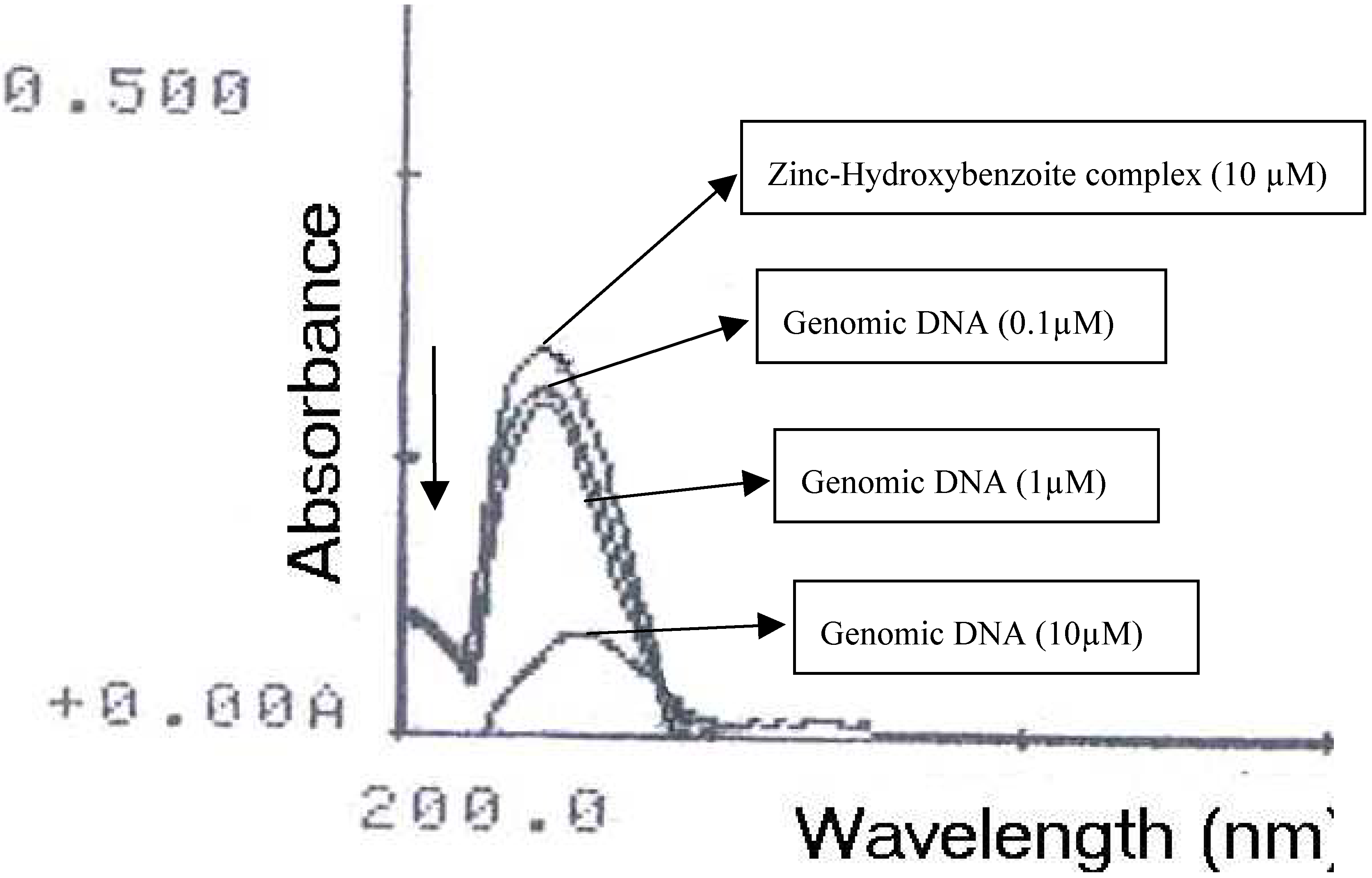
2.1 Interaction between [Zn(H20)6] (p-HO-C6H4COO)2. 2H20 complex and genomic DNA

We examined the effect of increasing concentration of Zinc-Hydroxybenzoite, [Zn(H20)6](p-HO-C6H4COO)2·2H20, at pH 7.4 on genomic DNA. The changes in both intensity, mobility, and other small fragments were monitored by agarose gel electrophoresis. Genomic DNA originally appeared as a bright streaking band at pH 7.4 (Fig. 1), indicating that the molecular mass of the DNA covered a wide range of values. When it was allowed to interact with [Zn(H20)6](p-HO-C6H4COO)2·2H20 at pH 7.4, it was found that although the unreacted DNA band was not very bright, there was a pronounced increase in intensity of the band for most of the concentration of [Zn(H20)6](p-HO-C6H4COO)2·2H20. Gel electrophoresis of unreacted genomic DNA at pH 7.4 gave two bands corresponding to supercoiled Form I and singly-nicked Form II (Fig. 1a) with the Form I band being not more prominent. Electrophoretic mobility of the bands was found to increase slightly with the increase in concentration of [Zn(H20)6](p-HO-C6H4COO)2·2H20. The increase in mobility could be due to a change in conformation of the DNA. The concentrations of the metal complex were varied from 0.1, 1 and 10mM. The actual changes in intensity of the bands with the increase in concentration of [Zn(H20)6](p-HO-C6H4COO)2·2H20 were as follows. First, it was found that (as in the case of unreacted DNA), the band at 0.1mM [Zn (H20)6] (p-HO-C6H4COO)2·2H20 concentration was less brighter than untreated DNA (Fig. 1a). The other two bands at next higher concentrations 1mM and 10mM had much more brightness compared to the lower concentration and untreated DNA (Fig.1a). The electrophoretic mobility of the band was found to increase slightly as the concentrations of [Zn(H20)6](p-HO-C6H4COO)2·2H20 was increased from 0.1, 1, and 10mM (Fig.1) [7]. We examined effect of incubating time period on the interaction between the complex and DNA. In the same way, the mixtures were incubated for 12 h following which the reaction was stopped by rapid cooling to 0 ºC. we found that untreated DNA band was bright. There was a pronounced decrease in intensity of the band for most of the concentration of [Zn(H20)6](p-HO-C6H4COO)2·2H20. The band at 10mM [Zn(H20)6](p-HO-C6H4COO)2·2H20 concentration was faint than untreated DNA (Fig. 1c). The other two bands at next lower concentrations 0.1 and 1mM had also faint bands compared to untreated DNA (Fig. 1c). The electrophoretic mobility of the band was found to increase slightly as the concentrations of [Zn(H20)6](p-HO-C6H4COO)2·2H20.
When genomic DNA was allowed to interact with zinc-Hydroxybenzoite complex in presence of ascorbate, the intensity of the band was found to decrease slightly as the concentration of the complex was increased (Fig.1b). In the case of Lane 1 (Fig 1b) shows that intensity of the band is found to decrease and also the mobility of band is decreased. Compared to lane2, lane3, and lane4 the increase in mobility could be due to a change in conformation of the DNA. As the concentration of the complex was increased, the mobility of the band increased slightly over the concentration range from 1, and 10 mM. The decrease in intensity and the increase in electrophoretic mobility suggest a reduction in the size of the DNA molecule due to its partial cleavage for short term incubation time period and except untreated DNA, all bands were disappeared for long period incubation 12 h (Fig. 1d). The results show that [Zn(H20)6](p-HO-C6H4COO)2·2H20 in the presence of ascorbate are somewhat more damage to genomic DNA.
In addition to the above study, the binding of Zinc-Hydroxybenzoite with genomic DNA was examined. Absorption titration can monitor the interaction of a metal complex and DNA. In general, complex bound to DNA through intercalation usually results in hypochromism and red shift, due to the strong stacking interaction between aromatic chromophore of the complex and the base pairs of DNA [23]. The absorption spectra of the complex in the absence and presence of DNA are illustrated in Fig. 2. The electronic absorption spectra of the complex is similar in shape to that of Zinc-Hydroxybenzoite, [Zn(H20)6](p-HO-C6H4COO)2·2H20. In the UV region, the intense absorption bands observed in the Zn (II) complex are attributed to intraligand pp* transition of the coordinated groups [23]. With increasing DNA concentration, the hypochromism increases and is accompanied by a red shift in the UV (UV-visible) band of the complex. In order to compare quantitatively the binding strength of the three concentrations of the complex, the intrinsic binding constants Kb of them with DNA were obtained by monitoring the changes in absorbance at 271, 273 and 268 nm for complex with increasing con­centration of DNA [23]. The intrinsic binding constants Kb of complex can be obtained from the decay of the absorbance. In general, a planar extension of the intercalative ligand would increase the strength of the interaction of the complex with DNA [23]. This significant difference in DNA binding affinity of complex can be understood as a result of the fact that the ligands display a more planar [23].
Figure 1. (a) Interaction between [Zn(H20)6](p-HO-C6H4COO)2·2H20 and genomic DNA in TAE buffer at pH 7.4 in air, and incubating for 3 h. Lane 4: untreated genomic DNA (100 ng); lanes 1-3: DNA + [Zn(H20)6](p-HO-C6H4COO)2·2H20 with 0.1, 1, and 10mM, respectively. (b) Interaction between [Zn(H20)6] (p-HO-C6H4COO)2. 2H20 and genomic DNA in presence of ascorbate in TAE buffer at pH 7.4 in air, and incubating for 3 h. Lane 4: untreated genomic DNA (100 ng); lanes 1-3: DNA + [Zn(H20)6](p HO-C6H4COO)2·2H20 with 0.1, 1, and10mM, respectively. (c) Interaction between [Zn(H20)6](p-HOC6H4COO)2·2H20 and genomic DNA in TAE buffer at pH 7.4 in air, and incubating for 12 h. Lane 1: untreated genomic DNA (100 ng); lanes 2-4: DNA + [Zn(H20)6](p-HO-C6H4COO)2·2H20 with 0.1, 1, and 10mM, respectively. (d) Interaction between [Zn(H20)6](p-HO-C6H4COO)2·2H20 and genomic DNA in presence of ascorbate in TAE buffer at pH 7.4 in air, and incubating for 12 h. Lane 1: untreated genomic DNA (100 ng); lanes 2-4: DNA + [Zn(H20)6](p-HO-C6H4COO)2·2H20 with 0.1, 1, and10mM, respectively.
Ijms 07 00111 g001
Figure 2. Absorption spectra of Zinc-Hydroxybenzoite complex (10 μM) in the absence (top) and presence of genomic DNA (0.1μM, 1μM, and 10μM). Arrow (thick) shows that the absorbance changes upon increasing DNA concentrations.
Ijms 07 00111 g002

2.2 Conclusion

When genomic DNA were allowed to interact with Zinc-Hydroxybenzoite, [Zn(H20)6](p-HO-C6H4COO)2·2H20, complex, it was found that the metal complex caused repairing to DNA. The results suggest that covalent binding of the metal complex caused a change in the conformation of genomic DNA such that more of ethidium bromide intercalated and hence an increase in intensity of the band was generally observed. The decrease in intensity of the band is believed to one or both of the following two reasons: (1) a change in conformation of the DNA due to its binding with the metal complex such that less ethidium bromide can intercalate within DNA and (2) some damage to DNA brought about by its covalent binding with the metal complex. The authors suggested that the strong binding was to N7 positions of guanine whereas the weak binding was due to the cooperatvity of the transition of DNA to a new double-helical conformation [24]. The results described in this study show that changing the ligand environment can modulate the binding property of the complex with DNA.

3. Experimental Section

3.1 Materials

Peripheral blood samples (6-9 ml) were collected into EDTA-tubes from cattle. DNA samples were isolated from the leukocytes by commercial kit (MBI Fermantas®-Genomic DNA Purification Kit #K0512, USA) using the salting out DNA extraction method. Isolated DNA concentrations were measured spectrophotometrically (Spectramax®Plus 384, Molecular Devices, USA) and DNA samples were concentrated at 100 ng/µl prior to process. All common chemicals, solvents were purchased from Aldrich and Sigma.

3.2 Synthesis

[Zn(H20)6](p-HO-C6H4COO)2·2H20, ([Zn(II)(hexaaqua)]bis(para-hydroxybenzoiate)dihydrate), was synthesized according to literature [22].

3.3 Methods

3.3.1 [Zn(H20)6](p-HO-C6H4COO)2·2H20 Genomic DNA Binding

For these series of experiments, [Zn(H20)6](p-HO-C6H4COO)2·2H20, were used as the source of reactive. The complex solutions were prepared in MilliQ water and were sterilized by passing through a Millipore filter. The pH of the solutions was adjusted to 7.4 by adding slowly NaOH solution. Solution of genomic DNA in the buffer consisting of 1mM Tris-HCI at pH 7.5, 1mM NaCI and 1mM EDTA were used [24]. Appropriate volume of the complex was added to 5µl of genomic DNA and the total volume was made up to 100 µl by adding MilliQ water so that the concentration of the complex ranged from 0.1, 1, and 10 mM while that of DNA remained unchanged in terms of nucleotide. The mixtures were then incubated at 37 ºC for 3 h and 12 h and the reaction was quenched by rapid cooling to 0 ºC. At the end of incubation, 6µl of loading dye (0.25% bromophenol blue in 40% sucrose solution) was added to the mixtures.

3.3.2 Gel Electrophoresis

Gel electrophoresis of genomic DNA was carried out with 100 µl reaction mixture. Agarose gel (1.5% w/v) in TBE buffer (45mM Tris, (Tris (hydroxymethyl) aminomethane), 45mM boric acid, and 1mM EDTA, pH 8.0) containing 0.5 µml-1 of ethidium bromide were prepared. Then, 15 µl of each of the incubated the complexes-DNA mixtures was loaded on to the gel and electrophoresis was carried out under TBE buffer system at 50 V for 1 h. At the end of electrophoresis the gel was visualized under UV light using a Bio-Rad Trans illuminator. The illuminated gel was photographed by using a Polaroid Camera (a red filter and Polaroid type of film was used) [24].

3.3.3 Interaction between [Zn(H20)6](p-HO-C6H4COO)2·2H20 complex and genomic DNA

Genomic DNAs were allowed to interact with Zinc-Hydroxybenzoite, [Zn(H20)6](p-HO-C6H4COO)2·2H20, complex. In order to compare the effect of interaction of the metal complex between genomic DNA, two sets of electrophoretic assay were carried out. Zinc-Hydroxybenzoite was also allowed to react with genomic DNA in the presence of ascorbate, followed by electrophoretic assay of DNA as described earlier. The mixture with pH adjusted to7.4 by adding 0.01M NaOH were left standing overnight at room temperature. Appropriate volumes of the mixture were added to 1µl of genomic DNA so that the concentration of the complex was varied from 0.1, 1, and 10mM. The mixtures were incubated for 3 h and 12 h following which the reaction was stopped by rapid cooling to 0ºC. At the end of incubation the mixtures were electrophorsed as described earlier.

Acknowledgements

The Authors thank Associate Professor H. Metin Erdogan of the Veterinary School, Kafkas University, for his help in the preparation of DNA. This work was supported by Kafkas University, Kars, Turkey.

References and Notes

  1. Bray, T.M.; Bettger, W.J. The physiological role of zinc as an antioxidant. Free Radic. Biol. Med. 1990, 8, 281–291. [Google Scholar] [CrossRef]
  2. Powell, S.R. The antioxidant properties of zinc. J. Nutr. 2000, 130, 1447S–1454S. [Google Scholar]
  3. Dreosti, I.E. Zinc and the gene. Mutate. Res. 2001, 475, 161–167. [Google Scholar] [CrossRef]
  4. Cousins, R.J. A role of zinc in the regulation of gene expression. Proc. Nutr. Soc. 1998, 57, 307–311. [Google Scholar] [CrossRef]
  5. Falchuk, K.H. The molecular basis for the role of zinc in developmental biology. Mol. Cell Biochem. 1998, 188, 41–48. [Google Scholar] [CrossRef]
  6. Prasad, A.S. Zinc deficiency in humans: a neglected problem. J. Am. Coll. Nutr. 1998, 17, 542–543. [Google Scholar] [CrossRef]
  7. Prasad, A.S.; Kucuk, O. Zinc in cancer prevention. Cancer Metastasis Rev. 2002, 21, 291–295. [Google Scholar] [CrossRef]
  8. Prasad, A.S. Zinc deficiency. BMJ. 2003, 326, 409–410. [Google Scholar] [CrossRef]
  9. Erkkila, K.E.; Odom, D.T.; Barton, J.K. Recognition and reaction of metallointercalators with DNA. Chem. Rev. 1999, 99, 2777–2796. [Google Scholar] [CrossRef]
  10. Metcalfe, C.; Thomas, J.A. Kinetically inert transition metal complexes that reversibly bind to DNA. Chem. Soc. Rev. 2003, 32, 215–224. [Google Scholar] [CrossRef]
  11. Haq, I.; Lincoln, P.; Suh, D.; Norden, B.; Choedhry, B.Z.; Chaires, J.B. Interaction of Delta A- [Ru (PHEN)(2)DPPZ](2+) and Lambda-[Ru(Phen)(2)DPPZ](2+) WITH DNA - A Caloimetric and Equilibrium Binding Study. J. Am. Chem. Soc. 1995, 17, 4788–4796. [Google Scholar]
  12. Arturo, S.; Giampaolo, B.; Giuseppe, R.; Maria, L.G.; Salvatore, T. The interaction of native DNA with Iron (III)-N, N-ethylene-bis (salicylideneiminato)-chloride. J. Inorg. Biochem. 2004, 98, 589–594. [Google Scholar]
  13. Maribel, N.; Efren, C.F.; Anßbal, S.; Mercedes, F.M.; Pedro, S.; Dwight, A.; Edgar, M. Design of copper DNA intercalators with leishmanicidal activity. J. Biol. Inorg. Chem. 2003, 8, 401–408. [Google Scholar]
  14. Catherine, H.; Marguerite, P.; Michael, R.; Heinz, G.; Stephanie, S.; Bernard, M. Preparation, characterization and crystal structures of manganese (II), iron (III) and copper (II) complexes of the bis[di-1,1-(2-pyridyl) ethyl] amine (BDPEA) ligand; evaluation of their DNA cleavage activities. J. Biol. Inorg. Chem. 2001, 6, 14–22. [Google Scholar] [CrossRef]
  15. Kumar, C.V.; Barton, J.K.; Turro, N.J. Photophysics of Ruthenium Complexes Bound to Double Helical DNA. J. Am. Chem. Soc. 1985, 107, 5518–5523. [Google Scholar] [CrossRef]
  16. Xu, H.; Zheng, K.C.; Deng, H.; Lin, L.J.; Zhang, Q.L.; Ji, L.N. Effects of ligand planarity on the interaction of polypyridyl Ru (II) complexes with DNA. Dalton. Trans. 2003, 3, 2260–2268. [Google Scholar]
  17. Mahadevan, S.; Palaniandavar, M. Electrochemical study of the enantioselective interaction of Tris(phen)Ru (II) with calf thymus DNA. Inorg. Chim. Acta. 1997, 254, 291–302. [Google Scholar]
  18. Xu, H.; Zheng, K.C.; Deng, H.; Lin, L.J.; Zhang, Q.L.; Ji, L.N. Interaction of Ru(II) Complex with Yeast tRNA Studied by Isothermal Clarometry. New J. Chem. 2003, 27, 1255–1263. [Google Scholar] [CrossRef]
  19. Mozaffar, A.; Elham, S.; Bijan, R.; Leila, H. Thermodynamic and spectroscopic study on the binding of cationic Zn(II) and Co(II) tetrapyridinoporphyrazines to calf thymus DNA: the role of the central metal in binding parameters. New J. Chem. 2004, 28, 1227–1234. [Google Scholar] [CrossRef]
  20. Chaires, J.B. Energetics of drug-DNA binding. Biopolymers 1998, 44, 201–215. [Google Scholar] [CrossRef]
  21. Zhang, H.; Liu, C.-S.; Bu, X.-H.; Yang, M. Synthesis, crystal structure, cytotoxic activity and DNA-binding properties of the copper (II) and zinc (II) complexes with 1-[3-(2-pyridyl) pyrazol-1-ylmethyl] naphthalene. J. Inorg. Biochem. 2005, 99, 1119–1125. [Google Scholar] [CrossRef]
  22. Shnulin, A.N.; Nadzhatov, G.N.; Amiraslanov, I.R.; Usubaliyev, B.T.; Mamedov, K.S. Diaquabis (N, N-diethylnicotinamide-N1)bis(2-hydroxybenzoato-O)cobalt(II). Koordinasionnaya Khimiya 1981, 7, 1409. [Google Scholar]
  23. Wang, X-L.; Chao, H.; Li, H.; Hang, X-L.; Liu, Y-J.; Tan, L-F.; Ji, L-N. DNA Interactions of Cobalt (III) Mixed-Polypridyl Complexes Containing Asymmetric Ligands. J. Inorg. Biochem. 2004, 98, 1143–1150. [Google Scholar] [CrossRef]
  24. Hossaian, Z.; Huq, F. Studies on the Interaction Between Ag+ and DNA. J. Inorg. Biochem. 2002, 91, 398–404. [Google Scholar] [CrossRef]

