Editorial

Expression, Characterization and Synergistic Interactions of Myxobacter Sp. AL-1 Cel9 and Cel48 Glycosyl Hydrolases

by
Norma Ramírez-Ramírez
1,
Eliel R. Romero-García
1,
Vianney C. Calderón
1,
Claudia I. Avitia
1,
Alfredo Téllez-Valencia
2 and
Mario Pedraza-Reyes
1,*
1
Instituto de Investigación en Biología Experimental (IIBE), Facultad de Química, Universidad de Guanajuato, P.O. Box 187, Guanajuato, Gto. 36050, Mexico
2
Instituto de Ciencias de la Salud, Universidad Autónoma del Estado de Hidalgo, Abasolo 600, Pachuca, Hgo 42000, Mexico
*
Author to whom correspondence should be addressed.
Int. J. Mol. Sci. 2008, 9(3), 247-257; https://0-doi-org.brum.beds.ac.uk/10.3390/ijms9030247
Submission received: 2 December 2007 / Revised: 24 January 2008 / Accepted: 1 February 2008 / Published: 29 February 2008
(This article belongs to the Special Issue Biofuels R&D: Securing the Planet's Future Energy Needs)

Abstract

:
The soil microorganism Myxobacter Sp. AL-1 regulates in a differential manner the production of five extracellular cellulases during its life cycle. The nucleotide sequence of a cel9-cel48 cluster from the genome of this microorganism was recently obtained. Cel48 was expressed in Escherichia coli to generate a His6-Cel48 protein and the biochemical properties of the pure protein were determined. Cel48 was more efficient in degrading acid-swollen avicel (ASC) than carboxymethylcellulose (CMC). On the other hand, cel9 was expressed in Bacillus subtilis from an IPTG-inducible promoter. Zymogram analysis showed that after IPTG-induction, Cel9 existed in both the cell fraction and the culture medium of B. subtilis and the secreted protein was purified to homogeneity by FPLC-ionic exchange chromatography. The exocellobiohydrolase Cel48 showed a synergism of 1.68 times with the endocellulase Cel9 during ASC degradation using an 8.1-fold excess of Cel48 over Cel9. Western blot analysis revealed that both proteins were synthesized and secreted to the culture medium of Myxobacter Sp. AL-1. These results show that the cel9-cel48 cluster encodes functional endo- and exo-acting cellulases that allows Myobacter Sp. AL-1 to hydrolyse cellulose.

1. Introduction

Myxobacter Sp. AL-1 regulates in a temporal fashion the production of at least five extracellular cellulases with different molecular weights. A 29 kDa protein, part of this enzyme battery, was purified to homogeneity and showed to possess chitosanase-cellulase activity [1]. Furthermore, a gene encoding a family 9 extracellular cellulase was cloned from the genome of Myxobacter Sp. AL-1 [2]. Sequences located downstream of cel9 showed the existence of a contiguous partial reading frame, which putatively coded for another cellulase, termed cel48 [2]. The first gene of this cluster, cel9, was successfully expressed in Escherichia coli and the biochemical properties of the pure protein were very similar as those exhibited by related cellulases produced by thermophilic bacteria [2]. Moreover, results of a Gapped-blast alignment analysis revealed that Cel9 is a modular enzyme composed of N-terminal catalytic and C-terminal type IIIc cellulose binding domains, respectively [3].
The complete degradation of cellulose occurs with the action of endo-1,4-β-glucanases and exo-1,4-β-glucanases. The endo-acting enzymes attack the cellulose chain internally whereas the exo-1,4-β-glucanases attack the reducing or non-reducing end of the cellulose chain to generate cellobiose or glucose [4, 5]. The ability of Myxobacter Sp. AL-1 to grow in CMC as a sole carbon source (Our unpublished results) and the presence in this microorganism of a cel9-cel48 cluster suggest that this soil microorganism possesses the enzymatic machinery to degrade cellulose. However, since the full nucleotide sequence of cel48 was not available, the biochemical properties of its encoding product remained unknown. In this communication, we report the expression of cel48 in E. coli and the purification of its encoding product as a His6-Cel48 protein. His6-Cel48 showed to possess biochemical properties as those described for cellobiohydrolases from bacterial origin belonging to the family 48 of the glycosyl hydrolases. Moreover, the expression of cel9 from an IPTG-inducible promoter was carried out in Bacillus subtilis. The recombinant Cel9 protein was secreted to the culture medium of B. subtilis cells and was purified to homogeneity by FPLC. Results from a Western-blot analysis revealed that Cel48 as well as Cel9 are produced and secreted by Myxobacter Sp. AL-1; moreover, both proteins acted together, in a synergistic manner, to process the degradation of acid-swollen avicel.

2. Methodology

Bacterial strains, plasmids and growth conditions

Strains used in this study are shown in Table 1. Media used was Luria-Bertani (LB) [6]. When necessary antibiotics were added to media at the following final concentrations: ampicillin (Amp), 100 μg/mL; kanamycin (Kan), 10 μg/mL. Cells were grown in liquid media with vigorous aeration or on solid media at 37°C. Growth in the cultures was determined by measuring the optical density at 600 nm using an Ultrospec Pharmacia spectrophotometer.

Genetic and molecular biology techniques

Transformation of E. coli, small and large scale preparation of plasmid DNA, enzymatic manipulations and agarose gel electrophoresis were performed by standard techniques [7]. PCR products for nucleic acid sequencing were obtained with homologous oligonucleotides and Vent DNA polymerase (New England Biolabs, Beverly, MA).

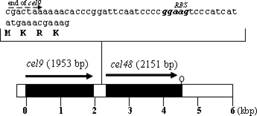
Plasmid construct to overexpress cel48 and generate a His6-Cel48 protein

The open reading frame of cel48 was amplified by PCR, using plasmid pPERM123 (Table 1) with specific oligonucleotide primers designed to insert BamHI and SalI restriction sites into the cloned DNA. The PCR fragment was first introduced into PCR-Blunt-II-TOPO plasmid to generate pPERM400 which was replicated into E. coli XL-10 Gold KanR (Stratagene, La Jolla, CA). pPERM400 was digested with SalI and BamHI and the cel48 ORF fragment was inserted in-frame into the BamHI/SalI site of the expression vector pQE30 (QIAGEN Inc. Valencia, CA); the resulting construction pPERM407 was introduced into E. coli XL-10 Gold KanR (Stratagene, La Jolla, CA) generating strain E. coli PERM407. The proper in-frame insertion of the cel48 fragment was assessed by both restriction analysis and DNA sequencing.

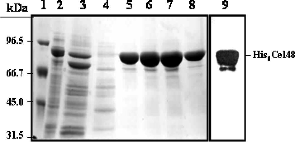
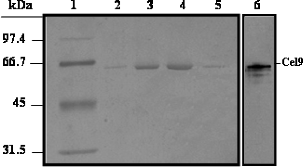
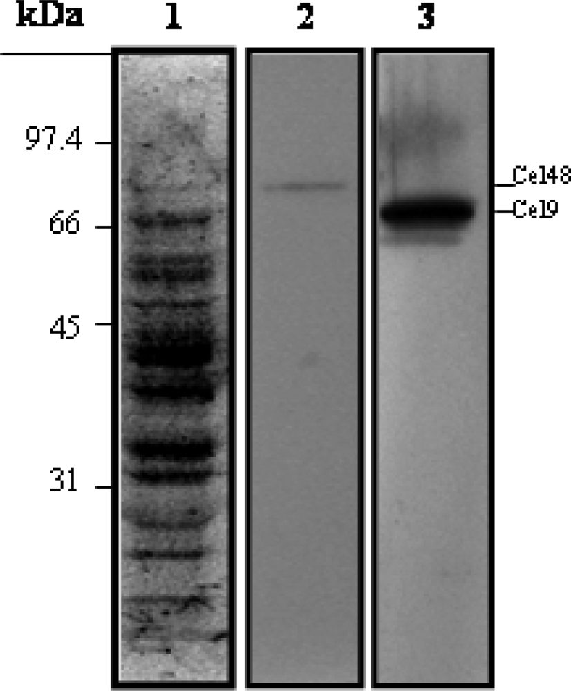
Purification of His6-Cel48 and Cel9

E. coli PERM407 was grown in 50 ml of LB medium supplemented with Amp to an OD600nm of 0.5. Expression of the cel48 gene was induced during 2 h, at 37°C by addition of isopropyl-β-D-thio-galactopyranoside (IPTG) to 0.2 mM. Cells were collected by centrifugation and washed two times with 50 mM Tris-HCl (pH 6.0, 10 mL), 150 mM NaCl (buffer A). The cells were disrupted in buffer A (10 mL) containing lysozyme (5 mg/mL) for 1 h at 37°C. The cell homogenate was subjected to centrifugation (27,200 × g) to eliminate undisrupted cells and cell debris and the supernatant was applied to a 5 mL Ni-NTA-Agarose (QIAGEN Inc.) column, previously equilibrated with buffer A. The column was washed with buffer A (50 mL) and buffer A containing 10 mM imidazole (50 mL) and the protein bound to the resin was eluted with buffer A (6 mL) containing imidazole (200 mM); 4 mL fractions were collected during this last step. Purified fractions were analyzed by sodium dodecyl sulfate polyacrylamide gel electrophoresis (SDS-PAGE) as previously described [8].
The protein secreted to the culture medium by B. subtilis PERM272 after 12 h of IPTG induction was precipitated with ammonium sulfate and the lyophilized precipitate was subjected to purification on a Mono Q HR 16/10 anion-exchange column as previously described [2].

Immunological procedures

Proteins present in the culture medium of B. subtilis PERM272 and cell extract from E. coli PERM407 were separated in SDS 10% polyacrylamide gels. The proteins bands corresponding to Cel9 and His6-Cel48, respectively, were cut out from the gel, homogenized, mixed 1:1 with Freud adjuvant, and injected into New Zealand White rabbits. The antiserums were collected 12 weeks after the first immunization. Western blot analyses were performed with the antiserums diluted 10,000-fold and then processed with an ECL (Enhanced chemiluminescence) western blotting analysis system (Amersham Pharmacia, UK).

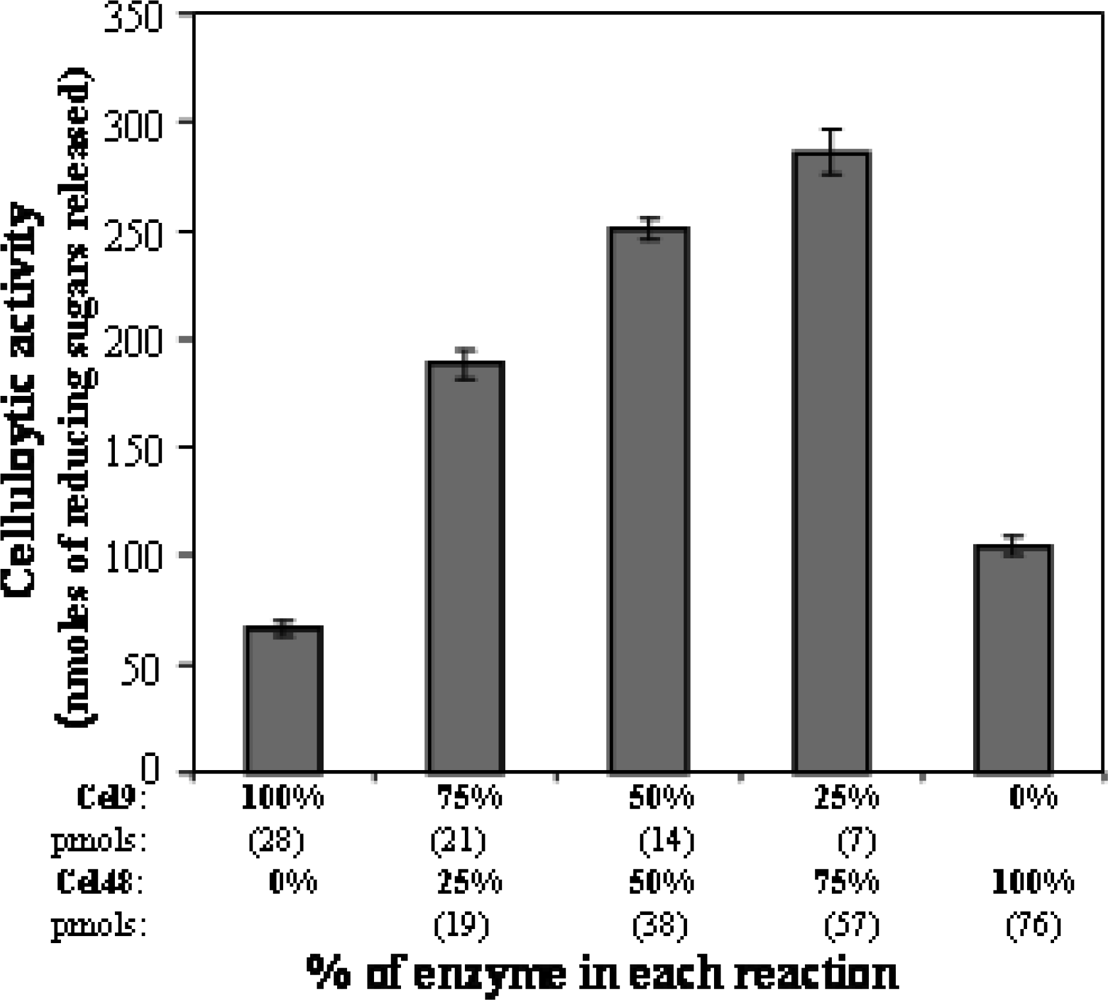
Enzyme and protein assays

Cellulase activity was determined by measuring the enzymatic release of reducing sugars as previously described [9, 10]. Acid-swollen avicel (ASC) and CMC were used at a final concentration of 0.5% and 1%, respectively. p-Nitrophenyl-β-D-cellobioside (p-NP-Glc2) and p-nitrophenyl-β-D-cellotrioside (p-NP-Glc3) were used at a final concentration of 4 mM. Units of enzyme activity were reported as nmols of reducing sugars released/min/mg of protein. ASC was prepared as previously described [11].

SDS-PAGE and cellulase zymography

Electrophoresis in SDS-polyacrylamide gels was carried out as previously described by Laemmli [8]. Gels were stained with Coomassie blue R-250. Zymograms of cellulase activity were performed in SDS-polyacrylamide gels containing CM-cellulose according to a previously described protocol [2].

3. Results and Discussion

Since regions lying downstream of cel9 showed the presence of a second partial ORF, encoding a putative cellulase, it was of further interest to characterize this gene. Thus, the full DNA sequence of this second gene was obtained by PCR walking using homologous oligonucleotides and the results revealed an ORF of 2151 bp (GenBank accession number 778327) encoding a protein with a predicted molecular mass of 81 kDa and a theoretical isoelectric point of 5.4 (Figure 1). A Gapped-blast analysis (results not shown) showed that this protein possessed identity with cellobiohydrolases of bacterial origin grouped in the family 48 of the glycosyl hydrolases [12], therefore this gene was termed cel48. Interestingly, Cel48C a 118 kDa cellobiohydrolase from Paenibacillus sp. BP-23 [13] contains a type 3a cellulose-binding module as well as a fibronectin-III domain on its C-terminal end which are absent in Cel48.
To determine the biochemical and biophysical properties of Cel48, this protein was produced and purified from E. coli cells transformed with plasmid pPERM407. Cell-free extracts of the transformed cells showed the presence of a protein of around 81 kDa (Figure 2, Lane 2), which was subsequently purified on a Ni-NTA-agarose (Figure 2, Lanes 5–8) and was recognized by an anti-Cel48 antibody in the cell free extracts of this E. coli strain (Figure 2, Lane 9).
Although the recombinant His6-Cel48 protein catalyzed the hydrolysis of ASC and CMC, the enzyme was more active against the former substrate (Table 2). It has been previously described that family 48 cellobiohydrolases catalyze the hydrolysis of cellulosic substrates such as ASC in a slow manner [9, 13]. In agreement with this observation, maximum hydrolysis of ASC and CMC by His6-Cel48 occurred after 18–24 h at pH 6 (Results not shown).
Moreover, the recombinant enzyme showed to be very stable at least for 2 h periods, at 23, 37 and 45°C; however, the enzyme was rapidly inactivated at 60°C or higher temperatures (Results not shown). On the other hand, as shown in Table 2, no activity of His 6-Cel48 was detected against avicel and the oligomeric substrates p-nitrophenyl β-D-cellobioside (p-NP-Glc2) and p-nitrophenyl β-D-cellotrioside (p-NP-Glc3). Thus, in terms of substrate specificity, His6-Cel48 resembled the catalytic domain of Cel48A from the thermophile bacterium Thermomonospura fusca (Cel48Acd), since the recombinant form of this enzyme showed activity against ASC and CMC but was inactive against cello-oligosaccharides [9]. Other properties such as optimal pH and thermal stability were also shared by both enzymes.
The synergistic degradation of cellulose by endoglucanases and exoglucanases has been previously documented [1317]. It has been proposed that the synergistic reaction between both classes of enzymes involves a mechanism in which the endoglucanases attack the amorphous portions of cellulose followed by the action of exoglucanases which release cellobiose from the internal nicks [14].
The existence of contiguous cel9 and cel48 genes encoding endo- and exo-acting cellulases suggested that Myxobacter Sp. AL-1 possessed the enzymatic machinery to degrade cellulose. To investigate whether Cel48 together with Cel9 catalyzed the degradation of ASC in a synergistic manner it was necessary to purify Cel9. To this end, the protease and β-glucanase deficient strain B. subtilis 1A751 (Table 1) was used as a host to express cel9. Thus, cel9 was cloned in the plasmid pDG148 [18] under the control of the IPTG-inducible Pspac promoter and the resulting construction was introduced in B. subtilis 1A751 generating the strain B. subtilis PERM272. As shown in Figure 3, addition of IPTG to a mid logarithmic phase culture of strain B. subtilis PERM272 induced around five times the levels of CM-cellulase expression. The maximum levels of CM-cellulase activity were reached 10 h after addition of the inducer and these values were maintained during the following 14 h. SDS-PAGE and zymogram analysis revealed the presence of a protein with the predicted molecular mass of Cel9 that also possessed CM-cellulase activity in the culture medium of B. subtilis PERM272.
Since B. subtilis was able to synthesize and secrete Cel9 we used a culture of strain B. subtilis PERM272 induced with IPTG for 12 h as a source to purify this protein. To this end, after separating the cells, the secreted proteins were concentrated by ammonium sulfate precipitation and then subjected to purification on a Mono-Q HR 16/10 anion-exchange column. After applying a linear salt gradient, Cel9 was solved on this column as a single peak of CM-cellulase activity which was eluded with around 0.12 M NaCl (Results not shown). SDS-PAGE analysis showed that Cel9 was purified to apparent homogeneity after this chromatographic step (Fig. 4). Following this protocol, Cel9 was purified five times with a 60% recovery (Results not shown). Results shown in Figure 4 (Lane 6) revealed that the ~66 kDa purified protein was specifically recognized by an anti-Cel9 antibody in the supernatant of the IPTG-induced strain B. subtilis PERM272.
Having available purified samples of both Cel9 and Cel48 it was further investigated whether these proteins catalyzed the degradation of ASC in a synergistic manner. To this end, either individual samples or different ratio’s mixtures of Cel9 and Cel48 were incubated with ASC at a final concentration of 0.5% during 18 h at 40°C. A synergistic interaction between Cel9 and Cel48 during ASC degradation was observed since the activity of all mixtures were significantly higher than that of either enzyme assayed alone (Fig. 5). The enzyme Cel48 showed a synergism of 1.68 times with the endoglucanase Cel9 during ASC degradation using an 8.1-fold excess of Cel48 over Cel9 (Figure 5).
Taken these results together is reasonable to conclude that the genetic information present in the cel9-cel48 cluster of Myxobacter Sp. AL-1 encodes functional enzymes which catalyze synergistically the degradation of cellulose. However, as noted above, both, Cel9 and Cel48 were synthesized in heterologous hosts and the existence of these enzymes in Myxobacter Sp. AL-1 remained to be demonstrated. Regarding this issue, previous results of a zymogram analysis suggested that a ~70 kDa protein secreted into the culture medium of Myxobacter Sp. AL-1 could correspond to Cel9 [2]. However, to date the production of Cel48 by Myxobacter Sp. AL-1 has not been proven. Thus, the polyclonal antibodies generated against Cel9 and Cel48 were used to localize both proteins in the cell culture of this microorganism. The results shown in Figure 6 reveled that the anti-Cel9 and anti-Cel48 antibodies were able to recognize proteins with molecular masses of around 67 and 82 kDa, respectively, in the supernatant of a 24 h culture of Myxobacter Sp. AL-1. These results strongly suggest that Cel9 and Cel48 are produced and secreted by Myxobacter Sp. AL-1 to process the hydrolysis of cellulose.
In conclusion, the results described in this work add new evidence on the existence of the enzymatic machinery to process the synergistic degradation of cellulose in the soil microorganism Myxobacter Sp. AL-1.

Acknowledgments

This work was supported by the University of Guanajuato and by grant 43644 from the Consejo Nacional de Ciencia y Tecnología (CONACYT) of México to Mario Pedraza-Reyes. Norma Ramírez and Eliel R. Romero-García were supported by fellowships from CONACYT. The authors wish to thank to Silvia Mellado Reyes for technical assistance

References

  1. Pedraza-Reyes, M; Gutiérrez-Corona, F. The bifunctional enzyme chitosanase-cellulase produced by the gram-negative microorganism Myxobacter sp. AL-1 is highly similar to Bacillus subtilis endoglucanases. Arch. Microbiol. 1997, 168, 321–327. [Google Scholar]
  2. Avitia, CI; Castellanos-Juárez, FX; Sánchez, E; Téllez-Valencia, A; Fajardo-Cavazos, P; Nicholson, W; Pedraza-Reyes, M. Temporal secretion of a multi-cellulolytic system in Myxobacter Sp. AL-1: Molecular cloning and heterologous expression of cel9 encoding a modular endocellulase clustered in an operon with cel48, an exocellobiohydrolase gene. Eur. J. Biochem. 2000, 267, 7058–7064. [Google Scholar]
  3. Téllez-Valencia, A; Sandoval-Carrillo, A; Pedraza-Reyes, M. The non-catalytic amino acid Asp446 is essential for enzyme activity of the modular endocellulase Cel9 from Myxobacter sp. AL-1. Current Microbiol. 2003, 46, 307–310. [Google Scholar]
  4. Béguin, P; Aubert, J. The biological degradation of cellulose. FEMS Microbiol. Rev. 1994, 13, 25–28. [Google Scholar]
  5. Teeri, TT. Crystalline celullose degradation: new insight into the function of cellobiohydrolases. TIB TECH. 1997, 15, 160–167. [Google Scholar]
  6. Miller, JH. Experiments in molecular genetics; Cold Spring Harbor Laboratory: Cold Spring Harbor, NY, 1972. [Google Scholar]
  7. Sambrook, J; Fritsch, EF; Maniatis, T. Molecular cloning: a laboratory manual, 2nd ed; Cold Spring Harbor Laboratory: Cold Spring Harbor, NY, 1989. [Google Scholar]
  8. Laemmly, UK. Cleavage of structural proteins during the assembly of the head of bacteriophage lambda T4. Nature 1970, 227, 680–685. [Google Scholar]
  9. Irwin, DC; Sheng, S; Wilson, DB. Cloning, expression and characterization of a family 48 exocellulase, Cel48A, from Thermobifida fusca. Eur. J. Biochem. 2000, 267, 4988–4997. [Google Scholar]
  10. Ghuysen, J; Tipper, DJ; Strominger, JL. Enzymes that degrade bacterial cell walls. In Methods in Enzymology; Newfield, EF, Ginsberg, V, Eds.; Academic Press: New York, NY, 1996; pp. 685–689. [Google Scholar]
  11. Sakamoto, R; Arai, M; Murao, S. Enzymatic properties of hydrocellulase from Aspergillus acuelatus. J. Ferment. Technol. 1984, 62, 561–567. [Google Scholar]
  12. Coutinho, PM; Henrissat, B. Carbohydrate Active Enzymes Server. 1999. available at http://www.cazy.org/CAZY/fam/acc_GH.html.
  13. Sánchez, MM; Javier Pastor, FI; Díaz, P. Exo-mode of action of cellobiohydrolase Cel48C from Paenibacillus sp-BP-23. A unique type of cellulase among Bacillales. Eur. J. Biochem. 2003, 270, 2913–2919. [Google Scholar]
  14. Wood, TM; McCrae, SI. Synergism between enzymes involved in the solubilization of native cellulose. Adv. Chem. Ser. 1979, 181, 181–209. [Google Scholar]
  15. Lynd, LR; Weimer, PJ; W. van Zyl, H; Pretorius, IS. Microbial cellulose utilization: fundamentals and biotechnology. Microbiol. Mol. Biol. Rev. 2002, 66, 506–577. [Google Scholar]
  16. Murashima, K; Kosugi, A; Doi, RH. Synergistic effects on crystalline cellulose degradation between cellulosomal cellulases from Clostridium cellulovorans. J. Bacteriol. 2002, 184, 5088–5095. [Google Scholar]
  17. Qi, M; Jun, H-S; Forsberg, CW. Characterization and synergistic interactions of Fibrobacter succinogenes glycoside hydrolases. Appl. Environ. Microbiol. 2007, 73, 6098–6105. [Google Scholar]
  18. Sun, D; Stragier, P; Setlow, P. Identification of a new sigma factor which allows RNA polymerase to transcribe the sspE gene and other forespore specific genes during sporulation of Bacillus subtilis. Genes Dev. 1989, 3, 141–149. [Google Scholar]
Figure 1. Genetic organization of the cel9-cel48 cluster of Myxobacter Sp. AL. 1. Solid arrows show the ORFs of cel9 and cel48, respectively. A steam loop structure downstream of cel48 (white) represent a putative transcriptional terminator. The end of cel9 is shown with a discontinuous arrow. The presumptive ribosome binding site of cel48 is shown in bold italics.
Ijms 09 00247 f1
Figure 2. SDS-PAGE analysis of His6-Cel48 purification through a Ni-NTA-agarose column. Expression and purification of His6-Cel48 from E. coli PERM407 was performed as described in Methodology. (Lane 1, molecular weight markers; Lane 2, E. coli PERM407 lysate; Lane 3, flow through; Lane 4, protein fraction eluded with 10 mM imidazol; Lanes 5–8, fractions eluted from the column with 200 mM imidazol; Lane 9, ~100 μg of protein from an E. coli PERM407 lysate were separated on a SDS–12% polyacrylamide gel and transferred to nitrocellulose membranes. The blot was probed with a polyclonal anti-Cel48 rabbit antibody which was diluted 10,000-fold and then processed with an ECL Western-blotting analysis system.)
Ijms 09 00247 f2
Figure 3. Analysis of cel9 induction in a B. subtilis strain harboring a Pspac-cel9 construction. A. B. subtilis PERM272 was grown at 37°C in LB medium to an OD600nm of 0.5. At this point the culture was divided in two equal subcultures; one of the subcultures was left as a control (●, ○) and the other was supplemented with 2 mM IPTG (■, □). Aliquots (1 mL) were taken at different times from both subcultures; cells were separated by centrifugation and the supernatant used to determine CM-cellulase activity (○, □) as described in Methodology. (●, ■) OD600nm. B. Samples (~100 μg protein) collected at different times from the supernatant of the IPTG-induced subculture were analyzed on zymograms as described in Methodology.
Ijms 09 00247 f3
Figure 4. SDS-PAGE analysis of Cel9 purification through a Mono Q HR 16/10 column. Fractions (15 μL) from the peak of CM-cellulase activity from a Mono Q HR 16/10 anion-exchange column (Lanes 2–5) were analyzed on a SDS-12% polyacrylamide gel. (Lane 1, Molecular weight markers. Lane 6, ~100 μg of protein from the supernatant of an IPTG-induced B. subtilis PERM272 culture were separated on a SDS–12% polyacrylamide gel and transferred to a nitrocellulose membrane. The blot was probed with a polyclonal anti-Cel9 rabbit antibody which was diluted 10,000-fold and then processed with an ECL Western-blotting analysis system.)
Ijms 09 00247 f4
Figure 5. Cellulase activity (nmols of reducing sugars released) against ASC by mixtures of Cel9 and Cel48 after 18 h of incubation at 40°C. Purified Cel9 and Cel48 were used alone or combined at the indicated ratios in the assay mixtures (500 μL). The pmols of each enzyme used in the assay mixtures are indicated in parenthesis.
Ijms 09 00247 f5
Figure 6. Westen blot analysis of the synthesis of Cel9 and Cel48 during Myxobacter Sp. AL-1 growth. 100 μg of protein from the culture medium collected at 24 h were separated by SDS–PAGE and transferred to nitrocellulose membranes. The blots were stainned with Ponceau’s red (Lanes 1) and probed with polyclonal anti-Cel48 (Lane 2) or anti-Cel9 (Lane 3) rabbit antibodies which were diluted 10,000-fold and then processed with an ECL Western-blotting analysis system.
Ijms 09 00247 f6
Table 1. Bacterial strains used in this study.
Bacterial strainGenotype and descriptionReference or source
E. coli XL10-Gold KanR{Tetr Δ(mcrA) 183, Δ(mcrBC-hsd SMR-mrr); Kan 173 endA1 sup E44 thi-1 recA1 gyrA96 relA1 lacHte [F’ proAB lacIqZDM15 Tn10 (Tetr) Tn5 (Kanr) Amy]}(Stratagene, La Jolla, CA.)
E. coli DH5α{deoR [φ80dlacΔ(lacZ)M15] recA1 endA1 gyrA96 hsdR17 (rk, mk+) phoA supE44 thi-1 relA1 Δ(lacIZYA-argF)U169}.Laboratory stock
E. coli PERM123E. coli DH5α containing plasmid pPERM123 (pBR322 with a 6 kb-EcoRI-EcoRI cel9-cel48 containing fragment).[2]
B. subtilis 1A751eglSΔ102, bglT/bglSΔEV, npr, aprE, hisBGSCa
B. subtilis PERM272B. subtilis IA751 containing plasmid pDG148 with An IPTG-inducible Pspac-cel9 construction[2]
E. coli PERM407E. coli XL10-Gold containing plasmid pQE30 with an IPTG inducible PT5-His6-cel48 constructionThis study
aBGSC (Bacillus genetic stock center)
Table 2. Substrate specificity of recombinant His6-Cel48.
SubstrateSpecific Activity
ASC10.5a
CMC2.75
AvicelNDb
p-NP-Glc2ND
p-NP-Glc3ND
aIndividual values are the mean of triplicate assays, where values did not deviate by more than 15% of the mean;
bND, not detected

