
4-[(4-Chlorophenyl)carbamoyl]butanoic Acid
by
 , , ,
, , ,
Bibi Hanifa
1, 
Muhammad Sirajuddin
1,*,
Ahmed Bari
2,
See Mun Lee
3 ,
,
Kong Mun Lo
3 and
Edward R. T. Tiekink
3,* 1
Department of Chemistry, University of Science and Technology, Bannu 28100, Pakistan
2
Department of Pharmaceutical Chemistry, College of Pharmacy, King Saud University, Riyadh 11451, Saudi Arabia
3
Research Centre for Crystalline Materials, School of Medical and Life Sciences, Sunway University, No. 5 Jalan Universiti, Bandar Sunway 47500, Selangor Darul Ehsan, Malaysia
*
Authors to whom correspondence should be addressed.
Molbank 2021, 2021(2), M1209; https://0-doi-org.brum.beds.ac.uk/10.3390/M1209
Submission received: 14 April 2021
/
Revised: 28 April 2021
/
Accepted: 30 April 2021
/
Published: 6 May 2021
(This article belongs to the Section Structure Determination)
Abstract
:The X-ray crystal structure determination of the glutaric acid-amide derivative, 4-ClC6H4N(H)C(=O)(CH2)3C(=O)OH (1), is described. The backbone of the molecule adopts an extended, all-trans configuration but the terminal carboxylic acid and phenyl resides are twisted out of the plane through the bridging atoms, as seen in the torsion angles of O(carboxylic acid)–C(m)–C(m)–C(m) [13.9(5)°] and C(m)–N–C(p)–C(p) [47.1(4)°]; m = methylene and p = phenyl. The most striking feature of the molecular packing is the formation of supramolecular tapes mediated by carboxylic acid-O–H⋯O(carbonyl) and amide-N–H⋯O(amide) hydrogen bonding.
1. Introduction
The title compound, 4-ClC6H4N(H)C(=O)(CH2)3C(=O)OH (1), Scheme 1, was investigated crystallographically in continuation of recent structural studies on related glutaric acid amides [1,2,3,4]. Compound (1) was originally reported in 1957 in the context of potential anti-bacterial activity [5] and has been subsequently investigated for the inhibition of sunflower seedling development [6] and antinociceptive properties [7]. Further, (1) and related derivatives have been employed as intermediates in reactions leading to the synthesis of bis-pyrazolo-1,4-dihydro-pyridines [8] and 2,2′-(N-phenylpiperidine- 2,6-diylidene)dimalononitriles [9]. Herein, the spectroscopic and crystallographic characterization of (1) is described.
2. Results and Discussion
Colorless crystals of (1) were prepared in 81% yield from the 1:1 reaction of 4-chloro aniline and glutaric anhydride, and was characterized by IR, 1H and 13C{1H} NMR, and UV spectroscopy; original spectra are found in the Supplementary Materials. In the IR spectrum, characteristic absorptions were apparent, for example, at 1689, 1662, and 1436 cm−1, assigned to ν(amide C=O), ν(COasym), and ν(COsym), respectively. The 1H NMR-recorded DMSO-d6 solution revealed broad resonances at δ 12.10 and 10.03 ppm, ascribed to OH and NH, respectively. In the 13C{1H} NMR (DMSO-d6), downfield resonances at δ 174.6 and 171.3 ppm were assigned to CO2 and C(=O), respectively. In the UV (acetonitrile) spectrum, the three well-defined absorptions observed at 250.0, 200.6, and 191.2 nm are ascribed to n-π*(C=O), π-π*(C=O), and π-π*(C=C) transitions, respectively. Additional characterization of (1) was achieved through X-ray crystallography.
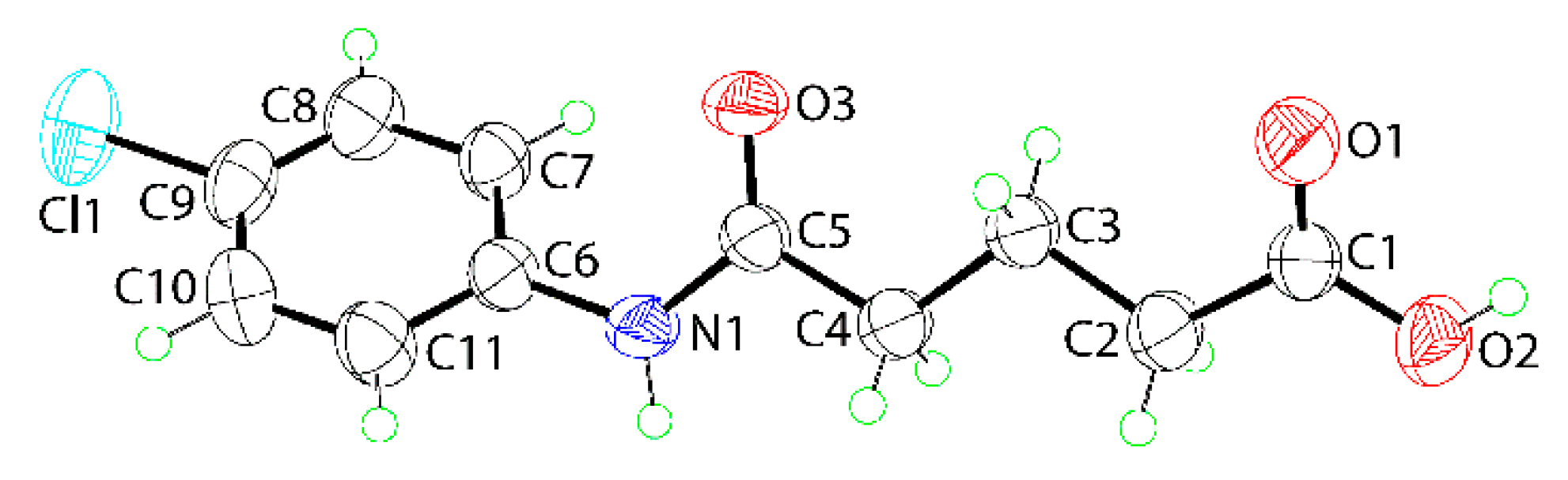
The molecular structure of (1) is shown in Figure 1. The confirmation of the carboxylic acid assignment is readily seen in the values of the C1–O1 and C1–O2 bond lengths of 1.226(4) and 1.301(4) Å, and in the supramolecular association, as detailed below. The sequence of C1–C2–C3–C4 [−171.7(3)°], C2–C3–C4–C5 [−173.6(3)°], C3–C4–C5–N1 [−171.0(3)°], and C4–C5–N1–C6 [179.1(3)°] torsion angles indicates that the backbone of the molecule has an all-trans configuration and an extended conformation. However, there are twists at the extremities of the molecule, as noted in the O1–C1–C2–C3 [13.9(5)°] and C5–N1–C6–C7 [47.1(4)°] torsion angles.
The most closely related structure in the literature is that of the 3,5-dichlorophenyl analog [3]. Here, the basic features as noted for (1) are evident but there is a notable twist about the C3–C4 bond, as seen in the value of the C2–C3–C4–C5 torsion angle of 78.5(2)°.
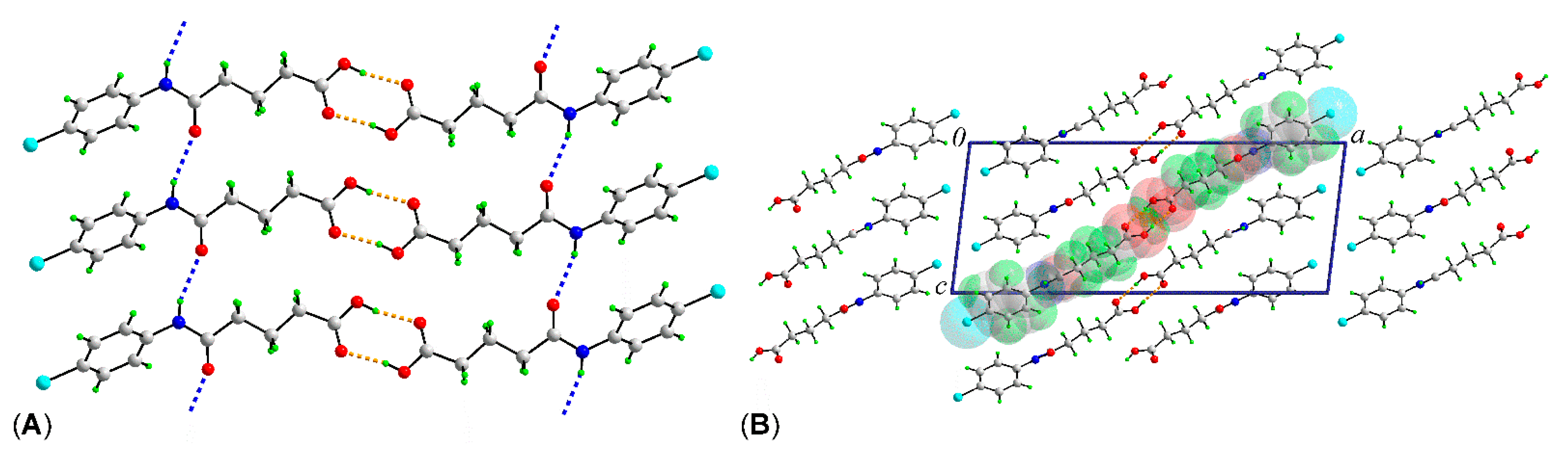
As anticipated from the chemical composition, there are significant hydrogen-bonding interactions at play in the molecular packing in the crystal of (1); geometric parameters describing these are given in the caption to Figure 2. The carboxylic acid residues assemble about a center of inversion to form eight-membered {⋯OCOH}2 synthons within two-molecule aggregates. As shown in Figure 2a, the two-molecule aggregates are assembled into a supramolecular tape via amide-N–H⋯O(amide) hydrogen bonds. In accordance with the distance criteria in PLATON [10], the tapes, which lie parallel to (1 0 1), pack in the crystal without directional interactions between them, as shown in Figure 2b. A similar supramolecular tape was observed in the crystal of the aforementioned 3,5-dichlorophenyl analog [3].
In conclusion, the X-ray crystallographic study established an all-trans configuration in the bridge linking the carboxylic acid and phenyl residues, with each of the latter being twisted out of the plane through the rest of the molecule, in particular the phenyl ring. Supramolecular tapes, two molecules wide, feature in the molecular packing mediated by carboxylic acid-O–H⋯O(carbonyl) hydrogen bonding, to form two-molecule aggregates, and amide-N–H⋯O(amide) hydrogen bonding which links the former into the tape.
3. Materials and Methods
3.1. General Information
All standard chemicals and solvents were sourced from Macklin (Pudong, Shanghai, China) and Sigma (Saint Louis, MO, USA) and used without further purification. The melting point was determined on a BioCote melting point apparatus (Staffordshire, UK). Elemental analyses were performed on a PerkinElmer CHNS 2400 instrument (Waltham, MA, USA). The FTIR spectrum was measured on a Thermo Nicolet-6700 spectrophotometer (Vienna, Austria) from 4000–450 cm−1. The 1H and 13C{1H} NMR spectra were recorded in DMSO-d6 solution on a Bruker Avance 500 MHz NMR (Billerica, MA, USA) spectrometer with chemical shifts relative to tetramethylsilane. The optical absorption spectrum was obtained from an acetonitrile solution (4.14 × 10⁻⁵ M) in the range 185–340 nm on a Shimadzu UV-3600 plus UV/VIS/NIR (Shimadzu Corporation, Kyoto Prefecture, Japan) spectrophotometer.
3.2. Synthesis and Characterization of (1)
4-Chloro aniline (0.64 g, 5 mmol) and glutaric anhydride (0.57 g, 5 mmol) were dissolved separately in about 10–15 mL analytical grade toluene. The two solutions were then slowly mixed and stirred at room temperature until the appearance of precipitate. This was filtered, washed with a minimal amount toluene (to remove any unreacted reactants), and then with water (to remove any glutaric acid that may have formed during the reaction). The desired compound was air-dried and recrystallized in the solvent mixture ethanol/acetone (1:1 v/v) to yield colorless crystals after one week. Yield: 81%. M.pt.: 138–139 °C. Anal. Calc. for C11H12ClNO3: C, 54.67; H, 5.01; N, 5.80%. Found: C, 54.19; H, 5.00; N, 5.87%. FTIR: 3305 (m) ν(N–H), 3189 (w) ν(O–H), 1689 (s) ν(amide C=O), 1662 (s) ν(COasym), 1589 (m) ν(C=C), 1436 (m) ν(COsym). 1H NMR (DMSO-d6): δ 12.10 (s, 1H, OH), 10.03 (s, 1H, NH), 7.63 (d, 2H, H7,11, 3JHH = 5 Hz), 7.35 (d, 2H, H8,10, 3JHH = 5 Hz), 2.36 (t, 2H, H4, 3JHH = 5 Hz), 2.28 (t, 2H, H2, 3JHH = 5 Hz), 1.81 (quint, 2H, H3, 3JHH = 5 Hz). 13C{1H} NMR (DMSO-d6): δ 174.6 (C1), 171.4 (C5), 138.7 (C6), 129.0 (C8,10), 126.9 (C9), 121.0 (C7,11), 35.8 (C4), 33.4 (C2), 20.8 (C3). UV (acetonitrile; nm, L.cm−1.M−1): λabs = 250.0 (ε = 21,541; n-π*(C=O)), 200.6 (27,476; π-π*(C=O)), 191.2 (67,181; π-π*(C=C)).
3.3. Crystallography
Intensity data for colorless crystal of (1) (0.04 × 0.11 × 0.17 mm) were measured at T = 294(2) K on a XtaLAB Synergy Dual AtlasS2 (Rigaku Polska SP. Z O O, Wrocław, Poland) diffractometer fitted with Cu Kα radiation (λ = 1.54184 Å) using ω-scans in the θmax range 3.7°–67.1°. Data reduction, including absorption correction, was accomplished with CrysAlis Pro [11]. Of the 13,024 reflections measured, 2014 were unique (Rint = 0.054), and of these, 1692 data satisfied the I ≥ 2σ(I) criterion of observability. The structure was solved by direct methods [12] and refined (anisotropic displacement parameters and C-bound H atoms in the riding model approximation) on F2 [13]. The O–H and N–H atoms were located from a difference map and refined with O–H and N–H distance restraints of 0.82 ± 0.01 and 0.86 ± 0.01 Å, respectively. A weighting scheme of the form w = 1/[σ2(Fo2) + (0.087P)2 + 1.129P] was introduced, where P = (Fo2 + 2Fc2)/3). Based on the refinement of 151 parameters, the final values of R[I ≥ 2σ(I)] and wR (all data) were 0.067 and 0.195, respectively. The molecular structure diagram was generated with ORTEP for Windows [14] and the packing diagram using DIAMOND [15].
Crystal data for C11H12ClNO3 (1): M = 241.67, monoclinic, P21/c, a = 24.2349(19) Å, b = 4.8027(5) Å, c = 9.7993(7) Å, β = 96.863(7)°, V = 1132.40(17) Å3, Z = 4, Dx = 1.418 g cm−3, F(000) = 504 and μ = 2.940 mm−1. CCDC deposition number: 2077071.
Supplementary Materials
Figure S1: FTIR spectrum of 4-[(4-chlorophenyl)carbamoyl]butanoic acid (1), Figure S2: 1H NMR spectrum of 4-[(4-chlorophenyl)carbamoyl]butanoic acid (1), Figure S3: 13C{1H} NMR spectrum of 4-[(4-chlorophenyl)carbamoyl]butanoic acid (1), and Figure S4: UV spectrum of 4-[(4-chlorophenyl)carbamoyl]butanoic acid (1). Crystallographic data for (1) in crystallographic information file (CIF) format. CCDC 2077071 also contains the supplementary crystallographic data for this paper. These data can be obtained free of charge via http://www.ccdc.cam.ac.uk/conts/retrieving.html.
Author Contributions
Synthesis, formal analysis, writing—review, B.H.; conceptualization, writing—review and editing, M.S.; acquisition of NMR and CHN data, writing—review, A.B.; crystallography, writing—review, K.M.L.; formal analysis, writing—review, S.M.L., writing—review and editing, E.R.T.T. All authors have read and agreed to the published version of the manuscript.
Funding
The crystallographic aspects of this research were funded by Sunway University Sdn Bhd, grant number GRTIN-IRG-01-2021.
Institutional Review Board Statement
Not applicable.
Informed Consent Statement
Not applicable.
Data Availability Statement
Not applicable.
Conflicts of Interest
The authors declare no conflict of interest.
References
- Hanifa, B.; Sirajuddin, M.; Lo, K.M.; Tiekink, E.R.T. Crystal structure of 4-[(4-methoxy-2-nitrophenyl)carbamoyl]butanoic acid, C12H14N2O6. Z. Kristallogr. New Cryst. Struct. 2020, 235, 1435–1437. [Google Scholar] [CrossRef]
- Hanifa, B.; Sirajuddin, M.; Lo, K.M.; Tiekink, E.R.T. Crystal structure of 4-[(2-methoxyphenyl)carbamoyl]butanoic acid, C12H15NO4. Z. Kristallogr. New Cryst. Struct. 2020, 235, 1481–1483. [Google Scholar] [CrossRef]
- Sirajuddin, M.; Hanifa, B.; Ullah, S.; Lo, K.M.; Tiekink, E.R.T. Crystal structure of 4-[(3,5-dichlorophenyl)carbamoyl]butanoic acid, C11H11Cl2NO3. Z. Kristallogr. New Cryst. Struct. 2020, 235, 1495–1497. [Google Scholar] [CrossRef]
- Sirajuddin, M.; Hanifa, B.; Ullah, S.; Lo, K.M.; Tiekink, E.R.T. Crystal structure of 4-[(3-methoxyphenyl)carbamoyl]butanoic acid, C12H15NO4. Z. Kristallogr. New Cryst. Struct. 2020, 235, 1519–1521. [Google Scholar] [CrossRef]
- Evans, D.W.S.; Roberts, J.C. Synthesis of potential antibacterial agents. Part III. Derivatives of some αα′-dialkylglutaric acids. J. Chem. Soc. 1957, 2104–2106. [Google Scholar] [CrossRef]
- Larsen, S.P.; Scholes, V.P.; Skinner, C.G. Effect of 4’-chloroglutaranilic acid on growth and development of sunflower seedlings. Am. J. Bot. 1974, 61, 290–295. [Google Scholar] [CrossRef]
- Stiz, D.S.; Souza, M.M.; Golin, V.; Neto, R.A.; Corrêa, R.; Nunes, R.J.; Yunes, R.A.; Cechinel-Filho, V. Antinociceptive properties of N-aryl-glutaramic acids and N-aryl-glutarimides. Pharmazie 2000, 55, 942–944. [Google Scholar] [PubMed]
- Weißenfels, M.; Kaubisch, S. Über umsetzungen von glutarsäureimiden und glutaramidsäuren mit dem Vilsmeier-Haack-reagens und folgereaktionen. Z. Chem. 1982, 22, 23–24. [Google Scholar] [CrossRef]
- Dhivare, R.S.; Rajput, S.S. Microwave synthesis and antimicrobial influence of some novel 2,2′-(N-phenylpiperidine-2,6-diylidene)dimalononitrile compounds derived from n-phenyl glutarimides. Heterocycl. Lett. 2016, 6, 443–451. [Google Scholar] [CrossRef]
- Spek, A.L. checkCIF validation ALERTS: What they mean and how to respond. Acta Crystallogr. E 2020, 76, 1–11. [Google Scholar] [CrossRef] [PubMed] [Green Version]
- Rigaku Oxford Diffraction. CrysAlis PRO; Rigaku Corporation: Oxford, UK, 2017. [Google Scholar]
- Sheldrick, G.M. A short history of SHELX. Acta Crystallogr. A 2008, 64, 112–122. [Google Scholar] [CrossRef] [PubMed] [Green Version]
- Sheldrick, G.M. Crystal structure refinement with SHELXL. Acta Crystallogr. C 2015, 71, 3–8. [Google Scholar] [CrossRef] [PubMed]
- Farrugia, L.J. WinGX and ORTEP for Windows: An update. J. Appl. Crystallogr. 2012, 45, 849–854. [Google Scholar] [CrossRef]
- Brandenburg, K.; Putz, H. DIAMOND; Crystal Impact GbR: Bonn, Germany, 2006. [Google Scholar]
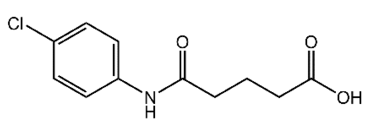
Scheme 1.
Chemical diagram for 4-[(4-chlorophenyl)carbamoyl]butanoic acid (1).

Figure 1.
The molecular structure of (1) showing atom labeling and displacement ellipsoids at the 50% probability level.
Figure 1.
The molecular structure of (1) showing atom labeling and displacement ellipsoids at the 50% probability level.

Figure 2.
Molecular packing in the crystal of (1): (A) a view of the supramolecular tape featuring O–H...O and N–H...O hydrogen bonding, shown as orange and blue dashed lines, respectively, and (B) a view of the unit-cell contents shown in projection down the b-axis; one tape has been highlighted in space-filling mode. Selected intermolecular parameters: O2–H2o...O1i: [H2o...O1i = 1.84(4) Å, O2...O1i = 2.651(4) Å and angle at H2o = 169(5)° for symmetry operation (i) 1-x, 1-y, 2-z], N1–H1n...O3ii [H1n...O3ii = 2.208(19) Å, N1...O3ii = 3.027(4) Å and angle at H1n = 159(3)° for (ii) x, 1+y, z].

Publisher’s Note: MDPI stays neutral with regard to jurisdictional claims in published maps and institutional affiliations. |
© 2021 by the authors. Licensee MDPI, Basel, Switzerland. This article is an open access article distributed under the terms and conditions of the Creative Commons Attribution (CC BY) license (https://creativecommons.org/licenses/by/4.0/).
Share and Cite
MDPI and ACS Style
Hanifa, B.; Sirajuddin, M.; Bari, A.; Lee, S.M.; Lo, K.M.; Tiekink, E.R.T. 4-[(4-Chlorophenyl)carbamoyl]butanoic Acid. Molbank 2021, 2021, M1209. https://0-doi-org.brum.beds.ac.uk/10.3390/M1209
AMA Style
Hanifa B, Sirajuddin M, Bari A, Lee SM, Lo KM, Tiekink ERT. 4-[(4-Chlorophenyl)carbamoyl]butanoic Acid. Molbank. 2021; 2021(2):M1209. https://0-doi-org.brum.beds.ac.uk/10.3390/M1209
Chicago/Turabian StyleHanifa, Bibi, Muhammad Sirajuddin, Ahmed Bari, See Mun Lee, Kong Mun Lo, and Edward R. T. Tiekink. 2021. "4-[(4-Chlorophenyl)carbamoyl]butanoic Acid" Molbank 2021, no. 2: M1209. https://0-doi-org.brum.beds.ac.uk/10.3390/M1209
Note that from the first issue of 2016, this journal uses article numbers instead of page numbers. See further details here.