
N,N-Bis(hexyl α-d-acetylmannosyl) Acrylamide


Department of Materials Science and Engineering, Nagoya Institute of Technology, Gokiso, Showa-ku, Nagoya 466-8555, Japan
*
Author to whom correspondence should be addressed.
Molbank 2021, 2021(3), M1255; https://0-doi-org.brum.beds.ac.uk/10.3390/M1255
Submission received: 7 June 2021
/
Revised: 26 June 2021
/
Accepted: 20 July 2021
/
Published: 22 July 2021
(This article belongs to the Collection Molecules from Side Reactions)
Abstract
:Glycosyl monomers for the assembly of multivalent ligands are typically synthesized using carbohydrates with biological functions and polymerizable functional groups such as acrylamide or styrene introduced into the carbohydrate aglycon, and monomers polymerized using a radical initiator. Herein, we report the acryloylation of 6-aminohexyl α-d-mannoside and its conversion into the glycosyl monomer bearing an acrylamide group. The general acryloylation procedure afforded the desired N-hexyl α-d-acetylmannosyl acrylamide monomer as well as an unexpected compound with a close Rf value. The compounds were separated and analyzed by nuclear magnetic resonance spectroscopy and mass spectrometry, which revealed the unknown compound to be the bivalent N,N-bis(hexyl α-d-acetylmannosyl) acrylamide monomer, which contains two hexyl mannose units and one acrylamide group. To the best of our knowledge, this side reaction has not previously been disclosed, and may be useful for the construction of multivalent sugar ligands.
1. Introduction
Many biological events in organisms are associated with carbohydrates on cell surfaces [1,2,3,4]. Carbohydrates assemble into clusters in raft domains and on polysaccharides and proteins to acquire high affinities for carbohydrate-binding proteins (CBMs). The clusters, as multivalent ligands, interact with CBMs to induce biological events [5,6,7]. Multivalency, which is important for carbohydrate–protein interactions, has also been imitated by synthetic polymers that contain carbohydrates; this affinity-enhancing effect of a multivalent ligand is known as the “cluster effect”. The polymer generally contains carbohydrates with pendants on the polymer back bones, which are generally chosen to be acrylamide [8,9], styrene [10,11], or norbornene [12,13]. A polymerizable carbohydrate monomer consists of a carbohydrate, a linker, and a polymerizable functional group. Carbohydrates include monosaccharides as well as oligosaccharides as ligands. The linker controls the flexibility and the binding space in the carbohydrate—protein interaction, while polymerizable functional groups modulate solubility and polymer structure, and these units are chosen to suit the research purpose. Studies into the preparation of glycomonomers and glycopolymers has continued to date with the aim of clarifying the biological mechanism and applying them to devices and biomaterials [14,15,16,17].
In this study, a mannosyl monomer was prepared using a general method in which 6-aminohexyl α-d-acetylmannoside was acryloylated to afford N-hexyl acetylmannosyl acrylamide. Surprisingly, a previously undocumented side-reaction was observed during acryloylation, the product of which was separated by silica-gel column chromatography and identified by nuclear magnetic resonance (NMR) spectroscopy and mass spectrometry (MS) to be N,N-bis(hexyl α-d-acetylmannosyl) acrylamide, which is a bivalent monomer containing two mannose residues. The discovery of this unusual bivalent monomer is described herein.
2. Results and Discussion
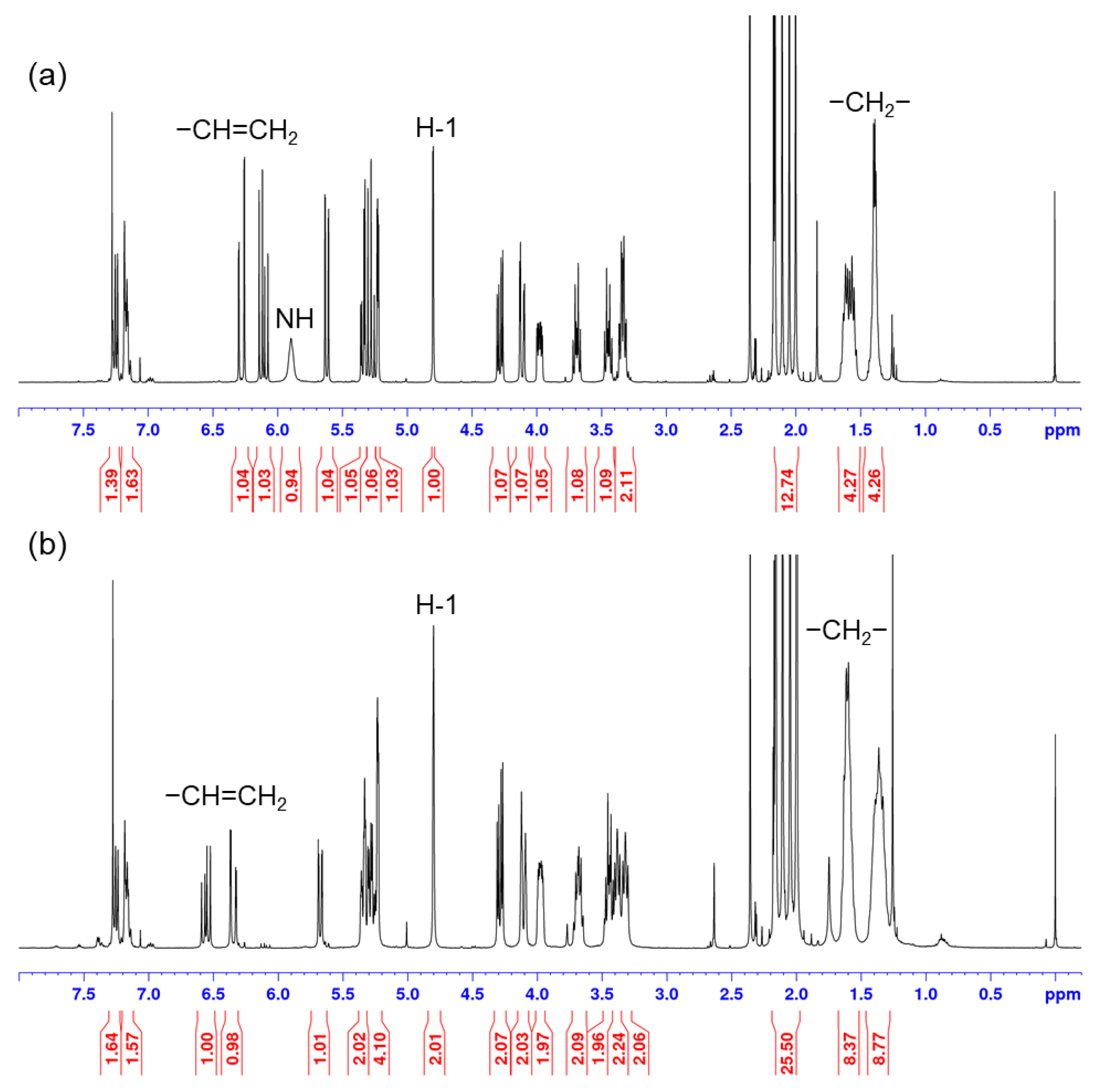
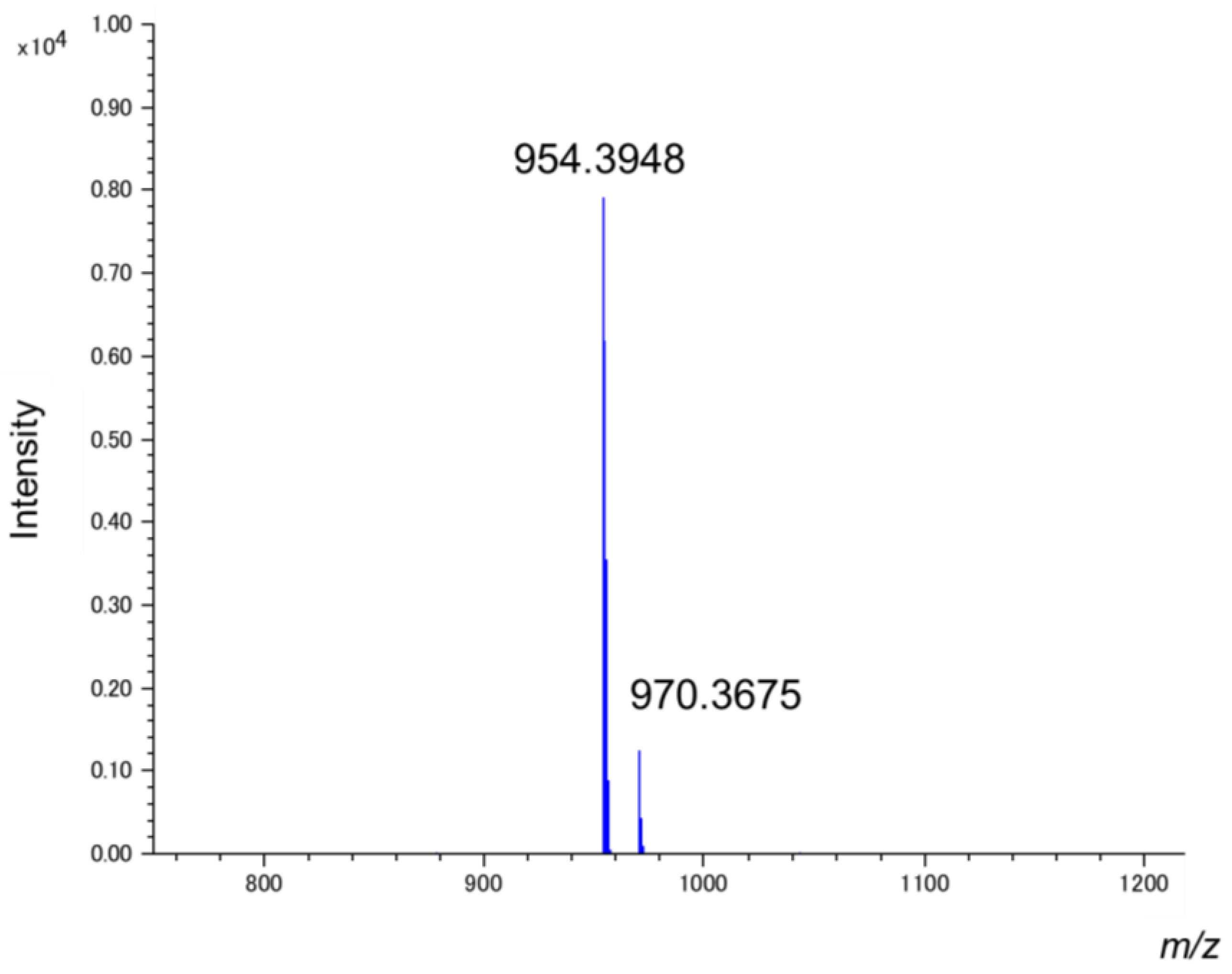
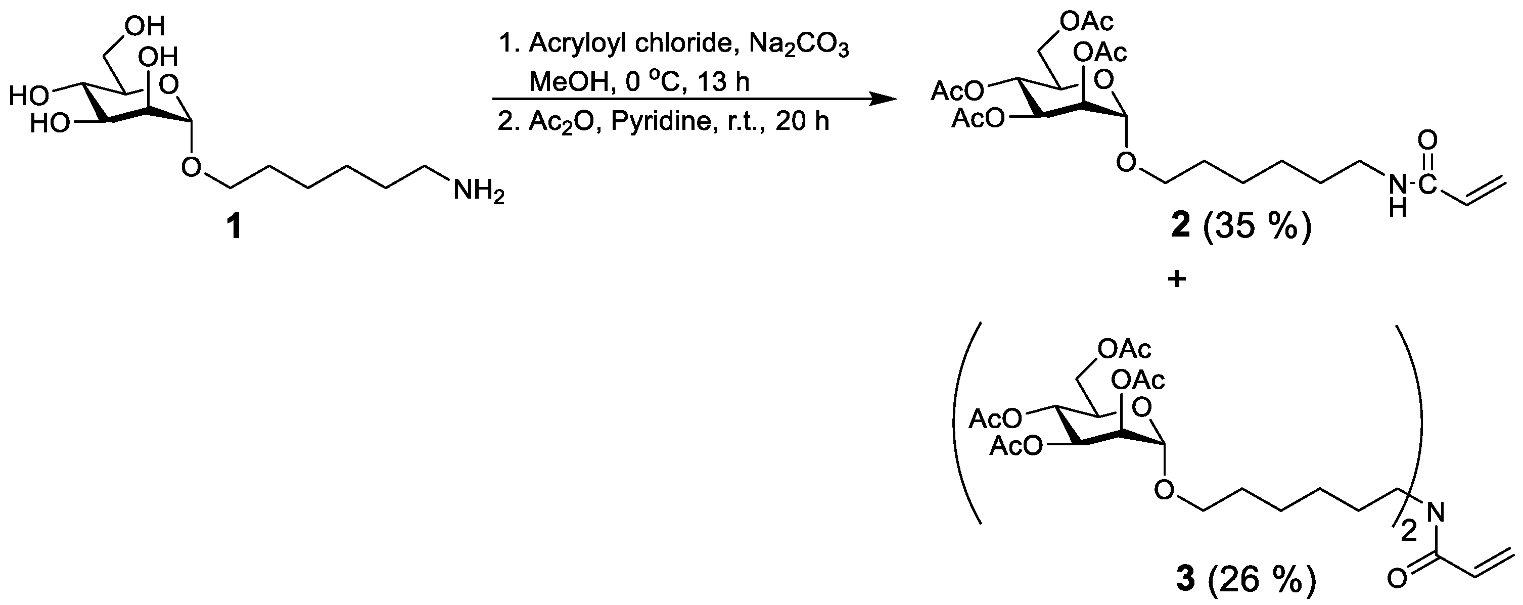
6-Aminohexyl α-d-mannoside (1) was prepared from d-mannose in five steps, which included acetylation, 1-deactylation, introduction of the trichloroacetimidate, glycosylation with 6-aminohexan-1-ol, and deacetylation. Mannoside (1) was converted into a mannosyl monomer bearing an acrylamide group by treatment with acryloyl chloride and sodium carbonate in methanol at 0 °C for 13 h, followed by acetylation (Scheme 1). The reaction mixture was evaporated and separated by silica-gel chromatography to provide two compounds that were subjected to NMR spectroscopy (Figures S1 and S2) and matrix assisted laser desorption ionization-time of flight (MALDI-TOF) MS (Figure S3). Their 1H NMR spectra showed similar peaks; however, integration of the sugar and alkyl peaks of the compound of higher Rf revealed the presence of two 6-aminohexyl α-d-mannoside units per acryloyl group. Furthermore, the amide peak was absent in the NMR spectrum of this compound (Figure 1). In the IR spectrum, the N–H stretching vibration was also not observed (Figure S4). The mass spectrum of this unknown compound is displayed in Figure 2.
The above-mentioned results suggest that the unexpected compound contains two hexyl mannose units and one acryloyl group, from which we concluded that the acryloylation of 1 gave N-hexyl α-d-acetylmannosyl acrylamide (2) and N,N-bis(hexyl α-d-acetylmannosyl) acrylamide (3), which were isolated as pure syrups in yields of 35% and 26%, respectively, along with a considerable amount of a mixture of the two. It is interesting to note that the analogous side reaction that forms the bisamide was not observed in the case of 2-aminoethyl α-d-mannoside and the acryloylation for N-hexyl α-d-acetylmannosyl acrylamide (2). Therefore, N,N-bis(hexyl α-d-acetylmannosyl) acrylamide (3) was given only in the acryloylation for 6-aminohexyl α-d-mannoside (1). To the best of our knowledge, this is the first report of such a side reaction using the general acryloylation procedure. While the reaction mechanism is not clear, the bivalent monomer bearing the two sugar units may be useful for the preparation of multivalent glycopolymers.
3. Materials and Methods
3.1. General
All reagents were purchased from FUJIFILM Wako Pure Chemical Corporation (Osaka, Japan). Methanol was prepared by storage over molecular sieves (3Å) that were activated under vacuum at 200 °C. Analytical thin-layer chromatography (TLC) was performed using Merck silica gel 60 F254 plates (layer thickness: 0.25 mm, Darmstadt, Germany). TLC plates were dipped in 85:10:5 (v/v/v) methanol/resorcinol/concentrated sulfuric acid and/or 0.7% ninhydrin/ethanol, followed by heating for a few minutes for visualization purposes. Column chromatography was performed using silica gel (Silica gel 60 N, spherical neutral, particle size 63–210 μm, Kanto Chemical, Tokyo, Japan). 1H and 13C NMR spectra were acquired on an AVANCE 400 Plus spectrometer (Bruker, Rheinstetten, Germany) in chloroform-d and reported in δ relative to tetramethylsilane (0.00 ppm for 1H and 77.0 ppm for 13C). MALDI-TOF mass spectra were recorded on a Jeol JMS-S3000 spectrometer (Tokyo, Japan) using 2,5-dihydroxybenzoic acid as the matrix. The Fourier transform infrared (FTIR) spectra were measured on a Jasco FT-IR 4100 spectrometer (Tokyo, Japan) and reported as wavenumber (cm–1). Pellet samples for FTIR were fabricated using KBr.
3.2. Acryloylation of 6-Aminohexyl α-d-Mannside (1)
6-Aminohexyl α-d-mannside 1 (976 mg, 3.13 mmol) was dissolved in methanol (21.0 mL). Sodium carbonate was added to the solution and the mixture was stirred at 0 °C, after which acryloyl chloride (380 μL, 4.70 mmol) was slowly added dropwise over 10 min and then stirred at 0 °C for 13 h. The mixture was evaporated and acetic anhydride (11.0 mL, 117 mmol) and pyridine (11.0 mL, 136 mmol) were added at room temperature, stirred for 20 h, and then evaporated. The residue was extracted with chloroform and washed successively with aqueous 1 M hydrochloric acid, aqueous sodium hydrogen carbonate, and brine, dried over anhydrous sodium sulfate, filtered, and then evaporated. The residue was purified by silica gel chromatography three times using 5:1 (v/v) toluene/acetone as the eluent to give 2 (553 mg, 35%), 3 (391 mg, 26%), and a mixture of 2 and 3 (231 mg; 2:3 = 2.7:1).
Analytical data for 2. Rf 0.54 (toluene/acetone, 1:1); 1H NMR (400 MHz, CDCl3) δ 6.28 (dd, J-CH=,Htrans = 16.4 Hz, JHtrans,Hcis = 1.6 Hz, 1 H, =CHtransH), 6.11 (dd, J-CH=,Hcis = 10.4 Hz, 1 H, =CH−), 5.90 (s, 1 H, NH), 5.62 (dd, 1 H, =CHHcis), 5.34 (dd, J2,3 = 3.6 Hz, J3,4 = 10.0 Hz, 1 H, H-3), 5.28 (dd, J4,5 = 19.8 Hz, 1 H, H-4), 5.22 (dt, J1,2 = 1.6 Hz, 1 H, H-2), 4.80 (d, 1 H, H-1), 4.28 (dd, J5,6a = 5.6 Hz, J6a,6b = 12.2 Hz, 1 H, H-6a), 4.11 (dd, J5,6b = 2.4 Hz, 1 H, H-6b), 3.98 (dt, 1 H, H-5), 3.69 (dt, JOCHa, 2CH2 = 9.6 Hz, JOCHa, OCHb = 6.4 Hz, 1 H, −OCHaHb−), 3.45 (dt, JOCHa, 2CH2 = 6.4 Hz, 1 H, −OCHaHb−), 3.33 (dq, JCHaNH, CHbNH = 3.2 Hz, JCH2NH, 5CH2 = 7.2 Hz, 2 H, −CH2NH−), 2.16 (s, 3 H, COCH3), 2.11 (s, 3 H, COCH3), 2.05 (s, 3 H, COCH3), 2.00 (s, 3 H, COCH3), 1.65–1.53 (m, 4 H, 2CH2, 5CH2), 1.43–1.38 (m, 4 H, 3CH2, 4CH2); 13C NMR (101 MHz, CDCl3) δ 170.6, 170.1, 170.0, 169.7, 165.5, 130.9, 129.0 (toluene), 128.2 (toluene), 125.2 (toluene), 126.0, 97.5, 69.6, 69.1, 68.4, 66.2, 62.5, 39.4, 30.9 (toluene), 29.4, 29.0, 26.6, 25.9, 20.8, 20.7, 20.7.
Analytical data for 3. Rf 0.64 (toluene/acetone, 1:1); 1H NMR (400 MHz, CDCl3) δ 6.55 (dd, J-CH=,Htrans = 16.8 Hz, JHtrans,Hcis = 2.4 Hz, 1 H, =CHtransH), 6.35 (dd, J-CH=,Hcis = 10.4 Hz, 1 H, =CH−), 5.68 (dd, 1 H, =CHHcis), 5.36–5.22 (m, 6 H, H-2, H-3, H-4), 4.80 (s, 2 H, H-1), 4.30 (dd, J5,6a = 5.2 Hz, J6a,6b = 12.0 Hz, 2 H, H-6a), 4.11 (d, 2 H, H-6b), 3.97 (dt, 2 H, H-5), 3.68 (dt, JOCHa, 2CH2 = 9.6 Hz, JOCHa, OCHb = 6.8 Hz, 2 H, −OCHaHb−), 3.48–3.30 (m, 6 H, −OCHaHb−, −CH2NH-), 2.16 (s, 6 H, COCH3), 2.11 (s, 6 H, COCH3), 2.05 (s, 6 H, COCH3), 2.00 (s, 6 H, COCH3), 1.63–1.60 (m, 8 H, 2CH2, 5CH2), 1.39–1.33 (m, 8 H, 3CH2, 4CH2); 13C NMR (101 MHz, CDCl3) δ 170.6, 170.6, 170.1, 169.9, 169.8, 169.7, 169.7, 165.9, 127.8, 129.0 (toluene), 128.2 (toluene), 127.6, 97.5, 69.7, 69.1, 68.4, 68.1, 66.2, 62.5, 48.0, 46.5, 29.6, 29.2, 27.7, 26.8, 26.7, 25.9, 25.9, 20.9, 20.7, 20.7, 20.7; IR (KBr, cm−1): 2937 (C−H), 2862 (C−H), 1751 (C=O), 1647 (C=C), 1611 (C=O), 1433 (C−H), 1373 (C−H); MALDI-TOF-MS m/z: [M+Na]+ Calcd for C43H65N1Na1O21+: 954.3941, found: 954.3948; [M+K]+ Calcd for C43H65K1N1O21+: 970.3681, found: 970.3675.
Supplementary Materials
The following are available online. 1H and 13C NMR spectra of 2 and 3 (Figures S1 and S2), and MALDI-TOF-mass and FTIR spectra of 3 (Figures S3 and S4).
Author Contributions
Conceptualization, A.M. and H.Y.; Investigation, S.O. and A.M.; Writing—original draft preparation and review and editing, A.M. All authors have read and agreed to the published version of the manuscript.
Funding
This research received no external funding.
Institutional Review Board Statement
Not applicable.
Informed Consent Statement
Not applicable.
Data Availability Statement
The data presented in this study are available in this article and Supplementary Materials.
Conflicts of Interest
The authors declare no conflict of interest.
References
- Ohtsubo, K.; Marth, J.D. Glycosylation in Cellular Mechanisms of Health and Disease. Cell 2006, 126, 855–867. [Google Scholar] [CrossRef] [PubMed] [Green Version]
- Varki, A. Biological Roles of Glycans. Glycobiology 2017, 27, 3–49. [Google Scholar] [CrossRef] [PubMed] [Green Version]
- Lakshminarayanan, A.; Richard, M.; Davis, B.G. Studying Glycobiology at the Single-Molecule Level. Nat. Rev. Chem. 2018, 2, 148–159. [Google Scholar] [CrossRef]
- Cummings, R.D. Stuck on Sugars–How Carbohydrates Regulate Cell Adhesion, Recognition, and Signaling. Glycoconj. J. 2019, 36, 241–257. [Google Scholar] [CrossRef] [PubMed]
- Becer, C.R. The Glycopolymer Code: Synthesis of Glycopolymers and Multivalent Carbohydrate–Lectin Interactions. Macromol. Rapid Commun. 2012, 33, 742–752. [Google Scholar] [CrossRef] [PubMed]
- Miyagawa, A.; Kurosawa, H.; Watanabe, T.; Koyama, T.; Terunuma, D.; Matsuoka, K. Synthesis of Glycoconjugate Polymer Carrying Globotriaose as Artificial Multivalent Ligand for Shiga Toxin-Producing Escherichia Coli O157: H7. Carbohydr. Polym. 2004, 57, 441–450. [Google Scholar] [CrossRef]
- Abdouni, Y.; Yilmaz, G.; Becer, C.R. Sequence and Architectural Control in Glycopolymer Synthesis. Macromol. Rapid Commun. 2017, 38, 1700212. [Google Scholar] [CrossRef] [PubMed]
- Li, J.; Zacharek, S.; Chen, X.; Wang, J.; Zhang, W.; Janczuk, A.; Wang, P.G. Bacteria Targeted by Human Natural Antibodies Using α-Gal Conjugated Receptor-Specific Glycopolymers. Bioorg. Med. Chem. 1999, 7, 1549–1558. [Google Scholar] [CrossRef]
- Miyagawa, A.; Kasuya, M.C.Z.; Hatanaka, K. Inhibitory Effects of Glycopolymers Having Globotriose and/or Lactose on Cytotoxicity of Shiga Toxin 1. Carbohydr. Polym. 2007, 67, 260–264. [Google Scholar] [CrossRef]
- Serizawa, T.; Yasunaga, S.; Akashi, M. Synthesis and Lectin Recognition of Polystyrene Core−Glycopolymer Corona Nanospheres. Biomacromolecules 2001, 2, 469–475. [Google Scholar] [CrossRef]
- Miura, Y.; Koketsu, D.; Kobayashi, K. Synthesis and Properties of a Well-Defined Glycopolymer via Living Radical Polymerization. Polym. Adv. Technol. 2007, 18, 647–651. [Google Scholar] [CrossRef]
- Lee, S.-G.; Brown, J.M.; Rogers, C.J.; Matson, J.B.; Krishnamurthy, C.; Rawat, M.; Hsieh-Wilson, L.C. End-Functionalized Glycopolymers as Mimetics of Chondroitin Sulfate Proteoglycans. Chem. Sci. 2010, 1, 322–325. [Google Scholar] [CrossRef] [PubMed]
- Miyagawa, A.; Yamamura, H. Synthesis of β-1,3-Glucan Mimics by β-1,3-Glucan Trisaccharyl Monomer Polymerization. Carbohydr. Polym. 2020, 227, 115105. [Google Scholar] [CrossRef] [PubMed]
- Miyagawa, A.; Watanabe, M.; Igai, K.; Kasuya, M.C.Z.; Natori, Y.; Nishikawa, K.; Hatanaka, K. Development of Dialyzer with Immobilized Glycoconjugate Polymers for Removal of Shiga-Toxin. Biomaterials 2006, 27, 3304–3311. [Google Scholar] [CrossRef] [PubMed]
- Kiessling, L.; Grim, L.C.; Glycopolymer, J. Probes of Signal Transduction. Chem. Soc. Rev. 2013, 42, 4476–4491. [Google Scholar] [CrossRef] [PubMed] [Green Version]
- Ma, Z.; Zhu, X.X. Copolymers Containing Carbohydrates and Other Biomolecules: Design, Synthesis and Applications. J. Mater. Chem. B 2019, 7, 1361–1378. [Google Scholar] [CrossRef] [PubMed]
- Pramudya, I.; Chung, H. Recent Progress of Glycopolymer Synthesis for Biomedical Applications. Biomater. Sci. 2019, 7, 4848–4872. [Google Scholar] [CrossRef] [PubMed]
Scheme 1.
Acryloylation of 6-aminohexyl α-d-mannside (1).

Figure 1.
1H NMR spectra of (a) N-hexyl α-d-acetylmannosyl acrylamide (2) and (b) N,N-bis(hexyl α-d-acetylmannosyl) acrylamide (3).
Figure 1.
1H NMR spectra of (a) N-hexyl α-d-acetylmannosyl acrylamide (2) and (b) N,N-bis(hexyl α-d-acetylmannosyl) acrylamide (3).

Figure 2.
MALDI-TOF mass spectrum of N,N-bis(hexyl α-d-acetylmannosyl) acrylamide (3).

Publisher’s Note: MDPI stays neutral with regard to jurisdictional claims in published maps and institutional affiliations. |
© 2021 by the authors. Licensee MDPI, Basel, Switzerland. This article is an open access article distributed under the terms and conditions of the Creative Commons Attribution (CC BY) license (https://creativecommons.org/licenses/by/4.0/).
Share and Cite
MDPI and ACS Style
Miyagawa, A.; Ohno, S.; Yamamura, H. N,N-Bis(hexyl α-d-acetylmannosyl) Acrylamide. Molbank 2021, 2021, M1255. https://0-doi-org.brum.beds.ac.uk/10.3390/M1255
AMA Style
Miyagawa A, Ohno S, Yamamura H. N,N-Bis(hexyl α-d-acetylmannosyl) Acrylamide. Molbank. 2021; 2021(3):M1255. https://0-doi-org.brum.beds.ac.uk/10.3390/M1255
Chicago/Turabian StyleMiyagawa, Atsushi, Shinya Ohno, and Hatsuo Yamamura. 2021. "N,N-Bis(hexyl α-d-acetylmannosyl) Acrylamide" Molbank 2021, no. 3: M1255. https://0-doi-org.brum.beds.ac.uk/10.3390/M1255
Note that from the first issue of 2016, this journal uses article numbers instead of page numbers. See further details here.