
Review of the Rheotanytarsus muscicola Species Group from China (Diptera: Chironomidae) †


1
Joint Laboratory of Xianju National Park & Taizhou University, Taizhou University, Taizhou 318000, China
2
College of Life Sciences, Taizhou University, Taizhou 318000, China
3
Tianjin Key Laboratory of Conservation and Utilization of Animal Diversity, Tianjin Normal University, Tianjin 300387, China
4
College of Life Sciences, Nankai University, Tianjin 300071, China
*
Author to whom correspondence should be addressed.
†
urn:lsid:zoobank.org:act:049C6E91-FA03-4FA4-9C5A-49BA0AF478ED.
Diversity 2022, 14(5), 383; https://0-doi-org.brum.beds.ac.uk/10.3390/d14050383
Submission received: 9 April 2022
/
Revised: 3 May 2022
/
Accepted: 10 May 2022
/
Published: 12 May 2022
(This article belongs to the Section Animal Diversity)
Abstract
:The Rheotanytarsus muscicola species group is generally considered to be a taxonomically difficult group of non-biting midges (Diptera: Chironomidae). In this study, we review the R. muscicola species group based on the adult males from China using morphology and DNA barcodes. Rheotanytarsus ferringtoni Lin & Yao sp. n. is described and figured, and four species (Rheotanytarsus falcipedius Kyerematen, Andersen & Sæther, 2000; Rheotanytarsus fluminis Kawai & Sasa, 1985; Rheotanytarsus illiesi Siebert, 1979; and Rheotanytarsus photophilus Goetghebuer, 1921) new to China are redescribed, figured and discussed. An updated key to known adult males of the R. muscicola species group is presented. Our study shows concordance between morphological species concepts and DNA barcodes.
1. Introduction
Rheotanytarsus Thienemann & Bause, 1913 (Diptera: Chironomidae) is one of the most species-rich genera of the tribe Tanytarsini in the subfamily Chironominae, containing more than 100 described species worldwide [1,2,3,4,5,6,7,8,9,10,11,12]. Larvae of Rheotanytarsus are rheobiontic, occurring in large rivers and the littoral of lakes [13]. Rheotanytarsus has been revised and divided into 15 species groups over the last few decades [9]. According to a cladistic analysis using morphological data by Sæther & Kyerematen in 2001 [9], the Rheotanytarsus muscicola species group was erected, including 15 species in Afrotropical, Oriental and Holarctic regions [6,14]. Prior to this study, two species of the Rheotanytarsus muscicola species group were recorded in China [2,5].
Over the last decades, the DNA barcode corresponding to the 658 bp fragment of the mitochondrial gene cytochrome c oxidase I (COI) has been identified as the core of a global bio-identification system at the species level [15,16] and has proven to be useful in the delimitation of non-biting midge species and provided important evidence to confirm new species [17,18,19,20,21,22,23,24,25,26,27,28]. The morphological similarity between species within the R. muscicola species group has likely led to misidentifications and underestimation of species diversity in the past. In general, DNA barcode data and morphology can give a good estimation in closely related species.
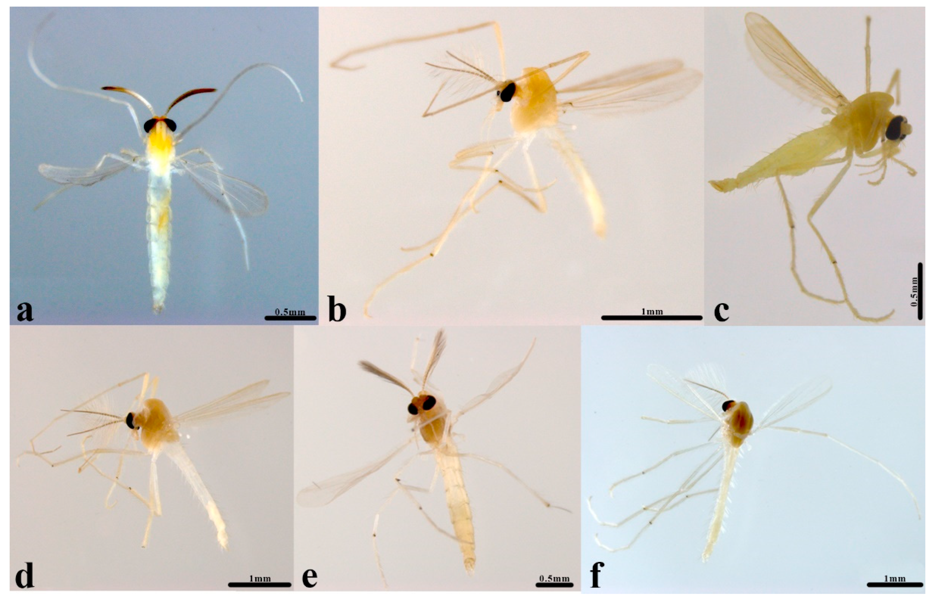
In this study, we review species within the Rheotanytarsus muscicola species group (Figure 1) from China, diagnosing and describing them based on morphology and DNA barcodes. An updated key to known adult males of the R. muscicola species group is presented.
2. Materials and Methods
2.1. Taxon Sampling and Identification
A total of 16 specimens of the Rheotanytarsus muscicola species group were collected from China. Adult males were collected by using malaise trap and light trap. The material examined was stored in 85% ethanol and mounted on slides following the procedure specified in [2,10,29,30]. Specimens were identified using taxonomic revisions and species descriptions [3]. The morphological nomenclature follows Sæther [31]. Color is described as observed in specimens mounted in Euparal on microscopy slides. Digital photographs of slide-mounted specimens were taken with 300 dpi resolution using a Nikon Digital Sight DS-Fil camera mounted on Nikon Eclipse 80i compound microscope using the software NIS-Elements F v.4.60.00. The voucher specimens were deposited at the College of Life Sciences, Nankai University, Tianjin, China (NKU).
2.2. Molecular Laboratory Work
Before slide preparation, genomic DNA of most specimens was extracted from the head-thorax using Qiagen DNA Blood and Tissue Kit according to the manufacture’s instruction. PCR amplifications of COI barcodes with the universal primers LCO1490 and HCO2198 [32] were performed following the protocol from Lin et al. [18]. Sanger sequencing of the purified PCR products was carried out on the ABI 3730 at the BGI (Beijing, China). In addition, genomic DNA extraction from three legs, PCR amplification, and high-throughput sequencing of the specimens were conducted at the Canadian Centre for DNA Barcoding (CCDB, University of Guelph, Canada) using standard high-throughput protocols [33]. DNA samples are deposited at the College of Life Sciences, Nankai University, Tianjin, China, and the CCDB.
2.3. DNA Barcode Analysis
Raw sequences were edited and assembled in Geneious Prime version 2021.0.3 and aligned using MUSCLE [34] implemented in MEGA 11.0 [35] to check stop codons. To obtain DNA barcodes, we searched for public COI barcodes of the Rheotanytarsus muscicola species group that were longer than 500 base pairs with a lack of stop codons in the Barcode of Life Data System (BOLD, http://www.boldsystems.org/ (accessed on 30 March 2022)) [36]. In total, a dataset entitled “DNA barcodes of Rheotanytarsus muscicola species group (DS-MUSCICO)”, including 22 COI barcodes of Rheotanytarsus muscicola species group, was generated on BOLD (30 March 2022), with 16 COI barcodes representing six species originating from this study, while the single COI barcode of Rheotanytarsus guanacastensis Kyerematen and Andersen 2002 was publicly acquired from BOLD. All COI sequences were applied to the Barcode Index Number (BIN) system on BOLD [37]. In addition, a neighbor-joining (NJ) [38] tree was constructed based on the 22 COI barcodes using the Kimura 2-Parameter (K2P) model [39] with 1000 bootstrap replicates and pairwise deletion in MEGA.
3. Results and Discussion
3.1. DNA Barcode Analysis
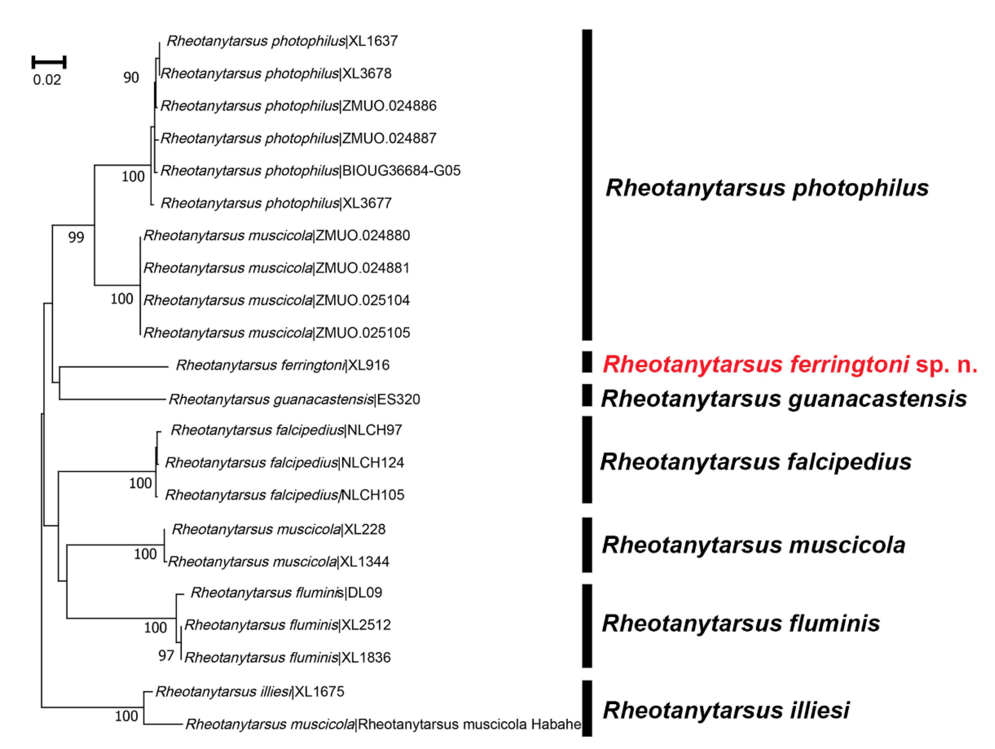
The aligned 22 COI sequences ranged from 589 to 658 base pairs, including 12 sequences with a full barcode length of 658 base pairs. These 22 sequences were assigned to eight BINs, including four new BINs. The NJ tree (Figure 2) based on COI DNA barcodes of the Rheotanytarsus muscicola species group revealed seven well-separated clusters representing seven morphospecies. The new species Rheotanytarsus ferringtoni sp. n. separated from R. guanacastensis by more than 12% divergence in COI barcodes. Interestingly, COI sequences of R. muscicola were grouped into three different clades. Unfortunately, we had no access to examine the vouchers of R. muscicola from Finland (BIN: BOLD:AAW4702) and Xinjiang, China (BIN: BOLD:ADG9631). Since R. illiesi keys to the same BIN (BOLD:ADG9631), we could confirm that the larva named as “R. muscicola” (BOLD Sample ID: Rheotanytarsus muscicola Habahe) from Xinjiang was misidentified. The BIN (BOLD:AAW4702) included four individuals from Finland (BOLD Sample ID: ZMUO.024880, ZMUO.024881, ZMUO.025104 and ZMUO.025105), with 7% genetic distance to the R. photophilus. Although we didn’t examine the vouchers from Finland, we believe that R. photophilus includes two BINs resulting from deep intraspecific divergence. In general, the average intraspecific divergence in COI p-distance (4–6%) is slightly higher than other insects, and the maximum intraspecific divergence can be up to 10% [18].
3.2. Taxonomy
Adult males of the species in the Rheotanytarsus muscicola species group have the following morphological characters: the anal tergite bands developed and of V-type, and the basal tergite bands also present (medially joined in R. foliates); the anal crest of V-type (except in R. phaselus and R. ferringtoni sp. n.); superior volsella with posterior extension (except in R. lamellatus, R. quadratus and R. spinicornus); median volsella relatively short, often not reaching the apex of the superior volsella (except in R. photophilus and R. remus) and with apical plate(s); gonostylus abruptly tapered in apical portion or at the base (emended diagnosis) and often with parallel sided apical portion [6,9].
3.2.1. Rheotanytarsus falcipedius Kyerematen, Andersen & Sæther, 2000
Rheotanytarsus falcipedius Kyerematen, Andersen & Sæther, 2000: 241.
Specimens examined. Two males, China, Guangdong, Ruyuan Yao Autonomous County, Nanling National Nature Reserve, 24.903° N, 113.048° E, 688 m a.s.l., 27.Ⅷ.2020, light tarp, leg. X. L. Lin (BOLD sample ID and NKU: NLCH97, NLCH105); one male, China, Guangdong, Ruyuan Yao Autonomous County, Nanling National Nature Reserve, Laopengyidui, 24.928° N, 113.018° E, 1020 m a.s.l., 27.Ⅷ.2020, light tarp, leg. X. L. Lin (BOLD sample ID and NKU: NLCH124).
Diagnosis. The adult male can be distinguished from known species of Rheotanytarsus by the following combination of characters: antenna with 13 flagellomeres; anal tergite bands of V-type, well developed and separated; anal point constricted in the middle, with rounded apex, and anal crests developed; superior volsella with pronounced apical and inner extensions; digitus with a triangular projection apically, and with one seta placed on the tubercle in the middle; median volsella of Z-type; gonostylus abruptly tapered apically and slightly curved.
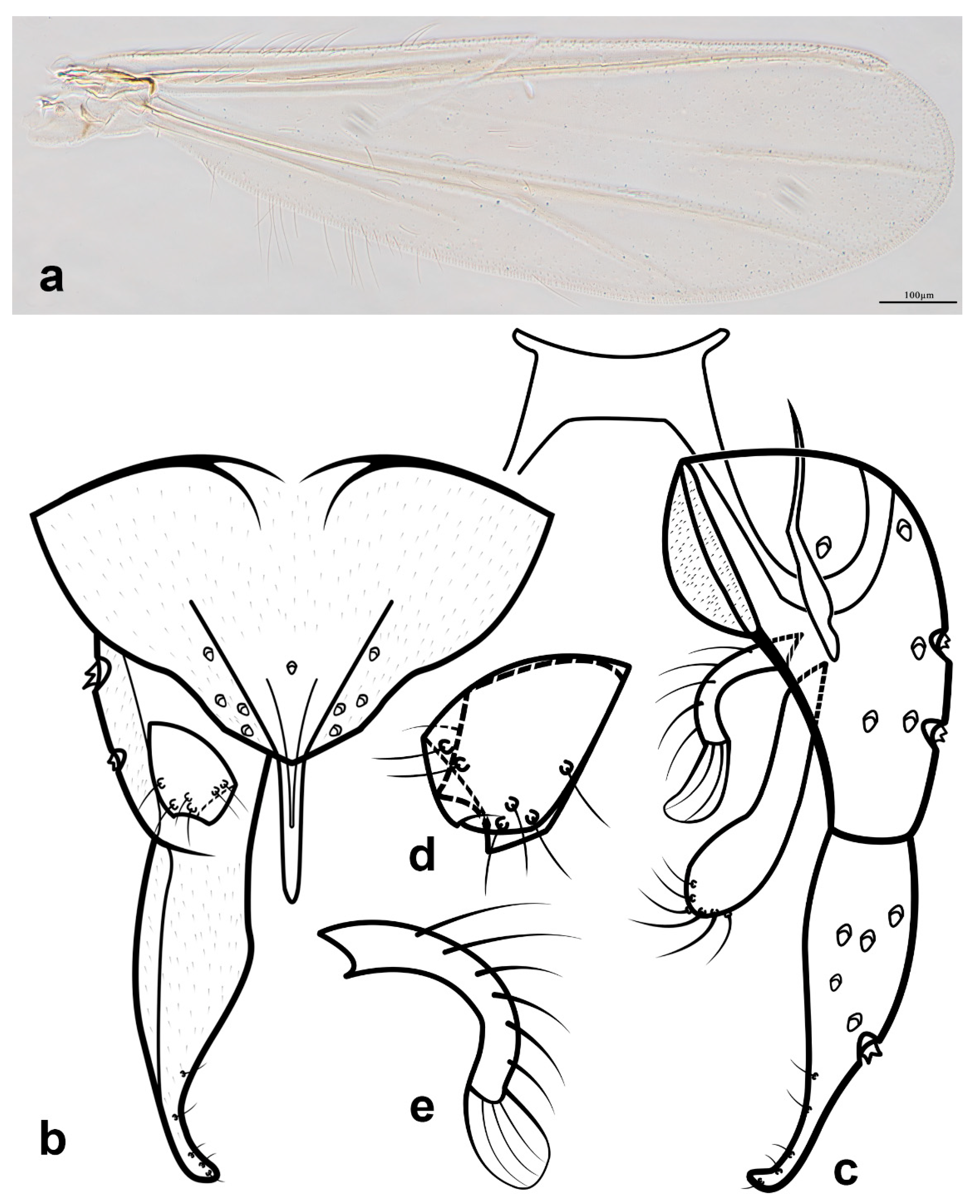
Description. Adult male (n = 3). Total length 2.12–2.31, 2.18 mm. Wing length 1.22–1.53, 1.44 mm. Total length/wing length 1.51–1.73, 1.69. Wing length/length of profemur 1.70–2.24, 1.92.
Coloration (Figure 1a). Thorax and abdomen yellow, legs brown.
Head. Antenna with 13 flagellomeres, ultimate flagellomere 253–277, 265 μm long. AR 0.54–0.61, 0.59. Temporal setae 6–7, 7. Clypeus with 15–16, 16 setae. Tentorium 91–95, 93 μm long, 17–24, 20 μm wide. Palpomere lengths (in μm): 24–29, 27; 26–28, 27; 85–92, 89; 91–95, 93; the fifth used for DNA extraction. Third palpomere with 2 sensilla clavata distally.
Thorax. Dorsocentrals 6–7, 7; acrostichals 7–8, 8; prealars 1. Scutellum with 6 setae. Halteres with 5–6, 6 setae.
Wing (Figure 3a). VR 1.46–1.51, 1.48. Brachiolum with one seta, Sc bare, R with 15–18, 16 setae, R1 with 21–25, 23 setae, R4+5 with 38–44, 41 setae, M1+2 with 33–37, 35 setae, M3+4 with 21–28, 25 setae, false vein with 62–68, 65 setae, Cu with 11–12, 12 setae, Cu1 with 14–15, 15 setae, PCu with 30–35, 32 setae, An with 21–29, 24 setae, remaining veins bare. Cell r4+5 with c. 200 setae, m with 2–3, 3 setae, m1+2 with c. 200 setae, m3+4 with c.110 setae, cu+an with c. 100 setae.
Legs. Fore leg bearing single tibial spur. Combs of mid tibias well separated, each bearing a spur. Tarsomere 1 of mid leg with 3–4, 4 sensilla chaetica. Lengths (in μm) and proportions of legs as in Table 1.
Hypopygium (Figure 3b–e). Tergite IX 86–90, 88 μm long, with 2–3, 3 median setae at the base of anal point, anal tergal bands of V-type, well developed and separated. Anal point 27–32, 30 μm long, constricted in the middle, bearing 2–3, 3 lateral setae on each side; crests of V-type, well developed. Transverse sternapodeme 24–33, 29 μm long, with oral projections. Phallapodeme 60–63, 62 μm long. Gonocoxite 85–93, 90 μm long. Gonostylus 72–77, 75 μm long, abruptly tapered and slightly curved apically. Superior volsella (Figure 3d) 33–39, 35 μm long, with apical and inner extensions, with two anteromedian setae and four dorsal setae. Digitus with triangular projections, bearing one seta located on cylindrical tubercle in the middle. Median volsella (Figure 3e) of Z-type, 24–32, 28 μm long, relatively short, not reaching the apex of superior volsella, with two short subulate setae fused into a narrow plate. Inferior volsella 48–53, 50 μm long, slightly swollen apically, with microtrichia. HR 1.18–1.21, 1.20. HV 2.91–3.00, 2.94.
Remarks. R. falcipedius is recorded in China for the first time. The Chinese specimens fit well with the original description [7].
Distribution. China (Guangdong) and Thailand.
3.2.2. Rheotanytarsus ferringtoni Lin & Yao sp. n.
http://zoobank.org/NomenclaturalActs/049C6E91-FA03-4FA4-9C5A-49BA0AF478ED (accessed on 30 March 2022).
Type material. Holotype: male, China, Yunnan, Baoshan, Longyang, Mangkuan, Gaoligongshan National Nature Reserve, 25.3105556° N, 98.795000° E, 1475 m a.s.l., 22.Ⅴ.2018, light trap, leg. X.-L. Lin (BOLD Sample ID and NKU: XL916).
Etymology. Named after Prof. Leonard C. Ferrington Jr., for his outstanding contribution to the knowledge of Chironomidae; noun in nominative case.
Diagnosis. The adult male can be distinguished from known species of Rheotanytarsus by the following combination of characters: antenna with 13 flagellomeres, and AR 0.43; LR1 2.25; anal tergite bands of V-type, well developed and separated; anal point slightly constricted in the middle, with rounded apex, and anal crests basally fused to form an arc and apically opened; digitus with wavy inner margin, and with one seta placed on the tubercle in the middle; superior volsella with a pronounced eagle’s beak-like extension; median volsella of Z-type; gonostylus abruptly tapered at the base, and slightly curved.
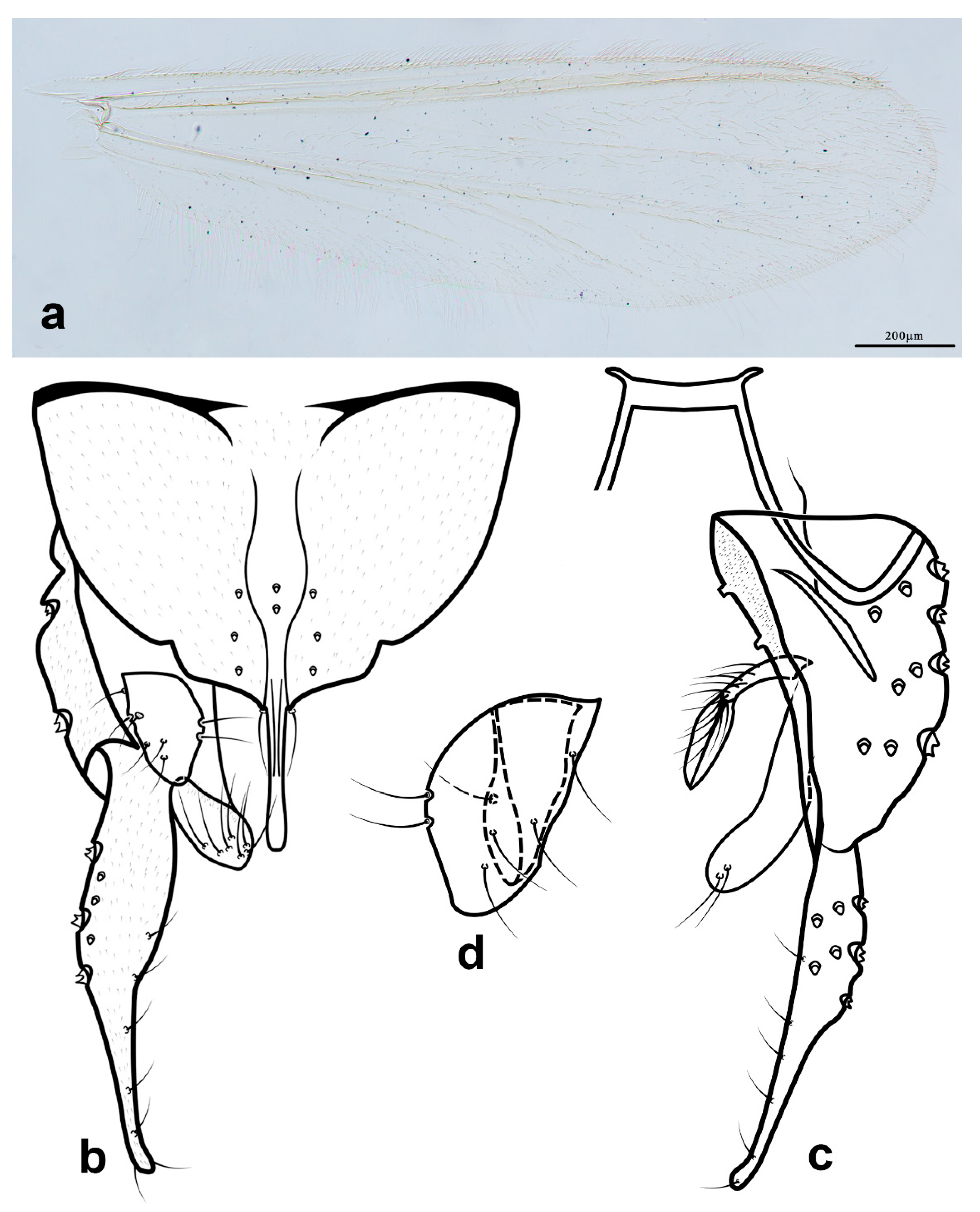
Description. Adult male (n = 1). Total length 2.44 mm. Wing length 1.44 mm. Total length/wing length 1.69. Wing length/length of profemur 1.90.
Coloration (Figure 1b). Thorax and legs brown, abdomen pale yellow.
Head. Antenna with 13 flagellomeres, ultimate flagellomere 232 μm long. AR 0.43. Temporal setae 7. Clypeus with 15 setae. Tentorium 93 μm long, 20 μm wide. Palpomere lengths (in μm): 31, 29, 113, 105, 212. Pm5/Pm3 1.88. Third palpomere with 2 sensilla clavata distally.
Thorax. Dorsocentrals 8; acrostichals 10; prealars 1. Scutellum with 6 setae. Halteres with 8 setae.
Wing (Figure 4a). VR 1.45. Brachiolum with one seta, Sc bare, R with 21 setae, R1 with 28 setae, R4+5 with 64 setae, RM with one seta, M1+2 with 64 setae, M3+4 with 38 setae, false vein with 89 setae, Cu with 21 setae, Cu1 with 24 setae, PCu with 52 setae, An with 30 setae, remaining veins bare. Cell r4+5 with c. 200 setae, m with six setae, m1+2 with c. 200 setae, m3+4 with 130 setae, cu+an with 127 setae.
Legs. Fore leg bearing single tibial spur, 23 μm long. Combs of mid tibia 22 μm wide with 21 μm long spur, and 17 μm wide with 31 μm long spur; combs of hind tibia 21 μm wide with 36 μm long spur, 29 μm wide with 39 μm long spur. Tarsomere 1 of mid leg with five sensilla chaetica. Lengths (in μm) and proportions of legs as in Table 2.
Hypopygium (Figure 4b–e). Tergite IX 78 μm long, with four median setae at the base of anal point, anal tergal bands of V-type, well developed and separated. Anal point 38 μm long, slightly constricted in the middle, bearing three lateral setae on each side; crests basally circular and apically opened. Transverse sternapodeme 39 μm long, with oral projections. Phallapodeme 72 μm long. Gonocoxite 105 μm long. Gonostylus 88 μm long, gradually tapered and slightly curved, with an oval setose area on the ventral side. Superior volsella (Figure 4d) 39 μm long, with an apical eagle’s beak-like extension, with two anteromedian setae and four dorsal setae. Digitus with wavy inner margin, bearing one seta located on cylindrical tubercle at the middle. Median volsella of Z-type (Figure 4e), 33 μm long, relatively short, not reaching the apex of inferior volsella, with three short subulate setae fused into a narrow oval plate. Inferior volsella 61 μm long, basally pronounced swollen and constricted in the middle, with microtrichia. HR 1.19. HV 2.77.
Female and immatures unknown.
Remarks. The new species resembles Rheotanytarsus falcipedius Kyerematen, Andersen & Sæther, 2000, by having similar shapes of superior volsella and median volsella, but can be separated from the latter species by the following combination characters: AR 0.43; superior volsella with a straight inner margin; gonostylus abruptly tapered at the base; whereas AR 0.52–0.57, superior volsella with an inner extension; gonostylus abruptly tapered in apical portion in R. falcipedius.
3.2.3. Rheotanytarsus fluminis Kawai & Sasa, 1985
Rheotanytarsus fluminis Kawai & Sasa 1985: 20.
Examined specimens. One male, China, Hainan, Lingshui, Diaoluo Mountain Forestry Bureau, Nanxi Forest Farm, 18.66862° N, 109.92361° E, 22.Ⅳ.2008, light trap, leg. Y. Fu (BOLD sample and NKU: DL09).
Diagnosis. The adult male can be distinguished from known species of Rheotanytarsus by the following combination of characters: antenna with 13 flagellomeres; anal point with parallel-sided apex; digitus rectangular, and with one seta in the middle; superior volsella with a beak-like extension; median volsella markedly curved, with an oblong plate apically; inferior volsella curved and apically swollen; gonostylus curved and abruptly tapered in apical portion.
Description. Adult male (n = 1). Total length 1.54 mm. Wing length 1.09 mm. Total length/wing length 1.41. Wing length/length of profemur 1.65.
Coloration (Figure 1c). Thorax, abdomen and legs yellowish green.
Head. Temporal setae 7. Tentorium 89 μm long, 31 μm wide. Palpomere lengths (in μm): 28, 25, 88, 107, 193. Pm5/Pm3 2.19. Third palpomere with 2 sensilla clavata distally. Antennas lost during collection.
Thorax. Dorsocentrals 7; acrostichals 9; prealars 1. Scutellum with 4 setae. Halteres with 6 setae.
Wing (Figure 5a). VR 1.61. Brachiolum with one seta, Sc bare, R with 16 setae, R1 with 22 setae, R4+5 with 39 setae, RM with one seta, M1+2 with 39 setae, M3+4 with 25 setae, false vein with 77 setae, Cu with 14 setae, Cu1 with 16 setae, PCu with 43 setae, An with 16 setae, remaining veins bare. Cell r4+5 with c. 230 setae, m with nine setae, m1+2 with c. 250 setae, m3+4 with c. 110 setae, cu+an with c. 130 setae.
Legs. Fore leg bearing single tibial spur, 29 μm long. Both combs of mid tibia and both combs of hind tibia with a spur. Lengths (in μm) and proportions of legs as in Table 3.
Hypopygium (Figure 5b–e). Tergite IX 86 μm long, with one median setae at the base of anal point, anal tergal bands of V-type and separated. Anal point 36 μm long with parallel-sided apex; crests of V-type. Transverse sternapodeme 37 μm long, with oral projections. Phallapodeme 34 μm long. Gonocoxite 99 μm long. Gonostylus 88 μm long, abruptly tapered distally and curved. Superior volsella (Figure 5d) 30 μm long, with an apical beak-like extension, with two anteromedian setae and four dorsal setae. Digitus broad and rectangular, bearing one seta located in the middle. Median volsella (Figure 5e) 48 μm long, markedly curved and not reaching the apex of inferior volsella, with subulate setae and four subulate setae fused into an oblong plate. Inferior volsella 62 μm long, curved and swollen apically, with microtrichia. HR 1.13. HV 1.74.
Remarks. R. fluminis is recorded in China for the first time. The Chinese specimen fits well with the original description [30].
Distribution. Japan and China (Hainan).
3.2.4. Rheotanytarsus illiesi Siebert, 1979
Rheotanytarsus illiesi Siebert, 1979: 165.
Examined specimens. One male, China, Inner Mongolia, Hulumbuir, Honghuaer’ji, 49.930766° N, 123.567751° E, 428 m a.s.l. 2.Ⅷ.2016, light trap, leg. C. Song (BOLD sample ID and NKU: XL1675).
Diagnosis. The adult male can be distinguished from known species of Rheotanytarsus by the following combination of characters: antenna with 13 flagellomeres and with relatively high antennal ratio; inner tibial comb of hind tibia with a spur; anal point slightly swollen apically; superior volsella with a relatively blunt projection; median volsella markedly curved, with an elongate plate apically; inferior volsella curved; gonostylus abruptly tapered in apical portion.
Description. Adult male (n = 1). Total length 3.11 mm. Wing length 1.76 mm. Total length/wing length 1.77. Wing length/length of profemur 1.85.
Coloration (Figure 1d). Thorax and legs brown, abdomen, pale yellow.
Head. Antenna with 13 flagellomeres, ultimate flagellomere 534 μm long. AR 0.99. Temporal setae 10. Clypeus with 17 setae. Tentorium 124 μm long, 26 μm wide. Palpomere lengths (in μm): 42, 43, 132, 134; the fifth used for DNA extraction. Third palpomere with 2 sensilla clavata distally.
Thorax. Dorsocentrals 9; acrostichals 9; prealars 1. Scutellum with 10 setae. Halteres with 7 setae.
Wing (Figure 6a). VR 1.34. Brachiolum with one seta, Sc bare, R with 26 setae, R1 with 32 setae, R4+5 with 61 setae, M1+2 with 35 setae, M3+4 with 35 setae, false vein with 93 setae, Cu with 22 setae, Cu1 with 28 setae, PCu with 61 setae, An with 32 setae, remaining veins bare. Cell r4+5 with c. 300 setae, m with 20 setae, m1+2 with c. 250 setae, m3+4 with c. 200 setae, cu + an with c. 150 setae.
Legs. Fore leg bearing single tibial spur. Combs of mid and hind tibias well separated, each bearing a spur. Tarsomere 1 of mid leg with four sensilla chaetica. Lengths (in μm) and proportions of legs as in Table 4.
Hypopygium (Figure 6b–d). Tergite IX 113 μm long, with eight around the base of anal point and two setae covered by the anal point, anal tergal bands of V-type and separated. Anal point 43 μm long constricted in the middle; crests with two separated arcs. Transverse sternapodeme 38 μm long, with pronounced oral projections. Phallapodeme 90 μm long. Gonocoxite 121 μm long. Gonostylus 127 μm long, abruptly tapered distally. Superior volsella (Figure 6d) 44 μm long, with a blunt projection, with two anteromedian setae and four (left) or six (right) dorsal setae. Digitus thumb-shaped, bearing one seta located on cylindrical tubercle in the middle. Median volsella 59 μm long, markedly curved, nearly reaching the apex of superior volsella, with an oblong plate densely covered setae on the ventral side. The plate was well illustrated in the original descriptive [10]. Inferior volsella 77 μm long, curved with microtrichia and ten setae apically. HR 0.95. HV 2.45.
Remarks. The adult male of R. illiesi is recorded in China for the first time. The Chinese specimen fits well with the original description [10], but the body size is smaller than the German specimens. A larva from Xinjiang was associated with the adult male (Figure 2) by DNA barcode, but was misidentified as R. muscicola morphologically and without access to examine this voucher.
Distribution. Germany and China (Inner Mongolia, Xinjiang).
3.2.5. Rheotanytarsus muscicola Thienemann, 1929
(Figure 1e)
Rheotanytarsus muscicola Thienemann, 1929: 114; Lehmann 1970: 362; Reiss 1971: 208, Wang & Zheng 1993: 90; Wang & Guo 2004: 9.
Specimens examined. One male, China, Liaoning, Shenyang, Benxi, Wangtian Cave Scenic Area, 41.192° N, 125.267° E, 254 m a.s.l., 2.Ⅸ.2014, light tarp, leg. C Song (BOLD sample ID and NKU: XL228). One male, China, Hebei, Baoding, Baoding, Zijingguan, Juma River, 39.428° N, 115.17° E, 521 m a.s.l., 8.Ⅴ.2018, light tarp, leg. X. L. Lin (BOLD sample ID and NKU: XL1344).
Diagnosis. This species can be separated from other members of the group by the following combination of characters: inner tibial comb of hind tibia without a spur; median volsella of S-type; and the base of anal point with only one seta.
Distribution. Europe, north Africa, Canada, China (Fujian, Guangdong, Hebei, Liaoning, Shaanxi, Shandong, Yunnan).
3.2.6. Rheotanytarsus Photophilus Goetghebuer, 1921
(Figure 1f)
Rheotanytarsus photophilus Goetghebuer, 1921: 115; Lehmann 1970: 365.
Specimens examined. One male, China, Jilin, Dunhua, Liuding mountain scenic area, 16. Ⅶ.2016, light trap, leg. C. Song (BOLD sample & NKU: XL3678).
Diagnosis. The adult male can be distinguished from known species of Rheotanytarsus by the following combination of characters: antenna with 13 flagellomeres and with relatively high antennal ratio; inner tibial comb of hind tibia with a spur; superior volsella with a projection; median volsella markedly of S-type, nearly reaching the apex of inferior volsella; inferior volsella slightly curved; gonostylus abruptly tapered in apical portion.
Description. Adult male (n = 1). Total length 2.95 mm. Wing length 1.73 mm. Total length/wing length 1.71 Wing length/length of profemur 1.90.
Coloration (Figure 1d). Thorax yellow with brown stripes, abdomen and legs pale yellow.
Head. Antenna with 13 flagellomeres, ultimate flagellomere 525 μm long. AR 1.03. Temporal setae 10. Clypeus with 20 setae. Tentorium 130 μm long, 33 μm wide. Palpomere lengths (in μm): 35, 40, 125, 125, 245. Third palpomere with 2 sensilla clavata distally.
Thorax. Dorsocentrals 10; acrostichals 11; prealars 1. Scutellum with 4 setae. Halteres with 2 setae.
Legs. Fore leg bearing single tibial spur. Combs of mid and hind tibias well separated, each bearing a spur. Tarsomere 1 of mid leg with three sensilla chaetica. Lengths (in μm) and proportions of legs as in Table 5.
Hypopygium. Anal point 40 μm long constricted in the middle. Transverse sternapodeme 48 μm long. Phallapodeme 85 μm long. Gonocoxite 125 μm long. Gonostylus 115 μm long, abruptly tapered distally. Superior volsella with a projection, with two anteromedian setae and four dorsal setae. Median volsella 70 μm long, relatively long and of S-type, nearly reaching the apex of inferior volsella, with distinct flaky setae apically. Inferior volsella 75 μm long, slightly curved and with microtrichia. HR 1.09. HV 2.57.
Remarks. R. photophilus is recorded in China for the first time. The Chinese specimen fits well with the description of the revision by Lehmann [3]. The hypopygium has been well illustrated by Lehmann [3]. R. photophilus shows a large intraspecific divergence (up to 7%) in COI barcode (Figure 2).
Distribution. Belgium, Finland, Germany and China (Jilin).
3.2.7. Rheotanytarsus quadratus Wang & Guo, 2004
Rheotanytarsus quadratus Wang & Guo, 2004: 9; Hazra et al. 2016: 12.
Specimens examined. Type materials: Holotype, male, China, Fujian, Nanping, Maodi, Mangdangshan, 23.IX.2002, light tarp, leg. Z. Liu (NKU: 20805). Paratype, male, China, Fujian, Yongtai, 17.Ⅸ.2002, light tarp, leg. Z. Liu (NKU: 20395).
Diagnosis. This species can be separated from other members of the group by the following combination of characters: relatively high AR; superior volsella rectangular.
Distribution. China (Fujian).
3.3. Key to Known Adult Males of the Rheotanytarsus Muscicola Species Group
| 1 Superior volsella without a posterior extension | 2 |
| _ Superior volsella with a posterior extension | 4 |
| 2 Superior volsella rectangular | R. quadratus Wang & Guo |
| _ Superior volsella oval or ovoid | 3 |
| 3 AR 0.31–0.38; median volsella with subulate setae fused into a plate | R. spinicornus Hazra, Brahma & Sanyal |
| _ AR 0.45–0.87; median volsella with lamelliform setae fused into a plate | R. lamellatus Reiss |
| 4 Median volsella relatively long, reaching the apex of inferior volsella | 5 |
| _ Median volsella relatively short, not reaching the apex of inferior volsella | 6 |
| 5 Digitus rectangular and reaching beyond the superior volsella; median volsella slightly curved | R. remus Kyerematen a& Sæther |
| _ Digitus triangular and not reaching beyond the superior volsella; median volsella of S-type | R. photophilus Goetghebuer |
| 6 Superior volsella reniform with digitiform extension | R. phaselus Kyerematen, Andersen & Sæther |
| _ Superior volsella not reniform | 7 |
| 7 Median volsella slightly or not curved | 8 |
| _ Median volsella markedly curved | 9 |
| 8 Superior volsella subquadrangular; anal point with parallel-sided apex | R. kusii Kyerematen |
| _ Superior volsella subtriangular; anal point constricted in the middle | R. foliatus Kyerematen & Andersen |
| 9 Median volsella of L-type | R. guanacastensis Kyerematen & Andersen |
| _ Median volsella of other types | 10 |
| 10 Apical plate of median volsella absent | R. thunesi Kyerematen & Andersen |
| _ Apical plate(s) of median volsella present | 11 |
| 11 Apical portion of gonostylus relatively thin | 12 |
| _ Apical portion of gonostylus relatively thick | 14 |
| 12 Anal point with parallel-sided apex, gonostylus curved apically | R. fluminis Kawai & Sasa |
| _ Anal point constricted in the middle; gonostylus not curved apically | 13 |
| 13 AR 0.86–1.04; median volsella with simple setae fused into an egg-shaped plate | R. illiesi Siebert |
| _ AR 0.36–0.39; median volsella with lamelliform setae fused into a broad plate | R. subtilis Kyerematen & Andersen |
| 14 Gonostylus abruptly tapered at the base | R. ferringtoni Lin & Yao sp. n. |
| _ Gonostylus abruptly tapered in apical portion | 15 |
| 15 The extension of superior volsella relatively blunt; the inner tibial comb of the hind leg without a spur | R. muscicola Thienemann |
| _ The extension of superior volsella relatively sharp; the inner tibial comb of the hind leg with a spur | R. falcipedius Kyerematen, Andersen & Sæther |
4. Conclusions
Our study shows concordance between morphological species concepts and DNA barcodes. After reviewing the Rheotanytarsus muscicola species group from China using DNA barcodes, we detected and described one new species and four species new to China, and provided updated keys to the adult male.
Author Contributions
Methodology, X.-L.L.; software, Y.Y. and W.-B.L.; investigation, X.-L.L. and Y.Y.; data curation, X.-L.L.; writing—original draft preparation, X.Q. and Y.Y.; writing—review and editing, C.-C.Y., X.-L.L. and X.-H.W.; supervision, C.-C.Y., X.-L.L. and X.-H.W.; project administration, X.-L.L.; funding acquisition, X.Q. and X.-L.L. All authors have read and agreed to the published version of the manuscript.
Funding
This research was funded by the National Natural Science Foundation of China, grant numbers 32070481, 32170473, and 31900344, and the Zhejiang Provincial Natural Science Foundation of China, grant number LY22C040003.
Institutional Review Board Statement
Not applicable.
Data Availability Statement
A list of all species, specimens, their individual images, georeferences, primers, sequences and other relevant laboratory data of all 22 specimens are available through the dataset “DNA bar-codes of Rheotanytarsus muscicola species group (DS-MUSCICO)” in the Barcode of Life Data System (http://www.boldsystems.org, BOLD (accessed on 30 March 2022)).
Acknowledgments
We are grateful to the Canadian Centre for DNA Barcoding (CCDB, Guelph, Ontario, Canada) for DNA sequencing.
Conflicts of Interest
The authors declare no conflict of interest.
Abbreviations
| An | anal vein |
| an | cell below anal vein |
| AR | antennal ration, ratio of length of apical elongated flagellomere plus any flagellomeres distal to it divided by combined length of the more basal flagellomeres |
| BV | Bein-Verhältnis ratio, length of (femur + tibia + ta1)/length of (ta2 + ta3 + ta4 + ta5) |
| Cu | cubitus |
| cu | cell below Cu vein |
| Cu1 | cell below Cu1 vein |
| fe | femur |
| HR | hypopygium ration, length of gonocoxite/length of gonostylus |
| HV | hypopygium value, total length/length of gonostylus times ten |
| LR | leg ration, length of tarsomere 1/length of tibia |
| m | cell below median vein |
| M, M1+2, M3+4 | media and its branches |
| m1+2 | cell below M1+2 vein |
| m3+4 | cell below M3+4 vein |
| P1 | fore leg |
| P2 | mid leg |
| P3 | hind leg |
| PCu | postcubital vein |
| R | Radius |
| R1 | Radius 1 vein |
| R4+5 | Radius 4 + 5 vein |
| RM | apparent radius to media cross vein |
| r4+5 | cell below R4+5 vein |
| Sc | subcosta |
| SV | Schenkel-Schiene-Verhältnis ratio, length of (femur + tibia)/length of ta1 |
| ti | tibia |
| ta1 | tarsomere 1 |
| ta2 | tarsomere 2 |
| ta3 | tarsomere 3 |
| ta4 | tarsomere 4 |
| ta5 | tarsomere 5 |
| VR | Venarum ration, length of Cubitus/length of Media |
References
- Wang, X.-H.; Zheng, L.-Y. Taxonomic studies on Chironomidae from China VI. Genus Rheotanytarsus Thienemann & Bause (Diptera: Chironomidae). Acta Sci. Nat. Univ. Nankaiensis 1993, 1, 89–93. [Google Scholar]
- Wang, X.-H.; Guo, Y.-H. A review of the genus Rheotanytarsus Thienemann & Bause from China (Diptera: Chironomidae: Tanytarsini). Zootaxa 2004, 650, 1–19. [Google Scholar]
- Lehmann, J. Revision der europäischen Arten (Imagines♂♂ und Puppen♂♂) der Gattung Rheotanytarsus BAUSE (Diptera, Chironomidae). Zool Anz 1970, 185, 343–378. [Google Scholar]
- Moubayed, Z. Chironomids from running waters of Thailand: Description of Rheotanytarsus thailandensis sp.n. and Tanytarsus thaicus sp.n. (Dipt., Chironomidae). Hydrobiologia 1990, 203, 29–33. [Google Scholar] [CrossRef]
- Yao, Y.; Liu, W.-B.; Wang, X.-H.; Lin, X.-L. Rheotanytarsus baihualingensis and R. diaoluoensis (Diptera: Chironomidae), two new species from Oriental China. Ann. Zool. Fenn. 2022, 59, 149–154. [Google Scholar] [CrossRef]
- Hazra, N.; Brahma, S.; Sanyal, K. New Species of Rheotanytarsus Thienemann and Bause (Diptera: Chironomidae: Tanytarsini) from Darjeeling–Sikkim, Himalaya, India, with Revised Keys to the Adult Males and Pupae of the Species of the Oriental Region. Psyche 2016, 2016, 1–14. [Google Scholar] [CrossRef] [Green Version]
- Kyerematen, R.A.; Sæther, O.A. A review of Afrotropical Rheotanytarsus Thienemann et Bause, 1913 (Diptera: Chironomidae). Tijdschr. Voor Entomol. 2000, 143, 27–70. [Google Scholar] [CrossRef] [Green Version]
- Cranston, P.S. Revision of Australian Rheotanytarsus Thienemann & Bause (Diptera: Chironomidae), with emphasis on immature stages. Invertebr. Syst. 1997, 11, 705–734. [Google Scholar]
- Sæther, O.A.; Kyerematen, R.A. Towards phylogeny and zoogography of the genus Rheotanytarsus Thienemann et Bause, 1913 (Diptera: Chironomidae). Tijdschr. Voor Entomol. 2001, 144, 73–117. [Google Scholar] [CrossRef]
- Siebert, M. Two new chironomids (Diptera: Chironomidae) from Germany and Austria. Aquat. Insects 1979, 1, 165–168. [Google Scholar] [CrossRef]
- Reiss, F. Ein Beitrag zur ostpalaearktischen Chironomidenfauna (Diptera) am Beispiel einiger Tanytarsini-Arten aus der Mongolei und Ostsibirien. Entomol. Tidskr. 1971, 92, 198–212. [Google Scholar]
- Moubayed-Breil, J.; Langton, P.H.; Ashe, P. Rheotanytarsus dactylophoreus, a new mountain species from streams in the Eastern Pyrenees and Corsica (Diptera: Chironomidae). Fauna Nor. 2012, 31, 167. [Google Scholar] [CrossRef] [Green Version]
- Epler, J.H.; Ekrem, T.; Cranston, P.S. The larvae of Chironominae (Diptera: Chironomidae) of the Holarctic region—keys and diagnoses. In Chironomidae of the Holarctic Region: Keys and Diagnoses, Part 1: Larvae; Cederholm, L., Ed.; Insect Systematics and Evolution, Supplement: Lund, Sweden, 2013; Volume 66, pp. 387–556. [Google Scholar]
- Kyerematen, R.A.K.; Andersen, T. Rheotanytarsus Thienemann et Bause (Diptera: Chironomidae) from Central America and Mexico. Stud. Neotrop. Fauna Environ. 2002, 37, 23–51. [Google Scholar] [CrossRef]
- Hebert, P.D.N.; Cywinska, A.; Ball, S.L. Biological identifications through DNA barcodes. Proc. R. Soc. Lond. B Biol. Sci. 2003, 270, 313–321. [Google Scholar] [CrossRef] [Green Version]
- Hebert, P.D.N.; Ratnasingham, S.; deWaard, J.R. Barcoding animal life: Cytochrome c oxidase subunit 1 divergences among closely related species. Proc. R. Soc. Lond. B Biol. Sci. 2003, 270, S96–S99. [Google Scholar] [CrossRef] [Green Version]
- Lin, X.-L.; Stur, E.; Ekrem, T. Exploring genetic divergence in a species-rich insect genus using 2790 DNA Barcodes. PLoS ONE 2015, 10, e0138993. [Google Scholar] [CrossRef] [Green Version]
- Lin, X.-L.; Stur, E.; Ekrem, T. DNA barcodes and morphology reveal unrecognized species of Chironomidae (Diptera). Insect Syst. Evol. 2018, 49, 329–398. [Google Scholar] [CrossRef] [Green Version]
- Lin, X.-L.; Yu, H.-J.; Wang, X.-H.; Bu, W.-J.; Wang, X.-H.; Yan, C.-C.; Liu, W.-B. New or little-known Boreoheptagyia (Diptera: Chironomidae) in China inferred from morpholgy and DNA barcodes. Zookeys 2021, 1040, 187–200. [Google Scholar] [CrossRef]
- Lin, X.L.; Mo, L.; Bu, W.J.; Wang, X.H. The first comprehensive DNA barcode reference library of Chinese Tanytarsus (Diptera: Chironomidae) for environmental DNA metabarcoding. Divers. Distrib. 2021, 27, 1932–1941. [Google Scholar] [CrossRef]
- Lin, X.L.; Jiang, K.; Liu, W.B.; Liu, W.; Bu, W.J.; Wang, X.H.; Mo, L. Toward a global DNA barcode reference library of the intolerant nonbiting midge genus Rheocricotopus Brundin, 1956. Ecol. Evol. 2021, 11, 12161–12172. [Google Scholar] [CrossRef]
- Song, C.; Lin, X.-L.; Wang, Q.; Wang, X.-H. DNA barcodes successfully delimit morphospecies in a superdiverse insect genus. Zool. Scr. 2018, 47, 311–324. [Google Scholar] [CrossRef]
- Anderson, A.M.; Stur, E.; Ekrem, T. Molecular and morphological methods reveal cryptic diversity and three new species of Nearctic Micropsectra (Diptera: Chironomidae). Freshw. Sci. 2013, 32, 892–921. [Google Scholar] [CrossRef] [Green Version]
- Lin, X.-L.; Yu, H.-J.; Zhang, R.-L.; Wang, X.-H. Polypedilum (Cerobregma) heberti sp. n. (Diptera: Chironomidae) from Gaoligong Mountains, Yunnan, China. Zootaxa 2019, 4571, 255–262. [Google Scholar] [CrossRef]
- Lin, X.-L.; Yu, H.-J.; Wang, Q.; Bu, W.-J.; Wang, X.-H. DNA barcodes and morphology confirm a new species of Rheocricotopus (Psilocricotopus) orientalis group (Diptera: Chironomidae). Zootaxa 2020, 4768, 282–290. [Google Scholar] [CrossRef]
- Qi, X.; Wang, X.-H.; Andersen, T.; Lin, X.-L. A new species of Manoa Fittkau (Diptera: Chironomidae), with DNA barcodes from Xianju National Park, Oriental China. Zootaxa 2017, 4231, 398–408. [Google Scholar] [CrossRef]
- Gadawski, P.; Montagna, M.; Rossaro, B.; Giłka, W.; Pešić, V.; Grabowski, M.; Magoga, G. DNA barcoding of Chironomidae from the Lake Skadar region: Reference library and a comparative analysis of the European fauna. Divers. Distrib. 2022. [Google Scholar] [CrossRef]
- Michailova, P.; Lencioni, V.; Nenov, M.; Nikolov, S. Can DNA barcoding be used to identify closely related Clunio Haliday, 1855 species (Diptera: Chironomidae, Orthocladiinae). Zootaxa 2021, 4927, 1–8. [Google Scholar] [CrossRef] [PubMed]
- Sæther, O.A. Some Nearctic Podonominae, Diamesinae, and Orthocladiinae (Diptera: Chironomidae). Bull. Fish. Res. Bd Can. 1969, 170, 1–154. [Google Scholar]
- Kawai, K.; Sasa, M. Seven new species of chironomid midges (Diptera, Chironomidae) from the Ohta River, Japan. JPN J. Limnol. (Rikusuigaku Zasshi) 1985, 46, 15–24. [Google Scholar] [CrossRef] [Green Version]
- Sæther, O.A. Glossary of chironomid morphology terminology (Diptera: Chironomidae). Entomol. Scand. Suppl. 1980, 14, 1–51. [Google Scholar]
- Folmer, O.; Black, M.; Hoeh, W.; Lutz, R.; Vrijenhoek, R. DNA primers for amplification of mitochondrial cytochrome c oxidase subunit I from diverse metazoan invertebrates. Mol. Mar. Biol. Biotechnol. 1994, 3, 294–299. [Google Scholar]
- deWaard, J.R.; Ivanova, N.V.; Hajibabaei, M.; Hebert, P.D.N. Assembling DNA barcodes: Analytical protocols. In Methods in Molecular Biology: Environmental Genetics; Cristofre, M., Ed.; Humana Press Inc.: Ottawa, ON, Canada, 2008; pp. 275–293. [Google Scholar]
- Edgar, R.C. MUSCLE: Multiple sequence alignment with high accuracy and high throughput. Nucleic Acids Res. 2004, 32, 1792–1797. [Google Scholar] [CrossRef] [Green Version]
- Tamura, K.; Stecher, G.; Kumar, S. MEGA11: Molecular evolutionary genetics analysis version 11. Mol. Biol. Evol. 2021, 38, 3022–3027. [Google Scholar] [CrossRef]
- Ratnasingham, S.; Hebert, P.D.N. BOLD: The Barcode of Life Data System (www.barcodinglife.org). Mol. Ecol. Notes 2007, 7, 355–364. [Google Scholar] [CrossRef] [Green Version]
- Ratnasingham, S.; Hebert, P.D.N. A DNA-based registry for all animal species: The barcode index number (BIN) system. PLoS ONE 2013, 8, e66213. [Google Scholar] [CrossRef] [Green Version]
- Saitou, N.; Nei, M. The neighbor-joining method: A new method for reconstructing phylogenetic trees. Mol. Biol. Evol. 1987, 4, 406–425. [Google Scholar]
- Kimura, M. A simple method for estimating evolutionary rates of base substitutions through comparative studies of nucleotide sequences. J. Mol. Evol. 1980, 16, 111–120. [Google Scholar] [CrossRef]
Figure 1.
Adult males of Rheotanytarsus muscicola species group. (a) Rheotanytarsus falcipedius, dorsal view; (b) Rheotanytarsus ferringtoni sp. n., lateral view; (c) Rheotanytarsus fluminis, lateral view; (d) Rheotanytarsus illiesi, lateral view; (e) Rheotanytarsus muscicola, ventral view; (f) Rheotanytarsus photophilus, dorsal view.

Figure 2.
Neighbor-joining tree for seven species of the Rheotanytarsus muscicola species group based on K2P distance in DNA barcodes. Numbers on branches represent bootstrap support (>70%) based on 1000 replicates; scale equals K2P genetic distance.
Figure 2.
Neighbor-joining tree for seven species of the Rheotanytarsus muscicola species group based on K2P distance in DNA barcodes. Numbers on branches represent bootstrap support (>70%) based on 1000 replicates; scale equals K2P genetic distance.

Figure 3.
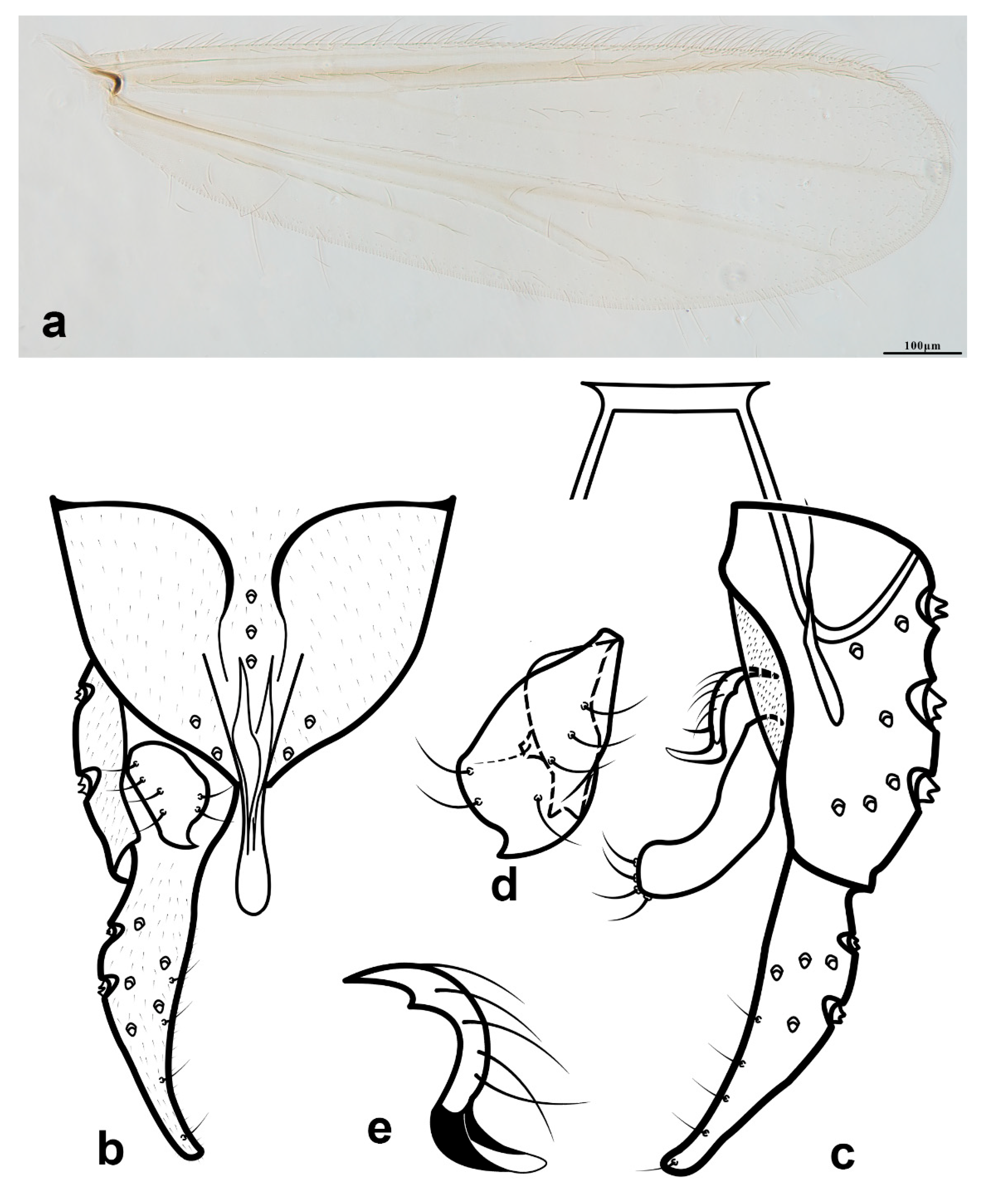
Rheotanytarsus falcipedius: (a) wing; (b) hypopygium, dorsal view; (c) hypopygium, ventral view; (d) superior volsella; (e) median volsella.
Figure 3.
Rheotanytarsus falcipedius: (a) wing; (b) hypopygium, dorsal view; (c) hypopygium, ventral view; (d) superior volsella; (e) median volsella.

Figure 4.
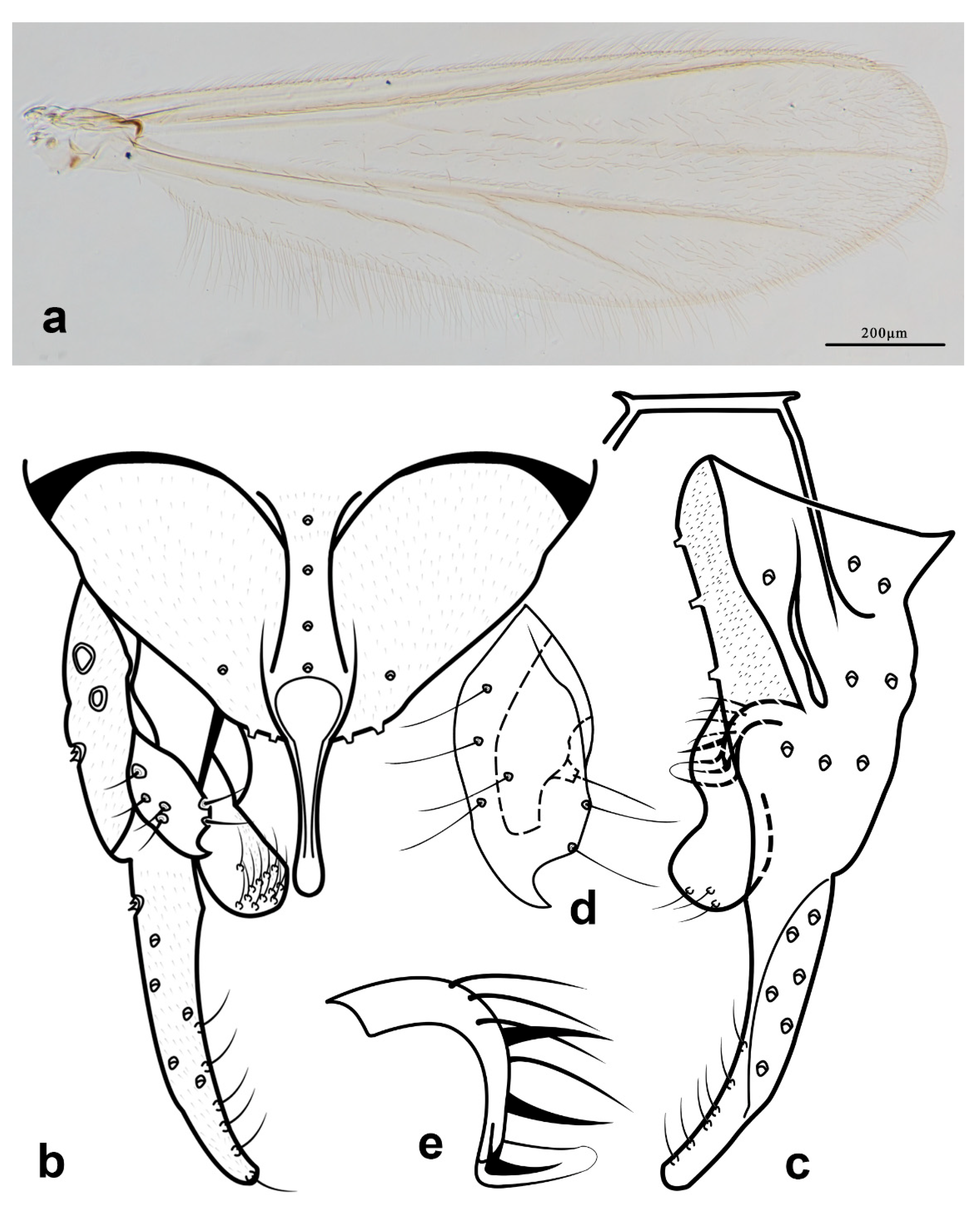
Rheotanytarsus ferringtoni sp. n.: (a) wing; (b) hypopygium, dorsal view; (c) hypopygium, ventral view; (d) superior volsella; (e) median volsella.
Figure 4.
Rheotanytarsus ferringtoni sp. n.: (a) wing; (b) hypopygium, dorsal view; (c) hypopygium, ventral view; (d) superior volsella; (e) median volsella.

Figure 5.
Rheotanytarsus fluminis: (a) wing; (b) hypopygium, dorsal view; (c) hypopygium, ventral view; (d) superior volsella; (e) median volsella.
Figure 5.
Rheotanytarsus fluminis: (a) wing; (b) hypopygium, dorsal view; (c) hypopygium, ventral view; (d) superior volsella; (e) median volsella.

Figure 6.
Rheotanytarsus illiesi: (a) wing; (b) hypopygium, dorsal view; (c) hypopygium, ventral view; (d) superior volsella.
Figure 6.
Rheotanytarsus illiesi: (a) wing; (b) hypopygium, dorsal view; (c) hypopygium, ventral view; (d) superior volsella.


Table 1.
Lengths (in μm) and proportions of legs of Rheotanytarsus falcipedius Kyerematen, Andersen & Sæther, 2000, adult male (n = 3).
Table 1.
Lengths (in μm) and proportions of legs of Rheotanytarsus falcipedius Kyerematen, Andersen & Sæther, 2000, adult male (n = 3).
| fe | ti | ta1 | ta2 | ta3 | |
|---|---|---|---|---|---|
| P1 | 688–683, 674 | 334–347, 339 | 808–826, 817 | 407–427, 411 | 322–345, 330 |
| P2 | 642–659, 648 | 432–458, 449 | 253–272, 267 | 129–141, 130 | 87–98, 91 |
| P3 | 679 (1) | - | - | - | - |
| ta4 | ta5 | LR | BV | SV | |
| P1 | 275–296, 283 | 119–131, 123 | 2.38–2.42, 2.41 | 1.55–1.61, 1.60 | 1.24–1.25, 1.24 |
| P2 | 51–63, 57 | 42–52, 46 | 0.59 | 3.92–4.29, 4.21 | 4.11–4.25, 4.16 |
| P3 | - | - | - | - | - |
Table 2.
Lengths (in μm) and proportions of legs of Rheotanytarsus ferringtoni Lin & Yao sp. n., adult male (n = 1).
Table 2.
Lengths (in μm) and proportions of legs of Rheotanytarsus ferringtoni Lin & Yao sp. n., adult male (n = 1).
| fe | ti | ta1 | ta2 | ta3 | ta4 | ta5 | LR | BV | SV | BR | |
|---|---|---|---|---|---|---|---|---|---|---|---|
| P1 | 758 | 414 | 932 | 451 | 361 | 326 | 139 | 2.25 | 1.65 | 1.26 | 2.14 |
| P2 | 715 | 524 | 322 | 169 | 115 | 72 | 52 | 0.61 | 3.83 | 3.85 | 4.75 |
| P3 | 780 | 641 | 509 | 280 | 256 | 177 | 83 | 0.79 | 2.42 | 2.79 | 5.63 |
Table 3.
Lengths (in μm) and proportions of legs of Rheotanytarsus fluminis Kawai & Sasa 1985, adult male (n = 1).
Table 3.
Lengths (in μm) and proportions of legs of Rheotanytarsus fluminis Kawai & Sasa 1985, adult male (n = 1).
| fe | ti | ta1 | ta2 | ta3 | ta4 | ta5 | LR | BV | SV | |
|---|---|---|---|---|---|---|---|---|---|---|
| P1 | 661 | 293 | - | - | - | - | - | - | - | - |
| P2 | 602 | 424 | - | - | - | - | - | - | - | - |
| P3 | 660 | 516 | 376 | 212 | 152 | 191 | 119 | 0.73 | 2.30 | 3.13 |
Table 4.
Lengths (in μm) and proportions of legs of Rheotanytarsus illiesi Siebert 1979, adult male (n = 1).
Table 4.
Lengths (in μm) and proportions of legs of Rheotanytarsus illiesi Siebert 1979, adult male (n = 1).
| fe | ti | ta1 | ta2 | ta3 | ta4 | ta5 | LR | BV | SV | |
|---|---|---|---|---|---|---|---|---|---|---|
| P1 | 954 | 507 | 1189 | 547 | 425 | 343 | 138 | 2.35 | 1.82 | 1.23 |
| P2 | 923 | 688 | 405 | 205 | 127 | 96 | 66 | 0.59 | 4.08 | 3.98 |
| P3 | 1016 | 867 | 629 | 369 | 292 | 189 | 107 | 0.73 | 2.62 | 2.99 |
Table 5.
Lengths (in μm) and proportions of legs of Rheotanytarsus photophilus Goetghebuer, 1921, adult male (n = 1).
Table 5.
Lengths (in μm) and proportions of legs of Rheotanytarsus photophilus Goetghebuer, 1921, adult male (n = 1).
| fe | ti | ta1 | ta2 | ta3 | ta4 | ta5 | LR | BV | SV | |
|---|---|---|---|---|---|---|---|---|---|---|
| P1 | 910 | 480 | 1035 | 510 | 410 | 310 | 130 | 2.16 | 1.78 | 1.34 |
| P2 | 900 | 680 | 380 | 200 | 160 | 100 | 60 | 0.56 | 3.77 | 4.16 |
| P3 | 1010 | 850 | - | - | - | - | - | - | - | - |
Publisher’s Note: MDPI stays neutral with regard to jurisdictional claims in published maps and institutional affiliations. |
© 2022 by the authors. Licensee MDPI, Basel, Switzerland. This article is an open access article distributed under the terms and conditions of the Creative Commons Attribution (CC BY) license (https://creativecommons.org/licenses/by/4.0/).
Share and Cite
MDPI and ACS Style
Qi, X.; Yao, Y.; Liu, W.-B.; Yan, C.-C.; Wang, X.-H.; Lin, X.-L. Review of the Rheotanytarsus muscicola Species Group from China (Diptera: Chironomidae). Diversity 2022, 14, 383. https://0-doi-org.brum.beds.ac.uk/10.3390/d14050383
AMA Style
Qi X, Yao Y, Liu W-B, Yan C-C, Wang X-H, Lin X-L. Review of the Rheotanytarsus muscicola Species Group from China (Diptera: Chironomidae). Diversity. 2022; 14(5):383. https://0-doi-org.brum.beds.ac.uk/10.3390/d14050383
Chicago/Turabian StyleQi, Xin, Yuan Yao, Wen-Bin Liu, Chun-Cai Yan, Xin-Hua Wang, and Xiao-Long Lin. 2022. "Review of the Rheotanytarsus muscicola Species Group from China (Diptera: Chironomidae)" Diversity 14, no. 5: 383. https://0-doi-org.brum.beds.ac.uk/10.3390/d14050383
Note that from the first issue of 2016, this journal uses article numbers instead of page numbers. See further details here.