Article

Staphylococcus aureus Sequences from Osteomyelitic Specimens of a Pathological Bone Collection from Pre-Antibiotic Times

1
Department of Historical Anthropology and Human Ecology, Johann-Friedrich-Blumenbach Institute for Zoology and Anthropology, University of Goettingen, Goettingen 37073, Germany
2
Robert Koch Institute, National Reference Center for staphylococci and enterococci, Wernigerode 38855, Germany
*
Author to whom correspondence should be addressed.
Submission received: 27 July 2017 / Revised: 15 September 2017 / Accepted: 28 September 2017 / Published: 1 October 2017
(This article belongs to the Special Issue Ancient DNA)

Abstract

:
Staphylococcus aureus is a major pathogen causing osteomyelitis, amongst other diseases, and its methicillin-resistant form (MRSA) in particular poses a huge threat to public health. To increase our knowledge of the origin and evolution of S. aureus, genetic studies of historical microorganisms may be beneficial. Thus, the aim of this study was to investigate whether osteomyelitic skeletal material (autopsy specimens collected from the mid 19th century until the 1920s) is suitable for detecting historical S. aureus DNA sequences. We established a PCR-based analysis system targeting two specific genes of S. aureus (nuc and fib). We successfully amplified the historical S. aureus nuc and fib sequences for six and seven pre-antibiotic, osteomyelitic bone specimens, respectively. These results encourage further investigations of historical S. aureus genomes that may increase our understanding of pathogen evolution in relation to anthropogenically introduced antibiotics.

1. Introduction

Staphylococcus aureus is a major pathogen for both humans and animals, causing a variety of diseases ranging from minor skin and soft tissue infection to severe, life-threatening medical conditions like bacteraemia, endocarditis and toxic shock syndrome. Of all diseases caused by S. aureus, only osteomyelitis enters the archaeological record. The clinical picture is characterized by an increased irregular osteolytic destruction of the bone surface, the production of pus, and the formation of woven bone, which is deposited on the periosteal surface, thus enlarging the affected skeletal element. Although osteomyelitis is an unspecific clinical picture that can be caused by several agents, S. aureus is the most frequently isolated causative organism in diagnosed osteomyelitis cases [1]. S. aureus is the causative organism in 90% of the cases of modern diagnosed osteomyelitis infection (e.g., [2]); historical references indicate that this pathogen played a major role in historical osteomyelitis infections as well (e.g., [3]). Roberts and Manchester [4] assumed that primarily initiated infection of the throat, ear, or the chest resulted in bacteremia and that the pyogenic bacteria reached organs and bones via the bloodstream. Skinner and Keefer [5] estimated that, in pre-antibiotic times, the mortality rate due to bacteremia initiated by S. aureus exceeded 80%. The discovery of penicillin in 1928 by Sir Alexander Fleming [6] and its introduction into medical use in 1938 [7] laid the primary foundation for improving health care. However, only a few years later, the first penicillin resistant S. aureus strains were recognized [8]. These isolated strains acquired blaZ horizontally encoding ß-lactamase, which made them resistant to all β-lactamase susceptible penicillins. After the introduction of β-lactamase unsusceptible agent methicillin into medical use, approximately 20 years later, strains which were resistant against all known β-lactam agents (methicillin-resistant S. aureus MRSA) were described (e.g., [9,10,11]) and subsequently spread through hospitals and care facilities (reviewed in [12]). Since S. aureus became an increasing threat to human health, in the nineties, national (Paul-Ehrlich-Gesellschaft für Chemotherapie e.V. [13]) and international (European Antimicrobial Resistance Surveillance Network [14]) organisations started to monitor MRSA rates. Evaluating data from these organisations, after constantly increasing MRSA rates up until the beginning of the twenty-first century, the improvement in hygienic standards led to a decreasing trend over the last decade in Germany. Nevertheless, S. aureus still poses a huge threat to public health, as there are large differences in the frequency of MRSA-rates both between various German hospitals and across European countries.
To increase our knowledge about the origin and evolution of MRSA, Deurenberg and Stobberingh [15] recommended focusing on historical S. aureus strain collections. Several studies have shown that genes encoding resistances to multiple modern antibiotics are not only a result of the treatment with clinical antibiotics but can be found in genomes of ancient bacteria [16,17]. Those bacteria were found in different remote places never before in contact with human activities (e.g., caves and permafrost). Their genomic make-up is interpreted to prove the existence of a natural resistome [18,19] beyond modern human pharmaceutical influence [16,17]. If such a characteristic were to be proven in historical S. aureus this may be seen in the context of the emergence and persistence of MRSA-strains. Further, studies of microorganisms from ancient remains can also help us understand host–pathogen interactions in the context of the emergence of infectious diseases [20]. Generally, comparing the genetic profiles of modern bacterial isolates with those of historical strains increases our understanding of the transmission and spread of infectious diseases and the corresponding pathogens [21].
The invention of the polymerase chain reaction (PCR) three decades ago enabled ancient DNA (aDNA) studies. Only a short time later, the first paleomicrobiological applications were presented by Salo et al. [22], who reported the successful extraction and amplification of specific Mycobacterium tuberculosis sequences from the soft tissue of a 1000-year-old Peruvian mummy. Spigelman and Lemma [23] first extracted and amplified M. tuberculosis DNA from skeletal material using the PCR technique, which was later successfully applied to pathological and archaeological skeletal material (e.g., [24,25,26]). Furthermore, evidence of other infectious diseases and their causative pathogens, like Mycobacterium leprae (e.g., [27,28,29]), Yersina pestis (e.g., [30,31]), and Bartonella quintana (e.g., [32,33]) from archaeological material was provided.
To the best of our knowledge, there is no report of a successful DNA extraction and amplification of historical S. aureus DNA sequences from human remains. Thus, the aim of this study was to investigate whether the osteomyelitic skeletal material assembled in the pathological collection of the Department of Historical Anthropology and Human Ecology is suitable for detecting the presence of analysable S. aureus DNA sequences. Positive results would encourage further investigation of historical, pre-antibiotic S. aureus sequences to improve our understanding of the molecular events that led to the origin and rise of resistance to anthropogenically introduced antibiotics.

2. Materials and Methods

2.1. Materials

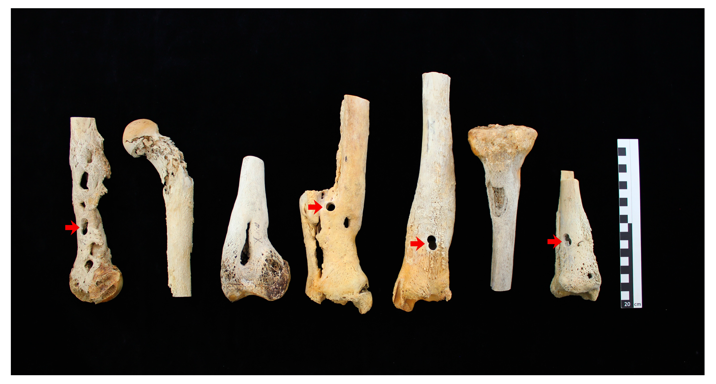
2.1.1. Osteomyelitic Bone Specimens

Bone specimens from seven different adult individuals (represented by few bones each) with apparent signs of an osteomyelitis infestation were chosen from the pathological collection of the Department of Historical Anthropology and Human Ecology (Figure 1). This large collection was assembled from autopsy specimens from the mid 19th century until the 1920s. The bones used in this study were permanently stored in the collections’ magazine, and have thus not been handled intensively over the last decades. Each bone was sampled at the diaphysis and four of the specimens were sampled additionally at a position close to the metaphysis that revealed morphologically detectable signs of osteomyelitis (Figure 1 arrows). Hence, eleven samples were investigated to prove the presence of S. aureus DNA sequences. Unlike archaeological skeletal material, the bone specimens of the pathological collection have never been in contact with soil-inhabiting bacteria, which can colonize skeletal material [34]. Nevertheless, the PCR system should be designed in a way that it is applicable to archaeological material, which has usually been buried. To test the PCR system in the presence of DNA from soil-inhabiting bacteria as well as PCR inhibitors like humic acids, which are present in every type of soil [35], several spiking experiments were performed (Supplementary Text S1).

2.1.2. Staphylococcus aureus Genomic DNA

To adjust the newly designed PCR-based analysis system to the requirements of degraded DNA samples, various control experiments were carried out (Supplementary Text S1). The genomic DNA of the S. aureus strain NCTC 8325 (accession CP000253.1 [36]) was used for initial spiking experiments and also served as a positive control for the PCR-amplification of the historical samples.

2.2. Methods

2.2.1. DNA Extraction

In order to remove possible cellular contamination from the sample surfaces, they were exposed to 6% sodium hypochlorite solution (Aug. Hedinger GmbH & Co. KG, Stuttgart, Germany) (e.g., [37,38]). Lysis and DNA extraction was performed according to a protocol as described by Frischalowski et al. [39]. For decalcification and lyses, 0.25 mg bone powder was incubated with 3900 µL EDTA (0.5 M, pH 8.0, Invitrogen™, Carlsbad, CA, USA) and 100 µL proteinase K solution in Tris/HCl (pH 7.5, 0.01 mol/L, 600 mAnson-U/mL, Merck, Darmstadt, Germany) for 18 h at 37 °C under constant rotation. An additional 50 µL proteinase K was added and a second lyses step, again under constant rotation, was performed for another 2 h at 56 °C. Afterwards, a 10 min incubation with 50 µL sodium dodecyl sulphate (10 mg/mL, Sigma-Aldrich®, St. Louis, MO, USA) was performed at 65 °C. After a 3 min centrifugation at 3300 rcf (Type 5430R, Eppendorf, Hamburg; Germany), the entire lysate of approximately 4100 µL was used for phenol-chloroform extraction. For this purpose, the entire lysate was transferred to 3 mL phenol (Carl Roth GmbH + Co. KG, Karlsruhe, Germany) and manually inverted for 6 min at room temperature. For phase separation, the extracts were incubated at 56 °C for 10 min. As much as possible of the aqueous phase was transferred to 4.5 mL chloroform (Carl Roth GmbH + Co. KG, Karlsruhe, Germany). Again, the samples were manually inverted for 6 min at room temperature and afterwards incubated at 56 °C for 10 min. The aqueous phase was then transferred to a mixture of 16 mL PB-Buffer (Qiagen, Hilden, Germany) and 100 µL sodium acetate buffer (pH = 5.2, 3 M Sigma-Aldrich®, St. Louis, MO, USA). The mixture was transferred to MinElute™ spin columns (Qiagen, Hilden, Germany) and DNA purification was performed via the QIAvac-System (Qiagen, Hilden, Germany) following the manufacturer’s protocol. The elution of the DNA from the MinElute™ spin columns (Qiagen, Hilden, Germany) was performed by using 20 µL RNAse free water (Qiagen, Hilden, Germany). After 5 min of incubation, centrifugation was performed at 13,000 rpm for 1 min. Elution was repeated twice to a total elution volume of 60 µL. The DNA extracts were stored frozen at −20 °C. For samples of the diaphysis at least two independent DNA extracts were prepared, for the samples close to the metaphysis one DNA extract each (Supplementary Text Table S1).

2.2.2. Primer Design

Ancient DNA analyses are highly dependent on optimal analysis conditions. This holds particularly true for the historical bone specimens investigated in this study, since they are expected to contain only traces of pathogenic DNA along with comparatively large quantities of human and microbial DNA, in case of the pathological collection due to maceration and taphonomic processes. Amplification sensitivity and specificity are mainly determined by primer characteristics [40]. Hence, the prerequisites for optimized primers—appropriate energy profile, avoidance of hairpins and primer dimer formation, and matching melting temperatures—were met. For the primer design for the amplification of specific sequences of the genes nuc [41] and fib [42] of S. aureus, the software PrimerSelect™ (DNASTAR, Madison, WI, USA) and the reference sequence of strain NCTC8325 (accession CP000253.1, cf. material-Staphylococcus aureus genomic DNA) was used. Primer specificity to the S. aureus complex was subsequently confirmed by BLASTN 2.2.32+ analysis [43,44] (Table 1). A cross reaction particularly with other Staphylococcus species (e.g., Staphylococcus epidermidis, common on the skin of humans) can be excluded.

2.2.3. Amplification of S. aureus DNA

The amplification reactions each consisted of a total volume of 25 µL with one part AmpliTaq Gold® 360 Master Mix (Applied Biosystems®, Waltham, MA, USA), 0.4 µM of each primer (forward and reverse), and a DNA inset varying between 0.5 and 6 µL (the DNA amount in the extracts varied because of the differential state of sample preservation). PCR conditions were as follows: initial phase at 94 °C for 7 min, 40 cycling steps consisting of 94 °C for 1 min, 46 °C for 1 min (nuc) or 50 °C (fib), and 72 °C for 1 min, and soak for 10 min at 10 °C.
Amplification of the X region of the protein A gene (spa typing) (e.g., [45,46]) and sequence analysis was done by the Robert Koch Institute as described by Strommenger et al. [47] (Supplementary Text S6).
The amplified PCR products were separated by gel electrophoresis. Run time was approximately 30 min at 120 V in a 2.5% gel in 1xTBE buffer.

2.2.4. Sequence Analysis of PCR Products

Sequencing of the purified amplification products (Supplementary Text S3) was carried out in a direct taq cycle sequencing approach with a total reaction volume of 20 µL. Each reaction mix consisted of one part BigDye®Terminator v1.1 5X Sequencing Buffer (Applied Biosystems®, Waltham, MA, USA), one part BigDye® Terminator v1.1 Ready Reaction Mix (Applied Biosystems®, Waltham, MA, USA), 0.24 µM primer (forward or reverse), and a PCR product inset varying between 0.5 and 5 µL. The PCR cycling conditions were as follows: initial phase at 96 °C for 10 min, 25 cycling steps consisting of 96 °C for 10 s, 50 °C for 5 s, and 60 °C for 4 min, and a final soak at 10 °C. Analysis of the purified sequencing products (Supplementary Text S3) was conducted via 3500 Genetic Analyzer (Applied Biosystem®, Waltham, MA, USA) and data processing was applied by using SeqA (Applied Biosystem®, Waltham, MA, USA) and the DNASTAR Lasergene 10 Core Suite software package (DNASTAR, Madison, WI, USA). The specificity of all amplified sequences was confirmed by BLASTN 2.2.32+ analysis [43,44].

2.2.5. Contamination Prevention

Concerning contamination, all aDNA analyses were conducted following a rigorous cleaning protocol and established laboratory standards that minimalize the contamination risk (e.g., [40,48]. All laboratory surfaces, instruments and bone samples underwent a strict cleaning protocol with 6% sodium hypochlorite solution (Aug. Hedinger GmbH & Co. KG, Stuttgart, Germany).
In order to easily detect possible external contamination with either human or even pathogenic DNA, there were three general strategies: (i) All DNA extracts of the bone specimens were checked for foreign human DNA via short tandem repeat typing (STR typing). Since STR typing only identifies human DNA and does not exclude a possible contamination with pathogenic DNA through a processor. (ii) The processors were screened for S. aureus colonization via culture of nasal swabs (Supplementary Text S4). In the case that none of the processors was a carrier of S. aureus, a late introduction of a modern contamination through a processor (e.g., contamination of a single tube during the PCR-setup) could be excluded. In the case that one of the processors had been a carrier of S. aureus, sequence analysis of the cultures would have been carried out allowing a differentiation e.g., through single nucleotide polymorphisms or spa typing. (iii) Several control samples and extraction blank controls were amplified with the S. aureus primer sets and the corresponding amplification parameters (Supplementary Text S1) to screen for possible S. aureus contaminants in extraction and PCR reagents.
The positive control sample of S. aureus was stored and handled separately from the historical DNA extracts during all steps in a working station (UV-bench DNA/RNA UV-Cleaner, UVT-S-AR) in a separate room and was always prepared last. Those precautions were taken to avoid a cross-contamination between the positive control sample (S. aureus strain NCTC8325) and the DNA extracts of the historical bone specimens.

2.2.6. STR Typing

STR typing was conducted with a miniSTR system [49] which was designed particularly for aDNA applications. Every DNA extract was tested twice, before and after amplifications were carried out for S. aureus analysis. The allele determination was performed by capillary electrophoresis via 3500 Series Genetic Analyzer (Applied Biosystems®, Waltham, MA, USA) and data processing was applied with the GeneMapper5 (Applied Biosystems®, Waltham, MA, USA) software (Supplementary Text S5).

3. Results

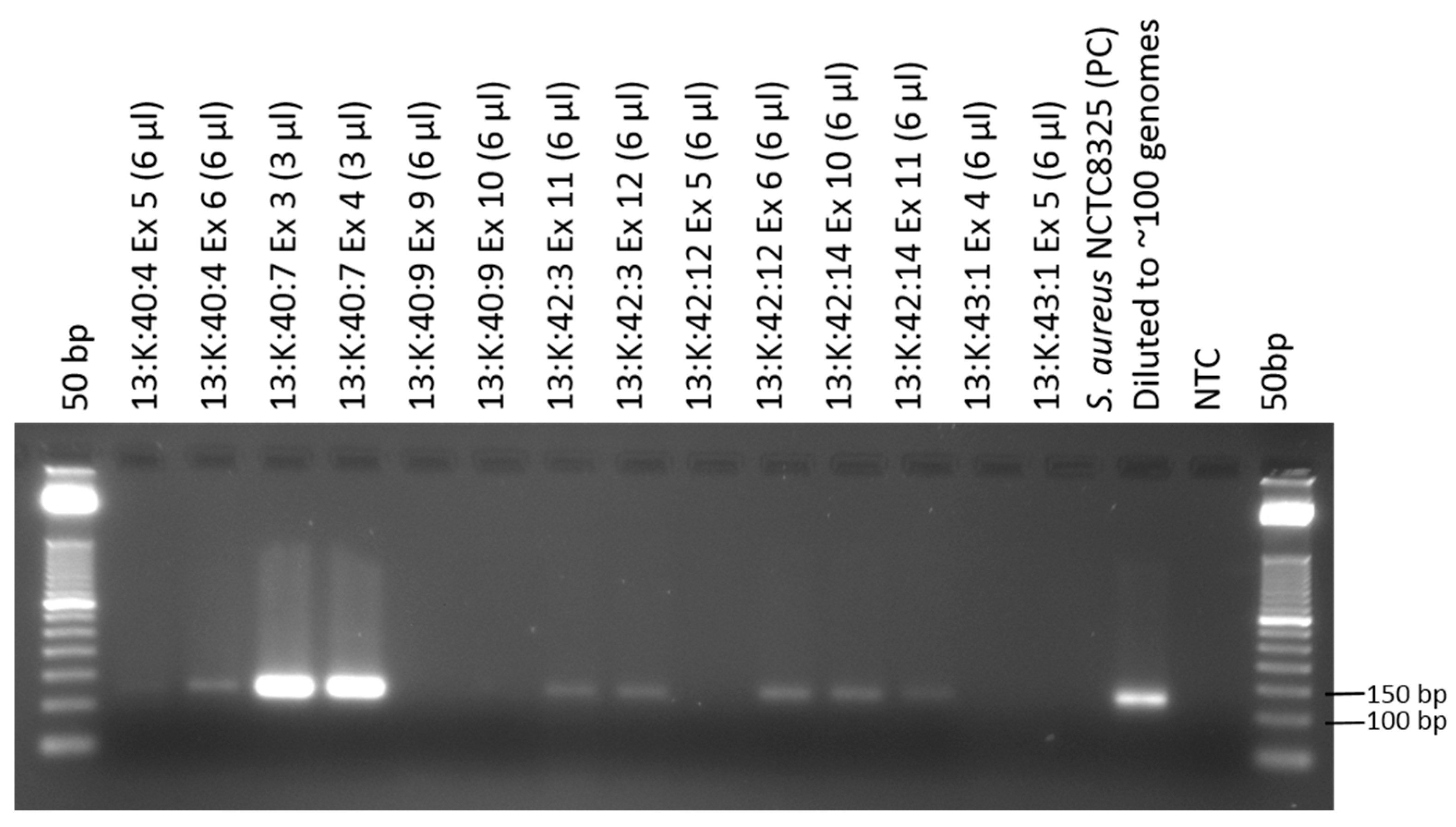
3.1. Amplification of Staphylococcus aureus Sequences from Osteomyelitic Bone Specimens

This study successfully conducted analyses of historical S. aureus sequences. The extraction revealed variable amounts of both human and pathogenic DNA from the different samples as judged by the differential brightness of amplification product in the respective gel pictures. The 122 bp fragments of the nuc gene was reproducibly amplified for eight out of eleven samples (Figure 2, Supplementary Figure S1, Table 2). The band intensity of most of the amplification products compared to that of the positive control indicates that there were less than 100 targets in the respective PCR. In contrast, the gel bands of 13:K:40:7 are much brighter than that of the positive control, indicating by far more than 100 targets in the reaction. The 111 bp fib fragment was amplified for ten out of eleven samples (Supplementary Figure S2, Table 2).
Concerning spa typing, only the sample 13:K:40:7 revealed an amplification product with a fragment length between 350 and 500 bp (Supplementary Text S6) visualized by gel electrophoresis. The spa type of this sample was determined as “t017” (Ridom StaphType, Ridom GmbH, [50]). Clustering analysis via the Based Upon Repeat Pattern algorithm [47] revealed an affiliation to the clonal complex “CC30”.

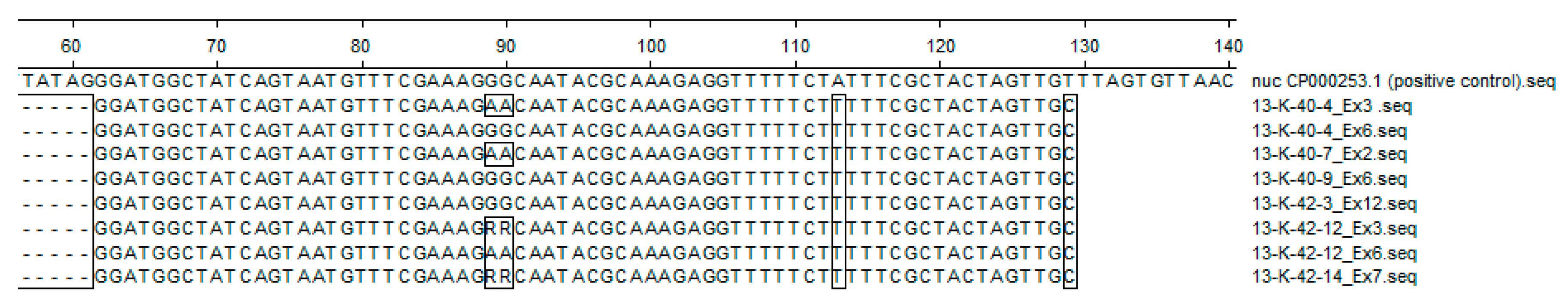
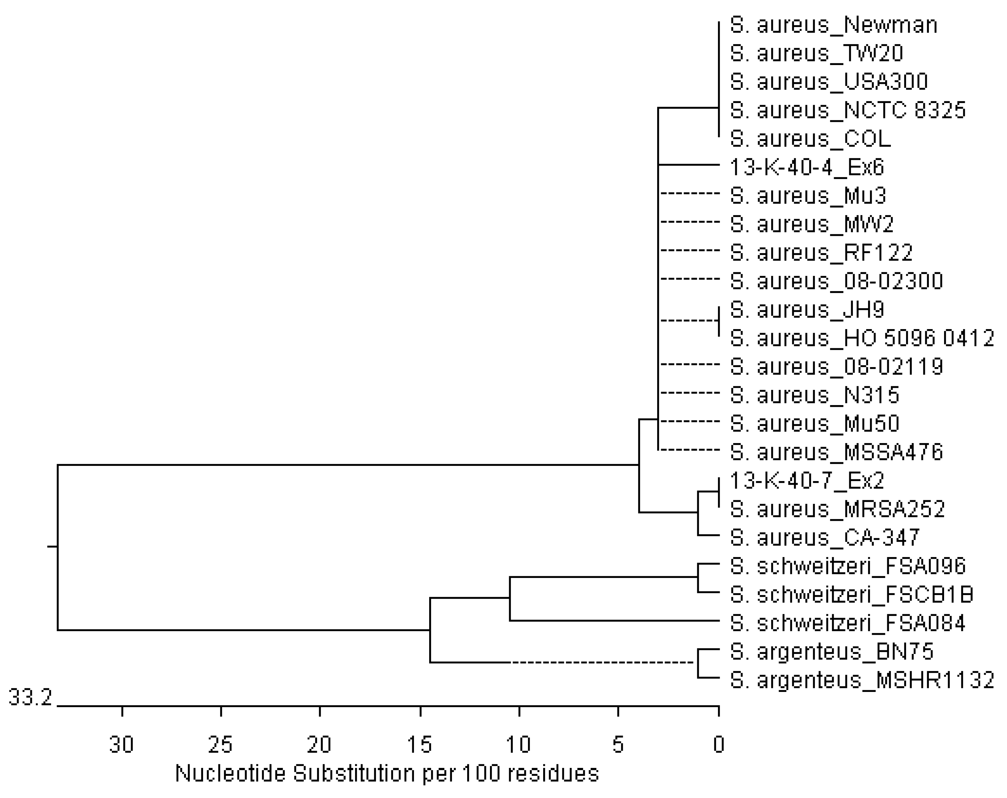
3.2. Sequencing of Staphylococcus aureus Sequences from Osteomyelitic Bone Specimens

For each bone sample revealing a positive signal in one of the PCR attempts for either the nuc (eight samples, Supplementary Figure S3) or fib locus (ten samples, Supplementary Figure S4), forward and reverse sequencing was performed. A BLASTN 2.2.32+ search [43,44] of the amplified historical sequences revealed sequence similarities to the reference sequences of the positive control sample NCTC 8325 between 97% and 99% for nuc and between 99% and 100% for fib, respectively. The alignment of the amplified historical nuc sequences and the genome of strain NCTC 8325 showed several polymorphisms (Figure 3). All amplified historical sequences reveal three single-base polymorphisms (C42T, A113T, and T129C) deviating from the reference sequences of the positive control sample NCTC 8325. Positions 89 to 90 revealed a polymorphic region where either double adenine or double guanine is present. For two amplified sequences (13:K:42:12_Ex3 and 13:K:42:14_Ex7), a double purine base (R = A or G) was determined. The alignment of the amplified fib sequences revealed only three base deviations in two historical samples (Supplementary Figure S5).
With the nuc sequencing results a phylogenetic analysis was performed. A comparison of the two nuc sequence variants obtained from the historical samples (13:K:40:4 Ex6 and 13:K:40:7 Ex2) with publically available sequences of S. aureus belonging to different clonal complexes was conducted. Furthermore, the sequences of Staphylococcus schweitzeri [51] and Staphylococcus argenteus [52,53] were used as an outgroup, as they both also belong to the S. aureus complex. Multiple alignments were performed using MegAlign™ (DNASTAR, Madison, WI, USA) and the phylogenetic relationship was visualized (Figure 4). As can be deduced from the phylogenetic tree, the 68 bp sequence variant as realised in 13:K:40:7_Ex2 clusters with the S. aureus strain MRSA 252 [54] and CA-347 [55]. The respective sequence characteristics of the 13:K:40:4_Ex6 cluster with the S. aureus isolates Mu3 [56], MW2 [57], RF122 [58], 08-02300 [59], JH9 [60], HO 5096 0412 [61], 08-02119 [59], N315 [62], Mu 50 [62], and MSSA476 [54].

3.3. STR Typing

Human STR typing of the historical DNA samples indicated that there were considerable differences in DNA preservation amongst the investigated bone specimens. When working with aDNA STR typing those differences may include variability in amplification success, allelic drop-out, preferential amplification of smaller STR systems (at the expense of the larger systems), or artefacts as a consequence of overamplified stutter products (e.g., [40,63,64]). The specimens 13:K:40:4, 13:K:40:7, and 13:K:42:3 revealed excellent DNA preservation resulting in full genetic fingerprints, whereas the remaining bone specimens (13:K:40:9, 13:K:42:12, 13:K:42:14, and 13:K:43:1) showed a poorer state of DNA preservation resulting in only a limited number of alleles (Supplementary Text Tables S4–S10).

4. Discussion

This study succeeded in finding specific DNA of S. aureus in seven investigated specimens from a pathological bone collection from historical times. Therefore, it is most likely that S. aureus was the causative organism of the morphologically manifested osteomyelitis infection. We succeeded in amplifying S. aureus-specific sequences of the two genes nuc and fib.
The investigation demonstrated that, as expected for aDNA analyses, different amounts of both pathogenic and human DNA were extracted from the pathological bone specimens. Due to the nature of DNA degradation, e.g., DNA fragmentation, human and pathogenic DNA were not quantified. We rather conducted initial spiking experiments with different amounts of S. aureus reference DNA demonstrating that approximately 10 to 50 genome equivalents are sufficient for a light band to be visible in the agarose gel picture (data not shown). Similar band intensities could be observed during the investigation of the DNA extracts of the pathological bone specimens, indicating that at least approximately 10–50 intact targets ought to be present in most of the historical extracts. Extracts of the bone specimen 13:K:40:7 were always amplified with a decreased DNA inset and yet revealed a brighter amplification product in gel electrophoresis (Figure 2, Supplementary Figures S1 and S2). The comparison of the band intensities of the spiking experiments with those of 13:K:40:7 revealed that by far higher amounts of pathogenic DNA was extracted and amplified from this sample (data not shown).
The evaluation of all amplified historical nuc sequences showed various polymorphisms differing from the nuc sequence of the strain NCTC 8325 (Figure 3). Hence, signals for historical S. aureus sequences are not a result of a contamination from the positive control. When analysing aDNA, contaminations should always be considered as a possible cause of positive signals during investigations. In this context, a processor in contact with the samples or the positive control is usually suspected of being the origin of a contamination. However, the spatial separation of the positive control from the historical samples makes a cross-contamination between them very unlikely. Furthermore, concerning S. aureus contamination through one of the processors (Flux, A.L. and Mazanec, J.), screening for S. aureus via a cultivation of swabs from the nasal mucosa on blood agar plates (Institute for Medical Microbiology, University Medical Center Goettingen) detected no colonization of the processors by the bacteria. In case of a contamination with DNA from one of the processors or from another bone specimen the detection of multiple allels in the STR typing results would have been expected. However, the comparison of the subsequent fingerprints showed that a contamination could be excluded (Supplementary Text Tables S4–S11). Therefore, it can be assumed that the results are authentic and not a result of a modern contamination.
Sequence variations of the nuc gene of 13:K:42:12, 13:K:42:14, and 13:K:40:4 were observed, within different samples (metaphysis and diaphysis) of the corresponding bone specimen, possibly indicating the presence of different S. aureus strains. Historically, S. aureus carriage as well as infection has been assumed to be monoclonal [65]. However, recent research data has indicated that polyclonal colonization is more common than previously anticipated [66,67]. During the investigation of the pathological samples sequencing of two different DNA extracts from one specimen (13:K:40:4, Figure 3) revealed different sequence characteristics, both being reproduced by forward and reverse sequencing reactions. This may be an indication that two different S. aureus strains colonized the individual in its lifetime. Specimen 13:K:42:12 also revealed two different sequence characteristics in two investigated extracts. It should be noted, however, that the sample 13:K:42:12 Ex3 revealed different characteristics in the forward and reverse amplified sequence that may be explained as a PCR artefact through e.g., base misincorporation. This assumption is strongly supported by the unusually high number of amplification cycles of the sample (in total, 70 cycling steps), because it was necessary to reamplify the PCR product before the sequencing reaction (Supplementary Text S3). The sequence of the sample 13:K:42:14 Ex7 revealed sequence characteristics similar to the sequences of sample 13:K:42:12 Ex3, but it was not reamplified before direct sequencing. The differences between the forward and reverse amplified sequences occurred repeatedly, making it difficult to evaluate whether the sequence characteristics can also be explained as a PCR artefact or as two sequences of different S. aureus strains being amplified.
Concerning spa typing, the amplification failure of the spa locus for the entire historical samples, except for 13:K.40:7, was not surprising since the spa amplicon size (approx. > 200 base pairs) is comparatively long with respect to aDNA analysis. Additionally, the primers for the spa locus were designed for the amplification of contemporarily isolated S. aureus DNA and do not meet aDNA requirements (Section 2.2.2). However, the spa type of 13:K.40:7 was determined as “t017” clustering to the clonal complex “CC30” (Section 3.1). Clonal complex 30 (CC30) had been involved in different pandemic ‘waves’ and was also part of the toxic shock syndrome epidemic. It includes the historic phage-type 80/81 strains and contemporary community, as well as hospital associated MRSA (CA-/HA-MRSA). Related methicillin-susceptible S. aureus (MSSA) are common human nasal colonizers worldwide [68].
Phylogenetic analysis shows different clustering for both sequence variants obtained from the historical samples (13:K:40:4 Ex6 and 13:K:40:7 Ex2) to the reference sequences of the S. aureus complex. Sequence 13:K:40:7 Ex2 clusters with MRSA252, which is in concordance with the determined spa type and clonal complex (CC30). The other historical sequence (13:K:40:4 Ex6) clusters in a group with S. aureus sequences belonging to various clonal complexes. This is not surprising, since there were no sequence variations within the amplified nuc sequences. Since the analysis was carried out with only 68 bp, further genome-wide analysis will be necessary to assess a robust phylogenetic relation between the historical sequences and those of contemporary S. aureus strains.
To the best of our knowledge, recent investigations focussing on analysing the genome of historical pathogens with PCR- or NGS-based methods were limited to archaeological skeletal material (e.g., [69,70,71,72,73,74,75,76,77]) or non-archaeological preserved soft tissues (e.g., [78,79,80]). The first successful investigation on non-archaeological, macerated bones assembled in the pathological collection was performed by Baron et al. [24]; we have now demonstrated yet again that these bone samples are suitable for extracting analysable amounts of both human and pathogenic DNA. In addition to archaeological material, these pathological samples are especially beneficial for the investigation of pathogens causing diseases limited to only single individuals in a skeletal series and not those leading to epidemics and mass burials in the archaeological context. Hence, these findings encourage further investigation of sample material from pathological collections and might establish a new approach to historical genomes of not only S. aureus but other pathogens causing the clinical picture of osteomyelitis, as well as other diseases. In the case of S. aureus, genome analysis of strains colonizing patients before the introduction of penicillin into medical use may increase our understanding of host–pathogen interactions and pathogen evolution in relation to anthropogenically introduced antibiotics.

Supplementary Materials

The following are available online at www.mdpi.com/1424-2818/9/4/43/s1. Flux et al. 2017 Supplementary Text Figure S1. Results of the PCR-based detection of the S aureus specific nuc sequence; Figure S2. Results of the PCR-based detection of the S aureus specific fib sequence; Figure S3. Electropherograms of all amplified historical nuc sequences; Figure S4. Electropherograms of all amplified historical fib sequences, Figure S5. Alignment of forward and reverse fib sequences of the historical bone specimens; Figure S6. Alignment of forward and reverse nuc sequences of the historical bone specimens; Figure S7. Alignment of two historical nuc sequences and reference sequences.

Acknowledgments

Raimond Lugert (Institute for Medical Microbiology, University Medical Center Goettingen, 37075 Goettingen, Germany) for the incubation of the nasal swabs and the morphological differentiation to detect a possible S. aureus colonization of Anna Lena Flux and Janine Mazanec. Verena Seidenberg (University of Goettingen, Johann-Friedrich-Blumenbach Institute for Zoology and Anthropology, Department of Historical Anthropology and Human Ecology, Goettingen, 37073, Germany) for assistance with primer design. Additionally, we would like to thank the reviewers for their critical discussion , which helped to improve the paper.

Author Contributions

Susanne Hummel conceived the experiments; Anna Lena Flux, Janine Mazanec and Susanne Hummel designed the experiments, Anna Lena Flux and Janine Mazanec performed the experiments; Birgit Strommenger and Susanne Hummel analyzed the data; Birgit Strommenger contributed the S. aureus positive control sample; Anna Lena Flux,Janine Mazanec, Birgit Strommenger and Susanne Hummel wrote the paper.

Conflicts of Interest

The authors declare no conflict of interest.

References

  1. Lew, D.P.; Waldvogel, F.A. Osteomyelitis. N. Engl. J. Med. 1997, 336, 999–1007. [Google Scholar] [CrossRef] [PubMed]
  2. Ortner, D.J. Infectious Diseases: Introduction, Biology, Osteomyelitis, Periostitis, Brucellosis, Glanders, and Septic Arthritis. In Identification of Pathological Conditions in Human Skeletal Remains; Ortner, D.J., Ed.; Academic Press: San Diego, CA, USA, 2003; ISBN 978-0-12-528628-2. [Google Scholar]
  3. Owen, W.B. The Diagnosis and Treatment of Osteomyelitis. Ann. Surg. 1936, 103, 1007–1014. [Google Scholar] [CrossRef] [PubMed]
  4. Roberts, C.; Manchester, K. The Archaeology of Disease; The History Press: Stroud, Great Britain, 2010; ISBN 978-0-7524-9497-5. [Google Scholar]
  5. Skinner, D.; Keefer, C.S. Significance of Bacteremia Caused by Staphylococcus aureus: A Study of One Hundred and Twenty-Two Cases and a Review of the Literature Concerned with Experimental Infection in Animals. Arch. Intern. Med. 1941, 68, 851–875. [Google Scholar] [CrossRef]
  6. Fleming, A. On the Antibacterial Action of Cultures of a Penicillium, with Special Reference to their Use in the Isolation of B. influenzæ. Br. J. Exp. Pathol. 1929, 10, 226–236. [Google Scholar] [CrossRef]
  7. Chain, E.; Florey, H.W.; Adelaide, M.B.; Gardner, A.D.; Heatley, N.G.; Jennings, M.A.; Orr-Ewing, J.; Sanders, A.G. Penicillin as a chemotherapeutic agent. Lancet 1940, 236, 226–228. [Google Scholar] [CrossRef]
  8. Rammelkamp, C.H.; Maxon, T. Resistance of Staphylococcus aureus to the Action of Penicillin. Proc. Soc. Exp. Biol. Med. 1942, 51, 386–389. [Google Scholar] [CrossRef]
  9. Jevons, M.P. “Celbenin”—Resistant Staphylococci. Br. Med. J. 1961, 1, 124–125. [Google Scholar] [CrossRef]
  10. Parker, M.T.; Jevons, M.P. A Survey of Methicillin Resistance in Staphylococcus aureus. Postgrad. Med. J. 1964, 40, 170–178. [Google Scholar] [CrossRef]
  11. Sutherland, R.; Rolinson, G.N. Characteristics of Methicillin-Resistant Staphylococci. J. Bacteriol. 1964, 87, 887–899. [Google Scholar] [PubMed]
  12. Peacock, S.J.; Paterson, G.K. Mechanisms of Methicillin Resistance in Staphylococcus aureus. Annu. Rev. Biochem. 2015, 84, 577–601. [Google Scholar] [CrossRef] [PubMed]
  13. Paul-Ehrlich-Gesellschaft für Chemotherapie, e.V. Studiengruppe Epidemiologie und Resistenzsituation bei Klinisch wichtigen Infektionserregern aus dem Ambulanten Versorgungsbereich bzw. aus dem dem Hospitalbereich Gegenüber Antibiotika. Available online: http://www.p-e-g.org/econtext/Berichte%20der%20Studien/ (accessed on 18 May 2017).
  14. European Antimicrobial Resistance Surveillance Network (EARS-Net). Annual Surveillance Reports on Antimicrobial Resistance. Available online: http://ecdc.europa.eu/en/antimicrobial-resistance/surveillance-and-disease-data/report (accessed on 18 May 2017).
  15. Deurenberg, R.H.; Stobberingh, E.E. The evolution of Staphylococcus aureus. Infect. Genet. Evol. J. Mol. Epidemiol. Evol. Genet. Infect. Dis. 2008, 8, 747–763. [Google Scholar] [CrossRef] [PubMed]
  16. D’Costa, V.M.; King, C.E.; Kalan, L.; Morar, M.; Sung, W.W.L.; Schwarz, C.; Froese, D.; Zazula, G.; Calmels, F.; Debruyne, R.; et al. Antibiotic resistance is ancient. Nature 2011, 477, 457–461. [Google Scholar] [CrossRef] [PubMed]
  17. Bhullar, K.; Waglechner, N.; Pawlowski, A.; Koteva, K.; Banks, E.D.; Johnston, M.D.; Barton, H.A.; Wright, G.D. Antibiotic resistance is prevalent in an isolated cave microbiome. PLoS ONE 2012, 7, e34953. [Google Scholar] [CrossRef] [PubMed]
  18. D’Costa, V.M.; McGrann, K.M.; Hughes, D.W.; Wright, G.D. Sampling the antibiotic resistome. Science 2006, 311, 374–377. [Google Scholar] [CrossRef] [PubMed]
  19. Wright, G.D. The antibiotic resistome: The nexus of chemical and genetic diversity. Nat. Rev. Microbiol. 2007, 5, 175–186. [Google Scholar] [CrossRef] [PubMed]
  20. Drancourt, M.; Raoult, D. Palaeomicrobiology: Current issues and perspectives. Nat. Rev. Microbiol. 2005, 3, 23–35. [Google Scholar] [CrossRef] [PubMed]
  21. Zink, A.R.; Reischl, U.; Wolf, H.; Nerlich, A.G. Molecular analysis of ancient microbial infections. FEMS Microbiol. Lett. 2002, 213, 141–147. [Google Scholar] [CrossRef] [PubMed]
  22. Salo, W.L.; Aufderheide, A.C.; Buikstra, J.; Holcomb, T.A. Identification of Mycobacterium tuberculosis DNA in a pre-Columbian Peruvian mummy. Proc. Natl. Acad. Sci. USA 1994, 91, 2091–2094. [Google Scholar] [CrossRef] [PubMed]
  23. Spigelman, M.; Lemma, E. The use of the polymerase chain reaction (PCR) to detect Mycobacterium tuberculosis in ancient skeletons. Int. J. Osteoarchaeol. 1993, 3, 137–143. [Google Scholar] [CrossRef]
  24. Baron, H.; Hummel, S.; Herrmann, B. Mycobacterium tuberculosis Complex DNA in Ancient Human Bones. J. Archaeol. Sci. 1996, 23, 667–671. [Google Scholar] [CrossRef]
  25. Donoghue, H.D.; Spigelman, M.; Zias, J.; Gernaey-Child, A.M.; Minnikin, D.E. Mycobacterium tuberculosis complex DNA in calcified pleura from remains 1400 years old. Lett. Appl. Microbiol. 1998, 27, 265–269. [Google Scholar] [CrossRef] [PubMed]
  26. Taylor, G.M.; Goyal, M.; Legge, A.J.; Shaw, R.J.; Young, D. Genotypic analysis of Mycobacterium tuberculosis from medieval human remains. Microbiology 1999, 145, 899–904. [Google Scholar] [CrossRef] [PubMed]
  27. Rafi, A.; Spigelman, M.; Stanford, J.; Lemma, E.; Donoghue, H.; Zias, J. Mycobacterium leprae DNA from ancient bone detected by PCR. Lancet Lond. Engl. 1994, 343, 1360–1361. [Google Scholar] [CrossRef]
  28. Haas, C.J.; Zink, A.; Pálfi, G.; Szeimies, U.; Nerlich, A.G. Detection of leprosy in ancient human skeletal remains by molecular identification of Mycobacterium leprae. Am. J. Clin. Pathol. 2000, 114, 428–436. [Google Scholar] [CrossRef] [PubMed]
  29. Donoghue, H.D.; Holton, J.; Spigelman, M. PCR primers that can detect low levels of Mycobacterium leprae DNA. J. Med. Microbiol. 2001, 50, 177–182. [Google Scholar] [CrossRef] [PubMed]
  30. Drancourt, M.; Aboudharam, G.; Signoli, M.; Dutour, O.; Raoult, D. Detection of 400-year-old Yersinia pestis DNA in human dental pulp: An approach to the diagnosis of ancient septicemia. Proc. Natl. Acad. Sci. USA 1998, 95, 12637–12640. [Google Scholar] [CrossRef] [PubMed]
  31. Raoult, D.; Aboudharam, G.; Crubézy, E.; Larrouy, G.; Ludes, B.; Drancourt, M. Molecular identification by “suicide PCR” of Yersinia pestis as the agent of Medieval Black Death. Proc. Natl. Acad. Sci. USA 2000, 97, 12800–12803. [Google Scholar] [CrossRef] [PubMed]
  32. Drancourt, M.; Tran-Hung, L.; Courtin, J.; de Lumley, H.; Raoult, D. Bartonella quintana in a 4000-year-old human tooth. J. Infect. Dis. 2005, 191, 607–611. [Google Scholar] [CrossRef] [PubMed]
  33. Grumbkow, P.V.; Zipp, A.; Seidenberg, V.; Fehren-Schmitz, L.; Kempf, V.A.J.; Gross, U.; Hummel, S. Brief communication: Evidence of Bartonella quintana infections in skeletons of a historical mass grave in Kassel, Germany. Am. J. Phys. Anthropol. 2011, 146, 134–137. [Google Scholar] [CrossRef] [PubMed]
  34. Herrmann, B.; Grupe, G.; Hummel, S.; Piepenbrink, H.; Schutkowski, H. Prähistorische Anthropologie: Leitfaden der Feld- und Labormethoden; Springer: Berlin Heidelberg, Germany, 1990; ISBN 0-387-52541-6. [Google Scholar]
  35. Ancient DNA: Recovery and Analysis of Genetic Material from Paleontological, Archaeological, Museum, Medical, and Forensic Specimens; Herrmann, B.; Hummel, S. (Eds.) Springer: New York, NY, USA, 1994; ISBN 13: 9780387943084. [Google Scholar]
  36. Gillaspy, A.F.; Worrell, V.; Orvis, J.; Roe, B.A.; Dyer, D.W.; Iandolo, J.J. The Staphylococcus aureus NCTC 8325 Genome. In Gram-Positive Pathogens, 2nd ed.; American Society of Microbiology: Washington, DC, USA, 2006; pp. 381–412. [Google Scholar] [CrossRef]
  37. Kemp, B.M.; Smith, D.G. Use of bleach to eliminate contaminating DNA from the surface of bones and teeth. Forensic Sci. Int. 2005, 154, 53–61. [Google Scholar] [CrossRef] [PubMed]
  38. Barta, J.L.; Monroe, C.; Kemp, B.M. Further evaluation of the efficacy of contamination removal from bone surfaces. Forensic Sci. Int. 2013, 231, 340–348. [Google Scholar] [CrossRef] [PubMed]
  39. Frischalowski, M.; Seidenberg, V.; Grosskopf, B.; Wulf, F.-W.; Hummel, S. Molekulargenetische Untersuchung des Verwandtschaftsverhältnisses von möglichen Mutter-Kind-Bestattungen aus dem frühneuzeitlichen Eldagsen. Nachrichten aus Niedersachsens Urgeschichte 2015, 84, 193–206. [Google Scholar]
  40. Hummel, S. Ancient DNA Typing: Methods, Strategies and Applications; Springer Science & Business Media: New York, NY, USA, 2003; ISBN 978-3-540-43037-7. [Google Scholar]
  41. Shortle, D. A genetic system for analysis of staphylococcal nuclease. Gene 1983, 22, 181–189. [Google Scholar] [CrossRef]
  42. Bodén, M.K.; Flock, J.I. Cloning and characterization of a gene for a 19 kDa fibrinogen-binding protein from Staphylococcus aureus. Mol. Microbiol. 1994, 12, 599–606. [Google Scholar] [CrossRef] [PubMed]
  43. Zhang, Z.; Schwartz, S.; Wagner, L.; Miller, W. A greedy algorithm for aligning DNA sequences. J. Comput. Biol. 2000, 7, 203–214. [Google Scholar] [CrossRef] [PubMed]
  44. Morgulis, A.; Coulouris, G.; Raytselis, Y.; Madden, T.L.; Agarwala, R.; Schäffer, A.A. Database indexing for production MegaBLAST searches. Bioinform. Oxf. Engl. 2008, 24, 1757–1764. [Google Scholar] [CrossRef] [PubMed]
  45. Frénay, H.M.; Theelen, J.P.; Schouls, L.M.; Vandenbroucke-Grauls, C.M.; Verhoef, J.; van Leeuwen, W.J.; Mooi, F.R. Discrimination of epidemic and nonepidemic methicillin-resistant Staphylococcus aureus strains on the basis of protein A gene polymorphism. J. Clin. Microbiol. 1994, 32, 846–847. [Google Scholar] [PubMed]
  46. Frénay, H.M.; Bunschoten, A.E.; Schouls, L.M.; van Leeuwen, W.J.; Vandenbroucke-Grauls, C.M.; Verhoef, J.; Mooi, F.R. Molecular typing of methicillin-resistant Staphylococcus aureus on the basis of protein A gene polymorphism. Eur. J. Clin. Microbiol. Infect. Dis. 1996, 15, 60–64. [Google Scholar] [CrossRef] [PubMed]
  47. Strommenger, B.; Kettlitz, C.; Weniger, T.; Harmsen, D.; Friedrich, A.W.; Witte, W. Assignment of Staphylococcus Isolates to Groups by spa Typing, SmaI Macrorestriction Analysis, and Multilocus Sequence Typing. J. Clin. Microbiol. 2006, 44, 2533–2540. [Google Scholar] [CrossRef] [PubMed]
  48. Llamas, B.; Valverde, G.; Fehren-Schmitz, L.; Weyrich, L.S.; Cooper, A.; Haak, W. From the field to the laboratory: Controlling DNA contamination in human ancient DNA research in the high-throughput sequencing era. STAR Sci. Technol. Archaeol. Res. 2017, 3, 1–14. [Google Scholar] [CrossRef]
  49. Seidenberg, V.; Schilz, F.; Pfister, D.; Georges, L.; Fehren-Schmitz, L.; Hummel, S. A new miniSTR heptaplex system for genetic fingerprinting of ancient DNA from archaeological human bone. J. Archaeol. Sci. 2012, 39, 3224–3229. [Google Scholar] [CrossRef]
  50. Harmsen, D.; Claus, H.; Witte, W.; Rothgänger, J.; Claus, H.; Turnwald, D.; Vogel, U. Typing of Methicillin-Resistant Staphylococcus aureus in a University Hospital Setting by Using Novel Software for spa Repeat Determination and Database Management. J. Clin. Microbiol. 2003, 41, 5442–5448. [Google Scholar] [CrossRef] [PubMed]
  51. Schaumburg, F.; Pauly, M.; Schubert, G.; Shittu, A.; Tong, S.; Leendertz, F.; Peters, G.; Becker, K. Characterization of a Novel Thermostable Nuclease Homolog (NucM) in a Highly Divergent Staphylococcus aureus Clade. J. Clin. Microbiol. 2014, 52, 4036–4038. [Google Scholar] [CrossRef] [PubMed]
  52. Holt, D.C.; Holden, M.T.G.; Tong, S.Y.C.; Castillo-Ramirez, S.; Clarke, L.; Quail, M.A.; Currie, B.J.; Parkhill, J.; Bentley, S.D.; Feil, E.J.; Giffard, P.M. A very early-branching Staphylococcus aureus lineage lacking the carotenoid pigment staphyloxanthin. Genome Biol. Evol. 2011, 3, 881–895. [Google Scholar] [CrossRef] [PubMed]
  53. Schuster, D.; Rickmeyer, J.; Gajdiss, M.; Thye, T.; Lorenzen, S.; Reif, M.; Josten, M.; Szekat, C.; Melo, L.D.R.; Schmithausen, R.M.; et al. Differentiation of Staphylococcus argenteus (formerly: Staphylococcus aureus clonal complex 75) by mass spectrometry from S. aureus using the first strain isolated from a wild African great ape. Int. J. Med. Microbiol. IJMM 2017, 307, 57–63. [Google Scholar] [CrossRef] [PubMed]
  54. Holden, M.T.G.; Feil, E.J.; Lindsay, J.A.; Peacock, S.J.; Day, N.P.J.; Enright, M.C.; Foster, T.J.; Moore, C.E.; Hurst, L.; Atkin, R.; et al. Complete genomes of two clinical Staphylococcus aureus strains: Evidence for the rapid evolution of virulence and drug resistance. Proc. Natl. Acad. Sci. USA 2004, 101, 9786–9791. [Google Scholar] [CrossRef] [PubMed]
  55. Stegger, M.; Driebe, E.M.; Roe, C.; Lemmer, D.; Bowers, J.R.; Engelthaler, D.M.; Keim, P.; Andersen, P.S. Genome Sequence of Staphylococcus aureus Strain CA-347, a USA600 Methicillin-Resistant Isolate. Genome Announc. 2013, 1. [Google Scholar] [CrossRef] [PubMed]
  56. Neoh, H.; Cui, L.; Yuzawa, H.; Takeuchi, F.; Matsuo, M.; Hiramatsu, K. Mutated Response Regulator graR Is Responsible for Phenotypic Conversion of Staphylococcus aureus from Heterogeneous Vancomycin-Intermediate Resistance to Vancomycin-Intermediate Resistance. Antimicrob. Agents Chemother. 2008, 52, 45–53. [Google Scholar] [CrossRef] [PubMed]
  57. Baba, T.; Takeuchi, F.; Kuroda, M.; Yuzawa, H.; Aoki, K.; Oguchi, A.; Nagai, Y.; Iwama, N.; Asano, K.; Naimi, T.; et al. Genome and virulence determinants of high virulence community-acquired MRSA. Lancet Lond. Engl. 2002, 359, 1819–1827. [Google Scholar] [CrossRef]
  58. Herron-Olson, L.; Fitzgerald, J.R.; Musser, J.M.; Kapur, V. Molecular Correlates of Host Specialization in Staphylococcus aureus. PLoS ONE 2007, 2, e1120. [Google Scholar] [CrossRef] [PubMed]
  59. Weber, R.E.; Layer, F.; Fuchs, S.; Bender, J.K.; Fiedler, S.; Werner, G.; Strommenger, B. Complete Genome Sequences of Two Methicillin-Sensitive Staphylococcus aureus Isolates Representing a Population Subset Highly Prevalent in Human Colonization. Genome Announc. 2016, 4. [Google Scholar] [CrossRef] [PubMed]
  60. Mwangi, M.M.; Wu, S.W.; Zhou, Y.; Sieradzki, K.; de Lencastre, H.; Richardson, P.; Bruce, D.; Rubin, E.; Myers, E.; Siggia, E.D.; et al. Tracking the in vivo evolution of multidrug resistance in Staphylococcus aureus by whole-genome sequencing. Proc. Natl. Acad. Sci. USA 2007, 104, 9451–9456. [Google Scholar] [CrossRef] [PubMed]
  61. Holden, M.T.G.; Hsu, L.-Y.; Kurt, K.; Weinert, L.A.; Mather, A.E.; Harris, S.R.; Strommenger, B.; Layer, F.; Witte, W.; de Lencastre, H.; et al. A genomic portrait of the emergence, evolution, and global spread of a methicillin-resistant Staphylococcus aureus pandemic. Genome Res. 2013, 23, 653–664. [Google Scholar] [CrossRef] [PubMed] [Green Version]
  62. Kuroda, M.; Ohta, T.; Uchiyama, I.; Baba, T.; Yuzawa, H.; Kobayashi, I.; Cui, L.; Oguchi, A.; Aoki, K.; Nagai, Y.; et al. Whole genome sequencing of meticillin-resistant Staphylococcus aureus. Lancet Lond. Engl. 2001, 357, 1225–1240. [Google Scholar] [CrossRef]
  63. Gill, P.; Whitaker, J.; Flaxman, C.; Brown, N.; Buckleton, J. An investigation of the rigor of interpretation rules for STRs derived from less than 100 pg of DNA. Forensic Sci. Int. 2000, 112, 17–40. [Google Scholar] [CrossRef]
  64. Advanced Topics in Forensic DNA Typing: Interpretation; Butler, J.M. (Ed.) Academic Press: San Diego, CA, USA, 2015; ISBN 978-0-12-405213-0. [Google Scholar]
  65. Kluytmans, J.; van Belkum, A.; Verbrugh, H. Nasal carriage of Staphylococcus aureus: Epidemiology, underlying mechanisms, and associated risks. Clin. Microbiol. Rev. 1997, 10, 505–520. [Google Scholar] [PubMed]
  66. Cespedes, C.; Saïd-Salim, B.; Miller, M.; Lo, S.-H.; Kreiswirth, B.N.; Gordon, R.J.; Vavagiakis, P.; Klein, R.S.; Lowy, F.D. The Clonality of Staphylococcus aureus Nasal Carriage. J. Infect. Dis. 2005, 191, 444–452. [Google Scholar] [CrossRef] [PubMed]
  67. Votintseva, A.A.; Miller, R.R.; Fung, R.; Knox, K.; Godwin, H.; Peto, T.E.A.; Crook, D.W.; Bowden, R.; Walker, A.S. Multiple-Strain Colonization in Nasal Carriers of Staphylococcus aureus. J. Clin. Microbiol. 2014, 52, 1192–1200. [Google Scholar] [CrossRef] [PubMed]
  68. DeLeo, F.R.; Kennedy, A.D.; Chen, L.; Wardenburg, J.B.; Kobayashi, S.D.; Mathema, B.; Braughton, K.R.; Whitney, A.R.; Villaruz, A.E.; Martens, C.A.; et al. Molecular differentiation of historic phage-type 80/81 and contemporary epidemic Staphylococcus aureus. Proc. Natl. Acad. Sci. USA 2011, 108, 18091–18096. [Google Scholar] [CrossRef] [PubMed]
  69. Suzuki, K.; Takigawa, W.; Tanigawa, K.; Nakamura, K.; Ishido, Y.; Kawashima, A.; Wu, H.; Akama, T.; Sue, M.; Yoshihara, A.; et al. Detection of Mycobacterium leprae DNA from Archaeological Skeletal Remains in Japan Using Whole Genome Amplification and Polymerase Chain Reaction. PLoS ONE 2010, 5, e12422. [Google Scholar] [CrossRef] [PubMed]
  70. Bouwman, A.S.; Kennedy, S.L.; Müller, R.; Stephens, R.H.; Holst, M.; Caffell, A.C.; Roberts, C.A.; Brown, T.A. Genotype of a historic strain of Mycobacterium tuberculosis. Proc. Natl. Acad. Sci. USA 2012, 109, 18511–18516. [Google Scholar] [CrossRef] [PubMed] [Green Version]
  71. Harbeck, M.; Seifert, L.; Hänsch, S.; Wagner, D.M.; Birdsell, D.; Parise, K.L.; Wiechmann, I.; Grupe, G.; Thomas, A.; Keim, P.; et al. Yersinia pestis DNA from Skeletal Remains from the 6th Century AD Reveals Insights into Justinianic Plague. PLoS Pathog. 2013, 9. [Google Scholar] [CrossRef] [PubMed] [Green Version]
  72. Schuenemann, V.J.; Singh, P.; Mendum, T.A.; Krause-Kyora, B.; Jäger, G.; Bos, K.I.; Herbig, A.; Economou, C.; Benjak, A.; Busso, P.; et al. Genome-wide comparison of medieval and modern Mycobacterium leprae. Science 2013, 341, 179–183. [Google Scholar] [CrossRef] [PubMed]
  73. Taylor, G.M.; Tucker, K.; Butler, R.; Pike, A.W.G.; Lewis, J.; Roffey, S.; Marter, P.; Lee, O.Y.-C.; Wu, H.H.T.; Minnikin, D.E.; et al. Detection and strain typing of ancient Mycobacterium leprae from a medieval leprosy hospital. PLoS ONE 2013, 8, e62406. [Google Scholar] [CrossRef] [PubMed] [Green Version]
  74. Mendum, T.A.; Schuenemann, V.J.; Roffey, S.; Taylor, G.M.; Wu, H.; Singh, P.; Tucker, K.; Hinds, J.; Cole, S.T.; Kierzek, A.M.; et al. Mycobacterium leprae genomes from a British medieval leprosy hospital: Towards understanding an ancient epidemic. BMC Genomics 2014, 15, 270. [Google Scholar] [CrossRef] [PubMed] [Green Version]
  75. Müller, R.; Roberts, C.A.; Brown, T.A. Genotyping of ancient Mycobacterium tuberculosis strains reveals historic genetic diversity. Proc. R Soc. B 2014, 281, 20133236. [Google Scholar] [CrossRef] [PubMed] [Green Version]
  76. Bos, K.I.; Herbig, A.; Sahl, J.; Waglechner, N.; Fourment, M.; Forrest, S.A.; Klunk, J.; Schuenemann, V.J.; Poinar, D.; Kuch, M.; et al. Eighteenth century Yersinia pestis genomes reveal the long-term persistence of an historical plague focus. eLife 2016, 5, e12994. [Google Scholar] [CrossRef] [PubMed]
  77. Feldman, M.; Harbeck, M.; Keller, M.; Spyrou, M.A.; Rott, A.; Trautmann, B.; Scholz, H.C.; Päffgen, B.; Peters, J.; McCormick, M.; et al. A High-Coverage Yersinia pestis Genome from a Sixth-Century Justinianic Plague Victim. Mol. Biol. Evol. 2016, 33, 2911–2923. [Google Scholar] [CrossRef] [PubMed]
  78. Taubenberger, J.K.; Reid, A.H.; Krafft, A.E.; Bijwaard, K.E.; Fanning, T.G. Initial genetic characterization of the 1918 “Spanish” influenza virus. Science 1997, 275, 1793–1796. [Google Scholar] [CrossRef] [PubMed]
  79. Devault, A.M.; Golding, G.B.; Waglechner, N.; Enk, J.M.; Kuch, M.; Tien, J.H.; Shi, M.; Fisman, D.N.; Dhody, A.N.; Forrest, S.; et al. Second-Pandemic Strain of Vibrio cholerae from the Philadelphia Cholera Outbreak of 1849. N. Engl. J. Med. 2014, 370, 334–340. [Google Scholar] [CrossRef] [PubMed]
  80. Maixner, F.; Krause-Kyora, B.; Turaev, D.; Herbig, A.; Hoopmann, M.R.; Hallows, J.L.; Kusebauch, U.; Vigl, E.E.; Malfertheiner, P.; Megraud, F.; et al. The 5300-year-old Helicobacter pylori genome of the Iceman. Science 2016, 351, 162–165. [Google Scholar] [CrossRef] [PubMed]
Figure 1. Bone specimens from seven different individuals with apparent signs of an osteomyelitis infestation. From left to right: 13:K:40:4 (femur, distal), 13:K:40:7 (femur, proximal), 13:K:40:9 (femur, distal), 13:K:42:3 (tibia and fibula, distal), 13:K:42:12 (tibia, distal), 13:K:42:14 (tibia, proximal), 13:K:43:1 (tibia, distal). Sampling sites: diaphysis all specimens, four specimens additionally close to the metaphysis (red arrows).
Diversity 09 00043 g001
Figure 2. Results of the PCR-based detection of S. aureus specific 122 bp nuc sequences after agarose gel electrophoresis (DNA inset bracketed). Gel bands are located between the 100 bp and 150 bp fragments of the DNA ladder, indicating that the sought amplification product of 122 bp was generated. 9/14 amplifications resulted in visible bands; the negative template control (NTC) shows no evidence of pathogenic DNA presence in the PCR reaction mix. As a positive control, DNA of the strain NCTC8325 (approximately 100 genomes) was used. Electrophoresis parameters: 8 μL PCR product with 2 μL loading dye, 4 μL size standard: 50 bp DNA ladder (Invitrogen™, Carlsbad, CA, USA), 2.5% gel, 120 V, exposure time: 1.00 s. For detailed description of the extract nomenclature see Supplementary Text Table S1.
Diversity 09 00043 g002
Figure 3. Alignment of the consensus nuc sequence of the historical bone specimens (primer binding sequence was removed) and the nuc gene of the S. aureus reference strain NCTC 8325 (CP000253.1, positive control sample). Bases differing from the reference nuc sequence are framed. Software: MegAlign™ (DNASTAR, Madison, WI, USA). For the alignment of all amplified forward (fwd.) and reverse (rev.) sequences and the corresponding electropherograms see Supplementary Figures S3 and S6.
Diversity 09 00043 g003
Figure 4. Phylogenetic relations based on the alignment (Figure S7 in supplementary data) of two historical nuc sequence variants of 13:K:40:4 Ex6 and 13:K:40:7 Ex2 and nuc sequences of seventeen contemporary S. aureus strains. S. schweitzeri and S. argenteus were used as the out group. Software: MegAlign™ (DNASTAR, Madison, WI, USA), Method: Jotun Hein.
Diversity 09 00043 g004
Table 1. Primer sequences for the S. aureus loci 1.
PositionNamePrimer LengthSequencePosition in S. aureus Reference Sequence (NCTC 8325, Accession CP000253.1)Product Length
ForwardS.aur nuc up 226-mer5’-GGCAATTGTTTCAATATTACTTATAG-3’800069…800094122 bp
ReverseS.aur nuc low28-mer5’-TTGAAACTACAACTAAAGTTAACACTAA-3’800163…800190
ForwardS.aur fib up20-mer5’-GAAGGATACGGTCCAAGAGA-3’1073163…1073128111 bp
ReverseS.aur fib low23-mer5’-AGGTGTTGAGTTAAATTTTGGTC-3’1073251…1073273
1 Using the amplification parameters described in this paper for both the nuc and the fib system, as little as 10 genomes/targets can be amplified and analysed by gel electrophoresis (data not shown). 2 The “T” at the sequence position seven of S.aur nuc up indicates a polymorphism (“C” instead of a “T”) at the primer binding site of S. aureus strain NCTC 8325 (positive control).
Table 2. Overall amplification success of S. aureus sequences visualized by gel electrophoresis (Figure 2, Supplementary Figures S1 and S2).
Bone SpecimenSampling Position
(Figure 1)
nucfib
13:K:40:4 diaphysis++
metaphysis++
13:K:40:7diaphysis++
13:K:40:9diaphysis++
13:K:42:3diaphysis++
metaphysis+
13:K:42:12diaphysis++
metaphysis++
13:K:42:14diaphysis++
13:K:43:1diaphysis
metaphysis+
+ = specific amplification product; − = no amplification product.

