
Titanium and Silicon Dioxide-Coated Fabrics for Management and Tuning of Infrared Radiation

 ,
,  ,
, 
Abstract
:1. Introduction
2. Materials and Methods
Sample Preparation
3. Structural Characterization
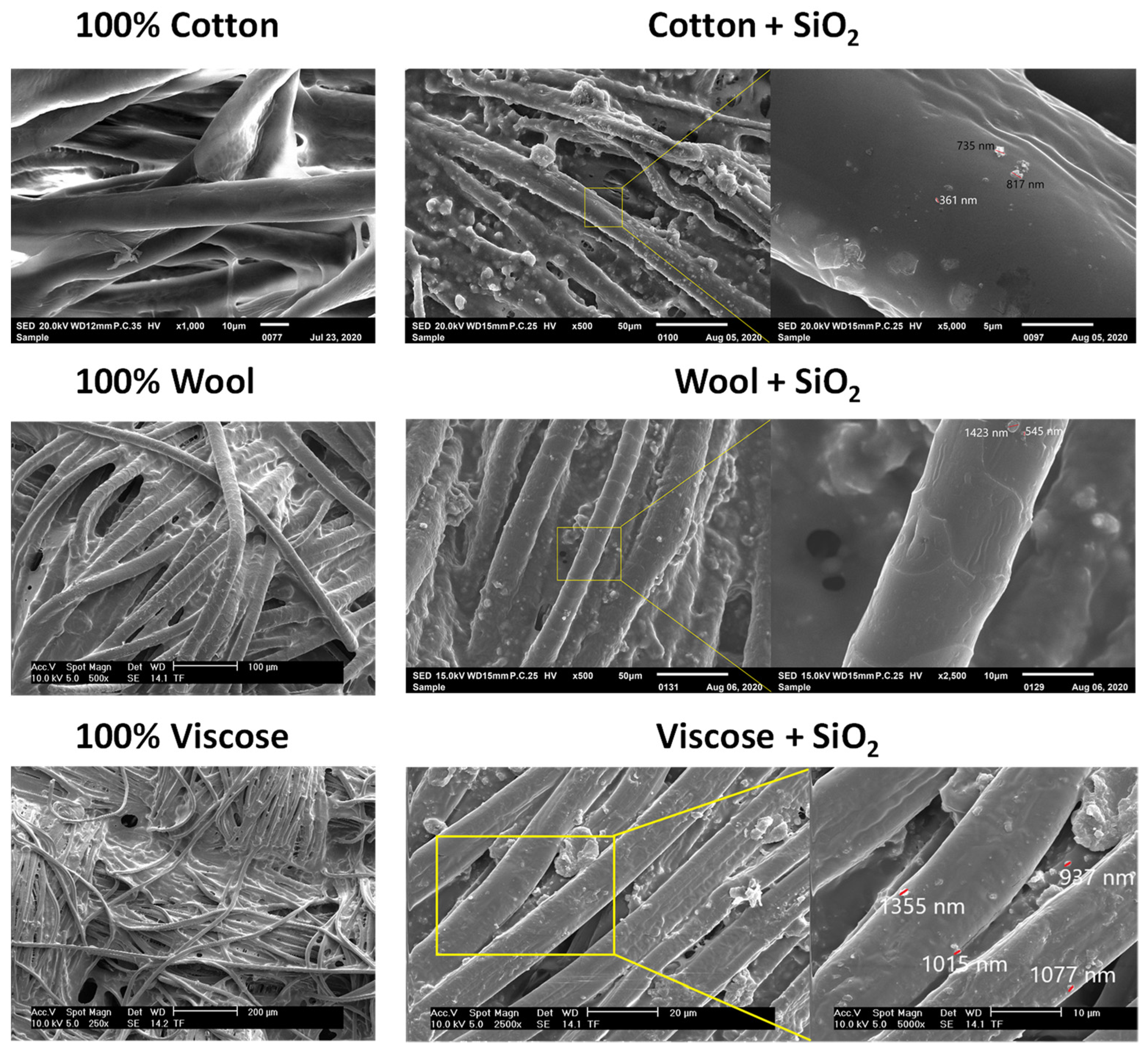
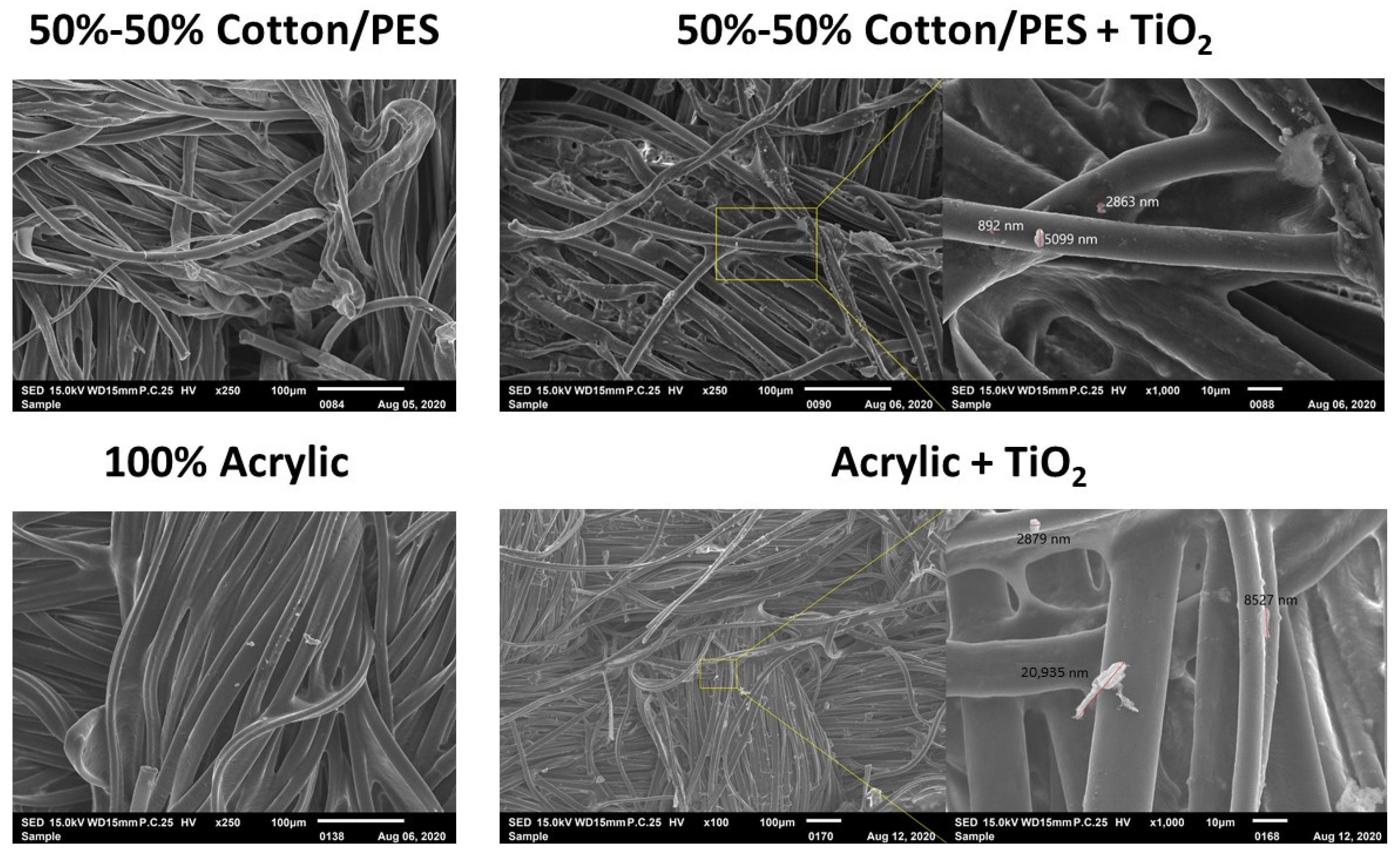
3.1. SEM Images
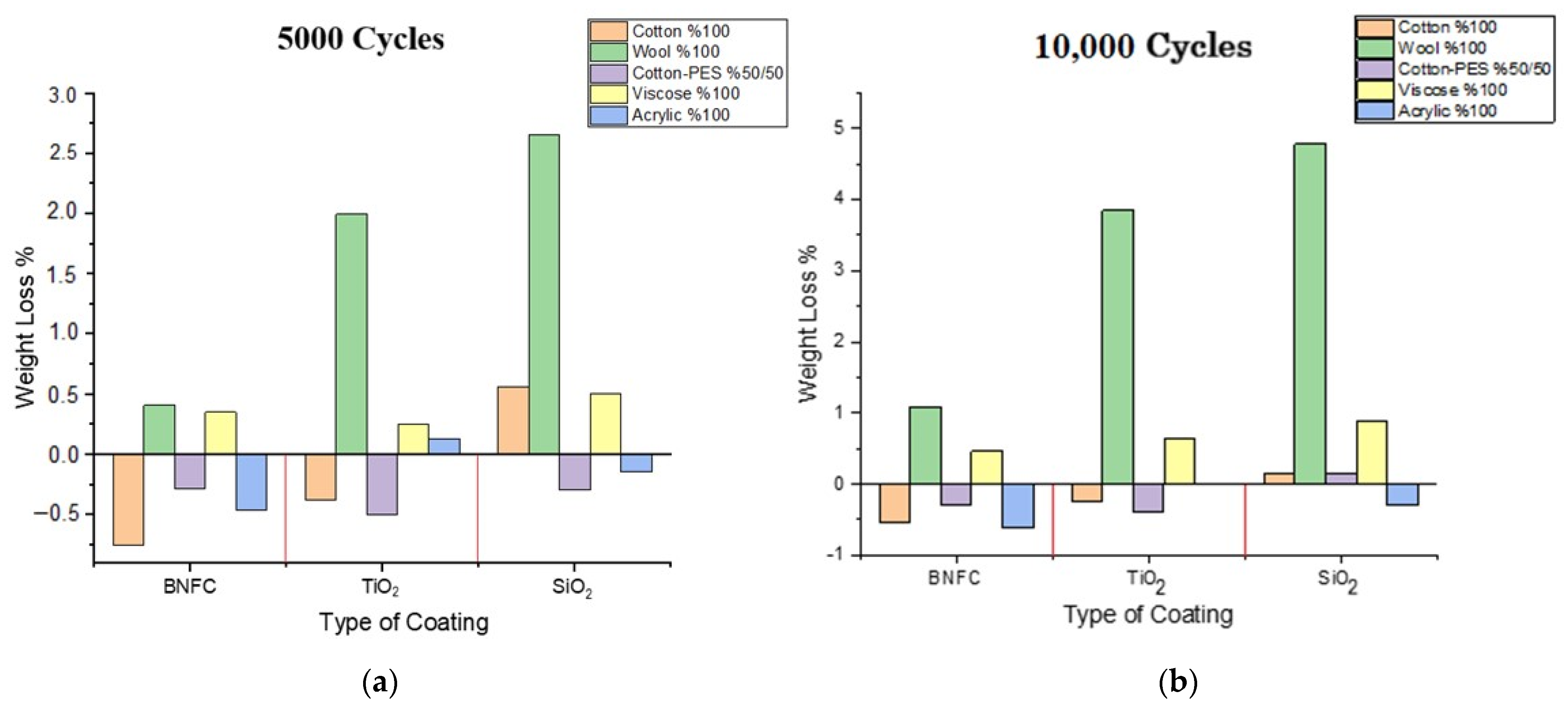
3.2. Abrasion Test
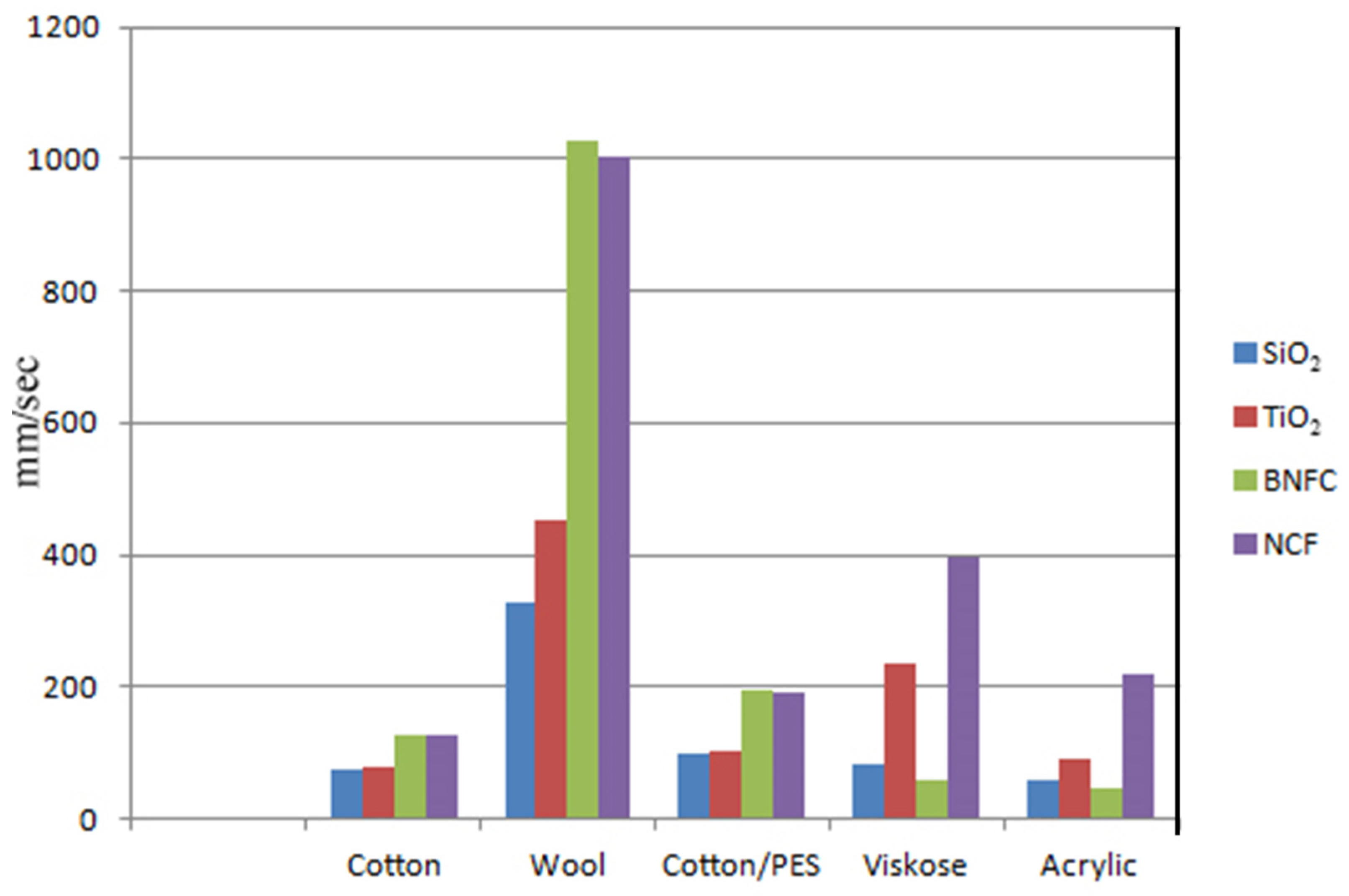
3.3. Air Permeability Test
4. Functional Characterization
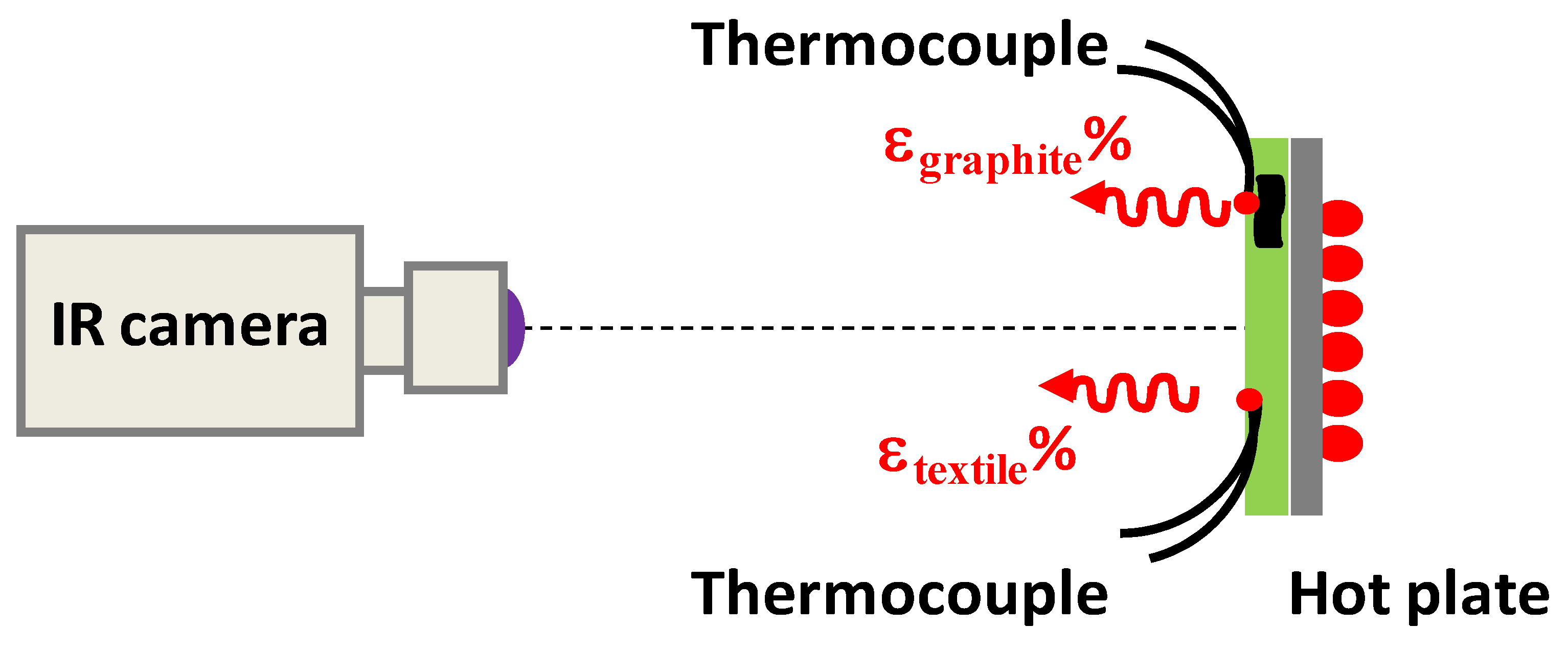
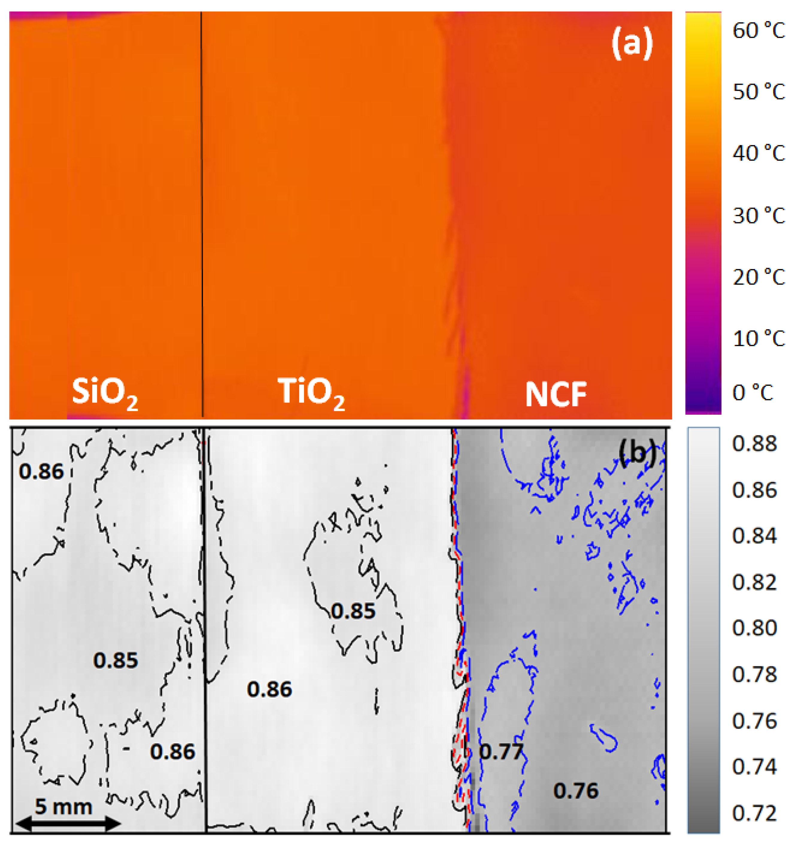
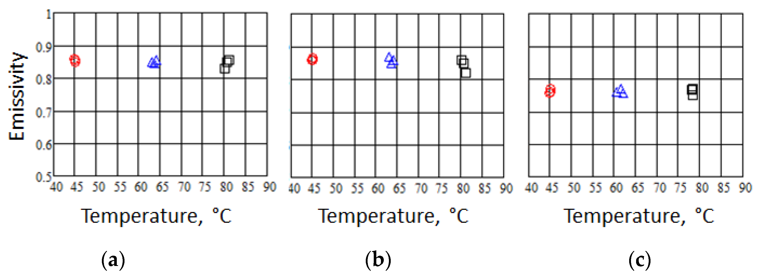
4.1. Infrared Emissivity Measurements
- (i)
- The IR signal from the heated sample comes from two main sources: the sample emittance and the environment. If the sample temperature is higher than the room temperature, the second becomes negligible, and consequently, the noise in the emissivity measurement decreases.
- (ii)
- The difference of at least 20 °C between sample and room temperatures guarantees the accurate emissivity measurement of textiles.
- (iii)
- The standard textile emissivity is expected to be constant with temperature in the range from 20 °C to 80 °C, and in conditions of low humidity. Therefore, it is convenient to estimate the sample emissivity by measuring the IR signals from heated samples at relatively high temperatures with respect to room temperature.
- (iv)
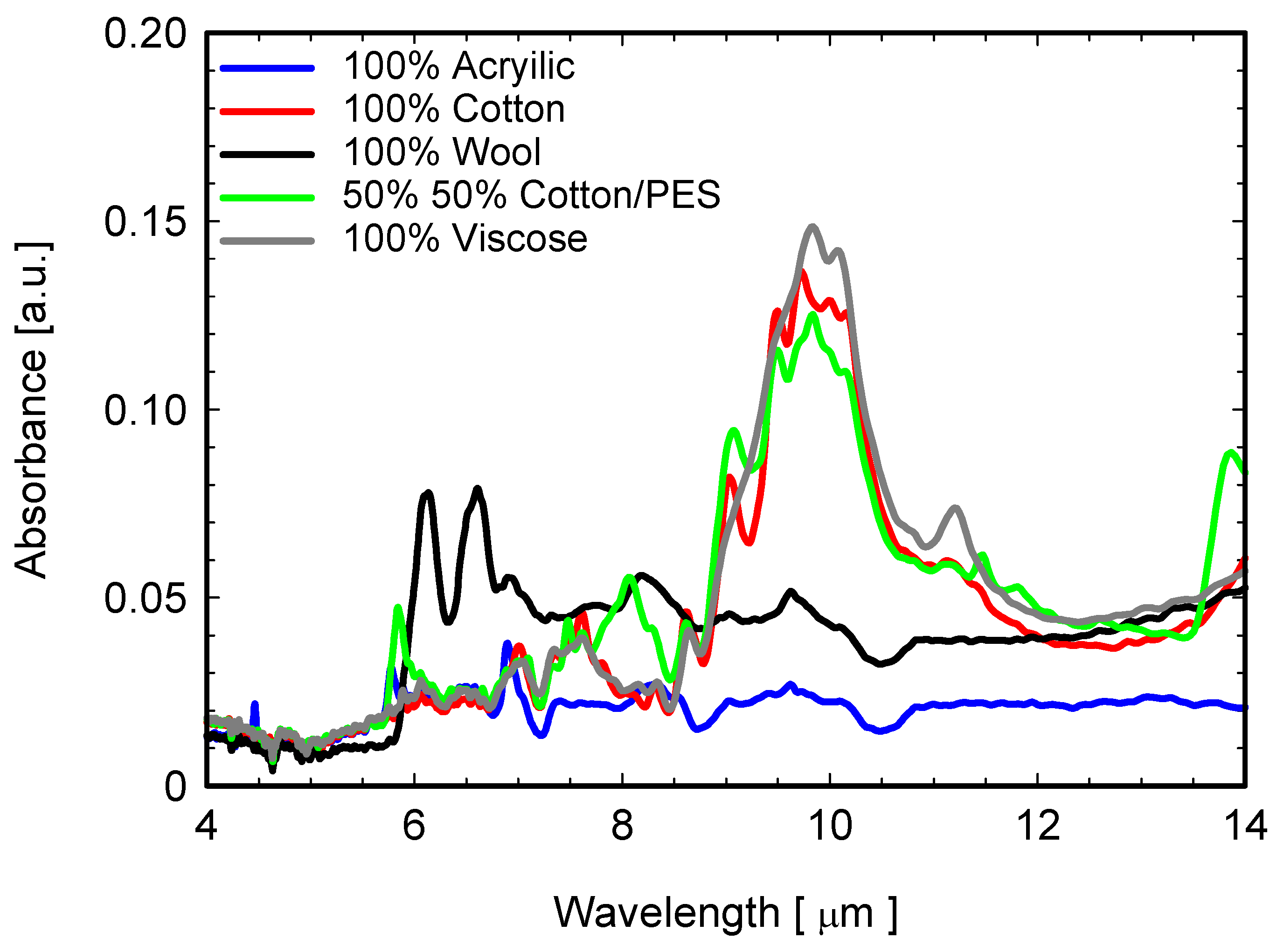
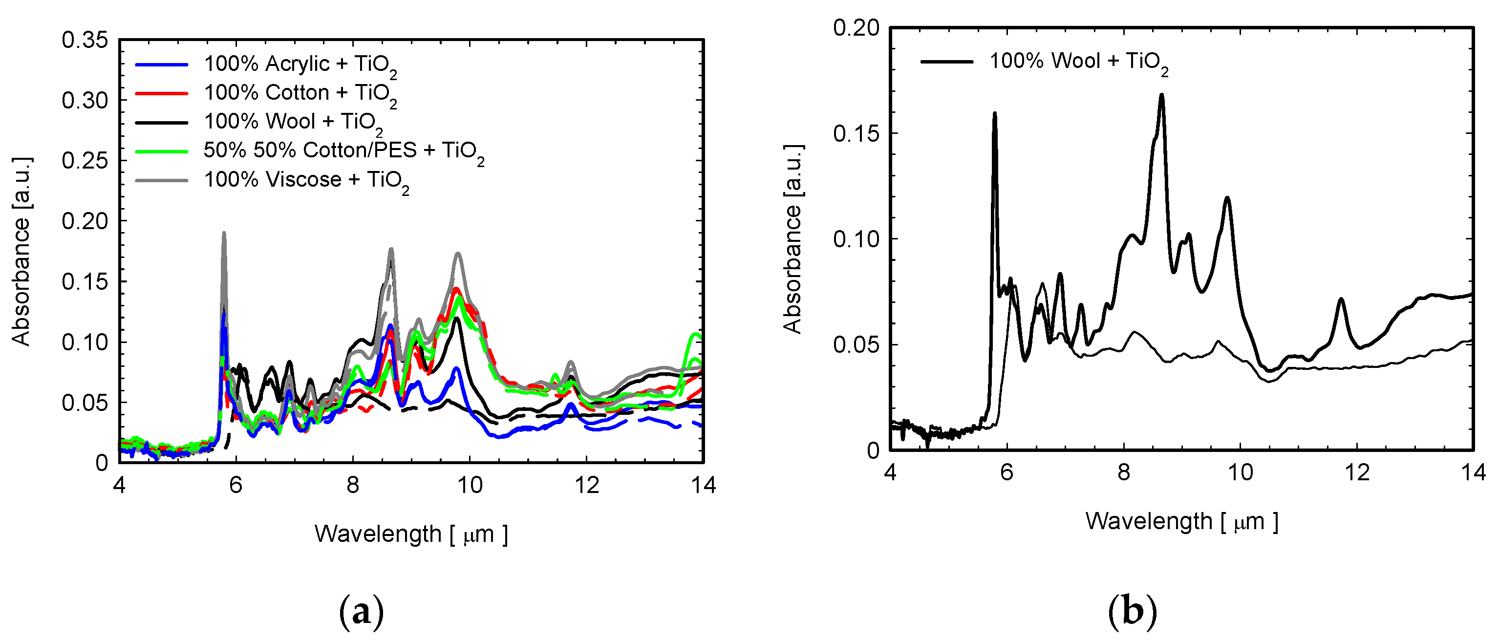
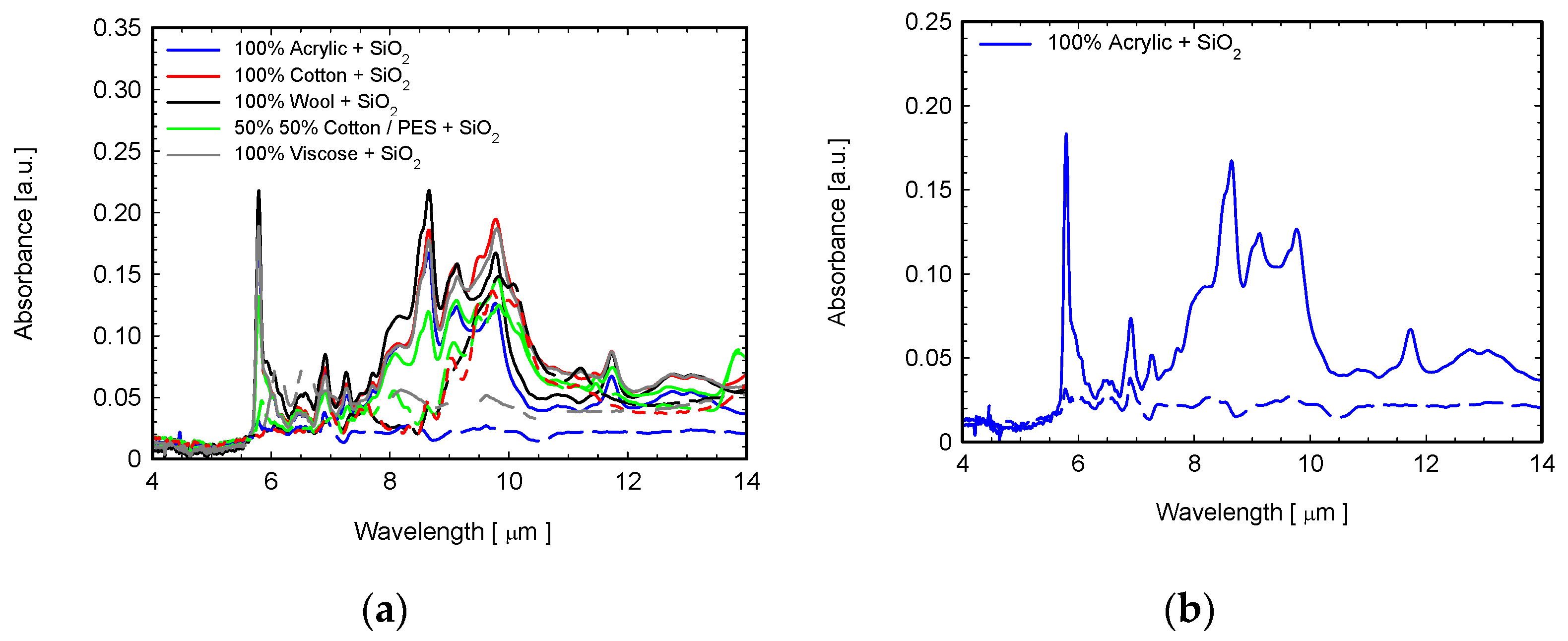
4.2. ATR-FTIR Analysis
5. Conclusions
Author Contributions
Funding
Institutional Review Board Statement
Informed Consent Statement
Data Availability Statement
Acknowledgments
Conflicts of Interest
References
- Vatansever, F.; Hamblin, M.R. Far infrared radiation (FIR): Its biological effects and medical applications. Photonics Lasers Med. 2012, 1, 255–266. [Google Scholar] [CrossRef] [PubMed] [Green Version]
- Dyer, J. Infrared functional textiles. In Functional Textiles for Improved Performance, Protection and Health; Pan, N., Sun, G., Eds.; Woodhead Publishing Ltd.: Sawston, UK, 2011; pp. 184–197. [Google Scholar]
- Shih, Y.H.; Lin, J.H.; Hsieh, C.T.; Lin, C.W.; Lou, C.W. Far-Infrared Nonwoven Fabrics Made of Various Ratios of Bamboo Fiber to Far-Infrared Fiber: Far-Infrared Emissivity and Mechanical Property Evaluations. In Proceedings of the 13th Asian Textile Conference, Geelong, Australia, 3–6 November 2015; pp. 830–834. [Google Scholar]
- Leung, T.K.; Shang, H.F.; Chen, D.C.; Chen, J.Y.; Chang, T.M.; Hsiao, S.Y.; Ho, C.K.; Lin, Y.S. Effects of far infrared rays on hydrogen peroxide-scavenging capacity. Biomed. Eng. Appl. Basis Commun. 2011, 23, 99–105. [Google Scholar] [CrossRef]
- Jeon, K.I.; Park, E.; Park, H.R.; Jeon, Y.J.; Cha, S.H.; Lee, S.C. Antioxidant activity of far-infrared radiated rice hull extracts on reactive oxygen species scavenging and oxidative DNA damage in human lymphocytes. J. Med. Food 2006, 9, 42. [Google Scholar] [CrossRef] [PubMed]
- Faisal, A.M.; Salaün, F.; Giraud, S.; Ferri, A.; Chen, Y.; Wang, L. Far-infrared emission properties and thermogravimetric analysis of ceramic-embedded polyurethane films. Polymers 2021, 13, 686. [Google Scholar] [CrossRef]
- Yuce, I. Far Infrared Ray Emitting Fabric and Yarns. Trak. Univ. J. Eng. Sci. 2017, 18, 145–151. [Google Scholar] [CrossRef] [Green Version]
- Larciprete, M.C.; Paoloni, S.; Voti, R.L.; Gloy, Y.S.; Sibilia, C. Infrared radiation characterization of several stainless steel textiles in the 3.5–5.1 μm infrared range. Int. J. Therm. Sci. 2018, 132, 168–173. [Google Scholar] [CrossRef]
- Larciprete, M.C.; Gloy, Y.S.; Voti, R.L.; Cesarini, G.; Leahu, G.; Bertolotti, M.; Sibilia, C. Temperature dependent emissivity of different stainless steel textiles in the infrared range. Int. J. Therm. Sci. 2017, 113, 130–135. [Google Scholar] [CrossRef]
- Larciprete, M.C.; Paoloni, S.; Orazi, N.; Mercuri, F.; Orth, M.; Gloy, Y.; Centini, M.; Voti, R.L.; Sibilia, C. Infrared emissivity characterization of carbon nanotubes dispersed poly (ethylene terephthalate) fibers. Int. J. Therm. Sci. 2019, 146, 106–109. [Google Scholar] [CrossRef]
- Leahu, G.; Voti, R.L.; Larciprete, M.C.; Sibilia, C.; Bertolotti, M.; Nefedov, I.; Anoshkin, I.V. Thermal Characterization of Carbon Nanotubes by Photothermal Techniques. Int. J. Thermophys. 2015, 36, 1349–1357. [Google Scholar] [CrossRef]
- Larciprete, M.C.; Paoloni, S.; Cesarini, G.; Sibilia, C.; Rubežienė, V.; Sankauskaitė, A. Thermo-regulating properties of textiles with incorporated microencapsulated Phase Change Materials. MRS Adv. 2020, 5, 1023–1028. [Google Scholar] [CrossRef]
- Pooley, M.A.; Anderson, D.M.; Beckham, H.W.; Brennan, J.F. Engineered emissivity of textile fabrics by the inclusion of ceramic particles. Opt. Express 2016, 24, 10556–10564. [Google Scholar] [CrossRef] [PubMed]
- Tao, Y.; Li, T.; Yang, C.; Wang, N.; Yan, F.; Li, L. The Influence of Fiber Cross-Section on Fabric Far-Infrared Properties. Polymers 2018, 10, 1147. [Google Scholar] [CrossRef] [PubMed] [Green Version]
- Kim, H.A.; Kim, S.J. Far-infrared emission characteristics and wear comfort property of ZrC-imbedded heat storage knitted fabrics for emotional garments. Autex Res. J. 2017, 17, 142–151. [Google Scholar] [CrossRef] [Green Version]
- Park, C.H.; Shim, M.H.; Shim, H.S. Far IR emission and thermal properties of ceramics coated fabrics by IR thermography. Key Eng. Mater. 2006, 321, 849–852. [Google Scholar] [CrossRef]
- Xiong, Y.; Huang, S.; Wang, W.; Liu, X.; Li, H. Properties and applications of high emissivity composite films based on far-infrared ceramic powder. Materials 2017, 10, 1370. [Google Scholar] [CrossRef] [PubMed] [Green Version]
- Hu, X.; Tian, M.; Qu, L.; Zhu, S.; Han, G. Multifunctional cotton fabrics with graphene/polyurethane coatings with far-infrared emission, electrical conductivity, and ultraviolet blocking properties. Carbon 2015, 95, 625–633. [Google Scholar] [CrossRef]
- Bahng, G.W.; Lee, J.D. Development of heat-generating polyester fiber harnessing catalytic ceramic powder combined with heat-generating super microorganisms. Text. Res. J. 2014, 84, 1220–1230. [Google Scholar] [CrossRef]
- Conrado, L.A.; Munin, E. Reduction in body measurements after use of a garment made with synthetic fibers embedded with ceramic nanoparticles. J. Cosmet. Dermatol. 2011, 10, 30–35. [Google Scholar] [CrossRef]
- Qiu, K.; Elhassan, A.; Tian, T.; Yin, X.; Yu, J.; Li, Z.; Ding, B. Highly flexible, efficient, and sandwich-structured infrared radiation heating fabric. ACS Appl. Mater. Interfaces 2020, 12, 11016–11025. [Google Scholar] [CrossRef]
- Cian, C.; Gianocca, V.; Barraud, P.A.; Guerraz, M.; Bresciani, J.P. Bioceramic fabrics improve quiet standing posture and handstand stability in expert gymnasts. Gait Posture 2015, 42, 419–423. [Google Scholar] [CrossRef] [Green Version]
- Sankauskaitė, A.; Rubežienė, V.; Kubilienė, D.; Abraitienė, A.; Baltušnikaitė-Guzaitienė, J.; Dubinskaitė, K. Investigation of Thermal Behavior of 3D PET Knits with Different Bioceramic Additives. Polymers 2020, 12, 1319. [Google Scholar] [CrossRef] [PubMed]
- Chung, J.; Lee, S. Development of nanofibrous membranes with far-infrared radiation and their antimicrobial properties. Fibers Polym. 2014, 15, 1153–1159. [Google Scholar] [CrossRef]
- Nhan, L.G.; Denby, E.F. The effect of humidity on the abrasion-resistance of wool fabric. J. Text. Inst. 1979, 70, 264–268. [Google Scholar] [CrossRef]
- Kara, S.; Yesilpinar, S.; Aksit, A. Permeability properties and abrasion resistance of coated polypropylene fabrics. Fibres Text. 2018, 2, 40–47. [Google Scholar]
- Brzeziński, S.; Kowalczyk, D.; Borak, B.; Jasiorski, M.; Tracz, A. Applying the sol–gel method to the deposition of nanocoats on textiles to improve their abrasion resistance. J. Appl. Polym. Sci. 2012, 125, 3058–3067. [Google Scholar] [CrossRef]
- Ding, Y.Y.; Xu, B.; Ge, F.Y.; Cai, Z.S. Robust Superhydrophobic and Photocatalytic Cotton Fabrics Based on TiO2-SiO2-PDMS Composite Coating. Key Eng. Mater. 2015, 671, 225–230. [Google Scholar] [CrossRef]
- Gürkan, P.Ü.; Atav, R. Yünlü Dokuma Kumaşlara Katma Değer Kazandırılması Amacıyla Atkıda Ipek veya Alpaka Iplik Kullanımının Araştırılması. 2018. Available online: http://acikerisim.nku.edu.tr/xmlui/handle/20.500.11776/2938 (accessed on 1 January 2019).
- Reis, A.B.; Yoshida, C.M.; Reis, A.P.C.; Franco, T.T. Application of chitosan emulsion as a coating on Kraft paper. Polym. Int. 2011, 60, 963–969. [Google Scholar] [CrossRef]
- Voti, R.L.; Leahu, G.; Larciprete, M.C.; Sibilia, C.; Bertolotti, M.; Nefedov, I.; Anoshkin, I.V. Photoacoustic Characterization of Randomly Oriented Silver Nanowire Films. Int. J. Thermophys. 2015, 36, 342. [Google Scholar] [CrossRef]
- ASTM E1933-99a; Standard Test Methods for Measuring and Compensating for Emissivity Using Infrared Imaging Radiometers. American Society for Testing and Materials International: West Conshohocken, PA, USA, 1999.
- Cesca, T.; Scian, C.; Petronijevic, E.; Leahu, G.; Li Voti, R.; Cesarini, G.; Macaluso, R.; Mosca, M.; Sibilia, C.; Mattei, G. Correlation between in situ structural and optical characterization of the semiconductor-to-metal phase transition of VO2 thin films on sapphire. Nanoscale 2020, 12, 851–863. [Google Scholar] [CrossRef] [Green Version]
- Centini, M.; Benedetti, A.; Larciprete, M.C.; Belardini, A.; Voti, R.L.; Bertolotti, M.; Sibilia, C. Midinfrared thermal emission properties of finite arrays of gold dipole nanoantennas. Phys. Rev. B 2015, 92, 205411. [Google Scholar] [CrossRef]
- Zhang, H.; Hu, T.L.; Zhang, J.C. Surface emissivity of fabric in the 8–14 μm waveband. J. Text. Inst. 2009, 100, 90–94. [Google Scholar] [CrossRef]
- Siadat, S.A.; Mokhtari, J. Diffuse reflectance behavior of the printed cotton/nylon blend fabrics treated with zirconium and cerium dioxide and citric acid in near-and short-wave IR radiation spectral ranges. Color Res. Appl. 2020, 45, 55–64. [Google Scholar] [CrossRef]
- Larciprete, M.; Orazi, N.; Gloy, Y.S.; Paoloni, S.; Sibilia, C.; Voti, R.L. In-plane thermal diffusivity measurements of polyethersulfone woven textiles by Infrared Thermography. Sensors 2022, 22, 940. [Google Scholar] [CrossRef] [PubMed]
- Larciprete, M.; Centini, M.; Voti, R.L.; Sibilia, C. Selective and tunable thermal emission in metamaterials composed of oriented polar inclusions. J. Opt. Soc. Am. B Opt. Phys. 2017, 34, 1459. [Google Scholar] [CrossRef]
- Olak-Kucharczyk, M.; Szczepańska, G.; Kudzin, M.H.; Pisarek, M. The photocatalytical properties of RGO/TiO2 coated fabrics. Coatings 2020, 10, 1041. [Google Scholar] [CrossRef]
- Abdel-Messih, M.F.; Ahmed, M.; El-Sayed, A.S. Photocatalytic decolorization of Rhodamine B dye using novel mesoporous SnO2–TiO2 nano mixed oxides prepared by sol–gel method. J. Photochem. Photobiol. A Chem. 2013, 260, 1. [Google Scholar] [CrossRef]
- Erdem, B.; Hunsicker, R.A.; Simmons, G.W.; Sudol, E.D.; Dimonie, V.L.; El-Aasser, M.S. XPS and FTIR surface characterization of TiO2 particles used in polymer encapsulation. Langmuir 2001, 17, 2664–2669. [Google Scholar] [CrossRef]
- Roshni, S.B.; Jayakrishnan, M.P.; Mohanan, P.; Surendran, K.P. Design and fabrication of an E-shaped wearable textile antenna on PVB-coated hydrophobic polyester fabric. Smart Mater. Struct. 2017, 26, 105011. [Google Scholar] [CrossRef]
- Silva, C.C.; Sampaio, M.; Marques, R.; Ferreira, L.; Tavares, P.; Silva, A.; Faria, J. Photocatalytic production of hydrogen from methanol and saccharides using carbon nanotube-TiO2 catalysts. Appl. Catal. B Environ. 2015, 178, 82–90. [Google Scholar] [CrossRef] [Green Version]
- Dhineshbabu, N.R.; Manivasakan, P.; Yuvakkumar, R.; Prabu, P.; Rajendran, V. Enhanced Functional Properties of ZrO2/SiO2 Hybrid Nanosol Coated Cotton Fabrics. J. Nanosci. Nanotechnol. 2013, 13, 4017–4024. [Google Scholar] [CrossRef]
- Dhineshbabu, N.R.; Arunmetha, S.; Manivasakan, P.; Karunakaran, G.; Rajendran, V. Enhanced functional properties of cotton fabrics using TiO2/SiO2 nanocomposites. J. Ind. Text. 2016, 45, 674. [Google Scholar] [CrossRef]
- Medeghini, F.; Rouxel, R.; Crut, A.; Maioli, P.; Rossella, F.; Banfi, F.; Vallée, F.; Del Fatti, N. Signatures of Small Morphological Anisotropies in the Plasmonic and Vibrational Responses of Individual Nano-objects. J. Phys. Chem. Lett. 2019, 10, 5372–5380. [Google Scholar] [CrossRef] [PubMed]
- Tomoda, M.; Voti, R.L.; Matsuda, O.; Wright, O.B. Tomographic reconstruction of picosecond acoustic strain propagation. Appl. Phys. Lett. 2007, 90, 041114. [Google Scholar] [CrossRef]
- Dehoux, T.; Wright, O.B.; Voti, R.L. Picosecond time scale imaging of mechanical contacts. Ultrasonics 2010, 50, 197. [Google Scholar] [CrossRef] [PubMed] [Green Version]


| Fabric Type | Weft Density (Piece/cm) | Warp Density (Piece/cm) | Surface Mass Density (g/m2) |
|---|---|---|---|
| 100% Cotton | 13 | 14 | 227 |
| 100% Wool | 14 | 15 | 194 |
| 100% Acrylic | 14 | 16 | 176 |
| 100% Viscose | 14 | 15 | 170 |
| 50-50% Cotton/PES | 14 | 15 | 212 |
| Chemical Used | Percent (%) | Bioceramic Particle Size and Purity Grade | Viscosity (Pa.s) |
|---|---|---|---|
| Acrylate (Rudolf Duraner AC111) | 20 | - | - |
| Water | 75.5 | - | - |
| Cross-linker (RUCO-COAT FX 8011) | 1.5 | - | - |
| Dispersing agent (Rudolf Duraner AD 719) | 0.5 | - | - |
| Thickener (RUCO-COAT TH 5020) | 1.5 | - | - |
| SiO2 | 1 | 55–75 nm, 98.5% | 9.3 |
| TiO2 | 1 | 17 nm, 99.9% | 6.2 |
| BNFC | - | - | 2.2 |
| NCF | BNFC | TiO2 | SiO2 | |
|---|---|---|---|---|
| Cotton (100%) | 0.80 | 0.81 | 0.84 | 0.85 |
| Wool (100%) | 0.75 | 0.84 | 0.83 | 0.81 |
| Cotton-PES (50-50%) | 0.84 | 0.85 | 0.83 | 0.83 |
| Viscose (100%) | 0.84 | 0.86 | 0.86 | 0.88 |
| Acrylic (100%) | 0.76 | 0.76 | 0.86 | 0.85 |
| NCF | BNFC | TiO2 | SiO2 | |
|---|---|---|---|---|
| Cotton (100%) | Reference | 0.03 | 0.05 | 0.07 |
| Wool (100%) | Reference | 0.01 | 0.06 | 0.12 |
| Cotton-PES (50-50%) | Reference | 0.03 | 0.04 | 0.03 |
| Viscose (100%) | Reference | 0.06 | 0.06 | 0.08 |
| Acrylic (100%) | Reference | 0.06 | 0.06 | 0.11 |
Publisher’s Note: MDPI stays neutral with regard to jurisdictional claims in published maps and institutional affiliations. |
© 2022 by the authors. Licensee MDPI, Basel, Switzerland. This article is an open access article distributed under the terms and conditions of the Creative Commons Attribution (CC BY) license (https://creativecommons.org/licenses/by/4.0/).
Share and Cite
Yuce, I.; Canoglu, S.; Yukseloglu, S.M.; Li Voti, R.; Cesarini, G.; Sibilia, C.; Larciprete, M.C. Titanium and Silicon Dioxide-Coated Fabrics for Management and Tuning of Infrared Radiation. Sensors 2022, 22, 3918. https://0-doi-org.brum.beds.ac.uk/10.3390/s22103918
Yuce I, Canoglu S, Yukseloglu SM, Li Voti R, Cesarini G, Sibilia C, Larciprete MC. Titanium and Silicon Dioxide-Coated Fabrics for Management and Tuning of Infrared Radiation. Sensors. 2022; 22(10):3918. https://0-doi-org.brum.beds.ac.uk/10.3390/s22103918
Chicago/Turabian StyleYuce, Ismail, Suat Canoglu, Sevhan Muge Yukseloglu, Roberto Li Voti, Gianmario Cesarini, Concita Sibilia, and Maria Cristina Larciprete. 2022. "Titanium and Silicon Dioxide-Coated Fabrics for Management and Tuning of Infrared Radiation" Sensors 22, no. 10: 3918. https://0-doi-org.brum.beds.ac.uk/10.3390/s22103918