The Influence of Body Composition on the Systemic Exposure of Paclitaxel in Esophageal Cancer Patients

 ,
,  , , ,
, , ,
Abstract
:1. Introduction
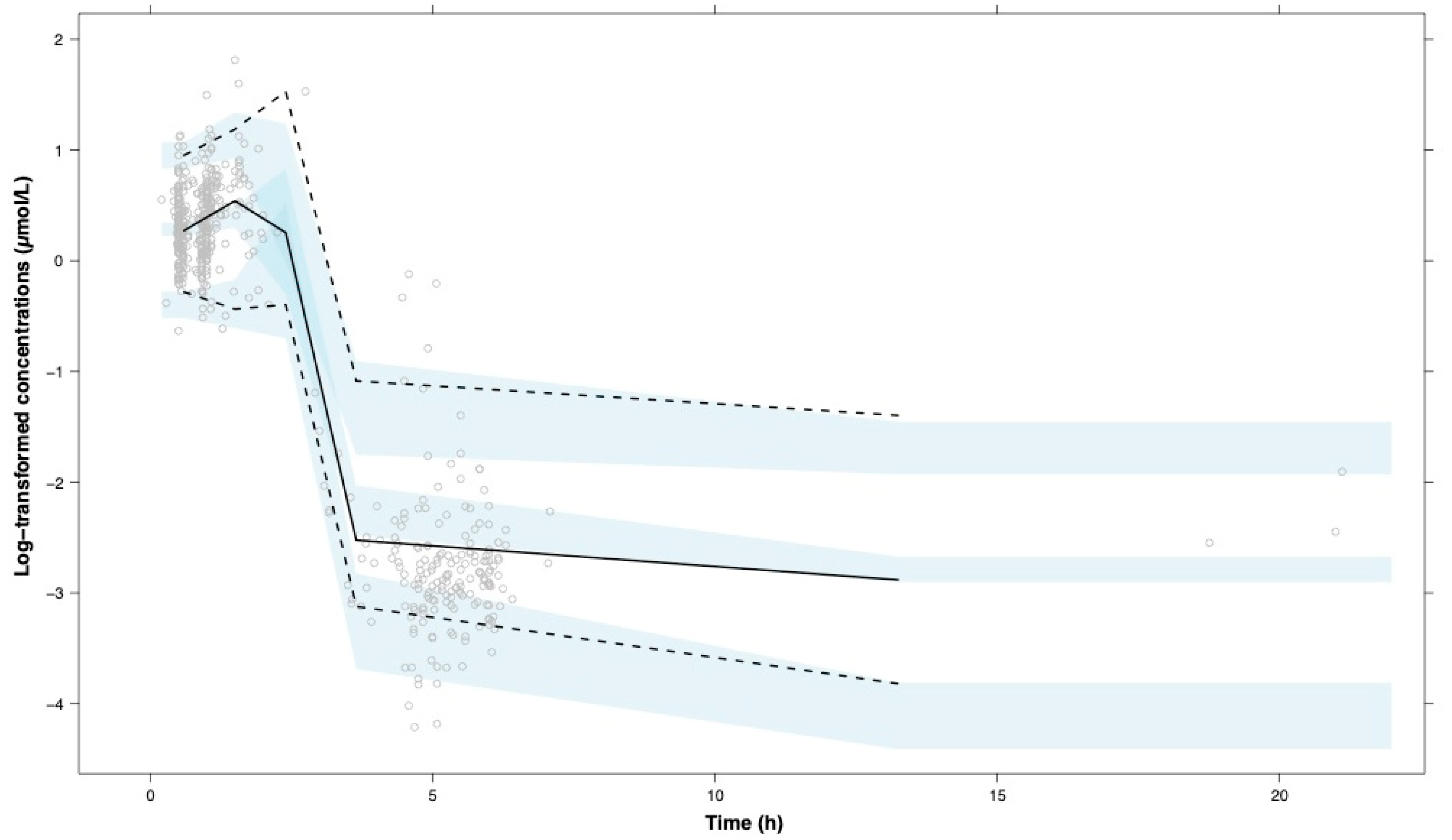
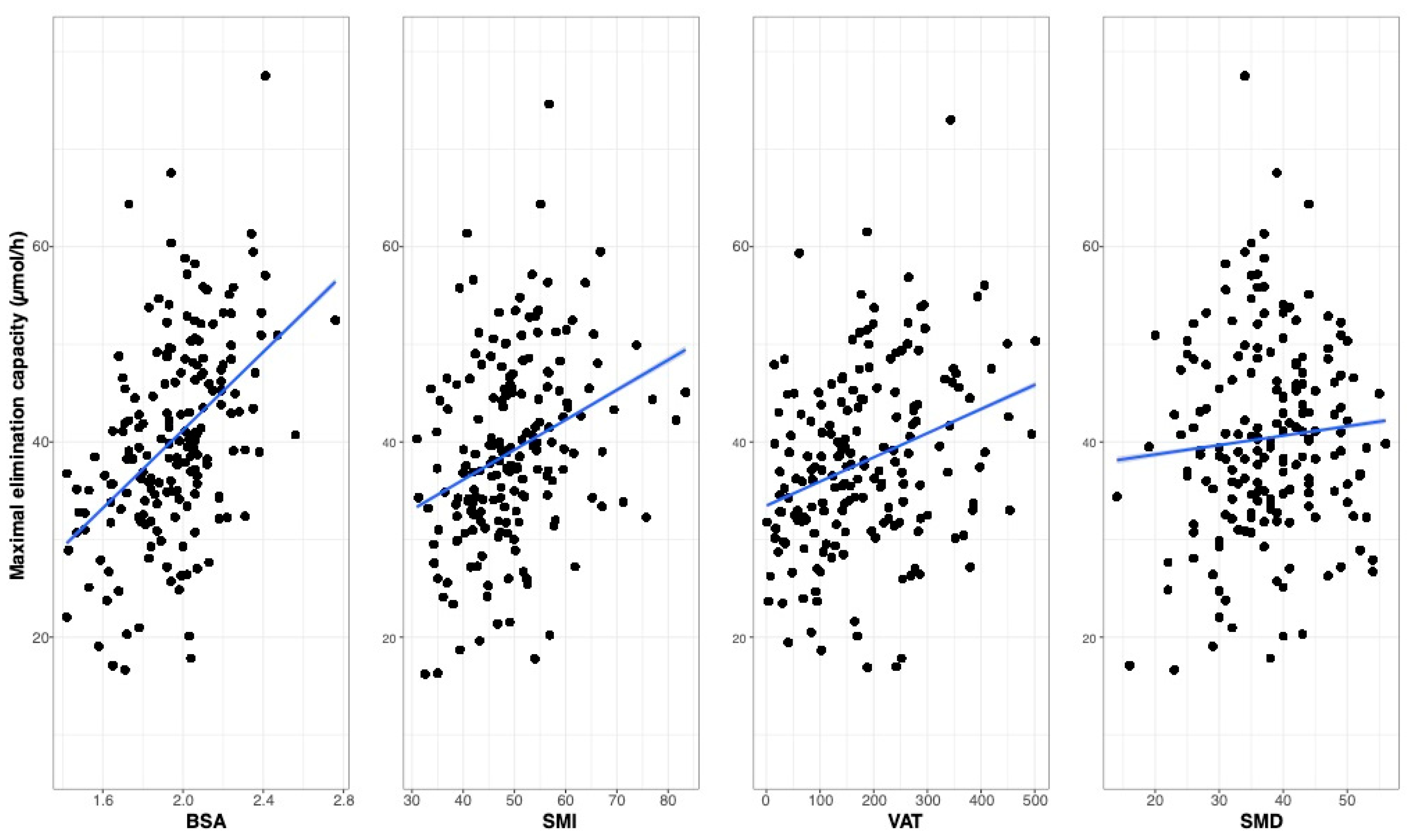
2. Results
3. Discussion
4. Materials and Methods
4.1. Patients
4.2. Body Composition Measurements
4.3. Paclitaxel Pharmacokinetics
4.4. Pharmacokinetic Model Evaluation and Covariate Analysis
5. Conclusions
Author Contributions
Funding
Institutional Review Board Statement
Informed Consent Statement
Data Availability Statement
Acknowledgments
Conflicts of Interest
References
- Stage, T.B.; Bergmann, T.K.; Kroetz, D.L. Clinical pharmacokinetics of paclitaxel monotherapy: An updated literature review. Clin. Pharm. 2017, 57, 7–19. [Google Scholar] [CrossRef] [PubMed] [Green Version]
- van Hagen, P.; Hulshof, M.C.; van Lanschot, J.J.; Steyerberg, E.W.; van Berge Henegouwen, M.I.; Wijnhoven, B.P.; Richel, D.J.; Nieuwenhuijzen, G.A.; Hospers, G.A.; Bonenkamp, J.J.; et al. Preoperative chemoradiotherapy for esophageal or junctional cancer. N. Engl. J. Med. 2012, 366, 2074–2084. [Google Scholar] [CrossRef] [PubMed] [Green Version]
- Toxopeus, E.L.; Talman, S.; van der Gaast, A.; Spaander, V.M.; van Rij, C.M.; Krak, N.C.; Biermann, K.; Tilanus, H.W.; Mathijssen, R.H.; van Lanschot, J.J.; et al. Induction chemotherapy followed by surgery for advanced oesophageal cancer. Eur. J. Surg. Oncol. 2015, 41, 323–332. [Google Scholar] [CrossRef] [PubMed]
- Baker, S.D.; Verweij, J.; Rowinsky, E.K.; Donehower, R.C.; Schellens, J.H.; Grochow, L.B.; Sparreboom, A. Role of body surface area in dosing of investigational anticancer agents in adults, 1991–2001. J. Natl. Cancer Inst. 2002, 94, 1883–1888. [Google Scholar] [CrossRef] [PubMed]
- Mathijssen, R.H.; de Jong, F.A.; Loos, W.J.; van der Bol, J.M.; Verweij, J.; Sparreboom, A. Flat-fixed dosing versus body surface area based dosing of anticancer drugs in adults: Does it make a difference? Oncologist 2007, 12, 913–923. [Google Scholar] [CrossRef] [PubMed]
- Prado, C.M.; Lima, I.S.; Baracos, V.E.; Bies, R.R.; McCargar, L.J.; Reiman, T.; Mackey, J.R.; Kuzma, M.; Damaraju, V.L.; Sawyer, M.B. An exploratory study of body composition as a determinant of epirubicin pharmacokinetics and toxicity. Cancer Chemother. Pharmacol. 2011, 67, 93–101. [Google Scholar] [CrossRef]
- Bins, S.; Ratain, M.J.; Mathijssen, R.H. Conventional dosing of anticancer agents: Precisely wrong or just inaccurate? Clin. Pharmacol. Ther. 2014, 95, 361–364. [Google Scholar] [CrossRef]
- Nieuweboer, A.J.; Hu, S.; Gui, C.; Hagenbuch, B.; Ghobadi Moghaddam-Helmantel, I.M.; Gibson, A.A.; de Bruijn, P.; Mathijssen, R.H.; Sparreboom, A. Influence of drug formulation on oatp1b-mediated transport of paclitaxel. Cancer Res. 2014, 74, 3137–3145. [Google Scholar] [CrossRef] [Green Version]
- Sparreboom, A.; Wolff, A.C.; Mathijssen, R.H.; Chatelut, E.; Rowinsky, E.K.; Verweij, J.; Baker, S.D. Evaluation of alternate size descriptors for dose calculation of anticancer drugs in the obese. J. Clin. Oncol. 2007, 25, 4707–4713. [Google Scholar] [CrossRef]
- Prado, C.M. Body composition in chemotherapy: The promising role of ct scans. Curr. Opin. Clin. Nutr. Metab. Care 2013, 16, 525–533. [Google Scholar] [CrossRef]
- Cespedes Feliciano, E.M.; Chen, W.Y.; Lee, V.; Albers, K.B.; Prado, C.M.; Alexeeff, S.; Xiao, J.; Shachar, S.S.; Caan, B.J. Body composition, adherence to anthracycline and taxane-based chemotherapy, and survival after nonmetastatic breast cancer. JAMA Oncol. 2019, 6, 264–270. [Google Scholar] [CrossRef] [PubMed]
- Hopkins, J.J.; Sawyer, M.B. A review of body composition and pharmacokinetics in oncology. Expert Rev. Clin. Pharmacol. 2017, 10, 947–956. [Google Scholar] [CrossRef] [PubMed]
- Joerger, M.; Kraff, S.; Huitema, A.D.; Feiss, G.; Moritz, B.; Schellens, J.H.; Beijnen, J.H.; Jaehde, U. Evaluation of a pharmacology-driven dosing algorithm of 3-weekly paclitaxel using therapeutic drug monitoring: A pharmacokinetic-pharmacodynamic simulation study. Clin. Pharm. 2012, 51, 607–617. [Google Scholar] [CrossRef]
- Kraff, S.; Nieuweboer, A.J.; Mathijssen, R.H.; Baty, F.; de Graan, A.J.; van Schaik, R.H.; Jaehde, U.; Joerger, M. Pharmacokinetically based dosing of weekly paclitaxel to reduce drug-related neurotoxicity based on a single sample strategy. Cancer Chemother. Pharmacol. 2015, 75, 975–983. [Google Scholar] [CrossRef]
- Crombag, M.B.S.; de Vries Schultink, A.H.M.; Koolen, S.L.W.; Wijngaard, S.; Joerger, M.; Schellens, J.H.M.; Dorlo, T.P.C.; van Erp, N.P.; Mathijssen, R.H.J.; Beijnen, J.H.; et al. Impact of older age on the exposure of paclitaxel: A population pharmacokinetic study. Pharm. Res. 2019, 36, 33. [Google Scholar] [CrossRef] [PubMed]
- Shachar, S.S.; Deal, A.M.; Weinberg, M.; Williams, G.R.; Nyrop, K.A.; Popuri, K.; Choi, S.K.; Muss, H.B. Body composition as a predictor of toxicity in patients receiving anthracycline and taxane-based chemotherapy for early-stage breast cancer. Clin. Cancer Res. 2017, 23, 3537–3543. [Google Scholar] [CrossRef] [Green Version]
- Joerger, M.; von Pawel, J.; Kraff, S.; Fischer, J.R.; Eberhardt, W.; Gauler, T.C.; Mueller, L.; Reinmuth, N.; Reck, M.; Kimmich, M.; et al. Open-label, randomized study of individualized, pharmacokinetically (pk)-guided dosing of paclitaxel combined with carboplatin or cisplatin in patients with advanced non-small-cell lung cancer (nsclc). Ann. Oncol. 2016, 27, 1895–1902. [Google Scholar] [CrossRef]
- de Graan, A.J.; Elens, L.; Smid, M.; Martens, J.W.; Sparreboom, A.; Nieuweboer, A.J.; Friberg, L.E.; Elbouazzaoui, S.; Wiemer, E.A.; van der Holt, B.; et al. A pharmacogenetic predictive model for paclitaxel clearance based on the dmet platform. Clin. Cancer Res. 2013, 19, 5210–5217. [Google Scholar] [CrossRef] [Green Version]
- de Graan, A.J.; Loos, W.J.; Friberg, L.E.; Baker, S.D.; van der Bol, J.M.; van Doorn, L.; Wiemer, E.A.; van der Holt, B.; Verweij, J.; Mathijssen, R.H. Influence of smoking on the pharmacokinetics and toxicity profiles of taxane therapy. Clin. Cancer Res. 2012, 18, 4425–4432. [Google Scholar] [CrossRef] [Green Version]
- Mosteller, R.D. Simplified calculation of body-surface area. N. Engl. J. Med. 1987, 317, 1098. [Google Scholar]
- Goodpaster, B.H.; Thaete, F.L.; Kelley, D.E. Composition of skeletal muscle evaluated with computed tomography. Ann. N. Y. Acad. Sci. 2000, 904, 18–24. [Google Scholar] [CrossRef] [PubMed]
- Shen, W.; Punyanitya, M.; Wang, Z.; Gallagher, D.; St-Onge, M.P.; Albu, J.; Heymsfield, S.B.; Heshka, S. Total body skeletal muscle and adipose tissue volumes: Estimation from a single abdominal cross-sectional image. J. Appl. Physiol. 2004, 97, 2333–2338. [Google Scholar] [CrossRef] [PubMed] [Green Version]
- van Vugt, J.L.; Levolger, S.; Gharbharan, A.; Koek, M.; Niessen, W.J.; Burger, J.W.; Willemsen, S.P.; de Bruin, R.W.; JN, I.J. A comparative study of software programmes for cross-sectional skeletal muscle and adipose tissue measurements on abdominal computed tomography scans of rectal cancer patients. J. Cachexia Sarcopenia Muscle 2017, 8, 285–297. [Google Scholar] [CrossRef] [PubMed]
- Sparreboom, A.; de Bruijn, P.; Nooter, K.; Loos, W.J.; Stoter, G.; Verweij, J. Determination of paclitaxel in human plasma using single solvent extraction prior to isocratic reversed-phase high-performance liquid chromatography with ultraviolet detection. J. Chromatogr. B Biomed. Sci. Appl. 1998, 705, 159–164. [Google Scholar] [CrossRef]
- Dosne, A.G.; Bergstrand, M.; Harling, K.; Karlsson, M.O. Improving the estimation of parameter uncertainty distributions in nonlinear mixed effects models using sampling importance resampling. J. Pharm. Pharm. 2016, 43, 583–596. [Google Scholar] [CrossRef] [Green Version]


| Parameters | Cohort |
|---|---|
| Number of patients (n) | 184 |
| Paclitaxel dose (mg/m2), median (range) | 70 (62–252) |
| Infusion time (h), median (range) | 0.9 (0.3–1.5) |
| Number of samples (n) | 550 |
| Per patient, median (range) | 3 (2–4) |
| BSA (m2), median (range) | 1.98 (1.42–2.76) |
| SMI (cm2/m2), median (range) | 48.5 (30.9–83.4) |
| VAT (cm2), median (range) | 165 (0.67–502) |
| SMD (HU), median (range) | 37 (14–56) |
| Gender, male, n (%) | 147 (80) |
| Age, median (range) | 64 (40–83) |
| Indication, n (%) esophageal cancer | 184 (100) |
| Paclitaxel treatment, n (%) | |
| Induction/palliative (3 weekly 175 mg/m2) | 7 (4) |
| Induction/palliative (weekly 100 mg/m2) | 45 (24) |
| Neoadjuvant (weekly 50 mg/m2) | 132 (72) |
| Bilirubin, total (μmol/L), median (IQR) | 7 (5–9) |
| Parameter (Unit) | Covariate Model BSA | Covariate Model SMI | Covariate Model VAT | Covariate Model SMD | ||||
|---|---|---|---|---|---|---|---|---|
| dOFV | REF | dOFV | +29 | dOFV | +34 | dOFV | +25 | |
| Estimate | 95% CI | Estimate | 95% CI | Estimate | 95% CI | Estimate | 95% CI | |
| VMEL (µmol/h) | 31.6 | 26.7–37.4 | 32.4 | 26.7–40.6 (11) | 31.9 | 26.0–39.0 | 31.6 | 34.7–38.9 |
| V1 (L) | 24.0 | 20.8–27.6 | 24.7 | 21.3–28.4 | 25.0 | 21.8–28.4 | 25.2 | 22.0–28.8 |
| V3 (L) | 267 | NA | 267 | NA | 267 | NA | ||
| KMEL (µmol/L) | 0.40 | 0.29–0.52 | 0.51 | 0.37–0.73 | 0.49 | 0.33–0.68 | 0.55 | 0.36–0.74 |
| VMTR (µmol/h) | 179 | 138–225 | 153 | 119–199 | 148 | 111–188 | 144 | 113–193 |
| KMTR (µmol/L) | 1.91 | 1.37–2.67 | 1.80 | 1.46–1.98 | 1.75 | 1.24–2.39 | 1.73 | 1.15–2.36 |
| K21 (h−1) | 2.34 | 1.85–2.96 | 2.16 | 1.69–2.72 | 2.09 | 1.64–2.65 | 2.15 | 1.70–2.69 |
| Q (L/h) | 20.7 | 18.0–23.3 | 19.8 | 17.0–23.0 | 19.6 | 17.0–22.3 | 19.9 | 16.6–23.0 |
| Body composition on VMEL | 1.25 | 1.03–1.51 | 0.34 | 0.24–0.44 | 0.09 | 0.06–0.12 | 0.03 | 0.02–0.04 |
| Age on VMEL | −0.30 | −0.39–0.22 | −0.30 | −0.32–0.30 | −0.50 | −0.65–0.34 | −0.28 | −0.36–0.20 |
| Gender on VMEL | 1.07 | 0.96–1.19 | 1.20 | 1.06–1.34 | 1.23 | 1.09–1.37 | 1.30 | 1.15–1.47 |
| Bilirubin on VMEL | −0.17 | NA | −0.17 | NA | −0.17 | NA | −0.17 | NA |
| Interindividual variability | ||||||||
| VMEL (%) | 24.3 | 20.8–28.3 | 28.6 | 24.9–33.3 | 27.4 | 24.0–32.4 | 29.8 | 25.5–34.9 |
| V1 (%) | 39.1 | 33.2–45.3 | 38.1 | 31.3–43.7 | 37.7 | 31.9–45.4 | 37.5 | 30.6–43.5 |
| Q (%) | 62.0 | 52.3–72.9 | 62.8 | 52.8–74.0 | 64.3 | 52.8–75.6 | 62.4 | 50.7–74.3 |
| Residual variability | ||||||||
| σprop (%) | 22.5 | 20.1–25.5 | 22.4 | 20.2–25.5 | 22.6 | 20.0–25.5 | 22.4 | 19.8–25.3 |
Publisher’s Note: MDPI stays neutral with regard to jurisdictional claims in published maps and institutional affiliations. |
© 2021 by the authors. Licensee MDPI, Basel, Switzerland. This article is an open access article distributed under the terms and conditions of the Creative Commons Attribution (CC BY) license (http://creativecommons.org/licenses/by/4.0/).
Share and Cite
van Doorn, L.; Crombag, M.-R.B.S.; Rier, H.N.; van Vugt, J.L.A.; van Kesteren, C.; Bins, S.; Mathijssen, R.H.J.; Levin, M.-D.; Koolen, S.L.W. The Influence of Body Composition on the Systemic Exposure of Paclitaxel in Esophageal Cancer Patients. Pharmaceuticals 2021, 14, 47. https://0-doi-org.brum.beds.ac.uk/10.3390/ph14010047
van Doorn L, Crombag M-RBS, Rier HN, van Vugt JLA, van Kesteren C, Bins S, Mathijssen RHJ, Levin M-D, Koolen SLW. The Influence of Body Composition on the Systemic Exposure of Paclitaxel in Esophageal Cancer Patients. Pharmaceuticals. 2021; 14(1):47. https://0-doi-org.brum.beds.ac.uk/10.3390/ph14010047
Chicago/Turabian Stylevan Doorn, Leni, Marie-Rose B. S. Crombag, Hánah N. Rier, Jeroen L. A. van Vugt, Charlotte van Kesteren, Sander Bins, Ron H. J. Mathijssen, Mark-David Levin, and Stijn L. W. Koolen. 2021. "The Influence of Body Composition on the Systemic Exposure of Paclitaxel in Esophageal Cancer Patients" Pharmaceuticals 14, no. 1: 47. https://0-doi-org.brum.beds.ac.uk/10.3390/ph14010047