Article

A Flavonoid-Rich Extract from Bergamot Juice, Alone or in Association with Curcumin and Resveratrol, Shows Protective Effects in a Murine Model of Cadmium-Induced Testicular Injury

1
Department of Biomedical, Dental Sciences and Morphofunctional Imaging, University of Messina, 98125 Messina, Italy
2
Department of Clinical and Experimental Medicine, University of Messina, 98125 Messina, Italy
3
Department of Chemical, Biological, Pharmaceutical and Environmental Sciences, University of Messina, 98166 Messina, Italy
4
Department of Pharmacy-Drug Sciences, University of Bari “Aldo Moro”, 70125 Bari, Italy
*
Author to whom correspondence should be addressed.
Pharmaceuticals 2021, 14(5), 386; https://0-doi-org.brum.beds.ac.uk/10.3390/ph14050386
Submission received: 6 April 2021 / Revised: 16 April 2021 / Accepted: 19 April 2021 / Published: 21 April 2021
(This article belongs to the Section Natural Products)

Abstract

:
It is known that cadmium damages testis structure and functionality. We examined the effects of nutraceuticals such as a flavonoid-rich extract of bergamot juice (BJe), alone or in association with curcumin (Cur) and resveratrol (Re), on mice testicular dysfunction caused by cadmium chloride (CdCl2). Controversial data on the protective effects of Cur and Re are available, while no evidence on the possible role of BJe exists. Adult male C57 BL/6J mice were administered with CdCl2 and treated with Cur, Re, or BJe alone or in combination for 14 days. Then, testes were removed and processed for molecular, structural, and immunohistochemical analyses. CdCl2 increased the mRNA of IL-1β, TNF-α, p53, and BAX while reduced that of Bcl-2 and induced tubular lesions and apoptosis of germinal cells. Cur, Re, and BJe at 40 mg/kg significantly improved all of these parameters and events, although BJe at 20 mg/kg showed a lower protective effect. The association of Cur, Re, and BJe at both doses of 50/20/20 and 100/20/40 mg/kg brought each parameter close to those of the control. Our results indicate that the nutraceuticals employed in this study and their associations exert a positive action against Cd-induced testicular injury, suggesting a possible protection of testis functionality in subjects exposed to environmental toxicants.

1. Introduction

Cadmium (Cd) is a nonessential transition metal without any physiological function in the human body, often considered a ubiquitous toxicant with carcinogenic activity. Cd, alongside arsenic, lead, mercury, and chromium, poses health risks [1,2]. Human exposure to Cd occurs either in the workplace or through inhalation or ingestion of Cd-contaminated materials [3]. Occupational exposure is typical of subjects working with pigments, glass, plastics, metal alloys, and nickel-Cd batteries [4], while the main sources of exposure for the general population are cigarette smoke and foods [5]. In humans, Cd induces toxic effects in various organs, including the testes [6]. In fact, male infertility with lower semen quality [7] and postponed puberty with reduced gonadal growth [8] were observed.
In animals, and particularly in rodents [9], after experimental Cd exposure, testicular lesions were observed, such as a significant reduction in testis weight and testosterone levels, and structural changes in both seminiferous tubules and Leydig cells [10,11,12,13].
Both oxidative stress and inflammation are key events involved in the toxic action of Cd [14]. Cd-induced inflammatory cascade is triggered by tumor necrosis factor (TNF)-α, a proinflammatory cytokine [13,15] that amplifies phlogistic response by stimulating the release of reactive oxygen species (ROS). These are extremely toxic to spermatozoa because of their high content of polyunsaturated fatty acids along with their limited ability to repair DNA [16].
In addition to directly inducing structural damage in the testis, oxidative stress is also capable of stimulating apoptosis both in vitro [17] and in vivo [12,18]. In fact, Cd has been shown to increase the expression of proapoptotic factors such B cell-lymphoma (Bcl)-2-associated X protein (Bax), of the key regulatory protein p53, and of caspase-3 as well as to reduce that of the antiapoptotic Bcl-2 [11,18,19,20,21]. It was also observed that germ cell apoptosis might be the consequence of a Cd-induced reduction in testosterone level [22] with detachment of immature germ cells from the seminiferous epithelium [23].
In recent years, several therapeutical approaches have been proposed to retrieve the toxic effects of Cd on male genital apparatus [6]. Among them, the role of antioxidants on the restoration of the seminiferous epithelium was evaluated. In this field, the phenolic compound curcumin (Cur), a major component of Curcuma longa L., showed protective effects against oxidative damage by inhibiting the production of TNF-α [24] and by scavenging ROS [25]. In this regard, in rodents challenged with Cd, Cur improved seminiferous tubules morphology [26,27] and significantly decreased the number of apoptotic germinal cells [28,29]. Additionally, resveratrol (Re), a polyphenol present in many plants and especially in red wine, showed antioxidant properties [30] as well a positive regulation of apoptosis and other zonula occludens protein expression, such as occludin [31,32]. In the testis, Re protected from oxidative stress [33], partially reversed the dysregulation of the apoptotic pathways [20], and improved sperm parameters and histopathological damage [34]. Recently, bergamot juice (BJ), obtained from the endocarp of Citrus bergamia Risso et Poiteau (bergamot) fruits, gained the attention of pharmaceutical companies interested in natural products and antioxidant bioactive compounds from dietary sources [35,36,37]. Indeed, it has been shown that BJ and its flavonoid-rich fraction (BJe) exert hypolipemic and hypoglycemic activity [38], and anticancer [39], anti-infective [40,41], neuroprotective [42], antioxidant, and anti-inflammatory effects [43,44,45,46]. However, no data are currently available on the potential role of BJe during Cd challenge.
In light of this background, the present study was designed to examine the effects of the nutraceuticals BJe, Cur, and Re, alone or in association, in a murine model of Cd-induced testicular damage.

2. Results

2.1. Testes and Body Weight along with Their Ratio

Mice testes and body weight (TW and BW, respectively) as well as their ratio (TW/BW) are shown in Table 1. As expected, CdCl2-challenged mice showed TW, BW, and TW/BW ratio significantly lower than the control (Table 1). Both doses of Cur slightly increased these parameters with respect to those found in CdCl2-injured mice that, however, remained significantly lower than the controls (Table 1). Conversely, in CdCl2 + Re treated mice, all parameters considered in this task showed a significant improvement in comparison to those assessed in CdCl2-subjected mice (p < 0.05) as occurs with BJe 40 mg/kg (p < 0.05 vs. CdCl2), whilst BJe 20 mg/kg did not significantly increase TW, BW, or their ratio (Table 1). Of note, the associations between BJe, Cur, and Re (Cur 50 mg/kg + Re 20 mg/kg + BJe 20 mg/kg as well as Cur 100 mg/kg + Re 20 mg/kg + BJe 40 mg/kg) significantly improved all parameters almost up to those of the controls (p < 0.05 vs. CdCl2), thus protecting mice and their testes from cadmium toxicity.

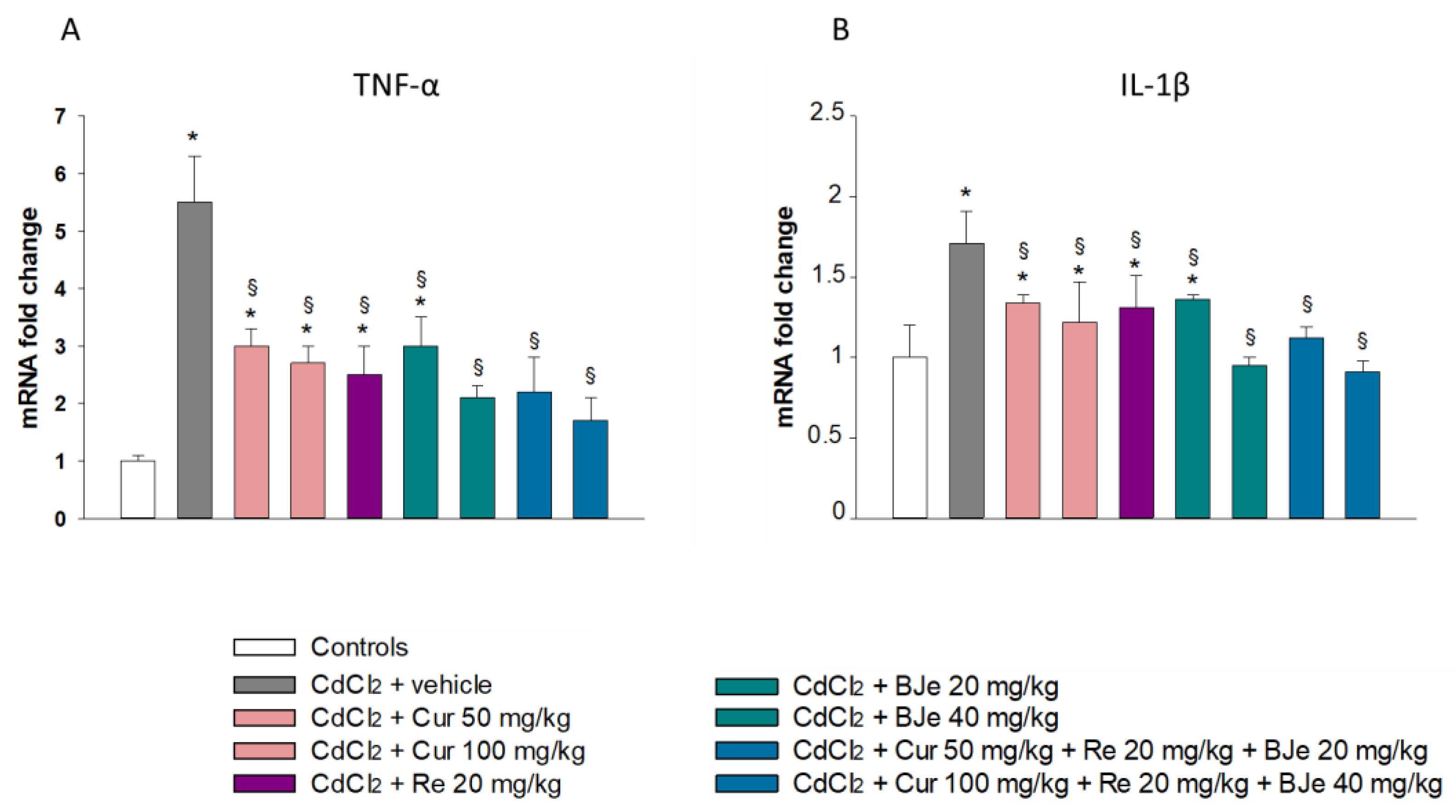
2.2. Effect of Nutraceuticals on Inflammatory Markers

In comparison to the untreated mice, at the doses employed in this study, treatment with BJe, Cur, and Re alone or in combination did not significantly affect either TNF-α or interleukin (IL)-1β gene expression (data not shown) mRNA. The data from real-time PCR analyses showed a significant upregulation of TNF-α and IL-1β in CdCl2-challenged animals when compared to control animals (Figure 1A,B). Notably, a significant reduction in TNF-α and IL-1β mRNA levels was found in the testes of all experimental groups of animals treated with the extracts compared to those from Cd-challenged animals. This reduction was particularly evident in the testes of rats treated with BJe 40 mg/kg and both Cur 50 mg/kg + Re 20 mg/kg + BJe 20 mg/kg and Cur 100 mg/kg + Re 20 mg/kg + BJe 40 mg/kg (Figure 1A,B).

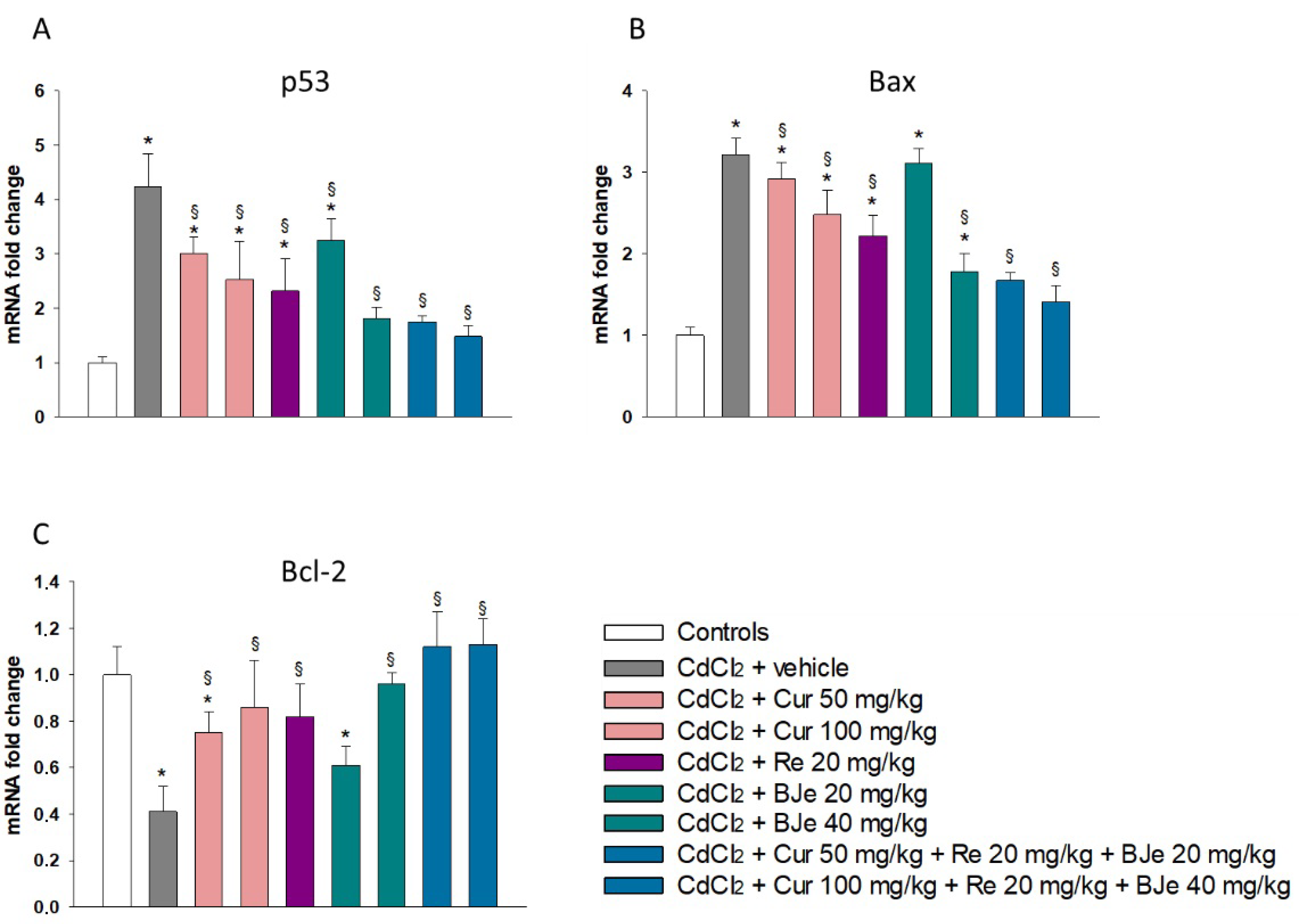
2.3. The Nutraceuticals Modulate the Expression of Apoptotic Markers

No significant difference was observed in mRNA levels of p53, BAX, and Bcl-2 among the control groups. Significant variations in all examined genes were found in the testes of CdCl2-challenged mice when compared to the control groups. In addition, the upregulation of p53 and BAX as well as the downregulation of Bcl-2 observed in CdCl2- subjected animals were significantly hindered by Cur, Re, and BJe, along with their associations at both higher and lower dosages, with the exception of BJe 20 mg/kg alone (Figure 2A,B).

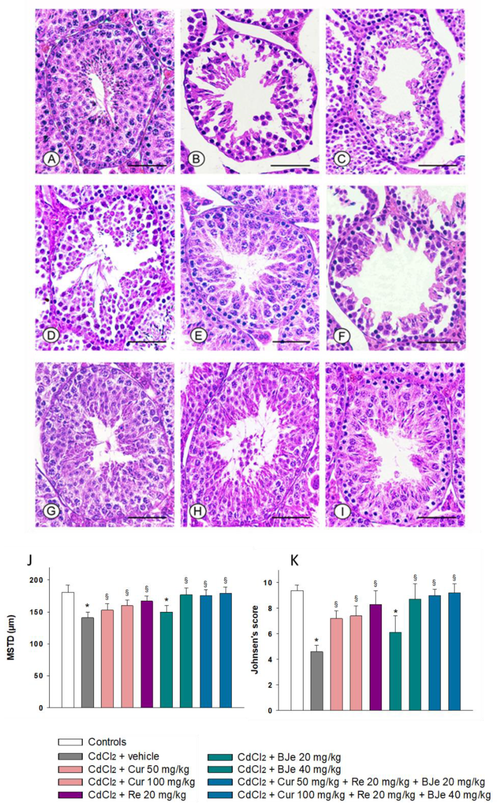
2.4. Effect of Nutraceuticals on Tubular Histological Organization

In all the control groups of animals, a normal morphology of both seminiferous tubules and extratubular compartment was observed. When the mice were challenged with CdCl2, the tubules had reduced MSTD and low Johnsen’s scores. The discontinuous seminiferous epithelium showed irregular clefts; it was formed only by spermatogonia, often detached from the basal membrane, and by spermatocytes. An evident edema was present in the extratubular compartment (Figure 3B,J,K). In mice challenged with CdCl2 and treated with Cur at 50 or 100 mg/kg, seminiferous tubules had a reduced MSTD and a lower Johnsen’s score compared to controls. The epithelium showed spermatocytes separated from spermatogonia and many large clefts. A marked edema was evident in the extratubular compartment (Figure 3C,D,J,K). In the testes of CdCl2 plus Re-treated mice, the seminiferous tubules had significantly higher MSTD and Johnsen’s scores and many elongated spermatids were present in the seminiferous epithelium. The extratubular edema was reduced (Figure 3E,J,K). In CdCl2 plus BJe alone in 20 mg/kg-treated mice, the seminiferous tubules had lower MSTD and Johnsen’s scores and the germinal epithelium showed many large clefts and many irregularly arranged spermatocytes. Evident hyperemia was present in the extratubular compartment (Figure 3F,J,K). In mice treated with CdCl2 plus BJe alone at 40 mg/kg and with both the associations (Cur 50 mg/kg + Re 20 mg/kg + BJe 20 mg/kg and Cur 100 mg/kg + Re 20 mg/kg + BJe 40 mg/kg), the seminiferous tubules had normal MSTD and Johnsen’s score and the germinal epithelium showed mature spermatozoa. In all groups, the extratubular compartment showed normal morphological organization (Figure 3G–K).

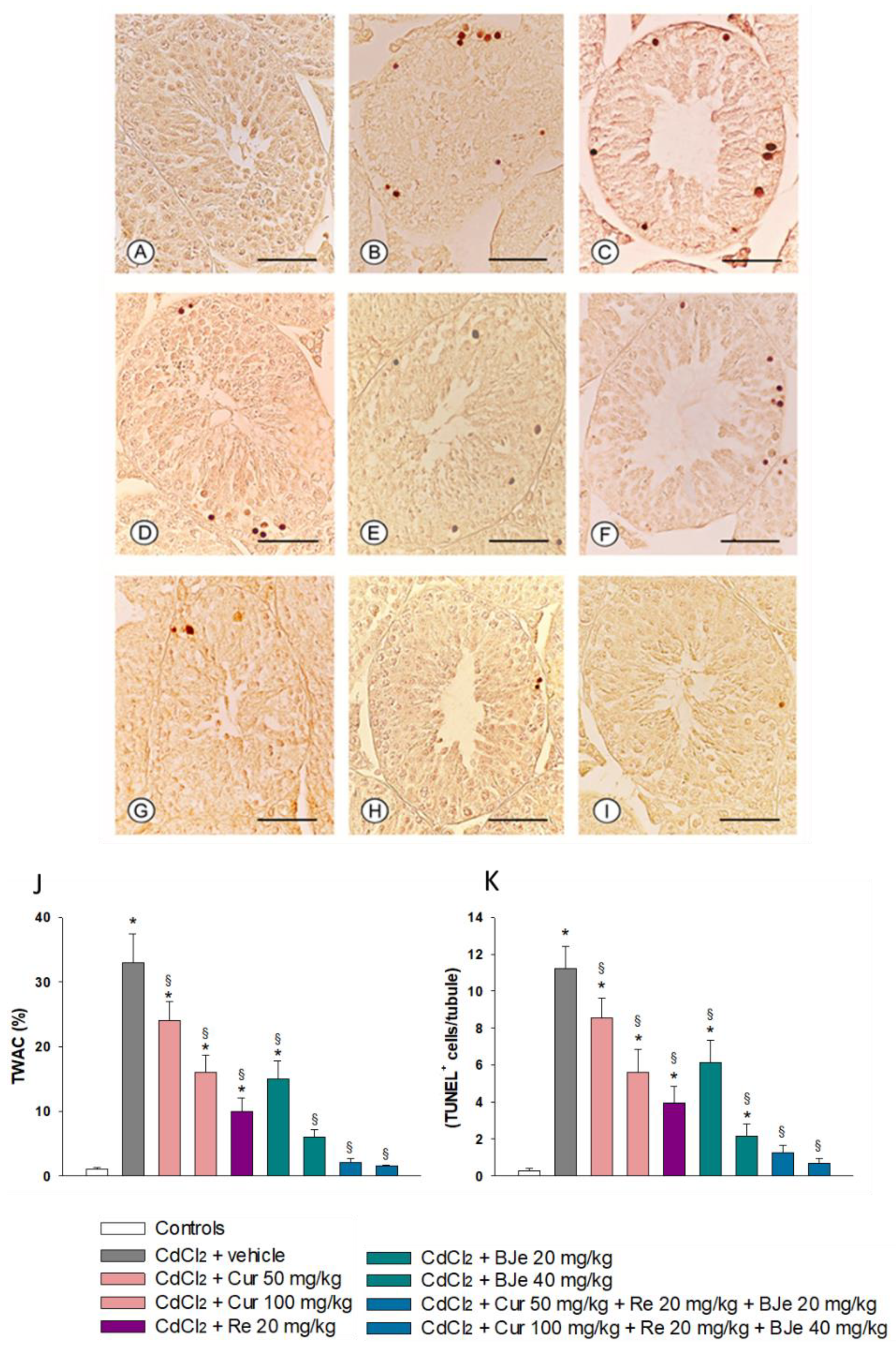
2.5. Measurement of Nutraceuticals Effects on Apoptosis with TUNEL Assay

The testes of all control animals showed the same morphological behavior when evaluated with TUNEL assay. Therefore, for clarity of results, a single image (Figure 4A) and single data for TWAC and the apoptotic index (Figure 4J,K) are provided as representative of the controls. No TUNEL-positive cells were observed in the seminiferous tubules in control animals. On the contrary, a large number of peripheral TUNEL-positive cells were seen in the seminiferous tubules of mice challenged with CdCl2 (Figure 4B). Both TWAC and apoptotic index were significantly higher if compared to the control group (Figure 4J,K). In mice challenged with CdCl2 and treated with Cur at 50 or 100 mg/kg, a marked reduction of TUNEL-positive cells was observed (Figure 4C,D), so that both TWAC and apoptotic index were significantly lower than those of CdCl2-subjected mice (Figure 4J,K). The treatment with Re of CdCl2-challenged mice further lowered the number of TUNEL positive cells (Figure 4E), as indicated by both TWAC and apoptotic index values (Figure 4J,K). In CdCl2 plus BJe alone in 20 mg/kg-treated mice, we observed a higher number of TUNEL-positive cells compared to the other treated groups (Figure 4F), along with greater TWAC and apoptotic index (Figure 4J,K), yet a lower number of TUNEL-positive cells compared to CdCl2-challenged mice. In CdCl2 plus BJe alone in 40 mg/kg-treated mice, the number of TUNEL-positive cells was significantly reduced (Figure 4G), as demonstrated also by TWAC and apoptotic index values (Figure 4J,K). In CdCl2-challenged mice treated with both associations (Cur 50 mg/kg + Re 20 mg/kg + BJe 20 mg/kg and Cur 100 mg/kg + Re 20 mg/kg + BJe 40 mg/kg), only isolated TUNEL-positive germ cells were observed in the periphery of the seminiferous tubules (Figure 4H,I). The TWAC and apoptotic index were close to that for the controls (Figure 4J,K).

3. Discussion

Recent studies examining the exposure-effect assessment linked chronic Cd challenge with adverse effects in almost every organ and tissue where Cd accumulates [47,48]. In particular, numerous studies have demonstrated Cd-induced reproductive dysfunctions, such as reduction in testis and body weight, damage of the blood–testis barrier, reduced germ cell adhesion with increased loss of immature cells, testosterone reduction, low sperm count, and consequent subfertility or infertility [7,11,12,13].
For the involved mechanisms, oxidative stress with overproduction of ROS has been reported to play a fundamental role [13,49]. Cd also enhances cellular inflammatory status through the increased release of pro-inflammatory mediators, in particular IL-1β and TNF-α [50]. In addition, Cd was found to potentiate apoptotic cascades and to alter the ratio between pro- and antiapoptotic proteins: in fact, the key regulatory protein p53 and Bax were upregulated while Bcl-2 was downregulated [20].
In the present study, a significant reduction in testis and body weight was observed after CdCl2 treatment. The adverse effect on body weight could be probably correlated to its harmfulness on many body systems, and the mechanism may be linked to the generation of free radicals [51]. With regard to the weight of the testis, it is mainly dependent on the germinal epithelium and the number of germ cells [52,53,54]. The significant decline demonstrated after CdCl2 challenge can be considered an important parameter of toxicity and it may be due to the histopathological damages in the seminiferous tubules (reduced MSTD and Johnsen’s score). Furthermore, mice treated with CdCl2 alone showed a significant upregulation of TNF-α, IL-1β, p53, and BAX; a significant down-regulation of Bcl-2; and a large number of TUNEL-positive germ cells, thus confirming a negative role of CdCl2 on the seminiferous epithelium.
In addition to the endogenous antioxidants produced by the body, pure and reliable exogenous antioxidants taken with nutrition, in particular from fruits, vegetables, and some medical plants, have been tested to minimize the functional and structural changes that occur in tissues after Cd challenge [47].
Among them, Cur, the main natural polyphenol found in the rhizome of Curcuma longa and in other Curcuma spp., showed protective effects against oxidative damage [29], as it inhibited the in vitro production of TNF-α [24] and scavenged ROS [25]. In Cd-treated rats, Cur administration improved the oxidative stress [27], partially protected seminiferous tubules morphology [26], and significantly decreased (but not to normal values) the number of apoptotic germinal cells [28]. In our study, Cur alone, particularly at 100 mg/kg, was able to increase the body and the testis weight to modulate the inflammatory and apoptotic pathways and to reduce the structural damages and the morphological parameters of the seminiferous tubules, thus playing a role in the treatment of human infertility.
Other studies have demonstrated the protective and therapeutic effects of other substances against different toxics in testes. In particular, Re showed a protective action against testicular toxicity as it upregulated Bcl2, downregulated p53 and Bax gene expression [20,54], and reduced inflammatory cascade [54]. Furthermore, marked improvements in sperm parameters and histopathological damages were observed in the Re-pretreated mice [34]. Indeed, our study confirmed the positive role of Re in Cd-challenged animals on both biochemical and structural parameters.
Citrus fruits are one of the most eaten fruits in the world and a great source of dietary flavonoids is well-known for their beneficial effects [55,56,57]. In this study, we demonstrated the protective effect of BJe, alone (40 mg/kg), or in combination (at the lowest dose of 20 mg/kg) to Cur and Re, against Cd-induced testicular injury. In particular, we showed that BJe reduced both apoptotic markers and cytokines evaluated in this study, thus protecting mice from Cd-induced testicular injury. Previous studies have demonstrated that BJe has antioxidant and anti-inflammatory properties [44,58], by interacting with both gene and protein targets linked to the apoptotic machinery and cytokine production [59,60]. This occurs directly or through specific signaling pathways such as NF-κB or SIRT-1 [45,61] that mediate the flavonoid actions within the cells. Therefore, we speculate that the bioactive compounds present in the phytocomplex can simultaneously modulate different molecular targets, acting in a multitarget mode of action.
The Cd toxicity determines peculiar damages in whole organisms, resulting in both acute and chronic poisoning [6]. Testes are particularly sensitive to Cd-induced injury, with the seminiferous epithelium showing morphological and functional changes, often irreversible. It has been suggested that oxidative stress, inflammation, and apoptosis play a central role in detrimental effects brought on by Cd in testes [62]. Therefore, the use of nutraceuticals able to counteract these damages might be the best preventive/therapeutic approach. In the present study, we documented the capability of BJe, alone or in combination with Re and Cur, to fight the oxidative and pro-inflammatory effects augmented by Cd as well as to reduce its proapoptotic stimuli, suggesting their potentiality towards heavy metal noxious action.
Very recently, Montano and coworkers [63] showed that an intervention based on Mediterranean diet and regular physical activity can determine an improvement of semen quality in healthy young men. This finding is in line with observational studies showing a positive association between the semen parameters and Mediterranean diet [64,65]. This positive association may be due to the antioxidant mechanisms of the various nutrients characterizing this dietary pattern [66] that could positively influence semen parameters [67,68,69]. These studies speak in favor of our results, indicating that a rational nutraceuticals supplementation may be a new reliable strategy in humans exposed to heavy metals, such as Cd, suggesting that the results of our research could be translated in clinical practice.
Of course, both short- and long-duration studies are required to determine the optimal doses of BJe alone or in association with Cur and Re to prove their safety and effectiveness against Cd toxicity in humans.

4. Materials and Methods

4.1. Ethical Approval

The standards for care and use of animal subjects as stated in the ARRIVE (Animal Research: Reporting In Vivo Experiments) guidelines were followed in the present work. All procedures were approved by the Italian Ministry of Health (authorization number 112/2017—PR) and by the Institutional Animal Care and Use Ethic Committee of the University of Messina (OPBA, #820/2016, 02/09/2016).

4.2. Experimental Protocol

One hundred and nineteen male C57 BL/6J mice (25–30 g) were purchased from Charles River Laboratories Italia srl (Lecco, Italy) and housed at the animal facility of the School of Medicine of the University of Messina, Messina, Italy. The animals were provided a standard diet ad libitum with free access to tap water and maintained on a 12 h light/dark cycle. The animals were randomly divided into 17 groups of 7 animals each. Nine groups were used as controls (0.9% NaCl (vehicle); corn oil (vehicle); Cur (50 mg/kg per os); Cur (100 mg/kg per os); Re (20 mg/kg per os); BJe (20 mg/kg per os); BJe (40 mg/kg per os); Cur (50 mg/kg per os) + Re (20 mg/kg per os) + BJe (20 mg/kg per os); and Cur (100 mg/kg per os) + Re (20 mg/kg per os) + BJe (40 mg/kg per os)). Eight groups were treated as follows: CdCl2 (2 mg/kg i.p.) + vehicle; CdCl2 + Cur (50 mg/kg per os); CdCl2 + Cur (100 mg/kg per os); CdCl2 + Re (20 mg/kg per os); CdCl2 + BJe (20 mg/kg per os); CdCl2 + BJe (40 mg/kg per os); CdCl2 + Cur (50 mg/kg per os) + Re (20 mg/kg per os) + BJe (20 mg/kg per os); and CdCl2 + Cur (100 mg/kg per os) + Re (20 mg/kg per os) + BJe (40 mg/kg per os). CdCl2, Re, and BJe were dissolved in 0.9% NaCl, while Cur was dispersed in corn oil. Cur, Re, and BJe were orally administered for 14 days, while CdCl2 was dispensed intraperitoneally (i.p.). The doses of CdCl2 as well as BJe, Re, and Cur employed in this study were chosen in agreement with previous findings [11,13,20,27,33,39,59,60,70]. Twenty-four hours after the last treatment, all mice were weighted and sacrificed with an overdose of ketamine and xylazine (75/10 mg/kg i.p. each). Orchidectomies were performed, and the testes were weighed and processed for molecular, histological, and immunohistochemical procedures.

4.3. Drugs and Chemicals

In the present study, we used the same BJe already employed in other experimental researches for which the quali-quantitative analysis of flavonoids content was reported in previous experimental studies [39,59]. The most abundant flavonoids of BJe are (mg/g) neohesperidin (94.00), naringin (92.4), melitidin (56.2), hesperetin (51.9), neoeriocitrin (48.6), and naringenin (27.3). Liquid BJe was provided by Agrumaria Corleone (Palermo, Italy) from bergamot fruits harvested in the Reggio Calabria province (Italy). Liquid BJe was dried in powder by lyophilization and kept at −20 °C. CdCl2, Cur, and Re were bought from Sigma-Aldrich Srl (Milan, Italy). All chemicals not otherwise mentioned were commercially available reagent grade.

4.4. Real-Time PCR Analyses

Total RNAs from testis samples belonging to animals of all of the challenged groups were extracted with the TRIzol LS reagent (Invitrogen, Carlsbad, CA, USA) according to the manufacturer’s protocol. Then, 2 µg of RNA from each sample was reverse transcribed into cDNA using the High-Capacity cDNA Archive Kit (Applied Biosystems, Life Technologies, Foster City, CA, USA). The expression of mRNA was assessed by real-time PCR, as previously described [39]. The sequences of primers employed for real-time PCR analyses are listed in Table 2. The real-time PCRs were carried out in 20 µL reactions containing 1xSYBR® Select Master Mix (Applied Biosystems, Life Technologies, Foster City, CA, USA), 0.2 µM of primers, and 25 ng RNA converted into cDNA. The analyses were performed in triplicate in a 96-well plate using a 7900HT Fast Real-Time PCR System (Applied Biosystems, Life Technologies, Foster City, CA, USA). Data were collected and analyzed using the 2-ΔΔCT relative quantification method, with β-actin used as endogenous control. The values are presented as fold changes relative to the control tissues. When the value was less than 1, it was converted into its negative inverse to report downregulated genes.

4.5. Histological Evaluation

The testes were fixed in a freshly prepared Bouin solution, dehydrated in graded ethanol, cleared in xylene, and embedded in paraffin (Paraplast, SPI Supplies, West Chester, PA, USA). Five micrometer sections were cut with a rotary microtome (RM2125 RT, Leica Instruments, Nussloch, Germany) and stained with hematoxylin and eosin (HE). All samples were photographed with a Nikon Ci-L (Nikon Instruments, Tokyo, Japan) light microscope using a digital camera Nikon Ds-Ri2 and saved as Tagged Image Format Files (TIFF) with the Adobe Photoshop CS software. All micrographs were examined at the same final magnification (800×) by two trained observers (AM and DP), who ignored to which of the experimental group mice belonged. Five microscopic fields (MFs), all including two entire seminiferous tubules with circular profiles from ten non-serial sections of each group, were evaluated. For the morphological assessment, the mean seminiferous tubule diameter (MSTD), expressed in micrometers, was calculated with the public domain ImageJ software (http://rsb.info.nih.gov/ij/; available in the public domain by the National Institutes of Health, Bethesda, MD, USA) using the function analyze > measure. Germinal epithelium was also evaluated with the Johnsen’s scoring system [71], as modified for rodents [72]. Briefly, a score of 10 to 1 was given to each tubule according to its epithelial organization: 10, full spermatogenesis; 9, many late spermatids and disorganized tubular epithelium; 8, few late spermatids; 7, no late spermatids, few early spermatids; 6, no late spermatids, arrest of spermatogenesis at the spermatid stage, disturbance of spermatid differentiation; 5, no spermatids, many spermatocytes; 4, no spermatids, few spermatocytes, arrest of spermatogenesis at the primary spermatocytes stage; 3, only spermatogonia; 2, no germ cells, Sertoli cells only; and 1, no seminiferous epithelial cells, tubular sclerosis.

4.6. Measurement of Apoptosis with Terminal Deoxynucleotidyl Transferase dUTP Nick End Labeling (TUNEL) Assay

An apoptosis detection kit (In situ Apoptosis Detection kit, Abcam, Cambridge, UK) was used for the TUNEL technique. From the same specimens used for histological evaluation, 5 μm sections were used: they were cleared in xylene and rehydrated in ethanol. After permeabilization with proteinase K, endogenous peroxidase activity was blocked with 3% H2O2 in methanol. The sections were incubated with terminal deoxynucleotidyl transferase, with biotin-labeled deoxynucleotides, with streptavidin-horseradish peroxidase conjugate, and then with the diaminobenzidine solution. The slides were photographed with a Nikon Ci-L light microscope using a digital camera Nikon Ds-Ri2. Two trained observers blindly evaluated 100 seminiferous tubules of each group to establish the percentage of tubules with apoptotic cells (%TWAC) and the mean number of TUNEL-positive cells per tubule, indicated as an apoptotic index [73].

4.7. Statistical Analysis

The values are expressed as mean ± standard error (SE). Statistical significance of the differences between group mean values was established using the Student’s t-test. Statistical evaluation of differences among groups was performed by ANOVA. Statistical analysis of histological scores was performed by the Mann–Whitney U test with Bonferroni correction. A p-value of ≤ 0.05 was considered statistically significant.

5. Conclusions

In conclusion, for the first time, we showed that BJe reduces the testicular damage induced by Cd through a mechanism involving its anti-inflammatory and antiapoptotic activities. Moreover, the results of our study indicate that the associations with both Cur and Re can amplify its protective effect, thus offering a new possible nutraceutical strategy to prevent and counteract Cd-induced testis lesions in humans exposed to heavy metals.

Author Contributions

M.N., A.M., H.R.M. and L.M. designed the study. D.P., N.F., G.S., G.P., J.F. and S.C. performed the experiments and analyzed the data. L.M., D.P., H.R.M. and M.N. drafted the manuscript. F.S., A.M., H.R.M., L.M. and M.N. critically revised the manuscript. All authors have read and agreed to the published version of the manuscript.

Funding

This research received no external funding.

Institutional Review Board Statement

All procedures were approved by the Italian Ministry of Health (authorization number 112/2017-PR) and by the Institutional Animal Care and Use Committee of the University Hospital of Messina, Messina, Italy. The standards for care and use of animal subjects as stated in the ARRIVE (Animal Research: Reporting In Vivo Experiments) guidelines were followed.

Informed Consent Statement

Not applicable.

Data Availability Statement

The datasets generated for this study are available upon request to the corresponding author.

Conflicts of Interest

All the authors declare no conflict of interest.

References

  1. GBD 2016 Risk Factors Collaborators. Global, regional, and national comparative risk assessment of 84 behavioural, environmental and occupational, and metabolic risks or clusters of risks, 1990–2016: A systematic analysis for the Global Burden of Disease Study 2016. Lancet 2017, 390, 1345–1422. [Google Scholar] [CrossRef] [Green Version]
  2. IARC Working Group on the Evaluation of Carcinogenic Risks to Humans. Arsenic, metals, fibres, and dusts. IARC Monogr. Eval. Carcinog. Risks Hum. 2012, 100 Pt C, 11–465. [Google Scholar]
  3. Lee, J.; Lim, K.T. Inhibitory effect of plant-originated glycoprotein (27 kDa) on expression of matrix metalloproteinase-9 in cadmium chloride-induced BNL CL.2 cells. J. Trace Elem. Med. Biol. 2011, 25, 239–246. [Google Scholar] [CrossRef] [PubMed]
  4. Morales, A.I.; Vicente-Sanchez, C.; Sandoval, J.M.; Egido, J.; Mayoral, P.; Arevalo, M.A.; Fernandez-Tagarro, M.; Lopez-Novoa, J.M.; Perez-Barriocanal, F. Protective effect of quercetin on experimental chronic cadmium nephrotoxicity in rats is based on its antioxidant properties. Food Chem. Toxicol. 2006, 44, 2092–2100. [Google Scholar] [CrossRef]
  5. Chunhabundit, R. Cadmium Exposure and Potential Health Risk from Foods in Contaminated Area, Thailand. Toxicol. Res. 2016, 32, 65–72. [Google Scholar] [CrossRef]
  6. Rinaldi, M.; Micali, A.; Marini, H.; Adamo, E.B.; Puzzolo, D.; Pisani, A.; Trichilo, V.; Altavilla, D.; Squadrito, F.; Minutoli, L. Cadmium, Organ Toxicity and Therapeutic Approaches: A Review on Brain, Kidney and Testis Damage. Curr. Med. Chem. 2017, 24, 3879–3893. [Google Scholar] [CrossRef] [PubMed]
  7. Benoff, S.; Hauser, R.; Marmar, J.L.; Hurley, I.R.; Napolitano, B.; Centola, G.M. Cadmium concentrations in blood and seminal plasma: Correlations with sperm number and motility in three male populations (infertility patients, artificial insemination donors, and unselected volunteers). Mol. Med. 2009, 15, 248–262. [Google Scholar] [CrossRef]
  8. Interdonato, M.; Pizzino, G.; Bitto, A.; Galfo, F.; Irrera, N.; Mecchio, A.; Pallio, G.; Ramistella, V.; de Luca, F.; Santamaria, A.; et al. Cadmium delays puberty onset and testis growth in adolescents. Clin. Endocrinol. 2015, 83, 357–362. [Google Scholar] [CrossRef]
  9. Siu, E.R.; Wong, E.W.; Mruk, D.D.; Sze, K.L.; Porto, C.S.; Cheng, C.Y. An occludin-focal adhesion kinase protein complex at the blood-testis barrier: A study using the cadmium model. Endocrinology 2009, 150, 3336–3344. [Google Scholar] [CrossRef] [PubMed] [Green Version]
  10. Monsefi, M.; Alaee, S.; Moradshahi, A.; Rohani, L. Cadmium-induced infertility in male mice. Environ. Toxicol. 2010, 25, 94–102. [Google Scholar] [CrossRef]
  11. Minutoli, L.; Micali, A.; Pisani, A.; Puzzolo, D.; Bitto, A.; Rinaldi, M.; Pizzino, G.; Irrera, N.; Galfo, F.; Arena, S.; et al. Flavocoxid Protects Against Cadmium-Induced Disruption of the Blood-Testis Barrier and Improves Testicular Damage and Germ Cell Impairment in Mice [corrected]. Toxicol. Sci. 2015, 148, 311–329. [Google Scholar] [CrossRef] [PubMed] [Green Version]
  12. Squadrito, F.; Micali, A.; Rinaldi, M.; Irrera, N.; Marini, H.; Puzzolo, D.; Pisani, A.; Lorenzini, C.; Valenti, A.; Laura, R.; et al. Polydeoxyribonucleotide, an Adenosine-A2A Receptor Agonist, Preserves Blood Testis Barrier from Cadmium-Induced Injury. Front. Pharmacol. 2016, 7, 537. [Google Scholar] [CrossRef] [Green Version]
  13. Benvenga, S.; Micali, A.; Pallio, G.; Vita, R.; Malta, C.; Puzzolo, D.; Irrera, N.; Squadrito, F.; Altavilla, D.; Minutoli, L. Effects of Myo-inositol Alone and in Combination with Seleno-Lmethionine on Cadmium-Induced Testicular Damage in Mice. Curr. Mol. Pharmacol. 2019, 12, 311–323. [Google Scholar] [CrossRef] [PubMed]
  14. Arafa, M.H.; Mohammad, N.S.; Atteia, H.H. Fenugreek seed powder mitigates cadmium-induced testicular damage and hepatotoxicity in male rats. Exp. Toxicol. Pathol. 2014, 66, 293–300. [Google Scholar] [CrossRef] [PubMed]
  15. Olszowski, T.; Baranowska-Bosiacka, I.; Gutowska, I.; Chlubek, D. Pro-inflammatory properties of cadmium. Acta Biochim. Pol. 2012, 59, 475–482. [Google Scholar] [CrossRef] [PubMed] [Green Version]
  16. Lanzafame, F.M.; la Vignera, S.; Vicari, E.; Calogero, A.E. Oxidative stress and medical antioxidant treatment in male infertility. Reprod. Biomed. Online 2009, 19, 638–659. [Google Scholar] [CrossRef] [Green Version]
  17. Angenard, G.; Muczynski, V.; Coffigny, H.; Pairault, C.; Duquenne, C.; Frydman, R.; Habert, R.; Rouiller-Fabre, V.; Livera, G. Cadmium increases human fetal germ cell apoptosis. Environ. Health Perspect. 2010, 118, 331–337. [Google Scholar] [CrossRef]
  18. Bu, T.; Mi, Y.; Zeng, W.; Zhang, C. Protective effect of quercetin on cadmium-induced oxidative toxicity on germ cells in male mice. Anat. Rec. 2011, 294, 520–526. [Google Scholar] [CrossRef]
  19. Al-Azemi, M.; Omu, F.E.; Kehinde, E.O.; Anim, J.T.; Oriowo, M.A.; Omu, A.E. Lithium protects against toxic effects of cadmium in the rat testes. J. Assist. Reprod. Genet. 2010, 27, 469–476. [Google Scholar] [CrossRef] [Green Version]
  20. Eleawa, S.M.; Alkhateeb, M.A.; Alhashem, F.H.; Bin-Jaliah, I.; Sakr, H.F.; Elrefaey, H.M.; Elkarib, A.O.; Alessa, R.M.; Haidara, M.A.; Shatoor, A.S.; et al. Resveratrol reverses cadmium chloride-induced testicular damage and subfertility by downregulating p53 and Bax and upregulating gonadotropins and Bcl-2 gene expression. J. Reprod. Dev. 2014, 60, 115–127. [Google Scholar] [CrossRef] [Green Version]
  21. Fouad, A.A.; Abdel-Aziz, A.M.; Hamouda, A.A.H. Diacerein Downregulates NLRP3/Caspase-1/IL-1beta and IL-6/STAT3 Pathways of Inflammation and Apoptosis in a Rat Model of Cadmium Testicular Toxicity. Biol. Trace Elem. Res. 2020, 195, 499–505. [Google Scholar] [CrossRef] [PubMed]
  22. El-Maraghy, S.A.; Nassar, N.N. Modulatory effects of lipoic acid and selenium against cadmium-induced biochemical alterations in testicular steroidogenesis. J. Biochem. Mol. Toxicol. 2011, 25, 15–25. [Google Scholar] [CrossRef] [PubMed]
  23. Blanco-Rodriguez, J.; Martinez-Garcia, C. Apoptosis precedes detachment of germ cells from the seminiferous epithelium after hormone suppression by short-term oestradiol treatment of rats. Int. J. Androl. 1998, 21, 109–115. [Google Scholar] [CrossRef]
  24. Mohebbati, R.; Anaeigoudari, A.; Khazdair, M.R. The effects of Curcuma longa and curcumin on reproductive systems. Endocr. Regul. 2017, 51, 220–228. [Google Scholar] [CrossRef] [Green Version]
  25. Priyadarsini, K.I. The chemistry of curcumin: From extraction to therapeutic agent. Molecules 2014, 19, 20091–20112. [Google Scholar] [CrossRef] [PubMed] [Green Version]
  26. Oguzturk, H.; Ciftci, O.; Aydin, M.; Timurkaan, N.; Beytur, A.; Yilmaz, F. Ameliorative effects of curcumin against acute cadmium toxicity on male reproductive system in rats. Andrologia 2012, 44, 243–249. [Google Scholar] [CrossRef]
  27. Momeni, H.R.; Eskandari, N. Curcumin protects the testis against cadmium-induced histopathological damages and oxidative stress in mice. Hum. Exp. Toxicol. 2020, 39, 653–661. [Google Scholar] [CrossRef]
  28. Aktas, C.; Kanter, M.; Erboga, M.; Ozturk, S. Anti-apoptotic effects of curcumin on cadmium-induced apoptosis in rat testes. Toxicol. Ind. Health 2012, 28, 122–130. [Google Scholar] [CrossRef]
  29. Muratoglu, S.; Dizakar, O.S.A.; Aktan, A.K.; Omeroglu, S.; Akbulut, K.G. The protective role of melatonin and curcumin in the testis of young and aged rats. Andrologia 2019, 51, e13203. [Google Scholar] [CrossRef]
  30. Xia, N.; Daiber, A.; Forstermann, U.; Li, H. Antioxidant effects of resveratrol in the cardiovascular system. Br. J. Pharmacol. 2017, 174, 1633–1646. [Google Scholar] [CrossRef] [Green Version]
  31. Wan, H.T.; Lai, K.P.; Wong, C.K.C. Comparative Analysis of PFOS and PFOA Toxicity on Sertoli Cells. Environ. Sci. Technol. 2020, 54, 3465–3475. [Google Scholar] [CrossRef]
  32. Galiniak, S.; Aebisher, D.; Bartusik-Aebisher, D. Health benefits of resveratrol administration. Acta Biochim. Pol. 2019, 66, 13–21. [Google Scholar] [CrossRef] [Green Version]
  33. Eybl, V.; Kotyzova, D.; Koutensky, J. Comparative study of natural antioxidants—curcumin, resveratrol and melatonin—in cadmium-induced oxidative damage in mice. Toxicology 2006, 225, 150–156. [Google Scholar] [CrossRef] [PubMed]
  34. Mitra, S.; Bhattacharyya, S.; Ray, S.; Saha, R.; Ghosh, P.; Rauth, S.; Mandal, S.; Banerjee, S.; Murmu, N. Resveratrol Alleviates Cadmium-Induced Damage and Overexpression of Epidermal Growth Factor Receptor and its Downstream Signaling Proteins in the Reproductive System of Male Swiss Albino Mice. J. Environ. Pathol. Toxicol. Oncol. 2016, 35, 73–90. [Google Scholar] [CrossRef]
  35. Mannucci, C.; Navarra, M.; Calapai, F.; Squeri, R.; Gangemi, S.; Calapai, G. Clinical Pharmacology of Citrus bergamia: A Systematic Review. Phytother. Res. 2017, 31, 27–39. [Google Scholar] [CrossRef] [PubMed]
  36. Cirmi, S.; Maugeri, A.; Ferlazzo, N.; Gangemi, S.; Calapai, G.; Schumacher, U.; Navarra, M. Anticancer Potential of Citrus Juices and Their Extracts: A Systematic Review of Both Preclinical and Clinical Studies. Front. Pharmacol. 2017, 8, 420. [Google Scholar] [CrossRef] [PubMed] [Green Version]
  37. Cirmi, S.; Navarra, M.; Woodside, J.V.; Cantwell, M.M. Citrus fruits intake and oral cancer risk: A systematic review and meta-analysis. Pharmacol. Res. 2018, 133, 187–194. [Google Scholar] [CrossRef] [Green Version]
  38. Mollace, V.; Sacco, I.; Janda, E.; Malara, C.; Ventrice, D.; Colica, C.; Visalli, V.; Muscoli, S.; Ragusa, S.; Muscoli, C.; et al. Hypolipemic and hypoglycaemic activity of bergamot polyphenols: From animal models to human studies. Fitoterapia 2011, 82, 309–316. [Google Scholar] [CrossRef] [PubMed]
  39. Navarra, M.; Femia, A.P.; Romagnoli, A.; Tortora, K.; Luceri, C.; Cirmi, S.; Ferlazzo, N.; Caderni, G. A flavonoid-rich extract from bergamot juice prevents carcinogenesis in a genetic model of colorectal cancer, the Pirc rat (F344/NTac-Apc(am1137)). Eur. J. Nutr. 2020, 59, 885–894. [Google Scholar] [CrossRef]
  40. Filocamo, A.; Bisignano, C.; Ferlazzo, N.; Cirmi, S.; Mandalari, G.; Navarra, M. In vitro effect of bergamot (Citrus bergamia) juice against cagA-positive and-negative clinical isolates of Helicobacter pylori. BMC Complement. Altern. Med. 2015, 15, 256. [Google Scholar] [CrossRef] [PubMed] [Green Version]
  41. Cirmi, S.; Bisignano, C.; Mandalari, G.; Navarra, M. Anti-infective potential of Citrus bergamia Risso et Poiteau (bergamot) derivatives: A systematic review. Phytother. Res. 2016, 30, 1404–1411. [Google Scholar] [CrossRef] [PubMed]
  42. Ferlazzo, N.; Cirmi, S.; Maugeri, A.; Russo, C.; Lombardo, G.E.; Gangemi, S.; Calapai, G.; Mollace, V.; Navarra, M. Neuroprotective Effect of Bergamot Juice in 6-OHDA-Induced SH-SY5Y Cell Death, an In Vitro Model of Parkinson’s Disease. Pharmaceutics 2020, 12, 326. [Google Scholar] [CrossRef] [PubMed] [Green Version]
  43. Ferlazzo, N.; Cirmi, S.; Calapai, G.; Ventura-Spagnolo, E.; Gangemi, S.; Navarra, M. Anti-Inflammatory Activity of Citrus bergamia Derivatives: Where Do We Stand? Molecules 2016, 21, 1273. [Google Scholar] [CrossRef] [PubMed] [Green Version]
  44. Ferlazzo, N.; Visalli, G.; Cirmi, S.; Lombardo, G.E.; Lagana, P.; di Pietro, A.; Navarra, M. Natural iron chelators: Protective role in A549 cells of flavonoids-rich extracts of Citrus juices in Fe3+-induced oxidative stress. Environ. Toxicol. Pharmacol. 2016, 43, 248–256. [Google Scholar] [CrossRef]
  45. Maugeri, A.; Ferlazzo, N.; de Luca, L.; Gitto, R.; Navarra, M. The link between the AMPK/SIRT1 axis and a flavonoid-rich extract of Citrus bergamia juice: A cell-free, in silico, and in vitro study. Phytother. Res. 2019, 33, 1805–1814. [Google Scholar] [CrossRef]
  46. Musumeci, L.; Maugeri, A.; Cirmi, S.; Lombardo, G.E.; Russo, C.; Gangemi, S.; Calapai, G.; Navarra, M. Citrus fruits and their flavonoids in inflammatory bowel disease: An overview. Nat. Prod. Res. 2020, 34, 122–136. [Google Scholar] [CrossRef]
  47. Unsal, V.; Dalkiran, T.; Cicek, M.; Kolukcu, E. The Role of Natural Antioxidants Against Reactive Oxygen Species Produced by Cadmium Toxicity: A Review. Adv. Pharm. Bull. 2020, 10, 184–202. [Google Scholar] [CrossRef] [Green Version]
  48. Zhang, H.; Reynolds, M. Cadmium exposure in living organisms: A short review. Sci. Total. Environ. 2019, 678, 761–767. [Google Scholar] [CrossRef]
  49. Almeer, R.S.; Soliman, D.; Kassab, R.B.; AlBasher, G.I.; Alarifi, S.; Alkahtani, S.; Ali, D.; Metwally, D.; Moneim, A.E.A. Royal Jelly Abrogates Cadmium-Induced Oxidative Challenge in Mouse Testes: Involvement of the Nrf2 Pathway. Int. J. Mol. Sci. 2018, 19, 3979. [Google Scholar] [CrossRef] [Green Version]
  50. Elmallah, M.I.Y.; Elkhadragy, M.F.; Al-Olayan, E.M.; Moneim, A.E.A. Protective Effect of Fragaria ananassa Crude Extract on Cadmium-Induced Lipid Peroxidation, Antioxidant Enzymes Suppression, and Apoptosis in Rat Testes. Int. J. Mol. Sci. 2017, 18, 957. [Google Scholar] [CrossRef] [Green Version]
  51. Rencuzogullari, N.; Erdogan, S. Oral administration of lycopene reverses cadmium-suppressed body weight loss and lipid peroxidation in rats. Biol. Trace Elem. Res. 2007, 118, 175–183. [Google Scholar] [CrossRef]
  52. Prahalathan, C.; Selvakumar, E.; Varalakshmi, P. Remedial effect of DL-alpha-lipoic acid against adriamycin induced testicular lipid peroxidation. Mol. Cell Biochem. 2004, 267, 209–214. [Google Scholar] [CrossRef]
  53. Aly, H.A.; Khafagy, R.M. Taurine reverses endosulfan-induced oxidative stress and apoptosis in adult rat testis. Food Chem. Toxicol. 2014, 64, 1–9. [Google Scholar] [CrossRef] [PubMed]
  54. Aly, H.A.A.; Eid, B.G. Cisplatin induced testicular damage through mitochondria mediated apoptosis, inflammation and oxidative stress in rats: Impact of resveratrol. Endocr. J. 2020, 67, 969–980. [Google Scholar] [CrossRef] [PubMed]
  55. Marino, A.; Paterniti, I.; Cordaro, M.; Morabito, R.; Campolo, M.; Navarra, M.; Cuzzocrea, S. Role of natural antioxidants and potential use of bergamot in treating rheumatoid arthritis. PharmaNutrition 2015, 3, 53–59. [Google Scholar] [CrossRef]
  56. Celano, M.; Maggisano, V.; de Rose, R.F.; Bulotta, S.; Maiuolo, J.; Navarra, M.; Russo, D. Flavonoid Fraction of Citrus reticulata Juice Reduces Proliferation and Migration of Anaplastic Thyroid Carcinoma Cells. Nutr. Cancer 2015, 67, 1183–1190. [Google Scholar] [CrossRef]
  57. Fusco, R.; Cirmi, S.; Gugliandolo, E.; di Paola, R.; Cuzzocrea, S.; Navarra, M. A flavonoid-rich extract of orange juice reduced oxidative stress in an experimental model of inflammatory bowel disease. J. Funct. Foods 2017, 30, 168–178. [Google Scholar] [CrossRef]
  58. Maugeri, A.; Cirmi, S.; Minciullo, P.L.; Gangemi, S.; Calapai, G.; Mollace, V.; Navarra, M. Citrus fruits and inflammaging: A systematic review. Phytochem. Rev. 2019, 18, 1025–1049. [Google Scholar] [CrossRef]
  59. Gugliandolo, E.; Fusco, R.; D’Amico, R.; Peditto, M.; Oteri, G.; di Paola, R.; Cuzzocrea, S.; Navarra, M. Treatment With a Flavonoid-Rich Fraction of Bergamot Juice Improved Lipopolysaccharide-Induced Periodontitis in Rats. Front. Pharmacol. 2018, 9, 1563. [Google Scholar] [CrossRef] [Green Version]
  60. Impellizzeri, D.; Cordaro, M.; Campolo, M.; Gugliandolo, E.; Esposito, E.; Benedetto, F.; Cuzzocrea, S.; Navarra, M. Anti-inflammatory and Antioxidant Effects of Flavonoid-Rich Fraction of Bergamot Juice (BJe) in a Mouse Model of Intestinal Ischemia/Reperfusion Injury. Front. Pharmacol. 2016, 7, 203. [Google Scholar] [CrossRef] [Green Version]
  61. Curro, M.; Risitano, R.; Ferlazzo, N.; Cirmi, S.; Gangemi, C.; Caccamo, D.; Ientile, R.; Navarra, M. Citrus bergamia Juice Extract Attenuates beta-Amyloid-Induced Pro-Inflammatory Activation of THP-1 Cells Through MAPK and AP-1 Pathways. Sci. Rep. 2016, 6, 20809. [Google Scholar] [CrossRef] [Green Version]
  62. Bhardwaj, J.K.; Panchal, H.; Saraf, P. Cadmium as a testicular toxicant: A Review. J. Appl. Toxicol. 2021, 41, 105–117. [Google Scholar] [CrossRef]
  63. Montano, L.; Ceretti, E.; Donato, F.; Bergamo, P.; Zani, C.; Viola, G.C.V.; Notari, T.; Pappalardo, S.; Zani, D.; Ubaldi, S.; et al. Effects of a Lifestyle Change Intervention on Semen Quality in Healthy Young Men Living in Highly Polluted Areas in Italy: The FASt Randomized Controlled Trial. Eur. Urol. Focus 2021. [Google Scholar] [CrossRef] [PubMed]
  64. Salas-Huetos, A.; James, E.R.; Aston, K.I.; Jenkins, T.G.; Carrell, D.T. Diet and sperm quality: Nutrients, foods and dietary patterns. Reprod. Biol. 2019, 19, 219–224. [Google Scholar] [CrossRef] [PubMed]
  65. Efrat, M.; Stein, A.; Pinkas, H.; Unger, R.; Birk, R. Dietary patterns are positively associated with semen quality. Fertil. Steril. 2018, 109, 809–816. [Google Scholar] [CrossRef]
  66. Aitken, R.J. Oxidative stress and the etiology of male infertility. J. Assist. Reprod. Genet. 2016, 33, 1691–1692. [Google Scholar] [CrossRef] [PubMed] [Green Version]
  67. Torres-Arce, E.; Vizmanos, B.; Babio, N.; Marquez-Sandoval, F.; Salas-Huetos, A. Dietary Antioxidants in the Treatment of Male Infertility: Counteracting Oxidative Stress. Biology 2021, 10, 241. [Google Scholar] [CrossRef]
  68. Santoro, M.; Aquila, S.; Russo, G. Sperm performance in oligoasthenoteratozoospermic patients is induced by a nutraceuticals mix, containing mainly myo-inositol. Syst. Biol. Reprod. Med. 2021, 67, 50–63. [Google Scholar] [CrossRef] [PubMed]
  69. Delbarba, A.; Arrighi, N.; Facondo, P.; Cappelli, C.; Ferlin, A. Positive effect of nutraceuticals on sperm DNA damage in selected infertile patients with idiopathic high sperm DNA fragmentation. Minerva Endocrinol. 2020, 45, 89–96. [Google Scholar] [CrossRef]
  70. Yang, S.H.; He, J.B.; Yu, L.H.; Li, L.; Long, M.; Liu, M.D.; Li, P. Protective role of curcumin in cadmium-induced testicular injury in mice by attenuating oxidative stress via Nrf2/ARE pathway. Environ. Sci. Pollut. Res. Int. 2019, 26, 34575–34583. [Google Scholar] [CrossRef]
  71. Johnsen, S.G. Testicular biopsy score count—A method for registration of spermatogenesis in human testes: Normal values and results in 335 hypogonadal males. Hormones 1970, 1, 2–25. [Google Scholar] [CrossRef] [PubMed]
  72. Mizuno, K.; Hayashi, Y.; Kojima, Y.; Kurokawa, S.; Sasaki, S.; Kohri, K. Influence for testicular development and histological peculiarity in the testes of flutamide-induced cryptorchid rat model. Int. J. Urol. 2007, 14, 67–72. [Google Scholar] [CrossRef] [PubMed]
  73. Tsounapi, P.; Saito, M.; Dimitriadis, F.; Kitatani, K.; Kinoshita, Y.; Shomori, K.; Takenaka, A.; Satoh, K. The role of K ATP channels on ischemia-reperfusion injury in the rat testis. Life Sci. 2012, 90, 649–656. [Google Scholar] [CrossRef] [PubMed]
Figure 1. Real-time PCR analysis for TNF-α (A) and IL-1β (B). * p < 0.05 versus control mice; § p < 0.05 versus CdCl2-treated mice.
Pharmaceuticals 14 00386 g001
Figure 2. Real-time PCR analysis for p53 (A), Bcl-2 (B), and BAX (C). * p < 0.05 versus control mice; § p < 0.05 versus CdCl2-treated mice.
Pharmaceuticals 14 00386 g002
Figure 3. Histological organization of the testes evaluated with hematoxylin–eosin stain in different groups of adult male mice (7 mice/group) (scale bar: 50 µm). (A): Control mice. The normal structure of both tubular and extratubular compartments is present. (B): Mice challenged with CdCl2. Testes with discontinuous epithelium and marked edema in the extratubular compartment are present. (C,D): Mice challenged with CdCl2 and treated with Cur at 50 or 100 mg/kg. Tubules with seminiferous epithelium formed by spermatocytes separated from spermatogonia are evident. (E): Testes of CdCl2 plus Re-treated mice. Significantly larger tubules with elongated spermatids can be observed. Extratubular edema is reduced. (F): Testes of CdCl2 plus BJe at 20 mg/kg-treated mice. The germinal epithelium shows many large clefts and many irregularly arranged spermatocytes. Hyperemia is present in the extratubular compartment. (GI): Testes from mice treated with CdCl2 plus BJe alone at 40 mg/kg and with both the extract associations Cur 50 mg/kg + Re 20 mg/kg + BJe 20 mg/kg and Cur 100 mg/kg + Re 20 mg/kg + BJe 40 mg/kg. In all groups, the germinal epithelium has a normal structure with many spermatids and mature spermatozoa. Additionally, the extratubular compartment shows normal morphological organization. The images are representative of the nine mice from each experimental group. (J): Quantitative evaluation of the mean tubular diameter in the different groups of mice. (K): Johnsen’s score in the different groups of mice. * p < 0.05 versus control; § p < 0.05 versus CdCl2-treated mice.
Pharmaceuticals 14 00386 g003
Figure 4. Assessment of apoptosis in mice testes with TUNEL staining technique (scale bar: 50 µm). (A): In control mice, no TUNEL-positive cells can be observed. (B): In the seminiferous epithelium of mice challenged with CdCl2 alone, a large number of peripheral TUNEL-positive germ cells are present. (C): Mice challenged with CdCl2 and treated with Cur at 50 mg/kg. Many TUNEL-positive germ cells are observed. (D): Mice challenged with CdCl2 and treated with Cur at 100 mg/kg. A reduction of TUNEL-positive cells is evident. (E): Mice challenged with CdCl2 and treated with Re. A further reduction in the number of TUNEL-positive cells is evident. (F): CdCl2 plus BJe in 20 mg/kg-treated mice. TUNEL-positive cells are more numerous. (G): In CdCl2 plus BJe alone in 40 mg/kg-treated mice, the number of TUNEL positive cells is significantly reduced. (H,I): In both extract associations Cur 50 mg/kg + Re 20 mg/kg + BJe 20 mg/kg and Cur 100 mg/kg + Re 20 mg/kg + BJe 40 mg/kg-treated mice, isolated or even no TUNEL-positive germ cells are observed in the periphery of the seminiferous tubules. The images are representative of the nine mice of each experimental group. (J): Tubules with apoptotic cells (TWAC) (expressed in %) in the different groups of mice. (K): Apoptotic index (apoptotic cells/tubule) in the different groups of mice. * p < 0.05 versus controls; § p < 0.05 versus CdCl2-treated mice.
Pharmaceuticals 14 00386 g004
Table 1. Cur, Re, BJe, and their combinations improved BW, TW, and TW/BW in mice exposed to CdCl2. All values are expressed as mean ± SEM; n = 7 animals for each group.
BW (g)TW (mg)TW/BW
Ratio
Controls30.2 ± 1.8108.2 ± 9.43.59
CdCl2 + vehicle24.3 ± 1.5 a71.2 ± 5.4 a2.93 a
CdCl2 + Cur 50 mg/kg27.1 ± 1.4 a,b83.2 ± 5.9 a,b3.07 a,b
CdCl2 + Cur 100 mg/kg27.8 ± 1.6 a,b88.9 ± 6.1 a,b3.19 a,b
CdCl2 + Re 20 mg/kg28.9 ± 1.1 b94.7 ± 6.5 b3.27 b
CdCl2 + BJe 20 mg/kg25.9 ± 1.4 a80.6 ± 5.2 a3.11 a
CdCl2 + BJe 40 mg/kg29.2 ± 1.3 b98.4 ± 5.8 b3.36 b
CdCl2 + Cur 50 mg/kg + Re 20 mg/kg + BJe 20 mg/kg28.8 ± 1.2 b100.3 ± 5.2 b3.48 b
CdCl2 + Cur 100 mg/kg + Re 20 mg/kg + BJe 40 mg/kg29.8 ± 1.4 b105.7 ± 6.1 b3.54 b
ap < 0.05 vs. control; b p < 0.05 vs. CdCl2 + vehicle.
Table 2. Oligonucleotide primers used for the quantitative real-time PCR analyses.
GenesForward Primer SequencesReverse Primer Sequences
BAXGCGAATTGGAGATGAACTCAGTTGAAGTTGCCATCA
BCL-2GTGGAGGAACTCTTCAGGTGACATCTCCCTGTTGAC
P53TGGAAGACAGGCAGACTTACTTGTAGTGGATGGTGGTA
IL-1βATCTATACCTGTCCTGTGTAATGAGCTTGTGCTCTGCTTGTG
TNF-αGTGGAACTGGCAGAAGAGATGAGAAGAGGCTGAGACA
iNOSGAGCGAGTTGTGGATTGTGCAGCCTCTTGTCTTTGA
β-ACTGCTGTGCTATGTTGCTCTATCGTTGCCAATAGTGATGA
Publisher’s Note: MDPI stays neutral with regard to jurisdictional claims in published maps and institutional affiliations.

