
Synthesis, Physicochemical Characteristics and Plausible Mechanism of Action of an Immunosuppressive Isoxazolo[5,4-e]-1,2,4-Triazepine Derivative (RM33)

 , , ,
, , ,
Abstract
:1. Introduction
2. Results
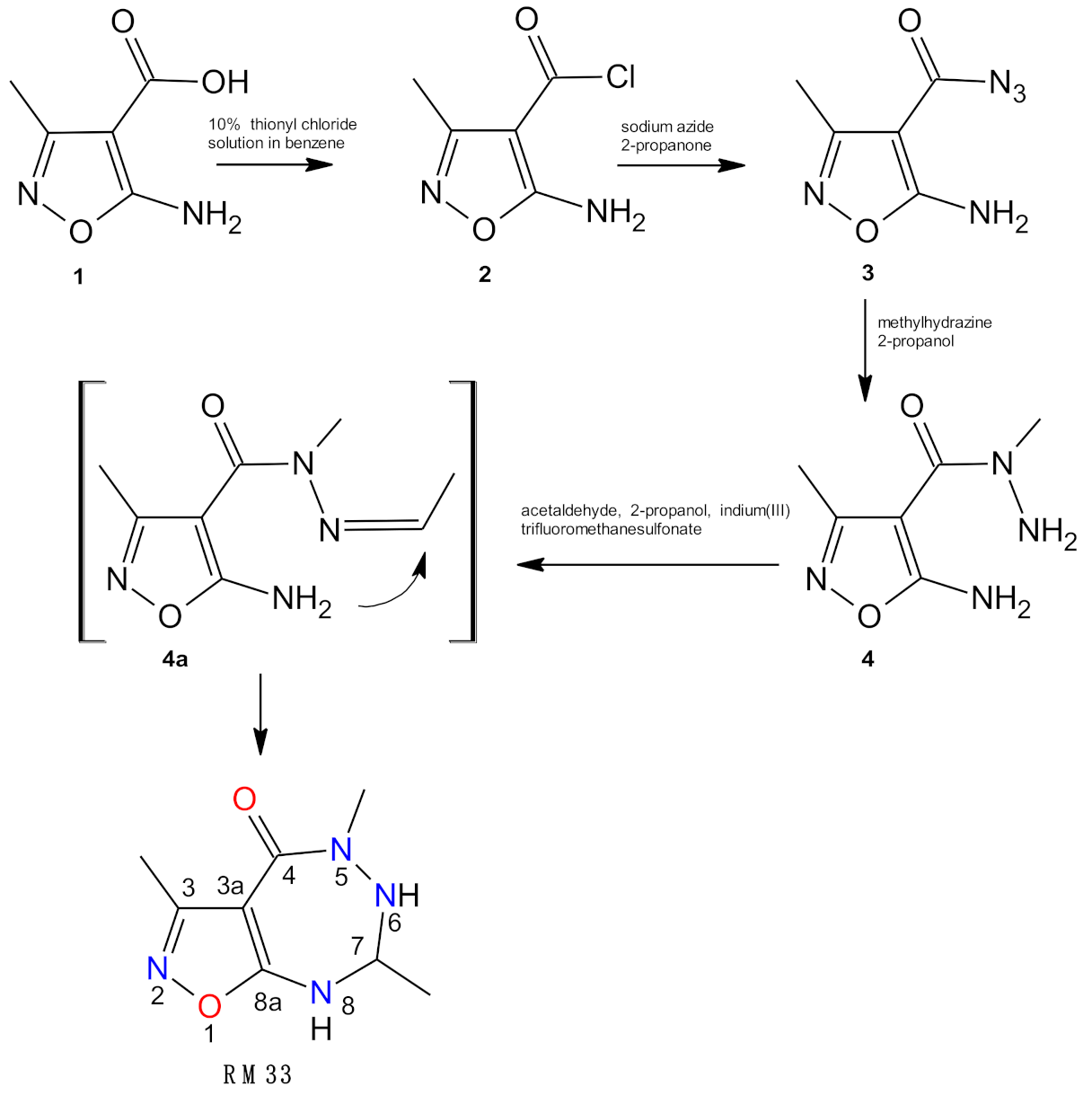
2.1. Chemistry
2.1.1. Proof and Analysis of the Structure of RM33 Compound
- Strong long range coupling through two bonds between protons of methyl group (2.21 ppm) at position 3 of system and the ring carbon 3 (161.1 ppm);
- Slightly weaker through three bonds between protons of methyl group (2.21 ppm) at position 3 of system with the bridged ring carbon 3a (88.0 ppm);
- Very weak, but noticeable, through four bonds between protons of methyl group (2.21 ppm) at position 3 of system with the bridged carbon 8a (166.2 ppm);
- Long range coupling through three bonds between protons of methyl group (2.94 ppm) at position 5 of the system and the carbonyl carbon atom (165.7 ppm) are also clearly visible.
2.1.2. Theoretical Prediction of Chemical Shifts of 1H NMR and 13C NMR Spectra of RM33
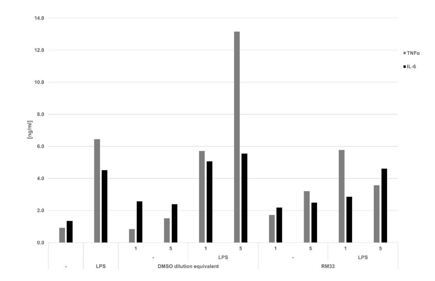
2.2. Activity of RM33 in Immunological Models In Vitro
3. Discussion
4. Materials and Methods
4.1. Chemistry
4.1.1. General Procedure for the Synthesis of 3,5,7-Trimethyl-5,6,7,8-Tetrahydro-4H-[1,2]Oxazolo[5,4-e][1,2,4]Triazepin-4-One—RM33
4.1.2. N-Deuteration of 3,5,7-Trimethyl-5,6,7,8-Tetrahydro-4H-[1,2]Oxazolo[5,4-e][1,2,4]Triazepin-4-One (RM33)
4.1.3. Computational Details about Theoretical Prediction of Chemical Shifts of 1H NMR and 13C NMR Spectra of RM33
4.2. Biology
4.2.1. Animals
4.2.2. Reagents
4.2.3. Cell Lines
4.2.4. Propagation of Cell Lines
4.2.5. Cell Toxicity Test
4.2.6. Isolation of Cells from the Lymphoid Organs
4.2.7. Evaluation of Viability of Cells from the Lymphoid Organs
4.2.8. Isolation of Peritoneal Exudates Cells and Cytokines Determination
4.2.9. Proliferation Tests
4.2.10. Colorimetric MTT Assay
4.2.11. Determination of Apoptosis
4.2.12. Total RNA Isolation
4.2.13. Reverse Transcription
4.2.14. Quantitative Analysis of Gene Expression by Real Time PCR
4.2.15. Determination of Cyclooxygenases in LPS-Stimulated Mouse Splenocytes
4.2.16. Statistics
Supplementary Materials
Author Contributions
Funding
Institutional Review Board Statement
Informed Consent Statement
Data Availability Statement
Acknowledgments
Conflicts of Interest
References
- Giomi, D.; Cordero, F.M.; Machetti, F. Comprehensive Heterocyclic Chemistry III; Katritzky, A.R., Ramsden, C.A., Scriven, E.F.V., Taylor, R.J.K., Eds.; Elsevier: Oxford, UK, 2008; pp. 365–486. [Google Scholar]
- Zhu, J.; Mo, J.; Lin, H.-Z.; Chen, Y.; Sun, H.-P. The recent progress of isoxazole in medicinal chemistry. Bioorg. Med. Chem. 2018, 26, 3065–3075. [Google Scholar] [CrossRef] [PubMed]
- Barmade, M.A.; Murumkar, P.R.; Sharma, M.K.; Yadav, M.R. Medicinal Chemistry Perspective of Fused Isoxazole Derivatives. Curr. Top. Med. Chem. 2016, 16, 2863–2883. [Google Scholar] [CrossRef] [PubMed]
- Zimecki, M.; Bąchor, U.; Mączyński, M. Isoxazole Derivatives as Regulators of Immune Functions. Molecules 2018, 23, 2724. [Google Scholar] [CrossRef] [PubMed] [Green Version]
- Herrmann, M.L.; Schleyerbach, R.; Kirschbaum, B.J. Leflunomide: An immunomodulatory drug for the treatment of rheumatoid arthritis and other autoimmune diseases. Immunopharmacology 2000, 47, 273–289. [Google Scholar] [CrossRef]
- Furst, D.E. Cyclosporin, leflunomide and nitrogen mustard. Baillieres Clin. Rheumatol. 1995, 9, 711–729. [Google Scholar] [CrossRef]
- McChesney, L.P.; Xiao, F.; Sankary, H.N.; Foster, P.F.; Sharma, S.; Haklin, M.; Williams, J.W. An evaluation of leflunomide in the canine renal transplantation model. Transplantation 1994, 57, 1717–1722. [Google Scholar] [CrossRef]
- Satyanarayana, P.S.; Jain, N.K.; Singh, S.; Kulkarni, S.K. Effect of selective inhibition of cyclooxygenase-2 on lipopolysaccha-ride-induced hyperalgesia. Inflammopharmacology 2004, 12, 57–68. [Google Scholar] [CrossRef]
- Chen, J.; Cong, X.; Zhan, X.; Zhou, Z.; Zheng, W. Effects of Parecoxib on Pain Threshold and Inflammatory Factors IL-1β, IL-6 and TNF-α in Spinal Cord of Rats with Bone Cancer Pain. J. Coll. Physicians Surg. Pak. 2019, 29, 528–531. [Google Scholar] [CrossRef] [PubMed]
- Ryng, S.; Machón, Z.; Wieczorek, Z.; Zimecki, M.; Glowiak, T. Synthesis and structure elucidation of 5-aminomethinimino-3-methyl-4-isoxazolecarboxylic acid phenylamides and their immunological activity. Arch. Pharm. 1997, 330, 319–326. [Google Scholar] [CrossRef]
- Mączyński, M.; Zimecki, M.; Drozd-Szczygieł, E.; Ryng, S. The synthesis, physicochemical properties and immunological activity of 5-amino-3-methylisoxazolo[5,4-d]4-pyrimidinone derivatives. Cell. Mol. Biol. Lett. 2005, 10, 613–623. [Google Scholar]
- Stosic-Grujicic, S.; Cvetkovic, I.; Mangano, K.; Fresta, M.; Maksimović-Ivanić, D.; Harhaji, L.; Popadic, D.; Momčilović, M.; Miljkovic, D.; Kim, J.; et al. A Potent Immunomodulatory Compound, (S,R)-3-Phenyl-4,5-dihydro-5-isoxasole Acetic Acid, Prevents Spontaneous and Accelerated Forms of Autoimmune Diabetes in NOD Mice and Inhibits the Immunoinflammatory Diabetes Induced by Multiple Low Doses of Streptozotocin in CBA/H Mice. J. Pharmacol. Exp. Ther. 2006, 320, 1038–1049. [Google Scholar] [CrossRef]
- Stojanović, I.; Cuzzocrea, S.; Mangano, K.; Mazzon, E.; Miljković, D.; Wang, M.; Donia, M.; Al Abed, Y.; Kim, J.; Nicoletti, F.; et al. In vitro, ex vivo and in vivo immunopharmacological activities of the isoxazoline compound VGX-1027: Modulation of cytokine synthesis and prevention of both organ-specific and systemic autoimmune diseases in murine models. Clin. Immunol. 2007, 123, 311–323. [Google Scholar] [CrossRef] [PubMed]
- Wagner, E.; Al-Kadasi, K.; Zimecki, M.; Sawka-Dobrowolska, W. Synthesis and pharmacological screening of derivatives of isoxazolo[4,5-d]pyrimidine. Eur. J. Med. Chem. 2008, 43, 2498–2504. [Google Scholar] [CrossRef]
- Andrzejak, V.; Muccioli, G.G.; Body-Malapel, M.; El Bakali, J.; Djouina, M.; Renault, N.; Chavatte, P.; Desreumaux, P.; Lambert, D.M.; Millet, R. New FAAH inhibitors based on 3-carboxamido-5-aryl-isoxazole scaffold that protect against experimental colitis. Bioorg. Med. Chem. 2011, 19, 3777–3786. [Google Scholar] [CrossRef]
- Leng, L.; Chen, L.; Fan, J.; Greven, D.; Arjona, A.; Du, X.; Austin, D.; Kashgarian, M.; Yin, Z.; Huang, X.R.; et al. A Small-Molecule Macrophage Migration Inhibitory Factor Antagonist Protects against Glomerulonephritis in Lupus-Prone NZB/NZW F1 and MRL/lpr Mice. J. Immunol. 2010, 186, 527–538. [Google Scholar] [CrossRef] [Green Version]
- Kankala, S.; Kankala, R.K.; Gundepaka, P.; Thota, N.; Nerella, S.; Gangula, M.R.; Guguloth, H.; Kagga, M.; Vadde, R.; Vasam, C.S. Regioselective synthesis of isoxazole–mercaptobenzimidazole hybrids and their in vivo analgesic and anti-inflammatory activity studies. Bioorg. Med. Chem. Lett. 2013, 23, 1306–1309. [Google Scholar] [CrossRef] [PubMed]
- Ghidini, E.; Capelli, A.; Carnini, C.; Cenacchi, V.; Marchini, G.; Virdis, A.; Italia, A.; Facchinetti, F. Discovery of a novel isoxazoline derivative of prednisolone endowed with a robust anti-inflammatory profile and suitable for topical pulmonary administration. Steroids 2015, 95, 88–95. [Google Scholar] [CrossRef]
- Rakesh, K.S.; Jagadish, S.; Balaji, K.S.; Zameer, F.; Swaroop, T.R.; Mohan, C.D.; Jayarama, S.; Rangappa, K.S. 3,5-Disubstituted Isoxazole Derivatives: Potential Inhibitors of Inflammation and Cancer. Inflammation 2015, 39, 269–280. [Google Scholar] [CrossRef]
- Banoglu, E.; Çelikoğlu, E.; Völker, S.; Olgaç, A.; Gerstmeier, J.; Garscha, U.; Çalışkan, B.; Schubert, U.S.; Carotti, A.; Macchiarulo, A.; et al. 4,5-Diarylisoxazol-3-carboxylic acids: A new class of leukotriene biosynthesis inhibitors potentially targeting 5-lipoxygenase-activating protein (FLAP). Eur. J. Med. Chem. 2016, 113, 1–10. [Google Scholar] [CrossRef]
- Russell, K.E.; Chung, K.F.; Clarke, C.J.; Durham, A.L.; Mallia, P.; Footitt, J.; Johnston, S.L.; Barnes, P.J.; Hall, S.R.; Simpson, K.D.; et al. The MIF Antagonist ISO-1 Attenuates Corticosteroid-Insensitive Inflammation and Airways Hyperresponsiveness in an Ozone-Induced Model of COPD. PLoS ONE 2016, 11, e0146102. [Google Scholar] [CrossRef]
- Płoszaj, P.; Regiec, A.; Ryng, S.; Piwowar, A.; Kruzel, M.L. Influence of 5-amino-3-methyl-4-isoxazolecarbohydrazide on selective gene expression in Caco-2 cultured cells. Immunopharmacol. Immunotoxicol. 2016, 38, 486–494. [Google Scholar] [CrossRef]
- Mączyński, M.; Artym, J.; Kocięba, M.; Kochanowska, I.; Ryng, S.; Zimecki, M. Anti-inflammatory properties of an isoxazole derivative—MZO-2. Pharmacol. Rep. 2016, 68, 894–902. [Google Scholar] [CrossRef]
- Elshemy, H.A.; Abdelall, E.K.; Azouz, A.A.; Moawad, A.; Ali, W.A.; Safwat, N.M. Synthesis, anti-inflammatory, cyclooxygenases inhibitions assays and histopathological study of poly-substituted 1,3,5-triazines: Confirmation of regiospecific pyrazole cyclization by HMBC. Eur. J. Med. Chem. 2017, 127, 10–21. [Google Scholar] [CrossRef]
- Mączyński, M.; Borska, S.; Mieszała, K.; Kocięba, M.; Zaczyńska, E.; Kochanowska, I.; Zimecki, M. Synthesis, Immunosuppressive Properties, and Mechanism of Action of a New Isoxazole Derivative. Molecules 2018, 23, 1545. [Google Scholar] [CrossRef] [Green Version]
- Singh, G.; Singh, G.; Bhatti, R.; Gupta, M.; Kumar, A.; Sharma, A.; Ishar, M.P.S. Indolyl-isoxazolidines attenuate LPS-stimulated pro-inflammatory cytokines and increase survival in a mouse model of sepsis: Identification of potent lead. Eur. J. Med. Chem. 2018, 153, 56–64. [Google Scholar] [CrossRef] [PubMed]
- Ryng, S.; Zimecki, M.; Maczyński, M.; Chodaczek, G.; Kocieba, M. Immunosuppressive activity of an isoxazolo[5,4-e]triazepine—Compound RM33. I. Effects on the humoral and cellular immune response in mice. Pharmacol. Rep. 2005, 57, 195–202. [Google Scholar] [PubMed]
- Zimecki, M.; Ryng, S.; Maczyński, M.; Chodaczek, G.; Kocieba, M.; Kuryszko, J.; Kaleta, K. Immunosuppressory activity of an isoxazolo[5,4-e]triazepine-compound RM-33 II. Effects on the carrageenan-induced inflammation. Pharmacol. Rep. 2006, 58, 236–241. [Google Scholar] [PubMed]
- Toledano, P.; Ait Itto, M.Y.; Hasnoui, A. 3-Mesityl-6-methyl-7-methylthio-4-phenyl-6H-isoxazolo[5,4-e][1,2,4]triazepine. Acta Cryst. C 1996, 52, 1230–1232. [Google Scholar] [CrossRef]
- Hasnaoui, A.; El Messaoudi, M.; Lavergne, J.-P. Nouvelles synthčses de systčmes bihétérocycliques II. Cycloaddition dipolaire-1,3 et condensation de carbčne sur des triazépines-1,2,4. Recl. Trav. Chim. Pays-Bas 2010, 104, 129–131. [Google Scholar] [CrossRef]
- Ryng, S.; Zimecki, M. Novel Leading Structure of Isoxazolotriazepinone Derivative and Method of Obtaining Same. Polish Patent PL193279, 31 January 2007. [Google Scholar]
- Shaw, G.; Sugowdz, G. The hydrogenation of 5-aminoizooxazoles. A new synthesis of pyrimidines. J. Chem. Soc. 1954, 665–668. [Google Scholar] [CrossRef]
- Regiec, A.; Gadzinski, P.; Płoszaj, P. New Methods for Preparing of Esters of 5-amino-3-methyl-4-isoxazolecarboxylic Acid. Polish Patent PL216764, 30 May 2014. (Chemical Abstracts CAN165:274203). [Google Scholar]
- Ryng, S.; Machoń, Z.; Głowiak, T. Synthesis and X-ray structure of new 5-amino-methyl-4-isoxazolecarboxylic acid azides. J. Chem. Crystallogr. 1994, 24, 483–488. [Google Scholar] [CrossRef]
- Regiec, A.; Wojciechowski, P.; Pietraszko, A.; Mączyński, M. Infrared spectra and other properties predictions of 5-amino-3-methyl-4-isoxazolecarbohydrazide with electric field simulation using CPC model. J. Mol. Struct. 2018, 1161, 320–338. [Google Scholar] [CrossRef]
- Hansen, M.B.; Nielsen, S.E.; Berg, K. Re-examination and further development of a precise and rapid dye method for measuring cell growth/cell kill. J. Immunol. Methods 1989, 119, 203–210. [Google Scholar] [CrossRef]
- Higham, A.D.; Sells, R.; Marshall-Clarke, S. Cyclosporin A has differential effects on the responses of murine B cells to TI antigens and B-cell mitogens. Immunology 1986, 59, 203–207. [Google Scholar]
- Espevik, T.; Nissen-Meyer, J. A highly sensitive cell line, WEHI 164 clone 13, for measuring cytotoxic factor/tumor necrosis factor from human monocytes. J. Immunol. Methods 1986, 95, 99–105. [Google Scholar] [CrossRef]
- Van Snick, J.; Cayphas, S.; Vink, A.; Uyttenhove, C.; Coulie, P.G.; Rubira, M.R.; Simpson, R.J. Purification and NH2-terminal amino acid sequence of a T-cell-derived lymphokine with growth factor activity for B-cell hybridomas. Proc. Natl. Acad. Sci. USA 1986, 83, 9679–9683. [Google Scholar] [CrossRef] [PubMed] [Green Version]
- Ferrari, G.; Terushkin, V.; Wolff, M.J.; Zhang, X.; Valacca, C.; Poggio, P.; Pintucci, G.; Mignatti, P. TGF-β1 Induces Endothelial Cell Apoptosis by Shifting VEGF Activation of p38MAPK from the Prosurvival p38β to Proapoptotic p38α. Mol. Cancer Res. 2012, 10, 605–614. [Google Scholar] [CrossRef] [Green Version]
- Zhong, J.; Lardinois, D.; Szilard, J.; Tamm, M.; Roth, M. Rat mesothelioma cell proliferation requires p38δ mitogen activated protein kinase and C/EBP-α. Lung Cancer 2011, 73, 166–170. [Google Scholar] [CrossRef] [PubMed]
- Risco, A.; del Fresno, C.; Mambol, A.; Alsina-Beauchamp, D.; MacKenzie, K.F.; Yang, H.-T.; Barber, D.F.; Morcelle, C.; Arthur, J.S.C.; Ley, S.C.; et al. p38 and p38 kinases regulate the Toll-like receptor 4 (TLR4)-induced cytokine production by controlling ERK1/2 protein kinase pathway activation. Proc. Natl. Acad. Sci. USA 2012, 109, 11200–11205. [Google Scholar] [CrossRef] [Green Version]
- Wada, M.; Canals, D.; Adada, M.; Coant, N.; Salama, M.F.; Helke, K.L.; Arthur, J.S.C.; Shroyer, K.R.; Kitatani, K.; Obeid, L.M.; et al. P38 delta MAPK promotes breast cancer progression and lung metastasis by enhancing cell proliferation and cell detachment. Oncogene 2017, 36, 6649–6657. [Google Scholar] [CrossRef] [Green Version]
- Lee, E.-S.; Ju, H.K.; Moon, T.C.; Lee, E.; Jahng, Y.; Lee, S.H.; Son, J.K.; Baek, S.-H.; Chang, H.W. Inhibition of Nitric Oxide and Tumor Necrosis Factor-.ALPHA. (TNF-.ALPHA.) Production by Propenone Compound through Blockade of Nuclear Factor (NF)-.KAPPA.B Activation in Cultured Murine Macrophages. Biol. Pharm. Bull. 2004, 27, 617–620. [Google Scholar] [CrossRef] [Green Version]
- Myers, M.J.; Deaver, C.M.; Lewandowski, A.J. Molecular mechanism of action responsible for carrageenan-induced inflammatory response. Mol. Immunol. 2019, 109, 38–42. [Google Scholar] [CrossRef]
- Stamm, C.E.; Collins, A.C.; Shiloh, M.U. Sensing ofMycobacterium tuberculosisand consequences to both host and bacillus. Immunol. Rev. 2015, 264, 204–219. [Google Scholar] [CrossRef] [Green Version]
- Coorens, M.; Schneider, V.A.F.; De Groot, A.M.; Van Dijk, A.; Meijerink, M.; Wells, J.M.; Scheenstra, M.R.; Veldhuizen, E.J.A.; Haagsman, H.P. Cathelicidins Inhibit Escherichia coli-Induced TLR2 and TLR4 Activation in a Viability-Dependent Manner. J. Immunol. 2017, 199, 1418–1428. [Google Scholar] [CrossRef] [Green Version]
- Malynn, B.; Romeo, D.T.; Wortis, H.H. Antigen-specific B cells efficiently present low doses of antigen for induction of T cell proliferation. J. Immunol. 1985, 135, 980–988. [Google Scholar] [PubMed]
- Chowdhury, M.G.; Maeda, K.; Furukawa, A.; Yasutomo, K.; Kagawa, S.; Himeno, K. B cells are required as APC for anti-gen-specific T cell proliferation but not for the differentiation or priming of those T cells. Tokushima J. Exp. Med. 1994, 41, 1–8. [Google Scholar]
- Mukaida, N.; Matsumoto, T.; Yokoi, K. Inhibition of neutrophil-mediated acute inflammatory injury by an antibody against interleukin-8 (IL-8). Inflamm. Res. 1998, 47, 151–157. [Google Scholar] [CrossRef]
- Sur, B.; Kang, S.; Kim, M.; Oh, S. Inhibition of Carrageenan/Kaolin-Induced Arthritis in Rats and of Inflammatory Cytokine Expressions in Human IL-1β-Stimulated Fibroblast-like Synoviocytes by a Benzylideneacetophenone Derivative. Inflammation 2019, 42, 928–936. [Google Scholar] [CrossRef] [Green Version]
- Chervenick, P.; Boggs, D.R.; Marsh, J.C.; Cartwright, G.; Wintrobe, M.M. Quantitative studies of blood and bone marrow neutrophils in normal mice. Am. J. Physiol. Content 1968, 215, 353–360. [Google Scholar] [CrossRef] [PubMed] [Green Version]
- Goodwin, J.S.; Ceuppens, J. Regulation of the immune response by prostaglandins. J. Clin. Immunol. 1983, 3, 295–315. [Google Scholar] [CrossRef] [PubMed]
- Kanda, N.; Koike, S.; Watanabe, S. IL-17 suppresses TNF-α-induced CCL27 production through induction of COX-2 in human keratinocytes. J. Allergy Clin. Immunol. 2005, 116, 1144–1150. [Google Scholar] [CrossRef] [PubMed]
- Nunn, S.; Nishikida, K. Advanced ATR Correction Algorithm; Thermo Fisher Scientific Inc.: Madison, WI, USA, 2008; pp. 1–4. [Google Scholar]
- Becke, A.D. Density-functional thermochemistry. IV. A new dynamical correlation functional and implications for exact-exchange mixing. J. Chem. Phys. 1996, 104, 1040–1046. [Google Scholar] [CrossRef]
- Lee, C.; Yang, W.; Parr, R.G. Development of the Colle-Salvetti correlation-energy formula into a functional of the electron density. Phys. Rev. B 1988, 37, 785–789. [Google Scholar] [CrossRef] [PubMed] [Green Version]
- Clark, T.; Chndrasekhar, J.; Spitznagel, G.W.; Schleyer, P. Efficient diffuse function-augmented basis sets for anion calculations. III. The 3-21+G basis set for first-row elements, Li-F. J. Comput. Chem. 1983, 4, 294–301. [Google Scholar] [CrossRef]
- Hariharan, P.C.; Pople, J.A. The influence of polarization functions on molecular orbital hydrogenation energies. Theor. Chem. Acc. 1973, 28, 213–222. [Google Scholar] [CrossRef]
- Krishnan, R.S.; Binkley, J.S.; Seeger, R.; Pople, J. Self-consistent molecular orbital methods. XX. A basis set for correlated wave functions. J. Chem. Phys. 1980, 72, 650–654. [Google Scholar] [CrossRef]
- Kendall, R.A.; Dunning, T.H., Jr.; Harrison, R.J. Electron affinities of the first-row atoms revisited. Systematic basis sets and wave functions. J. Chem. Phys. 1992, 96, 6796–6806. [Google Scholar] [CrossRef] [Green Version]
- Cossi, M.; Rega, N.; Scalmani, G.; Barone, V. Energies, structures, and electronic properties of molecules in solution with the C-PCM solvation model. J. Comput. Chem. 2003, 24, 669–681. [Google Scholar] [CrossRef]
- Frisch, M.J.; Trucks, G.W.; Schlegel, H.B.; Scuseria, G.E.; Robb, M.A.; Cheeseman, J.R.; Scalmani, G.; Barone, V.; Petersson, G.A.; Nakatsuji, H.; et al. Gaussian 16 (Revision, A.03); Gaussian, Inc.: Wallingford, CT, USA, 2016. [Google Scholar]
- Regiec, A.; Wojciechowski, P. Synthesis and experimental versus theoretical research on spectroscopic and electronic properties of 3-methyl-4-nitroisothiazole. J. Mol. Struct. 2019, 1196, 370–388. [Google Scholar] [CrossRef]
- Katritzky, A.R.; Akhmedov, N.G.; Doskocz, J.; Hall, C.D.; Akhmedova, R.G.; Majumder, S. Structural elucidation of nitro-substituted five-membered aromatic heterocycles utilizing GIAO DFT calculations. Magn. Reson. Chem. 2006, 45, 5–23. [Google Scholar] [CrossRef]
- Cheeseman, J.R.; Trucks, G.W.; Keith, T.A.; Frisch, M.J. A comparison of models for calculating nuclear magnetic resonance shielding tensors. J. Chem. Phys. 1996, 104, 5497–5509. [Google Scholar] [CrossRef]
- Matuszyk, J.; Cebrat, M.; Kalas, W.; Strzadala, L. HA1004, an inhibitor of serine/threonine protein kinases, restores the sensitivity of thymic lymphomas to Ca2+-mediated apoptosis through a protein kinase A-independent mechanism. Int. Immunopharmacol. 2002, 2, 435–442. [Google Scholar] [CrossRef]


| Method | 1H NMR | 13C NMR | ||
|---|---|---|---|---|
| Chemical Shift δ (ppm) | Deviation Δδ (ppm) | Chemical Shift δ (ppm) | Deviation Δδ (ppm) | |
| Experimental * ± SD ** | 1.26 ± 0.009 (CH3) | - | 11.6 ± 0.13 (CH3) 20.8 ± 0.13 (CH3) 37.3 ± 0.15 (N-CH3) 67.4 ± 0.01 (C7) 88.0 ± 0.014 (C3a) 161.1 ± 0.01 (C3) 165.8 ± 0.06 (CO) 166.2 ± 0.02 (C8a) | - - - - - - - - |
| 2.21 ± 0.00 (CH3) | - | |||
| 2.94 ± 0.00 (CH3) | - | |||
| 4.41 ± 0.013 (CH) | - | |||
| 5.94 ± 0.015 (NH) | - | |||
| 8.64 ± 0.002 (NH) | - | |||
| B3LYP/6-31+G(d,p) | 1.36 (CH3) | −0.10 | 15.66 (CH3) 22.55 (CH3) 38.19 (N-CH) 72.40 (C7) 90.77 (C3a) 163.82 (C3) 163.94 (CO) 161.79 (C8a) | −4.06 –1.75 –0.89 −5.00 −2.77 −2.72 1.85 4.41 |
| 2.28 (CH3) | −0.07 | |||
| 3.07 (CH3) | −0.13 | |||
| 4.70 (CH) | −0.29 | |||
| 3.79 (NH) | 2.15 | |||
| 5.58 (NH) | 3.06 | |||
| B3LYP/6-311++G(df,pd) | 1.41 (CH3) | −0.15 | 15.76 (CH3) 23.64 (CH3) 39.70 (N-CH) 75.73 (C7) 94.80 (C3a) 173.94 (C3) 175.26 (CO) 173.42 (C8a) | −4.16 −2.84 −2.40 −8.13 −6.80 −12.84 −9.46 −7.23 |
| 2.34 (CH3) | −0.13 | |||
| 3.08 (CH3) | −0.14 | |||
| 4.66 (CH) | −0.25 | |||
| 3.63 (NH) | 2.31 | |||
| 5.52 (NH) | 3.12 | |||
| B3LYP/aug-cc-pVTZ | 1.40 (CH3) | −0.14 | 15.85 (CH3) 23.30 (CH3) 38.68 (N-CH3) 75.30 (C7) 95.27 (C3a) 174.20 (C3) 175.44 (CO) 173.22 (C8a) | −4.25 −2.50 −1.38 −7.90 −7.27 −13.08 −9.64 −7.01 |
| 2.34 (CH3) | −0.13 | |||
| 3.09 (CH3) | −0.15 | |||
| 4.74 (CH) | −0.33 | |||
| 3.78 (NH) | 2.16 | |||
| 5.67 (NH) | 2.97 | |||
| Model | Effects of RM33 Administration | Reference |
|---|---|---|
| Humoral immune response | Inhibition of the inductive phase | [27] |
| Delayed type hypersensitivity | Inhibition of the inductive and effectual phase | [27] |
| Edema induced by complete Freund’s adjuvant | Decrease in footpad edema | [28] |
| LPS-induced endotoxemia | Significant inhibition of serum TNFα, small inhibitory effect on IL-6 and no effect on IL-10 serum level | [28] |
| Carrageenan reaction | Decreased serum TNFα, reduced: mast cell and macrophage infiltration and edema of the connective tissue | [28] |
| Organ | Increase in % of Apoptotic Cells | ||
|---|---|---|---|
| RM33 (μg/mL) | |||
| 2 | 10 | 50 | |
| Bone marrow | 0 | 0 | 0 |
| Thymus | 1 | 3 | 6 |
| Spleen | 1 | 1 | 1 |
| Organ | ERK-1 | ERK-2 | p38α | p38β | p38γ | p38δ | JNK |
|---|---|---|---|---|---|---|---|
| Bone marrow | 0.05 | 16.6 | 7.8 | 138.0 | 0.2 | 0.4 | 31.4 |
| Thymus | 2.7 | 6.7 | 11.9 | 31.7 | 4.5 | 70.9 | 92.8 |
| Spleen | 7.9 | 5.4 | 18.3 | 27.0 | 19.3 | 256.8 | 70.1 |
| Organ | Casp-3 | Casp-8 | Casp-9 | Bcl-2 | Fas | NFκB1 |
|---|---|---|---|---|---|---|
| Bone marrow | 2.0 | 0.0 | 23.1 | 1.9 | 19.2 | 4.8 |
| Thymus | 9.5 | 2.3 | 222.1 | 18.2 | 5.7 | 10.7 |
| Spleen | 5.4 | 1.5 | 198.9 | 0.8 | 22.2 | 8.8 |
| ERK-1 | ERK-2 | JNK | p38α | p38β | p38γ | p38δ | |
|---|---|---|---|---|---|---|---|
| RM33 | 1.7 | 0.8 | 0.9 | 0.3 | 0.5 | 0.1 | 3.7 |
| ERK-1 | ERK-2 | p38α | p38β | p38γ | p38ő | JNK | Casp-3 | Casp-8 | Casp-9 | NFκB1 | Bcl-2 | Fas |
|---|---|---|---|---|---|---|---|---|---|---|---|---|
| 0 | 5487 | 0 | 4629 | 1303 | 1177 | 902 | 295 | 1.46 | 0 | 3656 | 344 | 753 |
| Culture | Cox-2 (pg/mL) |
|---|---|
| Control | 456 |
| RM33 | 470 |
| LPS | 742 |
| LPS + RM33 | 1031 |
Publisher’s Note: MDPI stays neutral with regard to jurisdictional claims in published maps and institutional affiliations. |
© 2021 by the authors. Licensee MDPI, Basel, Switzerland. This article is an open access article distributed under the terms and conditions of the Creative Commons Attribution (CC BY) license (https://creativecommons.org/licenses/by/4.0/).
Share and Cite
Mączyński, M.; Regiec, A.; Sochacka-Ćwikła, A.; Kochanowska, I.; Kocięba, M.; Zaczyńska, E.; Artym, J.; Kałas, W.; Zimecki, M. Synthesis, Physicochemical Characteristics and Plausible Mechanism of Action of an Immunosuppressive Isoxazolo[5,4-e]-1,2,4-Triazepine Derivative (RM33). Pharmaceuticals 2021, 14, 468. https://0-doi-org.brum.beds.ac.uk/10.3390/ph14050468
Mączyński M, Regiec A, Sochacka-Ćwikła A, Kochanowska I, Kocięba M, Zaczyńska E, Artym J, Kałas W, Zimecki M. Synthesis, Physicochemical Characteristics and Plausible Mechanism of Action of an Immunosuppressive Isoxazolo[5,4-e]-1,2,4-Triazepine Derivative (RM33). Pharmaceuticals. 2021; 14(5):468. https://0-doi-org.brum.beds.ac.uk/10.3390/ph14050468
Chicago/Turabian StyleMączyński, Marcin, Andrzej Regiec, Aleksandra Sochacka-Ćwikła, Iwona Kochanowska, Maja Kocięba, Ewa Zaczyńska, Jolanta Artym, Wojciech Kałas, and Michał Zimecki. 2021. "Synthesis, Physicochemical Characteristics and Plausible Mechanism of Action of an Immunosuppressive Isoxazolo[5,4-e]-1,2,4-Triazepine Derivative (RM33)" Pharmaceuticals 14, no. 5: 468. https://0-doi-org.brum.beds.ac.uk/10.3390/ph14050468