Article

Bioactive Compounds from Euphorbia usambarica Pax. with HIV-1 Latency Reversal Activity

1
Interdisciplinary Excellence Centre, Department of Pharmacognosy, University of Szeged, H-6720 Szeged, Hungary
2
Department of Medicine, University of Utah School of Medicine, Salt Lake City, UT 84132, USA
3
Department of Medical Physiology, School of Medicine, University of Nairobi, Nairobi P.O. Box 30197-00100, Kenya
4
Department of Medical Chemistry, University of Szeged, H-6720 Szeged, Hungary
5
Interdisciplinary Centre of Natural Products, University of Szeged, H-6720 Szeged, Hungary
*
Authors to whom correspondence should be addressed.
Pharmaceuticals 2021, 14(7), 653; https://0-doi-org.brum.beds.ac.uk/10.3390/ph14070653
Submission received: 3 June 2021 / Revised: 27 June 2021 / Accepted: 30 June 2021 / Published: 7 July 2021
(This article belongs to the Special Issue Antiviral Compounds in Medicinal Plants)

Abstract

:
Euphorbia usambarica is a traditional medicine used for gynecologic, endocrine, and urogenital illnesses in East Africa; however, its constituents and bioactivities have not been investigated. A variety of compounds isolated from Euphorbia species have been shown to have activity against latent HIV-1, the major source of HIV-1 persistence despite antiretroviral therapy. We performed bioactivity-guided isolation to identify 15 new diterpenoids (19, 1417, 19, and 20) along with 16 known compounds from E. usambarica with HIV-1 latency reversal activity. Euphordraculoate C (1) exhibits a rare 6/6/3-fused ring system with a 2-methyl-2-cyclopentenone moiety. Usambariphanes A (2) and B (3) display an unusual lactone ring constructed between C-17 and C-2 in the jatrophane structure. 4β-Crotignoid K (14) revealed a 250-fold improvement in latency reversal activity compared to crotignoid K (13), identifying that configuration at the C-4 of tigliane diterpenoids is critical to HIV-1 latency reversal activity. The primary mechanism of the active diterpenoids 1214 and 21 for the HIV-1 latency reversal activity was activation of PKC, while lignans 26 and 27 that did not increase CD69 expression, suggesting a non-PKC mechanism. Accordingly, natural constituents from E. usambarica have the potential to contribute to the development of HIV-1 eradication strategies.

1. Introduction

Antiretroviral therapy (ART) durably blocks HIV-1 transcription by targeting viral enzymes; however, these drugs do not result in viral eradication due to the presence of replication-competent proviruses that are stably integrated into the genomes of a small population of long-lived memory T cells, known as the latent reservoir [1]. A promising strategy to address HIV-1 persistence is to use small molecules to reactivate latent proviruses in order to expose these cells to immune clearance and/or viral cytopathic effect. Natural products offer much promise regarding the discovery of new latency reversal agents (LRAs) for HIV-1 eradication [2,3,4].
The Euphorbia is one of the largest genera in Euphorbiaceae [5,6]. There are many bioactive secondary metabolites in the genus Euphorbia, including more than 20 different types of diterpenoids (abietane, atisane, casbane, daphnane, ingenane, jatrophane, karane, lathyrane, tigliane, and others) [7]. Moreover, sesquiterpenoids, triterpenoids, flavonoids, alkaloids, polyphenols, tannins, volatile compounds, and phytosterols have also been discovered in Euphorbia species, many of which are in active use as traditional medicines [8,9,10]. The pharmacological effects of Euphorbia species are related to anti-inflammatory [11], multidrug-resistance-reversing [12,13], antiviral [14,15], cytotoxic [16,17], anti-arrhythmic [18], antifungal [19], anti-thrombotic [20], antiallergic [21], and muscle relaxant [22] properties.
In past, several Euphorbia plants have previously been evaluated to determine their efficacy as LRAs [23,24,25,26,27,28,29,30]. For instance, Liu et al. reported the effects on HIV-1 transcription of ingenane esters 3-angeloylingenol and 3-(2-naphthoyl)ingenol from E. kansui, which can reactivate latent HIV with EC50 values at 4.2 and 2.4 nM, respectively [28]. Yan et al. published atisane diterpenoids euphorneroid D and ent-3-oxoatisan-16α,17-acetonide from E. neriifolia which showed anti-HIV-1 activities with EC50 values at 34 and 24 μM, respectively [29]. Valadão et al. established deoxyphorbol esters from E. umbellata which increased HIV-1 latency reactivation through NF-κB activation, nuclear translocation, and HIV-1 LTR promoter [30].
Euphorbia usambarica Pax. distributes mainly in East Africa [31] and is a large branching shrub as well as used as a traditional medicine for gynecologic, endocrine, and urogenital illnesses [32,33]. In our preliminary study, we found that the whole plant extract of E. usambarica showed a significant HIV-1 latency reversal activity. However, there was no study related to the chemical constituents and bioactivities of E. usambarica in the reported literature. In addition, the prevention and treatment of HIV infection and acquired immune deficiency syndrome (AIDS) are still the central issues around the world. Therefore, we would investigate the active constituents and the pharmacological effect of E. usambarica. Further, we sought to test its aqueous, and organic fractions for HIV-1 latency reversal activity and cytotoxicity. Dichloromethane and n-hexane fractions showed increased activity compared to the whole plant extract (EU) in dose-response analysis. Further sub-fractionation of the active fractions was followed by compounds purification and identification using multistep chromatography, NMR, and mass spectroscopy to yield 31 purified compounds. Six of those compounds demonstrated HIV-1 anti-latency activity. Extended dose-response curves were then generated for these compounds. Several of these compounds have no previously described anti-HIV-1 or anti-latency activity. These results support further exploration of medicinal plants, and Euphorbia species in particular, as sources of new means to address HIV-1 persistence.

2. Results

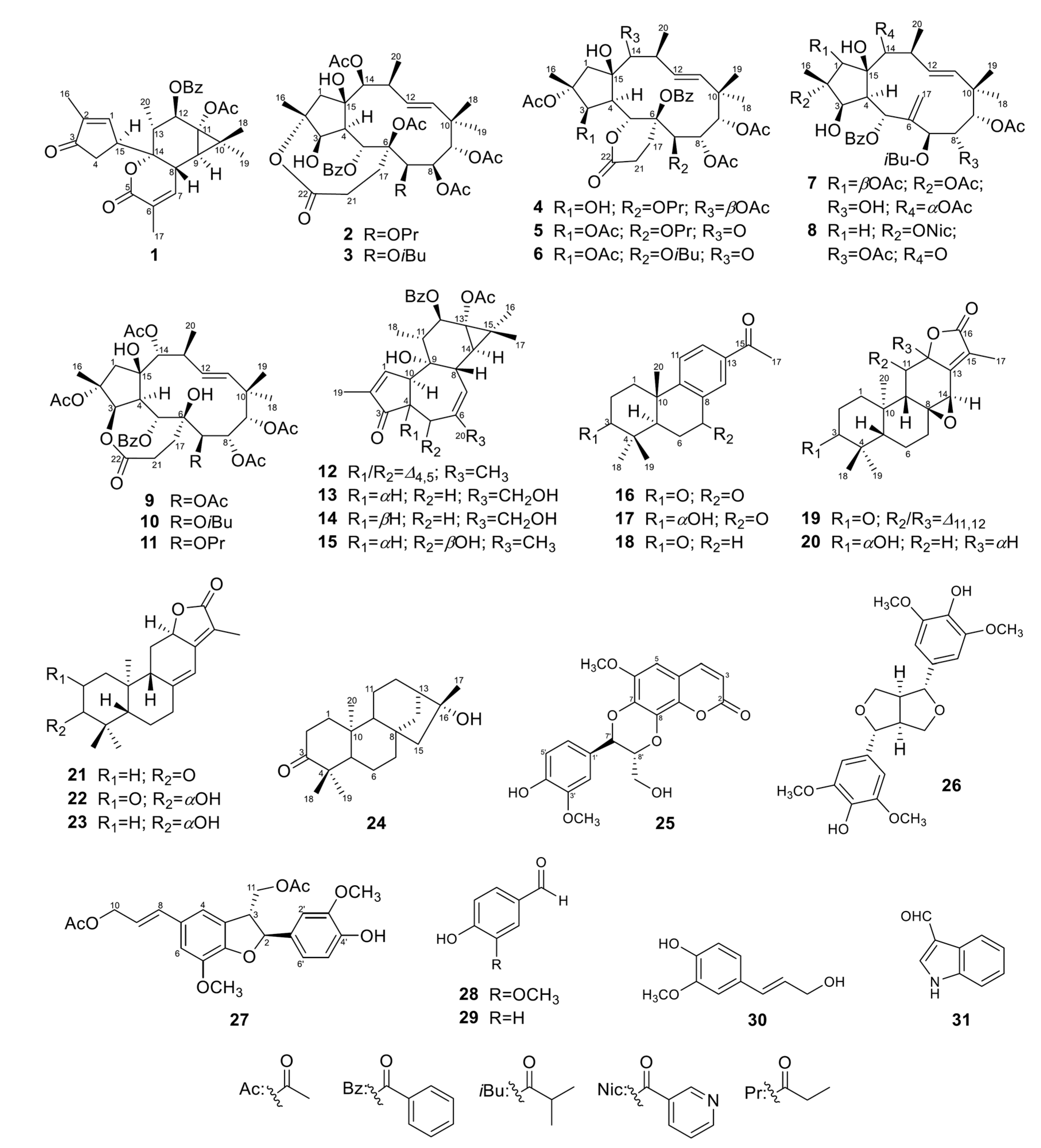
2.1. Structure Elucidation of New Compounds

The partitioned n-hexane (EU-H) and dichloromethane (EU-C) phases significantly improved upon reactivation efficacy compared to the EU. The EU-H reactivated latent HIV-1 to 91% at concentrations of 50 and 100 μg/mL. The EU-C phase reactivated latent HIV-1 up to 86% at 10 μg/mL concentration and 98% at 50 μg/mL. The partitioned ethyl acetate (EU-E) and water-soluble residue (EU-W) phases did not appear to have any activity (Figure 1A). Cell viability declined steeply above concentrations of 100 μg/mL. Significant toxicity at concentrations above 100 μg/mL limits conclusions about reactivation. The lower concentrations of the EU-H and EU-C fractions did not affect toxicity but markedly improved viral reactivation (Figure 1B). Due to the high reactivation ratio (86%) at the lowest tested concentration (10 μg/mL), the EU-C phase was selected for bioactivity-guided isolation. This led to identification of 15 new diterpenoids (19, 1417, 19, and 20) along with 16 known compounds (1013, 18, and 2131) (Figure 2).

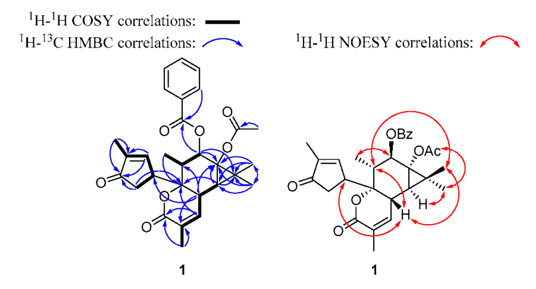
2.1.1. Euphordraculoate C (1)

Compound 1 was purified as a colorless gum with [ α ] D 28 −63 (c 0.05, CHCl3). The molecular formula was identified as C29H32O7 by HR-ESIMS m/z 493.2237 [M + H]+ (calcd. for C29H33O7 493.2221), indicating 14 unsaturated degrees. The 1H NMR spectrum of 1 revealed six methyls, an oxygenated methine, two unsaturated methines, and a monosubstituted aromatic group (Table 1). The 13C-JMOD spectrum of 1 evidenced 29 carbon signals, including six methyls, one methylene, seven olefinic methines, one oxygenated methine, four saturated methines, one quaternary, three olefinic quaternary, two oxygenated quaternary, and four carbonyl carbons (Table 2). According to the combination of the 1D and 2D NMR spectra, one benzoyloxy group (OBz) [δH 8.02 (2H), 7.58, 7.46 (2H); δC 166.2, 133.2, 130.4, 129.9 (2C), 128.6 (2C)] and one acetoxy (OAc) (δH 1.97; δC 170.4, 21.1) could be identified in 1. Based on the COSY and HSQC spectra of 1, a series of COSY correlations between an olefinic methine (δH 7.52, CH-1)/a methine (δH 3.80, CH-15)/methylene (δH 2.52, 2.24, CH2-4), together with the allylic four-bond coupling between H-1 and a methyl group [δH 1.74 (3H), CH3-16]. Key HMBC correlations from H-1 and H-16 to an olefinic quaternary carbon C-2 (δC 141.3) and a ketone carbon C-3 (δC 206.8), and H-4 to C-3, indicated the presence of an α-methyl-α,β-unsaturated cyclopentanone moiety. Moreover, a series of COSY correlations between a methine (δH 2.37, CH-8), an olefinic methine (δH 6.68, CH-7), and an allylic coupled methyl group [δH 2.02 (3H), CH3-17], together with the key HMBC correlations from H-7 to a carbonyl carbon C-5 (δC 163.4) and an oxygenated quaternary carbon C-14 (δC 85.8), H-8 to an olefinic quaternary carbon C-6 (δC 127.8), and H3-17 to C-5, C-6, and C-7, indicated the presence of an α-methyl-α,β-unsaturated-δ-lactone moiety. A gem-dimethylcyclopropane moiety could be identified by the key HMBC correlations from two methyl groups [δH 1.43 (3H), δC 16.5, CH3-18; δH 1.16 (3H), δC 24.8, CH3-19] to a methine C-9 (δH 1.02, δC 34.0), a quaternary carbon C-10 (δC 24.9), an oxygenated quaternary carbon C-11 (δC 63.4) and each other, and H-9 to C-10 and C-11. In addition, the 1H–1H COSY cross peak between H-8/H-9, a methyl group [δH 0.91 (3H), CH3-20]/a methine (δH 2.10, CH-13)/an oxygenated methine (δH 5.86, CH-12), as well as the HMBC correlations from H-7 to C-14, H-8 to C-11 and C-13, H-9 to C-11 and C-14, H-12 to C-10 and C-11, and C-13 to C-14, demonstrated the presence of a six-membered ring fusion with the gem-dimethyl-cyclopropane moiety at C-9 and C-11, and the α-methyl-α,β-unsaturated-δ-lactone moiety at C-8 and C-14. The HMBC correlations from H-4 to C-14 and H-8 to C-15 indicated that the α-methyl-α,β-unsaturated cyclopentanone moiety was linked to C-14. The OAc and OBz groups should be connected to C-11 and C-12, respectively, based on HMBC correlations (Figure 3). Additionally, comparing the NMR data of 1 with those of euphordraculoate A [34] suggested the same rare diterpenoid skeleton of both compounds. According to the NOESY cross-peaks between H-8/H-13, H-8/H3-18, H-8/H-15, H-9/H3-19, 11-OAc/H3-19, H-12/H3-20, and H-13/H3-18, as well as comparing with euphordraculoate A [34] and euphodendriane A [35], the relative configuration of 1 was established as shown on structural formula (Figure 3), and the compound was named as euphordraculoate C.

2.1.2. Usambariphane A (2)

Compound 2 was obtained as a white amorphous powder. Its molecular formula was calculated as C40H52O16 by the analysis of HR-ESIMS m/z 789.3327 [M + H]+ (calcd. for C40H53O16 789.3328). The NMR spectra of 2 revealed clearly four OAc (δH 2.56, δC 174.3, 21.4; δH 2.17, δC 170.2, 22.7; δH 2.14, δC 169.8, 21.5; δH 2.08, δC 170.0, 21.6), one OBz (δH 7.93 (2H), 7.52, 7.39 (2H), δC 164.5, 133.5, 129.7, 129.6, 128.8), one propionate group [δH 2.41 (2H), 1.16 (3H), δC 174.2, 27.7, 8.9; OPr], four methyls [δH 1.73 (3H), δC 19.1, CH3-16; δH 0.93 (3H), δC 26.0, CH3-18; δH 1.13 (3H), δC 21.7, CH3-19; δH 1.14 (3H), δC 23.2, CH3-20], a trans-disubstituted C=C (δH 5.49, δC 134.6, CH-11; δH 5.83, δC 133.0, CH-12), and a lactone carbonyl carbon (δC 175.1, C-22). Further, comparing the 1D NMR data of 2 (Table 1 and Table 2) with those of isoterracinolide A (10) [36], the skeleton of 2 was established as a dihomojatrophane type diterpenoid [7] with a double bond at 11,12 and a lactone moiety. An OH group was located at C-3 based on a 1H–1H COSY cross peak between H-3 and 3-OH, as well as the HBMC correlations from 3-OH to C-3 and C-4. Another OH group was connected to C-15 by the confirmation of the HMBC correlations from 15-OH to C-4, C-14, and C-15. Moreover, the HMBC correlations from H-5 to δC 164.5, H-7 to δC 174.2, H-8 to δC 170.0, H-9 to δC 170.2, H-14 to δC 174.3, indicated that OBz and OPr were located at C-5 and C-7 respectively, and three OAc were linked to C-8, C-9, and C-14 each. The last OAc was located apparently to C-6 based on NOESY correlations between the acetyl proton signal δH 2.14 (6-OAc) with H-5 and H-17a. The remaining lactone ring was proposed to be constructed between C-17 and C-2 in structure 2. According to the 13C signal value of C-6 (δC 92.5) of 2 was close to the signal in sororianolide A (δC 93.0, C-6-βOAc) and different from sororianolide B (δC 80.9, C-6-αOAc), suggesting the OAc at C-6 in 2 can be assigned as β-oriented [37]. Moreover, the NOESY correlations of H-3/H2-17, H-3/H-4, H-4/H-7, H-4/H-8, H-8/H3-19, H3-19/H-13, H3-19/H-14, H3-16/3-OH, H3-16/H-1b, H-1b/15-OH, 15-OH/H-9, and H-9/H3-18 indicted the configurations of 3β-OH, Hα-4, Hβ-5, 6β-OAc, 7β-OPr, 8β-OAc, 9α-OAc, 14β-OAc, 15-βOH, βCH3-16, and βCH3-20. Thus, the structure of 2 was established and named as usambariphane A.

2.1.3. Usambariphane B (3)

Compound 3 was obtained as a white amorphous powder. The molecular formula was determined as C41H54O16 based on HR-ESIMS m/z 803.3488 [M + H]+ (calcd. for C41H55O16 803.3485). The 1D NMR data (Table 1 and Table 2) of 3 were highly similar to those of 2, except for an isobutyryl group (OiBu) [δH 2.61, 1.21 (3H), 1.20 (3H), δC 176.4, 34.2, 18.8, 18.5] instead of propanoyl. The isobutyryl group was connected to C-7 in 3 based on the HMBC correlation from H-7 (δH 5.38) to the OiBu carbonyl carbon (δC 176.4). The NOESY correlations of 3 revealed the same relative configuration as that of 2. The structure of 3 was established and named as usambariphane B.

2.1.4. Usambariphane C (4)

Compound 4 was purified as a colorless crystal. The molecular formula was identified as C40H52O16 by HR-ESIMS m/z 789.3346 [M + H]+ (calcd. for C40H53O16 789.3328). Based on the comparison of the 1H and 13C NMR data (Table 1 and Table 2) for 4 with those of usambariphane B (2), the skeleton of 4 was suggested to be a C22 dihomojatrophane with a double bond at ∆11,12 (δH 5.41, δC 135.3, CH-11; δH 5.72, δC 134.1, CH-12) and a lactone moiety (δH 3.09, 2.40, δC 23.4, CH2-17; δH 2.65, 2.16, δC 26.0, CH2-21; δC 168.1, C-22). A δ-lactone ring was constructed at C-5 and C-6 supporting by the 1H–1H COSY cross peak between H2-17 and H2-21, as well as the HMBC correlations from H-5 to C-17 and C-22, H2-17 to C-5, C-6, and C-22, and H2-21 to C-6 and C-22. Moreover, the 1D NMR data of 4 were highly close to those of euphosorophane D [38] except for an OPr group [δH 2.57, 2.50, 1.22 (3H); δC 174.6, 27.5, 8.9] at C-7 according to an HMBC correlation from H-7 (δH 5.40) to δC 174.6. The NOESY cross-peaks of 4 demonstrated the same relative orientations to those of euphosorophane D [38]. Therefore, the structure of 4 was established and named as usambariphane C.

2.1.5. Usambariphane D (5)

Compound 5 was purified as a colorless crystal. The molecular formula was identified as C40H50O16 by HR-ESIMS m/z 787.3193 [M + H]+ (calcd. for C40H51O16 787.3172). The inspection of 1D (Table 1 and Table 2) and 2D NMR data suggested that compound 5 was a bishomojatrophane type diterpenoid with a double bond at 11,12 (δH 6.16, δC 137.4, CH-11; δH 5.43, δC 128.9, CH-12), a lactone moiety (δH 2.72, 2.01, δC 26.5, CH2-17; δH 3.43, 2.50, δC 29.1, CH2-21; δC 172.7, C-22), and a ketone unit δC 211.4 (C-14). A δ-lactone ring was constructed at C-5 and C-6 supporting by the 1H–1H COSY cross peak between H2-17 and H2-21, and the HMBC correlations from H-5 to C-17 and C-22, H2-17 to C-5, C-6, and C-22, and H2-21 to C-6 and C-22. The ketone unit in 5 was located at C-14 based on the HMBC correlations from H-1, H-12, H-13, and H3-20 to C-14, respectively. An OH group was connected to C-15 by the confirmation of HMBC correlations from 15-OH to C-4, C-14, and C-15. Moreover, four OAc [δH 2.26 (3H), δC 169.6, 22.4; δH 2.05 (3H), δC 169.1, 20.6; δH 2.03 (3H), δC 169.9, 20.9; δH 2.00 (3H), δC 170.0, 21.2], one OBz [δH 7.88 (2H), 7.65, 7.51 (2H), δC 165.8, 133.8, 130.6, 129.7 (2C), 128.5 (2C)], and one OPr [δH 2.49, 2.31, 1.23 (3H), δC 173.4, 27.6, 8.6] moieties were identified clearly by the examination of the NMR spectra. The HMBC correlations from H-3 to δC 169.1, H-7 to δC 173.4, H-8 to δC 170.0, and H-9 to δC 169.9, indicated the OPr was located at C-7, and three OAc were linked to C-3, C-8, and C-9, respectively. The location of the OBz at C-6 was confirmed by the NOESY correlations between the benzoyl proton signal δH 7.88 with H-5, H-8, and H-12. The last OAc was connected to C-2 based on the NOE cross-peak between the acetyl proton signal δH 2.26 with H3-16. The relative configuration of 5 was evaluated by the NOESY spectrum and comparison with a similar structure terracinolide J [39] to assign 2α-OAc, 3β-OAc, Hα-4, Hβ-5, 6β-OBz, 7β-OPr, 8α-OAc, 9α-OAc, βCH3-20, and 15-βOH. Above all, the structure of 5 was established and named as usambariphane D.

2.1.6. Usambariphane E (6)

Compound 6 was obtained as a colorless crystal. The molecular formula was identified as C41H52O16 by HR-ESIMS m/z 801.3356 [M + H]+ (calcd. for C41H53O16 801.3328). The 1D (Table 1 and Table 2) and 2D NMR data of 6 were almost identical with those of 5, except for the ester group at C-7. In 6, an OiBu [δH 2.63, 1.26 (3H), 1.22 (3H), δC 175.2, 34.5, 19.0, 18.1] was presented at C-7 as confirmed by the HMBC correlation from H-7 (δH 6.39) to δC 175.2. The NOESY correlations of 6 revealed the same relative configuration as that of 5. The structure of 6 was established and named as usambariphane E.

2.1.7. Usambariphane F (7)

Compound 7 was obtained as a colorless crystal. The molecular formula was identified as C39H52O15 by HR-ESIMS m/z 761.3383 [M + H]+ (calcd. for C39H53O15 761.3379). The 1D (Table 3) and 2D NMR spectra of 7 revealed four OAc [δH 2.24 (3H), δC 170.1, 21.0; δH 2.12 (3H), δC 170.9, 22.5; δH 2.06 (3H), δC 172.1, 20.9; δH 1.70 (3H), δC 172.2, 20.4], one OiBu [δH 2.55, 1.19 (3H), 1.14 (3H), δC 175.1, 34.0 19.6, 18.4], one OBz [δH 8.00 (2H), 7.56, 7.42 (2H), δC 165.4, 133.4, 130.1, 129.7 (2C), 128.8 (2C)], four methyls [δH 1.55 (3H), δC 17.1, CH3-16; δH 1.03 (3H), δC 27.6, CH3-18; δH 1.40 (3H), δC 23.4, CH3-19; δH 1.06 (3H), δC 23.9, CH3-20], a trans-disubstituted C=C (δH 5.93, δC 134.0, CH-11; δH 5.76, δC 130.9, CH-12), and an exocyclic methylene (δH 5.26, 5.10, δC 110.4, CH2-17). Further, the skeleton of 7 was established as a jatrophane type diterpenoid with two double bonds at 6,17 and 11,12 based on the series 1H–1H COSY correlations of H-3/H-4/H-5 and H-11/H-12/H-13/H-14 and H3-20, as well as the HMBC correlations from H-1 to C-2 and C-16, H-3 to C-1, C-2, C-4 and C-15, and H3-16 to C-1, C-2 and C-3, H-5 to C-3, C-4, C-6, C-15, C-17, H-7 to C-6 and C-9, H-8 to C-6 and C-10, H-9 to C-8 and C-11, H-11 to C-10 and C-13, H-12 to C-10, H-14 to C-1, C-4, C-12, C-13, and C-15, H2-17 to C-5, C-6, and C-7, H3-18 and H3-19 to C-9, C-10 and C-11, H3-20 to C-12, C-13, and C-14. The presence of the 3-OH group was deduced by the 1H–1H COSY cross-peak between H-3 and 3-OH, and the HMBC correlations from 3-OH to C-2, C-3, and C-4. Another OH group was located at C-8 by the COSY cross-peak between H-8 and 8-OH, and the HMBC correlations from 8-OH to C-7 and C-8. The third OH group was connected to C-15 by the confirmation of the HMBC correlations from 15-OH to C-1, C-4, and C-15. The HMBC correlations of H-1/δC 170.1 (OAc), H-5/δC 165.4 (OBz), H-7/δC 175.1 (OiBu), H-9/δC 172.1 (OAc), and H-14/δC 172.2 (OAc), demonstrated the locations of the acyl groups, and of necessity, the last OAc was located at C-2. The relative configuration of 7 was deduced by the NOESY spectrum. The H-4 and 15-OH in 7 can be assigned as α- and β-oriented, respectively, according to the comparison of the NMR data with those of known jatrophane-type diterpenoids [38,40]. The NOESY cross-peaks of H-1/H-4, H-3/H-4, and H-4/H-7 indicated the α-orientation of H-1, H-3, and H-7; meanwhile, the NOESY cross-peaks of H-5/15-OH, H-5/H-8, H-8/H3-19, H-9/H3-19, H-14/15-OH and H-14/H3-20 indicated the β-orientation of H-5, H-8, H-9, H-14, H3-19, and H3-20. Above all, the structure of 7 was established and named as usambariphane F.

2.1.8. Usambariphane G (8)

Compound 8 was obtained as a colorless crystal. The molecular formula was identified as C41H49O13N by HR-ESIMS m/z 764.3230 [M + H]+ (calcd. for C41H50O13N 764.3277), indicating 18 degrees of molecular unsaturation. The 1D (Table 3) and 2D NMR spectra of 8 revealed two OAc [δH 2.07 (3H), δC 169.7, 20.7; δH 2.00 (3H), δC 169.9, 20.8], an OiBu [δH 2.60, 1.23 (3H), 1.11 (3H), δC 175.8, 34.0 19.7, 18.4], an OBz [δH 8.06 (2H), 7.56, 7.44 (2H), δC 164.7, 133.4, 131.1, 130.0 (2C), 128.7 (2C)], a nicotinate group [δH 9.41, 8.79, 8.52, 7.39, δC 164.9, 153.4, 151.5, 137.6, 127.5, 123.2; ONic], four methyls [(δH 1.89 (3H), δC 20.9, CH3-16; δH 0.91 (3H), δC 26.5, CH3-18; δH 1.36 (3H), δC 23.2, CH3-19; δH 1.24 (3H), δC 19.6, CH3-20)], a trans-disubstituted C=C (δH 5.87, δC 137.9, CH-11; δH 5.57, δC 129.6, CH-12), ketone unit (δC 211.2, C-14), and an exocyclic methylene (δH 5.41, 5.16, δC 111.6, CH2-17). The skeleton of 8 was established as a jatrophane-type diterpenoid with two double bonds at 6,17 and 11,12, and the ketone unit at C-14 based on the series 1H–1H COSY correlations of H-3 (δH 4.76)/H-4 (δH 3.32)/H-5 (δH 5.67) and H-11/H-12/H-13 (δH 3.75)/H3-20; and the HMBC correlations from H2-1 (δH 2.95 and 2.27) to C-4 (δC 47.9), C-14, and C-15 (δC 89.0), H-3 to C-1(δC 51.4), C-2 (δC 92.0) and C-15, H-4 to C-15, H3-16 to C-1, C-2 and C-3 (δC 79.1), H-5 to C-3, C-6 (δC 144.9), and C-7 (δC 68.5), H-8 (δH 5.18) to C-6, C-7, C-9 (δC 80.6), and C-10 (δC 41.1), H-9 (δH 4.96) to C-10 and C-11, H-11 to C-9, C-10, C-13 (δC 44.4), C-18, and C-19, H-12 to C-10, H-13 to C-11, C-12, and C-14, H2-17 to C-5, C-6, and C-7, H3-18 and H3-19 to C-9 and C-10, H3-20 to C-12, C-13, and C-14. An OH group was located at C-3 based on a 1H–1H COSY cross-peak between H-3 and 3-OH (δH 3.57), and the HMBC correlations from 3-OH to C-3 and C-4. Another OH group was connected to C-15 by the confirmation of the HMBC correlations from 15-OH (δH 4.34) to C-1, C-4, and C-15. Furthermore, the HMBC correlations of H-5/δC 164.7 (OBz), H-7/δC 175.8 (OiBu), H-8/δC 169.9 (OAc), H-9/δC 169.7 (OAc), demonstrated the locations of these acyl groups, and thereby the ONic was located at C-2. According to the NOESY cross-peaks of H-1a/H-4, H-1a/H-13, H-3/H-4, H-4/H-7, H-4/H-13, H-5/H-8, H-5/15-OH, H-8/H3-19, H-9/H3-19, H3-19/H3-20, 3-OH/15-OH, 3-OH/H3-16, and comparison of the NMR data with those of (2R,3R,4R,5R,7S,8S,9S,11E, 13S,15R)-2,3,5,7,8,9,15-heptahydroxyjatropha-6(17),11-diene-14-one-2,3,8,9-tetraacetate-5-benzoate-7-(2-methylpropionate) [41] indicated the relative configuration of 8 as depicted on Figure 2. The structure of 8 was established and named as usambariphane G.

2.1.9. Isoterracinolides C (9)

Compound 9 was obtained as a white amorphous powder with a molecular formula of C39H50O16 determined based on HR-ESIMS m/z 775.3172 [M + H]+ (calcd. for C39H51O16 775.3172). The 1D (Table 3) and 2D NMR spectra of 9 revealed five OAc [δH 2.36 (3H), δC 172.0, 20.7; δH 2.19 (3H), δC 169.6, 22.9; δH 2.16 (3H), δC 170.4, 21.4; δH 2.15 (3H), δC 171.2, 21.7; δH 2.14 (3H), δC 171.0, 20.9], one OBz [δH 8.07 (2H), 7.57, 7.46 (2H), δC 168.3, 133.9, 130.1 (2C), 128.8 (2C), 128.6], four methyls [δH 1.75 (3H), δC 19.8, CH3-16; δH 0.98 (3H), δC 26.4, CH3-18; δH 1.04 (3H), δC 20.8, CH3-19; δH 1.11 (3H), δC 22.4, CH3-20], and a trans-disubstituted C=C (δH 5.50, δC 134.7, CH-11; δH 5.79, δC 134.1, CH-12), and a lactone carbonyl carbon (δC 173.5, C-22). Further, comparing the NMR data of 9 with those of isoterracinolide A (10) [36] indicated that the structure of 9 is very similarly to 10, except for the OiBu which was replaced in 10 by an OAc. The HMBC correlation from H-7 to δC 171.0 suggested that the OAc was located at C-7 in 9. Compound 9 was thus established and named as isoterracinolides C.

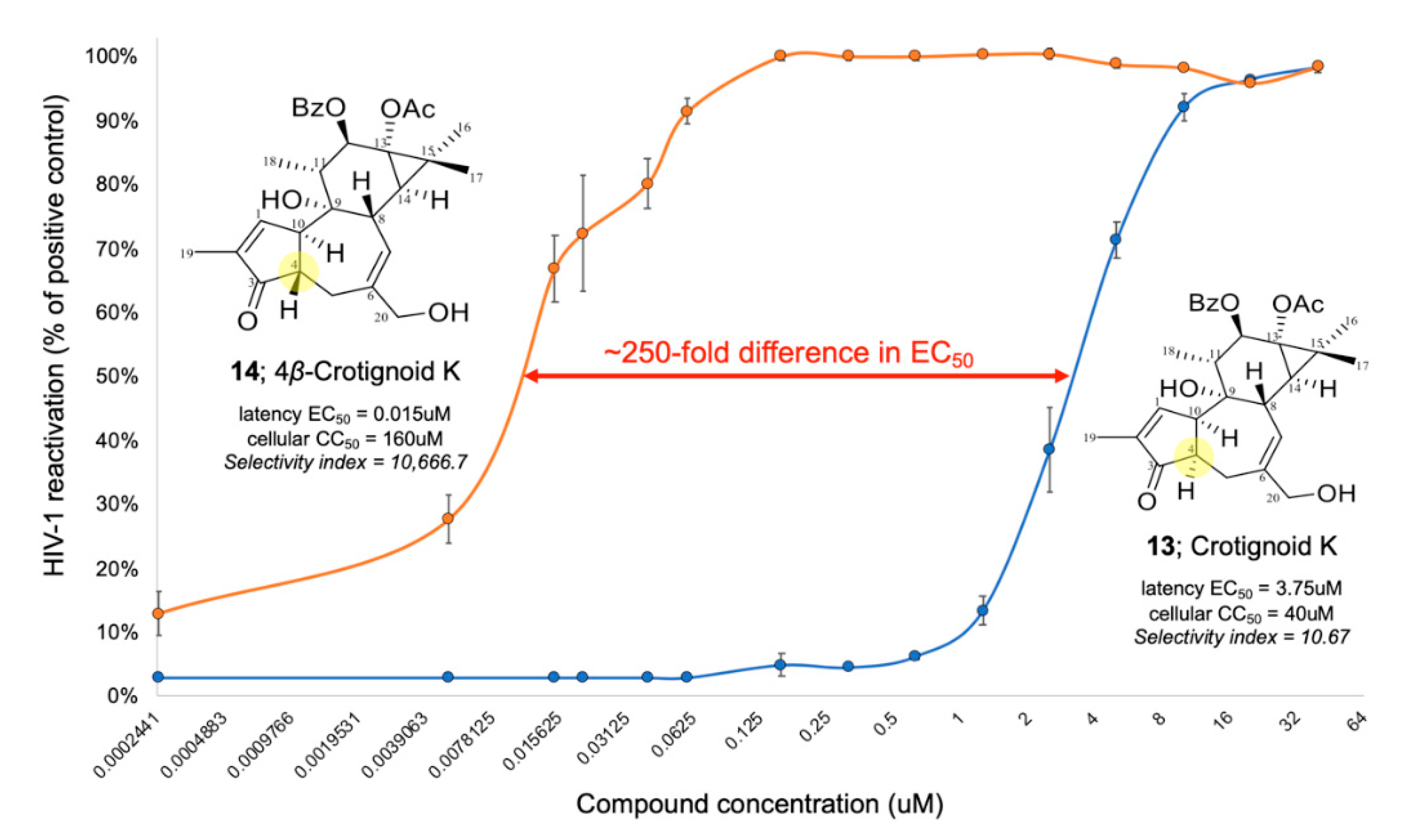
2.1.10. 4β-Crotignoid K (14)

Compound 14 was obtained as a white amorphous powder. The molecular formula was determined as C29H34O7 by HR-ESIMS m/z 495.2385 [M + H]+ (calcd. for C29H35O7 495.2377). The 1D (Table 4 and Table 5) and 2D NMR of 14 revealed one OAc [δH 2.14 (3H), δC 173.9, 21.3], one OBz [δH 8.02 (2H), 7.59, 7.47 (2H), δC 166.4, 133.4, 130.1, 129.9 (2C), 128.7 (2C)], four methyls [δH 1.21 (3H), δC 23.9, CH3-16; δH 1.33 (3H), δC 17.1, CH3-17; δH 0.98 (3H), δC 15.3, CH3-18; δH 1.73 (3H), δC 10.3, CH3-19], an oxygenated methylene [δH 4.05 (2H), δC 67.6, CH2-20], an oxygenated methine (δH 5.68, δC 77.8, CH-12), two unsaturated methines (δH 7.57, δC 159.7, CH-1; δH 5.56, δC 126.6, CH-7), and a ketone unit (δC 208.7, C-3). The interpretation of HMBC correlations suggested the skeleton of 14 was a tigliane-type diterpenoid [7] with an α-methyl-α,β-unsaturated cyclopentanone ring fused between C-4 and C-10, an OH (δH 5.62) connected to C-9, the OBz connected to C-12, the OAc connected to C-13 and a hydroxymethyl linked to C-6. Moreover, according to the NOESY correlations of H-4/H-8/H-11/H3-17 and H-12/H-14/9-OH/H3-18, as well as comparing the 1D NMR data of 14 with those of crotignoid K (13) [42] and 4-deoxyphorbol 12, 13-bis(isobutyrate) [43]. The structure of 14 was established as a 4β proton against the 4α proton of crotignoid K, thus named as 4β-crotignoid K.

2.1.11. Euphodendriane B (15)

Compound 15 was obtained as a white amorphous powder. The molecular formula was determined as C29H34O7 by HR-ESIMS m/z 495.2396 [M + H]+ (calcd. for C29H35O7 495.2377). The 1D (Table 4 and Table 5) and 2D NMR of 15 revealed one OAc [δH 2.11 (3H), δC 174.1, 21.2], one OBz [δH 8.06 (2H), 7.61, 7.49 (2H), δC 166.4, 133.4, 130.1, 129.9 (2C), 128.7 (2C)], five methyls [δH 1.20 (3H), δC 24.3, CH3-16; δH 1.33 (3H), δC 16.7, CH3-17; δH 1.16 (3H), δC 11.9, CH3-18; δH 1.83 (3H), δC 10.6, CH3-19; δH 1.90 (3H), δC 27.2, CH3-20], two oxygenated methines (δH 4.46, δC 71.1, CH-5; δH 5.73, δC 75.7, CH-12), two unsaturated methines (δH 7.06, δC 154.6, CH-1; δH 4.88 δC 125.5, CH-7), and a ketone unit (δC 207.5, C-3). The interpretation of HMBC correlations demonstrated that 15 was a tigliane-type diterpenoid with an α-methyl-α,β-unsaturated cyclopentanone ring fused between C-4 and C-10, with two OH (δH 5.92 and 5.95) connected to C-5 and C-9 respectively, one OBz connected to C-12, and one OAc connected to C-13 and 20-methyl group. The NMR data of 15 was highly close to those of euphodendriane A [35], except for the substitution at C-13 where an OiBu in euphodendriane A was replaced in 15 by the OAc. The relative configuration of 15 was deduced by inspection of the NOESY spectrum, showing the same orientations to euphodendriane A [35]. Thus, the structure of 15 was established and named as euphodendriane B.

2.1.12. 16-Nor-abieta-8,11,13-trien-3,7,15-trione (16)

Compound 16 was obtained as a colorless crystal with a molecular formula of C19H22O3 identified by HR-ESIMS m/z 299.1648 [M + H]+ (calcd. for C19H22O3 299.1642). The 1D (Table 4 and Table 5) and 2D NMR data of 16 revealed an acetyl moiety (δC 197.3, C-15; δH 2.64 (3H), δC 26.9, CH3-17), three methyls [δH 1.17 (3H), δC 25.2, CH3-18; δH 1.23 (3H), δC 21.7, CH3-19; δH 1.48 (3H), δC 22.8, CH3-20], three methylenes [δH 2.68, 2.05, δC 36.8, CH2-1; δH 2.91, 2.59, δC 34.6, CH2-2; δH 2.83, 2.75, δC 36.5, CH2-6], a methine (δH 2.36, δC 49.2, CH-5), a set of trisubstituted aromatic ring (δC 130.8, C-8; δC 158.2, C-9; δH 7.49 (d, J = 8.5), δC 125.2, CH-11; δH 8.17 (dd, J = 8.5, 2.5), δC 133.4, CH-12; δC 135.9, C-13; δH 8.57 (d, J = 2.5), δC 128.3, CH-14], two ketone units (δC 214.0, C-3; δC 197.4, C-7), and two quaternary carbons (δC 47.6, C-4; δC 38.3, C-10). The HMBC correlations of 16 from H2-1 to C-3, C-9, and C-20, H2-2 to C-3 and C-4, H-5 to C-1, C-4, C-9, and C-10, H2-6 to C-7, C-8, and C-10, H-12 to C-15, H-14 to C-7 and C-15, H3-17 to C-13, H3-18 and H3-19 to C-3, C-4, and C-5, and H3-20 to C-5 and C-10, suggested that 16 was an abietane-type diterpenoid [7] and was structurally similar to a known compound abieta-8,11,13-triene-3,7-dione [44,45], except for the substitution of the acetyl moiety at C-15–C-17. The relative configuration of 16 was the same as the typical abieta-8,11,13-triene diterpenoids [45] based on the NOESY correlations of H-5/H3-18 and H3-19/H3-20 as well as the comparison of the NMR data of 16 with those of literature [44,45]. The structure of 16 was identified as 16-nor-abieta-8,11,13-trien-3,7,15-trione.

2.1.13. 16-Nor-3β-hydroxy-abieta-8,11,13-trien-7,15-dione (17)

Compound 17 was obtained as a colorless crystal with a molecular formula of C19H24O3 identified by HR-ESIMS m/z 301.1803 [M + H]+ (calcd. for C19H25O3 301.1798). The inspection of 1D (Table 4 and Table 5) and 2D NMR data revealed that 17 was a 16-nor-abieta-8,11,13-triene diterpenoid [45]. The 1H and 13C NMR data of 17 was close to those of 16, except for a hydroxy group that was situated at C-3 (δC 78.0) by the confirmation of the 1H–1H COSY cross-peaks between H2-1/H2-2/H-3 as well as the HMBC correlations from H2-1 to C-3 and H3-18 to C-3. The H-3 [δH 3.37 (dd, J = 11.5, 4.0)] was identified as α-oriented by the NOESY correlations of H-3/H-5/H3-18 and the comparison of the proton signals for 17 with those of the similar compound 3β-hydroxy-abieta-8,11,13-trien-7-one [46]. The structure of 17 was identified as 16-nor-3β-hydroxy-abieta-8,11,13-trien-7,15-dione.

2.1.14. ent-8β,14β-Epoxyabieta-3-one-11,13(15)-dien-16,12-olide (19)

Compound 19 was purified as a colorless gum. The molecular formula was calculated as C20H24O4 by HR-ESIMS m/z 329.1753 [M + H]+ (calcd. for C20H25O4 329.1747). The 1D (Table 4 and Table 5) and 2D NMR data of 19 revealed four methyls [δH 2.09 (3H), δC 9.0, CH3-17; δH 1.17 (3H), δC 25.9, CH3-18; δH 1.09 (3H), δC 22.4, CH3-19; δH 0.95 (3H), δC 15.0, CH3-20], four methylenes [δH 2.05, 1.75, δC 38.1, CH2-1; δH 2.65, 2.37, δC 34.2, CH2-2; δH 1.79, 1.70, δC 21.7, CH2-6; δH 2.17, 1.68, δC 33.9, CH2-7], four methine (δH 1.67, δC 54.1, CH-5; δH 2.70, δC 50.1, CH-9; δH 5.44, δC 102.9, CH-11; δH 3.76, δC 54.5, CH-14), six quaternary carbons (δC 48.1, C-4; δC 61.0, C-8; δC 40.9, C-10; δC 148.1, C-12; δC 144.7, C-13; δC 126.2, C-15), and two carbonyl carbons (δC 215.0, C-3; δC 170.5, C-16). The structure of 19 was suggested an abietane-type diterpenoid with a ketone carbon at C-3, an epoxy ring fused at C-8 and C-14, a double bond at 11,12, and an α-methyl-α,β-unsaturated δ-lactone ring formed as D ring according to the analysis of the COSY cross-peaks of H2-1/H2-2, H2-5/H2-6/H2-7, and H-9/H-11, as well as the HMBC correlations from H2-1 to C-3, C-5, and C-10, H2-2 to C-3 and C-10, H-5 to C-4, C-18, C-19, and C-20, H2-7 to C-5 and C-8, H-9 to C-1, C-5, C-8, C-12, C-14, and C-20, H-11 to C-8, C-9, C-12, and C-13, H-14 to C-7, C-8, C-12, and C-13, H3-17 to C-13, C-15, and C-16, H3-18 and H3-19 to C-3 and C-4, together with H3-20 to C-1, C-9, and C-10. Comparison of the 13C NMR data with those of the related compounds jolkinolide A [47] and gelomulide C [48] further evidenced the presence of an ent-abietane skeleton with 8β,14β-epoxide in 19. Thus, 19 was established as ent-8β,14β-epoxyabieta-3-one-11,13(15)-dien-16,12-olide.

2.1.15. ent-8β,14β-Epoxyabieta-3α-hydroxy-13(15)-en-16,12-olide (20)

Compound 20 was purified as a colorless gum. The molecular formula was calculated as C20H28O4 by HR-ESIMS m/z 333.2067 [M + H]+ (calcd. for C20H29O4 333.2060). The 1H and 13C NMR data (Table 4 and Table 5) of 20 were close to those of 19 suggesting that the skeleton of 20 was an ent-abietane with 8β,14β-epoxide. Instead of the ketone carbon at C-3 in 19, a hydroxyl group was connected to C-3 (δC 78.6) based on the inspection of the 1H–1H COSY cross-peaks between H2-1/H2-2/H-3 together with the HMBC correlations from H-3 to C-1, C-4, C-18, and C-19. Saturated methylene (δH 2.27, 1.41, δC 24.0) and an oxygenated methine (δH 4.99, δC 75.6) were assigned to be at C-11 and C-12, respectively, according to the analysis of the 1H–1H COSY cross-peaks between H-9/H2-11/H-12 together with the HMBC correlations from H2-11 to C-8, C-10, and C-13, as well as H-12 to C-13 and C-15. The structure was found to be highly similar to the NMR features of gelomulide A [48], except for instead of the hydroxyl group at C-3 in 20. The configuration of 3-OH was deduced to be α-oriented as the proton signal of H-3 at 3.30 (dd, J = 12.0, 4.0 Hz) [49,50]. It was also supported by the NOESY correlations of H-3/H-5/H-9/H3-18. The structure of 20 was established as ent-8β,14β-epoxyabieta-3α-hydroxy-13(15)-en-16,12-olide.
The known compounds were identified as isoterracinolide A (10) [36], isoterracinolide B (11) [36], 12-O-benzoyl-13-acetoxy-4,20-dideoxyphorbol-4-ene (12) [51,52], crotignoid K (13) [42], 16-nor-abieta-8,11,13-trien-3,15-dione (18) [53], helioscopinolide E (21) [54], helioscopinolide C (22) [54], helioscopinolide A (23) [54], ent-kauran-16β-ol-3-one (24) [55], cleomiscosin A (25) [56], (+)-syringaresinol (26) [57], dimeric coniferyl acetate (27) [58], vanillin (28) [59], 4-hydroxybenzaldehyde (29) [60], coniferol alcohol (30) [61], and indole-3-carboxaldehyde (31) [62] by comparison of the NMR data with those of the literature.

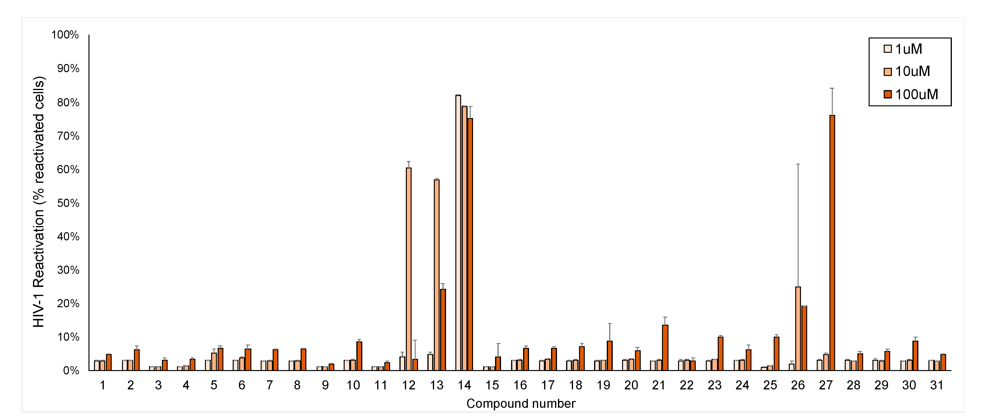
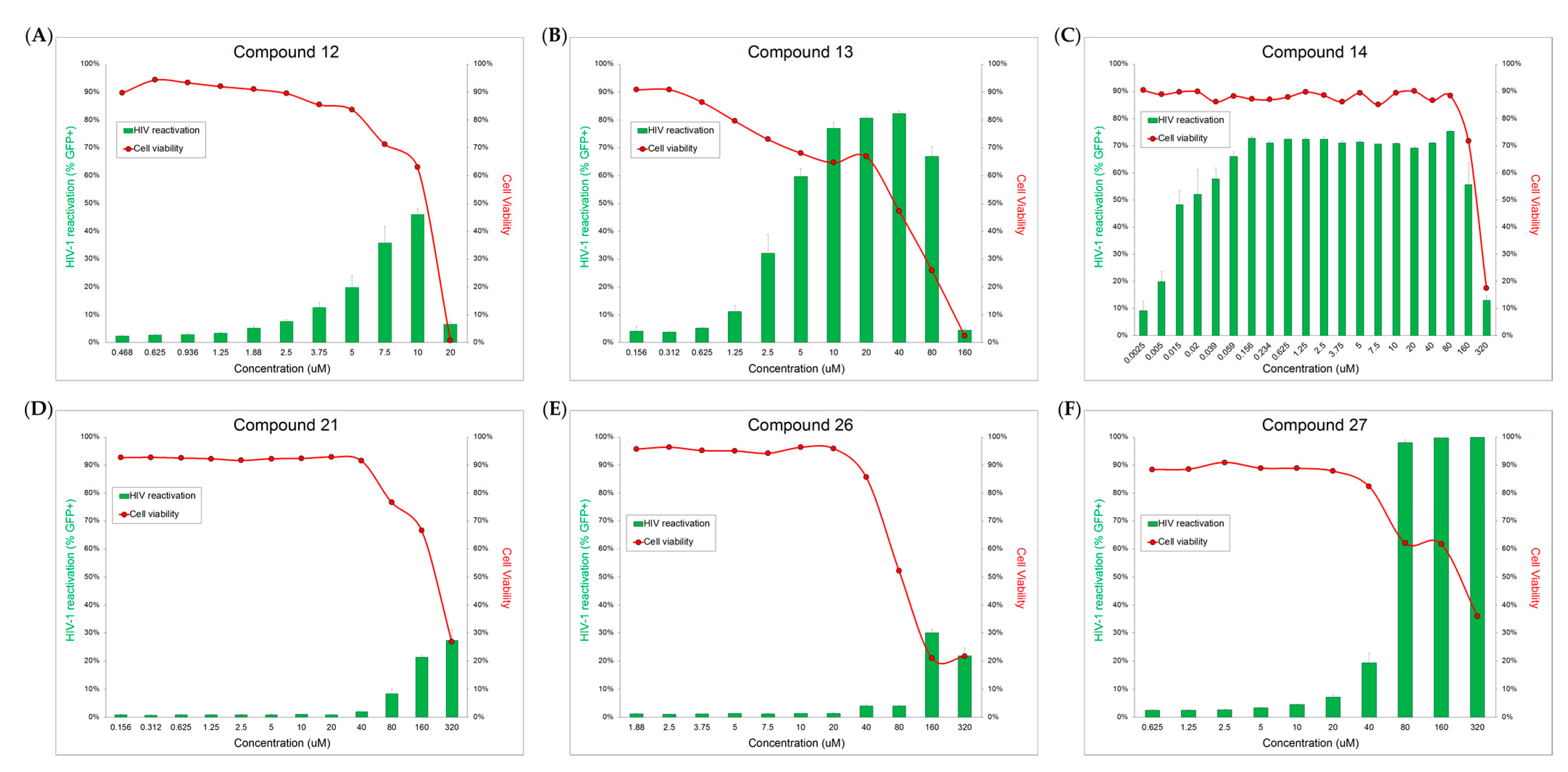
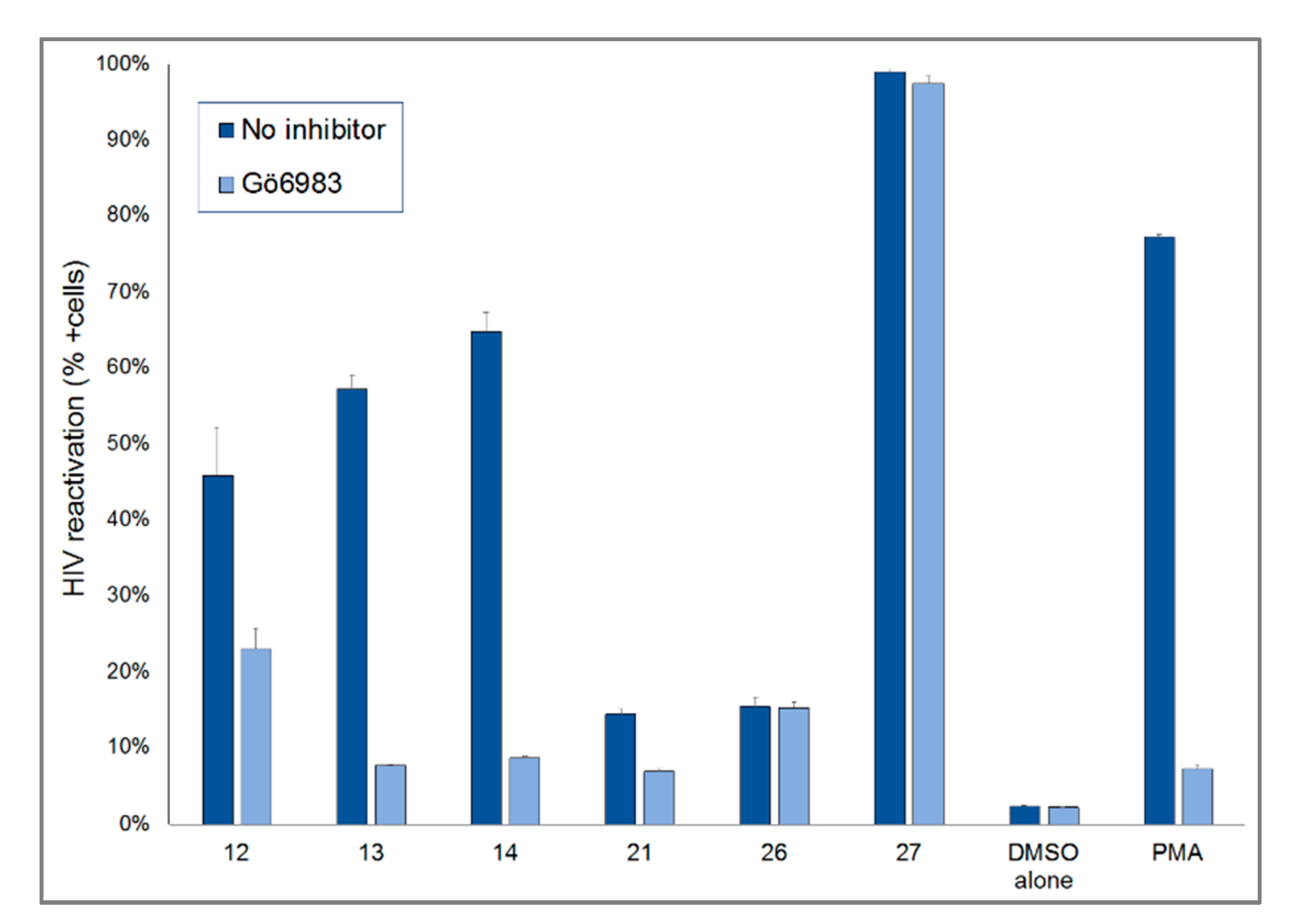
2.2. HIV-1 Latency Reversal Activity of Isolated Compounds in Vitro

Jurkat cells with a full-length integrated HIV-1 provirus that have been modified to contain a GFP coding region in place of the env gene (J-lat 10.6 cells) were used for HIV-1 anti-latency activity, cytotoxicity, and cellular activation testing. All 31 compounds were tested at 1, 10, and 100 μM. Through the GFP expression of J-lat 10.6 cells, it was determined that compounds 12, 13, 14, 21, 26, and 27 showed HIV-1 latency reversal activity (Figure 4). These compounds were further tested at additional concentrations to determine dose response and toxicity curves (Figure 5A–F). Cell viability for all isolated compounds is presented in Figure S106 in the supplementary material.
4β-Crotignoid K (14) showed high reactivation levels into nM concentrations, ~250-fold less than crotignoid K (13), which is a stereoisomer of 14, differing only in the configuration on C-4 (Figure 6). The striking difference between these compounds isolated from E. usambarica demonstrated a structure-activity relationship (SAR) of an important cellular trigger to induce HIV-1 proviral transcription. A similar SAR has recently been described between protein kinase C (PKC) agonists, 4-deoxyphorbol (4β-dPEA), phorbol myristate acetate (PMA), and prostratin [63].
Euphorbia species have been shown to be enriched for compounds capable of protein kinase C (PKC) activation in human cells [5,9,11,14,15]. In order to determine whether our active compounds were acting through PKC, we evaluated the latency reversal activity of each of these compounds in the presence and absence of a pan-PKC inhibitor, Gö6983 (Figure 7). Compounds 12, 13, 14, and 21 all showed reduced efficacy, indicating likely activation of PKC as their primary mechanism of action. In contrast, compounds 26 and 27 did not show a significant reduction in their activity when PKC was inhibited. In addition, these compounds did not increase CD69 expression (a hallmark of PKC activation), further suggesting an alternative (non-PKC) mechanism of latency reversal.

3. Discussion

In this study, 15 new diterpenoids, together with 16 known compounds, were isolated from the dichloromethane phase of methanolic extract of the medicinal plant E. usambarica. Compound 1 exhibited a 6/6/3-fused ring system with an α-methyl-α,β-unsaturated cyclopentanone moiety to construct a rare diterpenoid lactone, this skeleton was the second time discovered from nature [34]. The other compounds could be summarized in 4 types of diterpenoids, including jatrophanes (211), tiglianes (1215), abietanes (1623), and kaurane (24), alone with coumarinolignoid (25), lignan (26), coniferyl acetate (27), and benzenoids (2831). Especially, usambariphanes A (2) and B (3) displayed an unusual lactone ring constructed between C-17 and C-2 in the jatrophane structure, which is different from such lactone ring commonly constructed between C-17 and C-3 or between C-17 and C-5.
Furthermore, compounds 1214, 21, 26, and 27 showed significant HIV-1 latency reversal activity demonstrated by the GFP expression of J-lat 10.6 cells. 4β-Crotignoid K (14) showed the reactivation of HIV-1 latency at a very low concentration of EC50 about 0.015 μM and a higher CC50 concentration than 160 μM. The stereoisomer, crotignoid K (13), showed the EC50 and CC50 concentrations about 3.75 and 40 μM, respectively, indicating that 4β-crotignoid K (14) was provided with higher safety and efficacy. There is a 250-fold difference in EC50 and ~1000-fold difference in selectivity index (CC50/EC50) between these compounds. The structural difference between 13 and 14 is only in the relative configuration on C-4. However, they demonstrated dramatically different biological activity, indicating that the configuration on C-4 of tigliane-type diterpenoids is critical to HIV-1 latency reversal activity and likely reflects improved PKC activation. The primary mechanism of the active compounds 1214 and 21 for the HIV-1 latency reversal activity was activation of PKC.
Currently, LRAs are still under investigation and have not been approved by the US Food and Drug Administration (US-FDA). Therefore, the intensive study of LRAs is an important topic, especially to discover new candidates from natural sources. For example, a known LRA, ingenol, is isolated originally from Euphorbia peplus [64] and is a US-FDA-approved topical treatment for actinic keratosis (AK) [65], showed a significant effect in the reactivation of HIV-1 latency through the PKC pathway [65]. Both compound 14 and ingenol mebutate are Euphorbia diterpenoids and the potent PKC agonists. In addition, compound 14 presented the lower cytotoxicity, indicating 14 is a promising candidate for the development of an LRA.
In contrast, (+)-syringaresinol (26) and dimeric coniferyl acetate (27) did not increase CD69 expression, further suggesting a non-PKC mechanism of latency reversal that merits further exploration.

4. Materials and Methods

4.1. General Experimental Procedures

Optical rotation was performed on a Perkin-Elmer 341 polarimeter. 1D and 2D NMR spectra were recorded on a Bruker Avance DRX 500 spectrometer at 500 MHz (1H) and 125 MHz (13C). Chemical shifts were reported in parts per million (δ), and the coupling constants (J) were expressed in Hertz. The residual peaks of the deuterated solvents were taken as reference points. The NMR data were acquired and processed with MestReNova v12.0.0−20080 software. High-resolution MS spectra were acquired on an FTHRMS-Orbitrap (Thermo-Finnigan) mass spectrometer equipped with an ESI ion source in positive ionization mode. HPLC analyses were performed with a Shimadzu LC-10AS pump interface equipped with a Shimadzu SPD-10A UV–VIS detector (Shimadzu Inc., Kyoto, Japan) using Kinetex C18 column (5 μm, 100 Å, 250 × 4.6 mm), Kinetex Biphenyl column (5 μm, 100 Å, 250 × 4.6 mm), Kinetex XB-C18 column (5 μm, 100 Å, 250 × 10 mm), and/or Luna® Phenyl-Hexyl column (5 μm, 250 × 10 mm) (Phenomenex Inc., Torrance, CA, USA) using a mixture of acetonitrile–H2O or mixture of methanol–H2O as mobile phase. Rotational planar chromatography (RPC) was performed on self-coated silica plates (Kieselgel 60 GF254, 15 µm, Merck, Germany) using a Chromatotron apparatus (Harrison Research, Palo Alto, CA, USA). Silica gel (Kieselgel 60, 63–200 μm, Merck, Darmstadt, Germany), polyamide (MP Polyamide, 50−160 μm, MP Biomedicals, Irvine, CA, USA), and Sephadex LH-20 gel (Pharmacia Fine Chemicals AB, Uppsala, Sweden) were used for column chromatography (CC). Thin-layer chromatography (TLC) was carried out using silica gel (Kieselgel 60 F254, Merck) and RP-C18 (F254s, Merck) pre-coated plates, and the preparative TLC (pre-TLC) was performed on glass sheet silica gel pre-coated plates (20 × 20 cm, Kieselgel 60 F254, Merck). The compounds were detected with a developer (20% H2SO4 (v/v) with 5% vanillin (w/v) in ethanol) followed by heating (120 °C).

4.2. Plant Material

Euphorbia usambarica Pax. (Euphorbiaceae) was collected in Taita Taveta county, Kenya in 2019. Identification was performed by Peter Waweru Mwangi (Department of Medical Physiology, School of Medicine, University of Nairobi, Nairobi, Kenya). A voucher specimen (No. EU-001) has been deposited in the Herbarium of the Department of Pharmacognosy, University of Szeged, Szeged, Hungary.

4.3. Extraction and Isolation

The dried stem and root part (2.7 kg) were chopped and extracted with methanol (MeOH, 15 L) at room temperature. After removing the solvent, the crude methanolic extract (EU, 220.0 g) was dissolved in 50% MeOHaq and subjected to liquid–liquid partition to afford n-hexane (EU-H), dichloromethane (CH2Cl2, EU-C), ethyl acetate (EtOAc, EU-E), and water-soluble residue (EU-W) phases. The EU-C (25.7 g) was subjected to polyamide CC with MeOH–H2O mixture solvent system (40%, 60%, 80%, and 100% MeOHaq; EU-C-P1–P4). The EU-C-P1 (8.7 g) was further subjected to normal phase CC (silica gel, 63–200 μm) with a gradient solvent system of n-hexane–EtOAc–MeOH mixtures (from 40:5:1 to 0:8:1) to obtain ten subfractions (EU-C-P1-1–10) based on the TLC monitoring. EU-C-P1-2 (29.7 mg) was subjected to Sephadex LH-20 CC with the eluent of CH2Cl2–EtOAc–MeOH (1:1:6) to yield 6 subfractions (EU-C-P1-2/1–6), and EU-C-P1-2/2 was further separated by RP-HPLC on Kinetex XB-C18 column with an isocratic solvent system of MeCN–H2O (60:40, 2.0 mL/min) to yield compound 18 (1.1 mg). EU-C-P1-3 (895.5 mg) was separated by Sephadex LH-20 CC using CH2Cl2–EtOAc–MeOH (1:1:6) as eluent to obtain 5 subfractions (EU-C-P1-3/1–5). EU-C-P1-3/2 (245.5 mg) was further subjected to RPC (thickness 2 mm) using a gradient system of CH2Cl2–MeOH (from 100:0 to 15:1) to obtain 5 subfractions (EU-C-P1-3/2/1–5). Compound 21 (63.5 mg) was purified by recrystallization (MeOH) from EU-C-P1-3/2/1 (133.8 mg), and the residue of this fraction was further purified by RP-HPLC on Kinetex XB-C18 column with an isocratic system of MeCN–H2O (53:47, 2.0 mL/min) to yield compounds 16 (4.0 mg) and 19 (3.5 mg). EU-C-P1-3/2/2 (38.6 mg) was subjected to prep-TLC using CH2Cl2–MeOH (60:1) as the eluent to obtain 5 subfractions (EU-C-P1-3/2/2/1–5), then the second and third subfractions were purified by RP-HPLC on Kinetex XB-C18 column with an isocratic system of MeCN–H2O (65:35, 2.0 mL/min) to yield compounds 10 (4.7 mg) and 15 (1.0 mg), respectively. EU-C-P1-3/3 (178.5 mg) was subjected to RPC (thickness 2 mm) using a gradient system of n-hexane–CH2Cl2–MeOH (from 5:1:0 to 20:1) to obtain 9 subfractions (EU-C-P1-3/3/1–9), then the second subfraction (23.0 mg) was purified by RP-HPLC on Kinetex Biphenyl column with an isocratic system of MeOH–H2O (75:25, 1.0 mL/min) to yield compound 24 (2.7 mg). EU-C-P1-3/4 (39.3 mg) was separated by prep-TLC using CH2Cl2–MeOH (60:1) as eluent to yield compound 28 (11.0 mg). EU-C-P1-3/5 (10.2 mg) was purified by prep-TLC using CH2Cl2–MeOH (35:1) as eluent to yield compound 29 (1.8 mg). EU-C-P1-4 (1060.4 mg) was subjected to Sephadex LH-20 gel chromatography eluting with CH2Cl2–EtOAc–MeOH (1:1:6) to obtain 9 subfractions (EU-C-P1-4/1–9). EU-C-P1-4/2 (442.8 mg) was further separated by RPC (thickness 2 mm) using a gradient system of n-hexane–CH2Cl2–MeOH (from 1:1:0 to 10:1) to obtain 8 subfractions (EU-C-P1-4/2/1–9). EU-C-P1-4/2/2 (14.1 mg) was further purified by RP-HPLC on Kinetex XB-C18 column with an isocratic solvent system of MeCN–H2O (50:50, 2.0 mL/min) to yield compound 27 (4.6 mg). EU-C-P1-4/2/4 (68.6 mg) was further purified by RP-HPLC on Kinetex XB-C18 column with isocratic solvent system of MeCN–H2O (55:45, 2.0 mL/min) to yield compounds 1 (1.1 mg), 2 (4.0 mg), 3 (1.5 mg), 4 (2.0 mg), 5 (5.2 mg), 6 (2.5 mg), 7 (3.2 mg), 8 (1.4 mg), 9 (1.7 mg), 11 (3.7 mg), and 22 (2.2 mg). EU-C-P1-4/2/6 (60.4 mg) was purified by RP-HPLC on Kinetex XB-C18 column with an isocratic solvent system of MeCN–H2O (50:50, 2.0 mL/min) to yield compounds 12 (2.3 mg) and 13 (4.2 mg). EU-C-P1-4/2/7 (40.9 mg) was purified by RP-HPLC on Luna® Phenyl-Hexyl column with an isocratic system of MeCN–H2O (51:49, 2.0 mL/min) to yield compound 14 (1.4 mg). EU-C-P1-4/4 (218.3 mg) was further separated by RPC (thickness 2 mm) using a gradient system of CH2Cl2–MeOH (from 100:0 to 20:1) to obtain 6 subfractions (EU-C-P1-4/4/1–6). EU-C-P1-4/4/2 (52.4 mg) was further chromatographed by Sephadex LH-20 CC eluting with CH2Cl2–EtOAc–MeOH (1:1:6) to obtain 3 subfractions, then the second subfraction was purified by RP-HPLC on Kinetex XB-C18 column with an isocratic solvent system of MeCN–H2O (53:47, 2.0 mL/min) to yield compounds 17 (2.8 mg), 20 (2.7 mg), and 23 (14.2 mg). EU-C-P1-4/8 (11.6 mg) was purified by RP-HPLC on Kinetex XB-C18 column with an isocratic solvent system of MeCN–H2O (35:65, 2.0 mL/min) to yield compounds 30 (1.3 mg) and 31 (1.6 mg). EU-C-P1-7 (480.1 mg) was subjected to Sephadex LH-20 CC eluting with CH2Cl2–EtOAc–MeOH (1:1:6) to obtain 8 subfractions (EU-C-P1-7/1–8). EU-C-P1-7/6 was further purified by RP-HPLC on Kinetex XB-C18 column with an isocratic system of MeOH–H2O (48:52, 2.0 mL/min) to yield compound 26 (2.6 mg). Compound 25 (8.3 mg) was yielded by re-crystallization (MeOH) from EU-C-P1-7/7.

4.4. Physical Characteristic of New Compounds

Euphordraculoate C (1): Colorless gum; [ α ] D 28 −63 (c 0.05, CHCl3); the 1H and 13C NMR spectroscopic data, see Table 1 and Table 2; HR-ESIMS m/z 493.2237 [M + H]+ (calcd. for C29H33O7 493.2221), m/z 515.2046 [M + Na]+ (calcd. for C29H32O7Na 515.2040).
Usambaricinophane A (2): White amorphous powder; [ α ] D 28 −17 (c 0.20, CHCl3); the 1H and 13C NMR spectroscopic data, see Table 1 and Table 2; HR-ESIMS m/z 789.3327 [M + H]+ (calcd. for C40H53O16 789.3328), m/z 811.3169 [M + Na]+ (calcd. for C40H52O16Na 811.3148).
Usambaricinophane B (3): White amorphous powder; [ α ] D 28 −7 (c 0.07, CHCl3); the 1H and 13C NMR spectroscopic data, see Table 1 and Table 2; HR-ESIMS m/z 803.3488 [M + H]+ (calcd. for C41H55O16 803.3485), m/z 825.3333 [M + Na]+ (calcd. for C41H54O16Na 825.3304).
Usambaricinophane C (4): colorless crystal; [ α ] D 28 +34 (c 0.10, CHCl3); the 1H and 13C NMR spectroscopic data, see Table 1 and Table 2; HR-ESIMS m/z 789.3346 [M + H]+ (calcd. for C40H53O16 789.3328) m/z 811.3171 [M + Na]+ (calcd. for C40H52O16Na 811.3148).
Usambaricinophane D (5): Colorless crystal; [ α ] D 28 +54 (c 0.30, CHCl3); the 1H and 13C NMR spectroscopic data, see Table 1 and Table 2; HR-ESIMS m/z 787.3193 [M + H]+ (calcd. for C40H51O16 787.3172), m/z 809.3011 [M + Na]+ (calcd. for C40H50O16Na 809.2991).
Usambaricinophane E (6): Colorless crystal; [ α ] D 28 +54 (c 0.15, CHCl3); the 1H and 13C NMR spectroscopic data, see Table 1 and Table 2; HR-ESIMS m/z 801.3356 [M + H]+ (calcd. for C41H53O16 801.3328) m/z 823.3170 [M + Na]+ (calcd. for C41H52O16Na 823.3148).
Usambaricinophane F (7): Colorless crystal; [ α ] D 28 +15 (c 0.20, CHCl3); the 1H and 13C NMR spectroscopic data, see Table 3; HR-ESIMS m/z 761.3383 [M + H]+ (calcd. for C39H53O15 761.3379), m/z 783.3215 [M + Na]+ (calcd. for C39H52O15Na 783.3198).
Usambaricinophane G (8): Colorless crystal; [ α ] D 28 +15 (c 0.08, CHCl3); the 1H and 13C NMR spectroscopic data, see Table 3; HR-ESIMS m/z 764.3230 [M + H]+ (calcd. for C41H50O13N 764.3277), m/z 786.3098 [M + Na]+ (calcd. for C41H49O13NNa 786.3096).
Isoterracinolide C (9): White amorphous powder; [ α ] D 28 ‒2 (c 0.10, CHCl3); the 1H and 13C NMR spectroscopic data, see Table 3; HR-ESIMS m/z 775.31721 [M + H]+ (calcd. for C39H51O16 775.3172), m/z 797.3011 [M + Na]+ (calcd. for C39H50O16Na 797.2991).
4β-Crotignoid K (14): White amorphous powder; [ α ] D 28 +48 (c 0.05, CHCl3); the 1H and 13C NMR spectroscopic data, see Table 4 and Table 5; HR-ESIMS m/z 495.2385 [M + H]+ (calcd. for C29H35O7 495.2377), m/z 517.2202 [M + Na]+ (calcd. for C29H34O7Na 517.2197).
Euphodendriane B (15): White amorphous powder; [ α ] D 28 +12 (c 0.033, CHCl3); the 1H and 13C NMR spectroscopic data, see Table 4 and Table 5; HR-ESIMS m/z 495.2396 [M + H]+ (calcd. for C29H35O7 495.2377), m/z 517.2209 [M + Na]+ (calcd. for C29H34O7Na 517.2197).
16-Nor-abieta-8,11,13-trien-3,7,15-trione (16): Colorless crystal; [ α ] D 28 +15 (c 0.20, CHCl3); the 1H and 13C NMR spectroscopic data, see Table 4 and Table 5; HR-ESIMS m/z 299.1648 [M + H]+ (calcd. for C19H22O3 299.1642), m/z 321.1466 [M + Na]+ (calcd. for C19H22O3Na 321.1461).
16-Nor-3β-hydroxy-abieta-8,11,13-trien-7,15-dione (17): Colorless crystal; [ α ] D 28 −9 (c 0.20, CHCl3); the 1H and 13C NMR spectroscopic data, see Table 4 and Table 5; HR-ESIMS m/z 301.1803 [M + H]+ (calcd. for C19H25O3 301.1798), m/z 323.1623 [M + Na]+ (calcd. for C19H24O3Na 323.1618).
ent-8β,14β-Epoxyabieta-3-one-11,13(15)-dien-16,12-olide (19): Colorless gum; [ α ] D 28 +90 (c 0.20, CHCl3); the 1H and 13C NMR spectroscopic data, see Table 4 and Table 5; HR-ESIMS m/z 329.1753 [M + H]+ (calcd. for C20H25O4 329.1747), m/z 351.1572 [M + Na]+ (calcd. for C20H24O4Na 351.1567).
ent-8β,14β-Epoxyabieta-3α-hydroxy-13(15)-en-16,12-olide (20): Colorless gum; [ α ] D 28 +57 (c 0.20, CHCl3); the 1H and 13C NMR spectroscopic data, see Table 4 and Table 5; HR-ESIMS m/z 333.2067 [M + H]+ (calcd. for C20H29O4 333.2060), m/z 355.1886 [M + Na]+ (calcd. for C20H28O4Na 355.1880).

4.5. Cell Isolation and Culture

The HIV-1-infected Jurkat T cell line (J-Lat 10.6) was obtained in January 2019 from the NIH/ATCC HIV-1 Reagent Program (www.hivreagentprogram.org) and cultured in RPMI-based media supplemented with 10% fetal calf serum, 1% penicillin, and 1% streptomycin.

4.6. Flow Cytometry

After in vitro culture, J-lat cells were washed with phosphate-buffered saline (1× PBS) prior to staining with 0.1 µL fixable viability dye Live/Dead Aqua (Cat L34957, www.thermofisher.com) per 105 cells for 30 min at 4 °C. Simultaneously, J-lat cells were also stained with antibodies against CD69 conjugated to an APC fluorophore (APC anti-human CD69 antibody, biolegend.com). Cells were then washed and re-suspended in 1× PBS prior to flow cytometry acquisition evaluating for cellular viability, green fluorescent protein (GFP) expression, and CD69 expression. Flow cytometry was performed with a BD FACSCelesta or FACSCanoto flow cytometer with FACSDiva acquisition software (Becton Dickinson, Mountain View, CA) prior to analysis with FlowJo (TreeStar Inc., Ashland, OR, USA).

4.7. Compound Screening

Isolated compounds were resuspended in dimethyl sulfoxide (DMSO; Sigma-Aldrich, Burlington, USA), at a concentration of 10 mM, and diluted with PBS, and tested with J-lat 10.6 cells at concentrations of 100, 10, and 1 μM. J-lat 10.6 cells were tested with compounds at a concentration of 2.5 × 10⁵ cells/mL. We performed a minimum of three replicates of each condition for all experiments. Negative controls contained 1% DMSO to account for any effect of DMSO in the highest dilution of compounds. Additional dilutions were tested for those compounds that showed reactivation through increased GFP production in J-lat 10.6 cells.

4.8. Statistical Analysis

Statistical significance was analyzed using software from GraphPad Prism Version 7.0c (GraphPad Software, San Diego, CA, USA). The mean values and standard deviations for all replicate J-lat results were calculated and used to create Figure 4, Figure 5, Figure 6 and Figure 7. Where applicable, Students t-test was used to determine statistical significance of experimental mean results relative to negative controls.

5. Conclusions

In this study, 4β-crotignoid K (14) revealed a very higher effect improvement compared to crotignoid K (13), indicating that configuration at the C-4 of tigliane diterpenoids is critical to HIV-1 latency reversal activity. (+)-Syringaresinol (26) and dimeric coniferyl acetate (27) showed no exhibition of CD69 expression, suggesting a non-PKC mechanism of latency reversal. Our results provide insights into the stereochemistry importance of bioactive diterpenoids and suggest that isolated compounds from E. usambarica can further research and development into therapeutic strategies for HIV-1 management, particularly as reactivators of latent HIV-1.

Supplementary Materials

The following are available online at https://0-www-mdpi-com.brum.beds.ac.uk/article/10.3390/ph14070653/s1, Figures S1–S105: 1D-NMR, 2D-NMR, and HR-ESIMS spectra of compounds 19, 1416, 19, and 20. Figure S106: 24 h cell viability after exposure to the compounds 131.

Author Contributions

Conceptualization, Y.-C.T., A.M.S., and J.H.; methodology, A.M.S., and J.H.; software, Y.-C.T., N.K., R.A.N., J.E.B., and A.M.S.; validation, Y.-C.T., A.M.S., and J.H.; formal analysis, Y.-C.T., N.K., R.A.N., J.E.B., A.M.S., and J.H.; investigation, Y.-C.T., N.K., R.A.N., J.E.B., R.B., D.R., A.V., A.M.S., and J.H.; resources, P.W.M. and J.H.; data curation, Y.-C.T., and A.M.S.; writing—original draft preparation, Y.-C.T.; writing—review and editing, A.M.S. and J.H.; visualization, Y.-C.T., R.A.N., J.E.B., and A.M.S.; supervision, A.M.S. and J.H.; project administration, J.H.; funding acquisition, J.H. All authors have read and agreed to the published version of the manuscript.

Funding

This research was funded by the Economic Development and Innovation Operative Programme (GINOP-2.3.2-15-2016-00012) and the Ministry of Human Capacities, Hungary Grant (TUDFO/47138-1/2019-ITM FIKP). This work was supported by the National Research, Development and Innovation Fund (NKFI) under grant numbers K135845 and by ÚNKP-20-4 grant for co-author N. Kúsz.

Institutional Review Board Statement

The study was conducted according to the guidelines of the Declaration of Helsinki, and approved in 21 March 2021 by the Institutional Review Board of the University of Utah (IRB_0067637).

Informed Consent Statement

Informed consent was obtained from all subjects involved in the study.

Data Availability Statement

Data sharing not applicable.

Acknowledgments

T The following reagent was obtained through the NIH HIV Reagent Program, Division of AIDS, NIAID, NIH: J-Lat Full-Length Cells (10.6), ARP-9849, contributed by Eric Verdin. The authors appreciate the editorial assistance and comments by the editor and reviewers.

Conflicts of Interest

The authors declare no conflict of interest.

References

  1. Spivak, A.M.; Planelles, V. HIV-1 eradication: Early trials (and tribulations). Trends Mol. Med. 2016, 22, 10–27. [Google Scholar] [CrossRef] [PubMed] [Green Version]
  2. Andersen, R.J.; Ntie-Kang, F.; Tietjen, I. Natural product-derived compounds in HIV suppression, remission, and eradication strategies. Antivir. Res. 2018, 158, 63–77. [Google Scholar] [CrossRef] [PubMed]
  3. Cary, D.C.; Peterlin, B.M. Natural products and HIV/AIDS. AIDS Res. Hum. Retrovir. 2018, 34, 31–38. [Google Scholar] [CrossRef] [PubMed]
  4. Salehi, B.; Kumar, N.V.A.; Sener, B.; Sharifi-Rad, M.; Kilic, M.; Mahady, G.B.; Vlaisavljevic, S.; Iriti, M.; Kobarfard, F.; Setzer, W.N.; et al. Medicinal plants used in the treatment of human immunodeficiency virus. Int. J. Mol. Sci. 2018, 19, 1459. [Google Scholar] [CrossRef] [PubMed] [Green Version]
  5. Mwine, J.T.; Van Damme, P. Why do Euphorbiaceae tick as medicinal plants? A review of Euphorbiaceae family and its medicinal features. J. Med. Plants Res. 2011, 5, 652–662. [Google Scholar]
  6. Shi, Q.W.; Su, X.H.; Kiyota, H. Chemical and pharmacological research of the plants in genus Euphorbia. Chem. Rev. 2008, 108, 4295–4327. [Google Scholar] [CrossRef]
  7. Vasas, A.; Hohmann, J. Euphorbia diterpenes: Isolation, structure, biological activity, and synthesis (2008–2012). Chem. Rev. 2014, 114, 8579–8612. [Google Scholar] [CrossRef] [Green Version]
  8. Patil, S.B.; Naikwade, N.S.; Magdum, C.S. Review on phytochemistry and pharmacological aspects of Euphorbia hirta Linn. Asian J. Pharm. Res. Health Care 2009, 1, 113–133. [Google Scholar]
  9. Kumar, S.; Malhotra, R.; Kumar, D. Euphorbia hirta: Its chemistry, traditional and medicinal uses, and pharmacological activities. Pharmacogn. Rev. 2010, 4, 58–61. [Google Scholar] [CrossRef] [PubMed] [Green Version]
  10. Özbilgin, S.; Saltan Çitoğlu, G. Uses of some Euphorbia species in traditional medicine in turkey and their biological activities. Turk. J. Pharm. Sci. 2012, 9, 241–255. [Google Scholar]
  11. Lee, J.W.; Jin, Q.; Jang, H.; Lee, D.; Han, S.B.; Kim, Y.; Hong, J.T.; Lee, M.K.; Hwang, B.Y. Jatrophane and ingenane-type diterpenoids from Euphorbia kansui inhibit the LPS-induced NO production in RAW 264.7 cells. Bioorg. Med. Chem. Lett. 2016, 26, 3351–3354. [Google Scholar] [CrossRef] [PubMed]
  12. Redei, D.; Forgo, P.; Molnar, J.; Szabo, P.; Zorig, T.; Hohmann, J. Jatrophane diterpenoids with multidrug resistance-modulating activity from Euphorbia mongolica Prokh. Tetrahedron 2012, 68, 8403–8407. [Google Scholar] [CrossRef]
  13. Corea, G.; Di Pietro, A.; Dumontet, C.; Fattorusso, E.; Lanzotti, V. Jatrophane diterpenes from Euphorbia spp. as modulators of multidrug resistance in cancer therapy. Phytochem. Rev. 2009, 8, 431–447. [Google Scholar] [CrossRef]
  14. Nothias-Scaglia, L.F.; Retailleau, P.; Paolini, J.; Pannecouque, C.; Neyts, J.; Dumontet, V.; Roussi, F.; Leyssen, P.; Costa, J.; Litaudon, M. Jatrophane diterpenes as inhibitors of chikungunya virus replication: Structure-activity relationship and discovery of a potent lead. J. Nat. Prod. 2014, 77, 1505–1512. [Google Scholar] [CrossRef] [PubMed]
  15. Remy, S.; Litaudon, M. Macrocyclic diterpenoids from Euphorbiaceae as a source of potent and selective inhibitors of chikungunya virus replication. Molecules 2019, 24, 2336. [Google Scholar] [CrossRef] [Green Version]
  16. Islam, M.T. Diterpenes and their derivatives as potential anticancer agents. Phytother. Res. 2017, 31, 691–712. [Google Scholar] [CrossRef] [PubMed]
  17. Fattahian, M.; Ghanadian, M.; Ali, Z.; Khan, I.A. Jatrophane and rearranged jatrophane-type diterpenes: Biogenesis, structure, isolation, biological activity and SARs (1984–2019). Phytochem. Rev. 2020, 19, 265–336. [Google Scholar] [CrossRef]
  18. Kusz, N.; Orvos, P.; Bereczki, L.; Fertey, P.; Bombicz, P.; Csorba, A.; Talosi, L.; Jakab, G.; Hohmann, J.; Redei, D. Diterpenoids from Euphorbia dulcis with potassium ion channel inhibitory activity with selective G protein-activated inwardly rectifying ion channel (GIRK) blocking effect. J. Nat. Prod. 2018, 81, 2483–2492. [Google Scholar] [CrossRef]
  19. Rawal, M.K.; Shokoohinia, Y.; Chianese, G.; Zolfaghari, B.; Appendino, G.; Taglialatela-Scafati, O.; Prasad, R.; Di Pietro, A. Jatrophanes from Euphorbia squamosa as potent inhibitors of Candida albicans multidrug transporters. J. Nat. Prod. 2014, 77, 2700–2706. [Google Scholar] [CrossRef] [PubMed]
  20. Tsai, J.Y.; Redei, D.; Forgo, P.; Li, Y.; Vasas, A.; Hohmann, J.; Wu, C.C. Isolation of phorbol esters from Euphorbia grandicornis and evaluation of protein kinase c and human platelet-activating effects of Euphorbiaceae. Diterpenes. J. Nat. Prod. 2016, 79, 2658–2666. [Google Scholar] [CrossRef]
  21. Park, K.H.; Koh, D.; Lee, S.; Jung, I.; Kim, K.H.; Lee, C.H.; Kim, K.H.; Lim, Y. Anti-allergic and anti-asthmatic activity of helioscopinin-A, a polyphenol compound, isolated from Euphorbia helioscopia. J. Microbiol. Biotechnol. 2001, 11, 138–142. [Google Scholar]
  22. Islam, N.U.; Khan, I.; Rauf, A.; Muhammad, N.; Shahid, M.; Shah, M.R. Antinociceptive, muscle relaxant and sedative activities of gold nanoparticles generated by methanolic extract of Euphorbia milii. BMC Complementary Altern. Med. 2015, 15, 160. [Google Scholar] [CrossRef] [PubMed] [Green Version]
  23. Spivak, A.M.; Planelles, V. Novel latency reversal agents for HIV-1 cure. Annu. Rev. Med. 2018, 69, 421–436. [Google Scholar] [CrossRef] [PubMed] [Green Version]
  24. Wonderlich, E.R.; Subramanian, K.; Cox, B.; Wiegand, A.; Lackman-Smith, C.; Bale, M.J.; Stone, M.; Hoh, R.; Kearney, M.F.; Maldarelli, F.; et al. Effector memory differentiation increases detection of replication-competent HIV-l in resting CD4+ T cells from virally suppressed individuals. PLoS Pathog. 2019, 15, e1008074. [Google Scholar] [CrossRef] [PubMed]
  25. Cary, D.C.; Fujinaga, K.; Peterlin, B.M. Euphorbia kansui reactivates latent HIV. PLoS ONE 2016, 11, e0168027. [Google Scholar] [CrossRef] [PubMed] [Green Version]
  26. Pisano, M.B.; Cosentino, S.; Viale, S.; Spano, D.; Corona, A.; Esposito, F.; Tramontano, E.; Montoro, P.; Tuberoso, C.I.; Medda, R.; et al. Biological activities of aerial parts extracts of Euphorbia characias. Biomed. Res. Int. 2016, 2016, 1538703. [Google Scholar] [CrossRef] [PubMed] [Green Version]
  27. Rasmussen, T.A.; Tolstrup, M.; Sogaard, O.S. Reversal of latency as part of a cure for HIV-1. Trends Microbiol. 2016, 24, 90–97. [Google Scholar] [CrossRef] [PubMed]
  28. Liu, Q.; Li, W.; Huang, L.; Asada, Y.; Morris-Natschke, S.L.; Chen, C.H.; Lee, K.H.; Koike, K. Identification, structural modification, and dichotomous effects on human immunodeficiency virus type 1 (HIV-1) replication of ingenane esters from Euphorbia kansui. Eur. J. Med. Chem. 2018, 156, 618–627. [Google Scholar] [CrossRef] [PubMed]
  29. Yan, S.L.; Li, Y.H.; Chen, X.Q.; Liu, D.; Chen, C.H.; Li, R.T. Diterpenes from the stem bark of Euphorbia neriifolia and their in vitro anti-HIV activity. Phytochemistry 2018, 145, 40–47. [Google Scholar] [CrossRef]
  30. Valadão, A.L.C.; Pezzuto, P.; Silva, V.A.O.; Gonçalves, B.S.; Rossi, Á.D.; Cunha, R.D.; Siani, A.C.; Tostes, J.B.F.; Trovó, M.; Damasco, P.; et al. Reactivation of latent HIV-1 in vitro using an ethanolic extract from Euphorbia umbellata (Euphorbiaceae) latex. PLoS ONE 2018, 13, e0207664. [Google Scholar] [CrossRef] [Green Version]
  31. Thiselton-Dyer W.T. Euphorbia usambarica Pax. In Flora of Tropical Africa; L. Reeve & Co. Ltd.: London, UK, 1913; Volume 6, pp. 538–539. [Google Scholar]
  32. Schlage, C.; Mabula, C.; Mahunnah, R.L.A.; Heinrich, M. Medicinal plants of the Washambaa (Tanzania): Documentation and ethnopharmacological evaluation. Plant Biol. 2000, 2, 83–92. [Google Scholar] [CrossRef]
  33. Chrispin, F.S.; Innocent, J.E.Z.; Patrick, L.P.M.; Matti, N. Use of medicinal plants in the eastern arc mountains with special reference to the hehe ethnic group in the Udzungwa mountains, Tanzania. J. East Afr. Nat. Hist. 2008, 97, 225–254. [Google Scholar]
  34. Wang, L.; Yang, J.; Kong, L.M.; Deng, J.; Xiong, Z.J.; Huang, J.P.; Luo, J.P.; Yan, Y.J.; Hu, Y.K.; Li, X.N.; et al. Natural and semisynthetic tigliane diterpenoids with new carbon skeletons from Euphorbia dracunculoides as a Wnt signaling pathway inhibitor. Org. Lett. 2017, 19, 3911–3914. [Google Scholar] [CrossRef]
  35. Aljancic, I.S.; Pesic, M.; Milosavljevic, S.M.; Todorovic, N.M.; Jadranin, M.; Miosavljevic, G.; Povrenovic, D.; Bankovic, J.; Tanic, N.; Markovic, I.D.; et al. Isolation and biological evaluation of jatrophane diterpenoids from Euphorbia dendroides. J. Nat. Prod. 2011, 74, 1613–1620. [Google Scholar] [CrossRef]
  36. Marco, J.A.; Sanz-Cervera, J.F.; Yuste, A.; Jakupovic, J. Isoterracinolides A and B, novel bishomoditerpene lactones from Euphorbia terracina. J. Nat. Prod. 1999, 62, 110–113. [Google Scholar] [CrossRef]
  37. Huang, Y.; Aisa, H.A. Three new diterpenoids from Euphorbia sororia L. Helv. Chim. Acta 2010, 93, 1156–1161. [Google Scholar] [CrossRef]
  38. Hu, R.; Gao, J.; Rozimamat, R.; Aisa, H.A. Jatrophane diterpenoids from Euphorbia sororia as potent modulators against P-glycoprotein-based multidrug resistance. Eur. J. Med. Chem. 2018, 146, 157–170. [Google Scholar] [CrossRef] [PubMed]
  39. Corea, G.; Fattorusso, E.; Lanzotti, V.; Taglialatela-Scafati, O.; Appendino, G.; Ballero, M.; Sirnon, P.N.L.; Dumontet, C.; Di Pietro, A. Modified jatrophane diterpenes as modulators of multidrug resistance from Euphorbia dendroides L. Bioorgan. Med. Chem. 2003, 11, 5221–5227. [Google Scholar] [CrossRef] [PubMed]
  40. Lu, D.L.; Liu, Y.Q.; Aisa, H.A. Jatrophane diterpenoid esters from Euphorbia sororia serving as multidrug resistance reversal agents. Fitoterapia 2014, 92, 244–251. [Google Scholar] [CrossRef] [PubMed]
  41. Marco, J.A.; Sanz-Cervera, J.F.; Yuste, A.; Jakupovic, J.; Jeske, F. Jatrophane derivatives and a rearranged jatrophane from Euphorbia terracina. Phytochemistry 1998, 47, 1621–1630. [Google Scholar] [CrossRef]
  42. Zhang, D.D.; Zhou, B.; Yu, J.H.; Xu, C.H.; Ding, J.; Zhang, H.; Yue, J.M. Cytotoxic tigliane-type diterpenoids from Croton tiglium. Tetrahedron 2015, 71, 9638–9644. [Google Scholar] [CrossRef]
  43. Marco, J.A.; Sanz-Cervera, J.F.; Checa, J.; Palomares, E.; Fraga, B.M. Jatrophane and tigliane diterpenes from the latex of Euphorbia obtusifolia. Phytochemistry 1999, 52, 479–485. [Google Scholar] [CrossRef]
  44. Ara, I.; Siddiqui, B.S.; Faizi, S.; Siddiqui, S. Tricyclic diterpenoids from root bark of Azadirachta Indica. Phytochemistry 1990, 29, 911–914. [Google Scholar] [CrossRef]
  45. Corral, J.M.M.D.; Gordaliza, M.; Salinero, M.A.; Feliciano, A.S. 13C NMR data for abieta-8,11,13-triene diterpenoids. Magn. Reson. Chem. 1994, 32, 774–781. [Google Scholar] [CrossRef]
  46. Seca, A.M.L.; Silva, A.M.S.; Bazzocchi, I.L.; Jimenez, I.A. Diterpene constituents of leaves from Juniperus brevifolia. Phytochemistry 2008, 69, 498–505. [Google Scholar] [CrossRef]
  47. Lal, A.R.; Cambie, R.C.; Rutledge, P.S.; Woodgate, P.D. Ent-pimarane and ent-abietane diterpenes from Euphorbia fidjiana. Phytochemistry 1990, 29, 2239–2246. [Google Scholar] [CrossRef]
  48. Talapatra, S.K.; Das, G.; Talapatra, B. Stereostructures and molecular conformations of six diterpene lactones from Gelonium multiflorum. Phytochemistry 1989, 28, 1181–1185. [Google Scholar] [CrossRef]
  49. Wang, H.; Zhang, X.F.; Cai, X.H.; Ma, Y.B.; Luo, X.D. Three new diterpenoids from Euphorbia wallichii. Chin. J. Chem. 2004, 22, 199–202. [Google Scholar] [CrossRef]
  50. Choudhary, M.I.; Gondal, H.Y.; Abbaskhan, A.; Jahan, I.A.; Parvez, M.; Nahar, N.; Rahman, A. Revisiting diterpene lactones of Suregada multiflora. Tetrahedron 2004, 60, 7933–7941. [Google Scholar] [CrossRef]
  51. Song, Q.Q.; Rao, Y.; Tang, G.H.; Sun, Z.H.; Zhang, J.S.; Huang, Z.S.; Yin, S. Tigliane diterpenoids as a new type of antiadipogenic agents inhibit GR alpha-Dexras1 axis in adipocytes. J. Med. Chem. 2019, 62, 2060–2075. [Google Scholar] [CrossRef]
  52. Yin, S.; Huang, Z.; Rao, Y.; Tang, G.; Song, Q. Tigliane-type diterpenoid compounds and preparation method and application thereof. China Patent CN108689851A, 28 May 2018. [Google Scholar]
  53. Guo, K.; Liu, Y.C.; Liu, Y.; Luo, S.H.; Li, W.Y.; Li, X.N.; Li, S.H. Diversified abietane family diterpenoids from the leaves of Leucosceptrurn canum and their cytotoxic activity. Phytochemistry 2019, 157, 43–52. [Google Scholar] [CrossRef]
  54. Borghi, D.; Baumer, L.; Ballabio, M.; Arlandini, E.; Perellino, N.C.; Minghetti, A.; Vincieri, F.F. Structure elucidation of helioscopinolides D and E from Euphorbia calyptrata cell cultures. J. Nat. Prod. 1991, 54, 1503–1508. [Google Scholar] [CrossRef]
  55. Tori, M.; Arbiyanti, H.; Taira, Z.; Asakawa, Y. Terpenoids of the liverwort Frullanoides densifolia and Trocholejeunea sandvicensis. Phytochemistry 1993, 32, 335–348. [Google Scholar] [CrossRef]
  56. Arisawa, M.; Handa, S.S.; McPherson, D.D.; Lankin, D.C.; Cordell, G.A.; Fong, H.H.S.; Farnsworth, N.R. Plant anticancer agents XXIX. Cleomiscosin A from Simaba multiflora, Soulamea soulameoides, and Matayba arborescens. J. Nat. Prod. 1984, 47, 300–307. [Google Scholar] [CrossRef]
  57. Min, Y.D.; Choi, S.U.; Lee, K.R. Aporphine alkaloids and their reversal activity of multidrug resistance (MDR) from the stems and rhizomes of Sinomenium acutum. Arch. Pharm. Res. 2006, 29, 627–632. [Google Scholar] [CrossRef] [PubMed]
  58. Valcic, S.; Montenegro, G.; Timmermann, B.N. Lignans from Chilean propolis. J. Nat. Prod. 1998, 61, 771–775. [Google Scholar] [CrossRef] [PubMed]
  59. Bao, K.; Fan, A.X.; Dai, Y.; Zhang, L.; Zhang, W.G.; Cheng, M.S.; Yao, X.S. Selective demethylation and debenzylation of aryl ethers by magnesium iodide under solvent-free conditions and its application to the total synthesis of natural products. Org. Biomol. Chem. 2009, 7, 5084–5090. [Google Scholar] [CrossRef]
  60. Kim, H.; Ralph, J.; Lu, F.C.; Ralph, S.A.; Boudet, A.M.; MacKay, J.J.; Sederoff, R.R.; Ito, T.; Kawai, S.; Ohashi, H.; et al. NMR analysis of lignins in CAD-deficient plants. Part 1. Incorporation of hydroxycinnamaldehydes and hydroxybenzaldehydes into lignins. Org. Biomol. Chem. 2003, 1, 268–281. [Google Scholar] [CrossRef]
  61. Yao, C.S.; Lin, M.; Wang, L. Isolation and biomimetic synthesis of anti-inflammatory stilbenolignans from Gnetum cleistostachyum. Chem. Pharm. Bull. 2006, 54, 1053–1057. [Google Scholar] [CrossRef] [Green Version]
  62. Duarte, N.; Ferreira, M.J.U. Lagaspholones A and B: Two new jatropholane-type diterpenes from Euphorbia lagascae. Org. Lett. 2007, 9, 489–492. [Google Scholar] [CrossRef] [PubMed]
  63. De la Torre-Tarazona, H.E.; Jimenez, R.; Bueno, P.; Camarero, S.; Roman, L.; Fernandez-Garcia, J.L.; Beltran, M.; Nothias, L.F.; Cachet, X.; Paolini, J.; et al. 4-Deoxyphorbol inhibits HIV-1 infection in synergism with antiretroviral drugs and reactivates viral reservoirs through PKC/MEK activation synergizing with vorinostat. Biochem. Pharmacol. 2020, 177, 113937. [Google Scholar] [CrossRef]
  64. Hammadi, R.; Kúsz, N.; Dávid, C.Z.; Behány, Z.; Papp, L.; Kemény, L.; Hohmann, J.; Lakatos, L.; Vasas, A. Ingol and ingenol-type diterpenes from Euphorbia trigona Miller with keratinocyte inhibitory activity. Plants 2021, 10, 1206. [Google Scholar] [CrossRef]
  65. Jiang, G.; Maverakis, E.; Cheng, M.Y.; Elsheikh, M.M.; Deleage, C.; Mendez-Lagares, G.; Shimoda, M.; Yukl, S.A.; Hartigan-O’Connor, D.J.; Thompson, G.R.; et al. Disruption of latent HIV in vivo during the clearance of actinic keratosis by ingenol mebutate. JCI Insight 2019, 4, e126027. [Google Scholar] [CrossRef] [Green Version]
Figure 1. HIV-1 latency reversal activity of methanolic crude extract (EU), partitioned n-hexane (EU-H), dichloromethane (EU-C), ethyl acetate (EU-E), and water-soluble residue (EU-W) phases. (A) dose-response experiments conducted with Jurkat T cells that were latently infected with full-length HIV-1 reporter construct (J-Lat 10.6 cells), HIV-1 reactivation quantified as % of positive control (PMA); (B) cell viability of each sample at 10, 50, 100, 500, and 1000 μg/mL.
Pharmaceuticals 14 00653 g001
Figure 2. Structures of compounds 131 isolated from E. usambarica.
Pharmaceuticals 14 00653 g002
Figure 3. The 1H-1H COSY, key HMBC, and NOESY correlations of compound 1.
Pharmaceuticals 14 00653 g003
Figure 4. HIV-1 latency reversal activity of compounds 131 on J-Lat 10.6 cells in vitro.
Pharmaceuticals 14 00653 g004
Figure 5. HIV-1 latency reversal activity dose-response (green bar) and cytotoxicity (red curve) of active compounds 1214, 21, 26, and 27 at additional concentrations. (A) dose-response experiments of 12 in a series concentration from 0.468 to 20 μM; (B) dose-response experiments of 13 in a series concentration from 0.158 to 160 μM; (C) dose-response experiments of 14 in a series concentration from 0.0025 to 320 μM; (D) dose-response experiments of 21 in a series concentration from 0.156 to 320 μM; (E) dose-response experiments of 26 in a series concentration from 1.88 to 320 μM; (F) dose-response experiments of 27 in a series concentration from 0.625 to 320 μM.
Pharmaceuticals 14 00653 g005
Figure 6. HIV-1 reactivation dose-response for stereoisomers crotignoid K (13) and 4β-crotignoid K (14).
Pharmaceuticals 14 00653 g006
Figure 7. Effect of PKC inhibition on HIV-1 reactivation of compounds 1314, 21, 26, and 27.
Pharmaceuticals 14 00653 g007
Table 1. 1H NMR data of compounds 16 in CDCl3 at 500 MHz (δH in ppm, mult. J in Hz).
Position123456
17.52, br sa: 2.87, d (16.5)a: 2.89, d (17.0)a: 2.87, d (16.5)a: 2.76, d (16.0)a: 2.75, d (16.0)
b: 2.09, d (16.5)b: 2.08, d (17.0)b: 2.18, d (16.5)b: 2.04, d (16.0)b: 2.06, d (16.0)
3 4.26, dd (9.5, 3.5)4.25, dd (9.5, 3.5)4.52, dd (12.5, 4.0)5.80, dd (4.5, 1.0)5.82, dd (4.0, 1.0)
4a: 2.52, dd (18.5, 6.5) 2.53, m2.54, m2.63, m3.92, m3.92, m
b: 2.24, dd (18.5, 3.5)
5 6.53, d (2.0)6.55, d (2.0)6.09, m5.70, d (10.0)5.70, d (10.0)
76.68, dd (6.5, 1.5)5.39, s5.38, s5.40, s6.39, s6.39, s
82.37, br d (6.5)5.73, d (4.5)5.75, d (4.5)5.77, d (5.0)5.72, s5.75, s
91.02, d (2.0)4.88, d (4.5)4.91, d (4.5)4.83, d (5.0)4.99, s5.01, s
11 5.49, d (16.0)5.48, d (16.0)5.41, d (16.0)6.16, d (16.0)6.16, d (16.0)
125.86, d (10.5)5.83, dd (16.0, 10.0)5.85, dd (16.0, 9.5)5.72, dd (16.0, 10.0)5.43, dd (16.0, 10.0)5.41, dd (16.0, 10.0)
132.10, dq (10.5, 6.5)2.46, m2.46, m2.55, m3.97, m3.98, m
14 4.89, s4.89, s5.18, s
153.80, m
161.74, dd (2.5, 1.5)1.73, s1.73, s1.72, s1.56, s1.57, s
172.02, br sa: 3.31, ma: 3.33, ma: 3.09, ddd (15.5, 7.5, 2.5)a: 2.72, ma: 2.69, m
b: 2.12, mb: 2.06, mb: 2.40, mb: 2.01, mb: 1.98, m
181.43, s0.93, s0.93, s0.93, s0.98, s0.97, s
191.16, s1.13, s1.12, s0.96, s1.44, s1.43, s
200.91, d (6.5)1.14, d (6.5)1.14, d (7.0)1.15, d (7.0)1.23, d (7.0)1.23, d (6.5)
21 a: 2.36, ma: 2.35, dd (11.5, 7.5)a: 2.65, ma: 3.43, ma: 3.53, m
b: 2.27, mb: 2.28, dd (11.5, 5.0)b: 2.16, mb: 2.50, mb: 2.52, m
2-OAc 2.13, s2.26, s2.27, s
3-OAc 2.05, s 2.05, s
3-OH3.45, d (9.5)3.44, d (9.5)3.03, d (12.5)
5-OBz 7.93, m
7.52, m
7.39, m
7.93, m
7.53, m
7.39, m
6-OAc 2.14, s2.15, s
6-OBz 7.92, m
7.58, m
7.41, m
7.88, m
7.65, m
7.51, m
7.90, m
7.67, m
7.51, m
7-OiBu 2.61, h (7.0)
1.21, d (7.0)
1.20, d (7.0)
2.63, h (7.0)
1.26, d (7.0)
1.22, d (7.0)
7-OPr 2.41, q (7.5)
1.16, t (7.5)
2.57, m
2.50, m
1.22, t (7.5)
2.49, m
2.31, m
1.23, m
8-OAc 2.08, s2.09, s1.35, s2.00, s2.02, s
9-OAc 2.17, s2.17, s2.07, s2.03, s2.07, s
11-OAc1.97, s
12-OBz8.02, dd (8.5, 1.5)
7.58, m
7.46, dd (8.5, 7.5)
14-OAc 2.56, s2.56, s2.45, s
15-OH 3.07, s3.05, s4.61, s4.04, s4.08, s
Table 2. 13C NMR data of compounds 16 in CDCl3 at 125 MHz (δC in ppm).
Position123456
1160.249.14945.752.552.5
2141.389.589.590.187.787.7
3206.880.580.480.78079.8
437.943.343.144.945.245.1
5163.472.873.18573.272.9
6127.892.592.484.481.181.6
7141.470.870.569.1 e68.268.1
835.268.96969.2 e68.168.1
93479.478.978.881.781.7
1024.941.241.240.640.340.4
1163.4134.6134.6135.3137.4137.3
1276.2133132.9134.1128.9128.9
1338.937.437.43943.943.6
1485.881.681.582.2211.4211.6
1545.886.386.387.184.684.6
1610.219.1192018.518.5
1717.424.52423.426.526.6
1816.5262626.825.826
1924.821.7 a21.2 c21.923.223.1
2012.523.223.122.421.721.6
21 2628.32629.129.2
22 175.1174.9168.1172.7172.7
2-OAc 169.7 f, 22.7169.6 g, 22.4169.6 i, 22.4
3-OAc 169.1 g, 20.6 h169.2 i, 21.0 j
5-OBz 164.5, 133.5, 129.7, 129.6, 128.8164.3, 133.3, 129.6, 129.5, 128.7
6-OAc 169.8, 21.5 a169.6 d, 21.6
6-OBz 163.9, 133.8
130.1, 130.0
128.7
165.8, 133.81
30.6, 129.7
128.5
166.0, 133.8
130.7, 129.7
128.5
7-OiBu 176.4, 34.2
18.8, 18.5
175.2, 34.5
19.0, 18.1
7-OPr 174.2 b, 27.7, 8.9 174.6, 27.5, 8.9173.4, 27.6, 8.6
8-OAc 170, 21.6 a
169.9 d, 21.4 c

169.9 f, 21.0

170.0 g, 21.2 h

170.0 i, 21.1 j
9-OAc 170.2, 22.7169.8 d, 22.6169.8 f, 21.5169.9 g, 20.9 h170.1 i, 20.9 j
11-OAc170.4, 21.1
12-OBz166.2, 133.2
130.4, 129.9
128.6
14-OAc 174.3 b, 21.4 a174.3, 21.3 c170.8, 20.8
a–j: Exchangeable.
Table 3. 1H (500 MHz) and 13C (125 MHz) NMR data of compounds 79 in CDCl3 (δ in ppm).
Position7 8 9
δH, mult. (J in Hz)δCδH, mult. (J in Hz)δCδH, mult. (J in Hz)δC
15.46, s79.8a: 2.95, d (15.5)
b: 2.27, d (15.5)
51.4a: 2.83, d (16.5)
b: 2.24, d (16.5)
52.0
2 90.6 92.0 88.8
34.36, dd (10.5, 5.5)78.04.67, dd (10.0, 4.5)79.15.48, d (4.0)84.7
42.76, m41.43.32, m47.92.97, dd (4.0, 3.5)44.2
56.00, br s71.05.67, br s69.26.58, d (3.5)77.9
6 144.4 144.9 81.8
75.21, s68.85.41, br s68.55.27, s68.4
84.30, d (11.0)70.25.18, s70.75.71, d (6.5)70.0
94.79, s86.64.96, s80.64.96, d (6.5)78.4
10 40.1 41.1 40.7
115.93, d (16.5)134.05.87, d (15.5)137.65.50, d (16.0)134.7
125.76, d (16.5)130.95.57, dd (15.5, 9.5)129.65.79, d (16.0)134.1
132.76, m36.93.7544.42.69, m36.9
144.78, s76.9 211.25.04, s80.4
15 84.8 89.0 85.4
161.55, s17.11.89, s20.91.75, s19.8
17a: 5.26, s; b: 5.10, s110.4a: 5.41, s; b: 5.16, s111.6a: 1.85, m; b: 1.73, m32.0
181.03, s27.60.91, s26.50.98, s26.4
191.40, s23.41.36, s23.21.04, s20.8 e
201.06, d (7.0)23.91.24, d (6.5)19.61.11, d (7.0)22.4
21 a: 3.21, m; b: 2.33, m28.1
22 173.5
1-OAc2.24, s170.1, 21.0 a
2-OAc2.12, s170.9, 22.5 2.19, s169.6, 22.9
2-ONic 9.41, dd (2.0, 1.0)
8.79, dd (5.0, 2.0)
8.52, m; 7.39, m
164.9, 153.4
151.5, 137.6
127.5, 123.2
3-OH3.36, d (10.5) 3.57, d (10.0)
5-OBz8.00, m
7.56, m
7.42, m
165.4, 133.4
130.1, 129.7
128.8
8.06, m
7.56, m
7.44, m
164.7, 133.4
131.1, 130.0
128.7
8.07, m
7.57, m
7.46, m
168.3, 133.9
130.1, 128.8
128.6
6-OH 3.57, s
7-OAc 2.14, s171.0, 20.9 e
7-OiBu2.55, h (7.0)
1.19, d (7.0)
1.14, d (7.0)
175.1, 34.0
19.6, 18.4
2.60, h (7.0)
1.23, d (7.0)
1.11, d (7.0)
175.8, 34.0
19.7, 18.4
8-OAc 2.00, s169.9 c, 20.8 d2.15, s171.2, 21.7 f
8-OH3.15, d (11.0)
9-OAc2.06, s172.1 b, 20.9 a2.07, s169.7 c, 20.7 d2.16, s170.4, 21.4 f
14-OAc1.70, s172.2 b, 20.4 2.36, s172.0, 20.7 e
15-OH2.75, s 4.34, s 2.40, s
a–f: Exchangeable.
Table 4. 1H NMR data of compounds 1417, 19, and 20 in CDCl3 at 500 MHz (δH in ppm, mult. J in Hz).
Position141516171920
17.57, s7.06, br sa: 2.68, ma: 2.42, ma: 2.05, ma: 1.98, m
b: 2.05, mb: 1.73, mb: 1.75, mb: 1.24, m
2 a: 2.91, ma: 1.92, ma: 2.65, ddd (15.5, 14.0, 6.0)a: 1.73, m
b: 2.59, ddd (15.5, 5.5, 3.0)b: 1.87, mb: 2.37, ddd (15.5, 4.8, 3.2)b: 1.62, m
3 3.37, dd
(11.5, 4.0)
3.30, dd
(12.0, 4.0)
42.52, m3.13, dd (6.5, 4.5)
5a: 2.87, dd (18.5,9.5)4.46, dd
(11.5, 4.5)
2.36, dd
(14.0, 3.5)
1.88, m1.67, m1.05, m
b: 2.19, dd (18.5,4.0)
6 a: 2.83, dd
(17.5, 14.0)
a: 2.79, dd
(18.0, 13.5)
a: 1.79, ma: 1.79, m
b: 2.75, dd
(17.5, 3.5)
b: 2.77, dd
(18.0, 4.5)
b: 1.70, mb: 1.52, m
75.56, m4.88, br s a: 2.17, ma: 1.98, m
b: 1.68, mb: 1.66, m
82.46, t (5.5)2.06, m
9 2.70, d (5.0)1.95, m
103.28, m3.65, m
111.75, m1.86, dd
(10.5, 6.5)
7.49, d (8.5)7.47, d (8.0)5.44, d (5.0)a: 2.27, dd
(13.5, 5.5)
b: 1.41, m
125.68, d (10.0)5.73, d (10.5)8.17, dd
(8.5, 2.5)
8.14, dd
(8.0, 2.0)
4.99, ddd
(13.0, 5.5, 2.0)
13
141.14, d (5.5)0.89, d (6.5)8.57, d (2.5)8.55, d (2.0)3.76, br s3.77, s
15
161.21, s1.20, s
171.33, s1.33, s2.64, s2.63, s2.09, s 1.97, d (2.0)
180.98. d (6.5)1.16, d (6.5)1.17, s0.99, s1.17, s1.06, s
191.73, dd (2.5, 1.0)1.83, br s1.23, s1.08, s1.09, s0.88, s
204.05, m1.90, s1.48, s1.27, s0.95, s1.08, s
5-OH 5.92, d (11.5)
9-OH5.62, s5.95, s
12-OBz8.02, m
7.59, m
7.47, m
8.06, m
7.61, m
7.49, m
13-OAc2.14, s2.11, s
Table 5. 13C NMR data of compounds 1417, 19, and 20 in CDCl3 at 125 MHz (δC in ppm).
Position141516171920
1159.7154.636.835.938.138.7
2136.7144.334.627.534.227.3
3208.7207.5214.078.0215.078.6
444.456.347.639.148.139.0
529.871.149.248.354.153.6
6142.3138.036.536.121.720.8
7126.6125.5197.4 a198.533.934.8
842.340.2130.8130.861.061.0
978.078.7158.2159.950.149.2
1054.348.038.338.540.939.2
1142.843.5125.2124.8102.924.0
1277.875.7133.4133.2148.175.6
1365.565.4135.9135.5144.7155.6
1436.038.5128.3128.154.556.2
1526.025.1197.3 a197.4126.2128.9
1623.924.3 170.5174.1
1717.116.726.926.89.08.9
1815.311.925.227.625.929.1
1910.310.621.715.222.416.1
2067.627.222.823.315.019.3
12-OBz166.4
133.4
130.1
129.9
128.7
166.4
133.4
130.1
129.9
128.7
13-OAc173.9
21.3
174.1
21.2
a: Exchangeable.
Publisher’s Note: MDPI stays neutral with regard to jurisdictional claims in published maps and institutional affiliations.

