Review

Extracellular Vesicles in Skin Wound Healing

by
Deimantė Narauskaitė
1,†,
Gabrielė Vydmantaitė
1,†,
Justina Rusteikaitė
2,
Revathi Sampath
2,
Akvilė Rudaitytė
1,
Gabija Stašytė
1,
María Isabel Aparicio Calvente
1 and
Aistė Jekabsone
1,2,*
1
Laboratory of Pharmaceutical Sciences, Institute of Pharmaceutical Technologies, Faculty of Pharmacy, Medical Academy, Lithuanian University of Health Sciences, LT-50162 Kaunas, Lithuania
2
Preclinical Research Laboratory for Medicinal Products, Institute of Cardiology, Lithuanian University of Health Sciences, LT-50162 Kaunas, Lithuania
*
Author to whom correspondence should be addressed.
Contributed equally.
Pharmaceuticals 2021, 14(8), 811; https://0-doi-org.brum.beds.ac.uk/10.3390/ph14080811
Submission received: 30 July 2021 / Revised: 13 August 2021 / Accepted: 16 August 2021 / Published: 18 August 2021

Abstract

:
Each year, millions of individuals suffer from a non-healing wound, abnormal scarring, or injuries accompanied by an infection. For these cases, scientists are searching for new therapeutic interventions, from which one of the most promising is the use of extracellular vesicles (EVs). Naturally, EV-based signaling takes part in all four wound healing phases: hemostasis, inflammation, proliferation, and remodeling. Such an extensive involvement of EVs suggests exploiting their action to modulate the impaired healing phase. Furthermore, next to their natural wound healing capacity, EVs can be engineered for better defined pharmaceutical purposes, such as carrying specific cargo or targeting specific destinations by labelling them with certain surface proteins. This review aims to promote scientific awareness in basic and translational research of EVs by summarizing the current knowledge about their natural role in each stage of skin repair and the most recent findings in application areas, such as wound healing, skin regeneration, and treatment of dermal diseases, including the stem cell-derived, plant-derived, and engineered EVs.

Graphical Abstract

1. Introduction

Even though wound healing is a natural self-controlled process, some patients develop a chronic non-healing wound or abnormal scarring [1,2]. As a result, annually, millions of individuals suffer from injuries that are often accompanied by infection. In addition, the ageing population contributes to the increased number of impaired wound healing sufferers, and therefore, a demand for new treatment strategies is growing [3,4].
One of the most promising emerging wound healing approaches involves applying extracellular vesicles (EVs) [5]. They are secreted by various cells and appear essential players in regeneration-promoting intercellular communication [6]. EV-based signaling plays a crucial role in all four wound healing phases: hemostasis, inflammation, proliferation, and remodeling [7], suggesting their natural capacity could be exploited to stimulate the impaired healing process. In addition, EVs can be engineered for carrying specific cargo and used for targeted delivery by labelling them with particular surface proteins [8,9,10].
An increasing amount of experimental evidence defines the role of EVs in maintaining skin function, reverting ageing, or disease-related degeneration and immunity disorders, such as atopic dermatitis, hair regrowth, and impaired wound healing [11,12,13,14]. Also, some researchers investigate the regenerative capacity of EVs from sources other than human cell sources, such as milk, plants, or even bacteria. This review aims to promote EV research and regenerative applications by summarizing
(i).
the current knowledge about EV involvement in each stage of natural skin repair;
(i).
(ii). the current efforts applying EVs for skin regeneration, wound healing, and treatment of dermal diseases, including the cases of engineered EVs and those of non-human origin.

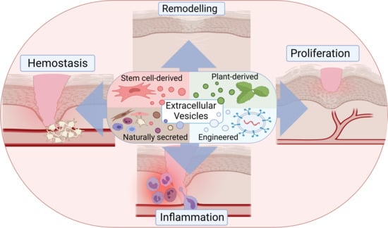
2. The Role of Extracellular Vesicles in Natural Wound Repair

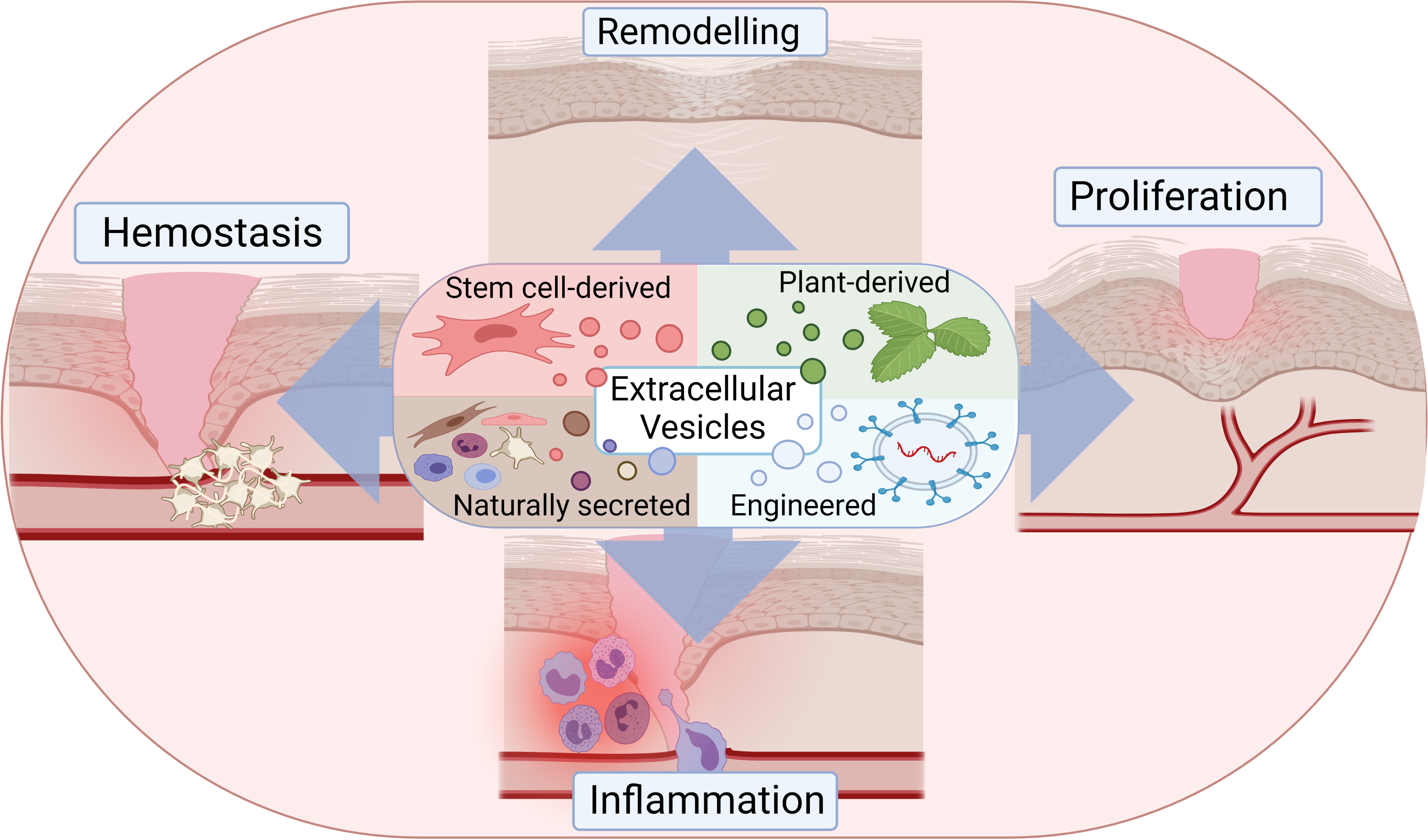
2.1. Physiology of Healthy and Wounded Skin

Human skin is the largest organ, covering the entire outer surface of the body. It makes a primary barrier against pathogens, UV rays, chemicals, and mechanical damage and regulates body temperature and the amount of water released into the environment. Skin functions are fulfilled by specialized cells found in three anatomically and functionally different skin layers: epidermis, dermis, and hypodermis (Figure 1) [15]. The epidermis consists of 95% keratinocyte cells (KCs) and 5% pigment melanin producing melanocytes. In addition, tactile epithelial Merkel cells, tissue-resident macrophages (Langerhans cells), and T resident memory cells are also present in this layer [16]. KCs are responsible for the formation of the epidermal water barrier by secreting lipids. Moreover, they express Toll-like receptors and produce inflammatory cytokines and chemokines (such as IL-1β, IL-8, and CCL20), which are crucial for pathogen pattern recognition and recruitment of leukocytes under skin injury conditions [17,18,19].
The dermis is the next layer after the epidermis and consists of two layers of connective tissue that merge without a clear boundary—stratum papillare and stratum reticulare. The papillary layer is the upper, thinner layer, composed of loose connective tissue, and contacts the epidermis. Next is the retinal layer, which is thicker, less cellular, and composed of dense connective tissue or bundles of collagen fibers [16,20]. The dermis contains sweat glands, hair, hair follicles, muscles, sensory neurons, and blood vessels. The vascular network supports the epidermis, hair follicles, and sweat glands with nutrients. It also plays a central role in the dermal inflammatory response after wounding via the recruitment of neutrophils, lymphocytes, and other inflammatory cells [21]. The dermis also has a groups other than blood-derived immune response cells, such as dermal dendritic cells, macrophages, CD4+ and CD8+ T resident memory cells, mast cells and Foxp3+ T regulatory cells, often located near the hair follicle [16]. The most widely distributed cells in the dermis are fibroblasts primarily responsible for extracellular matrix (ECM) production [22]. They make the layer rich in elastin, fibrillin, collagens (types I and III), and other ECM components, including growth factors. The ECM is responsible for skin mechanical properties such as elasticity and tensile strength. Collagen IV and integrin α6 are critical basement membrane components, contributing to the skin’s physical integrity [16].
The hypodermis, also called the subcutaneous tissue, is the deepest layer of skin and is composed of adipose cells, mesenchymal stem cells (MSCs), blood and lymph vessels [23]. It is endocrinologically and immunologically active. The subcutaneous layer of the skin is predominantly made of adipocytes and mesenchymal fibroblast precursor cells known as pre-adipocytes. The second most common cell type are immune cells, and nearly all immune cell types are included [24]. It contains fat lobes and some skin additives such as hair follicles, sensory neurons, and blood vessels. The subcutaneous adipose tissue underlies and functions as an energy reserve and also a constant source of growth factors to the dermis [25,26]. The adipose tissue MSCs (AdMSCs) are important regulators of tissue homeostasis; they are also explored for scientific and potential clinical purposes [23,27,28,29]. Numerous studies demonstrate that adipose tissue provides a worthy impact on the microenvironment by the secretion of bioactive factors with various functions [23]. The tissue is responsible for energy balance, lipid metabolism, insulin sensitivity, angiogenesis, immunomodulation, and inflammatory response [30].
The synchronized and efficient function of cells from all three layers of skin is needed to restore barrier function after damage. This process is characterized by four classic stages—hemostasis, inflammation, proliferation, and remodeling—which occur one after another, but also overlap. It consists of different cellular and molecular events that require the cooperation of various cell populations [31].
The first response to a wound is a constriction of blood vessels and fibrin clot formation. The most significant cells involved in this process are platelets. These megakaryocyte-derived, non-nucleated cell fragments circulate close to endothelial cells (ECs) during homeostasis. In case of vascular injury, they become activated by agonists released from damaged cells, e.g., collagen, adenosine diphosphate (ADP), von Willebrand factor, fibronectin, thrombin. This activation causes inside-out signaling pathways leading to the actin cytoskeleton and aggregation changes, resulting in platelet plug formation. Simultaneously activated platelets activate coagulation cascades, triggering prothrombin conversion to thrombin, cleaving fibrinogen into insoluble fibrin. Platelet plug and fibrin form a thrombus, which stops the bleeding [31,32,33]. Successful formation of a clot provides a temporary wound matrix for upcoming infiltration of immune cells serving the next phase of wound healing—inflammation.
Damaged cells and platelets release various “find me” signals (ATP, peptides, ECM components, cytokines, chemokines) at the site of the skin injury. Also, it is often colonized by microorganisms (bacteria and fungi) [34,35], so their molecular motifs are also present to activate immune responses. In the first few hours, the predominant cell line clearing the bed of the wound from remains, damaged cells, and bacteria are neutrophils. They constitute 50% of all cells at the wound site during the first day after tissue injury [36]. Within 48–96 h, monocytes are recruited and evolve into macrophages, whose main functions are phagocytosis of residual tissue and the rise of cytokine secretion and growth factors enhancing inflammatory response [31,37].
During the proliferation phase, granulation tissue is formed by activated fibroblasts, synthesizing ECM components, mainly collagen I. Thus, it provides a platform for new vessels and inflammatory cells [31,38]. A hypoxic state of the wound bed induces vascular EC activation, and this, in turn, initiates angiogenesis. Additionally, endothelial progenitor cells (EPC) are activated and participate in de novo blood vessel formation (vasculogenesis) by incorporating into vessels and differentiating into ECs. They are recruited by chemokines and transit to the circulation from the bone marrow, where they reside until vessel injury [39].
Concurrently with the proliferation state, ECM undergoes structural changes while fibroblasts differentiate to contractile myofibroblasts. These processes constitute the last phase of wound healing—remodeling. Ultimately, granulation tissue is replaced by connective tissue and wound healing results in closure [31,37,38].
For this successful restoration of the barrier, physiological functions must occur consistently, with adequate intensity and a specific duration. Any disruptions can cause an impaired wound healing.
Wounds exhibiting impaired healing usually have disorganized, delayed functioning of participant cells. Such a dysfunction causes pathological pro-inflammatory conditions and chronic wounds. Most cases of such chronic wounds are ulcers caused by diabetes, ischemia, venous stasis disease, or pressure [40].
Multiple local and systemic factors can cause impaired wound healing by affecting one or more phases of the process. Many of these factors are related. Local factors (e.g., oxygenation [41], infections [42]) directly influence the characteristics of the wound itself, while systemic factors (e.g., age [43], hormones [44], stress [45], diabetes [46]) are the overall health or disease state of the individual that affect the ability to heal. For example, the impaired healing in individuals with diabetes involves hypoxia and dysfunction in dermal and epidermal cells. Such healing abnormalities are caused by impaired angiogenesis, high levels of metalloproteases, damage from glycation end-products, and an increase in active oxygen species. In addition, the healing capacity in such cases is further reduced by host immune resistance and neuropathy [40]. Abnormal wound repair may result in various abnormalities, from excessive fibrosis and scarring to underhealing wounds, clinically typified by nonhealing chronic ulcers, posing a significant healthcare challenge [31].

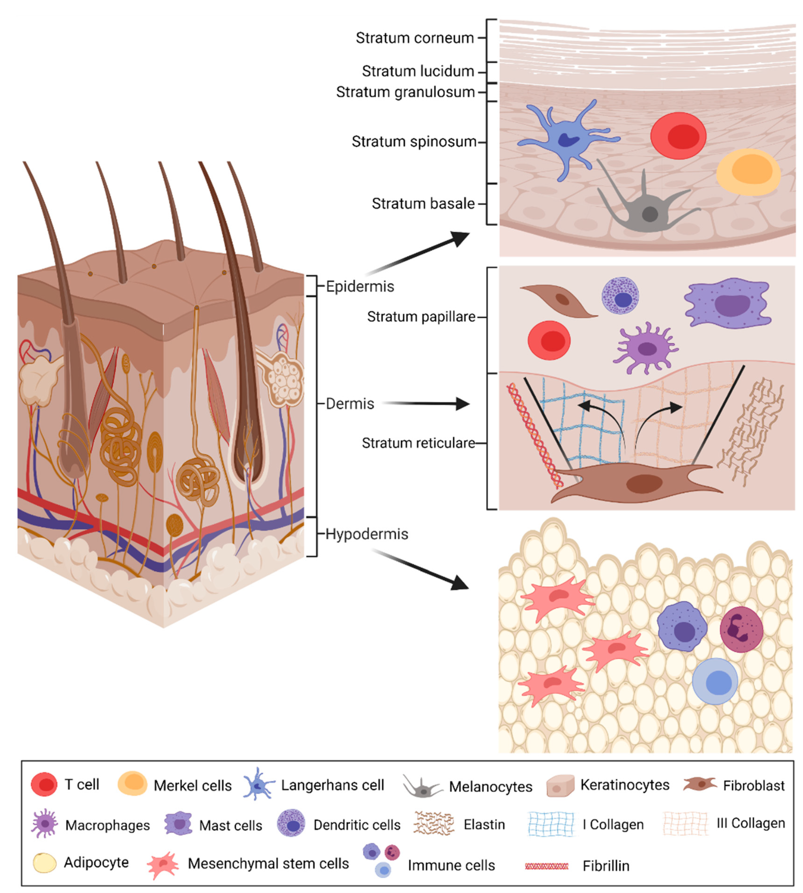
2.2. Extracellular Vesicles—Biogenesis, Composition, and Function

Pan and Johnstone were among the first to investigate EVs in 1983, stating that they evolved as a heterogenous family of membrane-surrounded vesicles originating from endosomes or plasma membranes [47]. Data collected over more than 30 years show EV presence almost in all bodily fluids [48,49,50,51,52,53,54] and suggest that EVs mediate cellular metabolism by transferring nucleic acids, lipids, and proteins, acting as signaling mediators in homeostatic or pathological processes [55,56,57].
The term EVs is applied for all secreted membrane vesicles, yet they are highly heterogeneous. According to the available EVs biogenesis data, they can be divided into three subpopulations: apoptotic bodies (50–5000 nm) produced throughout the cell death, exosomes (30–150 nm), formed during endosomal sorting and microvesicles (100–1000 nm in diameter), directly budding from the plasma membrane [58,59,60] (Figure 2).
Upon apoptosis, the cell passes through several morphological events. It starts with chromatin condensation, hereupon membrane budding, which results in the formation of apoptotic bodies with a cytosolic content [61]. Biogenesis of these EVs relies on caspase-mediated activation of Rho-associated protein kinase 1 (ROCK-1). It phosphorylates the myosin regulatory light chain and stimulates actomyosin contractile activity causing plasma membrane shedding of the cytoskeletal network and the formation of an apoptotic body [62,63]. It contains specific membrane rearrangements (e.g., phosphatidylserine (PS) exposure), which, under normal conditions, are recognized by macrophages, and therefore they are eliminated [64].
Newly discovered microvesicles and exosomes currently attract primary research interest. The most complex is the biogenesis of exosomes involving endosomal maturation and sorting machinery. It begins when an early endosome is formed from plasma membrane invagination. Afterwards, the endosome travels to the center of the cell, gradually changing the composition of the load it carries and the rearrangement of the membrane [65,66]. During this process, the early endosome transforms into the late endosome aggregating so called ”intraluminal vesicles” (ILVs). They are formed in the presence of multisubunit machinery—endosomal sorting complex required for transport (ESCRT)—which carries out budding and scission of the endosomal membrane. This canonical ESCRT pathway can intersect with the generation of ILVs carried out by other proteins. For example, protein syntenin combined with ESCRT accessory protein ALIX (ALG-2-interacting protein X) can engage cargos with the ESCRT-III complex proteins and promote membrane bending [67]. Besides, ESCRT-independent mechanisms also exist. They include the participation of membrane proteins tetraspanins [68,69] and sphingolipid ceramide [70,71]. In the process of ILV formation, cytosolic proteins, nucleic acids, and lipids are recruited. As the number of ILVs increases, the late endosome matures into the multivesicular body (MVB). Once formed, it either fuses with lysosome for degradation or with the cellular membrane releasing the ILVs as exosomes into the extracellular space [72].
The mechanisms of microvesicle biogenesis are still not understood. Some molecular mechanisms involved in the stages of EV biogenesis are common to both exosomes and microvesicle formation. These include the action of ceramide formed by sphingomyelinase and ESCRT proteins [73]. However, the component of ESCRT-I complex—tumour susceptibility gene protein 101 (TSG101)—can also participate in mechanistically different membrane budding from ILV formation. It was shown that TSG101 could be recruited to the cell surface by arrestin domain-containing protein 1 and promote direct membrane invagination [74]. Moreover, a unique mechanism of microvesicle biogenesis can be membrane phospholipid asymmetry rearrangement. It is mediated by Ca2+-dependent enzymes—calpain, gelsolin, phospholipid translocases, and scramblase, which promote the distribution of PS on the outer cell surface. Such membrane remodeling results in physical membrane flexion and actin skeletal restructuring leading to microvesicle detachment [75].
The protein composition of EVs in most cases depends on the mode of biogenesis. For instance, exosomes tend to be more enriched in tetraspanins CD37, CD53, CD63, CD81, CD82 [76,77], and ESCRT-associated proteins, such as TSG101, ALIX, and syntenin [67,78]. Moreover, chaperones, such as heat shock cognate 71 kDa and heat shock protein 90 (Hsp90), are abundantly found in exosomes. Data suggest that these proteins might promote the incorporation of cytosolic components to the exosomal membrane [79]. Additionally, 14-3-3 epsilon and pyruvate kinase M2 found the exosomes of most cell types, also contribute to protein sorting into exosomes [80]. Due to their plasma membrane origin, microvesicles tend to be enriched in proteins of a different repertoire, including integrins, P-selectin, and glycoprotein Ib [76,81]. Moreover, they carry more proteins with posttranslational modifications, such as glycoproteins or phosphoproteins, compared to exosomes [82]. Lastly, apoptotic bodies contain DNA-binding histones and are depleted in glycoproteins, which is in direct contrast to exosomes [83,84].
Irrespective of cell origin, proteins like tetraspanins, ALIX, TSG101, and heat-shock chaperones are commonly found in all EV subpopulations. They can consequently be used as general EVs markers [77,83]. In contrast, proteins within the mitochondria (e.g., aconitase), Golgi apparatus (e.g., GM130), endoplasmic reticulum (e.g., calreticulin), and some cytoplasmic proteins (e.g., α-tubulin) have appeared to be depleted in EVs isolated by differential centrifugation [85,86]. Overall, the absence might provide additional confirmation of the purity of the EV preparation.
The protein composition of different EV subtypes shows a substantial overlap, even if some proteins are more common in one than in another EV subtype [82]. In addition to this, overlapping sizes and similar morphology makes it challenging to characterize different EV types fully and standardize isolation methods. For these reasons, it is often unclear which subpopulation is responsible for any particular effect and articles exploring EV role focus on the potential functions rather than their origins. Therefore, specific markers for different EVs subpopulations and new standardized isolation methods remain to be determined.
Among other proteins, EVs might contain enzymes involved in lipid metabolism. For example, under hypoxic conditions, adipocytes secrete EVs containing increased levels of enzymes that participate in de novo lipogenesis, such as fatty acid synthase. After treatment with such EVs, adipocytes consume more lipid molecules, primarily because of fatty acid synthase delivered to cells by the EVs [87]. Additionally, EVs from RBL-2H3 basophil cells transport GTP dependent phospholipases D and A2 and bioactive lipids such as arachidonic acid and prostaglandin E2 [88].
Furthermore, EVs can transport nucleic acids: DNA within the size of 100 base pairs to 2.5 kilobase pairs [89,90,91,92] and RNA of fewer than 200 nucleotides. [50,93]. The DNA cargo in the EVs reflects the parental cells’ genomic DNA. For example, common gene mutations in cancer cells such as mutations in BRAF, epithelial growth factor receptor, KRAS and p53 were successfully detected in EVs derived from melanoma and pancreatic cancer cells [92]. Moreover, RNA was functional after EV-mediated transfer. Polyadenylated mRNAs participated in translation after uptake, which has been proved in recipient cells by translation assays [94,95,96]. The miRNAs transported between cells by EVs may regulate the translation of target mRNAs in recipient cells [94,97]. For example, EVs released from T cells can transfer specific miRNAs (such as miR-335) to recipient antigen-presenting cells [94].
Interestingly, some RNAs are systematically present in EVs, suggesting a specific mechanism of RNA incorporation into the vesicles [57,95,96]. Most likely, RNA is sorted into EVs due to particular sequence motifs. Indeed, the sequence motif of CUGCC in the binding site for miR-1289 was shown to promote the miRNA recruitment into EVs [98]. Studies suggest that this process might be carried out by heterogeneous nuclear ribonucleoprotein A2B1 (hnRNPA2B1), recognizing these specific sequence motifs and promoting miRNA packaging. In addition, specific miRNA binding is supervised by posttranslational modification of hnRNPA2B1 in the form of SUMOylation, which regulates protein stability and cellular trafficking [99]. The classification of miRNAs into EVs can also be driven by 3′ end posttranscriptional modifications because 3′ adenylated miRNAs are prevalent in cells, whereas 3′ uridylated miRNAs are characteristic of EVs miRNAs [100]. This data shows regulation after protein translation or transcription can modulate specific miRNA packaging into EVs.
Overall, findings have shown the substantial importance of EVs as an intercellular communication mediator.

2.3. Physiological Role of Extracellular Vesicles in Wound Healing

Recent in vivo studies reported EV presence in wounds and acknowledged their participation in normal healing [7,40]. This section will summarize the latest findings on EVs derived from “key-player” cells in skin repair under physiological conditions. The main results on the topic are listed in Table A1.

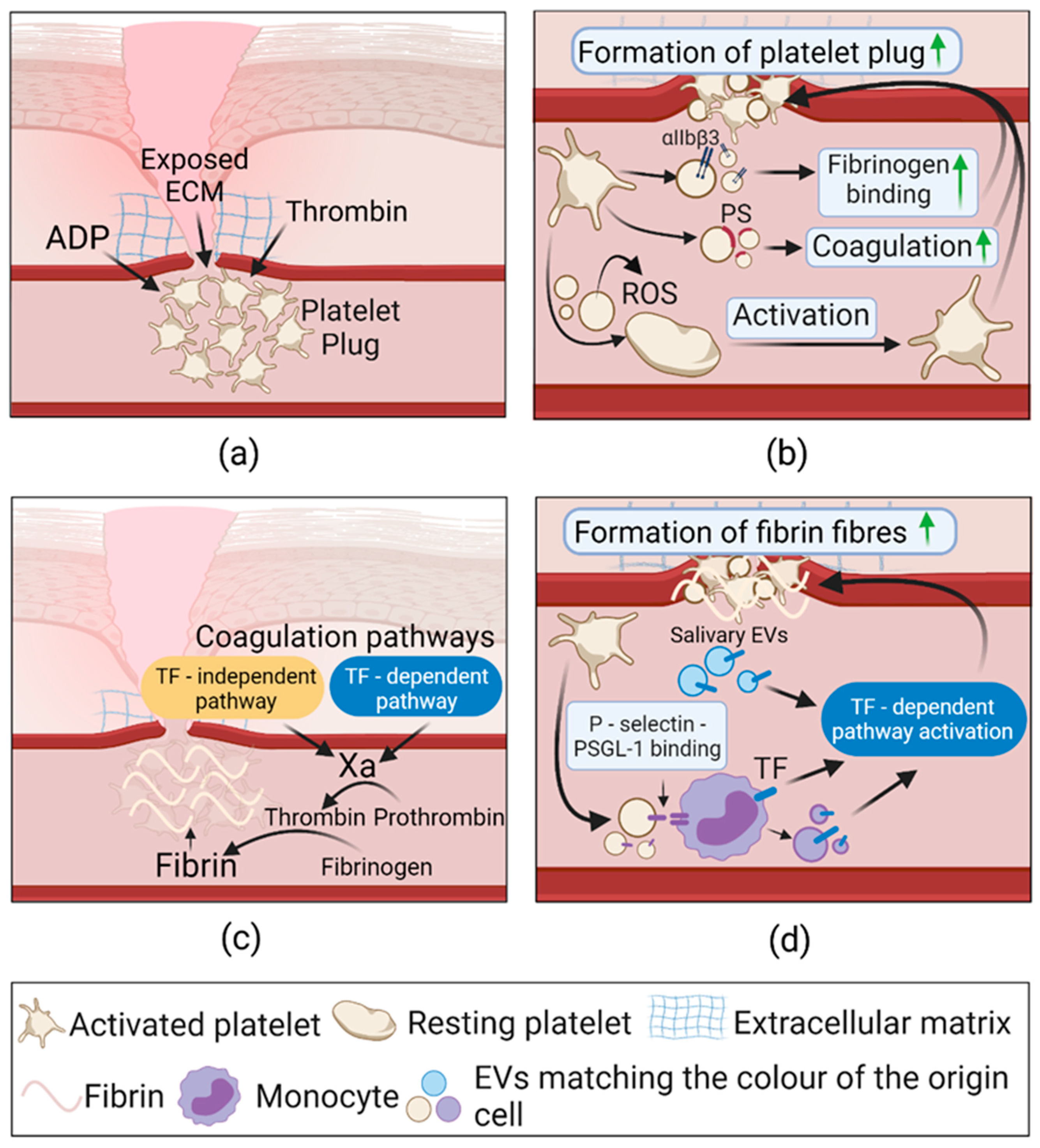
2.3.1. Extracellular Vesicles in Hemostasis

The EV role in hemostasis is schematically depicted in Figure 3. The most abundant EVs in blood circulation are platelet-derived EVs (PEVs) [101], and they significantly contribute to the regulation of hemostasis [60,102]. PEV procoagulant function mostly depends on platelet activation, e.g., PEVs from thrombin-activated platelets were more efficient in clot formation (p < 0.01) than PEVs from resting-state platelets (p < 0.05). PEV involvement in clot formation is mediated by the activated form of integrin αIIbβ3, which provides high affinity to fibrinogen and results in the construction of fibrin clots [103]. Additionally, activation of P2Y12 platelet receptors by ADP induces the release of PEVs, exposing the proinflammatory P-selectin and procoagulant PS [104]. The latter serves as a platform for procoagulant factors and promotes the generation of thrombin [105]. However, the plasma containing PEVs with exposed PS does not clot without the tissue factor (TF), which is known to be the primary initiator of blood coagulation. Upon vascular injury, TF forms a complex with coagulation factor VIIa, which, in turn, activates factor X, responsible for prothrombin conversion to thrombin [31]. In another study of sepsis-induced platelet activation, PEVs were shown to bind TF and Factor XII and evoke the formation of thrombin only in the presence of Factor VII and XII. The finding indicates that PEVs mediates both extrinsic (TF-dependent) and intrinsic (TF-independent) coagulation pathways [106]. Although TF presence in the PEVs of healthy individuals is still disputed [107,108], they can induce procoagulant effects indirectly by binding P-selectin to P-selectin glycoprotein ligand-1 (PSGL-1) on monocytes and cause TF exposure on their surface [38]. As an alternative, TF can be transferred via monocyte-derived EVs [109]. Interestingly, TF was also found in salivary EVs, implying their ability to facilitate hemostasis at the skin injury site upon the licking reflex [110].
In addition to the above-described coagulation factor transferring, a novel mechanism was proposed, introducing PEV ability to communicate components of NADPH oxidase (NOX-1). This study reported that PEVs from activated platelets generating superoxide in a NOX-1-dependent way, affect other platelets and enhance fibrinogen binding. It was also suggested that PEVs induce platelet activation via collagen receptor—GPVI [111].
The increasing amount of data shows that the EVs of various origins cooperate to ensure the successful formation of a platelet plug and fibrin fibers, which serve as a platform for subsequent infiltration of immune cells.

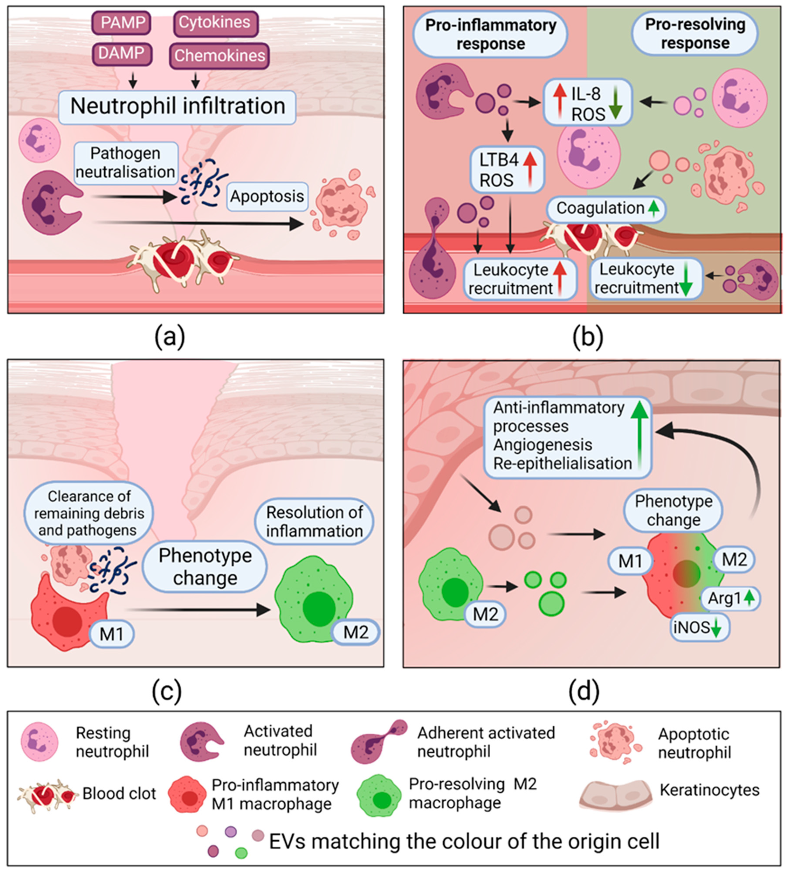
2.3.2. Extracellular Vesicles in Inflammation

The studies of neutrophil-derived EVs (NDEVs) show that they exert anti- and pro-inflammatory functions depending on environmental factors persisting at the time of EV biogenesis. It was reported that upon infectious conditions, NDEVs increase the production of ROS (p = 0.0371) and IL-8 (p = 0.0014) in other neutrophils. Additionally, they boost the expression of adhesion molecules E-selectin and VCAM-1 on ECs, indicating their activation. In contrast, resting-state NDEVs do not affect endothelium or alleviate its activation. Interestingly, EVs from apoptotic neutrophils also distinguish themselves with a potent procoagulant effect [112]. Another study has shown that EVs from activated neutrophils can also act in their own respect without transferring cargo to recipient cells. Evidently, NDEVs carry NOX-2 and, after directly interacting with pathogen-associated molecular patterns (PAMPs), increase ROS production in a receptor-dependent fashion p < 0.05). However, this does not apply to EVs from endothelium-attached neutrophils, which indicates NDEV heterogeneity due to neutrophil interaction with other cells. In addition, both NDEV subsets (from adherent and non-adherent neutrophils) generate leukotriene B4 (LTB4) and migrate towards a chemotactic gradient. ROS is a known protector against pathogens and can stimulate protective signaling pathways in other cells, while LTB4 is a chemo-attractant. These findings show that activated NDEVs mediate inflammation by producing “danger signals”. Additionally, adherent NDEVs activate pro-inflammatory gene expression in human umbilical vein endothelial cells (HUVECs), whereas non-adherent NDEVs act the opposite, promoting anti-inflammatory gene expression [113].
During the inflammatory phase of wound healing, macrophages have an essential role in transitioning from the inflammatory phase to the proliferative one. Macrophages undergo a phenotype change: from possessing pro-inflammatory properties to pro-resolving/healing properties, also referred to as M1 and M2 phenotypes, respectively [37,114]. The effect of EVs derived from macrophages of different phenotypes on cutaneous healing has been recently analyzed. It was found that M2 macrophage-derived EVs (M2-EVs) induce macrophage reprogramming from M1 to M2 phenotype; M2-EVs cause a complete absence of M1 marker—inducible nitric oxide synthase (iNOS) but induce M2 marker arginase—expression. Moreover, CCL24, CCL22, and MFG-E8 cytokines are identified as the main EVs compounds responsible for cell reprogramming [115]. Interestingly, EVs from wound edge keratinocytes (KCs-EVs) exhibit a similar role in the phenotypic change of macrophages. A brilliant study by Xiaoju Zhou and colleagues reported that these EVs, unlike uninjured skin EVs, expose a characteristic N-glycan composition on their surface and promote their uptake by wound macrophage cells. Consequently, wound edge KCs-EVs downregulate pro-inflammatory iNOS, CD74, TNF-α genes, and upregulate anti-inflammatory CL3, and this causes reprogramming to pro-resolving macrophage phenotypes. Moreover, the authors demonstrated that knocking down hnRNPA2B1 responsible for miRNA packing to EVs in KCs causes impaired wound closure and persistence of pro-inflammatory iNOS expressing macrophages in vivo. Essentially, miRNA packaging in KCs-EVs is crucial for resolving wound inflammation [7]. Once macrophages are reprogrammed, they accelerate fibroblast migration and ECs tube formation [115]. The inflammation phase and the role of EVs are depicted in Figure 4.
All this data indicates that EVs exert diverse anti- and pro-inflammatory effects modulating the inflammatory response. In addition, the stimulation of macrophage reprogramming has a vital role in the transition to the proliferating phase of wound healing.

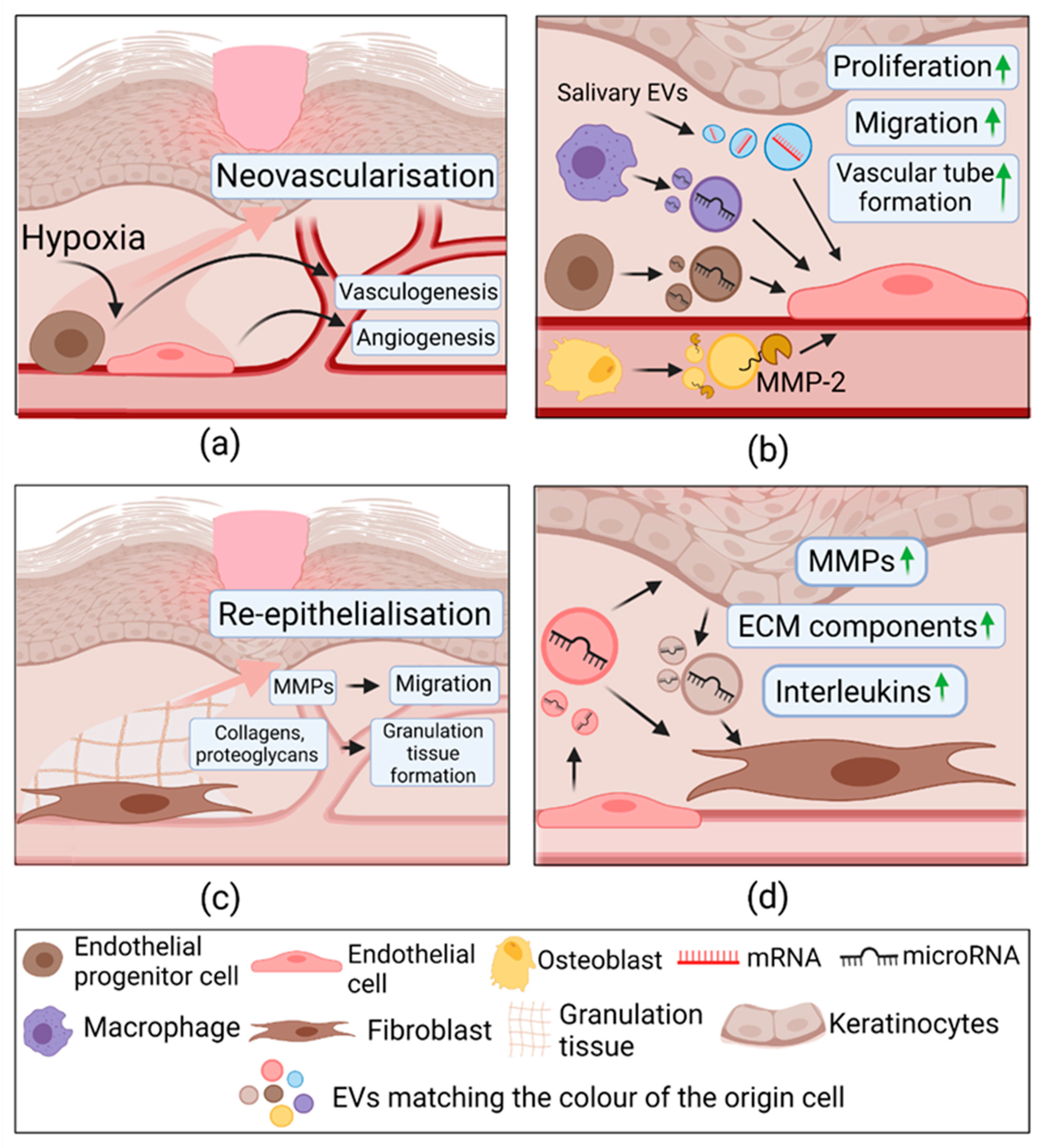
2.3.3. Extracellular Vesicles in Proliferation

Under wound healing conditions, the injury site is in hypoxia, therefore inducing activation of local vascular ECs. Once they are activated, the angiogenesis process begins, in which new blood vessels form from the existing ones. Some of the most critical angiogenic signals are Vascular Endothelial Growth Factor (VEGF), Fibroblast Growth Factor (FGF-1), and angiopoietins. In response to them, ECs increase permeability for extravasation of plasma proteins, which deposit provisional ECM. Proteolytic degradation carried out by matrix metalloproteinases (MMPs) remodels this ECM and enables migration of ECs, liberated from the basement membrane [116]. Additionally, EPCs are activated and participate in de novo blood vessel formation (vasculogenesis) by incorporating into vessels and differentiating into ECs. They are recruited by chemokines and transit through the circulation from the bone marrow, where they reside until vessel injury [39]. The proliferation phase and the role of EVs are represented in Figure 5.
Recently, studies have shown that EPCs’ released paracrine factors can induce activation of tissue-resident EC and suggest that this mechanism might be more significant in new vessel development than their direct differentiation [117]. Indeed, EVs from umbilical cord-derived EPCs induce pro-angiogenic effects in in vitro and in vivo healthy and diabetic rat wound models. They up-regulated a broad range of pro-angiogenic factor expression in vascular ECs; some of them include E-selectin, angiopoietin, FGF-1, cyclooxygenase 2 (COX-2), and cell cycle activator c-Myc [118,119]. The authors demonstrated that this effect depends on ERK1/2 signaling and speculated that miR-21, found in EVs, might be the culprit of its activation [119]. Moreover, EVs from bone-marrow-derived EPCs are enriched in miRNA-221-3p, which increases the expression of pro-angiogenic factors, including adhesion molecule PECAM-1 (p < 0.01), VEGF (p < 0.05), and cell proliferation marker Ki67 (p < 0.05) [120]. These findings suggest that EPCs-derived EVs (EPCs-EVs) promote angiogenesis by inducing ECs proliferation, motility, and tube formation.
However, EVs of an origin other than endothelial origin can also contribute to angiogenesis. For instance, EVs from macrophages (M-EVs) contain even higher concentrated VEGF, Wnt3a, and miR-130a than their parent cells, and some levels of miR-210 and miR-126 were also identified. These factors are known contributors to EC angiogenic activity and, as the authors suggest, might be responsible for EC proliferation, migration, and tube formation induced by M-EVs [121,122]. Furthermore, an interesting study of mature osteoblast-derived EVs demonstrated angiogenic capacities through the VEGF/ERK1/2 signaling pathway. It was shown that they carry metalloproteinase-2 (MMP-2), which is crucial for angiogenesis as it degrades ECM components that facilitate ECs migration [123]. Bobin and colleagues showed similar effects on angiogenesis of salivary EVs, yet demonstrated a novel mechanism of action. Saliva-EVs transfer mRNA of ubiquitin-conjugated enzyme E2O (UBE2O) that is overexpressed in ECs. UBE2O participates in ubiquitin-mediated proteolysis and decreases levels of SMAD6, a signal transducer known to be an angiogenesis suppressor. This effect resulted in pro-angiogenic cytokine bone morphogenetic protein 2 upregulation [124]. These discoveries revealed that macrophages, bone-forming cells, and saliva upon wound licking promote vessel formation and contribute to the wound healing process [121,122,123,124].
Fibroblasts play a vital role in skin structure formation. They clear a path by secreting proteases and migrate towards the wound site, where they synthesize collagen, proteoglycans, and other granulation tissue comprising components [38]. ECs-EVs can contribute to the process by mediating ECs-fibroblast or ECs-KCs crosstalk. EC-EVs derived from the plasma of healthy volunteers induce migration and proliferation and prevent senescence in diabetic skin fibroblasts through PI3K/Akt/mTOR signaling pathway. Additionally, in fibroblasts and epidermal keratinocyte-like cells (HaCaT), they promote nuclear translocation of transcriptional regulator Yes-associated protein (YAP) and subsequently, activation of its downstream effector—connective tissue growth factor (CTGF). This regulatory axis is known to participate in collagen deposition, fibroblast proliferation, and differentiation to myofibroblasts, which are critical in the remodeling phase of wound healing [125,126].
Notably, KCs-EVs strongly influence fibroblasts and may regulate several features in wound healing [127,128]. Ping Huang and colleagues reported that KC-EVs activate various signaling pathways, with the most prominent effect on ERK1/2. This pathway mediates induction of pro-migratory (MMP-1, MMP-3) and pro-angiogenic/pro-inflammatory (IL-6, IL-8) gene and protein level expression. Moreover, KCs-ECs suppress the expression of the MMP inhibiting proteins RECK and TIMP [128]. More than a third of genes regulated by KC-EVs participate in the signaling of transforming growth factor β (TGF-β), a vital contributor to wound healing. These molecular changes increase fibroblast migration and stimulate them to produce the endothelial tube formation promoting factors [127]. Authors also showed that a critical candidate for fibroblast regulation in KCs-EVs might be miR-21 [128].
These articles suggest that EVs released from cells during physiological wound healing contribute to neovascularization and epidermal layer reconstruction, which overlaps with the last healing phase—remodeling.

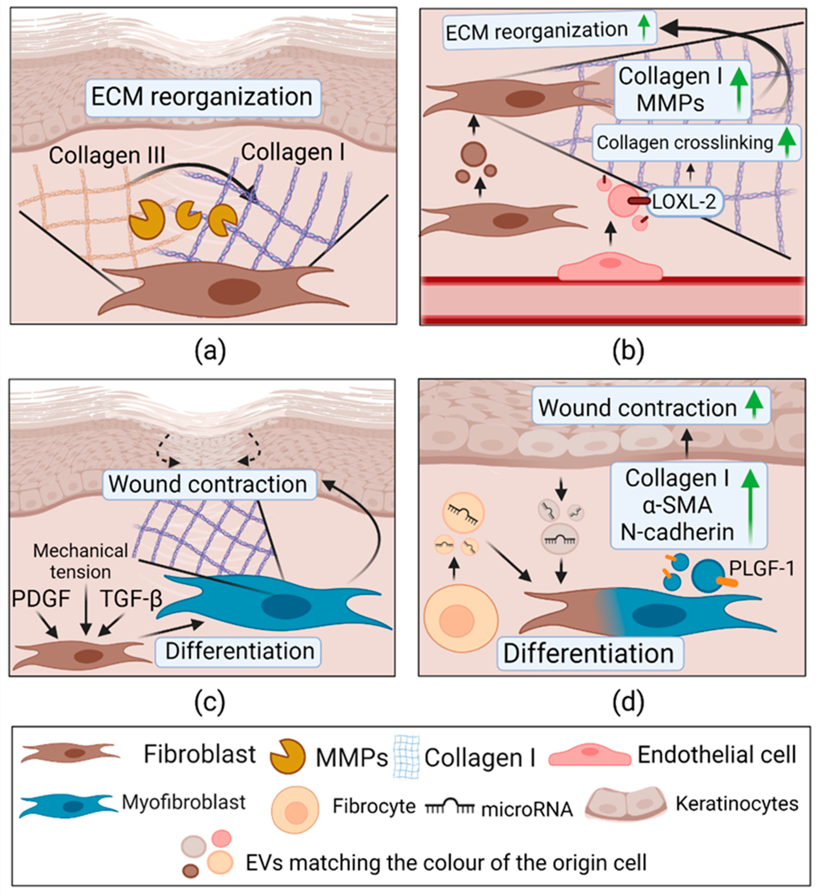
2.3.4. Extracellular Vesicles in Remodeling

The last phase of wound healing and EV’s importance in it are illustrated in Figure 6. Type III collagen is mainly synthesized in the early stages of wound healing, but eventually, it is replaced by type I—the dominant fibrillar collagen in the skin. During ECM reorganization, these components are specifically cleaved by MMP-1, MMP-8, and for final collagen maturation, it is modified by lysyl oxidase (LOX), resulting in covalent cross-linking and restoration of tensile strength [129]. Unsurprisingly, fibroblast-derived EVs contribute to ECM reorganization by increasing collagen I, MMP-1, and MMP-3 gene expression (p < 0.01) in other fibroblasts. This effect assists in migration and collagen deposition increase (p < 0.01) [130]. Furthermore, the study of Olivier G. de Jong and colleagues demonstrated ECs-EVs’ direct effect on ECM remodeling. It was shown that under hypoxic conditions, ECs release EVs exposing LOX family member lysyl oxidase-like 2 (LOXL2), which facilitates collagen I crosslinking and promotes collagen gel contraction [131].
Mechanical tension, TGF-β, and platelet-derived growth factor (PDGF) are considered to be initiators of fibroblast differentiation to a contractile, α smooth muscle actin (α-SMA) expressing myofibroblasts. Importantly, they synthesize large amounts of collagen I [132]. In addition to KCs-EV’s role in the proliferation phase, they also participate in remodeling by initiating the fibroblast differentiation. The treatment with KC-EVs upregulates gene expression and protein level of two known myofibroblast markers—α-SMA and N-cadherin [128]. A recent study showed that EVs from normal skin wound myofibroblasts stimulated collagen I production in cutaneous fibroblasts. This effect was caused by VEGF family member—placental growth factor 1 (PLGF-1)—abundantly found in myofibroblast EVs [133]. Moreover, a study by Adolf Geiger and colleagues showed a significant fibrocyte-derived EV (FDEV) role in wound healing [134]. These progenitor cells originate from bone marrow and acquire myofibroblast-like properties upon injury [135]. Evidence shows that FDEVs carry components such as Hsp-90α, total and activated signal transducer, and activator of transcription-3 (STAT3) [134]. Secreted HSP-90α is characterized by unique properties of promoting cell motility and re-epithelialization. It binds lipoprotein receptor-related protein-1 and activates the Akt signaling pathway [136]. Additionally, STAT3 can activate a broad range of signaling cascades regulating ECM remodeling, angiogenesis, and chemotaxis [137]. Besides these components, FDEVs are enriched in anti-inflammatory (miR124a, miR-125b), pro-angiogenic (miR-126, miR-130a, miR-132), and collagen deposition regulating (miR-21) mi-RNAs. Lastly, FDEVs increase (p < 0.01) α-SMA and collagen I expression in fibroblasts, most likely leading to differentiation [134].
The above-described evidence highlights the role of EVs in each wound healing phase. However, in the case of pathological wounding, their application has similar drawbacks. For example, EV assistance in coagulation or inflammation phases depends on specific cells‘ activation or interaction with other cells. Namely, the procoagulant role of PEVs relies on the activation of platelets with different stimulants (ADP, thrombin, collagen). Additionally, TF presence in EVs released from activated platelets remains unclear, meaning that EVs from these cells alone might not necessarily lead to coagulation, as well as complete wound healing. Moreover, pro-/anti-inflammatory functions of NDEVs may depend on neutrophil contact with ECs. In contrast, fibroblasts alone secrete EVs, which promote successful wound healing by activating several crucial processes. By transferring miR-21 and primarily activating ERK1/2 signaling pathways, the EVs induced angiogenesis, ECM reorganization, and differentiation to myofibroblasts, promoting wound contraction. The same miRNA and many others were detected in stem cells derived from bone marrow, specifically EPCs-EVs and FDEVs. Thus, their overall effect on wound healing is undoubted. For this reason, in the next chapter, we summarize the current evidence about the role of EVs, mostly from bone marrow-derived MSCs (BMSCs) and AdMSCs in skin barrier repairing.

3. Stem Cell-Derived Extracellular Vesicles in Skin Wound Healing

MSCs are multipotent mesenchymal stromal cells, which can differentiate into diverse cell types, for instance, adipocytes, osteocytes, chondrocytes, and ECs [138]. Due to immunosuppressive, anti-inflammatory, tissue recovering, and differentiation stimulating properties of the MSCs, they are used for cell therapy in regenerative medicine [139]. Cell therapy is based on injured tissue replacement and restoring of its biological functions [140]. However, using MSCs have some drawbacks: the requirement for a consistent source of stable phenotypic cells, a risk of immunological rejection and risk of tumour development [138]. Nevertheless, recent studies indicate that MSCs modulate tissue regeneration through released paracrine factors, and among them, EVs play a vital role [140]. They participate in main wound healing phases: help prevent inflammation, induce cell proliferation, new tissue formation, and maturation by transferring various biomolecules. Nowadays, MSC-derived EVs are considered novel non-cellular therapy, which can cut the safety limitations of cell therapy [140,141]. The effects of MSC-EVs on hemostasis are summarized in Table A2 and Figure 7.

3.1. Mesenchymal Stem Cell-Derived Extracellular Vesicles in Hemostasis

As described above, wound healing starts with blood clot formation, which leads to organism prevention and protection from loss of blood. It is a dynamic process based on platelet aggregation [142]. It is known that MSC-derived EVs have procoagulant properties, usually depending on their transferred cargo. In most cases, MSC-EV cargo resembles that of skin cell EVs released during wound healing.
EVs from MSCs might affect blood coagulation. Silachev with colleagues showed that in the presence of umbilical cord MSC-EVs, human blood clot formation time and lag period of spontaneous clotting is significantly reduced compared to the EV untreated group [143]. Also, the MSC-EVs experimental group had improved clot firmness and significantly increased blood clot area. The proteomic analysis demonstrates that both MSCs and MSC-EVs contain several well-known proteins participating in coagulation, such as CD9, PS, myosin-9, talin-1, histones, and cytoplasmic actin. CD9 is one of the most critical proteins in platelet activation initiation, platelet aggregate stability promotion, and fibrinogen binding enhancement. Moreover, umbilical cord MSCs were found to contain TF. However, it was not detected in MSC-EVs. Another exciting finding is that MSC-EVs contain annexin V, a protein characterized by participating in anticoagulant activities. Therefore, it might be speculated that the coagulation properties of EVs depend on pro- and anticoagulant proteins’ dynamic balance [143].
In addition, Chance et al. checked if EVs isolated from three-dimensional cultures have anticoagulant activities associated with the presence of procoagulant activity factors [144]. Scientists determined the procoagulant activity of monolayer and spheroid-cultured AdMSCs and BMSCs-derived EVs (AdMSCs-EVs and BMSC-EVs, respectively). Both EV groups were functionally thrombogenic. They significantly increased the peak of thrombin activity and decreased the time to reach it (p < 0.0001). Additionally, the total amount of generated thrombin in all EV groups was markedly increased. Moreover, this study confirmed that the procoagulant activity of EVs is associated with the expression of TF and PS on the surface of vesicles. Such procoagulant activity factors were identified in all the EV groups. However, AdMSC-EVs show greater PS expression, which leads to higher thrombin amounts compared to BMSC-EVs. Also, the strongest clots were formed in the group of treatment with the EVs derived from AdMSCs spheroids. On the other hand, BMSC-EVs demonstrated quicker clot initiation outcomes. In general, it is concluded that all examined EV types have a thrombogenic nature. Another similar study with AdMSC-EVs and BMSC-EVs confirmed that AdMSC-EVs have more significant procoagulant activity in whole human blood or human platelet-poor plasma, which corresponds to the level of TF expression [145]. These data suggest that the anticoagulant properties of MSC-EVs depend on the genes, regulating coagulation, levels of expression, despite cell culturing type.
Typically, proinflammatory cytokines trigger hemostatic activities [146]. Interestingly, AdMSC-EVs have procoagulant activity independent of proinflammatory stimulus [147]. Fiedler et al. investigated EVs from unstimulated AdMSCs and those treated with LPS and TNF proinflammatory substances. Also, clotting experiments were conducted with the EVs compared to reference plasma (a citrated human plasma), coagulation factor XII-deficient plasma, and coagulation factor VII-deficient plasma. In the reference plasma group, all EVs showed similar clotting times. However, in factor VII-deficient plasma, a clot did not form in all EV groups. Due to the absence of clots in factor VII-deficient plasma, authors predicted that EVs might contain TF, which has a role in the activation of VII factor-dependent extrinsic pathway of coagulation. Besides, in factor XII-deficient plasma, unstimulated and TNF-stimulated EVs groups demonstrated significantly increased (p < 0.05) clotting time. Prolonged clotting time may indicate that the EVs contain PS molecules, which provide a catalytic surface for factor XII activation. This factor plays an important role in the stimulation of the intrinsic coagulation pathway. Thus, AdMSC-EVs can participate in wound healing via different pathways independent of proinflammatory stimulus [147].
In general, MSC-EVs can maintain wound healing by balancing pro- and anticoagulant molecule supply and affecting blood clot formation pathways and kinetics. The vesicles contain several proteins and lipids, mainly TF and PS, responsible for EV procoagulant activity. In addition, MSC-EVs may participate in hemostasis by activating extrinsic and intrinsic pathways of coagulation. However, there is still a lack of studies indicating MSC-EVs’ role in wound hemostasis.

3.2. Mesenchymal Stem Cell-Derived Extracellular Vesicles in Inflammation

Neutrophils clean up the wound site from damaged cells and bacteria. However, macrophages continue the wound bed clearing by phagocytosis of residual tissue and increase the production of cytokines and growth factors, resulting in the enhanced inflammatory response. The key role of anti-inflammatory macrophage properties depends on their ability to switch their phenotype from M1 to M2. A broad range of studies (see Table A2) concluded that macrophages attenuated inflammation with immune modulation by shifting their phenotype after the internalization of stem cell EVs.
Xiaoning et al. checked if EVs isolated from BMSCs stimulated macrophage polarization [148]. In this case, in one of the experimental groups, BMSCs were treated with siRNA, which silenced the expression of the rab27a protein, a regulator of EVs secretion, thus inhibiting EVs release. Compared to the BMSC/siRNA group, macrophages cultured with EVs showed a higher level of M2 macrophages marker—CD206, and this proved the ability of BMSC-EVs to promote macrophage polarization. Furthermore, the EVs’ enhanced cutaneous wound healing in vivo, whereas the rab27a-silenced group had delayed healing. Also, scientists isolated EVs after BMSCs transfection with miRNA-223 mimics and inhibitors. Results indicated that BMSC-EVs, isolated after knockdown of miRNA-223 in BMSCs, reduced macrophage polarization from M1 to M2. Besides, pknox1, miRNA-223 target and regulator of macrophage polarization, gene expression in macrophages was altered, depending on treated BMSC-EVs type. The study revealed that miR-223 is transferred from EVs to macrophages and is responsible for a macrophage phenotype shift [148].
Another study used dermal fibroblasts treated with interferon-gamma (IFNγ) and tumour necrosis factor α (TNFα) as a cellular inflammation model to examine AdMSC-EVs’ anti-inflammatory role in wound healing [149]. Fibroblasts were co-cultured with peripheral blood mononuclear cells. After the addition of AdMSC-EVs, a change in macrophage phenotype from M1 to M2 was observed, demonstrated by a significant increase in expression of Arg1 and CD206, the markers of M2 cells. Moreover, various miRNAs (miR-34a-5p, miR-124-3p, miR-146a-5p) were detected in AdMSC-EVs, which are responsible for macrophage phenotype shift. Besides, the treatment of inflammatory cytokine-stimulated fibroblasts with AdMSC-EVs decreased the expression of inflammatory proteins TNFα, IL-6, and IL-8, while increased the expression of IL-10. Microarray experiments identified several miRNAs (miR-223, miR-203, miR-146a) present in AdMSC-EVs, which participate in various signaling pathways associated with wound healing by targeting factors such as myocyte-specific enhancer factor 2c (Mef2c), TNFα, and anti-inflammatory cytokine—IL-24. Authors hypothesized that the anti-inflammatory effect of AdMSC-EVs was caused by such miRNAs [149].
Liu recently characterized the mechanism of MSC-EV-induced macrophage phenotype change with colleagues [150]. The authors concluded that immunosuppression effects of melatonin-treated BMSC-EVs in diabetic wounds are reached by upregulating PTEN (phosphatase and tensin homolog) expression and inhibiting the phosphorylation of AKT (protein kinase B), i.e., by suppressing PTEN/AKT signaling pathway. Consequently, gene expression of proinflammatory IL-1β, TNFα, and iNOS (M1 macrophage markers) significantly decreased (p < 0.05). In contrast, M2 macrophage markers anti-inflammatory IL-10 and Arg1 gene expression raised after the EV treatment. Such EV-mediated balancing of inflammation-related biomolecules might lead to the reduction of prolonged inflammatory periods [150].
In addition, to macrophage phenotype change, AdMSC-EVs also increase (p < 0.05) the viability of KCs by suppressing apoptosis. It was shown in the HaCaT cell line after hydrogen peroxide exposure [151]. Treatment with EVs reduced expression of apoptosis-related proteins caspase-3 and IL-6 and elevated expression of inflammation-related biomolecules Bcl-2 and IL-10 (p < 0.05). Interestingly, the AdMSC-EVs internalization rate directly correlated with hydrogen peroxide concentration. It was observed that AdMSC-EV treatment increased miRNA-19b levels in HaCaT cells. This miRNA binds to inflammatory factor—chemokine CC motif ligand 1, resulting in activation of the TGF-β pathway, which inhibits the occurrence of inflammation.
Also, it was revealed that the AdMSC-EVs significantly inhibit ROS and inflammatory cytokine expression in EPCs [152]. The vesicles can also improve wound repairing by overexpressing the transcription factor nuclear factor-E2-related factor 2 (Nrf2), which protects cells against oxidative stress [152]. In another study, EVs isolated from umbilical cord MSCs proved their ability to decrease burn-caused inflammation by suppressed NF-κB activation and proinflammatory factors secretion via inhibition of the TLR4 pathway in macrophages [153]. Such findings suggest several new mechanisms of MSC-EVs in suppressing inflammation.
Overall, these data indicate that MSC-EVs can modulate inflammatory processes by changing macrophage phenotype, mainly by transferring miRNAs, resulting in a quicker inflammation stage in wound healing and protection from infections.

3.3. Extracellular Vesicles from Mesenchymal Stem Cells in Proliferation

During the proliferation stage, granulation tissue is formed, mainly made of ECM, fibroblasts, inflammatory cells, and blood vessels [154]. Fibroblasts and ECs play a vital role in the formation of this tissue. Also, in the late proliferation phase, the synthesis of a new epidermis occurs. Currently, there are a lot of studies (see Table A2) that indicate the effects of MSC-EVs in cell proliferation and migration activities, angiogenesis, and the epithelization processes during wound healing.
First of all, MSC-EVs affect fibroblast migration to the wound site. Cooper et al. conducted an interesting study measuring electrical current-treated fibroblast migration rate after incubation with AdMSC-EVs [155]. The experimental group treated with the EVs showed a similar fibroblast migration rate compared to the positive control treated with basic FGF (bFGF). In addition, the cellular migration rate value was doubled in the presence of twice higher concentrations of EVs. Besides, the significance of AdMSC-EVs cargo MALAT1 (metastasis-associated lung adenocarcinoma transcript 1, a long non-coding RNA) in maintaining fibroblast migration to wound sites was proved since EVs lacking MALAT1 decreased fibroblast migration rate by halving. Other authors determined that fibroblast migration is supported through downregulation of LATS2 (large tumour suppressor 2) levels by miR-135 transferred by human amnion MSC-EVs [156].
MSC-EVs do not just promote cell migration to the wound site but also improve granulation tissue formation. AdMSC-EVs internalized by fibroblasts increase N-cadherin, cyclin-1, PCNA, collagen I and III gene expression (p < 0.001), following in promotion of cell proliferation and inducing collagen synthesis in damaged tissue areas [157]. Another study suggests that AdMSC-EVs affect fibroblast proliferation, increase collagen, bFGF, TGF-β1 gene expression, and protein levels [158]. Additionally, Wang et al. showed that in vitro fetal dermal MSC-EVs promote fibroblast migration and proliferation and increase fibroblast activity detected as an elevation in mRNA expression of collagen, elastin, and fibronectin [159]. Since activated fibroblasts have regenerative effects, the authors investigated that in vivo, in a mouse wound model, the EV-treated group had higher collagen deposition, ECM synthesis, and a faster wound healing rate.
Recently, studies indicated several new MSC-EV cargos participating in proliferation stage activities. Previously described Wang et al. study revealed that after the treatment with EVs, fibroblasts showed increased expression of the components of the Notch pathway, responsible for the regulation of wound-healing-related-cell proliferation and migration [159]. In addition, a ligand of this pathway, Jagged 1, was detected in the EVs. These results determined that MSC-EVs promote fibroblast activity via the Notch signaling pathway by transferring Jagged 1. Qian with colleagues found that AdMSC-EVs accelerate wound healing through long non-coding RNA H19, miR-19b, and SRY-related high-mobility-group box 9 (SOX9) axis [160]. The EVs carried lncRNA H19 that inhibited mir-19b expression and upregulated SOX9, consequently activating the Wnt/β-catenin pathway followed by accelerated fibroblast proliferation, migration, and invasion into the wound bed [160]. Shabbir et al. determined that BMSC-EVs modulate wound healing by inducing the expression of cell cycle progression factors (c-myc, cyclin A1, cyclin D2), growth factors (HGF, IGF1, NGF, SDF1), and cytokines (IL-6) [161]. The authors figured out that MSC-EVs contain STAT3 and can transfer it to recipient cells inducing expression of mentioned genes and activation of signaling cascades, responsible for cell migration, proliferation, and angiogenesis in the wound site. All these findings suggest that EVs participating in different proliferation promoting signaling pathways due to the transferring of multiple cargos to the recipient cells.
It is essential to restore not only granulation tissue structure, but also its function. For this, new blood vessel formation is required. There are some publications indicating MSC-EV importance in new endothelial tube formation due to their proangiogenic activity in wound healing. AdMSC-EVs increase tube length and branches in vitro and in vivo via transferring miR-125a to ECs and inhibiting DLL4 expression [162]. Overexpression of miR-125a upregulated pro-angiogenic (Ang1 and Flk1) genes and downregulated anti-angiogenic (Vash1 and TSP1) gene expression in vitro. Another study investigating immortalized AdMSC line HATMSC1-derived EVs found that they increase proliferation and have proangiogenic properties on human ECs in a dose-dependent manner [163]. The EVs contain growth factors (EGF, bFGF) and pro- and anti-angiogenic factors (IL-8, VEGF, TIMP-1, and TIMP-2), also, several types of miRNAs: proangiogenic (miR-210, miR-296, miR-126, and miR-378) and antiangiogenic (miR-221, miR-222, miR-92a). It was determined that the expression of proangiogenic miRNAs was higher than antiangiogenic ones, resulting in shifting the balance to stimulate angiogenesis. The increased level of miR-296 expression upregulates VEGFR2 in ECs and leads to angiogenesis [163]. In other research, EVs from umbilical cord blood MSCs proved to enhance angiogenesis and accelerate the healing process in a mouse model [164]. The authors studied the expression level of some miRNA in EVs and found that the miR-21-3p was the most intensively expressed. In vitro, this miRNA promotes angiogenic effects by activating PI3K/Akt and ERK 1/2 pathway through the downregulation of miR-21 target genes PTEN and SPRY1 (sprouty homolog 1). Together these data indicate that MSC-EVs can transfer angiogenic signals through miRNAs.
However, angiogenic signals also can be transmitted by other biomolecules. Chun-Yuan Chen et al. analyzed the protein profile of EVs isolated from urine-derived stem cells (USCs) and found that the EVs showed higher levels of angiogenesis promoting protein DMTB 1 compared to the level of the protein in USCs. The study demonstrated the capability of EVs to stimulate angiogenesis through the transfer of DMTB1 protein to ECs [165]. Liu with colleagues determined that human umbilical cord MSC (HUMSC)-derived EVs promote cutaneous wound healing in rats after second-degree burns [166]. Data showed that after such EV application in vivo, the new epidermis was regenerated without a scar, and new vessels were nicely formed in the injury area compared to much worse results in the untreated control. The authors have found that angiopoietins (Ang-1 and Ang-2), the main regulators responsible for vascular maturation, remodeling and stability, were present in the EVs; however, expression of Ang-2 was more significant. Ang-1 participates in vessel stabilization, while Ang-2 regulates the interaction with cell-matrix by binding to integrin in new vessel growing. These biomolecules modulate angiogenesis activities via angiopoietin/TIE signaling pathway [166].
In parallel with new granulation tissue formation and angiogenesis, a new epidermis layer of skin is created. Here, KCs play a central role. First of all, they migrate to the wound edges and proliferate, starting re-epithelization. Experimental data show that MSC-EVs promote the activity of such wound edge KCs. Zhang with colleagues found that AdMSC-EVs support HaCaT cell migration and proliferation in vitro and accelerate wound healing in vivo [167]. They determined that such EVs activate the AKT/HIF-1α pathway, which leads to improved wound healing. Another study demonstrated the signaling is mediated by miR-21 and involves an increase in MMP-9 and TIMP-1 gene expression (p < 0.001) [168]. Other signaling pathways stimulated by AdMSC-EVs have also been identified, for example, that of Wnt/β-catenin; Wnt protein promotes nuclear translocation of β-catenin, increasing the expression of this protein, enhancing the proliferation of skin cells. The trigger of the pathway was the Wnt4 protein transferring to skin cells by the EVs, resulting in the increased expression of β-catenin in cultured KCs and stimulation of their migration and proliferation [169,170].
Pomatto et al. conducted an interesting study comparing AdMSC-EV and BMSC-EV activity in wound healing and their cargo content [171]. Both types of MSC-EVs similarly affected fibroblast and KCs migration; however, AdMSC-EVs more effectively stimulated ECs migration and vessel tube formation, while BMSC-EVs were more effective in promoting cell viability and proliferation. The comparison of miRNAs in AdMSC-EVs and BMSC-EVs demonstrated that 14 miRNA is present only in BMSCs-EVs, 70 miRNA—in AdMSC-EVs, and 99 miRNA was detected in both EV types. Based on these data, bioinformatics analysis, using miRpath tool, indicated that the miRNAs from both EV groups are involved in several signaling pathways: the EGFR receptor (ERBB2) signaling pathway triggering previously discussed P13K/Akt downstream signaling cascade; the ECM-receptor interaction, and adherent junction pathways, regulating cell adhesion and migration activities, and the MAPK signaling pathway, controlling cell viability, differentiation and proliferation. The TGF-β and the HIF-1α signaling pathway triggers were indicated in the AdMSC-EVs, but not in the BMSC-EV group. In addition, proteomics analysis revealed 38 proteins present in both EV types, 24 proteins—only in BMSC-EVs, and 41 proteins—in AdMSC-EVs. Panther pathway tool was used for identified significantly correlated pathways for detected proteins. In AdMSC-EVs, the proteins related to angiogenesis were Wnt, FGF, PDGF, TGF-β, and EGF receptor. In BMSC-EVs, detected proteins were linked to cell adhesion (integrin and cadherin) and metabolic processes. Together, these results confirm that EV cargo plays a vital role in maintaining specific biological activity, and occurring cargo differences correlate with the therapeutic effects in different EV types.
In general, all these articles suggest using MSC-EVs as a tool to accelerate wound healing as they participate in all the different proliferative stage processes. First, they stimulate fibroblast migration and proliferation, increasing collagen synthesis and other ECM components, favoring granulation tissue remodeling. Then, on top of that, they promote KCs activity, improving the re-epithelialization process. Finally, EVs also affect ECs, increasing the quantity and maturity of blood vessels and promoting faster wound irrigation.

3.4. Extracellular Vesicles from Mesenchymal Stem Cells in Remodelling

It is essential to restore injured tissue in the early healing stages by enhancing collagen production [154]. However, in later tissue remodeling phases, overexpressed collagen could lead to extensive scar formation. Therefore, during the last wound healing phase, tissue maturation takes place when collagen III is replaced with collagen I and dermal appendages (hair follicles, sweat glands) are formed. In addition to stimulation of KC migration and proliferation, MSC-EVs also positively affect tissue maturation and reduce scar formation (see Table A2).
Jiang with colleagues using full-thickness rat skin wound model, have demonstrated that after subcutaneous injection of BMSC-EVs, occurrence of cutaneous appendages, hair follicles, and sebaceous glands in the damaged tissue area significantly increased [172]. The authors noted that such EVs restored normal skin morphology and recovered its function; they significantly elevated the immunostaining intensity levels for α-SMA and VEGF, the critical mediators of angiogenesis and myofibroblasts markers. In addition, the authors found that BMSC-EVs participate in wound healing via inhibiting the TGF-β1/Smad signaling pathway; there were decreased mRNA levels of TGF-β1, Smad2, Smad3, Smad4, and increased those of TGF-β3 and Smad7 levels in the group treated with MSC-EVs compared to PBS control. In general, TGF-β1 is related to fibrosis, while TGF-β3 with anti-fibrotic or scarless activities is related to wound repair. These biomolecules have a vital role in regulating epidermal and dermal cell movement during wound healing. It is even assumed that changes in TGF-β1 and TGF-β3 expression can lead to scarless wound repair. However, another study suggests that AdMSC-EVs induce remodeling of ECM by enhancing the collagen III/collagen I ratio and improving MMP3 expression via protein kinase/mitogen-activated protein kinase (ERK/MAPK) signaling pathway, preventing myofibroblast differentiation to fibroblasts and promoting scarless wound healing [173].
At last, AdMSC-EVs improve skin elasticity and barrier integrity by increasing main skin barrier proteins filaggrin, loricrin, and AQP3 gene expression [174]. Moreover, MSCs-EVs from the umbilical cord and USC-EVs can reduce scar widths [164,165].
In general, MSC-EVs can facilitate tissue remodeling, activating fibroblast differentiation to contractile myofibroblasts and boosting skin appendage formation. Also, they improve skin mechanical properties, such as elasticity and barrier integrity. Finally, MSC-EVs reduce scars and promote scarless wound healing.

4. Plant-Derived Extracellular Vesicles

Plant-derived extracellular vesicles (plant derived-EVs) are membranous vesicles that characteristically resemble mammalian exosomes and have a diameter of 40–150 nm. Plant-derived EVs can communicate cross-kingdom (e.g., with mammalian cells), enter the cells, release their cargo, and modulate recipient cell response. In addition, plant-derived EVs, as opposed to mammalian exosomes, do not contain zoonotic or human pathogens [175]. Also, they differ in chemical profiles; their composition contains fewer proteins, and the lipid layer has no cholesterol [176]. Moreover, plant-derived EVs-based therapy strategies would be a safer and more economical alternative as they have lower toxicity, proper tissue-specific targeting, and significant potential for large-scale production [177,178]. Recent studies have investigated plants including grapefruit, grape, ginger, lemon, broccoli [176], and wheat [177], some of which were reported to have promising wound healing properties, summarized in Table A3.
Wound healing is a complex, multi-step process involving various biological responses from different types of cells, secreted mediators, and ECM elements. Throughout human history, plants were used as wound treatment remedies. There is a wide variety of herbs that exhibit wound healing potential. EVs were even found in the xylem and phloem of woody plants [161]. Also, the study on broccoli-derived EVs revealed they could penetrate deep into skin tissue [146]. Thus, plant-derived EVs are a new underexplored field, and they are a promising new biotechnological wound-care agent. A few studies that explored the plant-derived EVs identified the potential in wound healing properties.

4.1. Plant-Derived Extracellular Vesicles in Hemostasis

During the hemostasis stage in the wound healing process, blood vessels contract and blood coagulation and clot formation is initiated. Any deviance from the hemostatic balance may lead to health problems. For example, lack of proper clot formation may evoke excessive bleeding. To our current knowledge, there is no data regarding plant-derived EVs’ effect on hemostasis; however, from pharmacognosy and phytochemistry sciences, it is known that some plants have anti-bleeding properties [179]. Also, many examples of hemostatic plants, such as Rubia cordifolia (Manjistha) [180], Ageratum conyzoides, Alchornea cordifelia, Aspilia africana, Baphia nitida, Chromolaena odorata, Jathropha curcas, Landolphia owariensis are identified in the literature. Additionally, many more plants, for instance, Aloe spesiosa, Beta vulgaris, Dalbergia sissoo, Humulus lupulus, Salix alba, etc., were tested in vitro and identified as having hemostatic qualities [181]. Most of these plants and their healing properties are known for ages, and their herbal extracts were used in wound healing in traditional medicine [182]. Thus, there are limitless research opportunities in the plant-derived EVs field in hemostasis.

4.2. Actual and Predictive Role of Plant-Derived Extracellular Vesicles in Inflammation

The inflammatory stage of wound healing is a necessary natural phase, which may become harmful if prolonged. In this case, a chronic wound may develop, and anti-inflammatory therapeutic strategies should be considered. There are various studies that conducted research on various plant-derived EV effects on different cell lines and animal models, where anti-inflammatory properties were identified. Also, it is known that redox homeostasis is fundamental for proper wound healing, and plant-derived EVs were shown to modulate this balance. Even though a small amount of ROS is necessary for proper wound healing, the excess of ROS and reactive nitrogen species (RNS) leads to oxidative stress, which impairs wound repair and is thought to be related to chronic and non-healing wounds. Thus, modulation of anti-oxidant properties may be an essential strategy in the inflammatory wound healing stage [41].
Nicola Baldini et al. showed that nanovesicles derived from Citrus limon L. juice contain citrate, vitamin C, and short RNA sequences (20–30 bp). The incubation of human mesenchymal stromal cells with these plant-derived EVs in vitro resulted in EV uptake by the cells and the significant protective effect against oxidative stress. It is speculated that this may be due to the direct delivery of micronutrients that are well preserved inside the nanovesicle [183]. Likewise, Francesca Perut et al. demonstrated similar antioxidant effects of strawberry juice-derived EVs on mesenchymal stromal cells in a dose-dependent manner [184]. Additionally, blueberry-derived EVs reduced (p < 0.01) oxidative stress in rotenone-stimulated HepG2 cells and high-fat diet-fed C57BL/6 mice. After incubating rotenone-treated HepG2 cells with the blueberry-derived EVs, the level of ROS was decreased, mitochondrial membrane potential was increased, and cell apoptosis was prevented. The effects were mediated by stimulating the expression of Bcl-2 and heme oxygenase-1 and reducing the content of Bax. Also, the translocation of Nrf2, a critical transcription factor of antioxidative proteins, occurred from the cytoplasm to the nucleus in rotenone-treated HepG2 cells. In addition, the EVs increased the expression of antioxidant genes in hepatocytes of high-fat diet (HFD)-fed mice. Furthermore, the expression of two key transcription factors for de novo lipogenesis in the liver of HFD-fed mice was inhibited [185]. In another study conducted by Mariangela de Robertis et al., cellular uptake of blueberry-derived EVs was investigated on the EA.hy926 ECs line, and the protective effect against TNF-α-induced inflammatory gene expression and ROS generation was demonstrated [186].
Grape-derived EVs were shown to have protective effects against dextran sulfate sodium-induced colitis and mediate intestinal tissue remodeling [187]. Similarly, colitis was reduced, and intestinal wound repair was promoted by Curcuma Longa-derived EVs in the mice model [188]. Next to ginger and grapefruit EVs, anti-inflammatory and anti-oxidative properties on intestinal health and activation of Wnt signaling of carrot-derived EVs was shown in the study conducted by Jingyao Mu et al. [189]. EV-mediated lung inflammation was counteracted with ginger-derived EVs, which revealed anti-inflammatory therapeutic potential [190]. Thus, there is mounting evidence for plant-derived EVs as playing a role in the inflammatory wound healing stage, but more direct research is required to reveal their entire mechanism of action.

4.3. Plant-Derived Extracellular Vesicles in Proliferation

Proliferation follows and overlaps with the inflammation stage. During this phase, re-epithelization and angiogenesis occur and granulation tissue is formed [191]. Grapefruits` EVs in a dose-dependent manner increased HaCaT cells’ viability and cell migration and reduced intracellular ROS production. Additionally, treatment of HUVECs with grapefruit-derived EVs increased the tube formation capabilities [192]. Triticum aestivum, or in other words, common wheat, extracts are often used in traditional medicine for their natural healing properties. Wheatgrass juice–derived EVs significantly increased viability and migration of endothelial, epithelial, and dermal fibroblast cells in a dose-dependent manner, enhancing wound closure. Moreover, the EVs had an angiogenic effect stimulating ECs to increase vascularization and promote wound healing [177]. Ginger-derived EVs also induce intestinal wound healing by reducing the expression of hemopexin and altering the expression of other mitochondrial and cytoplasmic proteins such as heat shock protein, axin, and kinesin [176,193]. Currently, ginger-derived EVs with and without curcumin are being explored in inflammatory bowel disease in a clinical trial, which is in recruiting status (NCT04879810). Also, ginger-derived EVs are tested in a clinical trial for efficiency against colon cancer (NCT01294072). Even though ginger-derived EVs have reached the clinical trial stage, there is much unknown regarding various plant EV roles and effects in the proliferation phase so far.

4.4. Plant-Derived Extracellular Vesicles in Remodelling

During this last stage, which is also known as maturation, the scar is formed. Collagen is remodeled from type III to type I, and the wound fully closes. Also, cross-linking of collagen and apoptosis of unnecessary cells occurs [194]. However, there is no data regarding plant-derived EVs on wound remodeling and scarring. Still, several herbal extracts and active herbal compounds have been shown to reduce hypertrophic scar and keloid formation, such as onion extract, grapes, and peanut-derived resveratrol, epigallocatechin gallate from green tea and others [195]. Future research on whether EVs derived from these or other plants play a role in tissue remodeling in wound healing might provide new insights and potential therapeutic opportunities.

5. Therapeutical Application of Extracellular Vesicles for Skin Wound Healing

In the last few years, interest in MSC-derived EVs as a therapeutic tool has increased in regenerative medicine [138]. Recent studies showed promising applications of such EVs due to their cargo specificity, built on EVs’ secreted cell origin [196]. These nanovesicles can go through various biological barriers, including the blood-brain barrier; further, their cargo is well preserved and protected from degradation [197]. In comparison with stem cell therapy, EVs reduce the risk of immunogenicity, tumorigenesis, avoid cell differentiation to unexpected derivation. Moreover, it is possible to employ their cargo and achieve desired therapeutic effects [198]. All these properties are essential for considering EVs to maintain tissue regeneration processes.

5.1. Extracellular Vesicle-Loaded Scaffolds

Wound healing is a complex, dynamic, and highly regulated physiological process involving orchestrated activation of signaling pathways and molecular mechanisms to regenerate tissue microenvironment consisting of various cell types. To support the healing process, wound dressings are used to cover the wound and to provide protection against infection and mechanical stress, and promote tissue regeneration. Traditionally used wound dressings such as bandages, gauzes, and cotton are not effective enough as they fail to maintain an optimal level of moisture essential for wound healing, collagen synthesis and angiogenesis [199]. Due to advancements in regenerative medicine, modern wound dressings are made using biomaterials such as natural or synthetic polymers forming three-dimensional structures known as scaffolds. Biologically active compounds, cells or nanoparticles can be incorporated into the scaffolds creating bioactive wound dressings with improved biocompatibility and bioactivity [199,200]. Cell-derived EVs have a significant role in wound healing by mediating every step of this process [138]. Despite many advantages of using EVs in wound healing, there are some challenges. It is challenging to keep them in target sites due to their ability to travel far away from the application site. Also, topically applied EVs can have rapid clearance by fluids` and may be damaged by external factors [138,201]. To keep EVs at the wound site and promote longer-lasting and more efficient results of wound healing, they can be encapsulated into scaffolds.
The most attractive scaffolds for tissue regeneration are hydrogels. They are porous, hydrophilic polymeric structures obtained by physical or chemical cross-linking [201]. Ideal scaffolds should have the ability to absorb wound exudate without being dissolved. The porous structure is also needed to ensure a proper vascularization process, to maintain homeostasis and keep the normal tissue temperature and gas exchange at the wound site. Adhesiveness and biodegradability are other advantageous traits of ideal scaffolds [199,202]. Hydrogels usually consist of natural polymers, for instance, alginate, chitosan, collagen, and therefore, they are biocompatible with the natural tissues. In addition, these polymers have specific biological functions in wound healing. For instance, chitosan is a natural polysaccharide known to enhance the granulation of tissue formation, promote adsorption of fibrinogen and platelet adhesion, as well as collagen and hyaluronic acid formation [199,203]. Alginate, another natural polysaccharide used for scaffold formation, can stimulate cytokine production with monocytes. Collagen and its derivative gelatin, when used as a scaffold, facilitate cell adhesion and proliferation and provide structural support for connective tissue [199]. Synthetic polymers, such as polyethene glycol (PEG), poly(glycolic acid) (PGA), polyurethane (PU), or a combination of natural and synthetic ones, can also be used [202]. Biocompatible hydrogels are applied for the non-invasive and straightforward delivery of large amounts of EVs to the target site [138]. EVs can be encapsulated in hydrogels in three different ways: by mixing with polymers before the addition of cross-linking agents; physically added after hydrogel polymerization; used with polymers and crosslinkers at the same time during in situ mixings [201]. Examples of different types of EV-loaded scaffolds that may be used for wound healing are summarized in Table 1.
Nooshabadi et al. developed a chitosan-glycerol hydrogel loaded with EVs isolated from human endometrial stem cells (hEnSC) [204]. In vitro studies showed that EVs and CTF-glycerol hydrogels have synergistic effects on human skin fibroblast cell proliferation. In vivo experiments with mouse full-thickness excisional wound model resulted in a maximum level of fibrosis, vascularization, and epithelial thickness after treatment with CTF-glycerol-Exo hydrogel compared to control groups (CTF hydrogel and sterile paraffin gauze). Observed effects are attributed to hEnSC-derived EVs as they contain biomolecules such as growth factors VEGF, bFGF, and TGF-β1, which are involved in angiogenesis. Thus, the CTF-glycerol-Exo hydrogel could be used as an efficient scaffold for skin regeneration and wound healing.
One of the main functions of wound dressings is to protect the wound from bacterial infections, which negatively affect the wound healing process [200]. To combat this medical threat, Qian et al. produced an asymmetric composite made of wettable chitosan-silk fibroin/sodium alginate wound dressing with embedded silver nanoparticles (AgNPs) and EVs isolated from HUMSCs [205]. Silver nanoparticles are known to have broad-spectrum antibacterial activity and are often used in the clinic. It was revealed that the asymmetric modification of the dressing created two different surfaces: one hydrophilic and porous, and the other hydrophobic and smooth. This approach is beneficial for wound healing as the hydrophilic side can absorb excess fluids, maintain electrolyte balance, and release AgNPs-EVs over time, while the hydrophilic surface provides a physical barrier and protects against microorganisms. In vivo studies with Pseudomonas aeruginosa-infected full-thickness wound mouse model demonstrated that after seven-day treatment, new vessel formation was increased as evidenced by elevated expression of CD34 and alpha-smooth muscle actin (p < 0.05). Moreover, the dressing exhibited an antibacterial effect and increased cell proliferation. All these results indicate that CTS-SF/SA/AgNPs-EV wound dressings could be used to promote the healing of infected wounds.
Wang et al. developed methylcellulose-chitosan hydrogel (MC-CS) with embedded EVs isolated from placental MSCs (PMSC) [206]. Experiments with diabetic mouse full-thickness wound model revealed that ten-day treatment with the MC-CS-EV hydrogel promoted the activation of fibroblasts and KCs, accompanied by the formation of nerves and sweat glands. Moreover, results indicated faster re-epithelialization, angiogenesis, and inhibition of apoptosis. Thus, MC-CS-EV hydrogel can promote wound healing and recover skin structure by stimulating new tissue and vessel formation in a diabetic mouse model.
Another research group led by Zhao et al. developed gelatin methacryloyl (GelMA) hydrogel with encapsulated EVs from HUVECs [207]. In vitro studies with KCs and fibroblasts revealed HUVECs-EVs’ ability to accelerate the proliferation and migration of these cells. In vivo studies demonstrated that GelMA with HUVECs-EVs treatment accelerated wound re-epithelialization and angiogenesis. Moreover, after 14-day therapy, the wound with GelMA hydrogel with encapsulated HUVECs-EVs showed increased collagen deposition and collagen III and I expression (p < 0.05). Based on results, the GelMA wound dressing can provide sustained release of HUVECs-EVs and be used for wound repair.
Shiekh et al. created OxOBand wound dressing composed of antioxidant polyurethane and EVs isolated from AdMSCs [208]. In vitro experiments demonstrated Ad-SC-EVs capability to increase attachment, proliferation and migration rate of KCs and fibroblasts and reduce oxidative stress. In addition, studies using diabetic rat models showed accelerated wound closure, re-epithelialization, granulation tissue formation, angiogenesis, and collagen remodeling after treatment with OxOBand.
Despite hydrogels being one of the most commonly used scaffolds, other biomaterials can be used for wound dressings. Decellularized biomaterials are gaining interest in regenerative medicine. These biomaterials are obtained by the decellularization process where cells of living tissue are chemically or physically separated, creating an acellular ECM scaffold. One example of such a scaffold used for tissue and organ repair is the human acellular amniotic membrane (hAAM). Xiao et al. constructed a combination of hAAM with AdMSC-derived EVs [209]. In vitro, AdMSC-EVs promoted proliferation of HUVECs and human dermal fibroblast (HDF) cells and stimulated tube-forming ability of HUVECs and migration of HDFs. In vivo studies carried out in diabetic mouse models revealed that hAAM-EVs’ wound dressing accelerated wound closure, vascularization, collagen deposition, and regeneration of skin appendages such as hair follicles and sebaceous glands. To summarize, a scaffold made from hAAM is suitable as a delivery system for EVs to treat diabetic wounds.

5.2. Application of Extracellular Vesicles for Treatment of Dermal Diseases

EVs from different sources have shown various effects and potential in other dermal applications than wound healing. For instance, AdMSC-EVs have the ability to alleviate atopic dermatitis. Such nanovesicles enhance stratum corneum hydration, de novo ceramides synthesis and significantly reduce inflammatory cytokines (IL-4, IL-5, IL-13, TNF-α, IFN-γ, IL-17) secretion when applied in vivo [11]. Additionally, AdMSC-EVs were identified as capable of participating in skin brightening. This effect was tested with a cosmetic product containing AdMSCs-EVs in a prospective, split-face, double-blind, randomized placebo-controlled study with human volunteers with hyperpigmentation [210]. Results showed that the EVs reduce intracellular melanin levels and, as a result, cause a skin brightening effect. Besides, AdMSC-EVs alleviate photo-ageing by reducing ROS, MMP production, and collagen degradation [211].
Next to their skin health modulating properties, the EVs also have benefits in hair regrowth. A new strategy for androgenetic alopecia treatment to restore the hair follicles cycle is considered. MSC-EVs contain various growth factors and cytokines, which can be involved in hair regeneration [212]. They promote de novo morphogenesis of hair follicles and the dermal papilla formation, a vital component modulating follicle cell activity in hair regeneration via Wnt signaling [213]. TGF signaling cascades lead to hair follicle down-growth and hair follicle shape development [214]. Stem cell-secreted EVs also modulate hair re-growth through Fgf, Bmp, Shh, Notch signaling pathways, followed by dermal condensate (accumulation of stem cells) formation, activation and promotion of placode formation (spaced thickenings in the epidermis), and differentiation of hair shaft [212].
Regarding plant-derived EVs, a nanoparticle composition containing ginseng, pine tree leaf, Salvia militiorrhiza, and other plant-derived EVs were shown to have hair regrowth promoting effect by deeply penetrating the skin and providing nutrients, stimulating hair follicles and exerting anti-oxidant activity on the scalp [215]. Although not much research has been done regarding hair regrowth and plant EVs, the horizons are open for exploring the wide range of plants known to have hair growth modulating properties [216].

6. Future Perspectives

According to the resources discussed in this review, the most solid amount of experimental data on EV involvement in skin wound healing is about MSC-EVs, with AdMSC-EV leading among them (Figure 8). The role of EVs from activated platelets, neutrophils, and macrophages also attracts the interest of scientists, followed by that of keratinocytes.
Considering the future insights, besides further investigation of EVs from the less studied or unstudied sources, the EVs can be used as drug delivery matrices for the encapsulation of therapeutic agents, taking advantage of their biocompatibility. AdMSC-EVs loaded with miR-21-5p, a therapeutic candidate for diabetic wound healing, promote in vitro KCs proliferation and migration; in vivo increased re-epithelialization, formation and maturation of vessels and collagen remodeling [217]. In another study, EVs were derived from Human Embryonic Kidney (HEK293) cells and were engineered to contain miR-31-5p. It was shown that these designed EVs promoted healing of diabetic wounds by enhancing angiogenesis, fibrogenesis, and re-epithelization [218]. Thus, engineered designer EVs hold potential for drug delivery and cell-free applications in the wound healing treatment. However, although EV efficacy has been proven, the underlying mechanism is not fully clear. Further research is needed for more efficient AdMSC-EVs products and applications in clinical practice [219].
Regarding plant-derived EVs, they hold a high potential as drug delivery agents, as their use is safe and cost-effective. They provide stability and solubility while also not modifying the biological activity of a loaded cargo. In addition, plant-derived EVs can be harvested in large amounts, which makes them an attractive drug delivery system [220,221]. However, plant-derived EVs are poorly investigated compared to animal and human cell-derived ones, as reflected in Figure 8’s central and lower charts. The high heterogeneity of plant-derived EVs population and lack of specific markers make their characterization a challenge. Comprehensive omics analysis might help to identify potential characteristics and molecular profiles of EVs from different species [222]. Furthermore, future protocols with certain stimuli for specific plants species might be developed to produce naturally derived EVs containing qualities of interest, such as wound healing properties.
Both engineered and naturally derived EVs encapsulated in hydrogels are promising techniques for wound healing because they deliver EVs in the wound site and ensure prolonged and sustained release [223]. The scaffolding technologies are moving towards developing so-called smart hydrogels that can sense and respond to wound environmental parameters, such as pH, ROS levels, glucose concentrations, and so on [224]. By improving some properties of liposome-loaded and EV-loaded hydrogels, the release of these nanovesicles can be better controlled [225]. In response to specific stimulation (near-infrared laser irradiation or pH), substances in the scaffold are delivered in a tuned manner without instant explosive drug release. Future direction should be precise gradual administration of desired EVs at different stages of wound healing [224]. It has been shown that the combination of nanomedicine and oxygen-producing materials (such as SPO, CaO2, MgO2, H2O2) relieve wound hypoxia and improve chronic diabetic wound healing [226,227]. The next step might be developing new multi-functional biomaterials (e.g., improving hypoxia, enhancing angiogenesis, reducing oxidative stress) that would provide a balanced environment and regulate wound healing at all stages [224]. What is more, there is no data regarding smart hybrid nanovesicles (liposome and EVs hybrids) and their coupling with hydrogel systems [225]. Overall, self-healing injectable smart hydrogels combined with the smart designer EVs, either of mammalian or plant origin, might be the remedy in the wound treatment future.

Author Contributions

Conceptualization, D.N., G.V., M.I.A.C., A.J.; Formal analysis, D.N., G.V., A.J.; Writing—original draft preparation, D.N., G.V., J.R., R.S., A.R., G.S., M.I.A.C.; Writing—review and editing, D.N., G.V., J.R., R.S., A.R., G.S., A.J.; Visualization, D.N., G.V., G.S.; Supervision, A.J.; Project administration, D.N., A.J.; Funding acquisition, A.J. All authors have read and agreed to the published version of the manuscript.

Funding

A.J. and D.N. work was funded by the Lithuanian Research Council via the Researcher Group Project scheme; grant agreement No. S-MIP-21-25.

Institutional Review Board Statement

Not applicable.

Informed Consent Statement

Not applicable.

Data Availability Statement

Not applicable.

Acknowledgments

Illustrations were created using BioRender.com.

Conflicts of Interest

The authors declare no conflict of interest.

Appendix A

Table A1. The role of extracellular vesicles in different wound healing phases.
PhaseParental CellsRecipient CellsEffectsCargo/Signaling PathwayReference
HemostasisThrombin activated plateletsPlateletsFormation of fibrin in vitro ↑, bleeding time and blood loss in vivo ↓Activated form of integrin αIIbβ3[103]
ADP activated plateletsPlasmaFormation of fibrin ↑;
Provide pro-coagulant surface and bind PSGL-1
PS, P-selectin[104]
Sepsis activated plateletsPlasmaFormation of thrombin ↑;
Activate intrinsic and extrinsic coagulation pathways.
PS, Factor XII and TF.[106]
MonocytesCollagen-activated plateletsTransfer of molecules to platelet membraneTF, PSGL-1[109]
SalivaPlasmaFormation of fibrin ↑;
Activate TF-dependent coagulation pathway.
CD24, TF[110]
CRP or TRAP-6 activated plateletsPlateletsPlatelet activation ↑;
Generate superoxide and activate platelets via GPVI receptor, increase P-selectin exposure.
NOX-1[111]
InflammationOpsonized zymosan A activated neutrophilsNeutrophils, HUVECs↑ ROS, IL-8; E-selectin, VCAM-1Not determined[112]
Resting state neutrophilsNeutrophils, HUVECs, plasma↓ ROS, IL-8, ↑ CoagulationNot determined[112]
Apoptosing neutrophilsPlasma↑ CoagulationNot determined[112]
Non-adherent fMLF activated neutrophilsEVs alone
HUVECs
↑ ROS, ↑LTB4
Migrated towards chemotactic gradient
↓ STAT1, NFKBIZ, CCL8, CXCL6
NOX-2
LKHA4
5-LOX
[113]
Adherent fMLF activated neutrophilsEVs alone
HUVECs
↑ LTB4;
Migrated towards chemotactic gradient
IL1β, CCL3L1, STAT3
Not determined[113]
Bone marrow derived-IL-4 activated—M2 macrophagesBone marrow derived-IFN-γ activated—M1 macrophages
In vivo healthy mouse model
Macrophage reprogramming
M1 → M2
↓iNOS, ↑Arg1;
Reprogrammed M2 ↑ fibroblast migration, EC tube formation;
In vivo: ↑ wound healing
CCL24, CCL22, MFG-E8[117]
Mouse wound edge KCsEVs alone;
Mouse wound macrophages (M)
In vivo healthy mouse model
Glycan ions with high mannose, ↑M uptake;
M reprogramming
↓NOS2, CD74, TNFα;
↑ CL3;
In vivo: ↓accumulation of M; ↓iNOS, ↑Arg1; ↑Skin barrier-function
Not determined[7]
ProliferationHuman umbilical cord blood derived endothelial progenitor cellsHMEC-1
In vivo diabetic rat model
↑ Proliferation, migration, tube formation. ↑ ANG-1, E-selectin, FGF1, CXCL16, eNOS, VEGFA, VEGFR-2, IL8; ↓MMP-9.
In vivo: ↑ Wound healing
Not determined[118]
↑ Proliferation, migration, tube formation. ↑ FGF-1, IL8, IL6, VEGFA, COX-2; c-Myc, Id1, pERK1/2 expression gene and protein expression;
In vivo: ↑ Wound healing, angiogenesis
miR-21;
Activated ERK1/2
[119]
Murine bone marrow-derived ECsIn vivo healthy and diabetic mice↑ skin wound healing; ↑ VEGF, PECAM-1, Ki67.miR-221-3p[120]
Macrophages (RAW 264.7)Mouse endothelial cell line SVEC4-10EHR1;
In vivo healthy mouse model
↑ Proliferation, migration, tube formation
In vivo: ↑ Angiogenesis
VEGF, Wnt3a, miR-130a, miR-126, miR-210.[122]
Diabetic HUVECs
In vivo diabetic rat model
↑Angiogenesis, cell migration, proliferation via ↓ IL-6 and TNF-α production;
In vivo: ↓ IL-6, TNF-α; ↑ p-Akt, ↓MMP-9; ↑collagen deposition
Activated Akt/VEGF[121]
Murine mature osteoblastsBrain-derived endothelial cell line (bEnd.3)↑ Proliferation, migration, tube formation;
↑ p-VEGFR2, pERK1/2 expression, MMP-2
MMP-2
Activated:VEGF/ERK1/2
[123]
SalivaHUVECs
In vivo healthy mice
↑ Proliferation, migration, tube formation;
↓SMAD-6; ↑BMP2;
In vivo: ↑ wound healing
UBE2O mRNA[124]
Plasma ECsDiabetic skin fibroblasts;
HaCaT;
In vivo diabetic mouse model
↑Proliferation, migration;
↓ Senescence markers; ↑ YAP dephosphorylation and nuclear translocation
In vivo: ↑ wound healing, ↓fibroblast senescence
Activated PI3K/Akt/mTOR[125]
HaCaT; HEKa; NHEKHuman dermal fibroblasts↑ TGFBRII, CCN2, FGF2; laminin-111, collagen IV, IL-8, MMP-1, IL6
↑ IL-6; MMP-1; MMP-3; THBS protein expression; ↑migration, fibroblast-mediated endothelial tube formation.
TIMP3; TIMP4;
Activated: ERK1/2, Smad, p38; JNK.[127]
HEKaHuman foreskin fibroblasts
In vivo diabetic rat model
↑migration, fibroblast-mediated endothelial tube formation. ↑ IL-6, IL-8 gene and protein expression;
↓PTEN, RECK;
↑ α-SMA and N-cadherin;
↑ pERK1/2
In vivo: ↑ wound healing.
miR-21;
Activated: ERK1/2
[128]
RemodelingMouse fibroblast cell line (L-929)Mouse fibroblast cell line (L-929); mouse endothelial cell line SVEC4-10EHR1;
In vivo healthy mouse model
Collagen I, MMP-1, MMP-3;
↑ Proliferation, migration, tube formation;
Combination with fibrin glue ↑ wound healing, collagen deposition; VEGF
Not determined[130]
HMEC-1 under hypoxic conditionsECMLOX activity; Collagen gel contractionLOXL-2[131]
Human myofibroblasts from normal skin woundHuman skin fibroblasts↑ Migration, collagen I;PLGF-1, LTA, VEGF, IL-23[133]
Human fibrocytes stimulated with PDGF, TGF-β, FGF-1Diabetic human ECs, KCs, dermal fibroblasts;
In vivo diabetic mouse model
↑ Proliferation, migration, tube formation;
↑Collagen I, Collagen III, α-SMA in fibroblasts
In vivo: ↑ wound healing.
HSP-90α, total and activated STAT3, miR-124a, miR-125b, miR-126, miR-130a, miR-132, miR-21[134]
Abbreviations: 5-LOX-5-lipoxygenase, ADP-adenosine diphosphate, ANG-angiogenin, Arg-arginase, bEnd3-brain-derived endothelial cell line, BMP2-bone morphogenic protein 2, CCL3L1-(C-C motif chemokine ligand 3 like 1, CCL-chemokine CC motif ligand, c-myc-cellular myelocytomatosis oncogene, COX-cyclooxygenase, CRP-collagen-related peptide, CXCL-chemokine (C-X-C motif) ligand, ECs-endothelial cells, KCs-keratinocytes, ECM-extracellular matrix, eNOS-endothelial nitric oxide synthase, ERK-extracellular signal-regulated kinase, EVs-extracellular vesicles, FGF-fibroblast growth factor, GPVI-glycoprotein VI (platelet) receptor, HEKa-normal adult epidermal keratinocytes; HMEC-1-human microvascular endothelial cell line, HSP-90α-heat shock protein HSP 90-alpha, HUVEC-human umbilical vein endothelial cells, Id1-DNA-binding protein inhibitor, IFN—interferon, IL-interleukin, iNOS-inducible nitric oxide synthase, JNK-c-Jun N-terminal kinase, Ki67-proliferation marker protein, LKHA4-leukotriene A4 hydrolase, LTA-lymphotoxin alpha/tumour necrosis beta, LTB4-leukotriene B4, miR-microRNA, M1-pro-inflammatory macrophage, M2-anti-inflammatory macrophage, MFG-E8-milk fat globule epidermal growth factor 8, M-macrophage, NFKBIZ-NF-kappa-B inhibitor zeta, NHEK-newborn epidermal keratinocytes, NOS2-nitric oxide synthase, NOX–NADPH oxidase, p-Akt-phosphorylated protein kinase B, PECAM-platelet endothelial cell adhesion molecule-1, PLGF-placental growth factor, PSGL-P-selectin glycoprotein ligand, PS-phosphatidylserine, PTEN-phosphatase and tensin homolog, RECK-reversion-inducing cysteine-rich protein with Kazal motifs, ROSreactive oxygen species, SMAD6-mothers against decapentaplegic homolog 6, STAT-signal transducer and activator of transcription, SVEC4-10EHR1-mouse endothelial cell line, TF-tissue factor, TGFBRII-transmembrane serine/threonine kinase, THBS1-thrombospondin 1, TIMP-metalloproteinase inhibitor, TNF-tumour necrosis factor, TRAP-6-thrombin receptor activator peptide 6, UBE2O-ubiquitin-conj ugated enzyme E2O, VCAM-vascular cell adhesion molecule, VEGFA-vascular endothelial growth factor A, VEGFR-vascular endothelial growth factor receptor, VEGF-vascular growth factor, PSGL-1—P-selectin glycoprotein ligand-1, Wnt-wingless, YAP-Yes-associated protein, αIIbβ3-integrin alpha IIb/β3, α-SMA-α-smooth muscle actin. Italic font means changes in gene expression.
Table A2. The role of mesenchymal stem cell extracellular vesicles in wound healing.
PhaseEVs SourceEffectsEVs Molecules InvolvedSignaling PathwayReference
HemostasisUmbilical cord MSCsCoagulation activation
↑Clot firmness and area
↓Clotting time
↓Clot formation
↓Lag period of spontaneous clotting
PS, CD9, Histones, Myosin-9, Talin-1, cytoplasmic 1 and 2 actin, annexin VNot specified [143]
Adipose tissue MSCs
Bone marrow MSCs
↑Peak of thrombin activity
↑Thrombin generation
↓Thrombin activation times
Tendency to faster clot formation
PS, TFNot specified [144]
Adipose tissue MSCs
Bone marrow MSCs
Procoagulant activityTFNot specified [145]
Adipose tissue MSCsProcoagulant activityNot determinedExtrinsic and intrinsic [147]
InflammationBone marrow MSCsMacrophage polarization and reprogrammation M1→M2
↑CD206 M2 marker
IL-10 and ↓TNFα
In vivo: ↑Wound closure
miR-223pknox1 regulation [148]
Adipose tissue MSCsMacrophages polarization M1 → M2
Arg1 and ↑CD206 M2 markers
TNFα, ↓IL-6, ↓IL-8 and ↑IL-10,
TGFβ1, ↑TSG6, ↑collagen III and I, ↑fibrinonectin
In vitro: ↓Wound area
miR-34a-5p, miR-124-3p
miR-146a-5p, miR-132
miR-21, miR-29a
miR-223-3p, miR-203b-5p
Notch1
Mef2c
Targeting TNFα, IL-24
[149]
Bone marrow MSCsIL-1β, ↓TNFα, ↓iNOS, ↑IL-10, ↑Arg1
PTEN, inhibiting p-AKT,
↑M2/M1 ratio
In vivo: ↑Angiogenesis and ↑collagen synthesis
Not determinedPTEN/AKT [150]
Adipose tissue MSCs↑Bcl-2 and ↑IL10
↓C-caspase3 and ↓IL-6
↑Cell viability, ↓apoptosis
↑ KCs migration
miRNA-19bCCL1/TGF-β [151]
Adipose tissue MSCs↓ROS, ↓NOX1, ↓NOX4
↓IL-1β, ↓TNFα, ↓IL-6
↑SMP30, ↑VEGF, ↑p-VEGFR2
Nrf2Nrf2 overexpression [152]
Umbilical cord MSCs↓NF-κB activation
↓IL-1β, ↓TNFα and ↓IL-10
miR-181cTLR4 [153]
ProliferationAdipose tissue MSCs↑Fibroblast migration
↑Angiogenesis
In vivo: ↓ischemic wounds
MALAT1Not specified [155]
Human amnion MSCs↑Fibroblast migration, proliferation
↓E-Cadherin, ↓N-Cadherin, ↓LATS2, ↑αSMA
In vivo: ↑Wound healing, new granulation tissue, ↓inflammatory cells amount
miR-135aLATS2 [156]
Adipose tissue MSCs↑Fibroblast migration and proliferation,
N-cadherin, ↑cyclin-1, ↑PCNA, ↑collagen I and III
In vivo: ↑Cutaneous wound healing and ↑collagen synthesis
Not determinedNot specified [157]
Adipose tissue MSCs↑Fibroblast migration and proliferation, ↑collagen I and III, ↑MMP1, ↑bFGF, ↑TGF-β1, ↑p-Akt/Akt,
↑collagen I and III, ↑MMP1, ↑bFGF, ↑TGF-β1.
In vitro and in vivo: promote and optimize collagen deposition
Not determinedPI3K/Akt [158]
Fetal dermal MSCs↑Fibroblast migration, proliferation, viability and activity
In vitro: ↑collagen I and III, ↑elastin, ↑fibronectin-1, ↑αSMA
In vivo: ↑collagen deposition, ECM synthesis
Jagged 1Notch [159]
Adipose tissue MSCs↑Fibroblast migration, proliferation and invasionlncRNA H19Wnt/β-catenin
lncRNA H19/miR-19b/SOX9 axis
[160]
Bone marrow MSCs↑Fibroblast migration and proliferation, ↑tube formation
c-myc, ↑cyclin A1, ↑cyclin D2, ↑HGF, ↑IGF1, ↑NGF, ↑SDF1
STAT3Akt, ERK1/2 and STAT3 [161]
Adipose tissue MSCsIn vitro and in vivo: ↑angiogenesis
In vitro: ↑Tube length and branches number,
Ang1, ↑Flk1, ↓Vash1 and ↓TSP1
miR-125asuppress DLL4 expression [162]
Immortalized adipose MSCs line HATMSC1↑Proliferation and proangiogenic properties of ECsEGF, bFGF, IL-8, VEGF, TIMP-1, TIMP-2, miR-210 miR-296, miR-126 miR-378, miR-221, miR-222, miR-92aVEGFR2 [163]
Umbilical cord blood↑Number of new vessels, ↑tube length and branches amount, ↑wound closure, ↑collagen fibers, ↓scar widths, ↓PTENSPRY1miR-21-3p
miR-214-5p
miR-19b-5p
PI3K/Akt
ERK 1/2
[164]
Urine-derived stem cellsIn vitro: ↑Fibroblast proliferation and migration
In vivo: ↑Pro-angiogenesic effects (number and density of new vessels), ↓scar widths
DMTB1Not specified [165]
Human umbilical cord MSCs↑Migration, proliferation, and tube formation
In vivo: ↑wound closure,
↑angiogenesis
Angiopoietin-1 and 2Angiopoietin/
TIE
[166]
Adipose tissue MSCs↑KCs migration, proliferation, ↑p-AKT, ↑HIF-1α
In vivo: ↓Wound area
Not determinedAKT/HIF-1α [167]
Adipose tissue MSCs↑KCs proliferation and migration, ↑MMP-9, ↑TIMP2miRNA-21P13K/AKT [168]
Adipose tissue MSCs↑KCs proliferation and migration, ↓apoptosis
↑β-catenin
In vivo: ↓wound area
Not determinedWnt/β-catenin [169]
Umbilical cord MSCs↑Re-epithelizationWnt4Wnt/β-catenin [170]
Adipose tissue MSCs
Bone marrow MSCs
↑Fibroblast, KCs, ECs migration, proliferation, cell viability
↑Tube formation
↑Wound closure
CD73 and various miRNAsBoth types EVs:
EGFR receptor axis, PI3K/Akt,
MAPK, Wnt
Only AdMSCs-EVs: TGF-β and
HIF-1α
[171]
RemodelingBone marrow MSCs↓TGF-β1, ↓Smad2, ↓Smad3, ↓Smad4, ↑TGF-β3, ↑Smad7, α-SMA, ↑VEGF
↑KCs and fibroblast proliferation
In vivo: ↑wound healing, ↓wound area, ↑cutaneous appendages
Not determinedTGF-β/Smad [172]
Adipose tissue MSCsECM remodeling
↑rate of collagen III/collagen I, ↑TGFβ3/TGFβ1, ↑MMP3
↑Scarless wound healing
Not determinedERK/MAPK [173]
Adipose tissue MSCsIn vivo: ↑Skin elasticity and barrier integrity, ↑re-epithelization, ↑wound closure, ↑angiogenesis, ↑collagen synthesis
PCNA, VEGF, filaggrin, loricrin, AQP3TNFα
Not determinedNot specified [174]
Abbreviations: Ang-angiopoietin, AQP3-aquaporin-3, Arg1-arginase1, Bcl-2-B-cell lymphoma 2, bFGF-basic fibroblast growth factor, CCL-Chemokine (C-C motif) ligand, DLL4-delta like canonical Notch ligand 4, DMTB1-deleted in malignant brain tumors 1 protein, ECs-endothelial cells, KCs-keratinocytes, ECM-extracellular matrix, EGF-epidermal growth factor, EGRF-epidermal growth factor receptor, ERK-extracellular sig-nal-regulated kinases, EVs-extracellular vesicles, Flk1-receptor protein-tyrosine kinase, HGF-hepatocyte growth factor/scatter factor, HIF-1α-hypoxia-inducible factor 1-alpha, IGF1-insulin-like growth factor 1, IL-interleukin, Jagged1-protein jagged1 precursor, LATS2-large tumor suppressor kinase 2, lncRNA-long non-coding RNA, M1-pro-inflammatory macrophage phenotype, M2-anti-inflammatory mecrophage phenotype, MALAT1-metastasis associated lung adenocarcinoma transcript 1, MAPK-mitogen-activated protein mecrophage phenotype, MALAT1-metastasis associated lung adenocarcinoma transcript 1, MAPK-mitogen-activated protein kinase, Mef2c-myocyte-specific enhancer factor 2C, miR-microRNA, MSCs-mesenchymal stem cells, NF-κβ-nuclear factor kappa-light-chain-enhancer of activated B cells, NGF-nerve growth factor, Notch1-protein neurogenic locus notch homolog protein 1, NOX-NADPH oxidase, Nrf2-nuclear factor erythroid 2-related factor 2, p-AKT-phosphorylated protein kinase B, PCNA-proliferating cell nuclear antigen, MMPs-matrix metalloproteinases, PI3K-phosphatidylinositol 3-kinase, pknox1-PBX/Knotted 1 Homeobox 1, PS—phosphatidylserine, PTEN-phosphatase and tensin homolog, ROS-reactive oxygen species, SDF1-stromal cell-derived factor 1, SMP30-senescence marker protein 30, SOX9-SRY-related high-mobility-group box 9, SPRY1-protein sprouty homolog 1, STAT3-signal transducer and activator of transcription 3, TF—tissue factor, TGF-β-transforming growth factor beta, TIMP1-metallopeptidase inhibitor 1 precursor, TLR4-toll-like receptor 4, TNF-tumor necrosis factor, TSP1-thrombospondin 1, Vash1-tubulin carboxypeptidase 1, VEGFR2-vascular endothelial growth factor receptor 2, VEGF-vascular endothelial growth factor, α-SMA-α smooth muscle actin. Italic font means changes in gene expression.
Table A3. Plant-derived EV role in wound healing.
PhaseEVs SourceEffectsEVs Molecules InvolvedSignalling PathwayReference
InflammationCitrus limon L.Anti-oxidative effect on mesenchymal stromal cellsCitrate
Vitamin C
Short miRNAs with unknown function
Not specified [183]
Strawberry juice
Fragaria x ananassa
↓ROS productionVitamin C
Small RNAs (17–30 nt) miR166g
Not specified [184]
Blueberry↓Oxidative stress
↓ROS levels
↑Antioxidant genes expression ↑Bcl2 ↑HO-1 ↓Bax ↓FAS ↓ACC1
Not determinedNrf2 [185]
Blueberry↓TNFα induced ROS generation
↑cell viability
miR-156e, miR-162, and miR-319dInvolved in 340 canonical pathways, 121 KEGG pathways, and 121 GO Biological processes [186]
Grape↓DSS induced colitisNot determinedWnt/β-catenin pathway [187]
Curcuma Longa↓colitis ↑promote intestinal wound repair ↑HO-1 ↓IL-6 ↓IL-1β ↓TNF-αNot determinedNf-κB pathway [188]
Ginger↑HO-1 ↑IL-10 ↑IL-6 ↑nuclear factor like (erythroid-derived 2)Not determinedWnt/TCF4 [189]
Grapefruit↑nuclear factor like (erythroid-derived 2)Not determinedWnt/TCF4 [189]
Carrot↑nuclear factor like (erythroid-derived 2)Not determinedWnt/TCF4 [189]
Ginger↓lung inflammation ↓Nsp12aly-miR396a-5p and rlcv-miR-rL1-28-3pNK-κB [190]
ProliferationGrapefruit↑HaCaT cells’ viability ↑cell migration ↓intracellular ROS production ↑HUVECs tube formation capabilitiesNot determinedNot specified [192]
Wheat (Triticum aestivum)↑endothelial, epithelial and dermal fibroblast cells’ viability and migration
↑vascularization and angiogenesis
Not determinedNot specified [177]
Ginger↑intestinal wound healing
↓TNF-α, IL-6 and IL-1β
↑IL-10 and IL-22
phosphatidic acid, digalactosyldiacylglycerol, monogalactosyldiacyglycerol, actin and proteolysis enzymes, aquaporin and chloride channels, ~125 miRNAs, 6-gingerol, 6-shogaolNot specified [193]
Abbreviations: ACC1-acetyl-Coa carboxylase 1, Bax-BCL2-associated protein, Bcl-2-B-cell lymphoma 2, EVs-extracellular vesicles, FAS-Fas cell surface death receptor, HaCaT-human epidermal keratinocyte cell line, HO-1-heme oxygenase-1, HU-VECs-human umbilical vein endothelial cells, IL-interleukin, KEGG-Kyoto Encyclopedia of Genes and Genomes, miR-microRNA, NF-κβ-nuclear factor kappa-light-chain-enhancer of activated B cells, Nrf2-nuclear factor erythroid 2-related factor 2, Nsp12-SARS-CoV-2 nonstructural protein 12, ROS-reactive oxygen species, TNF-tumour necrosis factor.

References

  1. Frykberg, R.G.; Banks, J. Challenges in the Treatment of Chronic Wounds. Adv. Wound Care 2015, 4, 560–582. [Google Scholar] [CrossRef] [PubMed] [Green Version]
  2. Limandjaja, G.C.; Niessen, F.B.; Scheper, R.J.; Gibbs, S. Hypertrophic scars and keloids: Overview of the evidence and practical guide for differentiating between these abnormal scars. Exp. Dermatol. 2021, 30, 146–161. [Google Scholar] [CrossRef]
  3. Guest, J.F.; Vowden, K.; Vowden, P. The health economic burden that acute and chronic wounds impose on an average clinical commissioning group/ health board in the UK. J. Wound Care 2017, 26, 292–303. [Google Scholar] [CrossRef] [PubMed] [Green Version]
  4. Sen, C.K. Human Wounds and Its Burden: An Updated Compendium of Estimates. Adv. Wound Care 2019, 8, 39–48. [Google Scholar] [CrossRef] [PubMed] [Green Version]
  5. Wei, H.; Chen, Q.; Lin, L.; Sha, C.; Li, T.; Liu, Y.; Yin, X.; Xu, Y.; Chen, L.; Gao, W.; et al. Regulation of exosome production and cargo sorting. Int. J. Biol. Sci. 2020, 17, 163. [Google Scholar] [CrossRef]
  6. Chung, I.M.; Rajakumar, G.; Venkidasamy, B.; Subramanian, U.; Thiruvengadam, M. Exosomes: Current use and future applications. Clin. Chim. Acta 2020, 500, 226–232. [Google Scholar] [CrossRef]
  7. Clemmer, D.E.; Ghatak, S.; Zhou, X.; Brown, B.A.; Siegel, A.P.; El Masry, M.S.; Zeng, X.; Song, W.; Das, A.; Khandelwal, P.; et al. Exosome-mediated crosstalk between keratinocytes and macrophages in cutaneous wound healing. ACS Nano 2020, 14, 12732–12748. [Google Scholar] [CrossRef]
  8. Tran, P.H.L.; Xiang, D.; Tran, T.T.D.; Yin, W.; Zhang, Y.; Kong, L.; Chen, K.; Sun, M.; Li, Y.; Hou, Y.; et al. Exosomes and Nanoengineering: A Match Made for Precision Therapeutics. Adv. Mater. 2020, 32, 1904040. [Google Scholar] [CrossRef]
  9. Rahmati, S.; Shojaei, F.; Shojaeian, A.; Rezakhani, L.; Dehkordi, M.B. An overview of current knowledge in biological functions and potential theragnostic applications of exosomes. Chem. Phys. Lipids 2020, 226, 104836. [Google Scholar] [CrossRef]
  10. Meldolesi, J. Exosomes and Ectosomes in Intercellular Communication. Curr. Biol. 2018, 28, R435–R444. [Google Scholar] [CrossRef] [Green Version]
  11. Shin, K.O.; Ha, D.H.; Kim, J.O.; Crumrine, D.A.; Meyer, J.M.; Wakefield, J.S.; Lee, Y.; Kim, B.; Kim, S.; Kim, H.K.; et al. Exosomes from Human Adipose Tissue-Derived Mesenchymal Stem Cells Promote Epidermal Barrier Repair by Inducing de Novo Synthesis of Ceramides in Atopic Dermatitis. Cells 2020, 9, 680. [Google Scholar] [CrossRef] [Green Version]
  12. Wu, J.; Yang, Q.; Wu, S.; Yuan, R.; Zhao, X.; Li, Y.; Wu, W.; Zhu, N. Adipose-Derived Stem Cell Exosomes Promoted Hair Regeneration. Tissue Eng. Regen. Med. 2021, 18, 685–691. [Google Scholar] [CrossRef]
  13. Qiu, X.; Liu, J.; Zheng, C.; Su, Y.; Bao, L.; Zhu, B.; Liu, S.; Wang, L.; Wang, X.; Wang, Y.; et al. Exosomes released from educated mesenchymal stem cells accelerate cutaneous wound healing via promoting angiogenesis. Cell Prolif. 2020, 53, e12830. [Google Scholar] [CrossRef] [PubMed]
  14. Ha, D.H.; Kim, H.K.; Lee, J.; Kwon, H.H.; Park, G.H.; Yang, S.H.; Jung, J.Y.; Choi, H.; Lee, J.H.; Sung, S.; et al. Mesenchymal Stem/Stromal Cell-Derived Exosomes for Immunomodulatory Therapeutics and Skin Regeneration. Cells 2020, 9, 1157. [Google Scholar] [CrossRef] [PubMed]
  15. Gilaberte, Y.; Prieto-Torres, L.; Pastushenko, I.; Juarranz, Á. Anatomy and Function of the Skin. In Nanoscience in Dermatology; Elsevier: Amsterdam, The Netherlands, 2016; pp. 1–14. [Google Scholar] [CrossRef]
  16. Yousef, H.; Alhajj, M.; Sharma, S. Anatomy, Skin (Integument), Epidermis; StatPearls Publishing: Treasure Island, FL, USA, 2017. [Google Scholar]
  17. Medzhitov, R. Toll-like receptors and innate immunity. Nat. Rev. Immunol. 2001, 1, 135–145. [Google Scholar] [CrossRef] [PubMed]
  18. Köllisch, G.; Kalali, B.N.; Voelcker, V.; Wallich, R.; Behrendt, H.; Ring, J.; Bauer, S.; Jakob, T.; Mempel, M.; Ollert, M. Various members of the Toll-like receptor family contribute to the innate immune response of human epidermal keratinocytes. Immunology 2005, 114, 531–541. [Google Scholar] [CrossRef]
  19. Kennedy-Crispin, M.; Billick, E.; Mitsui, H.; Gulati, N.; Fujita, H.; Gilleaudeau, P.; Sullivan-Whalen, M.; Johnson-Huang, L.M.; Suárez-Fariñas, M.; Krueger, J.G. Human keratinocytes’ response to injury upregulates CCL20 and other genes linking innate and adaptive immunity. J. Investig. Dermatol. 2012, 132, 105–113. [Google Scholar] [CrossRef] [Green Version]
  20. Woodley, D.T. Distinct Fibroblasts in the Papillary and Reticular Dermis: Implications for Wound Healing. Dermatol. Clin. 2017, 35, 95–100. [Google Scholar] [CrossRef]
  21. Brown, T.M.; Krishnamurthy, K. Histology, Dermis; In StatPerls [Internet]. StatPearls Publishing, Treasure Island (FL). 2021. [Google Scholar]
  22. Cole, M.A.; Quan, T.; Voorhees, J.J.; Fisher, G.J. Extracellular matrix regulation of fibroblast function: Redefining our perspective on skin aging. J. Cell Commun. Signal. 2018, 12, 35–43. [Google Scholar] [CrossRef] [PubMed] [Green Version]
  23. Gaur, M.; Dobke, M.; Lunyak, V.V. Mesenchymal stem cells from adipose tissue in clinical applications for dermatological indications and skin aging. Int. J. Mol. Sci. 2017, 18, 208. [Google Scholar] [CrossRef] [PubMed] [Green Version]
  24. Mraz, M.; Haluzik, M. The role of adipose tissue immune cells in obesity and low-grade inflammation. J. Endocrinol. 2014, 222, R113–R127. [Google Scholar] [CrossRef] [Green Version]
  25. Chen, S.X.; Zhang, L.J.; Gallo, R.L. Dermal White Adipose Tissue: A Newly Recognized Layer of Skin Innate Defense. J. Investig. Dermatol. 2019, 139, 1002–1009. [Google Scholar] [CrossRef]
  26. Ezure, T.; Amano, S. Adiponectin and leptin up-regulate extracellular matrix production by dermal fibroblasts. Biofactors 2007, 31, 229–236. [Google Scholar] [CrossRef]
  27. Jackson, W.M.; Nesti, L.J.; Tuan, R.S. Concise review: Clinical translation of wound healing therapies based on mesenchymal stem cells. Stem Cells Transl. Med. 2012, 1, 44–50. [Google Scholar] [CrossRef] [Green Version]
  28. Naderi, N.; Combellack, E.J.; Griffin, M.; Sedaghati, T.; Javed, M.; Findlay, M.W.; Wallace, C.G.; Mosahebi, A.; Butler, P.E.; Seifalian, A.M.; et al. The regenerative role of adipose-derived stem cells (ADSC) in plastic and reconstructive surgery. Int. Wound J. 2017, 14, 112–124. [Google Scholar] [CrossRef]
  29. Jackson, W.M.; Nesti, L.J.; Tuan, R.S. Mesenchymal stem cell therapy for attenuation of scar formation during wound healing. Stem Cell Res. Ther. 2012, 3, 20. [Google Scholar] [CrossRef] [PubMed] [Green Version]
  30. Niu, P.; Smagul, A.; Wang, L.; Sadvakas, A.; Sha, Y.; Pérez, L.M.; Nussupbekova, A.; Amirbekov, A.; Akanov, A.A.; Gálvez, B.G.; et al. Transcriptional profiling of interleukin-2-primed human adipose derived mesenchymal stem cells revealed dramatic changes in stem cells response imposed by replicative senescence. Oncotarget 2015, 6, 17983. [Google Scholar] [CrossRef] [PubMed]
  31. Rodrigues, M.; Kosaric, N.; Bonham, C.A.; Gurtner, G.C. Wound healing: A cellular perspective. Physiol. Rev. 2019, 99, 665–706. [Google Scholar] [CrossRef] [PubMed]
  32. Joo, S.J. Mechanisms of platelet activation and integrin αIIβ3. Korean Circ. J. 2012, 42, 295–301. [Google Scholar] [CrossRef] [Green Version]
  33. Li, Z.; Delaney, M.K.; O’Brien, K.A.; Du, X. Signaling during platelet adhesion and activation. Arterioscler. Thromb. Vasc. Biol. 2010, 30, 2341–2349. [Google Scholar] [CrossRef] [Green Version]
  34. Bowler, P.G.; Duerden, B.I.; Armstrong, D.G. Wound microbiology and associated approaches to wound management. Clin. Microbiol. Rev. 2001, 14, 244–269. [Google Scholar] [CrossRef] [PubMed] [Green Version]
  35. Kalan, L.; Grice, E.A. Fungi in the wound microbiome. Adv. Wound Care 2018, 7, 247–255. [Google Scholar] [CrossRef] [PubMed]
  36. Gillitzer, R.; Goebeler, M. Chemokines in cutaneous wound healing. J. Leukoc. Biol. 2001, 69. [Google Scholar] [CrossRef] [Green Version]
  37. Sorg, H.; Tilkorn, D.J.; Hager, S.; Hauser, J.; Mirastschijski, U. Skin Wound Healing: An Update on the Current Knowledge and Concepts. Eur. Surg. Res. 2017, 58, 81–94. [Google Scholar] [CrossRef] [PubMed]
  38. Wang, P.H.; Huang, B.S.; Horng, H.C.; Yeh, C.C.; Chen, Y.J. Wound healing. J. Chin. Med. Assoc. 2018, 81, 94–101. [Google Scholar] [CrossRef] [PubMed]
  39. Balaji, S.; King, A.; Crombleholme, T.M.; Keswani, S.G. The Role of Endothelial Progenitor Cells in Postnatal Vasculogenesis: Implications for Therapeutic Neovascularization and Wound Healing. Adv. Wound Care 2013, 2, 283–295. [Google Scholar] [CrossRef] [Green Version]
  40. Guo, S.; DiPietro, L.A. Critical review in oral biology & medicine: Factors affecting wound healing. J. Dent. Res. 2010, 89, 219–229. [Google Scholar] [CrossRef]
  41. Dunnill, C.; Patton, T.; Brennan, J.; Barrett, J.; Dryden, M.; Cooke, J.; Leaper, D.; Georgopoulos, N.T. Reactive oxygen species (ROS) and wound healing: The functional role of ROS and emerging ROS-modulating technologies for augmentation of the healing process. Int. Wound J. 2015, 14, 89–96. [Google Scholar] [CrossRef]
  42. Edwards, R.; Harding, K.G. Bacteria and wound healing. Curr. Opin. Infect. Dis. 2004, 17, 91–96. [Google Scholar] [CrossRef]
  43. Gould, L.; Abadir, P.; Brem, H.; Carter, M.; Conner-Kerr, T.; Davidson, J.; Dipietro, L.; Falanga, V.; Fife, C.; Gardner, S.; et al. Chronic wound repair and healing in older adults: Current status and future research. Wound Repair Regen. 2015, 63, 427–438. [Google Scholar] [CrossRef] [Green Version]
  44. Hardman, M.J.; Ashcroft, G.S. Estrogen, not intrinsic aging, is the major regulator of delayed human wound healing in the elderly. Genome Biol. 2008, 9, R80. [Google Scholar] [CrossRef]
  45. Christian, L.M.; Graham, J.E.; Padgett, D.A.; Glaser, R.; Kiecolt-Glaser, J.K. Stress and wound healing. Neuroimmunomodulation 2007, 13, 337–346. [Google Scholar] [CrossRef] [Green Version]
  46. Patel, S.; Srivastava, S.; Singh, M.R.; Singh, D. Mechanistic insight into diabetic wounds: Pathogenesis, molecular targets and treatment strategies to pace wound healing. Biomed. Pharmacother. 2019, 112, 108615. [Google Scholar] [CrossRef]
  47. Pan, B.T.; Johnstone, R.M. Fate of the transferrin receptor during maturation of sheep reticulocytes in vitro: Selective externalization of the receptor. Cell 1983, 33, 967–978. [Google Scholar] [CrossRef]
  48. Kupsco, A.; Prada, D.; Valvi, D.; Hu, L.; Petersen, M.S.; Coull, B.; Grandjean, P.; Weihe, P.; Baccarelli, A.A. Human milk extracellular vesicle miRNA expression and associations with maternal characteristics in a population-based cohort from the Faroe Islands. Sci. Rep. 2021, 11, 5840. [Google Scholar] [CrossRef] [PubMed]
  49. Musante, L.; Bontha, S.V.; La Salvia, S.; Fernandez-Piñeros, A.; Lannigan, J.; Le, T.H.; Mas, V.; Erdbrügger, U. Rigorous characterization of urinary extracellular vesicles (uEVs) in the low centrifugation pellet—A neglected source for uEVs. Sci. Rep. 2020, 10, 3701. [Google Scholar] [CrossRef] [PubMed] [Green Version]
  50. Huang, X.; Yuan, T.; Tschannen, M.; Sun, Z.; Jacob, H.; Du, M.; Liang, M.; Dittmar, R.L.; Liu, Y.; Liang, M.; et al. Characterization of human plasma-derived exosomal RNAs by deep sequencing. BMC Genom. 2013, 14, 319. [Google Scholar] [CrossRef] [Green Version]
  51. Srinivasan, S.; Vannberg, F.O.; Dixon, J.B. Lymphatic transport of exosomes as a rapid route of information dissemination to the lymph node. Sci. Rep. 2016, 6, 24436. [Google Scholar] [CrossRef] [Green Version]
  52. Chiabotto, G.; Gai, C.; Deregibus, M.C.; Camussi, G. Salivary Extracellular Vesicle-Associated exRNA as Cancer Biomarker. Cancers 2019, 11, 891. [Google Scholar] [CrossRef] [Green Version]
  53. Guha, D.; Lorenz, D.R.; Misra, V.; Chettimada, S.; Morgello, S.; Gabuzda, D. Proteomic analysis of cerebrospinal fluid extracellular vesicles reveals synaptic injury, inflammation, and stress response markers in HIV patients with cognitive impairment. J. Neuroinflamm. 2019, 16, 254. [Google Scholar] [CrossRef] [PubMed] [Green Version]
  54. Vojtech, L.; Zhang, M.; Davé, V.; Levy, C.; Hughes, S.M.; Wang, R.; Calienes, F.; Prlic, M.; Nance, E.; Hladik, F. Extracellular vesicles in human semen modulate antigen-presenting cell function and decrease downstream antiviral T cell responses. PLoS ONE 2019, 14, e0223901. [Google Scholar] [CrossRef] [PubMed] [Green Version]
  55. Frühbeis, C.; Fröhlich, D.; Krämer-Albers, E.M. Emerging roles of exosomes in neuron-glia communication. Front. Physiol. 2012, 3, 119. [Google Scholar] [CrossRef] [PubMed] [Green Version]
  56. Marcilla, A.; Trelis, M.; Cortés, A.; Sotillo, J.; Cantalapiedra, F.; Minguez, M.T.; Valero, M.L.; Sánchez del Pino, M.M.; Muñoz-Antoli, C.; Toledo, R.; et al. Extracellular Vesicles from Parasitic Helminths Contain Specific Excretory/Secretory Proteins and Are Internalized in Intestinal Host Cells. PLoS ONE 2012, 7, e45974. [Google Scholar] [CrossRef] [PubMed]
  57. Barteneva, N.S.; Maltsev, N.; Vorobjev, I.A. Microvesicles and intercellular communication in the context of parasitism. Front. Cell. Infect. Microbiol. 2013, 3, 49. [Google Scholar] [CrossRef] [Green Version]
  58. Doyle, L.; Wang, M. Overview of Extracellular Vesicles, Their Origin, Composition, Purpose, and Methods for Exosome Isolation and Analysis. Cells 2019, 8, 727. [Google Scholar] [CrossRef] [Green Version]
  59. Zaborowski, M.P.; Balaj, L.; Breakefield, X.O.; Lai, C.P. Extracellular Vesicles: Composition, Biological Relevance, and Methods of Study. Bioscience 2015, 65, 783–797. [Google Scholar] [CrossRef] [Green Version]
  60. Kerris, E.W.J.; Hoptay, C.; Calderon, T.; Freishtat, R.J. Platelets and platelet extracellular vesicles in hemostasis and sepsis. J. Investig. Med. 2020, 68, 813–820. [Google Scholar] [CrossRef]
  61. Elmore, S. Apoptosis: A Review of Programmed Cell Death. Toxicol. Pathol. 2007, 35, 495. [Google Scholar] [CrossRef]
  62. Leverrier, Y.; Ridley, A.J. Apoptosis: Caspases orchestrate the ROCK “n” bleb. Nat. Cell Biol. 2001, 3, E91–E92. [Google Scholar] [CrossRef]
  63. Wickman, G.R.; Julian, L.; Mardilovich, K.; Schumacher, S.; Munro, J.; Rath, N.; Zander, S.A.; Mleczak, A.; Sumpton, D.; Morrice, N.; et al. Blebs produced by actin–myosin contraction during apoptosis release damage-associated molecular pattern proteins before secondary necrosis occurs. Cell Death Differ. 2013, 20, 1293–1305. [Google Scholar] [CrossRef]
  64. Martínez, M.C.; Freyssinet, J.M. Deciphering the plasma membrane hallmarks of apoptotic cells: Phosphatidylserine transverse redistribution and calcium entry. BMC Cell Biol. 2001, 2, 20. [Google Scholar] [CrossRef] [Green Version]
  65. Mathieu, M.; Martin-Jaular, L.; Lavieu, G.; Théry, C. Specificities of secretion and uptake of exosomes and other extracellular vesicles for cell-to-cell communication. Nat. Cell Biol. 2019, 21, 9–17. [Google Scholar] [CrossRef] [PubMed]
  66. Huotari, J.; Helenius, A. Endosome maturation. EMBO J. 2011, 30, 3481. [Google Scholar] [CrossRef] [PubMed]
  67. Baietti, M.F.; Zhang, Z.; Mortier, E.; Melchior, A.; Degeest, G.; Geeraerts, A.; Ivarsson, Y.; Depoortere, F.; Coomans, C.; Vermeiren, E.; et al. Syndecan–syntenin–ALIX regulates the biogenesis of exosomes. Nat. Cell Biol. 2012, 14, 677–685. [Google Scholar] [CrossRef] [PubMed]
  68. Van Niel, G.; Charrin, S.; Simoes, S.; Romao, M.; Rochin, L.; Saftig, P.; Marks, M.S.; Rubinstein, E.; Raposo, G. The tetraspanin CD63 regulates ESCRT-independent and dependent endosomal sorting during melanogenesis. Dev. Cell 2011, 21, 708. [Google Scholar] [CrossRef] [PubMed] [Green Version]
  69. Ghossoub, R.; Chéry, M.; Audebert, S.; Leblanc, R.; Egea-Jimenez, A.L.; Lembo, F.; Mammar, S.; Le Dez, F.; Camoin, L.; Borg, J.-P.; et al. Tetraspanin-6 negatively regulates exosome production. Proc. Natl. Acad. Sci. USA 2020, 117, 5913–5922. [Google Scholar] [CrossRef]
  70. Trajkovic, K.; Hsu, C.; Chiantia, S.; Rajendran, L.; Wenzel, D.; Wieland, F.; Schwille, P.; Brügger, B.; Simons, M. Ceramide triggers budding of exosome vesicles into multivesicular endosomes. Science 2008, 319, 1244–1247. [Google Scholar] [CrossRef]
  71. Verderio, C.; Gabrielli, M.; Giussani, P. Role of sphingolipids in the biogenesis and biological activity of extracellular vesicles. J. Lipid Res. 2018, 59, 1325–1340. [Google Scholar] [CrossRef] [PubMed] [Green Version]
  72. Grant, B.D.; Donaldson, J.G. Pathways and mechanisms of endocytic recycling. Nat. Rev. Mol. Cell Biol. 2009, 10, 597–608. [Google Scholar] [CrossRef] [Green Version]
  73. Stahl, P.D.; Raposo, G. Extracellular Vesicles: Exosomes and Microvesicles, Integrators of Homeostasis. Physiology 2019, 34, 169–177. [Google Scholar] [CrossRef]
  74. Nabhan, J.F.; Hu, R.; Oh, R.S.; Cohen, S.N.; Lu, Q. Formation and release of arrestin domain-containing protein 1-mediated microvesicles (ARMMs) at plasma membrane by recruitment of TSG101 protein. Proc. Natl. Acad. Sci. USA. 2012, 109, 4146–4151. [Google Scholar] [CrossRef] [Green Version]
  75. Piccin, A.; Murphy, W.G.; Smith, O.P. Circulating microparticles: Pathophysiology and clinical implications. Blood Rev. 2007, 21, 157–171. [Google Scholar] [CrossRef]
  76. Heijnen, H.F.G.; Schiel, A.E.; Fijnheer, R.; Geuze, H.J.; Sixma, J.J. Activated platelets release two types of membrane vesicles: Microvesicles by surface shedding and exosomes derived from exocytosis of multivesicular bodies and α-granules. Blood 1999, 94, 3791–3799. [Google Scholar] [CrossRef]
  77. Tauro, B.J.; Greening, D.W.; Mathias, R.A.; Ji, H.; Mathivanan, S.; Scott, A.M.; Simpson, R.J. Comparison of ultracentrifugation, density gradient separation, and immunoaffinity capture methods for isolating human colon cancer cell line LIM1863-derived exosomes. Methods 2012, 56, 293–304. [Google Scholar] [CrossRef]
  78. Morita, E.; Sandrin, V.; Chung, H.Y.; Morham, S.G.; Gygi, S.P.; Rodesch, C.K.; Sundquist, W.I. Human ESCRT and ALIX proteins interact with proteins of the midbody and function in cytokinesis. EMBO J. 2007, 26, 4215–4227. [Google Scholar] [CrossRef] [Green Version]
  79. Géminard, C.; De Gassart, A.; Blanc, L.; Vidal, M. Degradation of AP2 during reticulocyte maturation enhances binding of hsc70 and Alix to a common site on TFR for sorting into exosomes. Traffic 2004, 5, 181–193. [Google Scholar] [CrossRef]
  80. Buschow, S.I.; Van Balkom, B.W.; Aalberts, M.; Heck, A.J.; Wauben, M.; Stoorvogel, W. MHC class II-associated proteins in B-cell exosomes and potential functional implications for exosome biogenesis. Immunol. Cell Biol. 2010, 88, 851–856. [Google Scholar] [CrossRef]
  81. Dervin, F.; Wynne, K.; Maguire, P.B. Human Platelet Exosome Proteomics Leads to the Identification of WNT Positive Exosomes Which Impact Canonical WNT Signalling in Target Cells. Blood 2014, 124, 2758. [Google Scholar] [CrossRef]
  82. Palmisano, G.; Jensen, S.S.; Le Bihan, M.C.; Lainé, J.; McGuire, J.N.; Pociot, F.; Larsen, M.R. Characterization of membrane-shed microvesicles from cytokine-stimulated β-cells using proteomics strategies. Mol. Cell. Proteom. 2012, 11, 230–243. [Google Scholar] [CrossRef] [Green Version]
  83. Théry, C.; Boussac, M.; Véron, P.; Ricciardi-Castagnoli, P.; Raposo, G.; Garin, J.; Amigorena, S. Proteomic Analysis of Dendritic Cell-Derived Exosomes: A Secreted Subcellular Compartment Distinct from Apoptotic Vesicles. J. Immunol. 2001, 166, 7309–7318. [Google Scholar] [CrossRef] [Green Version]
  84. Escrevente, C.; Keller, S.; Altevogt, P.; Costa, J. Interaction and uptake of exosomes by ovarian cancer cells. BMC Cancer 2011, 11, 108. [Google Scholar] [CrossRef] [Green Version]
  85. Christianson, H.C.; Svensson, K.J.; Van Kuppevelt, T.H.; Li, J.P.; Belting, M. Cancer cell exosomes depend on cell-surface heparan sulfate proteoglycans for their internalization and functional activity. Proc. Natl. Acad. Sci. USA 2013, 110, 17380–17385. [Google Scholar] [CrossRef] [PubMed] [Green Version]
  86. Sinha, A.; Ignatchenko, V.; Ignatchenko, A.; Mejia-Guerrero, S.; Kislinger, T. In-depth proteomic analyses of ovarian cancer cell line exosomes reveals differential enrichment of functional categories compared to the NCI 60 proteome. Biochem. Biophys. Res. Commun. 2014, 445, 694–701. [Google Scholar] [CrossRef] [PubMed]
  87. Sano, S.; Izumi, Y.; Yamaguchi, T.; Yamazaki, T.; Tanaka, M.; Shiota, M.; Osada-Oka, M.; Nakamura, Y.; Wei, M.; Wanibuchi, H.; et al. Lipid synthesis is promoted by hypoxic adipocyte-derived exosomes in 3T3-L1 cells. Biochem. Biophys. Res. Commun. 2014, 445, 327–333. [Google Scholar] [CrossRef] [PubMed]
  88. Subra, C.; Grand, D.; Laulagnier, K.; Stella, A.; Lambeau, G.; Paillasse, M.; De Medina, P.; Monsarrat, B.; Perret, B.; Silvente-Poirot, S.; et al. Exosomes account for vesicle-mediated transcellular transport of activatable phospholipases and prostaglandins. J. Lipid Res. 2010, 51, 2105–2120. [Google Scholar] [CrossRef] [PubMed] [Green Version]
  89. Guescini, M.; Genedani, S.; Stocchi, V.; Agnati, L.F. Astrocytes and Glioblastoma cells release exosomes carrying mtDNA. J. Neural Transm. 2010, 117, 1. [Google Scholar] [CrossRef]
  90. Balaj, L.; Lessard, R.; Dai, L.; Cho, Y.J.; Pomeroy, S.L.; Breakefield, X.O.; Skog, J. Tumour microvesicles contain retrotransposon elements and amplified oncogene sequences. Nat. Commun. 2011, 2, 180. [Google Scholar] [CrossRef]
  91. Kahlert, C.; Melo, S.A.; Protopopov, A.; Tang, J.; Seth, S.; Koch, O.; Zhang, J.; Weitz, J.; Chin, L.; Futreal, A.; et al. Identification of doublestranded genomic dna spanning all chromosomes with mutated KRAS and P53 DNA in the serum exosomes of patients with pancreatic cancer. J. Biol. Chem. 2014, 289, 3869–3875. [Google Scholar] [CrossRef] [PubMed] [Green Version]
  92. Thakur, B.K.; Zhang, H.; Becker, A.; Matei, I.; Huang, Y.; Costa-Silva, B.; Zheng, Y.; Hoshino, A.; Brazier, H.; Xiang, J.; et al. Double-stranded DNA in exosomes: A novel biomarker in cancer detection. Cell Res. 2014, 24, 766–769. [Google Scholar] [CrossRef] [Green Version]
  93. Eirin, A.; Riester, S.M.; Zhu, X.Y.; Tang, H.; Evans, J.M.; O’Brien, D.; van Wijnen, A.J.; Lerman, L.O. MicroRNA and mRNA cargo of extracellular vesicles from porcine adipose tissue-derived mesenchymal stem cells. Gene 2014, 551, 55–64. [Google Scholar] [CrossRef] [Green Version]
  94. Mittelbrunn, M.; Gutiérrez-Vázquez, C.; Villarroya-Beltri, C.; González, S.; Sánchez-Cabo, F.; González, M.Á.; Bernad, A.; Sánchez-Madrid, F. Unidirectional transfer of microRNA-loaded exosomes from T cells to antigen-presenting cells. Nat. Commun. 2011, 2, 12859. [Google Scholar] [CrossRef] [Green Version]
  95. Skog, J.; Würdinger, T.; van Rijn, S.; Meijer, D.H.; Gainche, L.; Curry, W.T.; Carter, B.S.; Krichevsky, A.M.; Breakefield, X.O. Glioblastoma microvesicles transport RNA and proteins that promote tumour growth and provide diagnostic biomarkers. Nat. Cell Biol. 2008, 10, 1470–1476. [Google Scholar] [CrossRef]
  96. Valadi, H.; Ekström, K.; Bossios, A.; Sjöstrand, M.; Lee, J.J.; Lötvall, J.O. Exosome-mediated transfer of mRNAs and microRNAs is a novel mechanism of genetic exchange between cells. Nat. Cell Biol. 2007, 9, 654–659. [Google Scholar] [CrossRef] [Green Version]
  97. Redzic, J.S.; Balaj, L.; van der Vos, K.E.; Breakefield, X.O. Extracellular RNA mediates and marks cancer progression. Semin. Cancer Biol. 2014, 28, 14–23. [Google Scholar] [CrossRef] [Green Version]
  98. Bolukbasi, M.F.; Mizrak, A.; Ozdener, G.B.; Madlener, S.; Ströbel, T.; Erkan, E.P.; Fan, J.-B.; Breakefield, X.O.; Saydam, O. miR-1289 and “Zipcode”-like Sequence Enrich mRNAs in Microvesicles. Mol. Ther. Nucleic Acids 2012, 1, e10. [Google Scholar] [CrossRef]
  99. Villarroya-Beltri, C.; Gutiérrez-Vázquez, C.; Sánchez-Cabo, F.; Pérez-Hernández, D.; Vázquez, J.; Martin-Cofreces, N.; Martinez-Herrera, D.J.; Pascual-Montano, A.; Mittelbrunn, M.; Sánchez-Madrid, F. Sumoylated hnRNPA2B1 controls the sorting of miRNAs into exosomes through binding to specific motifs. Nat. Commun. 2013, 4, 2980. [Google Scholar] [CrossRef] [Green Version]
  100. Koppers-Lalic, D.; Hackenberg, M.; Bijnsdorp, I.V.; van Eijndhoven, M.A.J.; Sadek, P.; Sie, D.; Zini, N.; Middeldorp, J.M.; Ylstra, B.; de Menezes, R.X.; et al. Nontemplated nucleotide additions distinguish the small RNA composition in cells from exosomes. Cell Rep. 2014, 8, 1649–1658. [Google Scholar] [CrossRef] [Green Version]
  101. Arraud, N.; Linares, R.; Tan, S.; Gounou, C.; Pasquet, J.M.; Mornet, S.; Brisson, A.R. Extracellular vesicles from blood plasma: Determination of their morphology, size, phenotype and concentration. J. Thromb. Haemost. 2014, 12, 614–627. [Google Scholar] [CrossRef]
  102. Lopez, E.; Srivastava, A.K.; Burchfield, J.; Wang, Y.W.; Cardenas, J.C.; Togarrati, P.P.; Miyazawa, B.; Gonzalez, E.; Holcomb, J.B.; Pati, S.; et al. Platelet-derived- Extracellular Vesicles Promote Hemostasis and Prevent the Development of Hemorrhagic Shock. Sci. Rep. 2019, 9, 1–10. [Google Scholar] [CrossRef] [Green Version]
  103. Lee, J.H.; Jung, H.; Song, J.; Choi, E.S.; You, G.; Mok, H. Activated Platelet-Derived Vesicles for Efficient Hemostatic Activity. Macromol. Biosci. 2020, 20, 1900338. [Google Scholar] [CrossRef]
  104. Gasecka, A.; Nieuwland, R.; van der Pol, E.; Hajji, N.; Cwiek, A.; Pluta, K.; Konwerski, M.; Filipiak, K.J. P2y12 antagonist ticagrelor inhibits the release of procoagulant extracellular vesicles from activated platelets. Cardiol. J. 2019, 26, 782–789. [Google Scholar] [CrossRef] [Green Version]
  105. Owens, A.P.; MacKman, N. Microparticles in hemostasis and thrombosis. Circ. Res. 2011, 108, 1284–1297. [Google Scholar] [CrossRef] [PubMed]
  106. Wang, Y.; Zhang, S.; Luo, L.; Norström, E.; Braun, O.; Mörgelin, M.; Thorlacius, H. Platelet-derived microparticles regulates thrombin generation via phophatidylserine in abdominal sepsis. J. Cell. Physiol. 2018, 233, 1051–1060. [Google Scholar] [CrossRef]
  107. Van Es, N.; Bleker, S.; Sturk, A.; Nieuwland, R. Clinical Significance of Tissue Factor-Exposing Microparticles in Arterial and Venous Thrombosis; In Proceedings of the Seminars in Thrombosis and Hemostasis; Thieme Medical Publishers, Inc.: New York, NY, USA, 2015; Volume 41, pp. 718–727. [Google Scholar]
  108. Puhm, F.; Boilard, E.; MacHlus, K.R. Platelet Extracellular Vesicles: Beyond the Blood. Arterioscler. Thromb. Vasc. Biol. 2020, 41, 87–96. [Google Scholar] [CrossRef]
  109. Del Conde, I.; Shrimpton, C.N.; Thiagarajan, P.; López, J.A. Tissue-factor-bearing microvesicles arise from lipid rafts and fuse with activated platelets to initiate coagulation. Blood 2005, 106, 1604–1611. [Google Scholar] [CrossRef]
  110. Yu, Y.; Gool, E.; Berckmans, R.J.; Coumans, F.A.W.; Barendrecht, A.D.; Maas, C.; van der Wel, N.N.; Altevogt, P.; Sturk, A.; Nieuwland, R. Extracellular vesicles from human saliva promote hemostasis by delivering coagulant tissue factor to activated platelets. J. Thromb. Haemost. 2018, 16, 1153–1163. [Google Scholar] [CrossRef] [PubMed] [Green Version]
  111. Gaspar, R.S.; Ferreira, P.M.; Mitchell, J.L.; Pula, G.; Gibbins, J.M. Platelet-derived extracellular vesicles express NADPH oxidase-1 (Nox-1), generate superoxide and modulate platelet function. Free Radic. Biol. Med. 2021, 165, 395–400. [Google Scholar] [CrossRef]
  112. Kolonics, F.; Kajdácsi, E.; Farkas, V.J.; Veres, D.S.; Khamari, D.; Kittel, Á.; Merchant, M.L.; McLeish, K.R.; Lőrincz, Á.M.; Ligeti, E. Neutrophils produce proinflammatory or anti-inflammatory extracellular vesicles depending on the environmental conditions. J. Leukoc. Biol. 2021, 109, 793–806. [Google Scholar] [CrossRef]
  113. Dalli, J.; Montero-Melendez, T.; Norling, L.V.; Yin, X.; Hinds, C.; Haskard, D.; Mayr, M.; Perretti, M. Heterogeneity in neutrophil microparticles reveals distinct proteome and functional properties. Mol. Cell. Proteom. 2013, 12, 2205–2219. [Google Scholar] [CrossRef] [Green Version]
  114. Das, A.; Sinha, M.; Datta, S.; Abas, M.; Chaffee, S.; Sen, C.K.; Roy, S. Monocyte and Macrophage Plasticity in Tissue Repair and Regeneration. Am. J. Pathol. 2015, 185, 2596–2606. [Google Scholar] [CrossRef] [Green Version]
  115. Kim, H.; Wang, S.Y.; Kwak, G.; Yang, Y.; Kwon, I.C.; Kim, S.H. Exosome-Guided Phenotypic Switch of M1 to M2 Macrophages for Cutaneous Wound Healing. Adv. Sci. 2019, 6, 1900513. [Google Scholar] [CrossRef] [Green Version]
  116. Carmeliet, P.; Jain, R.K. Molecular mechanisms and clinical applications of angiogenesis. Nature 2011, 473, 298–307. [Google Scholar] [CrossRef] [Green Version]
  117. Kim, J.Y.; Song, S.H.; Kim, K.L.; Ko, J.J.; Im, J.E.; Yie, S.W.; Ahn, Y.K.; Kim, D.K.; Suh, W. Human cord blood-derived endothelial progenitor cells and their conditioned media exhibit therapeutic equivalence for diabetic wound healing. Cell Transplant. 2010, 19, 1635–1644. [Google Scholar] [CrossRef] [Green Version]
  118. Li, X.; Jiang, C.; Zhao, J. Human endothelial progenitor cells-derived exosomes accelerate cutaneous wound healing in diabetic rats by promoting endothelial function. J. Diabetes Complicat. 2016, 30, 986–992. [Google Scholar] [CrossRef]
  119. Zhang, J.; Chen, C.; Hu, B.; Niu, X.; Liu, X.; Zhang, G.; Zhang, C.; Li, Q.; Wang, Y. Exosomes derived from human endothelial progenitor cells accelerate cutaneous wound healing by promoting angiogenesis through Erk1/2 signaling. Int. J. Biol. Sci. 2016, 12, 1472–1487. [Google Scholar] [CrossRef]
  120. Xu, J.; Bai, S.; Cao, Y.; Liu, L.; Fang, Y.; Du, J.; Luo, L.; Chen, M.; Shen, B.; Zhang, Q. MiRNA-221-3p in endothelial progenitor cell-derived exosomes accelerates skin wound healing in diabetic mice. Diabetes Metab. Syndr. Obes. Targets Ther. 2020, 13, 1259–1270. [Google Scholar] [CrossRef] [Green Version]
  121. Li, M.; Wang, T.; Tian, H.; Wei, G.; Zhao, L.; Shi, Y. Macrophage-derived exosomes accelerate wound healing through their anti-inflammation effects in a diabetic rat model. Artif. Cells Nanomed. Biotechnol. 2019, 47, 3793–3803. [Google Scholar] [CrossRef] [Green Version]
  122. Gangadaran, P.; Rajendran, R.L.; Oh, J.M.; Hong, C.M.; Jeong, S.Y.; Lee, S.W.; Lee, J.; Ahn, B.C. Extracellular vesicles derived from macrophage promote angiogenesis In vitro and accelerate new vasculature formation In vivo. Exp. Cell Res. 2020, 394, 112146. [Google Scholar] [CrossRef]
  123. Tang, H.; He, Y.; Li, L.; Mao, W.; Chen, X.; Ni, H.; Dong, Y.; Lyu, F. Exosomal MMP2 derived from mature osteoblasts promotes angiogenesis of endothelial cells via VEGF/Erk1/2 signaling pathway. Exp. Cell Res. 2019, 383, 111541. [Google Scholar] [CrossRef]
  124. Mi, B.; Chen, L.; Xiong, Y.; Yan, C.; Xue, H.; Panayi, A.C.; Liu, J.; Hu, L.; Hu, Y.; Cao, F.; et al. Saliva exosomes-derived UBE2O mRNA promotes angiogenesis in cutaneous wounds by targeting SMAD6. J. Nanobiotechnol. 2020, 18, 68. [Google Scholar] [CrossRef]
  125. Wei, F.; Wang, A.; Wang, Q.; Han, W.; Rong, R.; Wang, L.; Liu, S.; Zhang, Y.; Dong, C.; Li, Y. Plasma endothelial cells-derived extracellular vesicles promote wound healing in diabetes through YAP and the PI3K/Akt/mTOR pathway. Aging 2020, 12, 12002–12018. [Google Scholar] [CrossRef]
  126. Lipson, K.E.; Wong, C.; Teng, Y.; Spong, S. CTGF is a central mediator of tissue remodeling and fibrosis and its inhibition can reverse the process of fibrosis. Fibrogenesis Tissue Repair 2012, 5, S24. [Google Scholar] [CrossRef] [Green Version]
  127. Huang, P.; Bi, J.; Owen, G.R.; Chen, W.; Rokka, A.; Koivisto, L.; Heino, J.; Häkkinen, L.; Larjava, H. Keratinocyte microvesicles regulate the expression of multiple genes in dermal fibroblasts. J. Investig. Dermatol. 2015, 135, 3051–3059. [Google Scholar] [CrossRef] [Green Version]
  128. Li, Q.; Zhao, H.; Chen, W.; Huang, P.; Bi, J. Human keratinocyte-derived microvesicle miRNA-21 promotes skin wound healing in diabetic rats through facilitating fibroblast function and angiogenesis. Int. J. Biochem. Cell Biol. 2019, 114, 105570. [Google Scholar] [CrossRef]
  129. Mathew-Steiner, S.S.; Roy, S.; Sen, C.K. Collagen in wound healing. Bioengineering 2021, 8, 63. [Google Scholar] [CrossRef]
  130. Oh, E.J.; Gangadaran, P.; Rajendran, R.L.; Kim, H.M.; Oh, J.M.; Choi, K.Y.; Chung, H.Y.; Ahn, B.C. Extracellular vesicles derived from fibroblasts promote wound healing by optimizing fibroblast and endothelial cellular functions. Stem Cells 2021, 39, 266–279. [Google Scholar] [CrossRef]
  131. De Jong, O.G.; van Balkom, B.W.M.; Gremmels, H.; Verhaar, M.C. Exosomes from hypoxic endothelial cells have increased collagen crosslinking activity through up-regulation of lysyl oxidase-like 2. J. Cell. Mol. Med. 2016, 20, 342–350. [Google Scholar] [CrossRef] [Green Version]
  132. Grotendorst, G.R.; Rahmanie, H.; Duncan, M.R. Combinatorial signaling pathways determine fibroblast proliferation and myofibroblast differentiation. FASEB J. 2004, 18, 469–479. [Google Scholar] [CrossRef] [Green Version]
  133. Arif, S.; Larochelle, S.; Moulin, V.J. PLGF-1 contained in normal wound myofibroblast-derived microvesicles stimulated collagen production by dermal fibroblasts. J. Cell Commun. Signal. 2020, 14, 427–438. [Google Scholar] [CrossRef]
  134. Geiger, A.; Walker, A.; Nissen, E. Human fibrocyte-derived exosomes accelerate wound healing in genetically diabetic mice. Biochem. Biophys. Res. Commun. 2015, 467, 303–309. [Google Scholar] [CrossRef]
  135. Suga, H.; Rennert, R.C.; Rodrigues, M.; Sorkin, M.; Glotzbach, J.P.; Januszyk, M.; Fujiwara, T.; Longaker, M.T.; Gurtner, G.C. Tracking the elusive fibrocyte: Identification and characterization of collagen-producing hematopoietic lineage cells during murine wound healing. Stem Cells 2014, 32, 1347–1360. [Google Scholar] [CrossRef] [Green Version]
  136. Bhatia, A.; O’Brien, K.; Chen, M.; Woodley, D.T.; Li, W. Keratinocyte-Secreted Heat Shock Protein-90alpha: Leading Wound Reepithelialization and Closure. Adv. Wound Care 2016, 5, 176–184. [Google Scholar] [CrossRef] [PubMed] [Green Version]
  137. Dauer, D.J.; Ferraro, B.; Song, L.; Yu, B.; Mora, L.; Buettner, R.; Enkemann, S.; Jove, R.; Haura, E.B. Stat3 regulates genes common to both wound healing and cancer. Oncogene 2005, 24, 3397–3408. [Google Scholar] [CrossRef] [Green Version]
  138. Akbari, A.; Jabbari, N.; Sharifi, R.; Ahmadi, M.; Vahhabi, A.; Seyedzadeh, S.J.; Nawaz, M.; Szafert, S.; Mahmoodi, M.; Jabbari, E.; et al. Free and hydrogel encapsulated exosome-based therapies in regenerative medicine. Life Sci. 2020, 249, 117447. [Google Scholar] [CrossRef] [PubMed]
  139. Yu, B.; Zhang, X.; Li, X. Exosomes Derived from Mesenchymal Stem Cells. Int. J. Mol. Sci 2014, 15, 4142–4157. [Google Scholar] [CrossRef] [PubMed] [Green Version]
  140. Álvarez-Viejo, M. Mesenchymal stem cells from different sources and their derived exosomes: A pre-clinical perspective. World J. Stem Cells 2020, 12, 100–109. [Google Scholar] [CrossRef]
  141. Rani, S.; Ryan, A.E.; Griffin, M.D.; Ritter, T. Mesenchymal stem cell-derived extracellular vesicles: Toward cell-free therapeutic applications. Mol. Ther. 2015, 23, 812–823. [Google Scholar] [CrossRef] [Green Version]
  142. Golchin, A.; Hosseinzadeh, S.; Ardeshirylajimi, A. The exosomes released from different cell types and their effects in wound healing. J. Cell. Biochem. 2018, 119, 5043–5052. [Google Scholar] [CrossRef]
  143. Silachev, D.N.; Goryunov, K.V.; Shpilyuk, M.A.; Beznoschenko, O.S.; Morozova, N.Y.; Kraevaya, E.E.; Popkov, V.A.; Pevzner, I.B.; Zorova, L.D.; Evtushenko, E.A.; et al. Effect of MSCs and MSC-Derived Extracellular Vesicles on Human Blood Coagulation. Cells 2019, 8, 258. [Google Scholar] [CrossRef]
  144. Chance, T.C.; Rathbone, C.R.; Kamucheka, R.M.; Peltier, G.C.; Cap, A.P.; Bynum, J.A. The effects of cell type and culture condition on the procoagulant activity of human mesenchymal stromal cell-derived extracellular vesicles. J. Trauma Acute Care Surg. 2019, 87, S74–S82. [Google Scholar] [CrossRef]
  145. Christy, B.A.; Herzig, M.C.; Montgomery, R.K.; Delavan, C.; Bynum, J.A.; Reddoch, K.M.; Cap, A.P. Pro-coagulant activity of human mesenchymal stem cells. J. Trauma Acute Care Surg. 2017, 83, S164–S169. [Google Scholar] [CrossRef] [PubMed]
  146. Margetic, S. Infl ammation and haemostasis. Biochem. Med. 2012, 22, 49–62. [Google Scholar] [CrossRef]
  147. Fiedler, T.; Rabe, M.; Mundkowski, R.G.; Oehmcke-Hecht, S.; Peters, K. Adipose-derived mesenchymal stem cells release microvesicles with procoagulant activity. Int. J. Biochem. Cell Biol. 2018, 100, 49–53. [Google Scholar] [CrossRef]
  148. He, X.; Dong, Z.; Cao, Y.; Wang, H.; Liu, S.; Liao, L.; Jin, Y.; Yuan, L.; Li, B.; Bolontrade, M.F. MSC-Derived Exosome Promotes M2 Polarization and Enhances Cutaneous Wound Healing. Stem Cells Int. 2019, 2019. [Google Scholar] [CrossRef] [Green Version]
  149. Heo, J.S.; Kim, S.; Yang, C.E.; Choi, Y.; Song, S.Y.; Kim, H.O. Human Adipose Mesenchymal Stem Cell-Derived Exosomes: A Key Player in Wound Healing. Tissue Eng. Regen. Med. 2021, 18, 537–548. [Google Scholar] [CrossRef]
  150. Liu, W.; Yu, M.; Xie, D.; Wang, L.; Ye, C.; Zhu, Q.; Liu, F.; Yang, L. Melatonin-stimulated MSC-derived exosomes improve diabetic wound healing through regulating macrophage M1 and M2 polarization by targeting the PTEN/AKT pathway. Stem Cell Res. Ther. 2020, 11, 259. [Google Scholar] [CrossRef]
  151. Cao, G.; Chen, B.; Zhang, X.; Chen, H. Human Adipose-Derived Mesenchymal Stem Cells-Derived Exosomal microRNA-19b Promotes the Healing of Skin Wounds Through Modulation of the CCL1/TGF-β Signaling Axis. Clin. Cosmet. Investig. Dermatol. 2020, 13, 957. [Google Scholar] [CrossRef] [PubMed]
  152. Li, X.; Xie, X.; Lian, W.; Shi, R.; Han, S.; Zhang, H.; Lu, L.; Li, M. Exosomes from adipose-derived stem cells overexpressing Nrf2 accelerate cutaneous wound healing by promoting vascularization in a diabetic foot ulcer rat model. Exp. Mol. Med. 2018, 50, 29. [Google Scholar] [CrossRef] [Green Version]
  153. Li, X.; Liu, L.; Yang, J.; Yu, Y.; Chai, J.; Wang, L.; Ma, L.; Yin, H. Exosome Derived From Human Umbilical Cord Mesenchymal Stem Cell Mediates MiR-181c Attenuating Burn-induced Excessive Inflammation. EBioMedicine 2016, 8, 72–82. [Google Scholar] [CrossRef] [Green Version]
  154. Sonnemann, K.J.; Bement, W.M. Wound repair: Toward understanding and integration of single-cell and multicellular wound responses. Annu. Rev. Cell Dev. Biol. 2011, 27, 237–263. [Google Scholar] [CrossRef] [Green Version]
  155. Cooper, D.R.; Wang, C.; Patel, R.; Trujillo, A.; Patel, N.A.; Prather, J.; Gould, L.J.; Wu, M.H. Human Adipose-Derived Stem Cell Conditioned Media and Exosomes Containing MALAT1 Promote Human Dermal Fibroblast Migration and Ischemic Wound Healing. Adv. Wound Care 2018, 7, 299–308. [Google Scholar] [CrossRef] [Green Version]
  156. Gao, S.; Chen, T.; Hao, Y.; Zhang, F.; Tang, X.; Wang, D.; Wei, Z.; Qi, J. Exosomal miR-135a derived from human amnion mesenchymal stem cells promotes cutaneous wound healing in rats and fibroblast migration by directly inhibiting LATS2 expression. Stem Cell Res. Ther. 2020, 11, 56. [Google Scholar] [CrossRef]
  157. Hu, L.; Wang, J.; Zhou, X.; Xiong, Z.; Zhao, J.; Yu, R.; Huang, F.; Zhang, H.; Chen, L. Exosomes derived from human adipose mensenchymal stem cells accelerates cutaneous wound healing via optimizing the characteristics of fibroblasts. Sci. Rep. 2016, 6, 32993. [Google Scholar] [CrossRef]
  158. Zhang, W.; Bai, X.; Zhao, B.; Li, Y.; Zhang, Y.; Li, Z.; Wang, X.; Luo, L.; Han, F.; Zhang, J.; et al. Cell-free therapy based on adipose tissue stem cell-derived exosomes promotes wound healing via the PI3K/Akt signaling pathway. Exp. Cell Res. 2018, 370, 333–342. [Google Scholar] [CrossRef]
  159. Wang, X.; Jiao, Y.; Pan, Y.; Zhang, L.; Gong, H.; Qi, Y.; Wang, M.; Gong, H.; Shao, M.; Wang, X.; et al. Fetal Dermal Mesenchymal Stem Cell-Derived Exosomes Accelerate Cutaneous Wound Healing by Activating Notch Signaling. Stem Cells Int. 2019, 2019, 2402916. [Google Scholar] [CrossRef]
  160. Qian, L.; Pi, L.; Fang, B.R.; Meng, X.X. Adipose mesenchymal stem cell-derived exosomes accelerate skin wound healing via the lncRNA H19/miR-19b/SOX9 axis. Lab. Investig. 2021. [Google Scholar] [CrossRef]
  161. Shabbir, A.; Cox, A.; Rodriguez-Menocal, L.; Salgado, M.; Van Badiavas, E. Mesenchymal Stem Cell Exosomes Induce Proliferation and Migration of Normal and Chronic Wound Fibroblasts, and Enhance Angiogenesis in Vitro. Stem Cells Dev. 2015, 24, 1635–1647. [Google Scholar] [CrossRef]
  162. Liang, X.; Zhang, L.; Wang, S.; Han, Q.; Zhao, R.C. Exosomes secreted by mesenchymal stem cells promote endothelial cell angiogenesis by transferring miR-125a. J. Cell Sci. 2016, 129, 2182–2189. [Google Scholar] [CrossRef] [PubMed] [Green Version]
  163. Krawczenko, A.; Bielawska-Pohl, A.; Paprocka, M.; Kraskiewicz, H.; Szyposzynska, A.; Wojdat, E.; Klimczak, A. Microvesicles from Human Immortalized Cell Lines of Endothelial Progenitor Cells and Mesenchymal Stem/Stromal Cells of Adipose Tissue Origin as Carriers of Bioactive Factors Facilitating Angiogenesis. Stem Cells Int. 2020. [Google Scholar] [CrossRef]
  164. Hu, Y.; Rao, S.-S.; Wang, Z.-X.; Cao, J.; Tan, Y.-J.; Luo, J.; Li, H.-M.; Zhang, W.-S.; Chen, C.-Y.; Xie, H. Exosomes from human umbilical cord blood accelerate cutaneous wound healing through miR-21-3p-mediated promotion of angiogenesis and fibroblast function. Theranostics 2018, 8, 1. [Google Scholar] [CrossRef]
  165. Chen, C.-Y.; Rao, S.-S.; Ren, L.; Hu, X.-K.; Tan, Y.-J.; Hu, Y.; Luo, J.; Liu, Y.-W.; Yin, H.; Huang, J.; et al. Exosomal DMBT1 from human urine-derived stem cells facilitates diabetic wound repair by promoting angiogenesis. Int. Publ. Theranostics 2018, 8, 1607–1623. [Google Scholar] [CrossRef]
  166. Liu, J.; Yan, Z.; Yang, F.; Huang, Y.; Yu, Y.; Zhou, L.; Sun, Z.; Cui, D.; Yan, Y. Exosomes Derived from Human Umbilical Cord Mesenchymal Stem Cells Accelerate Cutaneous Wound Healing by Enhancing Angiogenesis through Delivering Angiopoietin-2. Stem Cell Rev. Rep. 2015, 17, 305–317. [Google Scholar] [CrossRef]
  167. Zhang, Y.; Han, F.; Gu, L.; Ji, P.; Yang, X.; Liu, M.; Tao, K.; Hu, D. Adipose mesenchymal stem cell exosomes promote wound healing through accelerated keratinocyte migration and proliferation by activating the AKT/HIF-1α axis. J. Mol. Histol. 2020, 51, 375–383. [Google Scholar] [CrossRef]
  168. Yang, C.; Luo, L.; Bai, X.; Shen, K.; Liu, K.; Wang, J.; Hu, D. Highly-expressed micoRNA-21 in adipose derived stem cell exosomes can enhance the migration and proliferation of the HaCaT cells by increasing the MMP-9 expression through the PI3K/AKT pathway. Arch. Biochem. Biophys. 2020, 681, 108259. [Google Scholar] [CrossRef]
  169. Ma, T.; Fu, B.; Yang, X.; Xiao, Y.; Pan, M. Adipose mesenchymal stem cell-derived exosomes promote cell proliferation, migration, and inhibit cell apoptosis via Wnt/β-catenin signaling in cutaneous wound healing. J. Cell. Biochem. 2019, 120, 10847–10854. [Google Scholar] [CrossRef]
  170. Zhang, B.; Wang, M.; Gong, A.; Zhang, X.U.; Wu, X.; Zhu, Y.; Shi, H.; Wu, L.; Zhu, W.; Qian, H.; et al. HucMSC-Exosome Mediated-Wnt4 Signaling Is Required for Cutaneous Wound Healing. Stem Cells 2015, 33, 2158–2168. [Google Scholar] [CrossRef]
  171. Pomatto, M.; Gai, C.; Negro, F.; Cedrino, M.; Grange, C.; Ceccotti, E.; Togliatto, G.; Collino, F.; Tapparo, M.; Figliolini, F.; et al. Differential Therapeutic Effect of Extracellular Vesicles Derived by Bone Marrow and Adipose Mesenchymal Stem Cells on Wound Healing of Diabetic Ulcers and Correlation to Their Cargoes. Int. J. Mol. Sci. Artic. 2021, 22, 3851. [Google Scholar] [CrossRef]
  172. Jiang, T.; Wang, Z.; Sun, J. Human bone marrow mesenchymal stem cell-derived exosomes stimulate cutaneous wound healing mediates through TGF-β/Smad signaling pathway. Stem Cell Res. Ther. 2020, 11, 198. [Google Scholar] [CrossRef]
  173. Wang, L.; Hu, L.; Zhou, X.; Xiong, Z.; Zhang, C.; Shehada, H.M.A.; Hu, B.; Song, J.; Chen, L. Exosomes secreted by human adipose mesenchymal stem cells promote scarless cutaneous repair by regulating extracellular matrix remodelling. Sci. Rep. 2017, 7, 7066. [Google Scholar] [CrossRef]
  174. Zhou, Y.; Zhao, B.; Zhang, X.-L.; Lu, Y.; Lu, S.-T.; Cheng, J.; Fu, Y.; Lin, L.; Zhang, N.-Y.; Li, P.-X.; et al. Combined topical and systemic administration with human adipose-derived mesenchymal stem cells (hADSC) and hADSC-derived exosomes markedly promoted cutaneous wound healing and regeneration. Stem Cell Res. Ther. 2021, 12, 257. [Google Scholar] [CrossRef]
  175. Dad, H.A.; Gu, T.W.; Zhu, A.Q.; Huang, L.Q.; Peng, L.H. Plant Exosome-like Nanovesicles: Emerging Therapeutics and Drug Delivery Nanoplatforms. Mol. Ther. 2021, 29, 13–31. [Google Scholar] [CrossRef]
  176. Kocak, P.; Kala, E.Y.; Gunes, M.; Unsal, N.; Yilmaz, H.; Metin, B.; Sahin, F. Edible plant-derived exosomes and their therapeutic applicatons. J Biomed Imag Bioeng 2020, 4, 130–135. [Google Scholar]
  177. Şahin, F.; Koçak, P.; Yıldırım Güneş, M.; Özkan, İ.; Yıldırım, E.; Yağmur Kala, E. In Vitro Wound Healing Activity of Wheat-Derived Nanovesicles. Appl. Biochem. Biotechnol. 2010, 188, 381–394. [Google Scholar] [CrossRef] [PubMed]
  178. Kahroba, H.; Davatgaran-Taghipour, Y. Exosomal Nrf2: From anti-oxidant and anti-inflammation response to wound healing and tissue regeneration in aged-related diseases. Biochimie 2020, 171, 103–109. [Google Scholar] [CrossRef]
  179. Omar, G.; Abdallah, L.; Rahim, A.; Othman, R.; Barakat, A. Selected Wild Plants Ethanol Extracts Bioactivity on the Coagulation Cascade. J. Sci. Res. Rep. 2017, 13, 1–10. [Google Scholar] [CrossRef]
  180. Abu, M.; Nyeem, B.; Mannan, M.A. Rubia cordifolia-phytochemical and Pharmacological evaluation of indigenous medicinal plant: A review. Int. J. Physiol. 2018, 3, 766–771. [Google Scholar]
  181. Chamara, A.M.R.; Thiripuranathar, G. Assessment of Haemostatic Activity of Medicinal Plants Using In Vitro Methods: A Concise Review. J. Pharm. Biol. Sci. 2020, 15, 26–34. [Google Scholar] [CrossRef]
  182. Thakur, R.; Jain, N.; Pathak, R.; Sandhu, S.S. Review Article Practices in Wound Healing Studies of Plants. Evid. Based Complement. Altern. Med. 2011, 2011, 17. [Google Scholar] [CrossRef] [Green Version]
  183. Baldini, N.; Torreggiani, E.; Roncuzzi, L.; Perut, F.; Zini, N.; Avnet, S. Exosome-like Nanovesicles Isolated from Citrus limon L. Exert Antioxidative Effect. Curr. Pharm. Biotechnol. 2018, 19, 877–885. [Google Scholar] [CrossRef]
  184. Perut, F.; Roncuzzi, L.; Avnet, S.; Massa, A.; Zini, N.; Sabbadini, S.; Giampieri, F.; Mezzetti, B.; Baldini, N. Strawberry-derived exosome-like nanoparticles prevent oxidative stress in human mesenchymal stromal cells. Biomolecules 2021, 11, 87. [Google Scholar] [CrossRef] [PubMed]
  185. Zhao, W.J.; Bian, Y.P.; Wang, Q.H.; Yin, F.; Yin, L.; Zhang, Y.L.; Liu, J.H. Blueberry-derived exosomes-like nanoparticles ameliorate nonalcoholic fatty liver disease by attenuating mitochondrial oxidative stress. Acta Pharmacol. Sin. 2021. [Google Scholar] [CrossRef]
  186. De Robertis, M.; Sarra, A.; D’oria, V.; Mura, F.; Bordi, F.; Postorino, P.; Fratantonio, D. Blueberry-derived exosome-like nanoparticles counters the response to TNF-α-induced change on gene expression in ea.Hy926 cells. Biomolecules 2020, 10, 742. [Google Scholar] [CrossRef]
  187. Ju, S.; Mu, J.; Dokland, T.; Zhuang, X.; Wang, Q.; Jiang, H.; Xiang, X.; Deng, Z.-B.; Wang, B.; Zhang, L.; et al. Grape exosome-like nanoparticles induce intestinal stem cells and protect mice from DSS-induced colitis. Mol. Ther. 2013, 21, 1345–1357. [Google Scholar] [CrossRef] [Green Version]
  188. Zhang, M.; Merlin, D. Curcuma Longa-Derived Nanoparticles Reduce Colitis and Promote Intestinal Wound Repair by Inactivating the NF-ΚB Pathway. Gastroenterology 2017, 152, S567. [Google Scholar] [CrossRef]
  189. Mu, J.; Zhuang, X.; Wang, Q.; Jiang, H.; Deng, Z.-B.; Wang, B.; Zhang, L.; Kakar, S.; Jun, Y.; Miller, D.; et al. Interspecies communication between plant and mouse gut host cells through edible plant derived exosome-like nanoparticles. Mol. Nutr. Food Res. 2014, 58, 1561–1573. [Google Scholar] [CrossRef] [PubMed]
  190. Teng, Y.; Xu, F.; Zhang, X.; Mu, J.; Sayed, M.; Hu, X.; Lei, C.; Sriwastva, M.; Kumar, A.; Sundaram, K.; et al. Plant-derived exosomal microRNAs inhibit lung inflammation induced by exosomes SARS-CoV-2 Nsp12. Mol. Ther. 2021. [Google Scholar] [CrossRef] [PubMed]
  191. Cundell, J. Chapter Two—Diabetic Foot Ulcers: Assessment, Treatment, and Management. In Smart Bandage Technologies; Academic Press: New York, NY, USA, 2016; pp. 37–61. [Google Scholar] [CrossRef]
  192. Savci, Y.; Kirbas, O.K.; Bozkurt, B.T.; Abdik, E.A.; Tasli, P.N.; Sahin, F.; Abdik, H. Grapefruit-derived extracellular vesicles as a promising cell-free therapeutic tool for wound healing. Food Funct. 2021, 12, 5144–5156. [Google Scholar] [CrossRef] [PubMed]
  193. Zhang, M.; Viennois, E.; Prasad, M.; Zhang, Y.; Wang, L.; Zhang, Z.; Han, M.K.; Xiao, B.; Xu, C.; Srinivasan, S.; et al. Edible ginger-derived nanoparticles: A novel therapeutic approach for the prevention and treatment of inflammatory bowel disease and colitis-associated cancer. Biomaterials 2016, 101, 321–340. [Google Scholar] [CrossRef] [PubMed] [Green Version]
  194. Xue, M.; Jackson, C.J. Extracellular Matrix Reorganization During Wound Healing and Its Impact on Abnormal Scarring. Adv. Wound Care 2015, 4, 119–136. [Google Scholar] [CrossRef] [Green Version]
  195. Lingzhi, Z.; Meirong, L.; Xiaobing, F. Biological approaches for hypertrophic scars. Int. Wound J. 2020, 17, 405–418. [Google Scholar] [CrossRef]
  196. Goodarzi, P.; Larijani, B.; Alavi-Moghadam, S.; Tayanloo-Beik, A.; Mohamadi-Jahani, F.; Ranjbaran, N.; Payab, M.; Falahzadeh, K.; Mousavi, M.; Arjmand, B. Mesenchymal stem cells-derived exosomes for wound regeneration. Cell Biol. Transl. Med. 2018, 4, 119–131. [Google Scholar] [CrossRef]
  197. Cai, Y.; Liu, W.; Lian, L.; Xu, Y.; Bai, X.; Xu, S.; Zhang, J. Stroke treatment: Is exosome therapy superior to stem cell therapy? Biochimie 2020, 179, 190–204. [Google Scholar] [CrossRef]
  198. Casado-Díaz, A.; Quesada-Gómez, J.M.; Dorado, G. Extracellular Vesicles Derived From Mesenchymal Stem Cells (MSC) in Regenerative Medicine: Applications in Skin Wound Healing. Front. Bioeng. Biotechnol. 2020, 8, 146. [Google Scholar] [CrossRef] [Green Version]
  199. Negut, I.; Dorcioman, G.; Grumezescu, V. Scaffolds for wound healing applications. Polymers 2020, 12, 2010. [Google Scholar] [CrossRef]
  200. Kalantari, K.; Mostafavi, E.; Afifi, A.M.; Izadiyan, Z.; Jahangirian, H.; Rafiee-Moghaddam, R.; Webster, T.J. Wound dressings functionalized with silver nanoparticles: Promises and pitfalls. Nanoscale 2020, 12, 2268–2291. [Google Scholar] [CrossRef] [PubMed]
  201. Kusuma, G.D.; Hagemeyer, C.E.; Donnini, S.; Riau, A.K.; Mehta, J.S.; Ong, H.S.; Yam, G.H.F. Sustained Delivery System for Stem Cell-Derived Exosomes. Front. Pharmacol. 2019, 10, 1368. [Google Scholar] [CrossRef]
  202. Las Heras, K.; Igartua, M.; Santos-Vizcaino, E.; Hernandez, R.M. Chronic wounds: Current status, available strategies and emerging therapeutic solutions. J. Control. Release 2020, 328, 532–550. [Google Scholar] [CrossRef]
  203. Pan, Z.; Ye, H.; Wu, D. Recent advances on polymeric hydrogels as wound dressings. APL Bioeng. 2021, 5, 011504. [Google Scholar] [CrossRef]
  204. Nooshabadi, V.T.; Khanmohamadi, M.; Valipour, E.; Mahdipour, S.; Salati, A.; Malekshahi, Z.V.; Shafei, S.; Amini, E.; Farzamfar, S.; Ai, J. Impact of exosome-loaded chitosan hydrogel in wound repair and layered dermal reconstitution in mice animal model. J. Biomed. Mater. Res. Part A 2020, 108, 2138–2149. [Google Scholar] [CrossRef]
  205. Qian, Z.; Bai, Y.; Zhou, J.; Li, L.; Na, J.; Fan, Y.; Guo, X.; Liu, H. A moisturizing chitosan-silk fibroin dressing with silver nanoparticles-adsorbed exosomes for repairing infected wounds. J. Mater. Chem. B 2020, 8, 7197–7212. [Google Scholar] [CrossRef] [PubMed]
  206. Wang, C.; Liang, C.; Wang, R.; Yao, X.; Guo, P.; Yuan, W.; Liu, Y.; Song, Y.; Li, Z.; Xie, X. The fabrication of a highly efficient self-healing hydrogel from natural biopolymers loaded with exosomes for the synergistic promotion of severe wound healing. Biomater. Sci. 2020, 8, 313–324. [Google Scholar] [CrossRef] [PubMed]
  207. Zhao, D.; Yu, Z.; Li, Y.; Wang, Y.; Li, Q.; Han, D. GelMA combined with sustained release of HUVECs derived exosomes for promoting cutaneous wound healing and facilitating skin regeneration. J. Mol. Histol. 2020, 51, 251–263. [Google Scholar] [CrossRef]
  208. Shiekh, P.A.; Singh, A.; Kumar, A. Data supporting exosome laden oxygen releasing antioxidant and antibacterial cryogel wound dressing OxOBand alleviate diabetic and infectious wound healing. Data Br. 2020, 31, 105671. [Google Scholar] [CrossRef] [PubMed]
  209. Xiao, S.; Xiao, C.; Miao, Y.; Wang, J.; Chen, R.; Fan, Z.; Hu, Z. Human acellular amniotic membrane incorporating exosomes from adipose- derived mesenchymal stem cells promotes diabetic wound healing. Stem Cell Res. Ther. 2021, 12, 255. [Google Scholar] [CrossRef] [PubMed]
  210. Seung Cho, B.; Lee, J.; Won, Y.; Duncan, D.I.; Jin, R.C.; Lee, J.; Hoon Kwon, H.; Park, G.-H.; Hoseong Yang, S.; Cheol Park, B.; et al. cosmetics Skin Brightening Efficacy of Exosomes Derived from Human Adipose Tissue-Derived Stem/Stromal Cells: A Prospective, Split-Face, Randomized Placebo-Controlled Study. Cosmetics 2020, 7, 90. [Google Scholar] [CrossRef]
  211. Gentile, P.; Garcovich, S. biomedicines Adipose-Derived Mesenchymal Stem Cells (AD-MSCs) against Ultraviolet (UV) Radiation Effects and the Skin Photoaging. Biomedicines 2021, 9, 532. [Google Scholar] [CrossRef] [PubMed]
  212. Ajit, A.; Nair, M.D.; Venugopal, B. Exploring the Potential of Mesenchymal Stem Cell-Derived Exosomes for the Treatment of Alopecia. Regen. Eng. Transl. Med. 2021, 7, 119–128. [Google Scholar] [CrossRef]
  213. Saxena, N.; Mok, K.-W.; Rendl, M. An updated classification of hair follicle morphogenesis. Exp. Dermatol. 2019, 28, 332–344. [Google Scholar] [CrossRef] [Green Version]
  214. Millar, S.E. Molecular mechanisms regulating hair follicle development. J. Investig. Dermatol. 2002, 118, 216–225. [Google Scholar] [CrossRef]
  215. Kim, J.H. Nanoparticle Composition for Prevention of Hair Loss and Promotion of Hair Growth. U.S. Patent Application 12/520,951, 29 April 2010. [Google Scholar]
  216. Rathi, V.; Rathi, J.C.; Tamizharasi, S.; Kumar, A. Plants used for hair growth promotion: A review. Rev. Lit. Arts Am. 2008, 2, 185–187. [Google Scholar]
  217. Lv, Q.; Deng, J.; Chen, Y.; Wang, Y.; Liu, B.; Liu, J. Engineered Human Adipose Stem-Cell-Derived Exosomes Loaded with miR-21-5p to Promote Diabetic Cutaneous Wound Healing. Mol. Pharm. 2020, 17, 1723–1733. [Google Scholar] [CrossRef] [PubMed]
  218. Huang, J.; Yu, M.; Yin, W.; Liang, B.; Li, A.; Li, J.; Li, X.; Zhao, S.; Liu, F. Development of a novel RNAi therapy: Engineered miR-31 exosomes promoted the healing of diabetic wounds. Bioact. Mater. 2021, 6, 2841–2853. [Google Scholar] [CrossRef]
  219. An, Y.; Lin, S.; Tan, X.; Zhu, S.; Nie, F.; Zhen, Y.; Gu, L.; Zhang, C.; Wang, B.; Wei, W.; et al. Exosomes from adipose-derived stem cells and application to skin wound healing. Cell Prolif. 2021, 54, e12993. [Google Scholar] [CrossRef]
  220. Woith, E.; Fuhrmann, G.; Melzig, M.F. Extracellular vesicles—connecting kingdoms. Int. J. Mol. Sci. 2019, 20, 5695. [Google Scholar] [CrossRef] [Green Version]
  221. Raimondo, S.; Giavaresi, G.; Lorico, A.; Alessandro, R. Extracellular vesicles as biological shuttles for targeted therapies. Int. J. Mol. Sci. 2019, 20, 1848. [Google Scholar] [CrossRef] [Green Version]
  222. Urzì, O.; Raimondo, S.; Alessandro, R. Extracellular vesicles from plants: Current knowledge and open questions. Int. J. Mol. Sci. 2021, 22, 5366. [Google Scholar] [CrossRef]
  223. Huang, C.C.; Kang, M.; Shirazi, S.; Lu, Y.; Cooper, L.F.; Gajendrareddy, P.; Ravindran, S. 3D Encapsulation and tethering of functionally engineered extracellular vesicles to hydrogels. Acta Biomater. 2021, 126, 199–210. [Google Scholar] [CrossRef]
  224. Bai, Q.; Han, K.; Dong, K.; Zheng, C.; Zhang, Y.; Long, Q.; Lu, T. Potential applications of nanomaterials and technology for diabetic wound healing. Int. J. Nanomed. 2020, 15, 9717. [Google Scholar] [CrossRef] [PubMed]
  225. Elkhoury, K.; Koçak, P.; Kang, A.; Arab-Tehrany, E.; Ward, J.E.; Shin, S.R. Engineering Smart Targeting Nanovesicles and Their Combination with Hydrogels for Controlled Drug Delivery. Pharmaceutics 2020, 12, 849. [Google Scholar] [CrossRef] [PubMed]
  226. Ashammakhi, N.; Darabi, M.A.; Kehr, N.S.; Erdem, A.; Hu, S.K.; Dokmeci, M.R.; Nasr, A.S.; Khademhosseini, A. Advances in Controlled Oxygen Generating Biomaterials for Tissue Engineering and Regenerative Therapy. Biomacromolecules 2020, 21, 56–72. [Google Scholar] [CrossRef] [PubMed]
  227. Harrison, B.S.; Eberli, D.; Lee, S.J.; Atala, A.; Yoo, J.J. Oxygen producing biomaterials for tissue regeneration. Biomaterials 2007, 28, 4628–4634. [Google Scholar] [CrossRef] [PubMed]
Figure 1. Skin structure. The skin is composed of three layers. The upper layer of the epidermis consists of five micro-layers that are formed by the differentiation of keratinocytes. Melanocytes, Merkel cells, Langerhans cells, and T cells are also present in this layer. The dermis consists of two connective tissue layers that contain sweat glands, hair, hair follicles, muscles, sensory neurons, and blood vessels. The dermal fibroblasts produce an extracellular matrix making the layer rich in elastin, fibrillin, and collagens (I, III). It also has dendritic cells, macrophages, mast cells, and T cells. The lower layer of the hypodermis is rich in adipose cells, mesenchymal stem cells, and immune cells.
Pharmaceuticals 14 00811 g001
Figure 2. Biogenesis of extracellular vesicles. Upon apoptosis, caspases promote activation of Rho-associated protein kinase 1 (ROCK-1), which phosphorylates myosin regulatory light chain and stimulates actomyosin contractile activity, causing plasma membrane shedding and formation of apoptotic bodies. Exosomes are formed during endosomal sorting. During maturation of an early endosome, intraluminal vesicles (ILVs) are created in ESCRT—dependent or—independent (in the presence of tetraspanins or ceramides) manner. Late endosome with a multitude of ILVs is called the multivesicular body (MVB), which can either diffuse with lysosome for degradation or merge with plasma membrane releasing exosomes. A unique mechanism for microvesicle biogenesis involves Ca2+—dependent enzymes—calpain, gelsolin, phospholipid translocases, and scramblase, which promote the distribution of phosphatidylserine (PS) on the outer cell surface resulting in membrane remodeling and subsequent budding.
Pharmaceuticals 14 00811 g002
Figure 3. Supposed EV role in hemostasis phase of healthy wound healing. (a)—Platelet activation upon skin injury. Damaged cells release danger-signaling molecules such as ADP, collagen, and thrombin, causing changes in platelet cytoskeleton and inducing platelet plug formation, which temporarily stops the bleeding. (b)—Release of pro-coagulant EVs (PEVs) from activated platelets. PEVs’ pro-coagulant property is thought to be due to: an active form of αIIbβ3 integrin, having greater affinity to fibrinogen; (ii) exposure of phosphatidylserine (PS), which provides a platform for coagulation factors, (iii) transfer of reactive oxygen species (ROS) producing NOX-1, which enhances platelet activation. (c)—Formation of fibrin fibers. Tissue factor (TF) dependent and independent coagulation cascades meet in a common pathway, which results in fibrinogen conversion to fibrin. It binds to aggregated platelets and forms a thrombus. (d)—EV role in fibrin formation. TF-dependent coagulation pathway can be induced by PEVs transferring P-selectin, which causes TF exposure on monocyte membrane upon PSGL-1 binding. Alternatively, TF can be introduced by salivary and monocyte-derived EVs. The illustration is a simplified depiction based on the latest findings (see Table A1).
Pharmaceuticals 14 00811 g003
Figure 4. The role of extracellular vesicles (EVs) during the inflammation phase of wound healing. (a) Neutrophil cell recruitment. First immune cells to be recruited to the wound site are neutrophils. They respond to signals provided by damaged cells, microbes, and platelets (PAMP—pathogen-associated molecular patterns; DAMP—damage-associated molecular patterns; cytokines and chemokines). After they clear the wound of pathogens and cell remains, they become apoptotic. (b) Neutrophil–derived EVs’ (NDEVs) function depends on environmental conditions. Activated-state NDEVs promote reactive oxygen species (ROS), interleukin 8 (IL-8) production in other neutrophils, as well as directly induce ROS and leukotriene B4 synthesis in their turn. This results in the maintenance of a pro-inflammatory environment. In contrast, resting-state NDEVs act the opposite, while apoptotic NDEVs promote coagulation. Additionally, endothelium-attached NDEVs induce pro-inflammatory gene expression, while non-adherent NDEVs induce anti-inflammatory genes in endothelial cells. (c) Macrophage cell recruitment. Macrophages infiltrate the wound site and destroy remaining pathogens and apoptotic neutrophils. Pro-inflammatory M1 macrophages shift their phenotype to pro-resolving M2 phenotype. (d) EVs’ activity in macrophage phenotype change. Macrophages change phenotype when EVs from pro-resolving macrophages or wound edge keratinocytes transmit their active cargos. Consequently, the levels of inducible nitric oxide synthase (iNOS) and arginase (Arg1), M1 and M2 macrophage markers, are in control. Reprogrammed macrophages accelerate the transition to the proliferative stage of wound healing. The illustration is a simplified depiction based on the latest findings (see Table A1).
Pharmaceuticals 14 00811 g004
Figure 5. The role of extracellular vesicles (EVs) during the proliferation phase of wound healing. (a) Neovascularization. The injury site is in a state of hypoxia, therefore inducing activation of endothelial cells (ECs) and recruitment of endothelial progenitor cells (EPCs), which promote new vessel formation by two mechanisms—angiogenesis and vasculogenesis, respectively. (b) The variety of EVs contributes to neovascularization. Synthesis of critical pro-angiogenic factors is promoted by EVs derived from saliva, macrophages, EPCs, and osteoblasts. They stimulate ECs migration, proliferation, and vascular tube formation by transferring different cargos (mRNA, miRNA, MMPs). (c) Re-epithelialization. Fibroblasts are “key player” cells in this process. They clear a path by secreting matrix metalloproteinases (MMPs) and migrate towards the wound site, where they synthesize collagen, proteoglycans, and other granulation tissue comprising components. (d) EV role in re-epithelialization. EVs mediate crosstalk between ECs, keratinocytes (KCs), and fibroblasts. By transferring miRNA, EC, and KC-derived EVs, this promotes the release of extracellular matrix (ECM) components, MMPs involved in fibroblast migration, and interleukins promoting angiogenesis, KC, and macrophage migration. The illustration is a simplified depiction based on the latest findings (see Table A1).
Pharmaceuticals 14 00811 g005
Figure 6. The role of extracellular vesicles’ (EVs) role during the remodeling phase of wound healing. (a) Extracellular matrix (ECM) reorganization. Type III collagen, largely expressed in early granulation tissue, is replaced by dominant skin collagen—type I. For its reorganization, collagen and other ECM components are cleaved by matrix metalloproteinases (MMPs). “Key players” in this process are fibroblasts. (b) EVs’ role in ECM reorganization. Synthesis and modifications of crucial ECM reorganization components are activated by fibroblast and endothelial cell-derived EVs. Latter ones provide lysyl-oxidase-like 2 (LOXL-2) enzyme, catalyzing collagen crosslinking, and restoring tensile strength. (c) Myofibroblasts promote wound closure. PDGF, TGF-β, and mechanical tension initiate fibroblast differentiation to myofibroblasts, synthesizing large amounts of collagen I and promoting wound contraction. (d) EVs’ role in fibroblast differentiation. Both keratinocyte and fibrocyte-derived EVs carry miRNA and induce fibroblast differentiation to myofibroblast by increasing collagen I, α-SMA, and N-cadherin expression. In addition, myofibroblasts release EVs, which also contribute to wound closure by carrying placental growth factor 1 (PLGF-1). The illustration is a simplified depiction based on the latest findings (see Table A1).
Pharmaceuticals 14 00811 g006
Figure 7. The role of mesenchymal stem cell-derived extracellular vesicles (MSC-EVs) in wound healing. (a)—MSC-EVs in hemostasis. MSC-EVs contain pro- and anticoagulant factors, which balance and regulate blood coagulation. (b)—MSC-EVs in inflammation. MSC-EVs support anti-inflammatory processes, reducing reactive oxygen species (ROS) synthesis, alleviating apoptosis, and inducing macrophage phenotype change from pro-inflammatory (M1) to anti-inflammatory (M2). (c)—MSC-EVs in proliferation. MSC-EVs stimulate fibroblast migration and proliferation to the wound site, resulting in raised levels of extracellular matrix (ECM) components synthesis. Also, MSC-EVs can promote vascularization. (d)—MSC-EVs in remodeling. Bone marrow MSC-EVs (BMSC-EVs) increase collagen I production, α-smooth muscle actin (α-SMA) and fibroblast differentiation to myofibroblast; however, they decrease collagen III synthesis. Besides, BMSC-EVs boost new cutaneous appendage formation. Adipose mesenchymal stem cell extracellular vesicles (AdMSC-EVs) act opposite and lead to scar reduction.
Pharmaceuticals 14 00811 g007
Figure 8. The number of experimental research studies discussed in this review, distributed according to the EV source. MSC—mesenchymal stem cells, EC—endothelial cells, HMEC-1—human microvascular endothelial cell line, HATMSC1—immortalized adipose MSC line.
Pharmaceuticals 14 00811 g008
Table 1. EV-loaded scaffolds and their therapeutic effects in wound healing.
Scaffold MaterialsScaffold Formation and EV Loading MethodEVs SourceEvs CharacteristicsTherapeutic EffectsReferences
SizeSurface Marker
Mouse full-thickness excisional wound modelChitosan-glycerol hydrogelElectrostatic interaction between chitosan and glycerol groups; hydrogen-bonding interactions between the chitosan chains. EVs were mixed in to the scaffold mixtureHuman endometrial stem cell (hEnSC)40–150 nmCD63↑ angiogenesis, epidermal layer and tissue granulation formation[204]
Mouse infected full-thickness wound modelChitosan—silk fibroin/sodium alginate (CTS-SF/SA) dressing with incorporated silver nanoparticle-EVs composites (AgNPs-EVs)Lyophilized CTS-SF rehydrated, frozen and SA solution added on the surface of the CTS-SF dressing. AgNPs-EVs mixture was prepared by sonication and integrated into CTS-SF/SA by secondary freeze-dryingHuman umbilical cord MSCs 30–70 nmNo dataBroad-spectrum antimicrobial activity,
↑angiogenesis, collagen deposition and nerve repair,
oxygen and nutrient transfer to the wound was maintained due to moisture retention feature of the dressing
[205]
Diabetic mouse full-thickness wound modelMethylcellulose-chitosan hydrogelHydrogel was prepared by one pot mixing of aldehyde modified methyl-cellulose, chitosan grafted poly(ethylene glycol) and EVs. Self-healing properties of the hydrogel determined by dynamic Schiff base linkages between aldehyde and amino groupsPlacental MSCs About 62.5 nmCD9, CD63, CD81↑ migration of fibroblasts and KCs and other cells, angiogenesis, re-epithelialization. Inhibition of apoptosis[206]
Full-thickness cutaneous wound modelGelatin methacryloyl (GelMA) hydrogelGelMA was made by reaction between gelatin and methacrylic anhydride. The polymer was dialyzed and freeze-dried. EVs were incorporated by ultraviolet light-induced crosslinkingHUVECs50–140 nmCD9, CD63, CD81, HSP70↑ wound healing, angiogenesis, collagen deposition, re-epithelialization, migration and proliferation of KCs and fibroblasts[207]
Diabetic rat wound modelPolyurethane-based oxygen releasing antioxidant scaffold (PUAO-CPO)PUAO-CPO was made by synthesis of PUAO via addition of ascorbic acid to the backbone chain of polyurethane, and subsequent incorporation of calcium peroxide into PUAO cryogels. EVs were attached by incubation forming OxOBand wound dressing (PUAO-CPO-EXO)AdMSCs100–300 nmCD81↑ vascularization,
↑ KCs and fibroblast migration, proliferation,
↑ collagen remodeling, ↓ oxidative stress
[208]
Diabetic mouse skin wound modelHuman acellular amniotic membrane (hAAM)Decellularization of amniotic tissueAdMSCs47.7–150 nmCD9, CD81↑ wound healing ↑ vascularization, ↑ ECM production, collagen deposition[209]
Publisher’s Note: MDPI stays neutral with regard to jurisdictional claims in published maps and institutional affiliations.

