Review

Retinoids Delivery Systems in Cancer: Liposomal Fenretinide for Neuroectodermal-Derived Tumors

Laboratory of Experimental Therapies in Oncology, IRCCS Istituto Giannina Gaslini, 16147 Genoa, Italy
*
Authors to whom correspondence should be addressed.
These authors share first authorship.
These authors share last authorship.
Pharmaceuticals 2021, 14(9), 854; https://0-doi-org.brum.beds.ac.uk/10.3390/ph14090854
Submission received: 12 August 2021 / Revised: 23 August 2021 / Accepted: 24 August 2021 / Published: 26 August 2021
(This article belongs to the Special Issue Applications of Micro-Nanosystems in Retinoids Delivery)

Abstract

:
Retinoids are a class of natural and synthetic compounds derived from vitamin A. They are involved in several biological processes like embryogenesis, reproduction, vision, growth, inflammation, differentiation, proliferation, and apoptosis. In light of their important functions, retinoids have been widely investigated for their therapeutic applications. Thus far, their use for the treatment of several types of cancer and skin disorders has been reported. However, these therapeutic agents present several limitations for their widespread clinical translatability, i.e., poor solubility and chemical instability in water, sensitivity to light, heat, and oxygen, and low bioavailability. These characteristics result in internalization into target cells and tissues only at low concentration and, consequently, at an unsatisfactory therapeutic dose. Furthermore, the administration of retinoids causes severe side-effects. Thus, in order to improve their pharmacological properties and circulating half-life, while minimizing their off-target uptake, various retinoids delivery systems have been recently developed. This review intends to provide examples of retinoids-loaded nano-delivery systems for cancer treatment. In particular, the use and the therapeutic results obtained by using fenretinide-loaded liposomes against neuroectodermal-derived tumors, such as melanoma, in adults, and neuroblastoma, the most common extra-cranial solid tumor of childhood, will be discussed.

1. Introduction

Retinoids are a family of compounds related to vitamin A (retinol), essential for the life of all chordates. They are signaling molecules that, after binding to the nuclear retinoic acid receptors (RARs) and retinoic X receptors (RXRs), activate genetic networks involved in important biological and physiological processes, such as cell proliferation, cell differentiation, apoptosis, and fetal development [1,2,3]. The term “retinoids” was introduced in 1976 by Sporn and colleagues [4], and in 1981, the IUPAC-IUB Joint Commission on Biochemical Nomenclature (JCBN) defined retinoids: (i) compounds composed of four isoprenoid units joined head-to-tail; (ii) derived from a monocyclic molecule; (iii) containing five carbon-carbon double bonds and a functional terminal group at the end of the acyclic portion. According to this definition, the retinoids family includes the natural forms of vitamin A and their synthetic derivatives ([1] and http://publications.iupac.org) (accessed on 15 March 2021). To date, more than 1500 different related compounds have been discovered and tested. Retinoids have raised interest within the scientific community thanks to their beneficial effects in vision [5], skin disorders (acne, psoriasis, and keratinization disorders) [6], and cancer [7,8,9]. Specifically, in the oncology field, retinoids attracted researchers’ attention due to their known anti-tumor properties. In particular, they were demonstrated to be effective in inducing differentiation and/or apoptosis of tumor cells, as well as cell growth inhibition [10,11,12,13]. Moreover, they also showed chemo-preventive effects in experimental animal models of chemically-induced cancer [14]. The most frequently tested vitamin A derivatives in cancer medicine are represented by all-trans retinoic acid (ATRA, tretinoin), 9-cis retinoic acid (9-cis-RA, alitretinoin), and 13-cis retinoic acid (13-cis-RA, isotretinoin) [7]. Clinically, the most encouraging therapeutic results were obtained by the use of ATRA and 13-cis-RA following bone marrow transplantation in patients affected by acute promyelocytic leukemia and high-risk neuroblastoma (NB), respectively [15,16,17]. In contrast, the initial enthusiasm derived from several pre-clinical studies faded after their clinical application because of toxicity-driven limitations. Indeed, long-term and therapeutic-dosage administrations of natural retinoids caused liver toxicity, dry skin and irritation, bone damage, lipid alterations, and teratogenicity [7,18,19,20]. Further, retinoids are sensitive to oxygen, heat, and light and present poor solubility in water, all characteristics that reduce their bioavailability drastically and, consequently, their therapeutic efficacy [18]. Finally, they are characterized by a short lifetime due to the degradation by the cytochrome P450-dependent monooxygenase system [18]. Consequently, the development of less toxic and more bioavailable vitamin A-related compounds became necessary.
At present, there are four generations of retinoids differing from each other by the modifications of the vitamin A molecular structure [21,22,23] (Table 1). Among others, the synthetic derivative of ATRA, the amide analog of retinoic acid, N-(4-hydroxyphenyl)retinamide (fenretinide/4-HPR), belonging to the third generation of retinoids, deserved great attention (Table 1). Developed at the beginning to treat skin disorders, it was then investigated for its potential as a novel anti-cancer therapeutic. 4-HPR represented, indeed, one of the most promising drugs because of its favorable toxicological profile, characterized by minimal systemic toxicity, good tolerability, and high anti-tumor efficacy [24,25]. Due to its preferential accumulation in the mammary gland, it initially seemed very efficacious against breast cancer [26]. Successively, in vitro and pre-clinical experiments first and clinical trials later demonstrated that fenretinide was active against several types of cancers, including bladder, lung, ovary, prostate, melanoma, and NB [10,11,27]. Evidence shows that 4-HPR exerts anti-tumor effects on both premalignant cells by inhibiting the carcinogenesis process, and on transformed cells, by activating apoptosis, making fenretinide a promising compound for clinical application, both as a chemo-preventive agent and an anti-cancer drug [24,28].
A different strategy to reduce side-effects related to free retinoids administration while increasing their bioavailability and maximizing their therapeutic index is also represented by the design and development of appropriate Drug Delivery Systems. With this aim, several formulations loaded with retinoids have been developed [18,29].

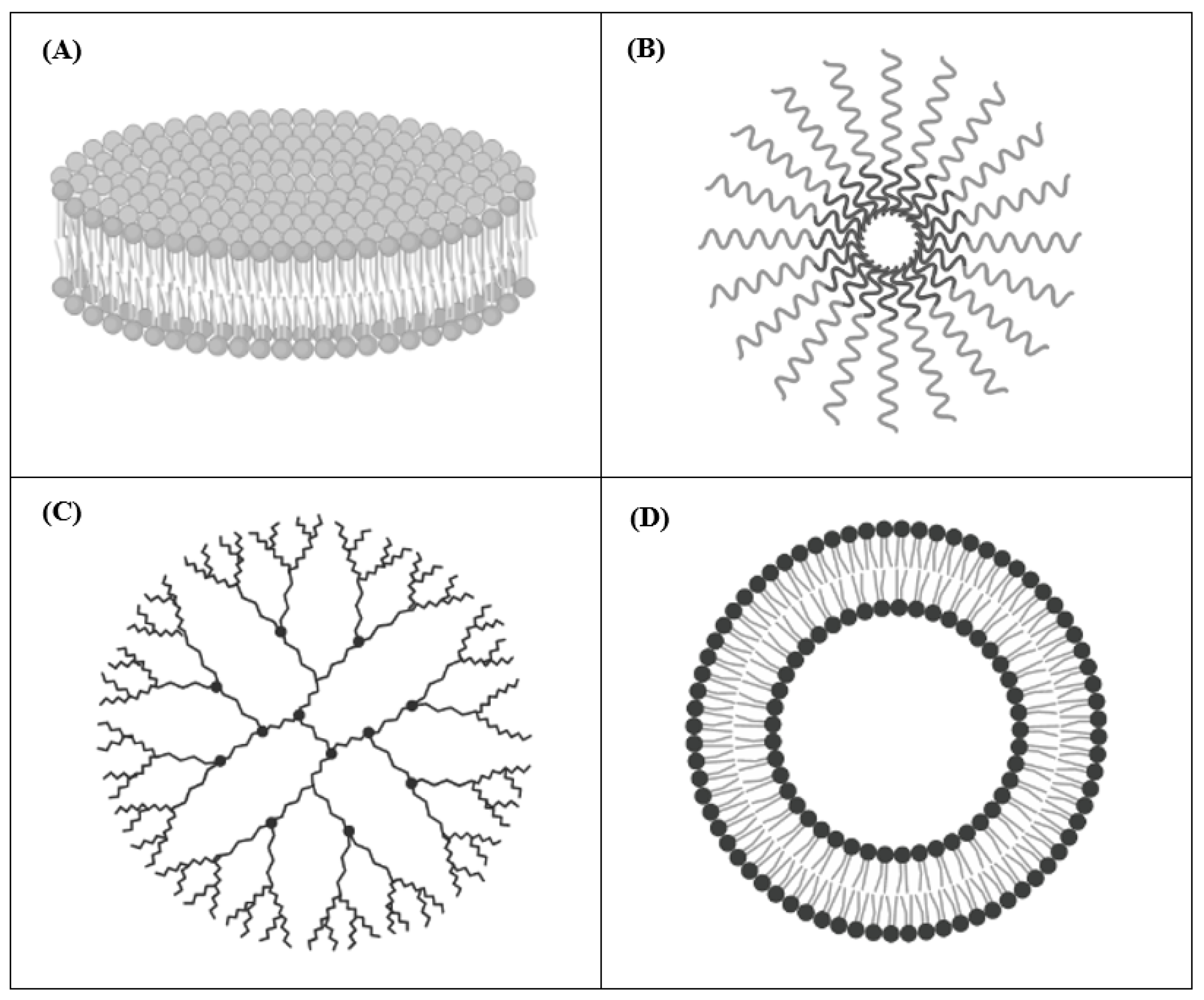
2. Drug Delivery Systems for Cancer Therapy

Drug Delivery Systems (DDSs) refer to formulations able to transport and deliver active molecules/drugs to cell/tissue targets in order to achieve a specific and hopefully increased therapeutic effect compared to the free drug while minimizing its potential side effects. The use of DDSs in cancer therapy lies in the possibility of increasing the therapeutic index of the encapsulated drugs by delivering them to tumor cells through both passive and active targeting. Passive targeting of tumor cells exploits the so-called “Enhanced Permeability and Retention” effect (EPR) [30]. It is well known that the newly formed blood vessels of solid tumors present altered permeability, rendering them more permeable compared to those of healthy tissues [30]. In these circumstances, the leaky blood vessels allow for the non-selective extravasation of macromolecules (larger than 40 kDa) and small particles (ranging from 50 to 500 nm) into the tumor stroma, finally leading to tumor cells killing [30,31]. However, the passive targeting capability of DDSs only leads to a modest increased delivery of the encapsulated drug to the target site, and it is strictly dependent on different factors such as size and circulation time of the carrier, as well as on tumor biology features, such as vascularity of the tumor and leakiness of the vessels [31]. On the other hand, the passive targeting capability of DDSs can be further optimized by coupling moieties (e.g., monoclonal antibodies, peptides etc.) on their external surface, with the aim to specifically recognize and target tumor-associated antigens [18,31,32,33,34].
The most relevant DDSs used in pre-clinical studies of cancer therapy are nanodisks (NDs), polymeric micelles, dendrimers, and liposomes (Figure 1). In these formulations, retinoic acids can be either entrapped into the inner core or mixed with the outer surface [18]. Below, some examples of each mentioned retinoids carrying DDSs are presented.

2.1. Nanodisks (NDs)

NDs are self-assembled nanoscale carriers composed of a phospholipid bilayer surrounded by amphipathic apolipoproteins that stabilize the structure, serving as a scaffold [35,36] (Figure 1A). This composition allows the encapsulation and the delivery of hydrophobic molecules, such as amphotericin B and ATRA [35,37]. NDs have the advantages of being very small in size (8–20 nm in diameter) and fully soluble in water [36]. Singh and colleagues used NDs to encapsulate ATRA for treating cell culture models of mantle cell lymphoma (MCL). Compared to free ATRA, they demonstrated that ATRA-NDs were more effective in inducing MCL cells apoptosis and G1 cell cycle arrest in vitro [38]. Then, they optimized the formulation by adding the single chain variable antibody fragment against CD20 on the surface in order to improve the selective targeting of CD20-positive MCL cells. In this case, NDs were loaded with either ATRA (ATRA-NDs) or curcumin (curcumin-NDs) and the combination therapy was able to induce higher tumor apoptosis compared to each single treatment [38,39]. Importantly, these anti-CD20 NDs, although developed for treating MCL cells, may be useful for any other CD20-expressing tumors [39]. In another study, Buehler et al. engineered vault nanoparticles in order to encapsulate ATRA, using a vault-binding lipoprotein complex that creates a lipid bilayer NDs [40]. Testing hepatocellular carcinoma cell viability after ATRA-vaults treatment, they demonstrated that ATRA-NDs caused increased tumor cells killing compared to that obtained by free ATRA [40].

2.2. Polymeric Micelles

Polymeric Micelles are composed of amphiphilic polymers, which self-associate when added to an aqueous solvent. After self-assembly in the aqueous environment, the hydrophilic polymers (e.g., poly(ethylene glycol), chitosan, dextran, and hyaluronic acids) face the aqueous medium forming a hydrophilic shell, while the hydrophobic ones (e.g., poly(lactide) (PLA), poly(caprolactone) (PCL), poly(lactide-co-glycolide) (PLGA), polyesters, poly(amino acids), and lipids) form the hydrophobic core (Figure 1B). Similar to NDs, Polymeric Micelles can be employed to encapsulate hydrophobic drugs [41]. The anti-cancer agents can be conjugated to the distal ends of polymer to prepare pharmacologically active polymeric systems that enhance solubility and stability of the conjugates, providing an opportunity for combined drug delivery [41]. Specifically, an efficient intracellular drug delivery system is represented by the use of biocompatible polymeric micelles (BPMs), which allow the administration of retinoic acid (RA), protecting RA from metabolic deactivation while reducing RA-mediated toxicity [42]. For instance, the apoptotic effects induced by RA, either free or encapsulated into BPMs, were compared on colon cancer cell lines. When loaded into BPMs, RA led to a stronger effect with respect to the free administration, also despite the lower dose used [42]. Furthermore, Orienti et al. developed a nano-micellar formulation entrapping 4-HPR into the inner core, called bionanofenretinide (Bio-nFeR) [43]. This system increased fenretinide bioavailability, showing anti-tumor activity against lung, colon, and melanoma cancer stem cells, both in vitro and in tumor xenografts. Interestingly, Bio-nFeR showed lower toxicity when compared to NCI-FeR, an oral formulation of 4-HPR, consisting of soft gelatin capsules, actually available at the National Cancer Institute, and administered in clinical trials [43].

2.3. Dendrimers

Dendrimers are polymeric molecules composed of multiple repetitive branches arising radially from a central core. The terminal groups of every branch provide modifiable functionalities. The number of repeated branching units determines the generation of the dendrimer [44,45] (Figure 1C). Dendrimers are widely used as carriers for the delivery of several therapeutics compounds, including retinoids [46]. They present several advantageous features such as high water solubility, monodispersity, biocompatibility, and low immunogenicity [47]. Moreover, pH-sensitive formulations have been developed in order to be stable at physiological pH and to dissociate in the acid environment of the endosomal and lysosomal tumor compartments, resulting in an enhanced cellular uptake into target cells. For instance, Wang et al. synthesized pH-sensitive nanoparticles based on poly(amidoamine) (PAMAM) dendrimers encapsulating ATRA. They tested the formulation in vitro on human hepatocellular liver carcinoma cells, demonstrating its ability to arrest tumor cell proliferation and increase tumor cell death, compared to free ATRA [48]. Yalçın et al. loaded gemcitabine together with ATRA into PAMAM dendrimer-coated magnetic nanoparticles (DcMNPs) in order to simultaneously target gemcitabine-resistant pancreatic cancer cells and pancreatic stellate cells (PSC), stromal cells that support tumorigenesis, and form a fibrotic barrier against therapeutic agents [49,50]. They firstly proved that the DcMNPs were successfully internalized by pancreatic cancer cell lines and by primary human PSC. Then, overcoming pancreatic cancer cell’s resistance to gemcitabine, showed that the increased gemcitabine- and ATRA-loaded DcMNPs accumulation into tumor cells and tumor stroma caused a significant cell death compared to that obtained by ATRA or gemcitabine administered separately [49].

2.4. Liposomes

Liposomes are spherical-shaped vesicles composed of a hydrophilic aqueous space surrounded by one or more phospholipid bilayers, making them similar to the cell membrane structure [51,52] (Figure 1D). They can entrap both hydrophobic and hydrophilic compounds. The ability of liposomes to encapsulate “drugs” characterized by different solubility in water and to specifically target organs, tissues, and cells makes them attractive candidates for drug delivery [53]. They can be classified on the basis of: (i) size; (ii) lipid composition; (iii) surface modification. Due to their good features, such as biocompatibility, biodegradability, and low toxicity, liposomes are the first DDSs that have been translated to clinical application [54,55,56]. Further, they are the most frequently used formulations for drugs encapsulation and, at present, several liposomal formulations have been approved by the FDA, and different products are available for clinical application (e.g., Doxil®, Ambisome®, DepoDur™, DaunoXome®, etc. [54,55,56]). Moreover, liposomes are the only nanosystems used in clinical trials for the delivery of retinoids in solid cancer (https://clinicaltrials.gov/) (accessed on 15 March 2021). To date, pre-clinical evaluations of retinoids-encapsulating liposomes have been testing against several types of cancer, including lung, thyroid, and liver cancers, as well as on neuroectodermal-derived tumors such as melanoma and NB [57,58,59,60,61,62]. In particular, the anti-tumor effects of cationic liposomes encapsulating ATRA were also tested in pre-clinical animal models of lung cancer [57]. Interestingly, in this study aimed at investigating ATRA-driven reactivation of the tumor suppressor protein retinoic acid receptor beta (RAR-β), it was shown that, compared to free ATRA, the treatment with ATRA-loaded liposomes led to an enhanced RAR-β expression, thus becoming a useful molecular target therapy for lung cancer [57]. In another study, with the aim to reduce ATRA photo-degradation during administration as a free drug, and consequently to increase its anti-cancer activity, a different liposomal formulation was developed [58]. The authors demonstrated that the liposomes protected ATRA and increased its anti-proliferative properties due to the improvement of its cellular uptake, becoming a useful formulation for the treatment of anaplastic thyroid carcinoma [58]. Moreover, Kawakami and colleagues demonstrated that ATRA incorporated into cationic liposomes was efficiently internalized into ATRA-resistant human lung cancer cells in vitro [59]. Specifically, the interaction between the positive charges of the liposomes and the negative charges of the tumor cell membranes allowed the specific internalization of ATRA, thus overcoming tumor cell resistance and producing pro-apoptotic and cytotoxic effects [59].
As already mentioned in the introduction, fenretinide (4-HPR) is a synthetic retinoic acid derivative, endowed with anti-tumor properties and characterized by favorable pharmacological profile, with lower systemic toxicity and better tissue distribution compared to its natural analogue [24]. Nevertheless, the main limitation for the clinical application of 4-HPR derives from its poor bioavailability [27]. Indeed, plasma levels of 4-HPR in patients receiving the maximum tolerated dose (MTD) of the drug (200 mg) were less than 1 µm, not sufficient to produce the desired anti-tumor effects [27]. To overcome such limitation and, consequently, to improve fenretinide performance, efforts to develop proper DDSs have been made in our laboratory. The results achieved, with particular attention on the use of liposomal fenretinide for neuroectodermal-derived tumors, are summarized below.

2.4.1. Neuroectodermal-Derived Tumors

During embryonic development, the three germ layers, endoderm, mesoderm, and ectoderm [63,64], give rise to all the tissues of the adult [65]. The endoderm creates the respiratory system, the digestive system, and some inner organs, such as the thyroid, the thymus, and the liver [66]. The mesoderm produces the musculoskeletal system, the cardiovascular system and the connective tissues [65]. The ectoderm gives rise to the surface ectoderm, the neural tube, and the neural crest [67]. The surface ectoderm generates the epidermis, the cutaneous annexes, the surface epithelium of the mouth and nose, the anterior pituitary gland, the tooth enamel, and the olfactory/optical/optic placodes [67]. The neural tube and the neural crest constitute the neuroectoderm, the first step in the development of the nervous system [68]. The neural tube produces the brain (rhombencephalon, mesencephalon, and prosencephalon), the spinal cord and the motor neurons, the retina, and the posterior pituitary [68]. The neural crest generates the pigment cells in the skin, the ganglia of the autonomic nervous system, the dorsal root ganglia, the facial cartilage, the pulmonary aortic septum of the developing heart and lungs, the ciliary body of the eye, the parafollicular cells (thyroid C cells) and the adrenal medulla [69,70]. In this contest, mutations in mature cells of neuroectoderm may reactivate the embryonic developmental pathways and initiate oncogenesis [71]. Among neuroectodermal-derived tumors, medullary thyroid carcinoma in the thyroid C cells, pheochromocytoma in the chromaffin cells of the adrenal medulla, ganglioneuroma in peripheral nervous system ganglia, malignant peripheral nerve sheath tumor (MPNST) in Schwann cells, melanoma in melanocytes and NB in sympathoadrenal precursors, are among the most frequently reported [71]. In particular, malignant melanoma, the most lethal form of skin cancer [72], affects adult population, with a mean age at diagnosis of 65 years, representing a clinical challenge. Indeed, although progresses have been obtained by immune check point blockade [73], the prognosis for metastatic and/or refractory disease is poor [74], and new therapeutic interventions appear necessary. NB is the most common extra-cranial solid tumor of pediatric age, and it accounts for approximately 15% of all oncology-related pediatric deaths [75,76]. Significant progress has been made in the cure of NB. However, despite the application of aggressive treatment strategies also including differentiation therapies based on the use of 13-cis retinoic acid, the clinical outcome for high-risk NB patients remains poor [77]. As for melanoma, new therapeutic approaches and more effective drugs are needed.

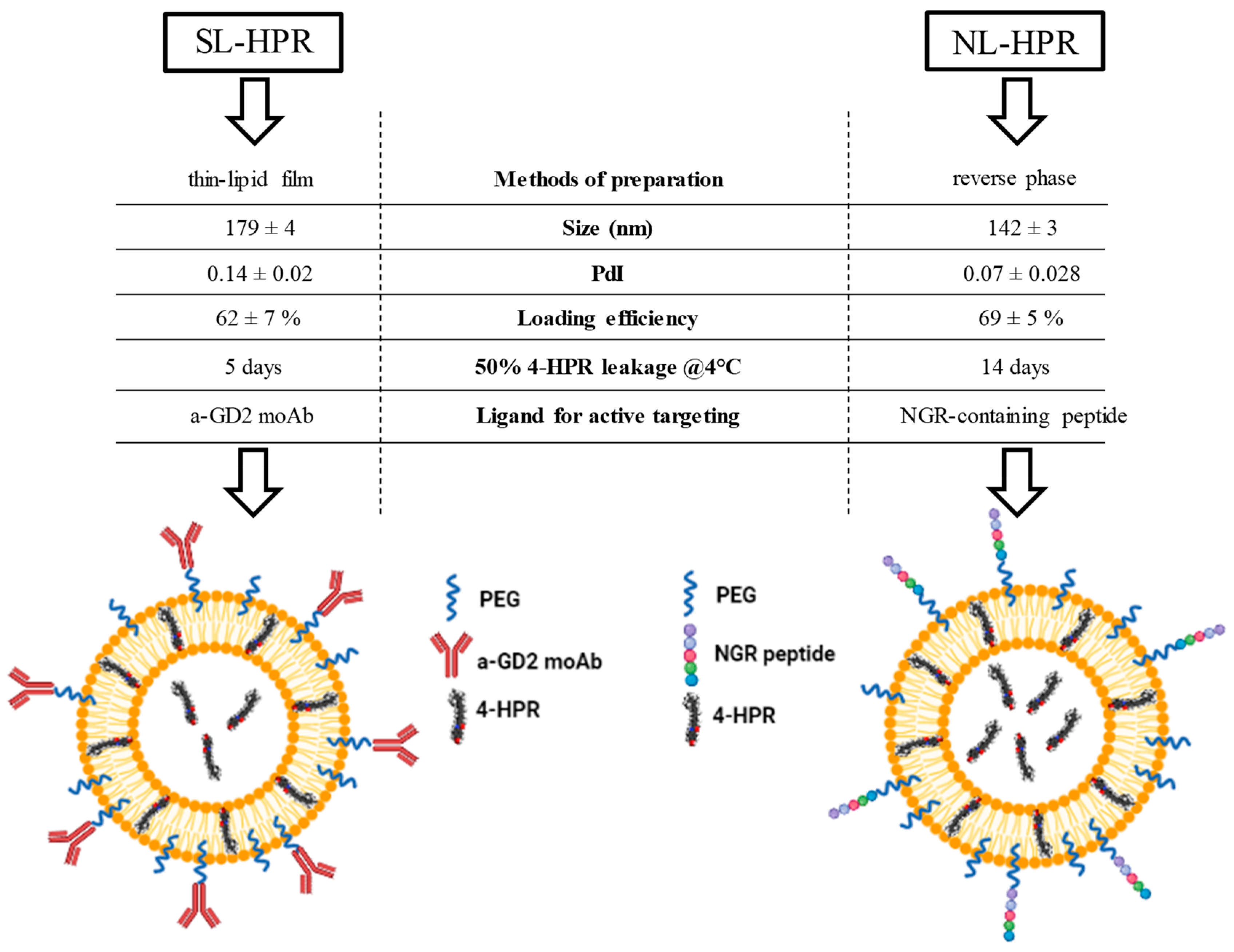
2.4.2. Development, Characterization, and Functionality of Fenretinide-Loaded Liposomes

Sterically stabilized, namely Stealth Liposomes (SL) encapsulating 4-HPR (SL-HPR) were developed and characterized [61,62]. SL are characterized by long circulation into the bloodstream, thanks to the use of the phospholipid 1,2-Distearoyl-sn-glycero-3-phosphorylethanolamine (DSPE) functionalized with polyethylene glycol (PEG), which confers to the liposomes stealth features. Following this method of composition, the recognition of liposomes by the cells of the reticulum endothelial system is reduced, in turn conferring them an increased blood circulation capability and an enhanced possibility to target the tumor [78,79]. SL-HPR showed a chemo-preventive action in the early stages of rat hepatocarcinogenesis, with anti-proliferative effects and apoptosis induction of initiated cells [60]. Moreover, as reported into details below, the same formulation, additionally decorated with an antibody recognizing the disialoganglioside GD2, exerted potent anti-tumor effects against the GD2-expressing neuroectodermal-derived tumors, melanoma and NB [61,62,80].
Attracted from the good properties demonstrated by 4-HPR and on the basis of its in vitro anti-tumor effect obtained when administered as free agent against NB and melanoma cells [10,11], at the end of 90’s, we focused on the development of SL-HPR. The aim was to enhance its bioavailability in vivo and, consequently, the anti-tumor potential. The idea to entrap 4-HPR into liposomes arose also from the fact that the in vitro anti-tumor effects mentioned above were obtained using a dosage 2–10 times higher than that reachable in vivo [81,82].

2.4.3. Fenretinide-loaded Liposomes for Tumor Targeting

SL-HPR were synthesized following the thin-film hydration method, which consists of making a thin lipid film by organic solvent removal, addition of the dispersion buffer and extrusion through 0.2–0.08 μm-pore size polycarbonate membranes, obtaining homogeneous small liposomes [83,84]. In order to increase tumor targeting, SL-HPR were then decorated with the anti-GD2 antibody (a-GD2 moAb), specifically recognizing the disialoganglioside GD2 highly expressed on the cell surface of melanoma and NB cells, and whose expression on healthy tissues is very limited and restricted to cerebellum and peripheral nerves [85,86]. This new formulation was consequently named GD2-Stealth ImmunoLiposomes (GD2-SIL-HPR) (Figure 2), and its functionality on neuroectodermal-derived tumors was firstly assessed on GD2-positive melanoma cells. By competition experiments, cellular association resulted specific and 10 to 15-fold higher compared to that obtained by the untargeted formulation [61]. From a therapeutic point of view, cell proliferation assays revealed that GD2-SIL-HPR were more effective in inhibiting melanoma cell proliferation, compared to both free 4-HPR and SL-HPR. As a note of worth, the anti-tumor efficacy was strictly dependent on the GD2 expression. Indeed, experiments performed on melanoma cells with different degree of GD2 expression showed different response rates to GD2-SIL-HPR treatment, with high response achieved in cell lines expressing high levels of GD2 [61].
The anti-tumor functionality of GD2-SIL-HPR was then evaluated, and extended, against GD2-expressing NB models. Again, in tumor cell binding and uptake studies, the formulation resulted highly selective for GD2-expressing NB cells [62]. Compared to non-targeted liposomes, cellular association of GD2 targeted SIL-HPR resulted 10- to 20-fold higher. As an important note about the carrier integrity, this formulation was able to maintain its ability to bind to GD2-positive cells for at least one week, when stored at 4 °C [61,62].
The in vitro anti-tumor efficacy of GD2-SIL-HPR was studied through cell proliferation experiments, which demonstrated that this formulation significantly inhibited NB cell proliferation compared to HPR, either free or encapsulated in untargeted liposomes. Also in this case, the degree of cell proliferation inhibition correlated with the extent of GD2 expression. NB cell lines expressing higher amount of GD2 better responded to the treatment with GD2-SIL-HPR [62]. A further confirmation of the specificity of the anti-tumor efficacy derived from the treatment of cell lines not expressing GD2. Cell proliferation of these cells was not affected by GD2-tageted-SIL-HPR [62].
Furthermore, to give a translational relevance to the findings obtained in vitro, the GD2-targeted liposomal formulation of HPR was then tested in a metastatic mouse model of human NB, where NBs cells are injected intravenously in the mice tail [87]. This model is clinically relevant, because it mimics both the dissemination of the disease at distant sites, characteristic of patients affected by stage M high-risk NB, and a state of minimal residual disease [87,88]. Treatment with GD2-SIL-HPR determined a complete inhibition of tumor micrometastases development in liver, kidney and ovaries, compared to mice receiving placebo (vehicle buffer only) or free HPR [62]. Survival studies confirmed and supported the above findings. While free HPR and SL-HPR treatments showed a partial anti-tumor response, GD2-SIL-HPR led to long-term survivors, paving the way for future clinical application [62].

2.4.4. Fenretinide-Loaded Liposomes for Tumor Vasculature Targeting

A further study conducted in our laboratory, was aimed at developing and testing a liposomal formulation of HPR, specifically targeted to the aminopeptidase N (CD13) receptor expressed by the neo-angiogenic vessels of solid tumors, including NB [33,89,90]. The idea to target tumor vessels within the mass of a solid NB tumor, arises from two main observations; first, high vascular index in NB is associated to poor prognosis [91,92]; second, therapeutic strategy based on the use of liposomal vectors decorated with tumor vasculature-homing and -penetrating peptides might help anti-cancer drugs to overcome many of the physiological barriers present in the abnormal tumor vasculature and on its interstitial matrix, likely increasing their therapeutic effects [34,93].
The new HPR-entrapping, sterically stabilized liposomal formulation, called hereafter NanoLiposomes-HPR (NL-HPR) was developed according to the reverse phase evaporation method [94] and its chemical and structural properties compared to those belonging to the liposomes synthesized and developed for tumor targeting, and following the thin-film hydration method. The reverse phase evaporation method was chosen with the aim to optimize the previously synthesized formulation and because the method used to prepare liposomes can also significantly impact on the physicochemical properties of the payload itself. Size of liposomes was here determined by the dynamic light scattering technology. Compared to SL-HPR, the average size of NL-HPR was smaller (179 ± 4 nm vs 142 ± 3 nm, respectively) and the HPR trapping efficiency was slightly increased (62 ± 7% vs 69 ± 5%, respectively) (Figure 2). The two formulations were also compared in terms of integrity of the vehicle. Importantly, leakage experiments demonstrated that NL-HPR were able to retain the entrapped drug for longer time with respect to SL-HPR. Specifically, while about 50% of HPR was released from SL-HPR in a time of 5 days, the same percentage of release from NL-HPR was reached in 14 days (Figure 2). In summary, the reverse phase evaporation method ameliorated all the liposomal features, rendering the new formulation potentially more performing.
NL-HPR were then decorated with an NGR-motif containing peptide that specifically targets CD13-positive neo-angiogenic tumor blood vessels [95] (Figure 2). Of note, coupling on the external surface of the targeting moiety did not alter the physicochemical properties of the resulting NGR-NL-HPR formulation [80].
NL-HPR and NGR-NL-HPR were then tested in the clinically relevant orthotopic mouse model of human NB, where a primary tumor mass and metastases at distant sites grow after tumor cells injection in the adrenal gland of mice [96,97]. When compared to control mice and those treated with free HPR, both NL-HPR and NGR-NL-HPR determined a significant increase in life span. However, NGR-NL-HPR led to a significant increase in life span if compared to the untargeted formulation, highlighting the impact of active targeting on the achieved anti-tumor effects. As a consequence, NGR-NL-HPR was the only therapy able to lead a 20% of long-term survivors [80]. These results were explained by the histological analyses performed on tumor samples derived from control and treated animals. As assessed by the staining with the proliferation marker Ki-67, NGR-NL-HPR determined a significant reduction of NB tumor growth compared to both free HPR and NL-HPR. In line with the known mechanism of action of HPR as apoptosis inducer [10,98], TUNEL assays performed on tissue sections from treated mice revealed that the percentage of apoptotic cells was drastically increased when HPR was delivered through the NGR-decorated liposomal formulation. Finally, immunofluorescence staining performed by using anti-CD31, -αSMA, -VEGF, -MMP-2 and -MMP-9 antibodies demonstrated a significant reduction of tumor angiogenesis and capability of tumor invasion, when HPR was administered through NL-HPR and, at a greater extent, through NGR-NL-HPR [80].

3. Discussion

Retinoids are a class of natural and synthetic compounds derived from vitamin A, which have raised interest within the scientific community thanks, among others, to their beneficial effects in cancer [7,8,9]. More specifically, the synthetic retinoic acid derivative fenretinide (4-HPR) is endowed with anti-tumor properties and characterized by a favorable pharmacological profile, with lower systemic toxicity and better tissue distribution compared to its natural analog [24]. However, despite substantial in vitro cytotoxicity, response rates in early clinical trials with 4-HPR have been less than anticipated, likely as the consequence of its low bioavailability. To improve bioavailability of 4-HPR, oral powder (LYM-X-SORB®, LXS) and intravenous lipid emulsion (ILE) formulations have been tested in early-phase clinical trials [27]. On the other hand, improving pharmacological properties and circulating half-life of retinoids while minimizing their off-target uptake, several delivery systems have been recently developed.
Here, the physicochemical features and the anti-tumor potential of 2 different, sterically stabilized, fenretinide-loaded liposomal formulations developed in our laboratory are summarized. Specifically, these two formulations were different from each other both for the method of preparation (thin-lipid film and reverse phase methods) and for the ligand associated at their external surface to obtain liposomes with the ability to selectively target either the tumor or the tumor vasculature of the neuroectodermal-derived melanoma and neuroblastoma (NB) tumors. Both formulations were found to have optimal physicochemical characteristics, allowing for improving the half-life of the encapsulated 4-HPR drug. Of note, the tumor vasculature-targeted formulation developed following the reverse phase method resulted slightly better than the formulation developed according to the thin-lipid film method in terms of size, polydispersity, drug loading efficiency and drug retention. Indeed, unlike the thin-lipid film method, in which the drug is added after the formation of the liposomes [61,62], the reverse phase method basically involves the initial mixing of the lipids with the fenretinide, the subsequent sonication, and the final evaporation. In this way, more drug was trapped both within the lipid core and at the level of the lipid bilayer itself (see Figure 2), making the vasculature-targeted nanoliposomes more drug-laden, and with a greater retention capacity. However, both 4-HPR-loaded formulations have been shown to exert a potent anti-tumor effect, both in vitro and in vivo. This therapeutic achievement, significantly higher than that obtained by the use of 4-HPR administered in free form or encapsulated in liposomes without targeting, might allow hypothesizing their clinical use in the future.
The use of liposomes as drug delivery has the main purpose of increasing the pharmacological half-life and, at the same time, trying to enhance the tumor-targeting, compared to that obtained by using the same drug but administered in free form. Regarding the delivery of retinoids by liposomes, this will not represent the panacea for all ills, nor will it totally abolish possible side effects. On the other hand, in the last 20 years, we were able to demonstrate that targeted liposomes can ameliorate the drug’s half-life and increase its therapeutic potential [34,87,96,99,100,101,102,103,104]. Moreover, targeted liposomal formulations are well tolerated, and the reduced side effects, compared to free drugs, were demonstrated [105].

Author Contributions

Conceptualization, C.B. and F.P.; writing—original draft preparation, V.B., E.C., C.B. and F.P.; writing—review and editing, V.B., E.C., E.G., P.P., M.V.C., M.P., C.B. and F.P.; funding acquisition, M.P. and F.P. All authors have read and agreed to the published version of the manuscript.

Funding

Ministero della Salute, Ricerca Corrente 2021 to Istituto Giannina Gaslini, Associazione Italiana per la Ricerca sul Cancro, Investigator Grant n. 18474 to M.P. and n. 24397 to F.P. V.B., and E.C. are recipients of Italian Foundation for neuroblastoma research and Associazione Oncologia Pediatrica E Neuroblastoma (OPEN) ONLUS fellowship.

Institutional Review Board Statement

Not applicable.

Informed Consent Statement

Not applicable.

Data Availability Statement

Data sharing not applicable.

Acknowledgments

Fabio Morandi for technical support. BioRender.com (accessed on 30 July 2021) was used for the realization of the figures.

Conflicts of Interest

The Authors declare no conflict of interest.

Abbreviations

4-HPRN-4-hydroxyphenyl retinamide (fenretinide)
NBneuroblastoma
SLStealth Liposomes
SILStealth ImmunoLiposomes

References

  1. Blomhoff, R.; Blomhoff, H.K. Overview of retinoid metabolism and function. J. Neurobiol. 2006, 66, 606–630. [Google Scholar] [CrossRef] [PubMed]
  2. Campo-Paysaa, F.; Marlétaz, F.; Laudet, V.; Schubert, M. Retinoic acid signaling in development: Tissue-specific functions and evolutionary origins. Genesis 2008, 46, 640–656. [Google Scholar] [CrossRef] [PubMed]
  3. Niederreither, K.; Subbarayan, V.; Dollé, P.; Chambon, P. Embryonic retinoic acid synthesis is essential for early mouse post-implantation development. Nat. Genet. 1999, 21, 444–448. [Google Scholar] [CrossRef]
  4. Sporn, M.B.; Dunlop, N.M.; Newton, D.L.; Henderson, W.R. Relationships between structure and activity of retinoids. Nature 1976, 263, 110–113. [Google Scholar] [CrossRef] [PubMed]
  5. Kiser, P.D.; Golczak, M.; Palczewski, K. Chemistry of the retinoid (visual) cycle. Chem. Rev. 2014, 114, 194–232. [Google Scholar] [CrossRef] [PubMed]
  6. Beckenbach, L.; Baron, J.M.; Merk, H.F.; Loffler, H.; Amann, P.M. Retinoid treatment of skin diseases. Eur. J. Dermatol. 2015, 25, 384–391. [Google Scholar] [CrossRef] [PubMed]
  7. Dobrotkova, V.; Chlapek, P.; Mazanek, P.; Sterba, J.; Veselska, R. Traffic lights for retinoids in oncology: Molecular markers of retinoid resistance and sensitivity and their use in the management of cancer differentiation therapy. BMC Cancer 2018, 18, 1059. [Google Scholar] [CrossRef]
  8. Bollag, W. Retinoids and cancer. Cancer Chemother. Pharmacol. 1979, 3, 207–215. [Google Scholar] [CrossRef] [PubMed]
  9. Zusi, F.C.; Lorenzi, M.V.; Vivat-Hannah, V. Selective retinoids and rexinoids in cancer therapy and chemoprevention. Drug Discov. Today 2002, 7, 1165–1174. [Google Scholar] [CrossRef]
  10. Ponzoni, M.; Bocca, P.; Chiesa, V.; Decensi, A.; Pistoia, V.; Raffaghello, L.; Rozzo, C.; Montaldo, P.G. Differential effects of N-(4-hydroxyphenyl)retinamide and retinoic acid on neuroblastoma cells: Apoptosis versus differentiation. Cancer Res. 1995, 55, 853–861. [Google Scholar]
  11. Montaldo, P.G.; Pagnan, G.; Pastorino, F.; Chiesa, V.; Raffaghello, L.; Kirchmeier, M.; Allen, T.M.; Ponzoni, M. N-(4-hydroxyphenyl) retinamide is cytotoxic to melanoma cells in vitro through induction of programmed cell death. Int. J. Cancer 1999, 81, 262–267. [Google Scholar] [CrossRef]
  12. Ferrara, F.F.; Fazi, F.; Bianchini, A.; Padula, F.; Gelmetti, V.; Minucci, S.; Mancini, M.; Pelicci, P.G.; Lo Coco, F.; Nervi, C. Histone deacetylase-targeted treatment restores retinoic acid signaling and differentiation in acute myeloid leukemia. Cancer Res. 2001, 61, 2–7. [Google Scholar] [PubMed]
  13. Liu, M.; Iavarone, A.; Freedman, L.P. Transcriptional activation of the human p21(WAF1/CIP1) gene by retinoic acid receptor. Correlation with retinoid induction of U937 cell differentiation. J. Biol. Chem. 1996, 271, 31723–31728. [Google Scholar] [CrossRef] [Green Version]
  14. Johnson, K.J. The retinoids: Biology, chemistry and medicine (second edition) Edited by M B Sporn, A B Roberts and the late D S Goodman. pp 311. Raven Press, NY. 1993. $250.00 ISBN 0-7817-0082-5. Biochem. Educ. 1994, 22, 56. [Google Scholar] [CrossRef]
  15. Hu, J.; Liu, Y.F.; Wu, C.F.; Xu, F.; Shen, Z.X.; Zhu, Y.M.; Li, J.M.; Tang, W.; Zhao, W.L.; Wu, W.; et al. Long-term efficacy and safety of all-trans retinoic acid/arsenic trioxide-based therapy in newly diagnosed acute promyelocytic leukemia. Proc. Natl. Acad. Sci. USA 2009, 106, 3342–3347. [Google Scholar] [CrossRef] [Green Version]
  16. Villablanca, J.G.; Khan, A.A.; Avramis, V.I.; Seeger, R.C.; Matthay, K.K.; Ramsay, N.K.; Reynolds, C.P. Phase I trial of 13-cis-retinoic acid in children with neuroblastoma following bone marrow transplantation. J. Clin. Oncol. 1995, 13, 894–901. [Google Scholar] [CrossRef]
  17. Reynolds, C.P.; Matthay, K.K.; Villablanca, J.G.; Maurer, B.J. Retinoid therapy of high-risk neuroblastoma. Cancer Lett. 2003, 197, 185–192. [Google Scholar] [CrossRef]
  18. Ferreira, R.; Napoli, J.; Enver, T.; Bernardino, L.; Ferreira, L. Advances and challenges in retinoid delivery systems in regenerative and therapeutic medicine. Nat. Commun. 2020, 11, 4265. [Google Scholar] [CrossRef]
  19. David, M.; Hodak, E.; Lowe, N.J. Adverse effects of retinoids. Med. Toxicol. Advers. Drug Exp. 1988, 3, 273–288. [Google Scholar] [CrossRef]
  20. LiverTox: Clinical and Research Information on Drug-Induced Liver Injury; National Institute of Diabetes and Digestive and Kidney Diseases: Bethesda, MD, USA, 2012.
  21. Khalil, S.; Bardawil, T.; Stephan, C.; Darwiche, N.; Abbas, O.; Kibbi, A.G.; Nemer, G.; Kurban, M. Retinoids: A journey from the molecular structures and mechanisms of action to clinical uses in dermatology and adverse effects. J. Dermatol. Treat. 2017, 28, 684–696. [Google Scholar] [CrossRef]
  22. Goncalves, A.; Estevinho, B.N.; Rocha, F. Formulation approaches for improved retinoids delivery in the treatment of several pathologies. Eur. J. Pharm. Biopharm. 2019, 143, 80–90. [Google Scholar] [CrossRef] [PubMed]
  23. Scott, L.J. Trifarotene: First Approval. Drugs 2019, 79, 1905–1909. [Google Scholar] [CrossRef] [PubMed]
  24. Torrisi, R.; Decensi, A. Fenretinide and cancer prevention. Curr. Oncol. Rep. 2000, 2, 263–270. [Google Scholar] [CrossRef] [PubMed]
  25. Kelloff, G.J.; Crowell, J.A.; Boone, C.W.; Steele, V.E.; Lubet, R.A.; Greenwald, P.; Alberts, D.S.; Covey, J.M.; Doody, L.A.; Knapp, G.G.; et al. Strategy and planning for chemopreventive drug development: Clinical development plans. Chemoprevention Branch and Agent Development Committee. National Cancer Institute. J. Cell Biochem. Suppl. 1994, 20, 55–62. [Google Scholar] [CrossRef]
  26. Mehta, R.G.; Moon, R.C.; Hawthorne, M.; Formelli, F.; Costa, A. Distribution of fenretinide in the mammary gland of breast cancer patients. Eur. J. Cancer 1991, 27, 138–141. [Google Scholar] [CrossRef]
  27. Cooper, J.P.; Reynolds, C.P.; Cho, H.; Kang, M.H. Clinical development of fenretinide as an antineoplastic drug: Pharmacology perspectives. Exp. Biol. Med. (Maywood) 2017, 242, 1178–1184. [Google Scholar] [CrossRef] [Green Version]
  28. Formelli, F.; Barua, A.B.; Olson, J.A. Bioactivities of N-(4-hydroxyphenyl) retinamide and retinoyl beta-glucuronide. FASEB J. 1996, 10, 1014–1024. [Google Scholar] [CrossRef]
  29. Trapasso, E.; Cosco, D.; Celia, C.; Fresta, M.; Paolino, D. Retinoids: New use by innovative drug-delivery systems. Expert Opin. Drug Deliv. 2009, 6, 465–483. [Google Scholar] [CrossRef]
  30. Torchilin, V. Tumor delivery of macromolecular drugs based on the EPR effect. Adv. Drug Deliv. Rev. 2011, 63, 131–135. [Google Scholar] [CrossRef]
  31. Attia, M.F.; Anton, N.; Wallyn, J.; Omran, Z.; Vandamme, T.F. An overview of active and passive targeting strategies to improve the nanocarriers efficiency to tumour sites. J. Pharm. Pharmacol. 2019, 71, 1185–1198. [Google Scholar] [CrossRef] [Green Version]
  32. Pastorino, F.; Marimpietri, D.; Brignole, C.; Di Paolo, D.; Pagnan, G.; Daga, A.; Piccardi, F.; Cilli, M.; Allen, T.M.; Ponzoni, M. Ligand-targeted liposomal therapies of neuroblastoma. Curr. Med. Chem. 2007, 14, 3070–3078. [Google Scholar] [CrossRef] [PubMed]
  33. Corti, A.; Pastorino, F.; Curnis, F.; Arap, W.; Ponzoni, M.; Pasqualini, R. Targeted Drug Delivery and Penetration Into Solid Tumors. Med. Res. Rev. 2012, 32, 1078–1091. [Google Scholar] [CrossRef] [PubMed]
  34. Pastorino, F.; Brignole, C.; Di Paolo, D.; Perri, P.; Curnis, F.; Corti, A.; Ponzoni, M. Overcoming Biological Barriers in Neuroblastoma Therapy: The Vascular Targeting Approach with Liposomal Drug Nanocarriers. Small 2019, 15, e1804591. [Google Scholar] [CrossRef]
  35. Redmond, K.A.; Nguyen, T.S.; Ryan, R.O. All-trans-retinoic acid nanodisks. Int. J. Pharm. 2007, 339, 246–250. [Google Scholar] [CrossRef] [PubMed] [Green Version]
  36. Ho, B.N.; Pfeffer, C.M.; Singh, A.T.K. Update on Nanotechnology-based Drug Delivery Systems in Cancer Treatment. Anticancer Res. 2017, 37, 5975–5981. [Google Scholar] [CrossRef] [Green Version]
  37. Ryan, R.O. Nanodisks: Hydrophobic drug delivery vehicles. Expert Opin. Drug Deliv. 2008, 5, 343–351. [Google Scholar] [CrossRef] [PubMed]
  38. Singh, A.T.; Evens, A.M.; Anderson, R.J.; Beckstead, J.A.; Sankar, N.; Sassano, A.; Bhalla, S.; Yang, S.; Platanias, L.C.; Forte, T.M.; et al. All trans retinoic acid nanodisks enhance retinoic acid receptor mediated apoptosis and cell cycle arrest in mantle cell lymphoma. Br. J. Haematol. 2010, 150, 158–169. [Google Scholar] [CrossRef] [PubMed] [Green Version]
  39. Stauffer, R.G.; Mohammad, M.; Singh, A.T. Novel Nanoscale Delivery Particles Encapsulated with Anticancer Drugs, All-trans Retinoic Acid or Curcumin, Enhance Apoptosis in Lymphoma Cells Predominantly Expressing CD20 Antigen. Anticancer Res. 2015, 35, 6425–6429. [Google Scholar]
  40. Buehler, D.C.; Toso, D.B.; Kickhoefer, V.A.; Zhou, Z.H.; Rome, L.H. Vaults engineered for hydrophobic drug delivery. Small 2011, 7, 1432–1439. [Google Scholar] [CrossRef] [Green Version]
  41. Ghosh, B.; Biswas, S. Polymeric micelles in cancer therapy: State of the art. J. Control. Release 2021, 332, 127–147. [Google Scholar] [CrossRef]
  42. Valerii, M.C.; Benaglia, M.; Caggiano, C.; Papi, A.; Strillacci, A.; Lazzarini, G.; Campieri, M.; Gionchetti, P.; Rizzello, F.; Spisni, E. Drug delivery by polymeric micelles: An in vitro and in vivo study to deliver lipophilic substances to colonocytes and selectively target inflamed colon. Nanomedicine 2013, 9, 675–685. [Google Scholar] [CrossRef]
  43. Orienti, I.; Salvati, V.; Sette, G.; Zucchetti, M.; Bongiorno-Borbone, L.; Peschiaroli, A.; Zolla, L.; Francescangeli, F.; Ferrari, M.; Matteo, C.; et al. A novel oral micellar fenretinide formulation with enhanced bioavailability and antitumour activity against multiple tumours from cancer stem cells. J. Exp. Clin. Cancer Res. 2019, 38, 373. [Google Scholar] [CrossRef] [Green Version]
  44. Lee, C.C.; MacKay, J.A.; Fréchet, J.M.; Szoka, F.C. Designing dendrimers for biological applications. Nat. Biotechnol. 2005, 23, 1517–1526. [Google Scholar] [CrossRef]
  45. Palmerston Mendes, L.; Pan, J.; Torchilin, V.P. Dendrimers as Nanocarriers for Nucleic Acid and Drug Delivery in Cancer Therapy. Molecules 2017, 22, 1401. [Google Scholar] [CrossRef] [PubMed] [Green Version]
  46. Tekade, R.K.; Dutta, T.; Tyagi, A.; Bharti, A.C.; Das, B.C.; Jain, N.K. Surface-engineered dendrimers for dual drug delivery: A receptor up-regulation and enhanced cancer targeting strategy. J. Drug Target. 2008, 16, 758–772. [Google Scholar] [CrossRef]
  47. Pan, J.; Attia, S.A.; Filipczak, N.; Torchilin, V.P. 10—Dendrimers for drug delivery purposes. In Nanoengineered Biomaterials for Advanced Drug Delivery; Mozafari, M., Ed.; Elsevier: Amsterdam, The Netherlands, 2020; pp. 201–242. [Google Scholar]
  48. Wang, Y.; Wang, H.; Lv, X.; Liu, C.; Qi, L.; Song, X.; Yu, A. Enhancement of all-trans retinoic acid-induced differentiation by pH-sensitive nanoparticles for solid tumor cells. Macromol. Biosci. 2014, 14, 369–379. [Google Scholar] [CrossRef] [PubMed]
  49. Yalcin, S.; Erkan, M.; Unsoy, G.; Parsian, M.; Kleeff, J.; Gunduz, U. Effect of gemcitabine and retinoic acid loaded PAMAM dendrimer-coated magnetic nanoparticles on pancreatic cancer and stellate cell lines. Biomed. Pharmacother. 2014, 68, 737–743. [Google Scholar] [CrossRef]
  50. Allam, A.; Thomsen, A.R.; Gothwal, M.; Saha, D.; Maurer, J.; Brunner, T.B. Pancreatic stellate cells in pancreatic cancer: In focus. Pancreatology 2017, 17, 514–522. [Google Scholar] [CrossRef] [PubMed]
  51. Akbarzadeh, A.; Rezaei-Sadabady, R.; Davaran, S.; Joo, S.W.; Zarghami, N.; Hanifehpour, Y.; Samiei, M.; Kouhi, M.; Nejati-Koshki, K. Liposome: Classification, preparation, and applications. Nanoscale Res. Lett. 2013, 8, 102. [Google Scholar] [CrossRef] [Green Version]
  52. Kopeckova, K.; Eckschlager, T.; Sirc, J.; Hobzova, R.; Plch, J.; Hrabeta, J.; Michalek, J. Nanodrugs used in cancer therapy. Biomed. Pap. Med. Fac. Univ. Palacky Olomouc. Czech. Repub. 2019, 163, 122–131. [Google Scholar] [CrossRef] [Green Version]
  53. Alavi, M.; Hamidi, M. Passive and active targeting in cancer therapy by liposomes and lipid nanoparticles. Drug Metab. Pers. Ther. 2019, 34. [Google Scholar] [CrossRef] [PubMed]
  54. Barenholz, Y. Doxil (R)—The first FDA-approved nano-drug: Lessons learned. J. Control. Release 2012, 160, 117–134. [Google Scholar] [CrossRef] [PubMed]
  55. Gabizon, A.A.; Patil, Y.; La-Beck, N.M. New insights and evolving role of pegylated liposomal doxorubicin in cancer therapy. Drug Resist. Updates 2016, 29, 90–106. [Google Scholar] [CrossRef]
  56. Bulbake, U.; Doppalapudi, S.; Kommineni, N.; Khan, W. Liposomal Formulations in Clinical Use: An Updated Review. Pharmaceutics 2017, 9, 12. [Google Scholar] [CrossRef]
  57. Viswanathan, S.; Berlin Grace, V.M.; Danisha, J.P. Enhancement of tumor suppressor RAR-β protein expression by cationic liposomal-ATRA treatment in benzo(a)pyrene-induced lung cancer mice model. Naunyn Schmiedebergs Arch. Pharmacol. 2019, 392, 415–426. [Google Scholar] [CrossRef]
  58. Cristiano, M.C.; Cosco, D.; Celia, C.; Tudose, A.; Mare, R.; Paolino, D.; Fresta, M. Anticancer activity of all-trans retinoic acid-loaded liposomes on human thyroid carcinoma cells. Colloids Surf. B Biointerfaces 2017, 150, 408–416. [Google Scholar] [CrossRef]
  59. Kawakami, S.; Suzuki, S.; Yamashita, F.; Hashida, M. Induction of apoptosis in A549 human lung cancer cells by all-trans retinoic acid incorporated in DOTAP/cholesterol liposomes. J. Control. Release 2006, 110, 514–521. [Google Scholar] [CrossRef]
  60. Simile, M.M.; Pagnan, G.; Pastorino, F.; Brignole, C.; De Miglio, M.R.; Muroni, M.R.; Asara, G.; Frau, M.; Seddaiu, M.A.; Calvisi, D.F.; et al. Chemopreventive N-(4-hydroxyphenyl)retinamide (fenretinide) targets deregulated NF-{kappa}B and Mat1A genes in the early stages of rat liver carcinogenesis. Carcinogenesis 2005, 26, 417–427. [Google Scholar] [CrossRef]
  61. Pagnan, G.; Montaldo, P.G.; Pastorino, F.; Raffaghello, L.; Kirchmeier, M.; Allen, T.M.; Ponzoni, M. GD2-mediated melanoma cell targeting and cytotoxicity of liposome-entrapped fenretinide. Int. J. Cancer 1999, 81, 268–274. [Google Scholar] [CrossRef]
  62. Raffaghello, L.; Pagnan, G.; Pastorino, F.; Cosimo, E.; Brignole, C.; Marimpietri, D.; Montaldo, P.G.; Gambini, C.; Allen, T.M.; Bogenmann, E.; et al. In vitro and in vivo antitumor activity of liposomal Fenretinide targeted to human neuroblastoma. Int. J. Cancer 2003, 104, 559–567. [Google Scholar] [CrossRef]
  63. Technau, U.; Scholz, C.B. Origin and evolution of endoderm and mesoderm. Int. J. Dev. Biol. 2003, 47, 531–539. [Google Scholar] [PubMed]
  64. Solnica-Krezel, L.; Sepich, D.S. Gastrulation: Making and shaping germ layers. Annu. Rev. Cell Dev. Biol. 2012, 28, 687–717. [Google Scholar] [CrossRef] [PubMed] [Green Version]
  65. Ferretti, E.; Hadjantonakis, A.K. Mesoderm specification and diversification: From single cells to emergent tissues. Curr. Opin. Cell Biol. 2019, 61, 110–116. [Google Scholar] [CrossRef] [PubMed]
  66. Zorn, A.M.; Wells, J.M. Vertebrate endoderm development and organ formation. Annu. Rev. Cell Dev. Biol. 2009, 25, 221–251. [Google Scholar] [CrossRef] [Green Version]
  67. Elshazzly, M.; Lopez, M.J.; Reddy, V.; Caban, O. Embryology, Central Nervous System. In StatPearls; StatPearls Publishing: Treasure Island, FL, USA, 2021. [Google Scholar]
  68. Darnell, D.; Gilbert, S.F. Neuroembryology. Wiley Interdiscip Rev. Dev. Biol. 2017, 6. [Google Scholar] [CrossRef] [Green Version]
  69. Pla, P.; Monsoro-Burq, A.H. The neural border: Induction, specification and maturation of the territory that generates neural crest cells. Dev. Biol. 2018, 444 Suppl. 1, S36–S46. [Google Scholar] [CrossRef]
  70. Krispin, S.; Nitzan, E.; Kalcheim, C. The dorsal neural tube: A dynamic setting for cell fate decisions. Dev. Neurobiol. 2010, 70, 796–812. [Google Scholar] [CrossRef]
  71. Maguire, L.H.; Thomas, A.R.; Goldstein, A.M. Tumors of the neural crest: Common themes in development and cancer. Dev. Dyn. 2015, 244, 311–322. [Google Scholar] [CrossRef]
  72. Erdei, E.; Torres, S.M. A new understanding in the epidemiology of melanoma. Expert Rev. Anticancer Ther. 2010, 10, 1811–1823. [Google Scholar] [CrossRef] [Green Version]
  73. Postow, M.A.; Callahan, M.K.; Wolchok, J.D. Immune Checkpoint Blockade in Cancer Therapy. J. Clin. Oncol. 2015, 33, 1974–1982. [Google Scholar] [CrossRef] [Green Version]
  74. Hamid, O.; Robert, C.; Daud, A.; Hodi, F.S.; Hwu, W.J.; Kefford, R.; Wolchok, J.D.; Hersey, P.; Joseph, R.; Weber, J.S.; et al. Five-year survival outcomes for patients with advanced melanoma treated with pembrolizumab in KEYNOTE-001. Ann. Oncol. 2019, 30, 582–588. [Google Scholar] [CrossRef]
  75. Brodeur, G.M. Neuroblastoma: Biological insights into a clinical enigma. Nat. Rev. Cancer 2003, 3, 203–216. [Google Scholar] [CrossRef]
  76. Maris, J.M. Recent advances in neuroblastoma. N. Engl. J. Med. 2010, 362, 2202–2211. [Google Scholar] [CrossRef] [Green Version]
  77. MacFarland, S.; Bagatell, R. Advances in neuroblastoma therapy. Curr. Opin. Pediatr. 2019, 31, 14–20. [Google Scholar] [CrossRef]
  78. Papahadjopoulos, D.; Allen, T.M.; Gabizon, A.; Mayhew, E.; Matthay, K.; Huang, S.K.; Lee, K.D.; Woodle, M.C.; Lasic, D.D.; Redemann, C.; et al. Sterically stabilized liposomes: Improvements in pharmacokinetics and antitumor therapeutic efficacy. Proc. Natl. Acad. Sci. USA 1991, 88, 11460–11464. [Google Scholar] [CrossRef] [PubMed] [Green Version]
  79. Allen, T.M.; Cullis, P.R. Liposomal drug delivery systems: From concept to clinical applications. Adv. Drug Deliv. Rev. 2013, 65, 36–48. [Google Scholar] [CrossRef] [PubMed]
  80. Di Paolo, D.; Pastorino, F.; Zuccari, G.; Caffa, I.; Loi, M.; Marimpietri, D.; Brignole, C.; Perri, P.; Cilli, M.; Nico, B.; et al. Enhanced anti-tumor and anti-angiogenic efficacy of a novel liposomal fenretinide on human neuroblastoma. J. Control. Release 2013, 170, 445–451. [Google Scholar] [CrossRef] [PubMed]
  81. Formelli, F.; Clerici, M.; Campa, T.; Di Mauro, M.G.; Magni, A.; Mascotti, G.; Moglia, D.; De Palo, G.; Costa, A.; Veronesi, U. Five-year administration of fenretinide: Pharmacokinetics and effects on plasma retinol concentrations. J. Clin. Oncol. 1993, 11, 2036–2042. [Google Scholar] [CrossRef] [PubMed]
  82. Formelli, F.; De Palo, G.; Costa, A.; Veronesi, U. Human transplacental passage of the retinoid fenretinide (4HPR). Eur. J. Cancer 1998, 34, 428–429. [Google Scholar] [CrossRef]
  83. Zhang, H. Thin-Film Hydration Followed by Extrusion Method for Liposome Preparation. Methods Mol. Biol. 2017, 1522, 17–22. [Google Scholar] [CrossRef]
  84. Allen, T.M.; Hansen, C. Pharmacokinetics of stealth versus conventional liposomes: Effect of dose. Biochim. Biophys. Acta 1991, 1068, 133–141. [Google Scholar] [CrossRef]
  85. Mujoo, K.; Cheresh, D.A.; Yang, H.M.; Reisfeld, R.A. Disialoganglioside-Gd2 on Human Neuroblastoma-Cells—Target Antigen for Monoclonal Antibody-Mediated Cytolysis and Suppression of Tumor-Growth. Cancer Res. 1987, 47, 1098–1104. [Google Scholar] [PubMed]
  86. Lammie, G.; Cheung, N.; Gerald, W.; Rosenblum, M.; Cordoncardo, C. Ganglioside gd(2) expression in the human nervous-system and in neuroblastomas—An immunohistochemical study. Int. J. Oncol. 1993, 3, 909–915. [Google Scholar] [CrossRef] [PubMed]
  87. Pastorino, F.; Brignole, C.; Marimpietri, D.; Sapra, P.; Moase, E.H.; Allen, T.M.; Ponzoni, M. Doxorubicin-loaded Fab’ fragments of anti-disialoganglioside immunoliposomes selectively inhibit the growth and dissemination of human neuroblastoma in nude mice. Cancer Res. 2003, 63, 86–92. [Google Scholar] [PubMed]
  88. Brignole, C.; Bensa, V.; Fonseca, N.A.; Del Zotto, G.; Bruno, S.; Cruz, A.F.; Malaguti, F.; Carlini, B.; Morandi, F.; Calarco, E.; et al. Cell surface Nucleolin represents a novel cellular target for neuroblastoma therapy. J. Exp. Clin. Cancer Res. 2021, 40, 180. [Google Scholar] [CrossRef] [PubMed]
  89. Corti, A.; Ponzoni, M. Tumor vascular targeting with tumor necrosis factor alpha and chemotherapeutic drugs. Signal. Transduct. Commun. Cancer Cells 2004, 1028, 104–112. [Google Scholar] [CrossRef]
  90. Di Matteo, P.; Arrigoni, G.L.; Alberici, L.; Corti, A.; Gallo-Stampino, C.; Traversari, C.; Doglioni, C.; Rizzardi, G.P. Enhanced expression of CD13 in vessels of inflammatory and neoplastic tissues. J. Histochem. Cytochem. 2011, 59, 47–59. [Google Scholar] [CrossRef] [PubMed] [Green Version]
  91. Eggert, A.; Ikegaki, N.; Kwiatkowski, J.; Zhao, H.; Brodeur, G.M.; Himelstein, B.P. High-level expression of angiogenic factors is associated with advanced tumor stage in human neuroblastomas. Clin. Cancer Res. 2000, 6, 1900–1908. [Google Scholar]
  92. Ribatti, D.; Ponzoni, M. Antiangiogenic strategies in neuroblastoma. Cancer Treat. Rev. 2005, 31, 27–34. [Google Scholar] [CrossRef]
  93. Ruoslahti, E.; Duza, T.; Zhang, L. Vascular homing peptides with cell-penetrating properties. Curr. Pharm. Des. 2005, 11, 3655–3660. [Google Scholar] [CrossRef]
  94. Szoka, F., Jr.; Papahadjopoulos, D. Procedure for preparation of liposomes with large internal aqueous space and high capture by reverse-phase evaporation. Proc. Natl. Acad. Sci. USA 1978, 75, 4194–4198. [Google Scholar] [CrossRef] [PubMed] [Green Version]
  95. Pasqualini, R.; Koivunen, E.; Kain, R.; Lahdenranta, J.; Sakamoto, M.; Stryhn, A.; Ashmun, R.A.; Shapiro, L.H.; Arap, W.; Ruoslahti, E. Aminopeptidase N is a receptor for tumor-homing peptides and a target for inhibiting angiogenesis. Cancer Res. 2000, 60, 722–727. [Google Scholar]
  96. Pastorino, F.; Brignole, C.; Marimpietri, D.; Cilli, M.; Gambini, C.; Ribatti, D.; Longhi, R.; Allen, T.M.; Corti, A.; Ponzoni, M. Vascular damage and anti-angiogenic effects of tumor vessel-targeted liposomal chemotherapy. Cancer Res. 2003, 63, 7400–7409. [Google Scholar] [PubMed]
  97. Loi, M.; Di Paolo, D.; Becherini, P.; Zorzoli, A.; Perri, P.; Carosio, R.; Cilli, M.; Ribatti, D.; Brignole, C.; Pagnan, G.; et al. The use of the orthotopic model to validate antivascular therapies for cancer. Int. J. Dev. Biol. 2011, 55, 547–555. [Google Scholar] [CrossRef] [PubMed]
  98. Wu, J.M.; DiPietrantonio, A.M.; Hsieh, T.C. Mechanism of fenretinide (4-HPR)-induced cell death. Apoptosis 2001, 6, 377–388. [Google Scholar] [CrossRef]
  99. Pastorino, F.; Brignole, C.; Marimpietri, D.; Pagnan, G.; Morando, A.; Ribatti, D.; Semple, S.C.; Gambini, C.; Allen, T.M.; Ponzoni, M. Targeted liposomal c-myc antisense oligodeoxynucleotides induce apoptosis and inhibit tumor growth and metastases in human melanoma models. Clin. Cancer Res. 2003, 9, 4595–4605. [Google Scholar] [PubMed]
  100. Pastorino, F.; Brignole, C.; Di Paolo, D.; Nico, B.; Pezzolo, A.; Marimpietri, D.; Pagnan, G.; Piccardi, F.; Cilli, M.; Longhi, R.; et al. Targeting liposomal chemotherapy via both tumor cell-specific and tumor vasculature-specific ligands potentiates therapeutic efficacy. Cancer Res. 2006, 66, 10073–10082. [Google Scholar] [CrossRef] [PubMed] [Green Version]
  101. Loi, M.; Marchio, S.; Becherini, P.; Di Paolo, D.; Soster, M.; Curnis, F.; Brignole, C.; Pagnan, G.; Perri, P.; Caffa, I.; et al. Combined targeting of perivascular and endothelial tumor cells enhances anti-tumor efficacy of liposomal chemotherapy in neuroblastoma. J. Control. Release 2010, 145, 66–73. [Google Scholar] [CrossRef]
  102. Di Paolo, D.; Brignole, C.; Pastorino, F.; Carosio, R.; Zorzoli, A.; Rossi, M.; Loi, M.; Pagnan, G.; Emionite, L.; Cilli, M.; et al. Neuroblastoma-targeted nanoparticles entrapping siRNA specifically knockdown ALK. Mol. Ther. 2011, 19, 1131–1140. [Google Scholar] [CrossRef] [Green Version]
  103. Loi, M.; Di Paolo, D.; Soster, M.; Brignole, C.; Bartolini, A.; Emionite, L.; Sun, J.; Becherini, P.; Curnis, F.; Petretto, A.; et al. Novel phage display-derived neuroblastoma-targeting peptides potentiate the effect of drug nanocarriers in preclinical settings. J. Control. Release 2013, 170, 233–241. [Google Scholar] [CrossRef] [Green Version]
  104. Di Paolo, D.; Pastorino, F.; Brignole, C.; Corrias, M.V.; Emionite, L.; Cilli, M.; Tamma, R.; Priddy, L.; Amaro, A.; Ferrari, D.; et al. Combined Replenishment of miR-34a and let-7b by Targeted Nanoparticles Inhibits Tumor Growth in Neuroblastoma Preclinical Models. Small 2020, 16, e1906426. [Google Scholar] [CrossRef] [PubMed]
  105. Cossu, I.; Bottoni, G.; Loi, M.; Emionite, L.; Bartolini, A.; Di Paolo, D.; Brignole, C.; Piaggio, F.; Perri, P.; Sacchi, A.; et al. Neuroblastoma-targeted nanocarriers improve drug delivery and penetration, delay tumor growth and abrogate metastatic diffusion. Biomaterials 2015, 68, 89–99. [Google Scholar] [CrossRef] [PubMed]
Figure 1. Generic structure of nanodisks (A), polymeric micelles (B), dendrimers (C), and liposomes (D).
Pharmaceuticals 14 00854 g001
Figure 2. Comparison of tumor- (left) and tumor vasculature- (right) targeted liposomal formulations of fenretinide. PEG: polyethylene glycol; a-GD2 moAb: anti-disialoganglioside GD2 monoclonal antibody; 4-HPR: N-(4-hydroxyphenyl)retinamide (fenretinide); NGR peptide: NGR-containing peptide; SL: Stealth Liposomes; NL: NanoLiposomes.
Pharmaceuticals 14 00854 g002
Table 1. Retinoids classification: retinoids are classified into four generations based on their chemical modification [20,21,22].
First GenerationSecond GenerationThird GenerationFourth Generation
Features
Non aromatics
Naturally occurring
Modifications in the polar end group
Mono-aromatics
Modifications in the cyclic ring
More lipophilics and bioavailable
Polyaromatics
Cyclized polyene side chain
Pyranones
Examples Pharmaceuticals 14 00854 i001 Pharmaceuticals 14 00854 i002 Pharmaceuticals 14 00854 i003 Pharmaceuticals 14 00854 i004
Publisher’s Note: MDPI stays neutral with regard to jurisdictional claims in published maps and institutional affiliations.

